19961021 131 · collection of information including ... molecular markers to monitor the...
TRANSCRIPT

AD
GRANT NUMBER DAMDl7-94-J-4065
TITLE: The Roles of TGF-Beta and TGF-Beta Signaling Receptorsin Breast Carcinogenesis
PRINCIPAL INVESTIGATOR: Xiao-Fan Wang, Ph.D.
CONTRACTING ORGANIZATION: Duke University Medical CenterDurham, North Carolina 27708
REPORT DATE: July 1996
TYPE OF REPORT: Annual
PREPARED FOR: CommanderU.S. Army Medical Research and Materiel CommandFort Detrick, Frederick, Maryland 21702-5012
DISTRIBUTION STATEMENT: Approved for public release;distribution unlimited
The views, opinions and/or findings contained in this report arethose of the author(s) and should not be construed as an officialDepartment of the Army position, policy or decision unless sodesignated by other documentation.
19961021 131

DISCLAIMER NOTICE
THIS DOCUMENT IS BESTQUALITY AVAILABLE. THE COPY
FURNISHED TO DTIC CONTAINEDA SIGNIFICANT NUMBER OF
COLOR PAGES WHICH DO NOT
REPRODUCE LEGIBLY ON BLACK
AND WHITE MICROFICHE.
1 Form Approved' REPORT DOCUMENTATION PAGE I OMB No. 0704-0188Public reporting burden for this collection of information is estimated to average 1 hour per response including the time for reviewing instructions, searching existing data sources,
gathering and maintaining the data needed, and completing and reviewing the co ect i on of information. Send comments re ardinn this burden estimate or any other aspect of thiscollection of information including auggestioos for reducing this burden to Washington Headquarters Services, Directorateror Information p0erations and Reports, 1215 JeffersonDavis Highway, Suite 1n04, Arlington, VA 22202-4302, and to the Office of Management and Budget, Paperwork Reduction Project 10704-01881, Washington, DC 20503.
1. AGENCY USE ONLY (Leave blank) 12. REPORT DATE 13. REPORT TYPE AND DATES COVERED
SJuly 1996 Annual (1 Jul 95 - 30 Jun 96)4. TITLE AND SUBTITLE 5. FUNDING NUMBERS
The Roles of TGF-Beta and TGF-Beta Signaling Receptorsin Breast Carcinogenesis DAMDI7-94-J-4065
6. AUTHOR(S)
Xiao-Fan Wang, Ph.D.
7. PERFORMING ORGANIZATION NAME(S) AND ADDRESS(ES) 8. PERFORMING ORGANIZATIONREPORT NUMBER
Duke University Medical CenterDurham, North Carolina 27708
9. SPONSORING/MONITORING AGENCY NAME(S) AND ADDRESS(ES) 10. SPONSORING/MONITORINGCommander AGENCY REPORT NUMBER
U.S. Army Medical Research and Materiel Command,Fort Detrick, Frederick, Maryland 21702-5012
11. SUPPLEMENTARY NOTES
12a. DISTRIBUTION / AVAILABILITY STATEMENT 12b. DISTRIBUTION CODE
Approved for public release; distribution unlimited
13. ABSTRACT (Maximum 200
The overall goal of this research project is to explore the roles of TGF-13 and componentsof its signaling pathways in the initiation, progression and metastasis of breast adenocarcinomasthrough an investigation of the disregulation of TGF-13 signal transduction. We have identifiedand isolated two cDNAs encoding members of the Dwarfins family and studied the TGF-13induced phosphorylation of these two molecules in' a normal mammary epithelial cell line.Further studies are underway to address the functional significance of the inducedphosphorylation in the mediation of the TGF-I.i growth inhibitory signal. We have alsoinvestigated the importance of a paracrine loop involving the interactions between mammaryepithelial and fibroblast cells. We specifically studied the regulation of IGFBP3 expression inthe presence of TGF-B. The results indicated that, in addition to its direct effect on the cell cycleprogression, TGF-3 may have a broader role in affecting the epithelial cell proliferation throughits induction of IGFBP3 in fibroblast, consequently blocking the action of mitogenic factors,such as IGF, in breast tissues. Results from further analysis in this direction will not onlysignificantly contribute to an understanding of the molecular events leading to breastcarcinogenesis, but also aid in the development of new therapeutics for breast cancer.
14. SUBJECT TERMS 15. NUMBER OF PAGES23
Breast Cancer, TGF-beta, IGF, IGFBP, Cell Cycle, Signal Transduct .16. PRICE CODE!
17. SECURITY CLASSIFICATION 18. SECURITY CLASSIFICATION 19. SECURITY CLASSIFICATION 20. LIMITATION OF ABSTRACTOF REPORT OF THIS PAGE OF ABSTRACT
UnclassifiedI Unclassified Unclassified UnlimitedNSN 7540-01-280-5500 Standard Form 298 (Rev. 2-89)
Prescribed by ANSI Std. Z39-182 298-102
FOREWORD
Opinions, interpretations, conclusions and recommendations arethose of the author and are not necessarily endorsed by the USArmy.
x Where copyrighted material is quoted, permission has beenobtained to use such material.
x Where material from documents designated for limitedIMribution is quoted, permission has been obtained to use thematerial.
x Citations of commercial organizations and trade names inthis report do not constitute an official Department of Armyendorsement or approval of the products or services of theseorganizations.
x In conducting research using ailmals, the investigator(s)adhered to the aGuide for the Care and Use of LaboratoryAnJM~1n., prepared by the Committee on Care and Use of LaboratoryAnimals of the Institute of Laboratory Resources, NationalResearch Council (NIH Publication No. 86-23, Revised 1985)
x For the protection of human subjects, the investigator(s)adhered to policies of applicable Federal Law 45 CFR 46.
x In conducting research utilizing recombinant DNA technology,the investigator(s) adhered to c=urrent guidelines promulgated bythe National Institutes of Health.
__ ITn the conduct of research utilizing recombinant DNA, theinvestigator(s) adhered to the NIH Guidelines for ResearchInvolving Recom inant DNA Molecules.
3 In the conduct of research involving hazardous organisms,the investigator(s) adhered to the CDC-NIH Guide for Biosafety inMicrobiological and Biomedical Laboratories.
P3 Sicns Date
3

Table of Contents
Cover Page 1
Report Documentation Page 2
Foreword 3
Table of Contents 4
Introduction 5-6
Progress Report (Body) 7-10
Conclusions 11
Figures 12-15
References 16-18
Appendix
4

A. INTRODUCTION
Breast cancer is the most common cancer in women in the United States. Endocrinetherapy has proven to be beneficial in approximately one-third of breast cancer patients.However, the tumors inevitably progress to a state of hormone insensitivity and no longerrespond to conventional endocrine therapies. Therefore, it is necessary to identify othermolecular markers to monitor the pathological process of the disease in order to better evaluatepatient prognosis and to elucidate molecular mechanism of breast cancer initiation, progressionand metastasis in order to develop new reagents for subsequent treatments. The proposed studyof TGF-B growth inhibitory signaling pathways in breast carcinogenesis will significantly benefitboth of these purposes.
The overall goal of this research project has been to explore the roles of TGF-B andcomponents of its signaling pathways in the initiation, progression and metastasis of breastadenocarcinomas through an investigation of the disregulation of TGF-B signal transduction.While breast cancers almost exclusively originate in epithelial cells, they cannot developsignificantly without manipulating and recruiting the activities of surrounding vasculature,matrix proteins, and stromal components. Of particular importance to the genesis andprogression of breast cancers are the stromal/epithelial interactions, as these interactions havebeen found to be important to both neoplastic and normal development of mammary tissue (1).Normal growth of breast epithelium is tightly regulated by a complex system of negative andpositive autocrine and paracrine controls. Involved in this control are the growth promotingfactors such as the hormones, estrogen and progesterone, the IGFs, and the growth inhibitingfactors such as the TGF-Bs. Although it is known that tissue growth homeostasis is maintainedby communication between the stromal and epithelial cells, the molecular details of theseinteractions are poorly understood. It is clear, however, that aberration of this homeostasis canlead to the development of a neoplastic state.
TGF-Bs are a group of multifunctional polypeptide hormones which play important rolesin many normal cellular functions including the regulation of proliferation, differentiation,extracellular matrix formation, cell adhesion and migration (2,3). Additionally, TGF-B acts toinhibit the proliferation of a variety of cell types, including breast epithelium, by playing apivotal role in maintaining negative autocrine and paracrine loops (4). Therefore, lesions toTGF-B signaling pathways that disrupt the negative growth regulation of breast epithelia maycontribute to mammary carcinogenesis and represent an obligatory step in neoplastic progressionof breast epithelia. Indeed, loss of TGF-13 responsiveness of MCF-7 breast cancer cells has beenshown to correlate with a loss of expression in these cells of the TGF-B signaling receptors (5).Loss of such autocrine control by TGF-13 represents an opportunity for malignant epithelia toincrease proliferation in response to other positive growth factors, like IGF, and hormones, likeestrogen.
Elucidation of signaling mechanism by the TGF-B receptor complex and the discovery ofa molecular link between the TGF-fý signal and the cell cycle control machinery havesignificantly advanced our knowledge of the molecular nature of the TGF-B growth-inhibitorysignaling pathway. However, the intracellular signaling cascade through which TGF-B signalsremains largely unknown. A number of molecules which interact with the receptor kinases andserve as in. vitro substrates have been isolated through the two-hybrid system in yeast (6-9). Thefunctional significance of those interactions, however, is completely unclear since thephosphorylation events have not been observed to be regulated by TGF-B in vivo and there is noindication of how these interacting molecules could potentially be involved in TGF-B signaling.In another study, a kinase, termed TAK which is similar to the MEK kinase in the MAP kinasesignaling pathway, was shown to affect TGF-P3 signaling when transiently overexpressed (10).The precise mechanism of TAK's involvement in TGF-13 signaling remains to be elucidated.
5

Genetic analyses in Drosophila and in C. elegans have led to the isolation of TGF-B-likepathway components, including ligands and receptors which are homologous to those identifiedin vertebrates (11). Recently, a group of molecules termed Dwarfins have been identified inthese invertebrates (MAD in Drosophila, SMA-2, SMA-3, and SMA-4 in C. elegans) whichpotentially serve as downstream effectors in the TGF-13-like signaling pathway, since mutationsin these genes cause the same mutant phenotype as the mutant receptors of the TGF-B-likeligands (12,13). Evidence which further supports the notion that the Dwarfins may be involvedin growth-inhibitory pathways, such as that of TGF-13, came from genetic analyses performed inhumans. A candidate tumor suppressor gene, DPC4, was found to be mutated in more than 50%of human pancreatic cancers (14). The molecular structure of the DPC4 gene product suggeststhat it belongs to the Dwarfin family with more than 60% sequence homology to the Mad andthe Sma genes. In addition, DPC4 contains two highly conserved domains, termed DH1 andDH2, which are shared by all known Dwarfins from invertebrate to mammals (13).
While genetic analyses have established that the Dwarfins function as potentialdownstream effectors of the signaling pathways for ligands of the TGF-B superfamily, thebiochemical nature of the signaling mechanism by which the Dwarfins transmit the TGF-B signalremains poorly understood. Furthermore, the potential involvement of Dwarfins in the control ofnormal cell proliferation as well as the pathological process of breast carcinogenesis in mammaryepithelium needs to be investigated. To address this question, we have recently isolated the fulllength cDNA clones for two murine Dwarfins, A & C, and examined their functionalinvolvement in the mediation of TGF-B growth-inhibitory signal in a murine mammary epithelialcell line NMuMg. The results from these studies are summarized in the next section (a preprintdescribing the findings is enclosed in the Appendix).
The insulin-like growth factors, IGF-I and IGF-II, are potent mitogens for a wide varietyof cells including breast tumor epithelium. The importance of the IGFs in breast cancer has beensupported by studies demonstrating that treatment of a breast cancer epithelial cell line, MCF-7,with monoclonal antibodies against the type I IGF receptor, blocks the mitogenic effects of bothIGF-I and IGF-II (1). Other studies have shown that antibody treatment against the type I IGFreceptor can actually inhibit the formation of mammary tumors grown in nude mice (15). Thesource of IGF production in mammary tissue is stromal fibroblasts, once again emphasizing theimportance of paracrine signaling between stromal and epithelial cells in the regulation of breastcancer epithelial cell growth.
The functions of IGF family is regulated by a series of binding proteins, termed IGFBPs,which have been shown to circulate in extracellular fluids and modulate the proliferative andmitogenic effects of IGFs on cells (16). By binding the IGFs with high affinity, the IGFBPs playpivotal roles in regulating the availability and bioactivity of IGFs. It has been furtherdemonstrated that IGFBPs can modulate cellular function in the absence of IGFs, presumably byspecific IGFBP cell-surface receptors (17). IGFBP-3 is one of six IGFBPs identified to date(18). Although structurally related, these IGFBPs differ in size, biochemical properties, IGFbinding preference, tissue-specificity, hormonal regulation, and presumably, physiologicalfunction. IGFBP-3 is expressed in most adult tissues, including mammary tissue, and representsthe major circulating IGFBP in adults (19). Consequently, IGFBP-3 present in the cellularmicroenvironment has the potential to directly alter local cell response to IGF-I and IGF-ll. Theexpression of IGFBP-3 has been shown to be induced in a variety of cell types, includingfibroblasts and breast cancer cells, by TGF-B, anti-estrogens, and retinoic acid (20-22). Thus,induction of IGFBP-3 by TGF-f3 in stromal fibroblasts to modulate the IGF mitogenic activity instimulating the proliferation of breast epithelial cells may represents a crucial paracrine effect ofTGF-B. To this end, we have initiated studies on this topic in the last year and some preliminaryresults generated are summarized in the next section.
6

B. PROGRESS REPORT
In the last twelve months, our work has been focused on two main areas: examination ofthe functions of one component of the TGF-f signaling pathway, Dwarfins, in the mediation ofTGF-13 growth inhibitory signal in mammary epithelial cells; and evaluation of a hypothesis thatthe induction of IGFBP-3 by TGF-13 in stromal fibroblasts is a mechanism by which TGF-Bregulates the growth of breast epithelial cells. These studies were proposed as Specific Aims 4 &5 in the original proposal (corresponding to Tasks V & VI in the Statement of Work) and theprogress in these areas are reported below.
To investigate the potential role of the dwarfins in TGF-B or TGF-B superfamily signalingpathways, we isolated two murine dwarfins, Dwf-A and Dwf-C by low stringency hybridization.The amino acid sequences of Dwf-A and Dwf-C are 92% homologous and 87% identical. Dwf-A and Dwf-C are about 75% homologous to the Drosophila dwarfin, MAD (12), and the C.elegans dwarfin, SMA-2 (13). Dwf-A and Dwf-C are only distantly related to the only otherknown mammalian dwarfin, DPC4 (60% homology; 40% identity), and thus likely represent adistinct family of mammalian dwarfins. Dwf-A and Dwf-C contain the characteristic dwarfinhomology domain 1 (DH1) and dwarfin homology domain 2 (DH2) motifs separated by aproline-rich linker region. A distinction between the mammalian dwarfins and SMA-2 is twoserine/threonine rich inserts in the linker region.
Genetic evidence suggests that a singleC. elegans TGF-B-like pathway requires threefunctionally non-redundant, yet highly related dwarfins, SMA-2, SMA-3 and SMA-4 (13).Because Dwf-A and Dwf-C are members of the same class of dwarfins, it was important todetermine if they are tissue-specific or differentially expressed. Northern analysis showed thatboth genes are ubiquitously expressed (data not shown). The mRNA message of Dwf-A is -3.5kb, while Dwf-C has two messages of -8 kb and -3 kb. This suggests that multiple dwarfinsmay be required to propagate TGF-13 or TGF-13 superfamily signals in mammals as well as C.elegans.
We have studied the regulation of endogenous Dwf-A and Dwf-C in two cell lines whichare growth inhibited by TGF-13, NMuMg (normal murine mammary epithelial line) and L6 (ratmyoblast line). Polyclonal antibodies were produced which preferentially recognize Dwf-A orDwf-C (data not shown). We used these antibodies to determine if the levels or phosphorylationstate of endogenous dwarfins were modulated by TGF-13. The expression levels of Dwf-A andDwf-C were unchanged at any time within 24 hours following TGF-B treatment (data notshown). However, TGF-B treatment of NMuMg cells results in rapid, but transientphosphorylation of both Dwf-A and Dwf-C (Fig. 2A, Appendix). Immunoprecipitation with theDwf-A antibody showed a TGF-1. induced phosphorylation of two specific proteins, Dwf-A anda cross-reacting protein of -54 kDa. The Dwf-C antibody revealed an induced phosphorylationof Dwf-C and a -54 kDa cross-reacting protein. Phosphorylation of Dwf-A and Dwf-C wasinduced 2-fold within 15 min and peaked at 3-fold by 30 min (Fig. 2A, Appendix). By 4 hoursthe phosphorylation state of all three proteins had returned to nearly basal levels. Quantitation ofthe cross-reacting 54 kDa band from cxDwf-A immunoprecipitates showed a 2.5-fold activationwithin 30 min, while the 54 kDa band from cxDwf-C immunoprecipitates showed a 5-foldactivation within 30 min. The difference in the extent of phosphorylation of the 54 kDa proteinin the two immunoprecipitates is due to the lack of basal phosphorylation in the absence of TGF-B treatment in the cxDwf-C immunoprecipitate (Fig. 2A, Appendix). The inducedphosphorylation of Dwf-A and Dwf-C was primarily on serine residues (-90%) with somethreonine phosphorylation (-10%) as determined by phosphoamino acid analysis (data notshown).

Immunoprecipitation of Dwf-A and Dwf-C from L6 cells gave nearly identical results asthose from NMuMg cells (data not shown). Dwf-A, Dwf-C and the 54-kDa protein werephosphorylated to a similar extent and with similar kinetics from both cell lines. Therefore,"dwarfin phosphorylation is a rapid response to TGF-3 in two responsive cell lines.
To ensure that TGF-B was capable of inducing phosphorylation of the dwarfins at morephysiological concentrations we determined the dose dependence of dwarfin phosphorylation inNMuMg cells. TGF-B at 10 pM resulted in a 2.5-fold increase in Dwf-A phosphorylation in 1hour with a peak of 3.5-fold at 100 pM (data not shown). Phosphorylation of Dwf-C wasincreased 3.5-fold by 10 pM TGF-B in 1 hour and peaked at 4.5-fold at 100 pM (data not shown).The 54 kDa protein in both Dwf-A and Dwf-C immunoprecipitates was also induciblyphosphorylated by 10 pM TGF-B and peaked at 100 pM. Thus, TGF-B is capable of modulatingthe phosphorylation state of endogenous dwarfins at concentrations which are physiologicallyrelevant.
TGF-B-like ligands from Drosophila and C. elegans are most closely related tomammalian bone morphogenetic proteins (BMPs) (11). BMPs have been shown to elicitmultiple effects on many different cell types including inhibition of cellular proliferation (11).BMP-2 has been shown to initiate its signaling cascade by binding a heteromeric complex oftransmembrane serine/threonine kinase receptors at the cell surface (23). This mechanism isanalogous to the TGF-B receptor system (24). However, the similarity between the cytoplasmicpathways that lead to the biological effects of TGF-Bi and BMP-2 is unknown. The NMuMg cellline is potently inhibited by BMP-2 (unpublished results) which afforded us the opportunity todetermine if dwarfin phosphorylation is induced by BMP-2. Phosphorylation of Dwf-A andDwf-C is induced 1.6-fold by 100 ng/ml BMP-2 within 15 min and peaks at 2.5-fold in 1 hour
"(Fig. 4A and B, Appendix). Interestingly, the 54 kDa protein which is inducibly phosphorylatedin response to TGF-B is not phosphorylated in BMP-2 treated NMuMg cells (Fig. 4A,Appendix). This is the first indication that TGF-B superfamily members may utilize overlappingyet distinct signaling mechanisms to achieve the same phenotypic effect. Efforts are underwayto clone and identify the 54 kDa protein which, based on its cross-reactivity, most likelyrepresents another murine dwarfin. The differential response of this protein to TGF-B and BMP-2 will facilitate elucidation of the differences in the intracellular pathways utilized by TGF-B andBMP-2.
To address the functional role of the dwarfins in TGF-13 signaling we cloned Dwf-A andDwf-C into mammalian expression vectors. Attempts to establish stable cell lines constitutivelyoverexpressing either Dwf-A in L6 cells or Dwf-C in NMuMg cells were unsuccessful. Thisresult was not unexpected since the dwarfins are suspected to have growth suppressive effectsbased on the tumor suppressor activity of DPC4 (14). Consequently, we used a modifiedtransient growth assay (25) to assess the ability of Dwf-A or Dwf-C to cause a growth arrestwhen transiently transfected into L6 cells. Overexpression of Dwf-A or Dwf-C causes 30-40%growth inhibition compared to 10% for control transfectants (Fig. 5, Appendix). Therefore,Dwf-A and Dwf-C, like DPC4, exhibit growth inhibitory properties, a phenotypic hallmark ofTGF-B.
While the dwarfins do not possess any known cataytic motifs, their DH1 and DH2domains are reminiscent of SH2 and SH3 domains which, in a variety of signaling pathways,
-modulate protein/protein interactions based on tyrosine phosphorylation and proline-richsequences, respectively (26). TGF-3 and BMP-2 induced phosphorylation of the dwarfins mayregulate protein/protein interactions in an analogous fashion for TGF-B superfamily signalingcascades.
As described in the last year's Annual Report, we have continued studies on theexamination of specific TGF[3 gene responses and growth responses, and the modulation of these
8

effects by the steroid hormones in breast cell lines (outlined in Specific Aim 3). We haveestablished a breast cell culture system to examine the interactions between the negative growthpromoting pathway induced by TGF-B and the positive signaling pathway(s) of the steroidhormones. We have maintained three separate MCF7 breast cancer cell sublines for our study ofTGF[ growth effects. MCF7 cells are ideal for our studies as they are still responsive tohormones and cytokines, thus representing an early stage of breast cancer. As breast cancersprogress, the cells involved typically become resistant to both positive and negative growthfactors which makes these later stage cancers immune to standard therapeutic methods.However, in mammary epithelium that is still responsive to growth factors, TGF-B acts primarilyas an inhibitor of cell proliferation. We have identified several MCF7 breast cancer cell linesthat show a 90% growth inhibition after TGF-B addition and a 10-fold proliferation increase afteraddition of steroid hormones (Figure 1). We have verified that these growth effects are duesolely to TGF-B and estrogen by using the breast cancer cell line, SKBR3, as a negative control.The SKBR3 line represents a late stage cancer since it is known to have lost responsiveness tothe signalling pathways of hormones and cytokines. Our cell culture system has revealed a pointof interaction between the two opposing signals. When MCF7 cells are exposed to both TGF-Band estrogen, the growth of the treated cells is intermediate between the two effects alone (Figure1). The cells are neither fully growth inhibited as if by TGF-B alone nor are they fully responsiveto the growth-promoting signal from estrogen Thus, the presence of estrogen blocks the TGF-0-growth inhibition, yet TGF-B blocks the full effect of estrogen-induced proliferation, resulting ina net 5-fold proliferation of the cells (Figure 1).
To determine the mechanism by which these two growth regulators interact, we haveinitially focused our attention on the cell cycle regulators (progress in this part of research wasreported in the last year's Annual Report). In other cell systems, the down regulation of the GIcyclin dependent kinases (specifically cdk2 and cdk4) and their cyclin partners (especially cyclinE) contribute to the growth inhibitory effects of TGF-B. The GI cyclins and cyclin dependentkinases (cdk's) play an essential role in the progression of a cell through the G1 phase of the cellcycle. Regulation does not occur solely by alterations in the cyclin/cdk expression levels. Inrecent years, treatment of different types of cells with growth-inhibiting substances such as TGF-B cause the increased accumulation of factors called cyclin dependent kinase inhibitors (CkI's)which inhibit cyclin/cdk activity. TGF-B has been shown to suppress the growth of severaldifferent cell types by inducing the expression of various cyclin/cdk inhibitors including, p15(27), p21 (28), and p2 7 (29,30).
In our laboratory, we have demonstrated that TGF-B induces the expression of the CkI's,p15 and p21, in human keratinocyte cells (HaCaT cells, 28,31). Induction of these proteins iscorrelated with TGF-B-induced growth inhibition in the HaCat cell line. To determine whetheror not TGF-B induces the expression of one or more of the CkIs in the MCF7 cells, we isolatedcytoplasmic mRNA and protein lysates from cells treated with TGF-B and assayed for anincrease in p15, p21, and p27 mRNA and protein levels. We have found that p15, not p21,appears to be a major component of TGF-B-mediated growth inhibition in MCF7 cells. In aslittle as 7 hours after TGF-B addition, the levels of p15 mRNA increase approximately 10-fold(Figure 2). As to be expected, the presence of estrogen does not increase p15 mRNA levels.However, when MCF7 cells are exposed to both estrogen and TGF-B, the positive growth signalsinitiated by estrogen block TGF-B's induction of p15 mRNA (Figure 2). This block in p15mRNA induction correlates with the growth effects observed in the MCF7 cells after TGF-B andestrogen addition. Thus, p15 represents an intersection of both pathways through which thegrowth of the breast epithelial cells may be manipulated. Interestingly, we have also found thatother mitogenic substances including the insulin-like growth factors (IGF1 and IGF2) block p15mRNA induction by TGF-B in a similar manner, supporting their roles as common mitogenicgrowth factors (Figure 2). We are currently investigating whether estrogen and the IGFs affectTGF-B-induced p15 expression through a direct or indirect mechanism. Continued examination
9

of this observation may lead to novel growth regulators that are affected during cancerprogression.
Our work on this project has progressed so rapidly that we have begun to investigate anadditional aspect of TGF-B-induced growth inhibition in the breast cancer epithelial cells. Theinduction of p15 in the MCF7 cells is an example of TGF-B's direct effect on the cell cyclemachinery in order to slow the growth of the breast cancer epithelial cells. In addition to theseeffects, TGF-B appears to have a broader effect on breast cell populations through paracrineactions. While breast cancers almost exclusively originate in epithelial cells such as the MCF7cells, they cannot develop significantly without the surrounding stromal components. Theinteractions between the stromal fibroblasts and the mammary epithelial are particularlyimportant in both neoplastic and normal development of mammary tissue (for review, see 32).Involved in these stromal/epithelial interactions are the growth promoting factors such as thesteroid hormones and the insulin-like growth factors (IGFs), and the growth inhibiting factorssuch as the TGF-B's. We have expanded our studies of TGFp3-induced growth inhibition in thebreast cancer by examining TGF-B's role in the stromal/epithelial paracrine loop. Specifically,we have begun to focus on the roles of TGF-R and IGFI in the progression of breast cancer.
The IGFs have been extensively characterized and have been shown by several groups tobe potent mitogens for breast epithelial cells. The importance of the IGFs in breast cancer hasbeen supported by studies in MCF7 cells demonstrating that antibodies against the type I IGFreceptor (recognized by both IGFs) block the mitogenic effects of both IGFs (32). Furthermore,type I IGF receptor antibodies reduce the formation of mammary tumors grown in nude mice(15). The stromal fibroblasts are the main source for IGF production in mammary tissue,emphasizing their importance in the development of breast tumors.
TGF-B has been found to induce the expression of insulin-like growth factor bindingprotein 3 (IGFBP3) in a variety of cell types, including fibroblasts and breast cancer cells (20-22). IGFBP3 belongs to a family of secreted proteins that modulate and suppress the growth-stimulatory effects of IGF1 and IGF2 (for review, see 16). By binding to the IGFs with highaffinity, the IGFBPs play pivotal roles in regulating the availability and bioactivity of the IGFs.IGFBP3 is expressed in most adult tissues, including mammary tissue, and it represents themajor circulating IGFBP in adults (16,19). We intend to test the hypothesis that astromal/epithelial paracrine loop exists between TGF-f3 IGFI, and IGFBP3.
We have established a collaboration with Dr. John Fowlkes from Duke Medical Center'sDepartment of Pediatric Endocrinology to test this potential paracrine role of TGF[ in breastcancer cells. Dr. Fowlkes has given us a fetal lung fibroblast cell line, MRC9, that secretesinsulin-like binding protein 3 (IGFBP3) in the presence of TGF-B. MRC9 cells are not growthinhibited by TGF-fB, yet TGF-13 induces a 20-fold increase in both IGFBP3 mRNA and protein inthese cells (Figures 3 and 4). We are currently using these cells to represent stromal fibroblastsin our cell culture system. Using the fibroblasts together with the epithelial cells, we intend todemonstrate that TGF-B suppresses the growth of the breast epithelial cells in two ways: 1.directly, by affecting the cell cycle machinery (as described above); and 2. indirectly, through aparacrine pathway, by inducing the fibroblasts to secrete IGFBP3 which, when released into theenvironment surrounding breast epithelial cells, block the IGF-mediated stimulation of theepithelial cells. In addition, we are interested in defining the mechanism(s) involved in theTGFP-induced expression of IGFBP3 in the MRC9 cells and in other fibroblasts. We havealready begun to identify the potential TGF-13 regulatory elements within the IGFBP3 promoterthat control its tissue specific induction in the fibroblasts (33,34). These additional project goalswill not only supplement our original investigation into the mechanisms of TGF-B growthinhibition of breast epithelial cells, but they will provide a novel cell culture model of the stromalfibroblast/epithelial interactions observed in clinical breast cancer cases.
10
C. CONCLUSIONS
Significant progress has been made in advancing the goal. described in the Specific Aim 4of the original proposal. Specifically, our studies have demonstrated that at least two membersof the dwarfin family, Dwf-A and Dwf-C, are rapidly phosphorylated in response to TGF-B andBMP-2. Furthermore, these proteins cause growth inhibition when overexpressed in amammalian cell line. Together with the genetic evidence from Drosophila and C. elegansimplicating Dwarfin family members in TGF-B-like signaling pathways, our results establish thedwarfins as effectors of growth inhibitory signals initiated by TGF-f3 and BMP-2 in mammals.Our results further suggest that overlapping, yet distinct dwarfins, may be involved in TGF-3superfamily signaling cascades. Future studies may reveal if members of the Dwarfins familyplay significant role in the pathological process of breast carcinogenesis, resembling the role ofDCP4, a member of the Dwarfins family, in pancreatic carcinogenesis.
As described in the second part of the report, cell growth is dictated by a delicate balancebetween positive and negative extracellular proliferative signals. Loss of this balance is a keyfactor leading to cancer. The goal of this part of research (Specific Aim 5 in the originalproposal) aims to determine the role of the antagonistic relationship between two opposinggrowth signals, IGF and TGF-f3, in mammary tumorigenesis. These two factors representopposing forces which contribute to the balance of proliferation and growth inhibition of normalmammary growth and development of normal mammary growth and development. As discussedabove, TGF-B acts to directly affect cell cycle progression and cause a Gl cell cycle arrest byactivating several cyclin dependent kinase inhibitors, including p15, p21, and p27. In addition tothese effects, TGF-B has a broader effect on cell populations through paracrine actions. It hasbeen recently shown that TGF-B can induce the expression of IGFBP-3, a protein which inhibitsthe action of the mitogenic IGFs. We have started to test this hypothesis by establishing a modelsystem in which the effects of TGF-13 through IGFBP-3 can be studied. We intend tosubsequently define the molecular mechanisms by which TGF-B induces IGFBP-3 by identifyingTGF-B regulatory elements in the IGFBP-3 promoter and the corresponding cellular componentsacting at these promoter elements.
D. APPENDIX
Yingling, J. M., Das, P., Savage, C., Zhang, M., Padgett, R. W., Wang, X.-F. (1996)Mammalian Dwarfins are phosphorylated in response to TGF-B and are implicated incontrol of cell growth. Proc. Nati. Acad. Sci. USA, in press.
11]

120000-
[] no addition100000* + TGF
"+ -Estrogen
o 80000 [] +EstJTGFP
60000-
40000-
20000-...
MCF7-1 MCF7-2 SKBR3
Tretmet wth 8 hursIncubation
Figure 1. Effect of TGFp3 and estrogen on MCF7 and SKBR3 cell proliferation.Cells were incubated for 48h with TGFfB, estrogen, TGFI3 and estrogen or withno treatment (see legend). After 48h, 4 jiC of 3H-thymidine was added to themedia, and the cells were incubated for an additional 3h. The cells were thenlysed and assayed for 3H--thymidine incorporation. Two MCF7 cell cloneswere used for the assay, MCF7-1 and MCF7-2.
12

MCF7 mRNA
0 +T +E +ET 0 +T +I +I,T
bp 1 2 3 4 5 6 7 8500_4-0-0 - - :.,:•. • , p15
350 -mRNA
240 No
undigestedGAPDII prbe
Ai .4K_._.GAPDH
control
Figure 2. Expression of p15 mRNA in MCF7 cells after simultaneous incubationwith TGFI3 and the mitogens, estrogen and IGF1. MCF7 cells were incubatedfor 26h (lanes 1 - 4) and 17h (lanes 5 - 8) with and without TGFp (lanes 1, 2, 5, and6), estrogen or IGF1 (lanes 3 and 7), and a combination of TGFI3 and estrogen (lane4) or IGF1 (lane 8). After incubation, cytoplasmic RNA was isolated andhybridized with both a p15 and a GAPDH control riboprobe. Protected RNAswere separated on a 6% polyacrylamide gel and exposed to film.
13

Yeast MRC9tRNA mRNA
bp 0 +Tb
400 - 10 IGFBP3350 n mRNA
240--
____ undigested00i GAPDH probe
S• GAPDHcontrol
Figure 3. Induction of IGFBP3 mRNA by TGFP inMRC9 Fibroblast cells. MRC9 cells were grownin the presence of TGFP for 8h. Cytoplasmic RNAwas isolated and hybridized with an IGFBP3 riboprobe.A control GAPDH probe was also used to verifyRNA levels used in the RNase Protection.
14

MRC9 Fibroblast
Cell Lysates
0 0 +T +T MW
--*--106
-."--- 69
• -d--IGFBP3-*-- 44
-- 28
Figure 4. Induction of IGFPB3 protein by TGFf in MRC9fibroblast cells. Cells were incubated in serum-free mediafor 48h prior to TGFI addition. The cells were then incubatedwith and without TGFp for an additional48h, lysed,electrophoresed on a 10% SDS-PAGE, transferred tonitrocellulose, and assayed for IGFBP3 protein.
15

E. REFERENCES
1. Cullen, K.J. and Lippman, M.E. (1992) Stromal-epithelial interactions in breast cancer. CancerTreat. Res. 61, 413-43 1.
2. Roberts, A.B. and Sporn, M.B. (1990) The transforming growth factor B, in: "Handbook ofexperimental pharmacology, peptide growth factors and their receptors". (M.B. Sporn andRoberts, A.B., eds.), Springer, Heidelberg.
3. Massague, J. (1990) The transforming growth factor-B family. Annu. Rev. Cell Biol. 6, 597-641.
4. Sporn, M.B. and Roberts, A.B. (1985) Autocrine growth factors and cancer. Nature 313, 745-47.
5. Sun, L.-Z., Wu, G., Willson, J.K.V., Zborowska, E., Yang, J., Rajkarunanayake, I, Wang, J.,Gentry, L., Wang, X.-F., Brattain, M.G. (1994) Expression of transforming growth factor B typeII receptor leads to reduced malignancy in human breast cancer MCF-7 cells. J. Biol. Chem.269, 26449-26455.
6. Wang, T., Donahoe, P.K., and Zervos, A. Specific interaction of type I receptors of the TGF-Bfamily with the immunophilin FKBP-12. Science, 265: 674-676, 1994.
7. Chen, R.-H., Miettinen, P.J., Maruoka, E.M., Choy, L., and Derynck, R. A (1995) WD-domainprotein that is associated with and phosphorylated by the type II TGF-B receptor. Nature, 377:548-552.
8. Kawabata, M., Imamura, T., Miyazono, K., Engel, M.E., and Moses, H.L. (1995) Interactionof the transforming growth factor-B type I receptor with farnesyl-protein transferase-a. J. Biol.Chem., 270: 29628-29631.
9. Wang, T., Danielson, P.D., Li, B.-Y., Shah, P.C., Kimn, S.D., and Donahoe, P.K. (1996) Thep2lRAS famesyltransferase a subunit in TGF-B and activin signaling. Science, 271: 1120-1122.
10. Yamaguchi, K., Shirakabe, K., Shibuya, H., Ihie, K., Oishi, I., Ueno, N., Taniguchi, E.,Nishida, E., and Matsumoto, K. (1995) Identification of a member of the MAPKKK family as apotential mediator of the TGF-B signal transduction. Science, 270: 2008-2011.
11. Kingsley, D.M. (1994) The TGF-B superfamily: new members, new receptors, and newgenetic tests of function in different organisms. Genes Devel., 8: 133-146.
12. Seekelsky, J.J., Newfeld, S.J., Raftery, L.A., Chartoff, E.H., and Gelbart, W.M. (1995)Genetic characterization and cloning of moters against dpp, a gene required for decapentaplegicfunction in Drosophila melanogaster. Genetics, 139: 1347-1357.
13. Savage, C., Das, P., Finelli, A.L., Townsend, S.R., Sun, C.-Y., Baird, S.E., and Padgett, R.W.(1996) Caenorhabditis elegans genes sma-2, sma-3, and sma-4 define a conserved family oftransforming growth factor b pathway components. Proc. Natl. Acad. Sci. USA, 93: 790-794.
14. Hahn, S.A., Schutte, M., Hoque, A.T.M.S., Moskaluk, C.A., da Costa, L.T., Rozenblum, E.,Weinstein, C.L., Fischer, A., Yeo, C.J., Hruban, R.H., and Kern, S.E. (1996) DPC4, a candidatetumor suppressor gene at human chromosome 18q21.1. Science, 271: 350-353.
16
15. Artega, C.L., Kitten, L., Coronado, E., Jacobs, S., Kull, F.C., Allred, D.C., and Osborne,C.K. (1989) Blockade of the type I somatomedin receptor inhibits growth of human breast cancercells in athymic mice. J. Clin. Invest. 84, 1418-1423.
16. Baxter, R.C. and Martin, J.L. (1989) Binding proteins for the insulin-like growth factors:structure, regulation, and function. Prog. Growth Factor Res. 1, 49-68.
17. Oh, Y., Muller, H.L., Pham, H., and Rosenfeld, R.G. (1993) Demonstration of receptors forinsulin-like growth factor binding protein-3 on Hs578T human breast cancer cells. J. Biol.Chem. 268, 26045-26048.
18. Conover, C.A., Clarkson, J.T., Durham, S.K.,and Bale, L.K. (1991) Cellular actions ofinsulin-like growth factor binding protein-3, in: "Current Directions in Insulin-like GrowthFactor Research", CRC Press, pp. 255-266.
19. Ellis, M.J.C., Singer, C., Hornby, A., Rasmussen, A., and Cullen, K.J. (1994) Insulin-likegrowth factor mediated stromal-epithelial interactions in human breast cancer. Br. Can. Res.Treat. 31, 249-261.
20. Gucev, Z.S., Oh, Y., Kelley, K.M.,and Rosenfeld, R.G. (1996) Insulin-like growth factorbinding protein 3 mediates retinoic acid- and transforming growth factor B2-induced growthinhibition in human breast cancer cells. Cancer Res. 56, 1545-1550.
21. Martin, J.L., Ballesteros, M. and Baxter, R.C. (1992) Insulin-like growth factor-1 (IGF-1)and transforming growth factor-B1 release IGF-binding protein-3 from human fibroblasts bydifferent mechanisms. Endocrinology 131, 1703-1710.
22. Huynh, H., Yang, X., and Pollak, M. (1996) Estradiol and antiestrogens regulate a growthinhibitory insulin-like growth factor binding protein 3 autocrine loop in human breast cancercells. J. Biol. Chem. 271, 1016-1021.
23. Liu, F., Ventura, F., Doody, J., and Massague, J. (1995) Human type II receptor for bonemorphogenic proteins (BMPs): Extension of the two-kinase receptor model to the BMPs. Mol.Cell Biol., 15: 3479-3486.
24. Wrana, J.L., Attisano, L., Wieser, R., Ventura, F., and Massague, J. (1994) Mechanism ofactivation of the TGF-b receptor. Nature, 370: 341-347.
25. DeGregori, J., Kowalik, T., and Nevins, J.R. (1995) Cellular targets for activation by theE2F1 transcription factor include DNA synthesis- and Gl/S-regulatory genes. Mol. Cell Biol.,15: 4215-4224.
26. Cohen, G.B., Ren, R., and Baltimore, D. (1995) Modular binding domains in signaltransduction proteins. Cell, 80: 237-248.
27. Hannon, G.J. and Beach, D. (1994) p15INK4B is a potential effector of TGF-B-induced cellcycle arrest. Nature, 371: 257-261.
28. Datto, M.B., Li, Y., Panus, J., Howe, D.J., Xiong, Y., and Wang, X-Y. (1995) TGF-Bmediated growth inhibition is associated with induction of the cyclin-dependent kinase inhibitor,p21. Proc. Natl. Acad. Sci. USA, 92: 5545-5549.
17
29. Polyak, K., Kato, J., Solomon, M.J., Sherr, C.J., Massague, J., Roberts, J.M., and Koff, A.(1994) p27Kipl, a cyclin-cdk inhibitor, links transforming growth factor-B and contact inhibitionto cell cycle arrest. Genes Devel., 8: 9-22.
30. Toyoshima, H. and Hunter, T. (1994) p27, a novel inhibitor of GI cyclin-cdk protein kinaseactivity, is related to p21. Cell 78, 67-74.
31. Li, J.-M., Nichols, M.A., Chandrasekharan, S., Xiong, Y. and Wang, X.-F. (1995)Transforming growth factor B activates the promoter of cyclin-dependent kinase inhibitorp151NK4B through an SpI consensus site, J. Biol. Chem. 270, 26750-26753.
32. Rohlik, Q.T., Adams, B., Kull, F.C., Jacobs, S. (1987) An antibody to the receptor forinsulin-like growth factor I inhibits the growth of MCF7 cells in tissue culture. BBRC 149, 276-281.
33. Cubbage, M.L., Suwanichkul, A. and Powell, D.R. (1990) Insulin-like growth factor bindingprotein-3: Organization of the human chromosomal gene and demonstration of promoter activity.J. Biol. Chem. 265, 12642-12649.
34. Buckbinder, B., Talbott, R., Velasco-Migel, S., Takenaka, I., Faha, B., Seizinger, B.R., andKley, N. (1995) Induction of the growth inhibitor IGF-binding protein 3 by p53. Nature 377,646-649.
18
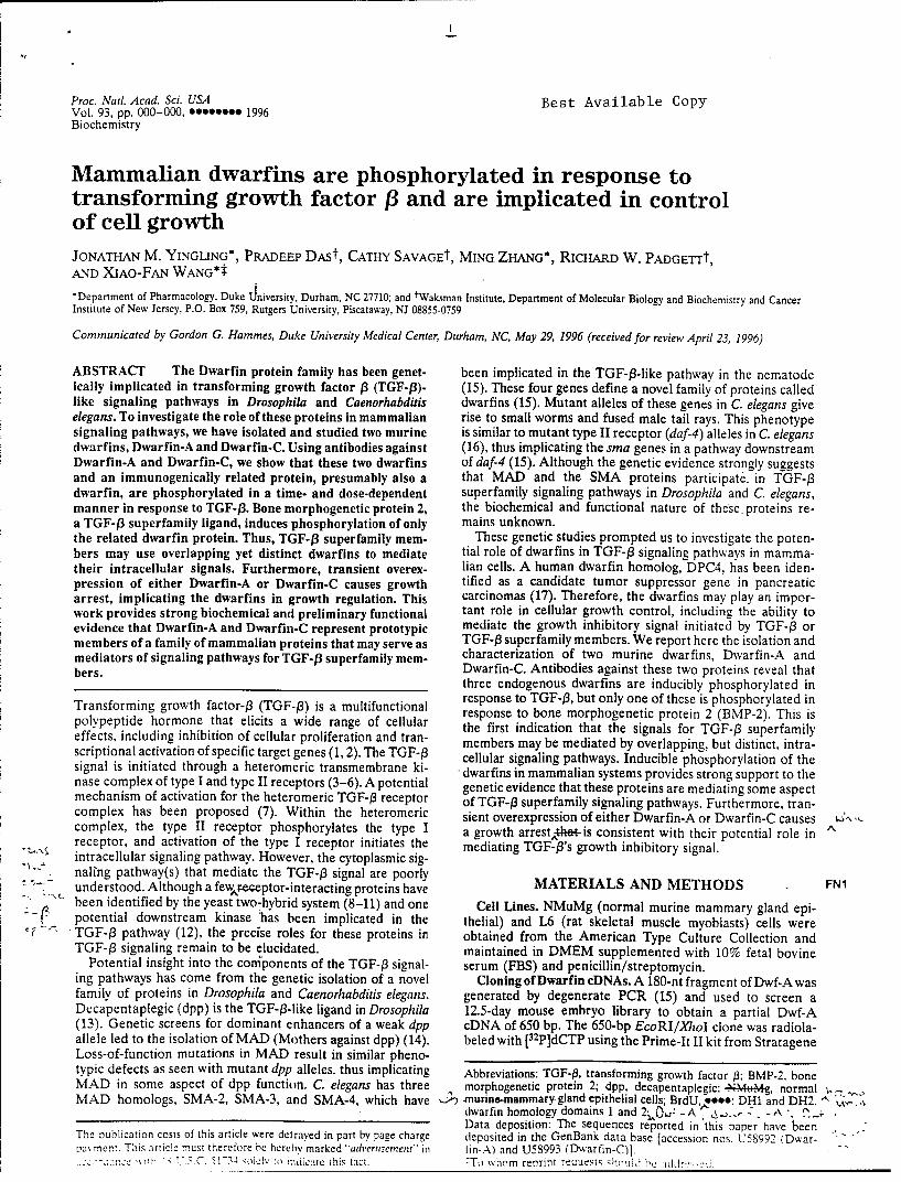
Proc. Natl. Acad. Sci. USA Best Available CopyVol. 93, pp. 000-000, oo09o0oo 1996Biochemistry
Mammalian dwarfins are phosphorylated in response totransforming growth factor 13 and are implicated in controlof cell growthJONATHAN M. YINGLING*, PRADEEP DASt, CATHY SAVAGEt, MING ZHANG*, RICHARD W. PADGE-rrt,AND XIAO-FAN WANG*t
*Department of Pharmacology, Duke dniversity, Durham, NC 27710; and tWaksman Institute, Department of Molecular Biology and Biochemistry and CancerInstitute of New Jersey, P.O. Box 759, Rutgers University, Piscataway, NJ 08855-0759
Communicated by Gordon G. Hammes, Duke University Medical Center, Durham, NC, May 29, 1996 (received for review April 23, 1996)
ABSTRACT The Dwarfin protein family has been genet- been implicated in the TGF-p3-like pathway in the nematodeically implicated in transforming growth factor /3 (TGF-13)- (15). These four genes define a novel family of proteins calledlike signaling pathways in Drosophila and Caenorhabditis dwarfins (15). Mutant alleles of these genes in C. elegans giveelegans. To investigate the role of these proteins in mammalian rise to small worms and fused male tail rays. This phenotypesignaling pathways, we have isolated and studied two murine is similar to mutant type II receptor (daf-4) alleles in C. elegansdwarfins, Dwarfin-A and Dwarfin-C. Using antibodies against (16), thus implicating the sma genes in a pathway downstreamDwarfin-A and Dwarfin-C, we show that these two dwarfins of daf-4 (15). Although the genetic evidence strongly suggestsand an immunogenically related protein, presumably also a that MAD and the SMA proteins participate. in TGF-A3dwarfin, are phosphorylated in a time- and dose-dependent superfamily signaling pathways in Drosophila and C. elegans,manner in response to TGF-f3. Bone morphogenetic protein 2, the biochemical and functional nature of these, proteins re-a TGF-j3 superfamily ligand, induces phosphorylation of only mains unknown.the related dwarfin protein. Thus, TGF-13 superfamily mem- These genetic studies prompted us to investigate the poten-bers may use overlapping yet distinct dwarfins to mediate tial role of dwarfins in TGF-13 signaling pathways in mamma-their intracellular signals. Furthermore, transient overex- lian cells. A human dwarfin homolog, DPC4, has been iden-pression of either Dwarfin-A or Dwarfin-C causes growth tified as a candidate tumor suppressor gene in pancreaticarrest, implicating the dwarfins in growth regulation. This carcinomas (17). Therefore, the dwarfins may play an impor-work provides strong biochemical and preliminary functional tant role in cellular growth control, including the ability toevidence that Dwarfin-A and Dwarfin-C represent prototypic mediate the growth inhibitory signal initiated by TGF-93 ormembers of a family of mammalian proteins that may serve as TGF-03 superfamily members. We report here the isolation andmediators of signaling pathways for TGF-j3 superfamily mem- characterization of two murine dwarfins, Dwarfin-A andbers. Dwarfin-C. Antibodies against these two proteins reveal that
three endogenous dwarfins are inducibly phosphorylated in
Transforming growth factor-P3 (TGF-J3) is a multifunctional response to TGF-03, but only one of these is phosphorylated inresponse to bone morphogenetic protein 2 (BMP-2). This ispolypeptide hormone that elicits a wide range of cellular the first indication that the signals for TGF-P3 superfamily
effects, including inhibition of cellular proliferation and tran- members may be mediated by overlapping, but distinct, intra-scriptional activation of specific target genes (1, 2). The TGF-3 cellular signaling pathways. Inducible phosphorylation of thesignal is initiated through a heteromeric transmembrane ki- dwarfins in mammalian systems provides strong support to thenase complex of type I and type II receptors (3-6). A potential genetic evidence that these proteins are mediating some aspectmechanism of activation for the heteromeric TGF-3 receptor of TGF-03 superfamily signaling pathways. Furthermore, tran-complex has been proposed (7). Within the heteromeric sient overexpression of either Dwarfin-A or Dwarfin-C causes -S",,complex, the type II receptor phosphorylates the type I a growth arrestýt-his consistent with their potential role in "receptor, and activation of the type I receptor initiates the mediating TGF-P's growth inhibitory signal."intracellular signaling pathway. However, the cytoplasmic sig-
' naling pathway(s) that mediate the TGF-03 signal are poorly MATERIALS AND METHODS FNI" -- understood. Although a few,-eceptor-interacting proteins have
been identified by the yeast two-hybrid system (8-11) and one Cell Lines. NMuMg (normal murine mammary gland epi--- a- potential downstream kinase 'has been implicated in the thelial) and L6 (rat skeletal muscle myoblasts) cells were
TGF-P3 pathway (12), the precise roles for these proteins in obtained from the American Type Culture Collection andTGF-/3 signaling remain to be elucidated. maintained in DMEM supplemented with 10% fetal bovine
Potential insight into the coniponents of the TGF-03 signal- serum (FBS) and penicillin/streptomycin.ing pathways has come from the genetic isolation of a novel Cloning ofDwarfin cDNAs. A 180-nt fragment of Dwf-Awasfamily of proteins in Drosophila and Caenorhabditis elegans. generated by degenerate PCR (15) and used to screen aDecapentaplegic (dpp) is the TGF-/3-like ligand in Drosophila 12.5-day mouse embryo library to obtain a partial Dwf-A(13). Genetic screens for dominant enhancers of a weak dpp cDNA of 650 bp. The 650-bp EcoRI/XhioI clone was radiola-allele led to the isolation of MAD (Mothers against dpp) (14). beled with [32 P]dCTP using the Prime-It II kit from StratageneLoss-of-function mutations in MAD result in similar pheno-typic defects as seen with mutant dpp alleles, thus implicating Abbreviations: TGF-/3, transforming growth factor 13; BMP-2, boneMAD in some aspect of dpp function. C. elegans has three morphogenetic protein 2; dpp, decapentaplegic: -NMtM- , normal .i-%.
MAD homologs, SMA-2, SMA-3, and SMA-4, which have ,-.muine-4nammary gland epithelial cells; BrdU oeeo: DHI and DH2 "dwarfin homology domains 1 and 2-,,0, - A A. - -. -- ". ,- -.Data deposition: The sequences reported in this paper have been
The publication costs of this article were del raved in part by page charge deposited in the GenBank data base [accession nos. U58992 IDwar- -pav, ment. This article must therefore he hereby marked "advernsernent" in tin-A) and U58993 (Dwarfin-C)]. -"
.. 'cc.- t.�, .h C. 11-t ,oielv lo indicate this fact. whom renrint recuests qh('l~l he :idd -.

2 Biochemistry: Yingling et al. Proc. Natl. Acad. Sci. USA 93 (1996)
and used to screen a kgtlO 8.5-day mouse embryonic library at Na2MoO 4, 1 mM DTT, IX protease inhibitors (5 .g/mllow stringency. Briefly, hybridization was at 42°C in 45% antipain, aprotinin, leupeptin, and trypsin inhibitor; 0.5 .gimlformamide, 5x standard saline phosphate/EDTA (SSPE; 0.18 pepstatin), and 1 mM phenylmethylsulfonyl fluoride on ice forM NaCI/10 mM phosphate, pH 7.4/1 mM EDTA), 5X Den- 15 min. Lysates were microcentrifuged at 14,000 rpm at 4°C for
-. hardt's solution, 0.5% SDS, and 100 tzg/ml salmon sperm 15 min. The resulting cell lysates were precleared with Pro-DNA for 16-20 hr. The filters were washed two times at( tein-A Sepharose for 30 min at 4°C. Lysates were then dividedin 2x standard saline citrate (SSC)/0. 1% SDS, one time afRT) for immunoprecipitation with Dwf-A or Dwf-C antibodies.in 0.5X SSC/0.1% SDS, and one time in 0.5x SSC/0.1% SDS Preimmune or immune sera (20 X of unpurified sera) wasat 55°C and were exposed overnight. Positive plaques were added with Protein-A Sepharose for 4 hr at 4°C. The immu-purified through quaternary screens before the insert was PCR noprecipitates were washed three times with lysis buffer beforeamplified using XgtlO-specific PCR primers (5' primer, AG- separation in SDS/8% polyacrylamide followed by autoradiog-CAAGTTCAGCCTGGTTAAG; 3' primer, 5-TTATGAG- raphy at room temperature.TATTTCTTCCAGGG). PCR products were subcloned into Transient Growth Arrest Assay. The following procedurepGEM-T (Promega) and partially sequenced with T7 and SP6 was adapted from DeGregori and coworkers (18). L6 cellsprimers. This approach isolated a Dwf-A cDNA of 1639 were plated in six-well trays on Poly-L-Lysine-coated coverslipsnucleotides and a Dwf-C cDNA of 2185 nucleotides. Both at 105 cells per well and allowed to attach overnight. CellFectincontain an open reading frame of 465 amino acids with a (GIBCO/BRL) was used to transfect the cells with 2 u.g ofpredicted molecular mass of 52 kDa. Subcloning and dele- pCMV LacZ and 8 Ag of vector alone, Dwf-A, or Dwf-C fortional analysis combined with automated sequencing yielded 10 hr in DMEM. The cells were incubated in 10% FBS for 36the nucleotide sequenc~eof Dwf-A and Dwf-C. hr before immunohistochemical staining. Media containing 20
Mammalian expression constructs were constructed using MM BrdU was added for the last 20 hr to metabolically label •'-"BamHI fragments containing full-length cDNAs for Dwf-A dividing cells. The cells were fixed in 4% paraformaldehyde in•'and Dwf-C generated by PCR using the following primer sets: PBS, permeabilized with MeOH/acetone, and stained with aDwf-A 5' primerACGCGGATCCGCGATGAATGTGAC- 1:500 dilution of rabbit anti-J3-galactosidase antibody (5
- -- CAGCTTG; Dwf-A 3' primer,.,CGCGGATCCGCGCA- Prime-3 Prime, Inc.) in 1% BSA/PBS for 60 min, followed by '"
GAGTTACCAGGTTTGGC; Dwf-C 5' primer•CGGGATC- a fluorescein isothiocyanate-conjugated goat anti-rabbit sec- •-'"CCGGAATTCCATGACGTCAATGGCCAGC; and Dwf-C ondary antibody (Boehringer Mannheim) at 1:500 in 1%
7 3' primer,,CGCGGATCCGCGGTAAGGCAAAGAAAT- BSA/PBS to stain transfected cells. After a second fixation andTCC. The resulting PCR products were subcloned into 2 M HCI permeabilization for 45 min, the cells were stainedpGEM-T and subsequently into pCMV5 and confirmed by with a mouse anti-BrdU antibody (Zymed) at 1:3 in 1%sequencingwithacytomegaloviruspromoter-specificsequenc- BSA/PBS followed by a goat anti-mouse Rhodamine-
_ ing primer: 5'-GCGGTAGGCGTGTACGG-3'. conjugated secondary antibody (Pierce) at 1:100 in 1% BSA/Northern Analysis. A,,multiple-tissue Northern blot PBS. Finally, 4 ug/ml Hoeschst 33342 in PBS was used to
(CLONTECH) containing 2 Mg of poly(A)÷ RNA was se- counter-stain the DNA. The coverslips were mounted inquentially probed with the 1.6-kb full-length Dwf-A cDNA DABCO and analyzed by fluorescence microscopy. .i., .;followed by the 2.1-kb full-length Dwf-C cDNA. The probes 2,.L. L.were labeled with [a-32P]dCTP by random priming (Strat- RESULTSagene) and hybridized for 16-20 hr at 42°C in 50% formamide.5x SSPE, 5x Denhardt's solution, 0.5% SDS, and 100 Mg/ml Cloning of Dwarfin-A and Dwarfin-C. Full-length cDNAssalmon sperm DNA. The filters were washed two times at RT for Dwf-A and Dwf-C were isolated by low stringency hybrid-in 2x SSC/0.1% SDS and two times in 0.2X SSC/0.1% SDS ization of a 8.5-day mouse embryonic library as described inat 60°C. Autoradiography was iat -800 C for 18-24 hr with Materials and Methods. The amino acid sequences of Dwf-Aintensifying screens. i and Dwf-C are 95% homologous and 90% identical. Both
Antibody Production. Bacterial expression constructs foi contain the characteristic dwarfin homology domain 1 (DH1)Dwf-A and Dwf-C were constructed using the BamHI frag- . and dwarfin homology domain 2 (DH2) motifs separated by aments from pGEM-T Dwf-A or Dwf-C, respectively, to clone proline-rich linker region (Fig. 1A; ref. 15). Dwf-A and Dwf-C F1in-frame into pGex 2T, generating glutathione S-transferase are about 80% homologous to the Drosophila dwarfin, MADfusion proteins. The resulting -85-kDa fusion proteins were (Fig. 1A; ref. 14), and the C. elegans dwarfin, SMA-2 (Fig. 1A;used as antigens for rabbit polyclonal antibody production. ref. 15). Dwf-A and Dwf-C are distantly related to the onlyPreimmune sera were obtained from each animal before the other known mammalian dwarfin, DPC4 (60% homologous,primary injection of antigen. Both the Dwf-A and Dwf-C 40% identity; ref. 17), and thus likely represent a distinct familyantibodies specifically recognize 56-kDa and 52-kDa proteins of mammalian dwarfins (Fig. 1B). Northern analysis showedon Western blots of NMuMg or L6 lysates (data not shown). that both genes are ubiquitously expressed (Fig. 1C). TheIn addition, transfection of Dwf-A or Dwf-C cDNA into COS mRNA message of Dwf-A is -3.5 kb, whereas Dwf-C has twocells generates a tight doublet around 52 kDa which cannot messages of -8 and -3 kb.account for the two bands seen by Western blotting of endog- TGF-13 Induces Phosphorylation of Endogenous Dwarfins.enous proteins (data not shown). Thus, the 56-kDa protein is We chose two cell lines, NMuMg (19) and L6 /which aremost likely immunogenically related to the dwarfins and potently growth inhibited by TGF-P,), as model systems torepresents an additional dwarfin family member. study endogenous dwarfin phosphorylation. Antibodies
In Vivo Phosphorylation of Endogenous Dwarfins. NMuMg against Dwf-A or Dwf-C were used to determine if the levelsor L6 cells were plated at 1.5 X 10⁶ cells per 100-mm dish and or phosphorylation state of endogenous dwarfins were mod-allowed to attach overnight. The cells were rinsed once with ulated by TGF-1. The expression levels of dwarfins werephosphate-free media (ICN) and then starved in phosphate- unchanged at any time within 24 hr after TGF-f3 treatment asfree media containing 0.5% dialyzed FBS (GIBCO/BRL) for determined by Western blot analysis (data not shown). How-1 hr and then labeled in 0.5% dFBS with 0.5 mCi/ml (1 Ci = ever, TGF-13 induced rapid, but transient, phosphorylation of
, 37 GBq) [32 P]orthophosphate for 4 hr. TGF-131 or BMP-2 was Dwf-A, Dwf-C, and a 56-kDa immunogenically related proteinadded at various concentrations for the indicated length of (Fig. 2h4). Fw2time at the end of the labeling period. Cells were rinsed twice Phosphorylation of Dwf-A and Dwf-C was induced within 15with ice-cold PBS and lysed in 50 mM Tris (pH 7.5), 100 mM min and peaked at 2.4-fold and 4-fold by 60 min. respectivelyNaCI. 50 mM NaF, 0.5% Nonidet P-40. 1 mM NaiVO4. 0.5 mM (Fig. 2B). The difference in the extent of phosphorviation of
Biochemistry: Yingling et al. Proc. Natl. Acad. Sci. USA 93 (1996) 3
A B C c
SMAnA
HDTi
1Prz~L MAD
L~P' VZ3T"R rc'GV M" 2A_
'.~ -,1 114 1S NH1.' 1 NT-DW2
GMI MAD SMA 2
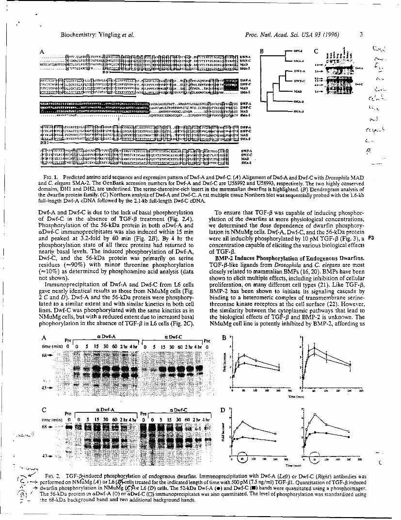
FrG. 1. Predicted amino acid sequence and expression pattern of Dwf-A and Dwf-C. (A) Alignment of Dwf-A and Dwf-C with Drosophila MADand C. elegans SMA-2. The GenBank accession numbers for Dwf-A and Dwf-C are U58992 and U58993, respectively. The two highly conserveddomains, DH1 and DH2, are underlined. The serine-threonine-rich insert in the mammalian dwarfmns is highlighted. (B) Dendrogram analysis ofthe dwarfin protein family. (C) Northern analysis of Dwf-A and Dwf-C. A rat multiple tissue Northern blot was sequentially probed with the 1.6-kbfull-length Dwf-A cDNA followed by the 2.1-kb full-length Dwf-C cDNA.
Dwf-A and Dwf-C is due to the lack of basal phosphorylation To ensure that TGF-f3 was capable of inducing phosphor-of Dwf-C in the absence of TGF-13 treatment (Fig. 2A). ylation of the dwarfins at more physiological concentrations,
Phosphorylation of the 56-kDa protein in both cxDwf-A and we determined the dose dependence of dwarfin phosphory-a•Dwf-C immunoprecipitates was also induced within 15 min lation in NMuMg cells. Dwf-A, Dwf-C, and the 56-kDa proteinand peaked at 3.2-fold by 60 mai (Fig. 2B). By 4 hr the were all inducibly phosphorylated by 10 pM TGF-j3 (Fig. 3), a gphosphorylation state of all three proteins had returned to concentration capable of eliciting the various biological effectsnearly basal levels. The induced phosphorylation of Dwf-A, of TGF-/3.Dwf-C, and the 56-kDa protein was primarily on serine BMP-2 Induces Phosphorylation of Endogenous Dwarfins.residues (,•90%) with minor threonine phosphorylation TGF-f3-1ike ligands from Drosophila and C. elegans are most(• 10%) as determined by phosphoamino acid analysis (data closely related to mammalian BMPs (16, 20). BMPs have beennot shown). shown to elicit multiple effects, including inhibition of cellular
Immunoprecipitation of Dwf-A and Dwf-C from L6 cells proliferation, on many different cell types (21). Like TGF-j3,gave nearly identical results as those from NMuMg cells (Fig. BMP-2 has been shown to initiate its signaling cascade by2 C and D). Dwf-A and the 56-kDa protein were phosphory- binding to a heteromeric complex of transmembrane serine-lated to a similar extent and with similar kinetics in both cell threonine kinase receptors at the cell surface (22). However,lines. Dwf-C was phosphorylated with the same kinetics as in the similarity between the cytoplasmic pathways that lead toNMuMg cells, but with a reduced extent due to increased basal the biological effects of TGF-f3 and BMP-2 is unknown. Thephophorylation in the absence of TGF-f3 in L6 cells (Fig. 2C). NMuMg cell line is potently inhibited by BMP-2, affording us
A a Dw- wf-.C BDtinmelmin)0 0 S IS 30 60 2hr4hrI 00 5 15 30 60 2hr4hr0
'T L
,,-/._-- FIGA. Prdce TGF- inoducid phsequenceatind expressiognou pattrfns of mmu ndpe ainwt Dwf-A. (A(Lenen ft or-an Dwf-C (Ritht antibophia MADan C.erfred ons NSMgA)2 The Ge.,6n aessio treate rs for the -in ated legtwf- tare Uih5899 and (5893 ng/ sp)ctively. Thentwtton hihl co- nservedthe dwarfin protsphorymily.n(Cn Nourth(e"rn anlyi ofD) s he5--aDf- o and Dwf-C. A(aJuliltsuNrhr blnds wasreequentitally probed wit thsheager.
.•); Te5-~ rti ncDwf-A and o'Dwf-C (i-s imunpecptae was tloqatttdhe level ofbal phosphorylation T nueta G- was capandardizeddusingphsor
Phsho-ao of the 68k agrudad an pro adteionaln bohackgroun bands. eemndteds eenec fdafnpopoy
a~fC imnpeptatswfas alsof- inueDihn1 i aini ~~ el.DfA w-,adte5- rti
and pemne at 0 5 IS 30b 60 min~b 0Fg 2B) By 4S 30 th0 wer~realidcbypohrltdby1 0(g.3,a3
phshrlto stt faltrepoen a eundt ocnrto aal feiiigtevrosbooia fet
DF-,and 2. e TGF- -k-irti aspiaiyo srn M-nduces phosphorylation of Endogenous dwarfins. muprcitinwthwfA(e)orwfCRgh)aibdswsr~ erformed on0% with (A) nor L6(~ lsthreatied fhortheindiatedlngt oGf time ith 500 nd pM(75 ogml Drosophil Qantiato of TlgF-3 induedms
- darfn posphrylpiation inNf~ (&')-&rand (D)cells.oTheL62cellDwfpAo(i)endtiofn, on)manydsfwerequntclltated sin a2) phshike ager.
T n ) w- n he 56-kDa protein inr cpw- 0)o hfC Cmuosphrecptae wabsiuntingtoatd The levoeli ope of phsh ryatinwsmstandried usine-
- ~~ the lls,~ bakrund bihanrduand etw nt adietio inalbckrouned bans.l tebooia fet fTG- n M- sukon h

4 Biochemistry: Yingling et al. Proc. Nati. Acad. Sci. USA 93 (1996)
a Dwf-A ,a Dwf-CPrel j Pre [ I
TGF-f1 (pM) 0 0 1 10 100 500 0 0 1 10 100 500
68-o-
43--o-
FIG. 3. TGF-13 phosphorylation of endogenous dwarfins is dose-dependent. Dwf-A and Dwf-C immunoprecipitation from NMuMg cells wasperformed as in Fig. 2 except various doses of TGF-f31 were added for 1 hr.
the opportunity to determine if dwarfin phosphorylation is studied two mammalian homologs of MAD and SMA-2 asF4 induced by BMP-2. As shown in Fig. 4 A and B, phosphory- potential downstream effectors of the TGF-f3 signaling path-
lation of the 56-kDa protein is induced by BMP-2 within 15 min way. We provide biochemical evidence that the dwarfin familyand peaks at 2.5-fold in 60 min. Interestingly, Dwf-A and of proteins is involved in TGF-f3 and BMP-2 signaling path-Dwf-C, which are inducibly phosphorylated in response to ways in mammalian systems. Furthermore, results from aTGF-P, are not phosphorylated in BMP-2-treated NMuMg modified transient growth assay and preliminary studies withcells (Fig. 4A). potentially dominant negative forms of Dwarfin-A (unpub.
Dwf-A and Dwf-C Are Implicated in Cell Growth Regula- lished data) strongly implicate a role for the dwarfins astion. Attempts to establish stable cell lines constitutively mediators of the TGF-f3 growth-regulatory signal.overexpressing either Dwf-A in L6 cells or Dwf-C in NMuMg Although the dwarfins do not contain any known catalyticcells were unsuccessful. This result was not unexpected be- motifs, their DH1 and DH2 domains are reminiscent of Srccause the dwarfins are suspected to have growth-suppressive homology 2 and 3 domains, which in a variety of signalingeffects based on the tumor suppressor activity of DPC4 (17). pathways modulate protein-protein interactions based on ty-Consequently, we used a modified transient growth assay (18) rosine phosphorylation and proline-rich sequences, respec-to assess the ability of Dwf-A or Dwf-C to cause a growth arrest tively (25). TGF-13 and BMP-2-induced phosphorylation of thewhen transiently transfected into L6 cells. Constitutive over- dwarfins may regulate protein-protein interactions in an anal-expression of Dwf-A or Dwf-C caused 30-40% growth inhi- ogous fashion for TGF-t3 superfamily signaling cascades. Re-bition compared with 10% for control vector transfectants cently, 14-3-3 proteins have been shown to be specific phos-
F5 (Fig. 5). Therefore, Dwf-A and Dwf-C, like DPC4, exhibit phoserine-binding proteins that are critical for the activation ofgrowth-inhibitory properties, implicating these dwarfin pro- signaling proteins (26). This suggests a novel role for serine-teins in cell growth regulation. threonine phosphorylation in the assembly of protein-protein
complexes required to transduce certain intracellular signals.
DISCUSSION Serine-threonine phosphorylation of the dwarfins may regu-late their ability to serve as adaptor molecules for other
Identification of downstream effectors for TGF-f3 or TGF-f3 effectors in the TGF-P pathway or regulate their ability tosuperfamily members has proven elusive. Mutagenesis studies specifically bind other intracellular proteins. These protein-in mammalian cells have yielded only receptor mutants (23, protein interactions may result in altered subcellular distribu-24), which suggests the existence of redundant pathways tion of the dwarfins. Preliminary immunofluorescence studiesdownstream of the receptors. Fortunately, TGF-13-like path- in NMuMg cells indicate that the dwarfins are predominantlyways exist in genetically tractable organisms to allow the use localized in the cytoplasm (unpublished data). Althoughof genetics to identify components of these signaling pathways. TGF-13 or BMP-2 treatment does not appear to cause aMany of these components (e.g., receptors, accessory mole- significant change in the subcellular distribution of the dwar-cules, and ligands) have been shown to have homologous fins, it is possible that a minor proportion of dwarfins thatcounterparts in vertebrate systems. Consequently, we have become phosphorylated accumulate at the membrane or trans-
A Pr I aDwf-A I r _ aDwf-C 7 B 3
time (min)O00 5 1 30 60 2hr 4hr 0 0 515 30 6 2hr 4hr68-
0 12 ISO 240 X
__ 4 4~ 444*4Timeimin) C.
- 4 '~FIG. 4. BMP-2Ainduced phosphorylation of endogenous Dwarfins. (A) Immunoprecipitation with Dwf-A (Left) or Dwf-C (Right) antibodies wasperformed on NMuMg cells treated for the indicated length of time with 100 ng/ml BMP-2. (B) Quantitation of BMP-2 induced dwarfinphosphorylation in NMuMg cells. The 52-kDa Dwf-A band (o) and the 56-kDa protein in otDwf-A (C) or aDwf-C (C3) immunoprecipitates werequantitated. The S2-kDa Dwf-C band that is not basally phosphoryiated in NMuMg cells is undetectable after BMP-2 treatment. The level ofF~hosphorvlatl(on was standardized usinj! the 68-kDa backtzround band and two additional background bands,

Biochemistry: Yingling et al. Proc. Natl. Acad. Sci. USA 93 (1996) 5
50 human TGF-031; Y. Xiong for automated sequencing assistance; J.
Heitman and M. Caron for critical review of the manuscript; C.
40 Bassing, D. Cortez, M. Datto, P. Hu, G. Reuther, and H. Symonds forhelpful discussion; and Y. Yu and H. Wang for technical help. J.M.Y.thanks M. L. Yingling for her constant support and encouragement.
S30 This work was supported by U.S. Army Breast Cancer ResearchProgram Grant DAMD17-945-4065 (X.-F.W.), National Institutes of
20 Health Grant GM47395 (R.W.P.); and Council for Tobacco ResearchGrant 3639 (R.W.P.). J.M.Y. was supported by a U.S. Army Breast
oR 10 Cancer Research Program Predoctoral Fellowship (DAMD17-94-J-4190), P.D. is a Busch Predoctoral Fellow, and C.S. is a New Jersey
Cancer Commission Postdoctoral Fellow; X.-F.W. is a LeukemiaMock Dwf-A Dwf-c Society Scholar.
FIG. 5. Dwf-A and Dwf-C cause a growth arrest in L6 cells. L6 cells 1. Massague, J. (1990) Annu. Rev. Cell Biol. 6, 597-641.
were transfected and immunohistochemically stained as described in 2. Roberts, A. B. & Sporn, M. B. (1990) in Handbook of Experi-
Materials and Methods. All the transfected cells (fluorescein isothio- mental Pharmacology, Peptide Growth Factors and their Receptors,
cyanate stained)on each coverslipwere scored for BrdU incorporation eds. Sporn, M. B. & Roberts, A. B. (Springer, Heidelberg), pp.
(Rhodamine stained). All (100%) of the nontransfected cells incor- 419-472.porate BrdU during the labeling period; therefore, the percentage of 3. Lin, H. Y., Wang, X.-F., Ng-Eaton, E., Weinberg, R. A. &
transfected cells that are BrdU-negative represents the percent growth Lodish, H. F. (1992) Cell 68, 775-785.
inhibition caused by transfection of the cDNA. Data shown are the 4. Wrana, J. L., Attisano, L., Carcamo, J., Zentella, A., Doody, J.,
mean t SD of at least three experiments. Laiho, M., Wang, X.-F. & Massague, J. (1992) Cell 71, 1003-1014.S. Franzen, P., ten Dijke,,,•e, Ichijo, H., Yamashita, H., Schulz, P., '
locate to the nucleus to fulfill their biological function. Indeed, Heldin, C.-H. & Miyazono, K. (1993) Cell 75, 681-692.
a human homolog of Dwf-A, MADR1, has recently been 6. Bassing, C. H., Yingling, J. M.,Howe, D. J.,Wang, T.,He,W. W.,
shown to be inducibly phosphorylated and to translocate to the Gustafson, M. L., Shah, P., Donahoe, P. K. & Wang, X.-F. (1994)
nucleus after BMP-2 treatment (27). Intriguingly, phosphor- Science 263, 87-89.
ylation of MADR1 appears to be BMP-2-specific in their 7. Wrana, J. L., Attisano, L., Weiser, R., Ventura, F. & Massague,
system, since neither TGF-P3 nor activin induces MADRI J. (1994) Nature (London) 370, 341-347.
phosphorylation. This apparent discrepancy may be due to 8. Wang, T. W., Donahoe, P. K. & Zervos, A. S. (1994) Science 265,
differences in experimental systems, overexpression of 674-676.
diffrencs inexpMADRi instead of endogenous dwarfins, or 9. Chen, R.H., Miettinen, P.J., Maruoka, E.M., Choy, L. &
epitope-tagged ll-typeispeadiffences i wa ys Derynck, R. (1995) Nature (London) 377, 548-552.
may represent cell-type specific differences in the pathways 10. Kawabata, M., Imamura, T., Miyazono, K., Engel, M. E. &
used by TGF-03 superfamily ligands. Clarification of this issue Moses, H. L (1995) J. Biol. Chem. 270, 29628-2963 1.
requires further study. 11. Wang, T. W., Danielson, P. D., Li, B.-y., Shah, P. C., Kim, S. D.
Our initial attempt to study TGF-P's ability to modulate the & Donahoe, P. K. (1996) Science 271, 1120-1122.
phosphorylation state of Dwf-A and Dwf-C involved transfec- 12. Yamaguchi, K., Shirakabe, K., Shibuya, H., Irie, K., Oishi, I.,
tion of hemagglutinin epitope-tagged cDNAs into COS or Ueno, N., Taniguchi, T., Nishida, E. & Matsumoto, K. (1996)
mink lung epithelial cells. Although both proteins are phos- Science 270, 2008-2011.
phorylated in these systems, the high level of constitutive 13. Padgett, R. W., St. Johnston, R. D. & Gelbart, W. M. (1987)
phosphorylation precluded detection of TGF-P induced Nature (London) 325, 81-84.
changes in Dwf-A or Dwf-C phosphorylation (unpublished 14. Sekelsky, J. J., Newfeld, S. J., Raftery, L. A., Chartoff, E. H. &
chta)Ingeestin gy thf e or eeasdafis M - n Gelbart, W. M. (1995) Genetics 139, 1347-1358.data). Interestingly, the C. elegans dwarfins, SMA-2 and 15. Savage, C., Das, P., Finelli, A. L., Townsend, S. R., Sun, C.-Y.,
SMA-3, are not phosphorylated when overexpressed in COS Baird, S. E. & Padgett, R. W. (1996) Proc. Natl. Acad. Sci. USA
cells. Therefore, the observed phosphorylation is likely a result 93, 790-794.
of phosphorylation by an associated kinase that is unable to 16. Estevez, M., Attisano, L., Wrana, J. L., Albert, P. S., Massague,
recognize SMA-2 or SMA-3 as substrates. The unique serine- J. & Riddle, D. L. (1993) Nature (London) 364, 644-649.
threonine-rich insert in the linker region of Dwf-A and Dwf-C 17. Hahn, S. A., Schutte, M., Shamsul Hoque, A. T. M., Moskaluk,
(Fig. 1A) may play a role in either kinase recognition or as C. A., da Costa, L. T., Rozenblum, E,, Weinstein, C. L., Fischer,
targets of phosphorylation by the associated kinase. Prelimi- A., Yeo, C. J., Hruban, R. H. & Kern, S. E. (1996) Science 271,
narv results indicate that neither Dwf-A nor Dwf-C are 350-353.the type I or type II TGF-3 receptor kinases 18. DeGregori, J., Kowalik, T. & Nevins, J. R. (1995) Mol. Cell Biol.
substrates of 15,4215-4224.(unpublished data), implicating an as yet unidentified kinase 19. Miettinen, P. J., Ebner, R., Lopez, A. R. & Derynck, R. (1994)
in phosphorylation of the dwarfins. J. Cell Biol. 127, 2021-2036.The differential phosphorylation of endogenous dwarfins in 20. Maliakal, J. C., Asahina, I., Hauschka, P. V. & Sampath, T. K.
response to TGF-P3 and BMP-2 suggests that the specific (1994) Growth Factors 11, 227-234.
dwarfins used by TGF-P superfamily members may vary. In 21. Yamashita, H., ten Dijke, P., Huylebroeck, D., Sampath, T. K.,
support of this notion, two related Xenopus dwarfins (Xmadl Andries, M., Smith, J. C., Heldin, C.-H. & Miyazono, K. (1995)
and Xmad2) have been shown to mediate either BMP-2/ J. Cell Biol. 130, 217-226.
BMP-4 signals or Vgl/activin/nodal signals, respectively (28). 22. Liu, F., Ventura, F., Doody, J. & Massague, J. (1995) Mol. Cell.
Thus, Dwf-A and Dwf-C may represent TGF-p3-specific dwar- Biol. 15, 3479-3486.
fins, whereas the 56-kDa dwarfin is shared by TGF-13 and 23. Boyd, F. & Massague, J. (1989) 1. Biol. Chem. 264, 2272-2278.
BMP-2. The differential response of these proteins to TGF-3 24. Laiho, M., Weis, F. M. B. & Massague, J. (1990)1. Biol. Chem.
and MP- wil failiateeluidaton f te dffernce inthe265, 185 18-18524.and BMP-2 will facilitate elucidation of the differences in the 25. Cohen, G. B., Ren, R. & Baltimore, D. (1995) Cell 80, 237-248.
intracellular pathways used by these two members of the 26. Muslin, A. J., Tanner, J. W., Allen, P. M. & Shaw, A. S. (1996)
TGF-f3 superfamily. Cell 84, 889-897.27. Hoodless, P.A., Haerry, T., Abdollah, S., Stapleton, M.,
We wish to thank S.-J. Lee forthe 12.5-day mouse embryonic library O'Connor, M. B., Attisano, L. & Wrana, J. L. (1996) Cell 85,
and B. Hogan for the 8.5-day mouse embryonic library; Genetics 489-500.
Institute for recombinant human BMP-2; Amgen for recombinant 28. Graff, J.M.,Bansal, A. & Melton, D. A. (1996) Cell85,479-487.