1999 internationaldictybase.org/dictyannualconference/dicty1999.pdf · 1999 international...
TRANSCRIPT

1

2
1999 INTERNATIONAL DICTYOSTELIUM CONFERENCE
BAR HARBOR, MAINE, U.S.A.COLLEGE OF THE ATLANTIC
AUGUST 14TH- 19TH, 1999
Robert Gundersen1 and David Knecht2, Organizers
1Department of Biochemistry, Microbiology, and Molecular Biology,University of Maine
2Department of Molecular and Cell Biology, University of Connecticut
AcknowledgementsThe organizers would like to thank the National Science Foundation, Developmental
Mechanisms Program and the Department of Biochemistry, Microbiology, and MolecularBiology at the University of Maine for their generous support of this meeting.
3
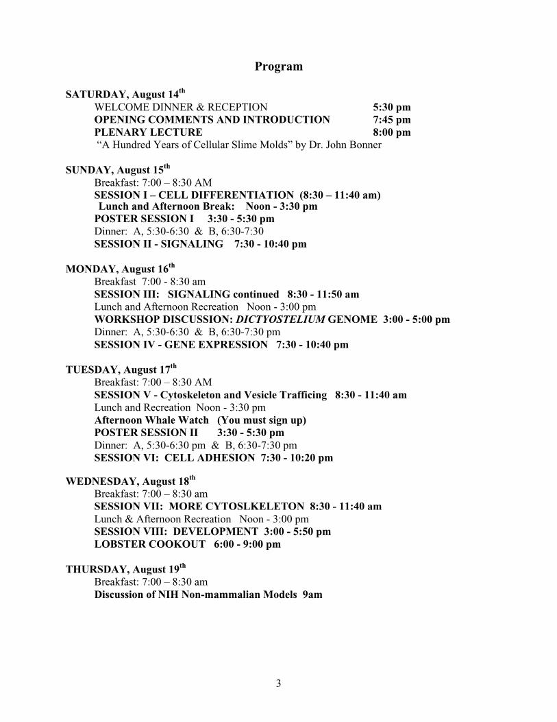
Program
SATURDAY, August 14th
WELCOME DINNER & RECEPTION 5:30 pmOPENING COMMENTS AND INTRODUCTION 7:45 pmPLENARY LECTURE 8:00 pm “A Hundred Years of Cellular Slime Molds” by Dr. John Bonner
SUNDAY, August 15th
Breakfast: 7:00 – 8:30 AMSESSION I – CELL DIFFERENTIATION (8:30 – 11:40 am)Lunch and Afternoon Break: Noon - 3:30 pm
POSTER SESSION I 3:30 - 5:30 pmDinner: A, 5:30-6:30 & B, 6:30-7:30SESSION II - SIGNALING 7:30 - 10:40 pm
MONDAY, August 16th
Breakfast 7:00 - 8:30 amSESSION III: SIGNALING continued 8:30 - 11:50 amLunch and Afternoon Recreation Noon - 3:00 pmWORKSHOP DISCUSSION: DICTYOSTELIUM GENOME 3:00 - 5:00 pmDinner: A, 5:30-6:30 & B, 6:30-7:30 pmSESSION IV - GENE EXPRESSION 7:30 - 10:40 pm
TUESDAY, August 17th
Breakfast: 7:00 – 8:30 AMSESSION V - Cytoskeleton and Vesicle Trafficing 8:30 - 11:40 amLunch and Recreation Noon - 3:30 pmAfternoon Whale Watch (You must sign up)POSTER SESSION II 3:30 - 5:30 pmDinner: A, 5:30-6:30 pm & B, 6:30-7:30 pmSESSION VI: CELL ADHESION 7:30 - 10:20 pm
WEDNESDAY, August 18th
Breakfast: 7:00 – 8:30 amSESSION VII: MORE CYTOSLKELETON 8:30 - 11:40 amLunch & Afternoon Recreation Noon - 3:00 pmSESSION VIII: DEVELOPMENT 3:00 - 5:50 pmLOBSTER COOKOUT 6:00 - 9:00 pm
THURSDAY, August 19th
Breakfast: 7:00 – 8:30 amDiscussion of NIH Non-mammalian Models 9am

1
1999 INTERNATIONAL DICTYOSTELIUM CONFERENCE
BAR HARBOR, MAINE, U.S.A.COLLEGE OF THE ATLANTIC
AUGUST 14TH- 19TH.
SATURDAY, August 14th
WELCOME DINNER & RECEPTION 5:30 pmOPENING COMMENTS AND INTRODUCTION 7:45 pmPLENARY LECTURE 8:00 pm “A Hundred Years of Cellular Slime Molds” by Dr. John Bonner
SUNDAY, August 15th
Breakfast: 7:00 – 8:30 AM
SESSION I – CELL DIFFERENTIATION (8:30 – 11:40 am)Chair: Harry MacWilliams
Cell-cycle regulation of the ribonucleotide reductase B gene of Dictyosteliumdiscoideum.
Harry MacWilliams, Pascale Gaudet, Claire Bonfils, and Adrian Tsang 15
Identification of pathways regulated by YakA that control growth, developmentand stress responses in Dictyostelium cells
Alexandre Taminato, Douglas Robinson, Marcos Navarro, Rodrigo Romanhole,Guokai Chen, James Spudich, Adam Kuspa and Glaucia Mendes Souza 16
Structure and function of a novel gene (dia1) specifically expressed duringtransition from growth to differentiation in Dictyostelium cells
Shigenori Hirose, Yuji Inazu, Soo-Cheon Chae and Yasuo Maeda 17
Altered Prestarvation Response in Two Dictyostelium MutantsMarc E. Colosimo and Eugene R. Katz 18
BREAK: 10:00 - 10:20 am
F-box Proteins and Mutants That CheatHerbert L. Ennis, Dee N. Dao, Stefan U. Pukatzki, and Richard H. Kessin 19
Nuclear translocation of the Dd-STATc protein in response to DIFMasahi Fukuzawa, Tsuyoshi Araki and Jeff Williams 20
Development in the Absence of DIF-1Christopher Thompson and Rob Kay 21
Tale of Two Transporters: Evidence for the involvement of TagA and RhTin cell-type proportioning.
J. Randall Good and Adam Kuspa 22

2
Lunch and Afternoon Break: Noon - 3:30 pm
POSTER SESSION I 3:30 - 5:30 pm
Dinner: A, 5:30-6:30 & B, 6:30-7:30
SESSION II - SIGNALING 7:30 - 10:40 pmChair: Peter Devreotes
Chemoattractant sensing in eukaryotic cells.C.A. Parent, W. M. Froehlich, J. Borleis, and P.N. Devreotes 23
Dynamic localization of a heterotrimeric G-protein _-subunit in living cellsTian Jin, Ning Zhang, Yu Long, Carol Parent, Peter Devreotes 24
Study of G-protein _-subunit from Dictyostelium discoideumNing Zhang, and Peter N. Devreotes 25
Role of G-Protein carboxyl methylation in signal transductionJ. Stock, Y. Chen and T. Cox. 26
BREAK 8:40 - 9:00 pm
CONTROL OF CHEMOTAXIS BY PI3 KINASE-REGULATED PATHWAYSRuedi Meili, Satoru Funamoto, Charlene Ellsworth, Nan Tang, and Richard A. Firtel 27
A p21-activated protein kinase, PAKa, is required for cytokinesis and theregulation of the cytoskeleton in Dictyostelium cells during chemotaxis.
Chang Y. Chung and Richard A. Firtel, 28
A temperature sensitive ACA mutant.J. Gross, K. Guo, H-J Kim, H. Otsuka. 29
Wave propagation in Dictyostelium discoideum slugsDirk Dormann & Cornelis J. Weijer 30
Light affects cAMP signaling and cell movement activity in Dictyosteliumdiscoideum
Kota Miura & Florian Siegert 31
3
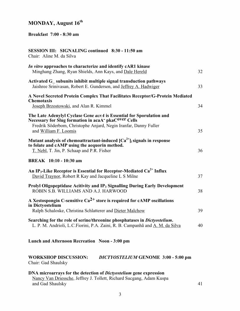
MONDAY, August 16th
Breakfast 7:00 - 8:30 am
SESSION III: SIGNALING continued 8:30 - 11:50 amChair: Aline M. da Silva
In vitro approaches to characterize and identify cAR1 kinaseMinghang Zhang, Ryan Shields, Ann Kays, and Dale Hereld 32
Activated G_ subunits inhibit multiple signal transduction pathwaysJaishree Srinivasan, Robert E. Gundersen, and Jeffrey A. Hadwiger 33
A Novel Secreted Protein Complex That Facilitates Receptor/G-Protein MediatedChemotaxis
Joseph Brzostowski, and Alan R. Kimmel 34
The Late Adenylyl Cyclase Gene acrA is Essential for Sporulation andNecessary for Slug formation in acaA- pkaCover Cells
Fredrik Söderbom, Christophe Anjard, Negin Iranfar, Danny Fullerand William F. Loomis 35
Mutant analysis of chemoattractant-induced [Ca2+]i signals in responseto folate and cAMP using the aequorin method.
T. Nebl, T. Jin, P. Schaap and P.R. Fisher 36
BREAK 10:10 - 10:30 am
An IP3-Like Receptor is Essential for Receptor-Mediated Ca2+ InfluxDavid Traynor, Robert R Kay and Jacqueline L S Milne 37
Prolyl Oligopeptidase Acitivity and IP3 Signalling During Early DevelopmentROBIN S.B. WILLIAMS AND A.J. HARWOOD 38
A Xestospongin C-sensitive Ca2+ store is required for cAMP oscillationsin Dictyostelium
Ralph Schaloske, Christina Schlatterer and Dieter Malchow 39
Searching for the role of serine/threonine phosphatases in Dictyostelium.L. P. M. Andrioli, L.C.Fiorini, P.A. Zaini, R. B. Campanhã and A. M. da Silva 40
Lunch and Afternoon Recreation Noon - 3:00 pm
WORKSHOP DISCUSSION: DICTYOSTELIUM GENOME 3:00 - 5:00 pmChair: Gad Shaulsky
DNA microarrays for the detection of Dictyostelium gene expressionNancy Van Driessche, Jeffrey J. Tollett, Richard Sucgang, Adam Kuspaand Gad Shaulsky 41
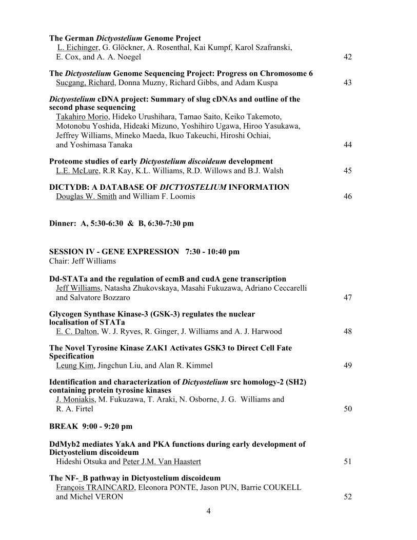
4
The German Dictyostelium Genome Project L. Eichinger, G. Glöckner, A. Rosenthal, Kai Kumpf, Karol Szafranski,E. Cox, and A. A. Noegel 42
The Dictyostelium Genome Sequencing Project: Progress on Chromosome 6Sucgang, Richard, Donna Muzny, Richard Gibbs, and Adam Kuspa 43
Dictyostelium cDNA project: Summary of slug cDNAs and outline of thesecond phase sequencing
Takahiro Morio, Hideko Urushihara, Tamao Saito, Keiko Takemoto,Motonobu Yoshida, Hideaki Mizuno, Yoshihiro Ugawa, Hiroo Yasukawa,Jeffrey Williams, Mineko Maeda, Ikuo Takeuchi, Hiroshi Ochiai,and Yoshimasa Tanaka 44
Proteome studies of early Dictyostelium discoideum developmentL.E. McLure, R.R Kay, K.L. Williams, R.D. Willows and B.J. Walsh 45
DICTYDB: A DATABASE OF DICTYOSTELIUM INFORMATIONDouglas W. Smith and William F. Loomis 46
Dinner: A, 5:30-6:30 & B, 6:30-7:30 pm
SESSION IV - GENE EXPRESSION 7:30 - 10:40 pmChair: Jeff Williams
Dd-STATa and the regulation of ecmB and cudA gene transcriptionJeff Williams, Natasha Zhukovskaya, Masahi Fukuzawa, Adriano Ceccarelliand Salvatore Bozzaro 47
Glycogen Synthase Kinase-3 (GSK-3) regulates the nuclearlocalisation of STATa
E. C. Dalton, W. J. Ryves, R. Ginger, J. Williams and A. J. Harwood 48
The Novel Tyrosine Kinase ZAK1 Activates GSK3 to Direct Cell FateSpecification
Leung Kim, Jingchun Liu, and Alan R. Kimmel 49
Identification and characterization of Dictyostelium src homology-2 (SH2)containing protein tyrosine kinases
J. Moniakis, M. Fukuzawa, T. Araki, N. Osborne, J. G. Williams andR. A. Firtel 50
BREAK 9:00 - 9:20 pm
DdMyb2 mediates YakA and PKA functions during early development ofDictyostelium discoideum
Hideshi Otsuka and Peter J.M. Van Haastert 51
The NF-_B pathway in Dictyostelium discoideumFrançois TRAINCARD, Eleonora PONTE, Jason PUN, Barrie COUKELLand Michel VERON 52
5
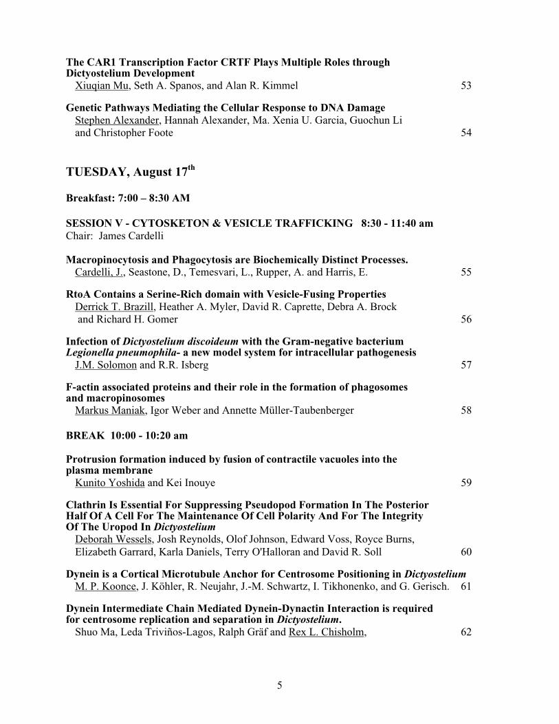
The CAR1 Transcription Factor CRTF Plays Multiple Roles throughDictyostelium Development
Xiuqian Mu, Seth A. Spanos, and Alan R. Kimmel 53
Genetic Pathways Mediating the Cellular Response to DNA DamageStephen Alexander, Hannah Alexander, Ma. Xenia U. Garcia, Guochun Liand Christopher Foote 54
TUESDAY, August 17th
Breakfast: 7:00 – 8:30 AM
SESSION V - CYTOSKETON & VESICLE TRAFFICKING 8:30 - 11:40 amChair: James Cardelli
Macropinocytosis and Phagocytosis are Biochemically Distinct Processes.Cardelli, J., Seastone, D., Temesvari, L., Rupper, A. and Harris, E. 55
RtoA Contains a Serine-Rich domain with Vesicle-Fusing PropertiesDerrick T. Brazill, Heather A. Myler, David R. Caprette, Debra A. Brock and Richard H. Gomer 56
Infection of Dictyostelium discoideum with the Gram-negative bacteriumLegionella pneumophila- a new model system for intracellular pathogenesis
J.M. Solomon and R.R. Isberg 57
F-actin associated proteins and their role in the formation of phagosomesand macropinosomes
Markus Maniak, Igor Weber and Annette Müller-Taubenberger 58
BREAK 10:00 - 10:20 am
Protrusion formation induced by fusion of contractile vacuoles into theplasma membrane
Kunito Yoshida and Kei Inouye 59
Clathrin Is Essential For Suppressing Pseudopod Formation In The PosteriorHalf Of A Cell For The Maintenance Of Cell Polarity And For The IntegrityOf The Uropod In Dictyostelium
Deborah Wessels, Josh Reynolds, Olof Johnson, Edward Voss, Royce Burns,Elizabeth Garrard, Karla Daniels, Terry O'Halloran and David R. Soll 60
Dynein is a Cortical Microtubule Anchor for Centrosome Positioning in DictyosteliumM. P. Koonce, J. Köhler, R. Neujahr, J.-M. Schwartz, I. Tikhonenko, and G. Gerisch. 61
Dynein Intermediate Chain Mediated Dynein-Dynactin Interaction is requiredfor centrosome replication and separation in Dictyostelium.
Shuo Ma, Leda Triviños-Lagos, Ralph Gräf and Rex L. Chisholm, 62
6
Lunch and Recreation Noon - 3:30 pmAfternoon Whale Watch (You must sign up)
POSTER SESSION II 3:30 - 5:30 pm
Dinner: A, 5:30-6:30 pm & B, 6:30-7:30 pm
SESSION VI: CELL ADHESION 7:30 - 10:20 pmChair: Richard Gomer
Simulation of stream breakup during Dictyostelium aggregationRichard H. Gomer 63
Aardvark: a _-catenin–like protein is required for axis formation and terminaldifferentiation during Dictyostelium morphogenesis.
Adrian J. Harwood, Mark Grimson, Richard, L Blanton & Juliet Coates 64
Novel Roles of Cell Adhesion Molecules during Development of DictyosteliumChi-Hung Siu, Tony Harris, Liansheng Hou, Jun Wang and Estella Wong 65
BREAK 9:00 - 9:20 pm
The Cell Adhesion Molecule gp150 is Encoded by the lagC Gene and isinvolved in the Formation of the Extracellular Matrix during DictyosteliumDevelopment
Jun Wang, Don Awrey, Lianseng Hou, William F. Loomis,Richard A. Firtel, Chi-Hung Siu 66
Dynamics of Rotation and Sorting in "Pancake" Aggregates of DictyosteliumHerb Levine, Willaim F. Loomis, Alistair Nicol, and Wouter-Jan Rappel 67
Regulation of Cell Adhesion and Cell-Type Proportioning by theDictyostelium Protein AmpA.
Timothy R. Varney, Elizabeth Casademunt, Chere' Petty, Beatrice Kondo,Jayne Dolman, and Daphne D. Blumberg 68
3D in vivo analysis of Dictyostelium mounds reveals directional sorting ofprestalk cells and defines a role for the myosin II regulatory light chainin prestalk cell sorting and tip protrusion
Patricia A. Clow, Tung-Ling L. Chen, Rex L. Chisholm, and James G. McNally 69

7
WEDNESDAY, August 18th
Breakfast: 7:00 – 8:30 am
SESSION VII: MORE CYTOSLKELETON 8:30 - 11:40 amChair: Robert Insall
Multiple Ras Pathways in Dictyostelium Movement and EndocytosisRobert Insall, Jonathan Chubb, Andrew Wilkins, Derek Fraser, Paul Fisher,Meenal Khosla and Gerry Weeks 70
Function of small G protein racB and racC in Dictyostelium.Eunkyung Lee, David J. Seastone, James A. Cardelli and David A. Knecht 71
Identification of a Ponticulin Gene FamilyC.G. Wilkerson*, L.L Antonette, T. Stawicki and A.L. Hitt 72
CHARACTERIZATION OF VILLIDIN, A VILLIN-LIKE PROTEIN FROMDICTYOSTELIUM
F. Rivero, A. Gloss, D. Rieger, R. Mueller, M. Schleicher, W.F. Loomis, A.A. Noegel 73
BREAK 10:00 - 10:20 am
DdABP1: A novel SH3 domain-containing actin-binding protein that bindsDictyostelium myosin I heavy chain kinase
Marc de la Roche, Amjad Mahasneh and Graham Côté 74
Evidence for a Functional Role of Myosin II in Rear RetractionGudrun E. Koehl and James G. McNally 75
Dynacortin, a genetic link between the global cortical tension generatingsystem of RacE and coronin and the equatorial cortical tension generatingsystem of cortexillin I and myosin II.
Douglas N. Robinson and James A. Spudich. 76
A mathematical model for cell movement in multicellular systems: Dictyosteliumaggregation and slug movement.
Eirikur Palsson and Hans Othmer 77
Lunch & Afternoon Recreation Noon - 3:00 pm
SESSION VIII: DEVELOPMENT 3:00 - 5:50 pmChair: Wolfgang Nellen
Functional analysis of a prokaryotic antisense system in DictyosteliumHenrik Martens, Jindrich Novotny, Beate Schnell and Wolfgang Nellen 78
8
Characterization of the cellulose synthase gene in DictyosteliumRichard L. Blanton, Danny Fuller, Negin Iranfar, Mark J. Grimson,and William F. Loomis 79
Contributions of Cellulose and a Cellulose-Binding Protein to Assembly of theDictyostelium Spore Coat
Yunyan Zhang, Ping Zhang, Richard L. Blanton, William F. Loomis,and Christopher M. West 80
BREAK 4:10 - 4:30 pm
Ribonucleotide Reductase Expression During DevelopmentPascale Gaudet1,2 and Adrian Tsang1,2,3 81
Macrocyst cycle in Dictyostelium discoideum: - A part of the genome is reserved for it.Hideko Urushihara, Hideki D. Shimizu, Hajime Shimizu, Toshihiro Hata, Miho Iijima 82
Membrane protein gp64 inhibits the whorl formation of Polysphondylium pallidumHiroshi Ochiai, Satoru Funamoto, Tamao Saito, Masumi Hirashima, Takahiro Sasaki 83
Expression of human gonadotropins in DictyosteliumMaarten H.K. Linskens, Elisa Vervoort, Petra Werler, Arno A.M. van Ravestein,Gijs F. Verheijden and Peter J.M van Haastert 84
LOBSTER COOKOUT 6:00 - 9:00 pm
THURSDAY, August 19th
Breakfast: 7:00 – 8:30 am
Morning free for checkout and sight seeing.
Have a safe trip home.
9
POSTER SESSION I : Sunday 3:30 - 5:30 pm
Screening for Dictyostelium cell death mutantsM. Adam, J.P. Levraud, C. Charrier, A. Richter, R.A. Olie, S. Cornillon, P. Golstein 85
GTPase-binding homology (GBH) domain protein isolated in a yeast 2-hybrid screenJeffrey A. Hadwiger and Kanchana Natarajan. 86
A Cell-Counting Factor Regulating Aggregate Size in Dictyostelium discoideumDebra A. Brock and Richard H. Gomer 87
Phosphothreonine and Phosphoserine Containing Proteins during Formation,Dormancy, and Germination of Dictyostelium discoideum spores
Dana C. Mahadeo, Tracy L. Marr, Dave N. Cervi, Yoshiro Kishi,Masazumi Sameshima, and David A. Cotter 88
Regulation of cbpA Expression During Dictyostelium DevelopmentJason Pun and Barrie Coukell 89
An F-Box Protein and Terminal DifferentiationDee N. Dao, Herbert L. Ennis, Jakob Franke and Richard H. Kessin 90
Transient expression of a mitochondorial gene cluster (dia3) including rps4 isessential for the phase-shift of Dictyostelium cells from growth to differentiation
Y. Inazu, S-C. Chae, and Y. Maeda 91
A cellular basis for division of labour in Dictyostelium discoideumSonia Kaushik and Vidyanand Nanjundiah 92
AN ANALYSIS OF CELL DEATH IN Dictyostelium discoideumTrupti. S. Kawli, Venkatesh, B.R., Gopal Pande and Vidyanand Nanjundiah 93
Developmental Analysis of the Dictyostelium LIM-only protein limBJoshua Krupp, Joseph Brzostowski, and Alan R. Kimmel 94
Identification and characterization of a new filamin binding proteinMonika Knuth, Si Jie Lu, Adam Kuspa and Angelika A. Noegel 95
The Characterization of a Major Myosin II Phosphatase in Dictyostelium discoideumM.M. Murphy, S.K. Levi, T.T. Egelhoff 96
Myosin Heavy chain kinases regulate Myosin filament assembly in DictyosteliumMaribel Rico, Paul Steimle, and Tom Egelhoff 97
Characterization of rasC expression during growth and developmentChinten James Lim, George B. Spiegelman and Gerry Weeks. 98
CELL CYCLE DEPENDENT REGULATION OF THE EXPRESSION OFEARLY DEVELOPMENTAL GENES
Sophia Lin, Hao-Jen Haung, and Catherine Pears 99
Identification and characterization of ß-COP, a major component of COPIcoated vesicles in Dictyostelium discoideum
Martina R. Mohrs, Klaus P. Janssen, Michael Schleicher, Angelika A. Noegel 100

10
Mutant rac1B expression in Dictyostelium: effects on growth, endocytosisand development.
R. K. Pope, T. Nebl, D. J. Seastone, J. Cardelli2, and E. J. Luna. 101
Human pyridoxal kinase gene complements Dictyostelium pyridoxal kinase knockoutKunde Guo and Peter C. Newell 102
Isolation of a novel gene, 227 , that regulates aggregative gene expressionNing Zhang, Yu Long, Hui sunny Chang, and Peter N. Devreotes 103
UBIQUITIN-MEDIATED PROTEOLYSIS IS ESSENTIAL FORDEVELOPMENT OF DICTYOSTELIUM DISCOIDEUM.
Stefan Pukatzki, Grant Otto, Herbert L. Ennis, Jakob Franke, Richard H. Kessin 104
snfA is a Dictyostelium Snf1/AMP kinase ortholog with a role in growthSandy Sung, Sabine A. Bisson, Shawn Kohler and Gregory J. Podgorski 105
Regulation of cell-cell adhesion by a cell-counting factorCeline Roisin and Richard H. Gomer 106
The action of lithium ions on glycogen synthase kinase-3 suggests its regulation bymagnesium and ATP
W. Jonathan Ryves and Adrian J. Harwood 107
Structures and molecular components of actin rods in dormant spores ofDictyostelium discoideum
M. Sameshima, Y. Kishi, D. Mahadeo, D. N. Cervi, and D. A. Cotter 108
Spore aging affects the pathways for actin tyrosine dephosphorylation upongermination of Dictyostelium discoideum
Y. Kishi, C. Clements, D. N. Cervi, D. A. Cotter and M. Sameshima. 109
mrpA: an ABC transporter involved in the starvation response ofDictyostelium discoideum
Sucgang, Richard, Ting-Fang Wang, and Adam Kuspa 110
The regulation of cAMP and cGMP signal transduction by a cell number-sensingfactor in Dictyostelium
Lei Tang, Bobby Y. Kwon, and Richard H. Gomer 111
Effects of Chimeric-Actin Expression on Cell Motility and Development ofDictyostelium discoideum
Kyoko Yoshioka, Osamu Kagami, Yoshimasa Kitagawa, Masafumi Hironoand Takafumi Mizuno 112
Novel G protein-coupled receptor homologs in Dictyostelium.Minghang Zhang and Dale Hereld 113
2000-fold inducible gene expression by a tetracycline-controlled transactivator inDictyostelium
Mieke Blaauw, Maarten H.K. Linskens and Peter J.M. van Haastert 114
A twelve transmembrane guanylyl cyclase regulated by GTPγS and Ca2+ ;a knock-out still aggregates

11
Jeroen Roelofs, Harriet Loovers, Ina Hummel, Lenie Snippe, Peter J.M. van Haastert 115
An F-Box/WD40 repeat-containing protein important for Dictyosteliumcell type proportioning, slug behavior,and culmination
Margaret K. Nelson, Alexandra Clark, Tomoaki Abe, Anson Nomura,Negendra Yadava, Chanin J. Funair, Keith A. Jermyn, Richard A. Firteland Jeffrey G. Williams 116
12
POSTER SESSION II : Tuesday 3:30 - 5:30 pm
Involvement of a novel gene, zyg1, in zygote formation of Dictyosteliummucoroides and D. discoideum cells
Aiko Amagai 117
A Protein from the Primary Wall of MacrocystsA. Thomas Weber and Steven J. Schreiner 118
Folate Receptors Of D. Discoideum Vegetative Amoebae: DistributionAnd Trafficking
Jared L. Rifkin 119
The extracellular factor inducing discoidin expression during development is different from CMF
Axel Kisters, Alexandra Kolbinger, Richard Gomer and Birgit Wetterauer 120
A study of cell type proportion in relation to slug morphology in DictyosteliumIsmael Rafols and Yasuji Sawada 121
Heat shock cognate 90 (Hsc90) as a phosphoprotein that may be involved intransition of Dictyostelium cells from growth to differentiation.
Kenji Saitoh, Tsuyoshi Morita and Yasuo Maeda 122
Related G_ subunits function antagonistically during development.Kanchana Natarajan, Casey A. Ashley, and Jeffrey A. Hadwiger 123
A Novel Factor(s) inducing the Morphogenesis of the Aggregation-minusMutant lacking ERK2
H. KUWAYAMA and M. MAEDA 124
A Two-Domain Structure for Dynein-Microtubule Interactions: UsingDictyostelium to Map Functional Domains of a Large Polypeptide.
M.P. Koonce and I. Tikhonenko 125
An esterase from Dictyostelium discoideum involved in phagosome processingAidong Yuan and Catherine P. Chia 126
A novel gene trapping method used poly A signal sequences: An application toDictyostelium genes
Kosuke Takeda, Tamao Saito, and Hiroshi Ochiai 127
Characterisation of Dictyostelium mutants lacking extracellular matrix proteins.Anne Early, Thomas A. Kerr, Michael W. Melkus, Mark J. Grimson,Alastair Morrison, Jeffrey G. Williams and Richard L. Blanton 128
Discovering the role of RasB in Dictyostelium discoideum growth.Brent Sutherland, Megan Delehanty, George Spiegelman and Gerald Weeks 129
Dictyostelium cells with mutations in lvsA have defects in cleavage furrow morphologyN.J. Gerald, E. Kwak, D. Larochelle, K. Vithalani, and A. DeLozanne 130
Cre/loxP-mediated recombination in DictyosteliumKirsten Kibler, Cunliu Su, and Gad Shaulsky 131
13
Expression Analysis of Developmental Genes using Microarrayed DNA on ChipsNegin Iranfar, Danny Fuller, Michael Laub and William F. Loomis 132
ORI-GENE; A tool for gene analysis based on evolutionary tree-Application tocDNA project of Dictyostelium discoideum.
Hideaki Mizuno, Akinori Sarai, Yoshimasa Tanaka. 133
Skp1 Isoforms are Differentially Modified and Compartmentalized in DictyosteliumChristopher M. West, Patana Teng-umnuay, Hanke van der Wel, Lauren Morges,Sharon Compton, John Erogul, Mark Sweetinburgh, Kathryn Dobson, Slim Sassi 134
Microtubule Number and Organization in Dictyostelium in the Presence and Absence of a GFP-α-tubulin.
C. Kuzmiak and M.Kimble 135
Identification Of The Gene For Polyphosphate Kinase In DictyosteliumMatthew D. Sims and Eugene R. Katz 136
SCAR interacts with RacC and Profilin and Regulates Endo-lysosomal and Phagosomal Processes.
Seastone, D., Bear, J., Saxe, K, and Cardelli, J. 137
The phase of the cell cycle, cellular Ca2+ and mitochondrial activity are theearly correlates of cell fate in Dictyostelium discoideum.
Azhar, M., Kennady, P. K., Pande, G., Espiritu, M., Holloman, W., Brazill, D.,Gomer, R. H., Kaushik, S., and Nanjundiah, V. 138
A STUDY OF THE ROLES OF G_2 IN Dictyostelium DEVELOPMENTALPROCESS BY TEMPERATURE-SENSITIVE G_2 MUTANTS.
Dayou Liu and Robert Gundersen 139
C-terminal, hexahistidine-tagged G_2: Comparison of GTPase activity betweenwild type and mutant _-subunits.
Anthony Semirale and Robert Gundersen 140
Analysis of cell motility using an under-agar folate chemotaxis assay: Contribution ofMyosin II to cortical integrity.
G. Laevsky and D.A. Knecht 141
Genes involved in the growth differentiation transition in DictyosteliumKarsten Riemann, Kathy Zeng, Katharina v. Löhneysen, Gerd Primpke,Birgit Wetterauer and Wolfgang Nellen 142
Isolation and characterization of putative antisense RNAsJuergen Oberstrass, Noemi Lukacs, Michael Bonin and Wolfgang Nellen 143
Phosphatidylinositol 3-Kinases Regulate Homotypic Fusion of Phagosomes andIntraphagosomal pH Changes but not Internalization of Particles.
Rupper, A., Grove, B., Rodriguez-Paris, J. and Cardelli, J. 144
Genetic Evidence that a Lysosomal Membrane Protein, DdLIMP and ProfilinInteract to Regulate F-Actin and Macropinocytosis in Dictyostelium.
Temesvari, L., Zhang, L., Janssen, K., Schleicher, M. and Cardelli, J. 145

14
Characterisation of cAR1,cAR2 and cAR3 expression in cAR receptor mutantsand their role in cell sorting.
N. Oswald and C. J. Weijer 146
Investigation of cAMP signalling using temperature sensitive ACA mutantsHitesh Patel & Cornelis J. Weijer 147
The opportunistic pathogen Pseudomonas aeruginosa kills Dictyostelium discoideum:a new genetic model system for studying host/pathogen interactions?
Stefan Pukatzki, Herbert L. Ennis, and Richard H. Kessin 148

15
Cell-cycle regulation of the ribonucleotide reductase B gene of Dictyostelium discoideum.
Harry MacWilliams1, Pascale Gaudet2, Claire Bonfils2, and Adrian Tsang2
1Zoologisches Institut, Ludwig-Maximilians-Universität, Munich and2Department of Chemistry and Biology, Concordia University, Montreal
The cell cycle is of central importance in the control of differentiation pathway choice inDictyostelium development. Molecular biological characterization of the Dictyostelium cellcycle has only begun, however; and few cell Dictyostelium cell cycle markers are available.Ribonucleotide reductase is essential for DNA synthesis, and is thus required by all cells,particularly during the S-phase. The enzyme consists of two subunits, and in most organisms thetranscription of one or the other genes is regulated by the cell cycle. The gene for theDictyostelium small subunit and its promoter have been described (Tsang et al, BBA 1309, 100-108 (1996); Bonfils et al, JBC, in press). During development, RNRb promotor activitycorrelates in space and time with classical descriptions of of cell cycle activity (Bonner andFrascella, J Exp Zool 121, 561 (1952); Durston and Vork, Exp Cell Res. 115, 454.457 (1978).
We find that in untransformed Ax2 cells, cell-cycle synchronized by cold release, the RNRbmessage rises to a peak at about one hour before S-phase, and thereafter falls to about one fifthof the peak value. When the same experiment is carried out using transformants carrying adestabilized β-galactosidase reporter driven by the 400-bp RNRb promoter, the endogenousRNRb message, the gal message, and the gal activity all show the pattern seen in the endogenousmessage in untransformed cells. Rather different results are obtained, however, in two otherexperiments with synchronized cells. If RNRb-destabilized-gal transformants are synchronizedby stationary phase release, the reporter activity is maximal 3 hours after S-phase. In cold-synchronization experiments using a destabilized GFP reporter and a low-flourescence growthmedium, the reporter fluorescence appears maximal several hours before S-phase.
One might thus be moved to suspect that the "cell cycle" regulation of RNRb is simply anartefact of the synchronization procedure. However, using double-staining for the destabilizedgal reporter and for BrdU, one can measure cell cycle regulation without synchronization. Suchexperiments show that in non-synchronized cells growing in HL-5, RNRb is strongly regulatedby the cell cycle and expressed one to several hours before the S-phase.
In mammalian cells, RNR belongs to a class of genes which are induced at or around the G1/S(„Start“) restriction point. In Dictyostelium, where G1 is believed to be absent and S-phase isvery short, our data suggest that the promoter is turned on in late G2. RNRb promoter activityin Dictyostelium may be a marker of the G2/M transition.

16
Identification of pathways regulated by YakA that control growth, development and stressresponses in Dictyostelium cells.
Alexandre Taminato, Douglas Robinson*, Marcos Navarro, Rodrigo Romanhole, Guokai Chen#,James Spudich*, Adam Kuspa# and Glaucia Mendes Souza.
Instituto de Quimica - Dep. Bioquimica - Universidade de Sao Paulo - Brazil*Stanford University – Dep. Biochemistry - USA#Baylor College of Medicine - Dep. Biochemistry - USA
YakA is a protein kinase involved in the regulation of the growth to developmenttransition in Dictyostelium discoideum. YakA controls the cell cycle by regulating the interval inbetween cell divisions and upon nutrient starvation inhibits expression of PufA, a negativeregulator of PKA-C mRNA translation. YakA’s inhibition of growth and vegetative geneexpression allows an increase in PKA-C levels that triggers development. In this work we willpresent evidence that YakA may be signalling growth arrest and entry into development byregulating the Ras pathway. High-copy suppression of yakA null cells revealed several membersof this pathway, including gefA, a Ras GTP-exchange factor, papA, a PDGF-associated proteinand stiA, a hsp90 co-chaperonin as genes regulated by YakA. A role for YakA in the regulationof stress responses has also been determined. yakA null cells are highly sensitive to oxidativestress. Several transcription factors were isolated as second-site suppressors of this phenotypeindicating that YakA may be a regulator of detoxifying and general stress responses. Theseinclude msnA, a gene similar to the general stress response transcription factor Msn2 from yeast,and keaA, similar to Keap1, an inhibitor of the transcription factor Nrf2 that regulates theexpression of genes encoding detoxifying enzymes in humans. Another suppressor, omsA, issimilar to the omnipotent suppressor from yeast S5, that regulates translational fidelity. This wasindicative of a higher rate of mutations in yakA null cells that was confirmed by measuringspontaneous rate of mutations in WT and yakA null cells. These results indicate a broad role forYakA in the regulation of gene expression in several responses of Dictyostelium cells whichinclude growth conditions, nutrient and oxidative stress and development.

17
Structure and function of a novel gene (dia1) specifically expressed during transition fromgrowth to differentiation in Dictyostelium cells
Shigenori Hirose, Yuji Inazu, Soo-Cheon Chae* and Yasuo Maeda
Biological Institute, Graduate School of Science, Tohoku University, Aoba, Sendai 980-8578,Japan*Present address: Center for Molecular Genetics, Department of Biology, University ofCalifornia at San Diego, La Jolla, CA92093, USA
In Dictyostelium discoideum Ax2, there is a specific check-point (PS-point) from which cells exitthe cell cycle to differentiate under starvation conditions. Using synchronized cells bytemperature-shift method and differential display method, we isolated a novel gene, dia1(differentiaion-associated gene 1), as one of genes specifically expressed in cells differentiatingfrom the PS-point. The dia1 mRNA has an open reading flame of 1,368bp, and codes for48.6kDa protein (DIA1) consisting of 455 amino acids. The DIA1 protein is highly serine-rich(21.3% of the total amino acid residues), and the serine residues are predominentaly located inthe C-terminal region (350-440a.a). After PSORT, the protein is predicted either to be secreted toextracellular space or to be integrated in cell membrane with at least two transmembranedomains. Unexpectedly, the overexpression of dia1 mRNA was found to impair celldifferentiation. That is, the dia1-overexpressing cells exhibited delayed aggregation on agersurface after starvation, and stopped their development at the migrating slug stage. Undersubmerged conditions, the starved cells initiated to aggregate with 1-2 hr delay, as comparedwith parental Ax2 cells. Surprisingly, however, the once-formed aggregates were dispersed tobecome round-shaped single cells during further incubation. In contrast to the dia1-overexpression, antisense-mediated gene inactivation of dia1 enhanced the progress of celldifferentiation. Although the results obtained seem to be somewhat strange, the biologicalsignificance is discussed, with reference to the dia1-function in growth/differentiation transition.

18
Altered Prestarvation Response in Two Dictyostelium Mutants
Marc E. Colosimo and Eugene R. Katz
Department of Molecular Genetics and Microbiology, SUNY Stony Brook, Stony Brook, NY11794
As wild type amoebae grow, they secrete an autocrine factor, prestarvation factor (PSF), thatallows them to measure the amount of food bacteria compared to their cell density.As the cells grow and consume the bacteria, the ratio of PSF to bacteria reaches a threshold,signaling the cells to prepare for eventual starvation. This prestarvation response (PSR) usuallyoccurs three to four generations before the end of exponentialgrowth leading to the accumulation of several aggregation genes, such as discoidin and α-mannosidase. Neither the mechanism by which the cells sense this signal nor the relationship ofthe prestarvation response to subsequent development is well understood. We have attempted togain a further insight into the response by studying two related mutants.The mutant HK19 (nysA201) appears to show an increased sensitivity to PSF. HK19,in the presence of bacteria, expresses discoidin and α-mannosidase during mid-loggrowth, three generations earlier than the wild type control. Nonetheless, HK19 is ableable to grow to normal densities. Discoidin production in HK19 is repressible by folate. Quiteinterestingly, when mid-log vegetatively growing (with bacteria) HK19 cells are exposed topulses of either cAMP or folate, clumping of cells and a greatly decreased growth rate are seen.HK19 forms normal fruiting bodies about an hour earlier than wild type.The mutant HK320 (nysA201,nocA320) shows a complex response to PSF. Growingamoebae appear to constitutively express PSR genes, although amoebae newly germinated fromspores require several generations of growth before beginning to express these genes. UnlikeHK19, discoidin expression in HK320 is not repressed by folate. In fact, discoidin expressioncontinues at a high level through most of development. CAMP pulses during vegetative growthdo not result in cell clumping.

19
F-box Proteins and Mutants That Cheat
Herbert L. Ennis, Dee N. Dao, Stefan U. Pukatzki, and Richard H. Kessin
Department of Anatomy and Cell Biology, Columbia University630 West 168th St., N.Y., N.Y. 10032 USA
Dictyostelium development is a cooperative event, but it is difficult to explain how itevolved. One of the problems, as pointed out by Buss and others, is that in the wild, parasiticforms Dictyostelium should arise that form only spore cells (Armstrong, 1984; Buss, 1982; Buss,1999). Such parasites, not having to contribute 20% to the stalk cells, would increase in thepopulation and eventually ruin the cooperativity that leads to fruiting body formation. There havebeen two reports that in freshly isolated populations of cells, variants exist that preferentiallymake spores in chimeras with wild-type cells (Filosa, 1962;Buss, 1982)
The mechanisms that these cells, which we call cheaters, use to avoid the stalk cell fate ina chimera could be based on manipulations of cell-cell communication. If such strains can beproduced in the laboratory, the basis of the parasitism, and the communication among cellsduring normal development can be studied in a way that is independent of previous approaches.
Insertional mutagenesis methods allowed us to create and select a mutant that cheats andto recover the affected gene. When developed alone, the terminal phenotype of the cheater A(chtA) mutant is a long slug that does not culminate to form a fruiting body. The slugs of a chtAmutant have expanded prespore regions. In chimeras with wild- type, chtA cells form onlyspores. Cheating is not passive – the chtA cells prevent the wild-type cells from becomingspores. See the accompanying poster for details.
The chtA gene codes for a developmentally regulated transcript that has the characteristicmotifs of F-box proteins, including an F-box and WD40 repeats. In other F-box proteins, thisdomain interacts with a specific complex that contains ubiquitinating enzymes, with WD40domains interact with one or more target proteins, thus bringing them into contact with theproteolysis machinery. The role of F-box protein in other organisms is to mediate the destructionof specific phosphorylated proteins, consequently we speculate that ChtA is essential for thedestruction of proteins that are involved in the decision to culminate. We present a model toexplain the cheating behavior.
Armstrong, D. P. (1984). Why don't cellular slime molds cheat? J. Theor. Biol. 109, 271-283.Buss, L. W. (1982). Somatic cell parasitism and the evolution of somatic tissue compatibility. Proc. Natl. Acad.Sci. USA 79, 5337-5341.Buss, L.W. (1999). Slime molds, ascidians, and the utility of evolutionary theory. Commentary. Proc. Natl. Acad.Sci. USA, in press.Filosa, M. F. (1962). Heterocytosis in cellular slime molds. Am. Naturalist XCVI (no. 887), 79-92.
20
Nuclear translocation of the Dd-STATc protein in response to DIF
Masahi Fukuzawa, Tsuyoshi Araki and Jeff Williams
The University of Dundee, MSI/WTB Complex, Dow St, Dundee DD1 5EH
Dd-STATc was isolated using the SH2 domain of Dd-STATa as a probe and the twoproteins are highly homologous in their terminal halves. However, Dd-STATa is nuclearlocalised in pstA cells in the slug tip while Dd-STATc is nuclear localised in the pstO cells andin ALC. Also, while Dd-STATa translocates to the nucleus in response to extracellular cAMP,Dd-STATc translocates to the nucleus in response to DIF. We have identified regions within Dd-STATc that direct its regulated movement to the nucleus and we have begun to identify proteinsthat interact with these sequences. These results will be discussed with respect to the mechanismof DIF action.
21
Development in the Absence of DIF-1
Christopher Thompson and Rob Kay
MRC Laboratory of Molecular Biology, Hills Road, Cambridge, CB2 2QH, UK
The signaling molecule DIF-1 has been proposed to play a central role in the control of stalk andspore differentiation during Dictyostelium development. However, most evidence comes fromstudies in cell culture because genetic approaches to the study of DIF biosynthesis have not beenwholly successful.
Two major questions thus remained unresolved: 1. Which cells make DIF and how is DIFbiosynthesis regulated? 2. What is the effect of development in the absence of DIF?
To address these issues, we set out to gain a biochemical understanding of the DIF biosyntheticpathway with the ultimate goal thus being the cloning and disruption of genes required for DIFbiosynthesis. We characterised enzyme activities implicated in DIF biosynthesis and cloned thegene for the final step in the pathway, the des-methyl-DIF-1 methyltransferase (dmtB). Theproduct of the gene appears to be indistinguishable from the endogenous activity by all testedcriteria and its developmental regulation closely mirrors DIF-1 biosynthesis. Furthermore there isno detectable des-methyl-DIF-1 methyltransferase activity in dmtB disruptants. The regulation ofdmtB thus gives a good measure of DIF biosynthesis. We find that DdmtB activity is localised inprespore cells, induced by cAMP and repressed by DIF thus giving rise to model for cell fatechoice and proportioning.
DIF-1 is undetectable in the dmtB knockout strain as assayed both by chlorine labelling andHPLC. However, examination of the mutant phenotype and expression of prestalk and presporemarkers is somewhat surprising. Firstly, development is slow and very much perturbed, resultingin the formation of long, thin and twisted fingers/slugs. However, eventually, rather abnormalculminants do form with most of the stalk tube on the substratum and the spore head onlyslightly raised. Secondly, not only are prespore and DIF-induced prestalk genes expressed atcomparable levels to the wild type, but also examination of the expression of specific markers ofcell types (by in situ hybridisation or in lacZ transformants) reveals that patterning isunperturbed. These results cannot be explained by a build up of the DIF-1 precursor des-methyl-DIF-1 as we have determined that it has no stalk cell inducing bioactivity. However, stalkinducing bioactivity in extracts of the mutant is comparable to wild type. We have performedpreliminary HPLC experiments to study the identity/nature of this activity which reveal it to benovel and absent (or at much lower levels) in the wild type. Furthermore, this novel activitydrives expression of DIF responsive genes through the same minimal DNA sequences requiredfor DIF action.
We have thus identified a novel activity which is upregulated in the DIFless mutant, thecharacterisation of which is ongoing. These results provide evidence that DIF function is criticalfor normal development and that compensatory mechanisms come into play in its absence. Thisgoes some way to explain why an analysis of DIF biosynthesis has remained intractable togenetic approaches.

22
Tale of Two Transporters: Evidence for the involvement of TagA and RhT in cell-typeproportioning.
J. Randall Good and Adam Kuspa
Department of Biochemistry, Baylor College of Medicine, Houston, TX 77030 USA
We have identified a prespore-specific transporter, RhT, with properties of the MDR-class of ATP-binding cassette (ABC) transporters. The activity is defined by its ability totransport the fluorescent dye rhodamine 123 (Rh123) and its inhibition by low temperature,energy poisons and standard inhibitors of MDR transporters, such as verapamil. All vegetativecells have the capacity to transport Rh123. During development, however, prestalk cells appearto lose their RhT activity. Thus, prestalk regions become stained in developing structures aftertreatment with Rh123, while prespore cells appear unstained. By examining RhT activity indissaggregated slug cells marked with GFP reporter genes, we found that cotB-expressing cellstransport Rh123 while cotB-negative cells do not, consistent with the in vivo staining pattern.To investigate the potential role for RhT in development, we examined the effects of RhTinhibitors on cell differentiation. The RhT inhibitors were found to induce stalk cell formation insubmerged culture with similar dose-response curves to their Rh123 transport inhibitionactivities. Cerulinin is an inhibitor of polyketide synthase which specifically inhibits DIFbiosynthesis and stalk cell induction in submerged culture (1). Cerulinin abolished the inductionof stalk cells by the RhT inhibitors, but this effect could be reversed by DIF. These results areconsistent with the notion that RhT inhibitors induce stalk cell formation by blocking the exportof endogenously synthesized DIF. We hypothesize that RhT is a prespore-specific DIFtransporter which is involved in the maintenance of the prestalk/prespore ratio for the followingreasons: 1) the RhT has the properties of an MDR subclass of ABC transporters which areknown to transport small lipophilic compounds; 2) DIF is chemically similar to the RhTsubstrate and inhibitors; 3) DIF inhibits the transport activity of RhT, consistent with substratecompetition; and 4) the RhT inhibitors induce stalk cell formation in submerged culture and thiseffect is blocked by a specific inhibitor of DIF biosynthesis.
We have also used PCR to identify genes that may encode MDR-like transporters. To date,we have identified over a dozen genes, including members of the MRP and Tag families that havebeen described previously (2,3). One of these genes, tagA, encodes a protein similar to TagB andTagC, the serine protease/membrane transporters thought to transport peptide signals important forcell differentiation (2, 4). TagA mRNA is detectable at 2 and 4 hours of development. TagA-nullcells aggregate into relatively normal mounds, but form tips precociously. At high cell densities onfilters, multiple tips form from a single aggregate. TagA-null cells produce 2.5-fold more ecmA-positive cells, compared with wild-type cells, and correspondingly fewer spores. Mosaic analysessuggest that the increased propensity of TagA-null cells to form prestalk cells is cell-autonomous.
1. R. R. Kay (1998). J. Biol. Chem. 273(5):2669-2675.2. G. Shaulsky et al. (1995). Genes & Devel. 9:1111-1122.3. R. Sucgang et al. (1999). Dictyostelium Meeting Abstract.4. C. Anjard et al. (1998). Devel. Biol. 193:146-155.

23
Chemoattractant sensing in eukaryotic cells.
C.A. Parent, W. M. Froehlich, J. Borleis, and P.N. DevreotesDepartment of Biological Chemistry, Johns Hopkins School of Medicine, Baltimore, MD 21205
Directional sensing is an essential physiological process displayed by many eukaryotic cells. Inphagocytic cells such as leukocytes and the free-living amoebae D. discoideum, chemotaxis ismediated by heterotrimeric guanine nucleotide binding protein (G protein)-linked signalingpathways. By exploiting the properties of the green fluorescent protein (GFP) to visualizesignaling components in live cells, we have previously shown that CRAC, a pleckstrin homology(PH) domain-containing protein, is recruited to the plasma membrane selectively at thestimulated edge of chemotaxing cells (C.A. Parent, et al. Cell 95: 81 (1998)). More recently, thePH domain of PKB was also shown to be selectively recruited to the plasma membrane (R.Meili, et al. EMBO J. 18: 2092 (1999)). We have now established that the highly dynamic natureof CRAC’s recruitment to the plasma membrane is dependent upon its PH domain. Moreover,this recruitment is inhibited by caffeine. In order to identify the binding site for the PH domain,we developed an in vitro assay where the GTP_S-mediated association of the PH domain withthe plasma membrane remains stable for up to 15 minutes. Using this assay we show thatinhibitors of PI3 kinase, Wortmannin and LY294002, block the association of the PH domainwith activated membranes. In order to assess if other PH domains behave like CRAC’s PHdomain, we fused GFP to the PH domain of the mammalian BTK (PHBTK-GFP) and GRP1(PHGRP1-GFP) proteins, transformed these constructs in AX3 cells, and observed the cellularlocalization of the fusion proteins under basal and stimulated conditions. Under basal conditions,the PHBTK-GFP cells showed an even cytosolic signal. However, contrary to the PHCRAC-GFP cells, the fluorescent signal did not change following receptor stimulation. Conversely, thePHGRP1-GFP cells showed fluorescent signal on the plasma membrane in both unstimulatedand stimulated cells. We conclude that (1) not all PH domain containing proteins are recruited tothe plasma membrane following chemoattractant receptor stimulation and (2) phosphorylatedinositol phospholipids are involved in the PH domain binding site.
24
Dynamic localization of a heterotrimeric G-protein _-subunit in living cells
Tian Jin*, Ning Zhang, Yu Long, Carol Parent, Peter Devreotes
Department of Biological Chemistry, Johns Hopkins University School of
Medicine, Baltimore, MD 21205
Chemotactic eukaryotic cells display different sensitivity to chemoattractants on theirsurface. Previous studies have shown that chemoattractant receptors are uniformlydistributed on the surface of the cells. We find G-protein _-subunits are present both inthe cytosol and on the plasma membrane. In highly polarized cells, chemotactic sensitivityand G-protein _-subunits associated with the cell surface are localized in a shallowanterior-posterior gradient. In addition, uniformly applied stimuli recruit PH-domainspreferentially to the anterior. When a loss in polarity is induced by treatment withinhibitors of actin polymerization, the membrane-associated _-subunits and the sensitivityof PH-domain recruitment become evenly distributed. Our observations suggest that thelocal concentration of G-proteins determines the relative chemotactic sensitivity ofpolarized cells, which provides a simple explanation to on fundamental question ofchemotaxis-why a chemotactic cell displays behavior polarity.

25
Study of G-protein _-subunit from Dictyostelium discoideumNing Zhang, and Peter N. Devreotes
G-protein mediated signal transduction pathways play a crucial role in the developmentalprogram of the simple eukaryotic organism Dictyostelium. In Dictyostelium, there areeleven G_s and a single G_ identified. We report here the purification of the G_-subunitand isolation of its cDNA. A greater than 20,000 fold of purification yielded about 2 _g G_.Degenerate PCR primers, based on the sequence of NH2-terminal 21 amino acids, weredesigned to isolate a DNA probe. The isolated probe was then used to isolate the full-lengthcDNA of G_. Its amino acid sequence consists of 68 residues. Like G_s from other species,it contains two and a half _-helix, which can form stable coiled-coil with NH2-terminal ofG_. There is a CXXL motif at COOH-terminal that can be modified by a lipid to targetG__ to the cytoplasmic membrane. Northern analysis suggests that G_ and G_ areexpressed in parallel throughout development. To study its function, we introduced CXXL-deleted G_(CDG) into wt to compete with endogenous G_. Overexpression of CXXL-deleted G_ shifts G__ to cytosol, which impairs ligand-induced actin polymerization andchemotaxis. However, G-protein mediated cAMP production and cGMP production arethe same as wt. Our data suggest that cytosolic G__ is capable of transducing “global”signaling, such as cAMP and cGMP production. Membrane localization of G__ is requiredfor cells to response to a gradient, or to give localized response.
26
Role of G-Protein carboxyl methylation in signal transduction
J. Stock, Y. Chen, and T. Cox.
All heterotrimeric G protein and most small molecular weight Ras-related G proteins arereversibly methyl esterified at carboxyl terminal prenylcysteine residues. In human neutrophils,increases in Rho/Rac modification have been associated with inflammatory responses andprenylcysteine methyltransferase inhibitors have been shown to be anti-inflammatory (Phillips etal., Science,1993, 259:977-980). A genetically tractable model system with neutrophil-likesignaling systems was required to further investigate the role of G-protein methylation in signaltransduction. Dictyostelium was the obvious choice. Results will be presented demonstratingcAMP-induced changes in G-protein methylation similar to those that have previously beenobserved in neutrophils in response to the chemotactic peptide fMetLeuPhe. The DictyosteliumG-protein methyltransferase has been cloned and sequenced. Expression of this gene increasesdramatically during early development and is then repressed subsequent to aggregation. Adeletion has been constructed, and the phenotype of a methylation-deficient strain has beencharacterized. Theresults are interpreted in terms of previously proposed "targeting" models for the role ofmethylation in eukaryotic signal transduction.
27
CONTROL OF CHEMOTAXIS BY PI3 KINASE-REGULATED PATHWAYS
Ruedi Meili, Satoru Funamoto, Charlene Ellsworth, Nan Tang, and Richard A. Firtel,
Department of Biology, Center for Molecular Genetics, UCSD, La Jolla CA 92093-0634Dictyostelium PI3 kinase is required for proper chemotaxis and rearrangements of the
actin cytoskeleton. Previously, we described the phenotypes of a null mutation of theDictyostelium homologue of mammalian Akt/PKB. These cells are unable to properly polarizein cAMP gradients and show abnormal chemotaxis. We demonstrated that the PH domain of theprotein rapidly and transiently translocates to the plasma membrane in the direction of the cAMPgradient. We believe that this transient translocation is important for the activation of Akt andprovides evidence for localized phospholipid binding sites at the plasma membrane beingessential for activating localized response at the plasma membrane and controlling directionalmovement.
We have additional evidence that the activation of PI3 kinase, possibly via a Raspathway, is essential for mediating Akt activation and controlling directional and localizedresponses within the plasma membrane. We show that point mutants in PI3 kinase that abrogateinteraction with Ras impair the activation of PI3 kinase. We demonstrate that in PI3 kinase nullmutants that Akt is unable to translocate to the plasma membrane, indicating an important rolefor PI3 kinase in mediating this response.
Further support for our findings and models derives from analysis of pdkA, a PH domain-containing protein that is required for proper activation of adenylyl cyclase and chemotaxis.pdkA, like Akt, translocates to the plasma membrane in response to cAMP signaling and ishighly localized in the area and direction of the chemoattractant source. This protein does nottranslocate in the PI3K double knockout mutant. Point mutations in the PH domain that shouldabrogate its interaction with PI(3,4,5)P3, the product of PI3 kinase, drastically affect translocate,arguing that activation of PI3K is required for this process.
We have analyzed an Akt/PKB-related gene, PKBR1. The kinase and C-terminaldomains are highly related to those of Akt including conserved phosphorylation sites by anupstream kinase. However, instead of an N-terminal PH domain, PKBR1 has a putativemyristoylation site. Tagged versions of the protein are localized in the plasma membrane,whereas tagged versions of protein that would block the ability of the protein to be myristolatedare cytoplasmic. We demonstrate that, like Akt, the protein is rapidly activated in response tocAMP signaling. This gene is expressed early in development but shows an increase ofexpression at the mound stage, the time at which bona fide Akt protein disappears from cells.Null mutants of PDKR1 arrest at the mound stage. Interestingly, expression of Akt from a GBFpromoter at the mound stage complements the PKBR1 null phenotype, suggesting they may havecommon downstream substrates. The relationship between activation by cAMP and PI3K willbe discussed.
A double knockout of Akt and PKBR1 results in cells that grow very slowly. Mostinterestingly, these cells are unable to polarize or move in cAMP gradients. This findingsuggests that the ability of akt null cells to move, although very poorly, may be due to partialcomplementary effects of PKBR1 that is expressed during the aggregation stage. These resultsfurther support the model that PKB and PKB-related genes are essential for controlling cellpolarity and chemotaxis.
28
A p21-activated protein kinase, PAKa, is required for cytokinesis and the regulation of thecytoskeleton in Dictyostelium cells during chemotaxis
Chang Y. Chung and Richard A. Firtel,
Department of Biology, Center for Molecular Genetics, University of California, San Diego;9500 Gilman Drive, La Jolla, CA 92093-0634
We have identified a Dictyostelium gene encoding a serine/threonine kinase, PAKa, thatis a member of the Ste20/PAK family of p21-(Rac/Cdc42)-activated protein kinase. Inchemotaxis assays, paka null cells produce many random, lateral pseudopodia, chemotax veryinefficiently, and have a much higher frequency of making wrong turns than wild-type cells. Wefind that PAKa co-localizes with myosin II to the cleavage furrow of cells undergoingcytokinesis and the posterior of polarized, chemotaxing cells, which is consistent with the defectsof the null strain in cytokinesis and cell movement. PAKa appears to be required for theformation and/or maintenance of the cortical cap of myosin II at the posterior of cells, as thismyosin cap is absent or significantly reduced in paka null cells. PAKa kinase activity is rapidlyand transiently stimulated in response to cAMP, suggesting that PAKa activity is highlyregulated. Our data suggest that PAKa regulates myosin II filament assembly in the posteriorcortex in response to chemotattractant stimulation, which is important to maintain polarity andrestrict the formation of random, lateral pseudopodia. As PAKa does not appear tophosphorylate myosin II, we suggest that PAKa functions, in part, to negatively regulate myosinII heavy chain kinase leading indirectly to an increase in myosin II assembly. Using a GFPfusion, we demonstrate that the N-terminal region lacking the CRIB and kinase domains issufficient for the subcellular localization of PAKa in live, chemotaxing cells. In response toreceptor-saturating concentrations of cAMP, which leads to a loss of cell polarization, PAKadelocalizes along the cortex of the whole cell. The activation of PAKa upon cAMP stimulationappears to be absent or delayed in pkb null cells. A PAKa mutant lacking the putative PKBphosphorylation site in the regulatory domain does not delocalize upon the loss of cellpolarization, which suggests the regulation of PAKa localization via PKB signaling pathway.We suggest that the localization of PAKa in a subdomain of the cell is an essential part of themechanism that controls polarization and chemotaxis of cells.

29
A temperature sensitive ACA mutant.
J. Gross, K. Guo, H-J Kim, H. Otsuka.
Dept. of Biochemistry, University of Oxford.
Until recently only two adenylyl cyclases had been identified in Dictyostelium, one of which,ACA, has been shown to be essential for cAMP signalling during aggregation. ACA has thetypical twelve membrane-spanning structure of mammalian forms of the enzyme and is activatedby a G-protein dependent mechanism initiated by binding of extracellular cAMP to plasmamembrane cAMP receptors. ACG has a single transmembrane-spanning domain and is thoughtto be an osmosensor expressed only during spore germination. In order to gain further insightinto the developmental roles of ACA we have isolated a temperature sensitive mutant from aPCR-mutagenised library kindly provided by Carole Parent and Peter Devreotes. The mutant(ACA-ts2) was obtained by transforming ACA null cells with a library of the autonomouslyreplicating expression vector pCP33 incorporating an ACA gene part of which had beenamplified by error prone PCR and which was driven by the actin15 promoter. The mutantaggregates and fruits when plated at 22C and remains flat when developed at 26.8C. We havetried to determine how far into development ACA is required by shifting the ts mutant at varioustimes from 22C to 26.8 degrees. We find that developing populations shifted to the non-permissive temperature at any time up to about 7 hours cease development and revert to a flatmorphology. On the other hand populations shifted after 7-10 hours continue development, formnormal migrating slugs and eventually perfectly normal fruiting bodies. We will report furtherobservations on the mutant.
30
Wave propagation in Dictyostelium discoideum slugs
Dirk Dormann & Cornelis J. Weijer
Department of Anatomy & Physiology, University of Dundee, UK
Based on the observation of periodic movement of cells in the prespore zones of slugs weproposed that their movement was controlled by propagating waves of cAMP that originated inthe tip. Up to now we were only able to visualise the associated optical density waves inDictyostelium mucoroides slugs. We report the presence of optical density waves in slugs ofDictyostelium discoideum. A periodic signal is generated in the slug tip and then relayed alongthe prespore region. This was observed in a number of strains including AX2 and the wild-typestrain NC4 but not in AX3 or DH1, possibly reflecting differences in the excitability of differentstrains. Wave velocity is slightly slower than in mounds, while wave period is considerablylonger. However, we found that there is a very close correlation between wave propagation andperiodic cell movement. That the signal waves are initiated in the tip was confirmed byexperiments in which the tip was removed. Waves already present in the prespore regionpropagated to the end, however no further waves were initiated, since the pacemaker in the tipwas missing. Isolated tips continued to migrate with optical density waves still visible. Theseresults support the view that cell movement in slugs is controlled by the propagation of achemotactic signal that originates in the slug tip. We will present further experimental evidenceusing a variety of mutants showing that this signal is almost certainly cAMP.

31
Light affects cAMP signaling and cell movement activity in Dictyostelium discoideum
Kota Miura & Florian Siegert
Zoologisches Institut, Universität München, Luisenstr. 14, 80333 München, Germany
(Video sequences will be presented.)
The multicellular slug stage of the cellular slime mould Dictyostelium discoideum responds in avery sensitive manner to external stimuli such as temperature and light. Within the migratingslug the behavior of up to 100,000 individual amoeba is coordinated by cell-cell signaling andchemotaxis. Two different hypotheses have been proposed to explain phototactic turning. Thedifferential speed hypothesis assumes that light locally speeds up cell movement in the tip thusleading to bending of the anterior zone towards the light source (Bonner, 1994). The tipactivation/inhibition hypothesis assumes that light acts directly on cell-cell signaling by shiftingthe position of the organizing center in the tip (Fisher, 1997). To examine these hypotheses, weinvestigated the influence of light irradiation on cell-cell signaling and cell movement atdifferent stages during multicellular development. Cell movement was observed using nearinfrared light that was inactive for phototaxis. We found that light acts directly on the cAMPsignaling system. While pre-aggregation cells did not change their movement activity by lightirradiation, aggregating cells changed their periodicity of cAMP signaling and slug tip cellsreleased cAMP upon light irradiation. Concomitant changes in cell velocity occurred only in slugcells. Thus, the effect of light on cell movement activity seems to be dependent on a modulationof cell-cell signaling. These results suggest that the two previously proposed hypotheses on slugphototaxis should be merged into a single hypothesis, in which changes in both cell-cellsignaling and cell movement orient a slug towards a light source.
Bonner, J.T. 1994. The migration stage of dictyostelium: Behavior without muscles or nerves. Fems Microbiol Lett.
120:1-2.
Fisher, P.R. 1997. Genetics of phototaxis in a model eukaryote, Dictyostelium discoideum.
Bioessays. 19:397-407.
32
In vitro approaches to characterize and identify cAR1 kinase
Minghang Zhang, Ryan Shields, Ann Kays, and Dale Hereld
Department of Microbiology and Molecular Genetics, University of Texas-Houston HealthScience Center, Houston, TX 77030
Like other G protein-coupled receptors, the aggregation-stage cAMP receptor ofDictyostelium, cAR1, undergoes reversible ligand-induced phosphorylation of C-terminaldomain serine residues. Our understanding of the function of cAR1 phosphorylation deriveslargely from previous studies of phosphorylation-deficient alleles. These revealed an importantrole for phosphorylation in lowering the receptor’s affinity for cAMP. In addition, ourobservation of a dominant mound-stage arrest when these alleles were over-expressed suggests arole for phosphorylation in inactivating cAR1 at this stage. Despite being highly correlated, themutant alleles demonstrated that cAR1 phosphorylation is not essential for the adaptation ofadenylyl cyclase activation and other transient cAR1-mediated responses.
Towards identifying the kinase responsible for cAR1 phosphorylation (“cAR1 kinase”),we are developing the means to detect and assay the cAR1 kinase in a cell-free system so that itmay be purified. We have demonstrated an activity in crude lysates and in washed membraneswhich phosphorylates the endogenous receptor with the appropriate ligand-dependency andspecificity and, furthermore, effects the electrophoretic shift seen in vivo. However, we have notyet found means to dissociate the activity from the receptor that would be suitable for areconstitution assay. We have, therefore, explored alternative substrates. Neither a syntheticpeptide, based on the major target of cAR1 phosphorylation, nor cAR1 expressed in insect cellmembranes served as a phosphate acceptor when incubated with Dictyostelium lysates. Incontrast, an enriched preparation of cAR1 (CHAPS-insoluble floating fraction or CHIFF) and aGST fusion protein containing the receptor’s C-terminal domain were both phosphorylated uponreconstitution by a soluble activity.
In a separate effort, we have begun to characterize cAR1 kinase in terms of its sensitivityto various Ser/Thr kinase inhibitors. In vivo cAR1 phosphorylation, monitored as anelectrophoretic shift on immunoblots, was found to be impaired by the cell-permeant inhibitorH89 in a dose-dependent manner (IC50=65 µM), well above the reported dose required for PKAinhibition but within the range of a variety of other protein kinases. Staurosporine and K-252a,on the other hand, did not result in inhibition of in vivo phosphorylation. We are extending thisinhibition profile for cAR1 kinase to better define which class of kinase it is and to assess therelevance of the in vitro activities described above.
33
Activated G_ subunits inhibit multiple signal transduction pathways.
Jaishree Srinivasan*, Robert E. GundersenA, and Jeffrey A. Hadwiger*
*Dept. of Microbiology and Molecular Genetics, Oklahoma State University, 306 Life SciencesEast, Stillwater, Oklahoma 74078.ADept. of Biochemistry, Microbiology, and Molecular Biology, University of Maine, OronoMaine 04469
Eukaryotic cells respond to many different environment signals by way of G protein-mediated signal transduction pathways and some of these pathways have profound influences oncell fate. Mutant G_ subunits with impaired GTPase activity are thought to alter signaltransduction presumably due the maintenance of an activated state. A mutation analogous tothose found in "activated" G_ subunits was introduced to Dictyostelium G_4 subunit gene(Q200L substitution) to determine if an altered G_4 subunit affected signal transduction.Expression of the G_4-Q200L subunit from a high-copy-number vector is detrimental to cellviability but expression of the subunit from a low-copy-number vector partially inhibited G_4-mediated responses to folic acid, including the accumulation of cyclic nucleotides andchemotactic cell movement. In addition, the G_4-Q200L subunit severely inhibited responses tocAMP, including cyclic nucleotide accumulation, cAMP chemotaxis, and cellular aggregation.Analogous mutations in the G_2 subunit (Q208L substitution) and the G_5 subunit (Q199L) alsowere found to inhibit both folic acid and cAMP chemotactic responses and aggregation. Allaggregation-defective G_ mutants were capable of multicellular development after a temporaryincubation at 4o C and this development was found to be dependent on wild-type G_4 function.

34
A Novel Secreted Protein Complex That Facilitates Receptor/G-Protein MediatedChemotaxis
Joseph Brzostowski, and Alan R. Kimmel,
Laboratory of Cellular and Developmental Biology, NIDDK, National Institutes of Health,Bethesda, MD 20892-2715, USA.
Dictyostelium and mammalian cells share many complex signaling networks mediated by7-transmembrane receptor/G-protein coupled pathways. The pulsatile release of cAMP duringthe early development of Dictyostelium directs the chemotatic migration of 105 cells intopatterned multicellular structures. The extracellular cAMP signal is transduced through themembrane by specific receptor/G-protein pathways that transiently activate adenylyl cyclase(AC). This response rapidly adapts to a persistent cAMP signal. While G__ is implicated in ACactivation, the mechanism for adaptation of the chemotatic response to cAMP is unknown. Weidentified a novel G_ (G_9) that is highly similar to G_ proteins belonging to the “inhibitory” G_subclass, although G_9 lacks the carboxy-terminal ADP-ribosylation site found in most membersof this class. Our data suggest that G_9 may participate in the adaptation pathway. Specifically,we created G_9-null strains and strains that express constitutively activated forms of G_9(G_9G38V or G_9Q196L). G_9-null cells form significantly smaller mounds relative to wild-typecells and are also hyperactivated for the cAMP response. These data are consistent with the lossof an inhibitory function in G_9-nulls. In contrast, both G_9G38V and G_9Q196L cells display anopposite phenotype relative to G_9-nulls. When developed on agar or in monolayer, G_9G38V
and G_9Q196L cells form extremely large aggregation territories, with streams stretching distancesgreater than two centimeters when developed at standard densities.
Dictyostelium is sensitive to a variety of secreted factors that regulate chemotaxis anddevelopment. Since the developmental defect of G_9-null strains occurs during earlydevelopment, we were interested if G_9-nulls responded differently to factors secreted by wild-type cells. We identified a new factor secreted by developing Dictyostelium that potentiates thechemotatic response to cAMP. G_9-nulls are hypersensitive to this factor, responding morequickly and at cell densities 8 times lower than wild-type when developed in monolayer. Theseresults again are consistent with the loss of an inhibitory response. Using this assay, we purifiedthe factor to homogeneity. Western blot analysis (anti-CMF, kind gift of R. Gomer), as well asother physical data, indicate that this factor is distinct from CMF. This new factor is aglycosylated multimeric complex; two proteins have been individually isolated and are beingsequenced. These intriguing observations suggest that G_9 is part of a complex signalingnetwork that mediates cell movement under conditions of varied cell density. This network mayprovide a model in which to understand the complexities of chemotaxis and signaling inDictyostelium and may be extended to neutrophil response and assembly of mammalian tissuesand organs during metazoan development.
35
The Late Adenylyl Cyclase Gene acrA is Essential for Sporulation and Necessary forSlug formation in acaA- pkaCover Cells
Fredrik Söderbom, Christophe Anjard, Negin Iranfar, Danny Fuller and William F. Loomis
Center for Molecular Genetics, Department of Biology, UCSD, La Jolla, CA. 92093
By screening morphological mutants generated by Restriction Enzyme Mediated Integration(REMI) we discovered a novel adenylyl cyclase gene, acrA, that is expressed at low levels ingrowing cells and at more than 25 fold higher levels during development. Growth anddevelopment up to the slug stage are unaffected in acrA- mutant strains but the cells make almostno viable spores and produce unnaturally long stalks. A few percent of the prespore cellsencapsulate into defective spores. Unlike wild type spores in which the galactose polysaccharideis sequestered from exogenously added ricin, acrA- spores bind ricin. The sporulation defect ofacrA- is cell autonomous indicating that wild type cells expressing ACR do not secrete a factorthat can rescue the mutant.
In wild type cells adenylyl cyclase activity increases during aggregation, dips during the slugstage and then increases considerably during terminal differentiation. The increase in activityfollowing aggregation fails to occur in acrA- cells. Moreover, the basal level of adenylyl cyclaseactivity observable in vegetative cells is missing in acrA- cells. Unlike ACA which is activatedby manganese and GTP_S, ACR is preferentially active in the presence of magnesium andunaffected by GTP_S. These properties of the acrA dependent enzyme activity are similar tothose of the vegetative activity observed by Kim, et al. (1998).
We have found that some of the defects in terminal differentiation in acrA- strains can beovercome by introducing a high-copy vector in which actin 15 drives expression of aconstitutively active form of acaA. The hundred fold increase in the number of viable spores inthis transformed line indicates that the major role of ACR in wild type cells is to producesufficient cAMP during culmination. However, spores formed in this transformed line fail toremain dormant indicating that the act15::acaA construct is not sufficient for spore maintenance.
Using homologous recombination of a disrupted form of acrA we generated a strain thatlacks both ACA and ACR and overexpresses PKA-C from the acaA- pkaCover strain of Wangand Kuspa (1997). Like the host strain, the double mutant strain grows well and can formmounds if plated at high cell density. However, further development is arrested and no spores areformed when both ACA and ACR are missing even if such cells are developed as chimeras withwild type cells. The mutant strain has no measurable adenylyl cyclase. It appears that either ACAor ACR is sufficient for the formation of migrating slugs, but when both are missing, cells arrestat the mound stage. Expression of cells type specific genes (ecmA and cotB) is markedlyreduced in cells lacking both adenylyl cyclases even though pkaC is overexpressed. Thepossibility of a feedback loop connecting PKA-C and ACR is being explored.
Although ACR is necessary for sporulation, it does not appear to be sufficient. Strainslacking the SDF-2 receptor histidine kinase, DhkA, fail to sporulate efficiently and yet have asmuch ACR activity as wild type strains during culmination. It appears that cAMP generated byACR is rapidly broken down by RegA unless the signal transduction pathway leading from SDF-2 to inhibition of this cytoplasmic phosphodiesterase is intact.
We will also describe the results of on-going experiments with a strain in which both rdeAand acrA are disrupted.

36
Mutant analysis of chemoattractant-induced [Ca2+]i signals in response to folate and cAMPusing the aequorin method.
T. Nebl1*, T. Jin2, P. Schaap3 and P.R. Fisher1
1School of Microbiology, La Trobe University, Bundoora, VIC 3083, Australia.2Department of Biological Chemistry, Johns Hopkins University School of Medicine, Baltimore, MD21205, USA.3Institute for Molecular Plant Sciences, Leiden University, Wassenaarseweg 64, Leiden, 2333 AL, TheNetherlands.* Present address: Department of Cell Biology, University of Massachusetts Medical School, Worcester,MA 01605, USA.
Recent evidence suggests that receptor-activated Ca2+ signals in response to cAMP proceed viamultiple G-protein-dependent and -independent Ca2+ mobilization pathways, possibly involvingdepletion of IP3-sensitive stores and/or increases in intracellular cGMP levels. Compared to thetransduction of chemotactic cAMP signals, the pathway(s) regulating folate-induced Ca2+
responses have been neglected. The present study was designed to genetically dissect andcompare the role of heterotrimeric G-proteins and the second messengers IP3 and cGMP inregulating receptor-activated [Ca2+]i signals in response to both folic acid and cAMP.
It was found that folic acid receptor-mediated changes in [Ca2+]i require the presence of G_4__protein, as shown by the inhibition of [Ca2+]i increase in aequorin-expressing G_ (LW6/AEQ)and G_4 null mutants (g_4-/AEQ) and the restoration with altered kinetics and temperature-sensitivity in G_ null mutants over-expressing wild-type (LW6/G_+AEQ) and temperature-sensitive G_ isoforms (LW6/G_ts+AEQ). This contrasts with the reported G-protein-independence of Ca2+ responses to cAMP in differentiated cells, which is confirmed bytemperature-shift experiments in LW6/G_ts+AEQ mutants. Neither folate nor cAMP-induced[Ca2+]i changes are significantly altered in a heteroautotrophic PLC null transformant (plc-/AEQ),suggesting that IP3-dependent Ca2+ release from intracellular stores and/or capacitative Ca2+ entryacross the plasma membrane play little or no role in chemotactic signal transduction. In contrast,[Ca2+]i changes elicited by both attractants are significantly prolonged in two stmF mutantslacking cGMP-specific phosphodiesterase activity (NP368/AEQ, NP377/AEQ). This confirmsan important role of cGMP in regulating the Ca2+ uptake and/or extrusion system ofDictyostelium amoebae. In response to cAMP stimuli, this cGMP-dependent component of theCa2+ response appears to be developmentally down-regulated.
We conclude that the folate-induced [Ca2+]i response requires a G_4__-coupled receptor and ismediated by cGMP, whereas cAMP receptor-activated [Ca2+]i changes appear regulated in a morecomplex manner by multiple pathways - one being cGMP-dependent (presumably G-proteindependent) and present at the early aggregation stage while the other is G-protein-independentand dominant once tight aggregates have formed.
37
An IP3-Like Receptor is Essential for Receptor-Mediated Ca2+ Influx
David Traynor, Robert R Kay and Jacqueline L S Milne
MRC Laboratory of Molecluar Biology, Hills Road, Cambridge, CB2 2QH, UK
During Dictyostelium development, rapid transient elevations in free cytoplasmic Ca2+ levels[Ca2+]i are associated with cell movement and aggregation whereas slower long-term increases in[Ca2+]i may mediate changes in gene expression and prestalk cell differentiation. Elevation of[Ca2+]i by the chemoattractants folate and cAMP is brought about by the activation of a singleCa2+ entry pathway which may involve the emptying of intracellular Ca2+ stores. In highereukaryotes, the IP3 receptor acts as an intracellular Ca2+ release channel mediating the emptyingCa2+ stores which, in turn, activates Ca2+ entry across the plasma membrane by the capacitativeentry pathway.
Starting from a clone provided by the Japanese EST project, the entire 9.8kb coding region ofa Dictyostelium IP3-like receptor (DdIP3R) was obtained and mapped to chromosome 6.Although database searches revealed the stongest homology to IP3 receptors, the Dictyosteliumgene also shared homology with ryanodine receptors which are another class of intracellular Ca2+
release channels. A DdIP3R null strain was generated by homologous recombination in Ax2.
The major biochemical phenotype of the DdIP3R null strain is that chemoattractant-mediated Ca2+ entry is abolished. Other agents such as arachidonic acid or calmidazolium, whichactivate Ca2+ entry in Ax2, also fail to stimulate Ca2+ entry in this mutant. However, cAMPsignal transduction responses, multicellular development and prestalk-prespore cell geneexpression are largely unaffected in the DdIP3R null strain.
In Ax2 cells, DIF-1 stimulates Ca 2+ entry with similar kinetics to that of cAMP, although themagnitude of this response is smaller. This rapid DIF-1 induced Ca2+ influx is also abolished inthe DdIP3R null mutant. Interestingly, DIF-1 mediated in vitro stalk cell formation is alsoimpaired in the DdIP3R null strain but spore cell formation is unaffected.
These observations suggest that the DdIP3R is an essential component of both chemoattractantand DIF-1-mediated Ca2+ entry in Dictyostelium. Future studies will address whether the DdIP3Ris a plasma membrane Ca2+ channel directly mediating Ca2+ entry or if it is located in discreteintracellular Ca2+ store(s), mediating Ca2 + influx indirectly via a mechanism similar tocapacitative entry.
38
Prolyl Oligopeptidase Acitivity and IP3 Signalling During Early Development
ROBIN S.B. WILLIAMS AND A.J. HARWOOD
MRC Laboratory for Molecular Cell Biology, UCL, WC1E 6BT, UK.
Although the therapeutic properties of lithium ions (Li+) are well known, with two majortargets being GSK-3 and inositol signalling, the mechanism of its action remains to bedetermined. To define sights of lithium action, we have isolated Li+ resistant mutants, one ofwhich results in the loss of the Dictyostelium prolyl oligopeptidase gene (dpoA). We find noevidence for an interaction with GSK-3, but instead find that loss of dpoA causes an increasedconcentration of IP3. This increase is necessary for aggregation in the presence of lithium and isconsistent with the involvement of IP3 during aggregation. The same increase in IP3 is induced inwild type cells by a prolyl oligopeptidase inhibitor, which acts over a short time period toincrease IP3 concentrations via an unconventional mechanism that involves enhanceddephosphorylation of inositol (1,3,4,5,6) pentakisphosphate (IP5). Preliminary data suggests thatthe mechanism of this is independent of G_ activity. Furthermore, two more mutants have beenisolated in this pathway: LisD, a armadillo repeat containing protein, and LisE. I will presentnew results to further describe the pathway and mechanism of dpoA in IP3 regulation duringearly development.
39
A Xestospongin C-sensitive Ca2+ store is required for cAMP oscillations in Dictyostelium
Ralph Schaloske, Christina Schlatterer and Dieter Malchow
Faculty of Biology, University of Konstanz, D-78457 Konstanz, FRG
Xestospongin C (XeC) is known to bind to the IP3-sensitive Ca2+ store in mammalian cells and
to inhibit non-competitively IP3-induced Ca2+ release. We found that XeC reduced Ca2+ uptake
into purified vesicle preparations and inhibited IP3- and Thapsigargin-induced Ca2+ release.
XeC caused a time-dependent increase of the cytosolic Ca2+ concentration and inhibitedreceptor-mediated Ca2+ influx in single cells as well as in cell suspension. cAMP-oscillationsand lightscattering spikes receded in the presence of XeC indicating that a functional IP3-
sensitive store is required for pulsatile cAMP-signalling and Ca2+ influx.

40
Searching for the role of serine/threonine phosphatases in Dictyostelium.
L. P. M. Andrioli, L.C.Fiorini, P.A. Zaini, R. B. Campanhã and A. M. da SilvaDepartamento de Bioquímica, Instituto de Química, Universidade de São Paulo, São Paulo, SP,Brasil
Previous work demonstrated that Dictyostelium extracts contain PP2A, PP2B and PP2C catalyticactivities with biochemical properties similar to those of their mammalian and yeastcounterparts. In contrast, no type 1 protein phosphatase (PP1) was detected during Dictyosteliumlife cycle even though PP1 activity has been shown to be present in all other organisms analyzed.Increasing effort are been devoted to the identification of Dictyostelium genes that code forprotein phosphatases. Such genes should be important tools to better understand the role of theseenzymes in Dictyostelium growth and development. Complete cDNA clones for PP2B, PP2Aand PP6 catalytic subunits were isolated but the role played by these enzymes in Dictyostelium isstill unclear. More recently a novel PP2C gene found in Dictyostelium was described as essentialfor its cell type differentiation.
Through the use of PCR with a pair of degenerated oligonucleotides primers derived fromconserved residues in the catalytic core of the PPP family we have isolated genomic DNAsequences encoding putative PP catalytic subunits. These clones were used as probes to screen acDNA library derived from Dictyostelium vegetative cells and we have isolated full lengthcDNAs for PP1 and PP4 catalytic subunits. The deduced aminoacid sequence from the cDNAthat encodes PP1 reveals an average identity of 80% with other PP1 sequences. The maindifference we have observed is the substitution of a conserved cysteine to a phenylalanine(Phe269). In all other known PP1 this cysteine is present in a motif of the catalytic domain thatpossibly interacts with enzyme inhibitors. Dictyostelium recombinant PP1 expressed in E.coli isactive and shows biochemical properties similar to those of a typical PP1. However mutation ofPhe269 to Cys renders the recombinant enzyme more active. PP1 activity seems to be essentialfor growth of Dictyostelium cells, since we have failed after many attempts to knock out the onlyDictyostelium PP1 gene. Taken together, these observations contrast with the failure in detectingbiochemically PP1 activity in Dictyostelium cell extracts. Hence the question is why is thisenzyme activity not demonstrable in Dictyostelium cell extracts? One possibility is the existenceof an inhibitory subunit tightly associated with the PP1 catalytic subunits in the cell extracts.
41
DNA microarrays for the detection of Dictyostelium gene expression
Nancy Van Driessche1,3, Jeffrey J. Tollett1,4, Richard Sucgang2, Adam Kuspa1,2,3 and GadShaulsky1,3
1Department of Molecular and Human Genetics, 2Department of Biochemistry3Program in Developmental Biology, 4Microarray Core Facility, Baylor College of Medicine,Houston TX, 77030
The expression of thousands of genes can be detected simultaneously with DNA microarrays. In thebasic form of the method, the target DNA is printed in a tight array on a glass slide (DNA chip). TheDNA chip is hybridized with fluorescent probes that are cDNAs synthesized from cellular RNA. Thefluorescence intensity is measured with a laser detector. The data is interpreted as the relative change inRNA accumulation between the experimental sample and a reference sample.
The Dictyostelium genome project at Baylor College of Medicine is producing sequence data as wellas short genomic DNA fragments that are cloned in plasmids. We tested whether these genomic DNAclones can be used as targets for expression arrays that will account for the entire genome.
Starting with a small array of several hundred probes, we have adapted the yeast microarray methoda
to the detection of gene expression in Dictyostelium. We optimized the system for target DNApreparation, for probe labeling, and for quantitation. The sensitivity of the arrays was confirmed bycomparing RNA samples from vegetative AX4 cells to RNA from developing cells. In addition, thearray was used successfully to detect differences between wild type AX4 cells and severaldevelopmental mutants (e.g. yakA, tagB, regA, and pkaC). Results from these analyses will bepresented.
The goal of establishing a microarray for the entire Dictyostelium genome is now within reach. Thechallenges are to make the technology accessible to the entire community and to develop tools forinterpretation and management of the datab.
a- Schena, M., Shalon, D., Davis, R.W., and Brown, P.O. (1995). Quantitative monitoring of gene expression patterns with acomplementary DNA microarray. Science 270, 467-470.
b- Sucgang, R., Shaulsky, G. and Kuspa, A. (1999). Toward the Functional Analysis of the Dictyostelium discoideumGenome. J. Euk. Micro. in press.
42
The German Dictyostelium Genome Project
1 L. Eichinger, 2 G. Glöckner, 2 A. Rosenthal, 2 Kai Kumpf, 2 Karol Szafranski, 3 E. Cox, and 1 A . A. Noegel1 Institut für Biochemie I, Universität zu Köln, D-50931 Köln, FRG2 IMB Jena, Dept for Genome Analysis, D-07745 Jena, FRG3 Princeton University, Dept. of Molecular Biology, Princeton, NJ 08544, USA
The first complete genome of a bacterial organism, Haemophilus influenzae, was published in1995. Now, four years later, complete genomic sequences of 15 eubacteria, 4 archaea, and 2eukaryotes are available to the scientific community. In addition, the determination of thegenomic sequence of a large number of diverse organisms from simple bacteria to homo sapiensis in progress and powerful techniques for studying the transcriptional activities of completegenomes are being further developed.
The Dictyostelium genome has a size of 34 Mb dispersed over six chromosomes ranging fromfour to seven Mb. Sequencing of the genome is funded by three sources, the DFG, the NIH, andthe EU. Consequently, the Dictyostelium genome project is an international collaborationbetween the Baylor College of Medicine, the Sanger Centre, and the Genome Sequencing CentreJena together with the Institute of Biochemistry I in Cologne. It was decided to shotgun sequenceand assemble reads from individual chromosomes that have been separated by pulsed field gelelectrophoresis. Libraries of chromosomes I, II, IV/V, and VI have been prepared by the SangerCentre in Hinxton. Currently chromosome II enriched libraries are being used for shotgunsequencing in Germany.
It is estimated that a 1x coverage of the genome will reveal sequence information forapproximately 90 % of all genes. Thus in the early stage of the project a light shotgunsequencing of the entire genome was performed as part of the German project. This approachresulted in an early overview of the total gene content and gene family distribution of D.discoideum. Results demonstrating the value of this approach will be presented.
All shotgun reads as well as the corresponding BLAST results are publicly available on ourwebsites (http://www.uni-koeln.de/dictyostelium/ and http://genome.imb-jena.de/dictyostelium/)and provide a valuable resource for the Dictyostelium community.
43
The Dictyostelium Genome Sequencing Project: Progress on Chromosome 6
Sucgang, Richard, Donna Muzny, Richard Gibbs, and Adam Kuspa
Department of Biochemistry/Human Genome Sequencing Center, Baylor Collegeof Medicine, 1 Baylor Plaza, Houston, TX 77030
Sequencing of the Dictyostelium genome is proceeding apace, using a chromosome-by-chromosome strategy. Together with the Sangre Center, the Baylor team is performing theshotgun sequencing and assembly of the 4.2 Mb Chromosome 6. Plasmid-based libraries wereconstructed from DNA fractions enriched for the chromosome. Assembly will be assisted bysequencing sub-libraries constructed from YACs that form the minimum tiling set forChromosome 6. The sequencing progress and the development of resources for the Dictyosteliumresearch community will be presented.
44
Dictyostelium cDNA project: Summary of slug cDNAs and outline of the second phasesequencing
Takahiro Morio (1), Hideko Urushihara (1), Tamao Saito (2), Keiko Takemoto (3), MotonobuYoshida (4), Hideaki Mizuno (1), Yoshihiro Ugawa (5), Hiroo Yasukawa (6), Jeffrey Williams(7), Mineko Maeda (8), Ikuo Takeuchi (9), Hiroshi Ochiai (2), and Yoshimasa Tanaka (1)
(1)Institute of Biological Sciences, Univ. Tsukuba, Japan, (2) Division of Biological Sciences,Graduate School of Science, Hokkaido Univ., Japan, (3) Institute for Virus Research, KyotoUniv., Japan, (4) Research Institute of Food Science, Kinki Univ., Japan, (5) EnvironmentalEducation Center, Miyagi University of Education, Japan, (6) Faculty of Engineering, ToyamaUniv., Japan, (7) Dept. of Anatomy and Physiology, Univ. Dundee, UK, (8) Department ofBiology, Graduate School of Science, Osaka Univ., Japan, (9) Novartis Foundation for thePromotion of Science, Japan.
The first phase of the cDNA project, the sequence analysis of slug stage cDNAs, has finished.During this phase of the project the cDNA inserts of almost 20000 clones were sequenced fromtheir 3'-ends. The cDNA sequences were classified into non-redundant EST groups based on the3' nucleotide sequences and representative clones, those with the longest insert cDNAs in aparticular EST group, were sequenced from their 5'-ends. The results are as follows;
Total clones selected 18832
Total obtained sequences 14459
Number of EST groups 5030
Identical to known Dictyostelium genes 477
Homologous to known genes 1155
Those representative clones with internal gaps, i.e. those where 5' and 3' reads did not overlap,
will now be sequenced using internal primers.
Two new activities will now be undertaken.
i) Large-scale expression analyses using the cloned cDNA in conjunction with micro- and/or macro-array
technology are planned.
ii) To obtain further sequence information from Dictyostelium genes, we have started the secondphase of the cDNA sequencing project. We plan to pick up 20000 cDNA clones prepared fromgrowing cells and cells at 6 hours of development.
The current status of this second phase of the sequencing project will be reported.

45
Proteome studies of early Dictyostelium discoideum development
L.E. McLure1, R.R Kay2, K.L. Williams3, R.D. Willows4 and B.J. Walsh1.1Australian Proteome Analysis Facility, Division of Environmental and Life Sciences, BuildingF7B, level 4, Macquarie University, Sydney, NSW, Australia2Medical Research Council, Laboratory of Molecular Biology, Cambridge, United Kingdom3Proteome Systems Limited, Sydney, NSW, Australia4Department of Biological Sciences, Division of Environmental and Life Sciences, MacquarieUniversity, Sydney, NSW, Australia
Dictyostelium discoideum has been widely used to examine biological pattern formationand morphogenesis due to its simplified process of development and cell differentiation. Ofparticular interest, is how the signal molecule differentiation-inducing factor (DIF), which haspreviously been shown to induce stalk cell differentiation (1), regulates the global proportions ofprestalk and prespore cells within the multicellular aggregate. A mutant D. discoideum strain,HM44, cannot produce DIF, and is unable to produce stalk cells. However, addition ofexogenous DIF to HM44 allows stalk cell differentiation to occur.
The proteome is the full complement of proteins expressed in an organism or tissue (2),and represents the effective print out of the organism’s genomic information. The approach takenhere was to use proteome technology to examine the global response of cellular proteinexpression in early HM44 prestalk cell differentiation and development (3). A new DIF-less D.discoideum mutant (strain HM1030) will also be analysed against the parent strain (Ax2) fordifferences in global protein expression.
Proteome separation technology using 2D-PAGE has proven a valuable approach toanalyse the global protein expression in a simple parallel process, providing reproducible andcomplete separation of complex protein mixtures. Differences between differential displays of aproteome on 2D gels represent key alterations in the amounts of individual proteins or in theactivity of the proteins regulated by post-translational modifications, eg phosphorylation,glycosylation. Proteins potentially involved in response to DIF in HM44 and HM1030 cantherefore be visualised and identified. This work has also studied, for the first time, the utility ofthe Dictyostelium expressed sequence tag (EST) database for protein identification. Proteomemaps of Dictyostelium (mound stage) and the Dictyostelium extracellular matrix will also bepresented.
Ref.(1) Kay, R.R. & Jermyn, K.A. (1983) A possible morphogen controlling differentiation inDictyostelium. Nature 303:242-244(2) Wilkins, M.R., Williams,K.L., Appel, R.D. & Hochstrasser, D.F. (Eds) Proteome Research:New Frontiers in Functional Genomics. Springer, Berlin 1997.(3) McLure, L.E., Wilkins, M.R., Kay, R.R., Walsh, B.J. and Williams, K.L. Proteome AnalysisOf Early Dictyostelium Discoideum Development: Identification Of Differentiation-InducingFactor (DIF) Regulated Proteins. (in preparation)
46
DICTYDB: A DATABASE OF DICTYOSTELIUM INFORMATION
Douglas W. Smith and William F. Loomis
Center for Molecular Genetics and Department of BiologyUniversity of California, San Diego; La Jolla CA 92093
The UCSD Dicty Web Site (www-biology.ucsd.edu/others/dsmith/dictydb/ html) nowincludes movies of Dictyostelium development, links to mitochonrial DNA, rDNA palindromes,and to the Dicty genome sequencing centers, and links to the Tsukuba cDNA program includingthe Groups of cDNA clones with statistical data. Chromosome Maps are presented using theGenomicView display developed for the Saccharomyces Genome Database. DictyDB is nowavailable on the Web at the Cornell Genome Center both via the enhanced, faster WebAce2 andAcePerl/ AceBrowser ACeDB servers. The two servers present data in complementary fashion,and AceBrowser is particularly suited for presentation of enhanced biological information.Links are provided to GenBank, SwissProt, Pfam and other databases, as well as to clone sites inTsukuba. An All Dicty BLAST server will soon be available at the San Diego SupercomputerCenter that will permit rapid queries of the raw Dicty genome sequence data from the Baylor,Jena, and Sanger genome centers as well as all the Dicty sequences at Genbank.
DictyDB_2.5 is scheduled for release in August, 1999. In addition to standard updatesthrough July, 1999, of Proteins, DNA and RNA sequences, Papers (at least through FrankeEndNote library Version 10A and Update 15A), and Dicty investigators, DictyDB_2.5 containssome 16,000 Tsukuba cDNA clones as Clone and Sequence subclasses. Data on knockoutmutant alleles of mapped genes are graphically presented. DictyDB is being extended to includeBLAST data, with links to BLAST hits, and identification of overlapped DNA sequences, withpresentation as subsequences. In addition to the Web ACeDB servers, DictyDB is available as astandalone Macintosh application using MacAce or as a Unix or Linex application using xace.xace has more features, including dot plots via Dotter and multiple sequence alignments viaBlixem, than does either MacAce or the Web ACeDB servers.
The effective user interface of ACeDB permits graphic displays of maps, sequences, and otherdata, as well as text displays of text and numerical information. Primary data objects are loci,clones, DNA and protein sequences, references, and authors. The genetic map shows mappedloci, clones, and mutant alleles for a given chromosome, in kb units. The DNA sequence displayviews sequence features, including exons, sites, and GeneFinder analyses. Users can easily"browse" information via hypertext links. New genomic sequence data are readily incorporatedusing .ace files created by the standard sequencing programs Phred, Phrap, and Consed, andACeDB has been used for large genome programs such as the C. elegans program.
DictyDB is intended to be a resource for examination and manipulation of Dictyosteliumgenetic and biological information. We intend to develop DictyDB into a sophisticated tool forprediction and representation of biochemical pathway and genetic network information of theDictyostelium life cycle.

47
Dd-STATa and the regulation of ecmB and cudA gene transcription
Jeff Williams, Natasha Zhukovskaya, Masahi Fukuzawa, Adriano Ceccarelli* and SalvatoreBozzaro*
The University of Dundee, MSI/WTB Complex, Dow St, Dundee DD1 5EH, UK* University of Torine, Dipartimento di scienze chliniche e biologiche,,Ospedale San Luigi Gonzaga, 10043-Orbassano, Torino, Italy
The STAT (Signal Transducer and Activator of Transcription) proteins lie at the end of manycytokine signalling pathways and are also important in mouse and fly development. Dd-STATa,the best characterised Dictyostelium STAT,translocates to the nucleus in response toextracellular cAMP and is localised in the nuclei of all cells at the tight aggregate stage. By theslug stage nuclear enrichment becomes confined to a subset of pstA cells located at the extremeslug tip. Analysis of a Dd-STATa null mutant, carried out in a collaboration with Rick Firtel'slaboratory (Mohanty et al., 1999, Development in press), has shown that Dd-STATa is thepreviously predicted repressor protein that prevents premature commitment to stalk celldifferentiation and that Dd-STATa is also required earlier in development for cells to becomechemotactically responsive. Recent analyses of specific promoter regions in two different genes,ecmB and cudA further suggest that Dd-STATa has multiple functions: 1) as a directcompetitive inhibitor of ecmB transcription and 2) as an activator of cudA.
1) In addition to acting as a long range repressor of the ecmB gene transcription, by binding tosites hundreds of nucleotides away from the activator region (Mohanty et al., 1999, Developmentin press), Dd-STATa may also reinforce this repressive action by directly competing for bindingto the activator site. The evidence for this comes from sequence identification of the ecmBactivator and characterisation of the protein that binds to this sequence. Band shift analysisshows that Dd-STATa binds avidly to the ecmB activator sequence in vitro but analysis of theDd-STATa null shows that in vivo activation of ecmB must be via a different protein. Band shiftanalysis using nuclear extracts from the Dd-STATa null has identified a candidate for thisactivator. One attractive interpretation of our data is that Dd-STATa and the newly discovered,putative activitor compete for binding at the ecmB activator locus in vivo.
2) The cudA gene encodes a nuclear protein of unknown function and has the most complexpattern of expression thus far described for a Dictyostelium gene. At the slug stage cudA isexpressed in the prespore cells and in the pstA* cells, a sub-set of the prestalk cells that comprisethe slug tip. The cudA mRNA is also present in pstAB cells, a cone of prestalk cells that form theinner part of the basal disc at culmination. Cap-site distal promoter sequences (the psp domain)direct expression in prespore cells while proximal sequences (the pstA*/AB domain) directexpression in pstA* and pstAB cells. The pstA*/AB domain contains a binding site for Dd-STATa and in the Dd-STATa null mutant the pstA*/AB domain is not utilised. Dd-STATa maytherefore serve as a direct activator of cudA transcription in pstA* cells and pstAB cells. The pspdomain of cudA contains a sub-region that has the potential to direct expression in all cellswithin the slug (the general activator region) and a counteracting sub-region that preventsexpression in the prestalk cells (the pst repressor region). A very similar form of prespore-specific regulation has recently been described in Adrian Tsang's laboratory for the rnrA gene(Bonfils et al., 1999J Biol Chem, 274, 29, 20384-20390) and the putative pst repressor regions ofcudA and rnrA share considerable sequence homology.
48
Glycogen Synthase Kinase-3 (GSK-3) regulates the nuclear localisation of STATa
E. C. Dalton, W. J. Ryves, R. Ginger*, J. Williams* and A. J. Harwood.
MRC-LMCB, University College London, Gordon Street, London, ENGLAND, WC1E 6BT.*The Wellcome Trust Building, University of Dundee, Dow St, Dundee, SCOTLAND, DD15EH.
GSK-3 is a highly conserved, multifunctional protein kinase that is required for bothDictyostelium and metazoan development. The STAT family of transcription factors whichmediate cytokine responses in mammalian cells and are required during Drosophiladevelopment, are also conserved between Dictyostelium and animals. The predicted sequence ofthe Dictyostelium STATa protein contains a cluster of putative GSK-3 phosphorylation sites. Inthis report, we demonstrate that the Dictyostelium homologue of GSK-3, GskA, phosphorylatesSTATa and regulates its nuclear localisation.
Our observations from both in vitro and in vivo experiments indicate the presence of a cluster ofoverlapping GskA phosphorylation sites within the STATa protein. As seen for other GSK-3substrates, these sites are generated by a prior phosphorylation event, either by a priming kinaseor overlapping GSK-3 phosphorylation. This leads to the incorporation of up to 6 phosphatemoieties into the STATa protein. We have generated mutants of STATa which either lack theGSK-3 phosphorylation sites or have acidic residue substitutions for phospho-serine. When cellsare stimulated with cAMP, STATa becomes phosphorylated on a tyrosine residue at the C-terminus, and enters the nucleus. We find increased nuclear staining in cells expressing a STATathat is missing the GSK-3 phosphorylation sites. This same increase is seen for the endogenousSTATa protein in GskA mutant cells. The opposite behaviour is seen for the STATa mutantcontaining acidic substitutions. In this case, very few nuclei contain STATa protein and thecytoplasm becomes brightly stained. To examine the mechanism by which GskA regulates thenuclear localisation of STATa, we treated cells with the nuclear export inhibitor leptomycin B(LMB). This treatment leads to the accumulation of STATa containing the acidic substitutions inthe nucleus. Our observations indicate that GskA regulates the nuclear localisation of STATa bycontrolling its rate of nuclear export.

49
The Novel Tyrosine Kinase ZAK1 Activates GSK3 to Direct Cell Fate Specification
Leung Kim, Jingchun Liu, and Alan R. Kimmel
Laboratory of Cellular and Developmental Biology, NIDDK (Bldg 6/B1-22),National Institutes of Health, Bethesda, Maryland 20892
The 7-TM cAMP receptors (CARs) regulate GSK3 protein kinase by parallel andantagonistic pathways to establish an anterior/posterior developmental axis. We had previouslyfound that receptor cAR4 inhibits GSK3 and prespore pathways. Harwood and coworkers hadfurther uncovered an activation pathway for GSK3, mediated by cAR3 that antagonizes cAR4and promotes posterior cell formation. In this study, we have identified a novel tyrosine kinaseZAK1, with separate serine/threonine- and tyrosine-specific catalytic motifs, that functionsdownstream of cAMP receptor signaling and that is required for activation of GSK3 duringdevelopment. To characterize the function of ZAK1, we generated zak1-nulls. zak1-nulls hadreduced prespore expression but expanded ecmB-lacZ spatial staining pattern, similar to thephenotypes of car3- and gsk3-nulls. Over-expression of ZAK1 activated prespore geneexpression above wild type levels, and ecmB expression was totally repressed. Over-expressedZAK1 in gsk3-null cells had no effect, suggesting the ZAK1 mediated antagonistic regulation ofprestalk and prespore pathways are GSK3 dependent. Consistent with these data, GSK3 kinaseactivity was reduced in zak1-nulls compared to wild type. zak1-nulls (and cAR3-nulls) wereunable to activate GSK3 in response to cAMP. Furthermore, purified, recombinant ZAK1specifically phosphorylated and activated GSK3 in vitro. We propose that ZAK1 liesdownstream of cAMP/CAR3-signaling to activate GSK3 for cell pattern formation inDictyostelium and speculate that similar mechanism may exist to activate GSK3 in other system.Experiments are in progress to identify the target phosphorylation site(s) inGSK3 and to determine the mechanism of ZAK1 mediated GSK3 activation.
50
Identification and characterization of Dictyostelium src homology-2 (SH2) containingprotein tyrosine kinases.
J. Moniakis1, M. Fukuzawa2, T. Araki2, N. Osborne1, J. G. Williams2 and R. A. Firtel1
1 University of California, San Diego, Center for Molecular Genetics, 9500 Gilman Drive LaJolla, California 92093-06342 Dept. of Anatomy and Physiology, University of Dundee, UK
Src homology-2 (SH2) domains and their associated proteins constitute critical signaltransduction components in metazoans. Such SH2 proteins are found in the regulation of manycellular processes, including growth, development, motility, metabolism, immune response, andgene transcription. The dynamic and reversible binding interactions of SH2 domain containingproteins with phosphotyrosines (pTyr) is co-ordinated by protein-tyrosine kinases (PTKs) andprotein-tyrosine phosphatases (PTPs). The discovery of three Dictyostelium discoideum STATssuggests that SH2 domains play crucial roles in Dictyostelium signaling pathways. Recent workby the Williams and Firtel laboratories has demonstrated that, as in metazoans, regulated tyrosinephosphorylation of STAT proteins controls spatial patterning during development. To learnmore about SH2 mediated signaling in Dictyostelium, we decided to identify and characterizetyrosine kinases that could function in these pathways.We employed a broad approach to identify novel tyrosine kinases in Dictyostelium. We madeuse of the Dictyostelium cDNA database and identified two putative kinases, which by BLASTanalysis are related to tyrosine kinases. A third was identified by PCR. The full length cDNAsfor these genes (SHK1-3) were isolated. All three have an N-terminal kinase domain related toDictyostelium tyrosine kinases. The C-terminal half of each protein has a SH2 domain. TheSH2 domain of SHK1 is closely related to the SH2 domain of mammalian STAT Induced STATInhibitor-2. The SH2 domain of SHK2 is related to that of src-kinases and the SH2 domain ofSHK3 shows little homology to any other known SH2 domains. The SH2 domains are followedby a potential site of tyrosine phosphorylation near the C-terminus. All three genes show similarpatterns of expression, with transcripts significantly up-regulated during mound formation.We have shown that these proteins function as tyrosine kinases. The kinase activity of SHK2 isconstant throughout development but that of SHK1 increases at 12 hours and remains highthroughout culmination. The kinases are found associated with the plasma membrane. Thislocalization is dependant on the SH2 domain as determined by an Arg to Ala substitution in anessential part of this domain required for phosphotyrosine binding. SHK2 is cytoplasmic invegetative cells but membrane associated in pulse cells, suggesting this response isdevelopmentally regulated. Overexpression of SHK2 results in a severe aggregation defectivephenotype. However, the SHK2 Arg to Ala substitution as well as kinase dead mutants do notexhibit this severe phenotype. SHK1 and 3 have been disrupted. SHK1 nulls form very smallplaques on bacteria and small fruiting bodies. SHK3 nulls form larger than normal streams andmounds. Putative SHK1/3 double knockouts and do not aggregateform small plaques anddevelop poorly. Possible roles of these genes in chemotaxis and STAT function will bediscussed.

51
DdMyb2 mediates YakA and PKA functions during early development of DictyosteliumdiscoideumHideshi Otsuka and Peter J.M. Van Haastert*
Department of Biochemistry, University of Groningen, Nijenborgh 4, 9747 AG Groningen, TheNetherlands
Dictyostelium development is induced by starvation leading to the induction of the adenylylcyclase ACA. Signaling with extracellular cAMP produced by ACA is essential for cellaggregation and gene expression during early development. Two kinases, YakA and cAMP-dependent protein kinase (PKA) have been reported as essential components for the induction ofACA expression. Previously we have shown that DdMyb2, a Dictyostelium homolog of the Mybfamily of transcription factors, is also necessary for ACA expression. We show here, usingmutants with either deletion or ectopic expression of YakA, PKA catalytic subunit (PKA-C) andDdMyb2, that DdMyb2 acts downstream of YakA and PKA to induce ACA expression. ACAexpression is dependent on DdMyb2 even in cells overexpressing PKA-C or YakA. Moreover,ectopic expression of DdMyb2 rescues ACA expression and cAMP relay response of pkaC- andyakA- null cells; pkaC- cells aggregate and yakA- cells can form fruiting bodies if DdMyb2 isectopicaly expressed. We also found that expression of YakA and DdMyb2 is dependent onPKA. We propose that starvation signals are mediated by PKA to upregulate RNA levels ofYakA and DdMyb2, and by YakA to activate DdMyb2.
52
The NF-_B pathway in Dictyostelium discoideumFrançois TRAINCARD1, Eleonora PONTE1, Jason PUN2, Barrie COUKELL2 and MichelVERON1
1: Unité de Régulation enzymatique des Activités cellulaires, CNRS URA 1773, Institut Pasteur, Paris, France.2: York University, Toronto, Ontario, Canada.
Using antibodies raised against components of the mammalian NF-_B pathway, we havedemonstrated the presence in Dictyostelium cells extracts of proteins homologous to Rel/NF-_Btranscription factors p50, p65 and p52/p100, to the I_B inhibitor I_B_ and to IKK kinases IIKK_and IKK_. Using gel shift experiments, we have demonstrated the presence in nuclear extracts ofdeveloping Dictyostelium cells of proteins binding to bona fide _B oligonucleotides and to a GC-rich _B-like sequence, lying in the promoter of cbpA, a developmentally regulated Dictyosteliumgene encoding the Ca2+-binding protein CBP1. Using immunofluorescence, we have shown aspecific nuclear translocation of the p65 and p50 homologues of the NF-_B transcription factorsas vegetatively growing cells develop to the slug stage. Taken together, our results demonstratethe presence of a complete NF-_B signal transduction pathway in Dictyostelium discoideum thatcould be involved in the developmental process.Traincard et al. JCS, in press
53
The CAR1 Transcription Factor CRTF Plays Multiple Roles through Dictyostelium DevelopmentXiuqian Mu, Seth A. Spanos, and Alan R. KimmelLCDB, NIDDK, NIH, Bethesda, MD 20892
Initiation of Dictyostelium development requires the induction of cAMP receptor 1 (CAR1)which relays the pulsatile extracellular cAMP signal to regulate chemotaxis and gene expression.Expression of the CAR1 gene is itself regulated by cAMP pulses. We have previously identifiedan element and a factor (CRTF) that binds to the CAR1 early promoter. We have purified CRTFto homogeneity by DNA-affinity chromatography and isolated its cDNA. The CRTF cDNA hasan open reading frame that encodes a protein of 876 amino acid residues with an estimatedmolecular weight of 100kDa and a novel Zn-finger DNA-binding motif. The CRTF mRNA isexpressed throughout development and CRTF-null strains created by homologous recombinationhave multiple developmental defects. In the early stages of development, CRTF-null cells aredefective in aggregation and expression of CAR1. Other early gene expression is alsocompromised. While CRTF-nulls will aggregate and develop when plated at high density, theirdevelopment is very delayed and extremely inefficient. <50% of the CRTF-null aggregatesappear to complete development after 48 hours, but they do not make mature spores. The defectof CRTF-nulls in spore maturation can not be recovered when developed in mixture with wildtype cells or with 8-Bromo-cAMP in monolayer cell culture assay. To our surprise, treatment ofCRTF-null cells with cAMP pulses can rescue CAR1 expression to wild type levels, indicatingthat there might be other factors involved in the regulation of CAR1 by cAMP. However, thecAMP pulses can not rescue the late stage developmental defects of CRTF-null cells. We haverescued CRTF-nulls by expression of HA-tagged CRTF driven the by actin 15 promoter. Withthese rescued strains, we are studying the mechanism of CRTF function by western blot andimmunofluorescence staining techniques.
54
Genetic Pathways Mediating the Cellular Response to DNA DamageStephen Alexander, Hannah Alexander, Ma. Xenia U. Garcia, Guochun Li and ChristopherFooteDivision of Biological Sciences, University of Missouri, Columbia, MO 65211
In order to protect themselves from genetic damage, cells have mechanisms for detecting thespecific type of damage and mounting the appropriate biochemical response. Often, genesencoding DNA repair enzymes also appear to have a role in the development of the organism.
The signal transduction mechanisms which activate the repair processes are essentially unknown.Moreover, previous work from our lab and others indicates that unique pathways exists for eachtype of DNA damage although common elements are shared.
Dictyostelium discoideum is unusually resistant to DNA damage by UV light, gamma irradiationand chemicals. In addition, its relatively simple developmental program makes is ideal fordetecting relationships between the pathways of DNA repair and morphogenesis. Earlier workfrom our laboratory identified genes homologous to those implicated in xeroderma pigmentosumand Cockayne=s syndrome in humans. The expression of these genes in Dictyostelium wasshown to be modulated by the onset of development, UV light and the antitumor drug cisplatin.Each condition results in different pattern of gene expression supporting the idea that a uniquesignal transduction pathway is involved for each condition.
We have undertaken several parallel approaches to understanding these processes, two of whichwill be described.
1) Evidence exists that reactive oxygen species (ROS) may play a role in mediating the cellularresponse to UV light. We have examined the enzymes that modulate the level of ROS in cells.We have identified two catalase genes and a Cu++/Zn++ superoxide dismutase gene, and studiedtheir developmental expression and response to DNA damage. Genetic alteration of theiractivities will allow us to modulate the levels of ROS in the cells.
2) We have used REMI to isolate mutants with increased resistance to cisplatin. These mutantsdefine several novel signaling pathways, and some of these mutants display alterations indevelopment. This work should help define why tumor cells frequently become resistant tocisplatin during the course of therapy.
Overall, Dictyostelium provides a powerful system for investigations that have directapplication to the biology of cancer and mutagenesis.

55
Macropinocytosis and Phagocytosis are Biochemically Distinct Processes.Cardelli, J., Seastone, D., Temesvari, L., Rupper, A. and Harris, E.Department of Microbiology and Immunology, LSU Medical Center, Shreveport, LA.
Macropinocytosis and phagocytosis are two morphologically similar processes that require thepolymerization of F-actin to internalize large volumes of fluid and particles, respectively. Ourlaboratory is investigating the molecular mechanisms that regulate these two processes. Bothprocesses require, in addition to F-actin, the activity of coronin, Rab7, myosin I, Scar, and theATPase proton pump. We propose a model that predicts that phosphatidylinositol (4,5)P2 is a keyearly substance required for both processes and that the activity of PI 3-kinase to generate PI(3,4,5)P3 drives macropinocytosis while the activity of phospholipase C to form DAG and IP3
drives phagocytosis. In addition, phagocytosis requires the activity of the small GTPases Rap1and RacC; proteins that appear to negatively regulate macropinocytosis. In contrast,macropinocytosis requires the activity of PI 3-kinase and the G-actin sequestering proteinprofilin; proteins that appear to negatively regulate phagocytosis. Together, our studies supportthe hypothesis that although macropinocytosis and phagocytosis share common proteins thatpolymerize F-actin and recruit vesicles from the endo-lysosomal pathway, the signallingpathways that initiate these two processes are biochemically distinct and competing.

56
RtoA Contains a Serine-Rich domain with Vesicle-Fusing PropertiesDerrick T. Brazill, Heather A. Myler, David R. Caprette, Debra A. Brock and Richard H. GomerHoward Hughes Medical Institute, Department of Biochemistry and Cell Biology, RiceUniversity, Houston, TX 77005-1892, USA
Little is known about how a set of undifferentiated cells can break symmetry and differentiateinto distinct cell types. In order to identify genes involved in this process in Dictyostelium, weemployed shotgun antisense to identify mutants which are unable to properly proportion intoprestalk and prespore cells (Wood et al., Development 122, 3677-3685, 1996). One of thesemutants, rtoA, has an increased percentage of prestalk cells.RtoA is a 40 kD protein with a possible transmembrane domain, a putative ATP/GTP bindingdomain, and 11 perfect repeats of a 10 amino acid-long serine-rich sequence which comprises60% of the protein. This serine-rich domain has a predicted random coil structure, which weconfirmed by circular dichroism and NMR. Light and electron microscopy, light scattering andfluorescence energy transfer all indicate that a recombinant protein containing this domain isable to catalyze the fusion of phospholipid vesicles, suggesting that RtoA functions to inducevesicle fusion.Western blots of fractionated cells indicate that RtoA is associated with the cytosol as well assmall subcellular structures, possibly vesicles. Using electron microscopy, we observe that cellslacking RtoA do not initiate the fusion of a specific subset of their vesicles. While the rate ofpinocytosis of fluorescently labeled dextran is normal in rtoA cells, the subsequent excretion ismarkedly reduced. In addition, the pinocytic vesicles are over acidified. Our data thus suggestthat RtoA directly causes vesicle fusion during processing of endosomal vesicles, and that thedisruption of this process alters exocytosis. We hypothesize that RtoA affects initial cell typechoice by regulating vegetative cell physiology. Gross et al. (Differentiation 38, 91-98, 1988)predicted that initial differentiation is regulated by the pH of specific vesicles, with acidificationfavoring development of prestalk cells, and alkalization promoting prespore differentiation. Thefact that rtoA cells have a higher percentage of prestalk cells and have a class of vesicles with alower average internal pH correlates beautifully with the Gross et al. prediction.
57
Infection of Dictyostelium discoideum with the Gram-negative bacterium Legionellapneumophila- a new model system for intracellular pathogenesisJ.M. Solomon and R.R. IsbergDepartment of Molecular Biology and Microbiology, Tufts University School of Medicine,Boston, MA 02111
L. pneumophila is a bacterial pathogen that lives and replicates inside of eukaryotic cells.L. pneumophila enters cells by phagocytosis, but the bacterium-containing phagosome evadesthe endocytic pathway and the bacterium is not digested. Instead, L. pneumophila replicateswithin the phagosome until the cell is filled with bacteria and bursts, initiating another round ofinfection. L. pneumophila normally exists as an intracellular parasite of fresh-water amoebae,but if inhaled by a person can cause Legionnaire's disease by growing within the macrophages ofthe lung.
We discovered conditions in which L. pneumophila grows inside of Dictyostelium just asit does in amoebae and macrophages. During the infection bacterial titer increases over 150 foldin four days. Electron microscopy shows that the bacteria are growing inside of Dictyostelium ina membrane-bound compartment. Characteristic of L. pneumophila infections, the replicativephagosome is associated with rough endoplasmic reticulum. L. pneumophila mutants that areunable to grow in amoebae and macrophages are also unable to grow in Dictyostelium.
We are utilizing Dictyostelium genetics to identify host cell functions required to supportgrowth of L. pneumophila. We are testing known Dictyostelium mutants for their effects ongrowth of L. pneumophila. G-beta mutants, myoA/B mutants, and cells that overexpressconstitutively active rab7 have no dramatic effect on bacterial growth. We are also selectingREMI mutagenized Dictyostelium for resistance to infection by L. pneumophila.
58
F-actin associated proteins and their role in the formation of phagosomes and macropinosomesMarkus Maniak*, Igor Weber^ and Annette Müller-Taubenberger^*MRC-LMCB, UCL, Gower St., London WC1E 6BT, England^Abt. Zellbiologie, MPI für Biochemie, 82152 Martinsried, Germany
Axenic strains of Dictyostelium are equally competent to grow on bacteria or liquid medium.Particles are taken up by phagocytosis and liquid medium is mainly internalised bymacropinocytosis. Both processes depend on the actin cytoskeleton for the protrusion of theplasma membrane. Less than one minute after the engulfment is complete the cytoskeletondissociates from the membrane of the freshly formed vesicle. Then the lumen of the vesicle isacidified within seconds by the action of a vacuolar-type proton-ATPase and digestion of thecontent begins.
The actin-binding proteins coronin and DAip1, both belonging to the family of WD40-repeatproteins, specifically contribute to the internalisation step at the plasma membrane. GFP-fusionsto coronin and DAip1 localise to the cortical cytoskeleton and are enriched at sites of protrusiveactivity, e.g. phagocytic cups and crown-like structures previously shown to be sites ofmacropinocytosis. In coronin mutants phagocytosis and macropinocytosis are strongly reducedand the phenotype of cells lacking DAip1 is very similar. CytochalasinA, a drug that caps thefast growing end of actin filaments and prevents actin polymerisation, also inhibits both theformation of fluid-filled or particle containing endosomes. On this basis, both DAip1 andcoronin may be involved in the regulation of actin polymerisation.
However, coronin and DAip1 may act by different mechanisms in the cell. From in vitro studiesof yeast coronin, this protein has been proposed to regulate the polymerisation at the barbed endof actin filaments. Yeast Aip1, on the other hand, interacts with cofilin, a protein thatdepolymerises actin from the pointed end. In Dictyostelium an actin depolymerising drug,latrunculinA, increases phagocytosis but not macropinocytosis at low concentrations. The sameeffect is observed in a cell-line overexpressing DAip1 20-fold. Therefore, it is conceivable thatDAip1 acts as an actin depolymerising protein in vivo, most likely by stimulating the activity ofDictyostelium cofilin.
59
Protrusion formation induced by fusion of contractile vacuoles into the plasma membraneKunito Yoshida* and Kei InouyeDepartment of Botany, Graduate School of Science, Kyoto University, Kyoto 606-8502, Japan
Mechanism of pseudopod extension of amoeboid cells has long been the subject of extensivestudies, but there still remains much controversy. This is mainly due to the diversity ofpseudopods, not only among different organisms but also within the same species. The difficultyof obtaining a homogenous population of pseudopods has hampered analytical studies ofpseudopod extension. In this study, we investigated the formation and extension of thecylindrical protrusion that is initiated by fusion of a contractile vacuole into the plasmamembrane in the presence of quinine. These protrusions, which had similarities with naturallyoccurring lobopodia, especially of cells at the slug stage, are experimentally inducible and can beidentified without ambiguity by the presence of contractile vacuole markers.
These protrusions were typically cylindrical with a round head and extended at a high speed,often exceeding 1 µm/sec. Fluorescent particles attached to the cell surface remained stationarywith respect to the substratum during locomotion. Immunofluorescence study revealed thatmarkers of the contractile vacuole membrane (calmodulin and V-ATPase) stayed at the proximalend of the protrusions while they extended. When contractile vacuole membranes had beenstained in vivo with a styryl dye RH795, the region of strong fluorescence remained restricted toa small region around the distal end of the extending protrusion. This observation rules outexocytotic insertion of intracellular vesicles into the leading edge as a major supply of themembrane lipid in this type of protrusion. The presence of up to 300 mM sorbitol did notsuppress extension of the protrusions, suggesting that passive influx of water into the cell cannotaccount for the protrusive force.
In mhc- cells, fusion of the contractile vacuole into the plasma membrane occurred but noprotrusion was formed. Observations with GFP-myosin cells showed that a cortex of myosin II,which was initially absent from the protrusion, gradually extended from the main body of the cellinto the protrusion, eventually surrounding the entire protrusion, when it ceased to elongate. Alayer of F-actin was observed underneath the plasma membrane of the protrusions. Addition ofcytochalasin A nearly abolished the actin layer in the protrusion and resulted in the formation ofspherical blebs rather than cylindrical protrusions. The rate of the flux of the cytoplasm from theoriginal cell body into the blebs in the presence of cytochalasin A was four-fold higher than thatinto cylindrical protrusions without cytochalasin A.. These observations suggest that actinfilaments play a role in maintaining the shape of protrusions, rather than providing protrusiveforce, in this type of protrusions.
We conclude that the protrusive force in this type of protrusions is generated by the contractionof the original cell body and transmitted by the resultant flow of the cytoplasm into theprotrusion.
*Present address: Dept. of Biology, University of Arkansas at Little Rock, Little Rock,AR72204, USA
60
Clathrin Is Essential For Suppressing Pseudopod Formation In The Posterior Half Of A Cell ForThe Maintenance Of Cell Polarity And For The Integrity Of The Uropod In DictyosteliumDeborah Wessels1, Josh Reynolds1, Olof Johnson1, Edward Voss1, Royce Burns1, ElizabethGarrard1, Karla Daniels1, Terry OíHalloran2 and David R. Soll11W.M. Keck Dynamic Image Analysis Facility Department of Biological Sciences University of Iowa, Iowa City, IA 522422Department of Cell Biology Duke University Medical Center Durham, NC 27710
Although the traditional role of clathrin has been in vesicle trafficking and the internalization ofreceptors, a novel role in cytokinesis was recently revealed in an analysis of a clathrin minusDictyostelium mutant. Mutant cells grown in suspension were demonstrated to be defective inassembling myosin II into a normal contractile ring (Niswonger and OíHalloran, 1997). To testwhether this defect reflected a more general one of cytoskeletal dysfunction, we analyzedclathrin-minus cells for cell shape, cell polarity, pseudopod formation, cell locomotion,chemotaxis, distribution of actin, microtubule organization and vesicle movement. Clathrin-minus cellscrawled and chemotaxed. They also exhibited a normal one minute velocity cycle, normallocalization of F-actin to pseudopods, normal organization of the microtubule organizer andassociated microtubule arrays, and normal vesicle motility. However, they abnormally extendedtwice as many pseudopods form the posterior half of the cell body when compared to wild typeor rescued cells. Clathrin minus cells also exhibited a decrease in the stability of both pseudopodand uropod morphology. These abnormalities resulted in a dramatic decrease in cellular polarity,a reduction in average velocity, an increase in the frequency of sharp turns and a reduction inchemotactic efficiency. Of all of the cytoskeletal and regulatory mutants of Dictyostelium so farmotion analyzed with 2D-DIAS and 3D-DIAS, the motility defects of clathrin-minus mutants aresome of the most extreme. These results are the first to suggest a role for clathrin in cell polarity,pseudopod formation and locomotion. The results demonstrate that clathrin, like myosin II, isnecessary for the suppression of pseudopod formation in the posterior two thirds of a crawlingcell, which is essential for efficient cell locomotion, efficient chemotaxis and the maintenance ofcell polarity. The results also demonstrate that clathrin is necessary for the integrity of theuropod and the stability of pseudopod morphology.

61
Dynein is a Cortical Microtubule Anchor for Centrosome Positioning in DictyosteliumM. P. Koonce1, J. Köhler2, R. Neujahr2, J.-M. Schwartz2, I. Tikhonenko1, and G. Gerisch3.1 Division of Molecular Medicine, Wadsworth Center, Albany, NY 12201-0509.2Max-Planck-Institut für Biochemie, D-82152 Martinsried, Germany
Dynein is a microtubule-based motor protein responsible for several aspects of organelletransport, cilia and flagellar bending, and mitotic spindle function. We show here that dyneinalso supports the interphase microtubule array and helps determine centrosome position.Overexpression of the dynein motor domain in Dictyostelium leads to a collapse of the interphasemicrotubule array, forming loose bundles of microtubules that often enwrap the nucleus. UsingGFP-tubulin to visualize microtubules in live cells, we show that the collapsed arrays remainassociated with the centrosomes and are highly motile. The centrosomes move at rates up to 2.5_m/sec and frequently make complete rotations along the inner surface of the plasma membrane.The bulk of the microtubule array trails behind, giving the appearance of a comet tail. This isstrikingly different from wild type cells where centrosomes show limited movement that is oftenconstrained by tension on the microtubule array. Centrosome motility in the mutant cells appearsto involve force-generating microtubule interactions at the cell cortex, with the rate and directionmost consistent with a dynein-mediated mechanism. We have mapped the dominant-negativeexpression effect to the carboxy-terminal region of the dynein heavy chain. This suggests aninteraction domain that regulates motor activity. We conclude that in Dictyostelium, dyneinfunctions to stabilize the interphase microtubule array and acts to maintain centrosome position.
62
Dynein Intermediate Chain Mediated Dynein-Dynactin Interaction is required forcentrosome replication and separation in Dictyostelium.
Shuo Ma, Leda Triviños-Lagos, Ralph Gräf# and Rex L. Chisholm*,
Dept of Cell and Molecular Biology and Robert H. Lurie Comprehensive Cancer Center,Northwestern University Medical School, 303 E. Chicago Ave., Chicago, IL 60611. # Adolf-Butenandt-Institut/Zellbiologie, Universitaet Muenchen, Schillerstr. 42 D-80336Muenchen/Germany
The intermediate chain of cytoplasmic dynein (IC) is thought to regulate dynein function. Invitro binding studies suggested that IC mediates the direct interaction between dynein anddynactin complex. In order to determine the physiological role of IC and the significance ofdynein-dynactin interaction, we inducibly expressed IC truncation mutants in wild typeDictyostelium AX3 cells and studied the phenotypes of these cell lines. IC_C, which lacks theC-terminal 373 amino acids, associated with dynactin, but was unable to associate with dyneinheavy chain, while IC_N, lacking the N-terminal 47 or 106 residues, bound to dynein heavychain but not dynactin complex. Both mutants interfered with wild type IC function bydisrupting the interaction between dynein and dynactin complex. Both resulted in abnormalGolgi localization, consistent with previous findings that dynein is required for perinuclear Golgidistribution. In addition, IC mutant expression resulted in disorganized interphase microtubule(MT) networks including large microtubule bundles, multiple cytoplasmic asters and MTsunconnected to an organizing center. The MT disorganization is accompanied by abnormalDNA content, suggesting a defect in mitosis. Immunofluorescence analysis revealed abnormalcentrosome size, location and number. Interestingly, these centrosomal defects correlate withmicrotubule abnormalities. Ultrastructural analysis of centrosomes in IC mutants showedaccumulation of large centrosomes typical of prophase during interphase as well as unusualpaired centrosomes suggesting defects in centrosome replication and separation. These resultsdemonstrate that cytoplasmic dynein function is required for the proper organization of theinterphase microtubule network and that this function requires dynein-dynactin interaction. Inaddition these studies suggest that cytoplasmic dynein is required for normal centrosomereplication and separation in Dictyostelium.

63
Simulation of stream breakup during Dictyostelium aggregation
Richard H. Gomer
Howard Hughes Medical Institute and Department of Biochemistry and Cell Biology, RiceUniversity, Houston, Texas, 77005-1892, USA
Understanding the regulation of fruiting body size in Dictyostelium is of major interest to our lab. Undersome conditions, aggregation streams containing too many cells to form a fruiting body of proper sizewill break up into smaller groups. We have found mutants with defects in the aggregation process. Inthese mutants, over- or under-secretion of a factor that regulates cell-cell adhesion causes changes ingroup size. This correlates with previous observations by other workers showing that in some casesalterations in cell-cell adhesion can cause alterations in group size.
To develop a quantitative understanding of how cell-cell adhesion might regulate group size, I wrote asimple computer simulation of an irregular ribbon of cells. Cells in the ribbon have a small randomcomponent in their motility, and are held in the stream by their adhesion to other cells. The adhesionincreases with the number of other cells a cell is touching. In addition, I modeled the cells as having achemoattraction to nearby cells. In the model, if the motility strength is greater than the adhesionstrength, the cell moves; otherwise it stays put. Cells that are not touching another cell (such as a cellthat used its random motility to move away from the stream) always chemotax towards nearby cells.The simulations indicate that if the adhesion is constantly high, the stream stays intact; if adhesion isconstantly low the stream disperses, but if the adhesion is low for a while and then increases, the streamtends to break up into groups. There is a rough correlation between the amount of adhesion and groupsize. We are currently examining how the temporal regulation of adhesion strength affects group size.
64
Aardvark: a _-catenin–like protein is required for axis formation and terminaldifferentiation during Dictyostelium morphogenesis.
Adrian J. Harwood, Mark Grimson*, Richard, L Blanton* & Juliet Coates.MRC Laboratory for Molecular Cell Biology, University College London& * Dept of Biological Sciences, Texas Tech University, Lubbock, USA.
In common to metazoan development, we have found that regulation of GSK-3 isrequired for correct Dictyostelium pattern formation. Metazoan GSK-3 acts within the Wntsignalling pathway and we are investigating whether other components of this pathway arerequired during Dictyostelium development. _-catenin is a downstream effector of GSK-3 and isrequired both for nuclear signalling and as a component of the adherens junction. We haveisolated a Dictyostelium gene, Aardvark (aar), that encodes a protein with a related structure to__-catenin. We have identified the presence of cell-cell junctions within the fruiting body andthese have an adherens junction morphology. Loss of aar disrupts these junctions. This is the firstreport of a non-metazoan adherens junction and indicates that Aar serves similar cytoskeletalfunctions to _-catenin.
The wild type fruiting body comprises of a spore head supported by a single stalk tube.Loss of Aar leads to the formation of super-nummary stalk tubes. The presence of these ectopicdevelopmental axes suggests that Aar regulates an “organiser” of stalk tube formation. Inaddition, we find that aar- cells have a reduced response to differentiation inducing extracellularsignals. We conclude that as with metazoan _-catenin, Aar plays both a structural and signallingrole during development.

65
Novel Roles of Cell Adhesion Molecules during Development of Dictyostelium
Chi-Hung Siu, Tony Harris, Liansheng Hou, Jun Wang and Estella Wong
Banting and Best Department of Medical Research, University of Toronto, Toronto, Ontario
M5G 1L6, Canada.
Multicellularity in Dictyostelium is achieved by the expression of cell adhesionmolecules. However, cells during chemotactic migration must constantly break and re-makeintercellular contacts, suggesting a highly dynamic process of adhesive interactions among thecells. An analysis of contact regions and membrane rafts isolated from cell aggregates revealedthat they both contained high levels of the GPI-anchored cell adhesion molecule gp80, butdevoid of the other cell adhesion molecules, DdCAD-1 and gp150. In addition, several majorproteins including comitin, porin and actin shared by both membrane fractions. Chemical cross-linking revealed that gp80 molecules were clustered in both rafts and contact regions.Significantly, the relative amount of membrane rafts was much reduced in gp80-null cells anddetergent-resistant contact regions became undetectable. Thus, gp80 clustering may be necessaryfor the coalescence of rafts into cytoskeleton-associated membrane contact regions and constitutethe basis for the dynamic adhesive interactions during cell streaming.
Immunofluorescence staining studies showed the co-localization of gp80 andgp150/LagC in the intercellular contact regions. Interestingly, a lower molecular form of gp150was found in isolated sheath material, indicating that it is a component of the extracellularmatrix. Furthermore, lagC-null cells failed to synthesize EcmA protein and cellulose and thus nosheath was formed, suggesting that gp150 might play a regulatory role in the formation of theextracellular matrix. In the absence of the slime sheath, gp80 was unable to maintain stable cell-cell contacts. Cell motility within these aggregates led to the shedding of gp80 complexes andthe dissociation of cells. In chimeras of wildtype and lagC-null cells, wildtype cells expressinggp150 sorted to the periphery of the aggregate and synthesized both EcmA protein and cellulose,resulting in the deposition of the extracellular matrix and the rescue of morphogenesis. Theseresults highlight the importance of the sheath in maintaining multicellularity and its role in theprogression of developmental events.

66
The Cell Adhesion Molecule gp150 is Encoded by the lagC Gene and is Involved in theFormation of the Extracellular Matrix during Dictyostelium Development
Jun Wang1, Don Awrey1, Lianseng Hou1, William F. Loomis2, Richard A. Firtel2, and Chi-HungSiu1
1Banting and Best Department of Medical, University of Toronto, Toronto, Ontario, Canada;2Department of Biology, University of California at San Diego, La Jolla, CA.
gp150 is a cell surface glycoprotein which mediates EDTA-resistant cell-cell adhesion inthe post-aggregation stages of Dictyostelium development. An analysis of its tryptic fragmentsby MALDI-TOF mass spectrometry led to the identification of gp150 as the product of the lagCgene, which was previously cloned based on a gene-disruption mutant strain with a block at theloose aggregate stage. Antibodies raised against GST-LagC fusion proteins recognizespecifically the gp150 protein, which is absent in lagC-null cells. gp150 is present in cell-cellcontact regions, consistent with its role as a cell adhesion molecule. gp150 exists in twomolecular weight forms, and subcellular fractionation indicates that the lower molecular weightform is a sheath component. Confocal microscopy revealed novel patterns of gp150 expressionduring development. Cells entering the loose aggregate last begin to express high levels ofgp150 and they move to the periphery of the aggregate to form a one-cell layer of gp150-positivecells. These cells are also actively involved in the synthesis of extracellular matrix components,such as EcmA protein and cellulose. Then they move upward from the basolateral regions,forming the tip structure of the tight mound. At later stages, gp150 is synthesized in bothanterior and posterior cells and become concentrated in the intercellular contact regionsthroughout the slug. Thus, the early expression of gp150 defines a novel layer of cells involvedin the synthesis of the sheath, which is a prerequisite for the formation of tight mounds andfurther morphogenesis.

67
Dynamics of Rotation and Sorting in "Pancake" Aggregates of Dictyostelium
Herb Levine, Willaim F. Loomis, Alistair Nicol, and Wouter-Jan Rappel
Departments of Physics and Biology, University of California, San Diego, La Jolla, CA 92093
The cellular dynamics of aggregation and sorting were followed in a novel set-up whichconstrained developing cells of Dictyostelium to the 2d plane between a cover-slip and anagarose layer. We used time lapse fluorescence microscopy of strains expressing GFP under thecontrol of either a prespore (cotB) or a prestalk (ecmA) promoter to analyze relative movementof the cell types over periods of 10 hours. Under these conditions the cells aggregate into groupscontaining up to one thousand cells each within which the cell types were fully intermingled.Rapid coherent rotational motion of the cells within such round "pancake" structures wasfollowed and quantitated. Subsequently, prestalk and prespore cells sorted out and formeddiscrete regions, typically leading to an elongated motile structure, the flattened equivalent of amigrating slug.
We have also observed rapid rotational movement in aggregated cells of the Wang-Kuspa strainthat carries a null mutation in the aggregation adenylyl cyclase (acaA-) and over expresses PKA-C (Rappel et al., 1999). Since these cells accumulate no measurable cAMP, it seems unlikely thatchemotaxis to cAMP is necessary for rotational movement. Model calculations show thatadhesive energies between the cells coupled with propulsive forces generated by the cytoskeletoncan account for our observations, without the need for any chemical wave guidance.
Rappel, W-J., Nicol, A., Sarkissian, A., Levine, H. and Loomis, W.F. (1999) Self-organizedvortex state in two-dimensional Dictyostelium dynamics. Phys. Rev. Letters (in press).
68
Regulation of Cell Adhesion and Cell-Type Proportioning by the Dictyostelium ProteinAmpA.
Timothy R. Varney, Elizabeth Casademunt, Chere' Petty, Beatrice Kondo, Jayne Dolman, andDaphne D. Blumberg
University of Maryland Baltimore County, Baltimore, MD 21250 USA
The Dictyostelium protein AmpA contains multiple repeats of cell adhesion regulatory domainsfound in the disintegrin and ornatin protein families. Although the two domain types arestructurally distinct, both function as anti-coagulant moieties first described in pit viper venoms(the disintegrins) and in leech saliva (the ornatins). When starved Dictyostelium cells are platedfor development, as much as 30% of the total AmpA protein is secreted. During morphogenesis,ampA reporter gene expression is restricted to the Anterior-Like Cell (ALC) population. ALCsplay important roles in regulating tissue proportioning, and also undergo dramatic cell migrationpatterns that facilitate slug migration and morphogenesis of the final culminant.We describe here the phenotypic characterization of an ampA- strain generated by targeted genereplacement. Axenically growing ampA- cells are smaller and tend to associate into clumps to agreater extent than wild type cells. The increase in ampA- cell-cell adhesion becomes moredramatic when cells are starved in non-nutrient buffer suspensions. Starved ampA- cells platedfor development are also more adhesive to the underlying substrate compared to wild type cells.The increase in cell-cell and cell-substrate adhesion may be responsible for the delays indevelopmental progression we observe in ampA- structures. The severity of the delay dependson prior growth conditions: axenically grown cells are delayed in tip extension, 2 day bacteria-fed cells are delayed at aggregation and arrest development at the mound stage, and 3 daybacteria-fed cells fail to initiate development. ampA- slugs make numerous sharp turns and loopsnot observed in wild type slugs, and leave a larger number of cells in their slime trails. Finally,structures formed by axenically grown ampA- cells show early and increased expression of ampA(ALC) and pspD (prespore) reporter genes, and a delayed expression of the ecmA (prestalk)reporter gene. Reporter gene analysis further shows that prestalk cells are delayed in migrationto form the tip in ampA- structures.Based on sequence homology, evidence that AmpA is secreted, the ampA- mutant phenotypes,and reporter gene expression patterns, we propose that AmpA functions as an anti-adhesiveprotein to prevent a premature increase in cell-cell adhesion and to facilitate the movements ofmigratory cells during Dictyostelium morphogenesis.

69
3D in vivo analysis of Dictyostelium mounds reveals directional sorting of prestalk cells anddefines a role for the myosin II regulatory light chain in prestalk cell sorting and tipprotrusion
Patricia A. Clow*, Tung-Ling L. Chen†, Rex L. Chisholm†, and James G. McNally*,1
*Department of Biology, Washington University, St. Louis, Missouri 63130;†Department of Cell and Molecular Biology, Northwestern University Medical School, 303 E.Chicago Ave., Chicago, Illinois 60611;present address: 1Laboratory of Receptor Biology and Gene Expression, National CancerInstitute, 41 Library Drive MSC 5055, Bethesda, Maryland 20892-5055
Two models have been proposed for how cell sorting occurs in Dictyostelium: chemotaxis ordifferential adhesion. To distinguish between these possibilities, we have used time-lapse 3Dmicroscopy to observe the motion of GFP-tagged prestalk and prespore cells. We found that oneclass of prestalk cells (ecmA-expressing cells) moved independently and directionally to form acluster. Such motion supports a chemotaxis model in which a long-range signal attracts many ofthe prestalk cells to the site of cluster formation. Surprisingly, the ecmA prestalk cluster that weobserved was initially found at a random location within the mound, defining an intermediatesorting stage not widely reported in Dictyostelium. The cluster then moved en masse to the topof the mound to produce the classic, apical pattern of ecmA prestalk cells. Migration of thecluster was also directional, suggesting the presence of another long-range guidance cue. Onceat the mound apex, the cluster continued moving upward leading to protrusion of the mound’stip. To investigate the role of the cluster in tip protrusion, we examined ecmA prestalk-cellsorting in a myosin II regulatory light chain (RLC) null in which tips fail to form. In RLC nullmounds, ecmA prestalk cells formed an initial cluster that began to move to the mound apex, butthen arrested as a vertical column that extended from the mound’s apex to its base. Mixingexperiments with wild-type cells demonstrated that the RLC-null ecmA prestalk-cell defect iscell autonomous. These observations define a specific mechanism for myosin’s function in tipformation, namely a mechanical role in the upward movement of the ecmA prestalk cluster.

70
Multiple Ras Pathways in Dictyostelium Movement and Endocytosis
Robert Insall, Jonathan Chubb, Andrew Wilkins, Derek Fraser†, Paul Fisher†, Meenal Khosla*
and Gerry Weeks*
School of Biochemistry, Birmingham University, Birmingham B15 2TT, UK†School of Microbiology, La Trobe University, Bundoora, Victoria 3083, Australia*Department of Microbiology and Immunology, UBC, 6174 University Blvd., Vancouver V6T1Z3, Canada
We have used gene disruptions to investigate the functions of several differentDictyostelium Ras proteins. The resulting phenotypes were unexpected, and in each caseinvolved cell movement.
As we have reported previously, loss of RasG causes cells to lose polarity, move slowly,and fail in cytokinesis. RasS, which had previously been assumed to be involved withaggregation, is in fact required for normal fluid-phase endocytosis and phagocytosis. VegetativerasS null cells also move unusually rapidly, which suggests that endocytosis and cell movementare reciprocally regulated. Lastly, and perhaps most surprisingly, loss of RasD caused almost nochanges in normal development - the timing of aggregation and cell-type specific differentiationwas the same as wild type, and cell-type proportions and patterning were also normal. Slugsfrom rasD- cells do, however, show a clear defect in phototaxis and thermotaxis.
We have also isolated a large family of RasGEFs, the proteins which activate Raspathways. Dictyostelium contains at least eight unpublished RasGEFs, in addition to aimless, thefirst to be isolated. We have prepared knockout strains for several of these genes and willdiscuss their phenotypes at the meeting.

71
Function of small G protein racB and racC in Dictyostelium.
Eunkyung Lee*, David J. Seastone^, James A. Cardelli^ and David A. Knecht*
* U-125, Department of Molecular and Cell Biology. University of Connecticut, Storrs, CT06269^ Department of Microbiology and Immunology, Louisiana State University Medical Center,1501 Kings Highway, Shreveport, LA 71130
Thus far 14 homologues of mammalian rac proteins have been identified inDictyostelium. It is unclear whether each of these genes has a unique function and whether allplay a role in actin cytoskeletal organization. One of the most well characterized rac genes isracE, which is involved in maintaining intact cytoskeletal structure and controlling cytokinesis1.RacC, on the other hand, has been shown to regulate polymerization of actin by inducingspherical actin containing protrusions called petalopods and to play a role in phagocytosis. Wecontinued to study RacC and, in addition, have begun to examine the function of several otherRac genes
We have studied the effect of racC on the cytoskeleton using cell lines conditionallyoverexpressing wild type (WT racC) and the constitutive active form of racC (CA racC).Alterations in the cytoskeleton were monitored by time lapse movies of cells also expressing aprobe which tracks dynamics of F-actin. This probe consists of the actin binding domain ofABP-120 fused to GFP (GFP-ABD. The ultra-structure of actin filaments and 3-dimensional cellshape was also analyzed by electron microscopy. In contrast with WT racC expressing cells,CA racC cells have an irregular cortex with almost no protrusions and a very flattened shape.Distictive vacuolar structures connected by tubular network were observed by CMFDA(chloromethyl fluorescein diamine) staining were greatly enlarged in CA racC expressing cells.Cells expressing CA racC also become multinucleated during prolonged induction.
RacB is highly homologous to RacC, however, cells expressing CA racB display adistinctive phenotype. Early in induction of CA racB, they contain numerous protrusions,become detached and start to form bleb-like protrusions resembling apoptotic membrane boiling.Later, the cells stop dividing and eventually die. On the other hand, WT racB expressing cellsare extremely flat, displaying extensive cortical flow.
We have also cloned the rest of the RacA gene. This gene is quite distint from any otherRac gene yet described. The gene encodes a xx ORF encoding a protein of predicted size of xxkd. The 5' end of the gene is homologous to other Rac genes, however the 3' extension has nosignificant homology to other sequences in the database. Antibody localization and expressionof the CA version of the protein are in progress to try to ascertain the function of this novel Racgene.
From these data, we propose that each rac protein will have a different role in regulatingcytoskeletal function and the shape of a cell.
72
Identification of a Ponticulin Gene Family.
C.G. Wilkerson*, L.L Antonette, T. Stawicki, and, A.L. Hitt,
Department of Biological Sciences, Oakland University, Rochester, MI 48309, *Michigan StateUniversity, East Lansing, MI 48824.
The major , high-affinity link between the cytoskeleton and the plasma membrane in vegetativeameoba is the small transmembrane protein ponticulin. Recently, three homologues of theponticulin gene have been identified as members of expressed sequence tag libraries(Kobayashi,A., Shimizu,H. and Urushihara,H. - Dictyostelium discoideum Sexual cDNA libraryproject, Yoshino,R., Morio,T. and Tanaka,Y. Developmental cDNA in Dictyosteliumdiscoideum(slug stage) and Morio,T., Urushihara,H., Saito,T., Ugawa,Y., Mizuno,H.,Yoshida,M., Yoshino,R., Mitra,B.N., Pi,M., Sato,T., Takemoto,K., Yasukawa,H., Williams,J.,Maeda,M., Takeuchi,I., Ochiai,H. and Tanaka,Y, in The Dictyostelium developmental cDNAproject: generation and analysis of expressed sequence tags from the first-finger stage ofdevelopment, DNA Res. 5 (6), 335-340 (1998)). These ests have been generated to mRNAisolated from different stages in the D. discoideum life-cycle: 1. fusion competent D. discoideumcells; 2. Slug stage cells; 3. First finger stage cells. The genes were identified by sequencehomology. The proteins encoded by these genes are all similar in size to ponticulin and arepredicted to have many of the same characteristics as ponticulin: a cleaved signal sequence, alipid anchor, transmembrane peptide domains and a -strand structure. In addition, theseproteins are all predicted to have six cysteines, of these five are absolutely conserved. The sixthcysteine has been shifted by two amino acid residues. Because of the sequence similarity andtheir predicted conserved structures, these genes are all members of the ponticulin gene family.We propose that these genes be referred to as ponB, ponC, and ponD to reflect their homology toponA the gene that encodes ponticulin. Currently we are investigating the relative abundanceand expression profiles of these genes. [Supported by - NIH R55AR44721, R01AR44721,Research Excellence Program in Biotechnology, Oakland University to ALH, HHMIUndergraduate Biological Sciences Fellowship to LLA]
73
CHARACTERIZATION OF VILLIDIN, A VILLIN-LIKE PROTEIN FROMDICTYOSTELIUM
F. Riveroa, A. Glossb, D. Riegerb, R. Muellera, M. Schleicherb, W.F. Loomisc, A.A. Noegela.a Institut fuer Biochemie I, Med. Fak., Universitaet zu Koeln, 50931 Koeln, Germany; b Institutfuer Zellbiologie, LMU Muenchen, 80333 Muenchen, Germany; c Center for MolecularGenetics, UCSD, La Jolla, CA 92093, USA
A large number of actin-binding proteins regulates the actin cytoskeleton in motile non-musclecells. Most of the filament fragmenting proteins share repetitive sequence motifs, of whichseverin contains three, whereas gelsolin and villin contain six domains. In addition villin harborsan extra C-terminal headpiece domain of 8.5 kDa which confers filament bundling activity.During the last Dictyostelium meeting we reported cloning and sequencing of a new gene whichcodes for a unique villin-like molecule of 1704 amino acids with a molecular mass of 190 kDaand a calculated pI of 6.9. Villidin is developmentally regulated; in early developmental stages itis only present in very small amounts, whereas the concentration increases at the beginning ofthe aggregation stage and reaches the maximum just before culmination. The protein seems to bepresent in the cytoplasm as well as at membranes.
The protein sequence shows four distinguishable elements: (1) The C-terminus exhibits 40%similarity to villin and protovillin1. The first of the six villin domains seems to be absent invillidin, but the typical headpiece is present. Sequence motifs known to bind F-actin can befound in the second domain as well as in the headpiece (KKEK-motif). (2) The N-terminus isnovel among villin-like proteins and harbors WD-40 repeats. These two elements withhomologies to other proteins are linked by (3) a PST-rich region and (4) a region without anyobvious structural or functional significance. To investigate the contribution of each of thesedomains to the subcellular distribution of villidin, GFP fusions have been generated and analyzedin vivo using confocal laser scanning microscopy. GFP fusions of the N-terminal domaincomprising the WD-40 repeats localize at macropinosomes and phagosomes, and dissociateshortly after the endosome is formed. Double staining with TRITC-phalloidin showscolocalization with actin at these regions. In aggregation competent cells this GFP fusionredistributes to newly formed pseudopods. In addition, in fixed cells the N-terminal GFP fusionappears to localize at membrane compartments. GFP fusions of the central region and,surprisingly, of the C-terminal domain, displayed no obvious enrichment, either duringendocytosis or during pseudopod formation.
Vilidin-minus mutants have been generated using a replacement vector. Complete absence ofvillidin did not affect growth and development.
1 Hofmann, A. et al. (1993). "The 100 kDa F-actin capping protein ofDictyostelium amoebae is a villin prototyp (Protovillin)". FEBS-Lett. 328: 71-76
74
DdABP1: A novel SH3 domain-containing actin-binding protein that binds Dictyosteliummyosin I heavy chain kinase.
Marc de la Roche, Amjad Mahasneh and Graham Côté
Department of Biochemistry, Queen’s University, Kingston, Ontario, Canada K7L 3N6
The D. discoideum myosin I heavy chain kinase (MIHCK) phosphorylates and activates thesmall, single-headed myosin I isozymes. MIHCK is a member of the p21-activated kinase(PAK) family, and consists of a proline-rich N-terminal domain, a CDC42/Rac binding domainand a C-terminal kinase domain. GFP-MIHCK fusion proteins expressed in D. discoideumlocalize to the cell periphery, but only if the N-terminal domain is present. To identify proteinsthat might interact with the N-terminus of MIHCK, a fragment consisting of residues 1-228 ofMIHCK was used as bait in a yeast two-hybrid screen of a D. discoideum library (gift of S. Lu,Baylor) . This screen identified a novel SH3 domain-containing protein that we have tentativelynamed DdABP1, since its overall structure is most similar to S. cerevisiae ABP1 (ScABP1) andmammalian SH3P7. DdABP1 consists of a 150 residue N-terminal actin binding domain thatshares 28-35% sequence identity with the actin binding domains of ScABP1, SH3P7 and D.discoideum coactosin. The actin binding domain of DdABP1 is followed by six highly acidic16-19 residue long repeats that have no counterpart in ScABP1 or SH3P7. The C-terminal SH3domain of DdABP1 shares 48% sequence identity with the SH3 domain of ScABP1 and,interestingly, 58% sequence identity with the SH3 domains of D. discoideum myoC and myoD.GST-DdABP1 has been cloned and expressed in bacteria and used to generate a rabbitpolyclonal antibody. When used to probe crude extracts of D. discoideum the antibody detectsproteins of 55 kDa and 18 kDa (presumably DdABP1 and coactosin). The anti-DdABP1antibody co-immunoprecipitates MIHCK, but not MIHCK lacking the N-terminus. Studies todetermine if the SH3 domains of DdABP1, myoB, myoC (gift of Dr. J. Hammer, NIH) andmyoD directly bind the N-terminus of MIHCK are underway (Funded by the MRC of Canada).
75
Evidence for a Functional Role of Myosin II in Rear Retraction
Gudrun E. Koehl and James G. McNally
Department of Biology, Washington University, St. Louis, MO and Laboratoy of ReceptorBiology and Gene Expression, NIH/NCI, Bethesda, MD
To test the function of myosin II in rear retraction of a cell, we have been examining GFPfusions of two myosin II mutants.Previous observations of a normal myosin heavy chain tagged with GFP (GFP-MHC) revealed a“C” to spot cycle, especially in cells moving in a mound (Clow and McNally (1999), Mol. Biol.of the Cell 10: 1309-1323). In directionally moving cells, a “C” forms at the rear cell cortex,possibly by myosin II polymerization. As the cell pulls in its rear, the “C” condenses into a spot,which subsequently disappears, possibly due to myosin II depolymerization.
To test the role of myosin polymerization (i.e. filament formation) in the “C” to spot and rearretraction, we examined a mutant with predominantly monomeric myosin (3xAsp). In GFP-3xAsp the “C” to spot transition still occurred in the mound, but the rear did not retract properlyand remained elongated for an extended period, leaving a “dangling dot” of GFP-3xAsp myosin.Our interpretation is that in 3xAsp myosin the “C” to spot still occurs because myosin monomers(or short filaments) can still move over actin. However, this “C” to spot is not productivebecause thick filaments are needed to exert enough force for proper retraction. Theseobservations provide the first direct functional evidence that myosin filaments are required forproper rear retraction of a moving cell.
After the cell rear is pulled in, myosin must be repositioned to start a new retraction cycle. Todetermine whether myosin depolymerization is required for this step, we examined a myosin thatconstitutively forms thick filaments (3xAla). In this mutant, the cortical myosin “C” resembledan “I”, demonstrating that the cell posterior is much less curved during rear retraction. Moreover,a myosin spot often persisted near the middle of a then curved “C” yielding a “winged spot”pattern. The 3xAla myosin spot was always larger than normal and often failed to dissipate.These observations show first that myosin depolymerization is required for dissolution of thespot in the normal myosin “C” to spot cycle, and second that the level of myosin polymerizationis important for the normal changes in shape of the cell posterior during retraction.
Based on these results, we propose the following working model: Filamentous myosin II binds tocortical actin at the cell rear. As it condenses from a “C” to a spot, myosin facilitates rearretraction by contracting the cortical actin meshwork. This contraction may help pull in the cellposterior and/or help break adhesion bonds embedded in the cortical actin meshwork. Myosinrepositioning is than facilitated by myosin depolimerization.
76
Dynacortin, a genetic link between the global cortical tension generating system of RacEand coronin and the equatorial cortical tension generating system of cortexillin I andmyosin II.
Douglas N. Robinson and James A. Spudich.
Stanford University Department of Biochemistry, Stanford, CA 94305
In order to gain insights into the mechanisms of cytokinesis, we have conducted a chemicalmutagenesis screen for cytokinesis deficient Dictyostelium strains. One strain, 11-5.1, hasdefects when grown on surfaces, has a highly penetrant cytokinesis defect when grown insuspension culture, and develops abnormally on Klebsiella lawns. Using librarycomplementation and multi-copy suppression analysis, we recovered four genes as suppressorsof 11-5.1. These genes include versions of cortexillin I, a novel protein named dynacortin,coronin and an ezrin-radixin-moesin family protein. Three clones of cortexillin I were recoveredindependently. Two were recovered by rescuing growth in suspension and one was recovered byrescuing development on Klebsiella lawns. All three clones are predicted to encode versions ofcortexillin I that are missing the amino-terminal actin-binding domain. The mRNA forcortexillin I is absent in 11-5.1, strongly suggesting that the defect in 11-5.1 is due to loss ofcortexillin I. This allows us to rename 11-5.1 as cortI11-5.1. Constructs that encode versions ofdynacortin were recovered two times independently by rescuing cortI11-5.1's growth insuspension defect. The full-length dynacortin gene encodes a novel 354 amino acid solublephosphoprotein that is 18% serine. The cortI11-5.1 rescuing clones of dynacortin encode only thecarboxy-terminal 181 amino acids of dynacortin. We overexpressed the full-length dynacortin incells and it induced a dominant cytokinesis defect in wild type, cortI11-5.1, and myosin-II nullcells, indicating that neither cortexillin I nor myosin-II is required for this dominant defect. Incontrast, overexpression of dynacortin in RacE null cells failed to induce this dominant defect,suggesting that RacE might be required for a part of dynacortin's functions. Since the cortI11-5.1
rescuing version of dynacortin encodes only the carboxy half of the protein, it suggests that thisversion might be antagonizing the normal dynacortin function. Antibodies raised againstdynacortin revealed that dynacortin localizes to the cortex of the cell but is enriched in dynamiccortical domains including pseudopodia, lamellipodia, filopodia, and the phagocytic cup. Asimilar localization pattern was observed for a GFP dynacortin fusion protein in live cellsverifying the localization. This pattern is remarkably reminiscent of the cortical localization ofcoronin and the regions of high filamentous actin content. Indeed, these proteins are "co-enriched" in the cortex. During cytokinesis, dynacortin localizes globally at the cell cortexsimilar to coronin and RacE. Intriguingly, the coronin construct that was recovered from thesuppression experiment expresses an antisense version of coronin and was discovered in a cellthat also contained a rescuing cortexillin I construct. This suggests that antagonism of coroninmight offer a selective advantage to a cortexillin I impaired cell. Together, the data favor amodel in which there is a global system for cell shape control and cortical tension generation thatincludes dynacortin, coronin, and RacE. During cytokinesis, the cortically localized cortexillin Iand myosin-II become enriched in the cleavage furrow where they generate a regional increase incortical tension that creates a tension gradient that leads to cell cleavage. If this system isimpaired, as in the case of cortI11-5.1, it can be compensated for if the global tension generatingsystem is reduced to restore the tension gradient. Therefore, the cleavage furrow does not appearto be a discrete structure but is a function of spatially restricted and globally distributed tensiongenerators in a continuous cortex.

77
A mathematical model for cell movement in multicellular systems: Dictyosteliumaggregation and slug movement.
Eirikur Palsson and Hans Othmer
Dept. of Mathematics, University of Utah
A mathematical model for cell movement in multicellular systems has been developed thatallows us to simulate and visualize, in three dimensions, individual cell movements in a numberof multicellular systems. These include cell movement during, aggregation and slug stage ofDictyostelium discoideum, embryogenesis, limb formation and wound healing.
The model is quite adaptable to a number of systems, due to the way it is designed: The buildingblocks of the model are individual cells, and each cell has certain given properties, notnecessarily the same for all cells. The basic properties are that a cell can deform under force(either stretch or compress), while conserving its volume, it also adheres to other cells and it cangenerate an active motive force. The response of a cell depends on its internal parameter state,and on the information it receives from its external environment, which includes neighbor cells,the extracellular matrix and chemical signals. The cell experiences forces from its surroundingand the net force on a cell is calculated by summing up all these forces. Each cell is then movedand deformed according to the given equations of motion, and the movement of all the cellsrepresents the collective movement of the entire tissue.
Here I will introduce the model and and show examples of its applications and compare theresults with experimental data. Among the simulations I will show is how different cell types sortout based solely on differences in adhesion. These results are compared to cell sortingexperiments done by Steinberg et al. (1, 2) using values for adhesion within the range of theexperimental values, and show that the model reproduces the experiments very well. I here showsimulations of Dictyostelium aggregation, where cells are moving chemotactically in response tocAMP waves. In these simulations one can see the stream formation and how the mound arisesdue to the inward motion of the cells towards the signaling center. I also show simulations of 2-Dslugs, and compare them to observations of 2-D slugs done by Bonner (3).
References[1] M. S. Steinberg, Reconstruction of tissues by dissociated cells,Science, 141:401-408, 1963.[2] R. Foty et al. Surface tension of embryonic tissues predicttheir mutual envelopment, Development, 122:1611-1620, 1996.[3] J. Bonner, A Way of following individual cells in the migrating slugsof Dictyostelium discoideum, PNAS, Vol. 95, pp. 9355-9359, 1998
78
Functional analysis of a prokaryotic antisense system in Dictyostelium
Henrik Martens, Jindrich Novotny, Beate Schnell and Wolfgang Nellen
Abt. Genetik, Univ. Kassel, Heinrich-Plett-Str. 40, 34132 Kassel, Germany
In E. coli, copy number of RI plasmids is controlled by the copA/T antisense system. The shortcopA transcript as well as the 5' located copT segment in the recA coding mRNA are highlystructured and it has been shown that these secondary structures are required for efficient sense -antisense interaction and thus for posttranscriptional recA silencing.In a Dictyostelium expression vector with _-galactosidase driven by the actin 6 promoter, wefused the copT sequence in frame 5' to the _-gal region. Since the vector carried the Blasticidinresistance, low copy number (approx. 1 to 5 copies per genome) transformants were isolated. In asecond step, copA, driven by the developmentally regulated discoidin promoter and integratedinto a high copy number Neomycin resistance vector was introduced. Analysis of _-galexpression showed a significant decrease in cells where copA expression was induced. Thisclearly demonstrated that a 5' inserted copA/T system could efficiently cause antisense mediatedgene silencing. We are now investigating the effect of mutations in the copA/T system whichmaintain complete complementarity but change secondary structure and have been shown to beapprox. 100fold less efficient in E. coli. With the advent of RNAi (interfering RNA) we are alsotrying to express double stranded copA/T RNA to investigate it's effect on copT-_-gal fusions.PTGS (posttranscriptional gene silencing) involves upf/smg genes (for RNAi), RdRp (forquelling) and possibly RNaseIII homologs for antisense functions. We have so far cloned anRdRp and RNaseIII gene from Dictyostelium and are now investigating the effects of functionalknock outs on antisense mediated gene silencing.

79
Characterization of the cellulose synthase gene in Dictyostelium
Richard L. Blanton*, Danny Fuller§, Negin Iranfar§, Mark J. Grimson*, and William F.Loomis§
*Department of Biological Sciences, Texas Tech University, Lubbock, TX 79409, U.S.A.§Center for Molecular Genetics, Department of Biology, University of >California San Diego,La Jolla, CA 92093, U.S.A.
Cellulose is a major component of the extracellular matrices formed during development of thesocial amoeba, Dictyostelium discoideum. Cellulose microfibrils are imbedded in the slimesheath that surrounds migrating slugs and are woven together to form the stalk tube of fruitingbodies. Moreover, thick cellulosic cell walls are laid down during terminal differentiation ofboth spores and stalk cells. We isolated two independent insertional mutants that fail toaccumulate cellulose at any stage of development and showed that the same gene was disruptedin both strains. The predicted product of this gene, dcsA, shows significant similarity tocellulose synthases of Acetobacter xylinum and Agrobacterium tumefaciens as well as putativeplant cellulose synthases. Cellulose synthase activity was completely missing in extracts of themutant strains but was recovered in strains transformed with a construct expressing the intactdcsA gene that rescues the mutant phenotype. Although the 120kD protein synthesized from thedcsA gene was present in these rescued strains from the start of development, cellulose was notprematurely synthesized and only accumulated at the normal time after the mound stage. Thus,the cellulose synthase encoded by dcsA appears to function together with other developmentallyregulated components to synthesize cellulose. It has long been noted that Dictyosteliumdiscoideum holds particular promise in understanding the mechanisms of eukaryotic cellulosesynthesis. Its promise derives from the inducibility of cellulose synthesis in Dictyostelium, theexistence of a well-characterized in vitro assay system for the enzyme activity, and theapplicability of a wide range of powerful molecular genetic methodologies. With theidentification of the gene for the catalytic subunit this potential can at last be realized. To theadvantages listed above can be added the presence of a single gene for the cellulose synthase,which will greatly facilitate the molecular genetic analysis of cellulose synthesis in thisorganism.
80
Contributions of Cellulose and a Cellulose-Binding Protein to Assembly of theDictyostelium Spore Coat
Yunyan Zhang, Ping Zhang, Richard L. Blanton*, William F. Loomis§ and Christopher M. West
Department of Anatomy and Cell Biology, University of Florida College of Medicine,Gainesville, FL 32610-0235 USA; *Department of Biological Sciences, Texas TechUniversity, Lubbock, TX 79409, USA; §Center for Molecular Genetics, Department of Biology,University of California San Diego, La Jolla, CA 92093, USA
In response to an appropriate trigger, the spore coat forms at the surface of each presporecell within 30 mins from 9 major proteins, a galuran, and cellulose. The proteins and galuran aredelivered from prespore vesicles (PSVs) by exocytosis, whereas cellulose is extruded de novoacross the plasma membrane. From the admixture of vesicle-derived molecules and nascentcellulose appears the trilaminar coat with cellulose fibrils comprising the central region.
SP85 is a multidomain coat protein whose C-terminal region binds cellulose in vitro. Tomap domains critical for targeting SP85 to the PSV and interacting with the coat in vivo, its N-and C-terminal regions, and a hybrid fusion of the N- and C-regions, were expressed in presporecells with N-terminal c-myc epitope tags. Immunofluorescence showed that only the N-terminalregion and the N/C-hybrid accumulated in PSVs, whereas only the C-terminal region and N/C-hybrid were incorporated into the coat after secretion. To determine if SP85 is important for theincorporation of other coat proteins, an SP85-null strain was created and found to mislocalize thecoat protein SP65 to the interspore matrix. In vitro binding studies demonstrated that the SP85 C-terminal region bound SP65 and cellulose simultaneously, and SP65 incorporation was partiallyrescued in vivo by the C-terminal region. SP85-null spores showed increased latent permeabilityto a fluorescent lectin probe and increased germination efficiency, suggesting that coat barrierfunctions were compromised. Dominant negative reductions in barrier functions also resultedfrom expression of the SP85 terminal regions, suggesting that SP85 cross-linking activities wereimportant in both the prespore vesicle and at the cell surface. Thus, separate domains of SP85specify prespore vesicle compartmentalization and coat incorporation, and additional domainscross-link SP65 to the coat and interact with other binding partners which contribute to coatbarrier functions.
The contribution of cellulose to the coat assembly process was investigated in strainDG1099, a cellulose synthetase-negative strain recovered from a REMI-screen.Imunolocalization revealed that DG1099 fails to exocytose proteins and galuran from the PSV.However, exocytosis was rescued by co-development with cellulose-normal cells. When co-developed with strains whose coat proteins lacked certain carbohydrate epitopes present on theDG1099 coat proteins, it was possible to track the fate of the secreted DG1099 proteins.DG1099-derived coat proteins, including SP85, became associated with the cell surfaces of bothcellulose-negative and cellulose-positive cells. Association with future cellulose-positive cellsappeared to precede cellulose deposition, which was seen by Calcofluor-induced fluorescence tostart at a single point and subsequently spread to enclose the entire amoeba. The cell surfacestructures of the cellulose-negative cells were fragile and shortly after their formation contractedinto small knots thereby expulsing the enclosed amoebae. In conclusion, cellulose has two rolesin spore coat formation. First, it provides a signal for exocytosis of coat proteins and the galuran,presumably by an indirect, transcellular mechanism. Second, it stabilizes a precoat which isformed by the exocytosed proteins and galuran.

81
Ribonucleotide Reductase Expression During Development
Pascale Gaudet1,2 and Adrian Tsang1,2,3
1Department of Chemistry and Biochemistry, 2Centre for Functional and Structural Genomics,and 3Department of Biology, Concordia University, 1455 de Maisonneuve W. Montreal, Quebec,Canada, H3G 1M8
Prior to differentiation, prespore cells undergo a wave of DNA synthesis. The role of thisdevelopmentally programmed burst of DNA synthesis is unknown. The enzyme ribonucleotidereductase catalyses the reduction of ribonucleotides to deoxyribonucleotides, the precursors inDNA synthesis. Temporally and spatially correlated with this wave of DNA synthesis is theelevated expression of the gene encoding the small subunit of ribonucleotide reductase, rnrB (1).We have examined the promoter of the rnrB gene. Deletion analysis showed that expression ofthis gene in vegetative cells involves an A/T-rich element whereas its expression in presporecells during development requires a region encompassing two G/C-rich elements, designated boxA and box B (2).
We have constructed a mutant in which the cis-regulatory elements responsible for theprespore expression of rnrB have been replaced with a marker conferring resistance toblasticidin. We are in the process of analyzing the effect of this mutation on development. Inaddition, we are identifying factors that interact with the cis-regulatory elements.
(1) Tsang, A., Bonfils, C., Czaika, G., Shtevi, A. and Grant, C. 1996 Biochim. Biophys. ACTA,1309: 100-108.
(2) Bonfils, C., Gaudet, P. and Tsang, A. 1999 J. Biol. Chem., (July 16) 274: 20384.
82
Macrocyst cycle in Dictyostelium discoideum: - A part of the genome is reserved for it.
Hideko Urushihara, Hideki D. Shimizu, Hajime Shimizu, Toshihiro Hata, and Miho IijimaInstitute of Biological Sciences, University of Tsukuba, Tsukuba-shi, 305-8572 Japan
Cells of Dictyostelium discoideum proliferate by fission whenever food supply is sufficient.When starved, they set some of the population aside high on the ground as spores at the sacrificeof others. When submerged, however, spore formation is no longer beneficial, so that they switchthe emergency mode to macrocyst formation for future possible germination. Inside the shelter ofmacrocyst walls, they occasionally exchange genetic materials, creating new types. This is thestory of macrocyst cycle, a sexual process in D. discoideum. We take it as a model system forfertilization in higher organisms and are studying molecular mechanisms of sexual cell fusion inD. discoideum. Here I discuss about our results with an attention to relationships between sexualand asexual development.
Common pathways: We have several lines of evidence that suggests shared mechanismsbetween asexual and sexual developmental processes. First, we isolated chemical and REMImutants that are defective in both fruiting body and macrocyst formation; One chemically-induced mutant, XMC2, is severely reduced in sexual fusion potency and is also aggregation-defective in fruiting body formation. A REMI-mutant TMC1 is fully potent for sexual cell fusionbut defective in post-fusion development of macrocysts. It showed the aggregationless phenotypein asexual development, and likely to be interfered at somewhere in the cAMP-mediatedsignaling pathway. Second, zygote-specific gene G9b we isolated by mRNAdd is induced bystarvation, and thus expressed during fruiting body formation. Thirdly, members of a clusteredmultigene family GP138 (Hata et al. in this meeting), which is responsible for sexual cell fusion,are differentially expressed in asexual and sexual processes but are closely related.
Specific pathways: Some of other mutants are completely normal in fruiting bodyformation, while defective in macrocyst formation. Two chemically induced independentmutants, XMC7 and XMC17, and a REMI mutant MCF1 belong to this category. The relevantgene for defective phenotype in MCF1 was isolated and named macA. This gene is 6.3 Kb longand encodes a protein with two membrane localization signals at N- and C-termini. It is anabsolutely novel gene, and we cannot find any homologous sequences in databases.Identification of the product is in progress.
Analysis of gamete cDNAs: We randomly sequenced gamete cDNAs and are analyzingtheir functions (Iijima et al. in this meeting). We succeeded in isolation of many genes specific togametes, but those indispensables for macrocyst development have not been obtained yet. Sinceraising antisense mutants and gene disruptants are laborious, we may make better use of thosemutants by establishing a collaboration system.

83
Membrane protein gp64 inhibits the whorl formation of Polysphondylium pallidum
Hiroshi Ochiai, Satoru Funamoto*, Tamao Saito, Masumi Hirashima, and Takahiro Sasaki
(Division of Biological Sciences, Graduate School of Science, Hokkaido University, Sapporo,Japan; * the present address, Center for Molecular Genetics, Department of Biology, UCSD, LaJolla, USA)
gp64 is a 36-cysteine containing and glycolipid (GPI) anchored membrane protein. Disruptantsof gp64 gene show very normal development in the light, whereas they show an unusualphenotype in the dark: they cause extensive branching on a lying stalked cell mass (the cell massremains in contact with the substratum, because of the lack of light). In the control cells, a cellmass shows a migratory slug without branching in the dark, because of the delay of developmentin the dark.
Assuming that gp64 functions as a repressor and is able to express permanently, then themulticellular mass of the transformant would continue to remain unmatured without branching.We linked the Dictyostelium ecmB promoter to the coding region of gp64, and introduced theconstruct into cells: in fact, the transformant synthesized gp64 continuously and constitutively,and showed a Dictyostelium fruiting body like phenotype without branching.
In chimeric organisms composed of only10% disruptants and 90% wild type cells, branchedfruiting body comprised 40% of the multicellular body at 36 hr in the dark, whereas in wild typeand the knockout cells, branched fruiting body comprised only 5% and 96%, respectively, underthe above conditions, suggesting that a diffusible molecule induces branching.
These results strongly suggests that gp64 represses the whorl formation of Polysphondylium.Currently, we are also analyzing four Dictyostelium gp64's homologs which also are cysteine-rich and GPI anchored proteins and preparing overproducing and antisense constructs for thesehomologs.

84
Expression of human gonadotropins in Dictyostelium
Maarten H.K. Linskens1, Elisa Vervoort1, Petra Werler1, Arno A.M. van Ravestein2, Gijs F.Verheijden2 and Peter J.M van Haastert1
1Cell Engineering Facility GBB, University of Groningen, Nijenborgh 4, 9747 AG Groningen,The Netherlands, 2N.V. Organon, P.O. Box 20, 5340 BH Oss, The Netherlands.
The gonadotropins human choriogonadotropin (hCG), luteinizing hormone (LH), and folliclestimulating hormone (FSH) regulate cellular and endocrine function of the reproductive organs.These complex glycoproteins consist of two subunits (_ and _). Each subunit contains severaldisulphide bonds, and N-linked glycosylation is required for folding and secretion of thesehormones. Until recently, these hormones were mainly expressed in mammalian cells likeChinese Hamster Ovary (CHO) cells.Dictyostelium is a host cell which provides simple genetics in combination with complex proteinsynthesis. We show that gonadotropins can be produced and secreted by this organism (1, 2).Furthermore, FSH and hCG produced by Dictyostelium bind to their human receptors and elicit abiological response comparable to the wild-type hormones.We constructed variant genes which contain regions with codons that are preferably used inDictyostelium. Results show that especially adaptation of the first 10 codons plays an importantrole in raising the expression level. Experiments mutagenizing the amino acids on position 94and 95 in the _ subunit of hCG demonstrates that structure-function analysis using randommutagenesis and screening of recombinant glycoprotein hormones is feasible (2). SinceDictyostelium can be used in medium throughput growing protocols (1,000-10,000’s), expressionof gonadotropins in Dictyostelium opens the way to the engineering of potential newgonadotropin analogues. The results presented here indicate that Dictyostelium may besuccesfully used as a heterologous expression host for screening purposes of complex humanglycoproteins.
(1) Heikoop, J.C., Grootenhuis, P.D., Blaauw, M., Veldema, J.S., van Haastert, P.J.M. andLinskens, M.H.K. (1998) Expression of a bioactive, single chain choriogonadotropin inDictyostelium discoideum. European Journal of Biochemistry, 256, 359-363(2) Linskens, M.H.K., Grootenhuis, P.D.J., Blaauw, M., Huisman-de Winkel, B., van Ravestein,A., van Haastert, P.J.M. and Heikoop, J.C.(1999)Random mutagenesis and screening of complex glycoproteins: expression of humangonadotropins in Dictyostelium discoideum. FASEB Journal, 13, 639-645
85
Screening for Dictyostelium cell death mutants
M. Adam, J.P. Levraud, C. Charrier, A. Richter, R.A. Olie, S. Cornillon, P. Golstein
Centre d’Immunologie (INSERM CNRS) de Marseille Luminy (CIML)case 906 13288 Marseille Cedex 9 France
Development generally implies elimination of a number of cells by a process called programmedcell death (PCD). PCD involves a cascade of ordered complex molecular steps in the dying celland has been described in a variety of eukaryotic and prokaryotic models. We have chosenDictyostelium as a model to study cell death; indeed it appears that PCD plays a great part in itsdevelopment during stalk cell formation. Previous studies in the laboratory have shown that stalkcells present hallmarks of death resembling those observed in other eukaryotes but PCD inDictyostelium is somewhat different from apoptosis (1).In order to identify genes involved in Dictyostelium cell death, a program of REMIelectroporation and PCD resistant mutants screen was set up using HMX44 as recipient strain.Transfected cells were tested for resistance to death upon differentiation conditions. Survivingcells (cell death mutants) were recovered by their ability to regrow in rich medium.As 10-20% of untranfected cells do not die, successive rounds of differentiation-regrowth arerequired to allow death resistant mutants to emerge from this background (2).Six cell death resistant mutants have been isolated so far using this approach. Sequences flankingplasmid insertion have been recovered; their analysis is under way.
Attemps are also made to reduce the background of untransfected surviving cells; twoapproaches are undertaken :* GSK-A inactivation : Harwood et al. (3) showed that knock out of GskA gene lead to theformation of an abnormal high number of stalk cells. Inactivation of this gene in HMX44 cellsbefore REMI electroporation might improve the stalk cell formation and thus reduce backgroundduring regrowth.* PKA overexpression : like GSK-A, PKA is a key enzyme in Dictyostelium development . Itsactivation can trigger both stalk and spore maturation. Overexpression of Dd PK2 under variouspromoters will be tested in HMX44 strain; the resulting strains might become our starting cells tostudy PCD.
(1) Cornillon S., Foa ., Davoust J., Buonavista N., Gross J.D., Golstein P. (1994) Programmedcell death in Dictyostelium J. Cell Science 107: 2691-2704
(2) Cornillon S., Olie R.A., Golstein P. (1998) An insertional mutagenesis approach toDictyostelium cell death Cell Death and Differentiation 5: 416-425
(3) Harwood A.J., Plyte S.E., Woodgett J., Strutt H., Kay R.R. (1995) Glycogen synthase kinase3 regulates cell fate in Dictyostelium Cell 80: 139-148

86
GTPase-binding homology (GBH) domain protein isolated in a yeast 2-hybrid screen.
Jeffrey A. Hadwiger and Kanchana Natarajan.Dept. of Microbiology and Molecular Genetics, Oklahoma State University, 306 Life SciencesEast, Stillwater, Oklahoma 74078
Elucidating the regulatory mechanisms of G protein-mediated signal transductionrequires the identification of components that interact with G protein subunits. A yeast 2-hybridsystem was used to search for proteins that interact with the Dictyostelium G_4 subunit. One ofthe genes, gipA, identified in this search encodes a protein with a GBH (GTPase-bindinghomology) domain near the carboxyl terminus. GBH domains have been found on several otherproteins that interact with GTPases such as ras proteins or small G proteins. The sequenceidentity between gipA and other GBH domain containing proteins is limited primarily to theGBH domain suggesting the gipA protein might represent a different class of GBH-domainproteins. The entire open reading frame of the gipA gene has been isolated from theDictyostelium genome. An insertional mutation within the gipA gene results in an aggregationdefective phenotype. The gipA mutant cells cannot chemotax to cAMP but retain thechemotactic responsiveness to folic acid. These phenotypes suggest the gipA protein mightfunction in cAMP responses by interacting with the G_2 subunit.

87
A Cell-Counting Factor Regulating Aggregate Size in Dictyostelium discoideum
Debra A. Brock and Richard H. Gomer
Howard Hughes Medical Institute, Department of Biochemistry and Cell Biology, RiceUniversity, Houston, TX 77251-1892, USA
During development, Dictyostelium cells form large aggregation streams which break up intogroups of roughly 2x10⁴ cells that then go on to form a fruiting body. The mechanism whichdetermines this group size or number is mostly unknown. Using shotgun antisense to generatemutants which are unable to regulate aggregate size, we previously identified and cloned theSmlA gene (Brock et al., Development 122, 2569-2578, 1996).smlA cells form very small fruiting bodies because they oversecrete an unknown factor whichcauses aggregation streams to break up groups of approximately 5x10³ cells. We have purifiedthis counting factor and find that it behaves as a complex of polypeptides with an effectivemolecular weight of 450 kD. One of the polypeptides is a novel, 40 kD hydrophobic protein wehave named countin. The countin gene is expressed in vegetative cells and throughoutdevelopment.In transformants with a disrupted countin gene, there is no detectable secretion of countingfactor, and the aggregation streams do not break up, resulting in huge aggregates (up to 2x10⁵
cells) and fruiting bodies. The large aggregate size seen in countin cells can be reversed bymixing them with 10% Ax4 cells. In addition, these cells can be induced to form smallaggregates by mixing them with 10% smlA cells. A similar effect can be observed when countincells are developed on filter pads soaked with conditioned medium (CM) from Ax4 or smlAcells. Finally, removing countin from smlA CM by incubating the CM with antibodies tocountin negates the ability of smlA CM to induce small aggregates.These experiments support the following hypothesis. To sense the number of cells in anaggregation stream, Dictyostelium cells secrete counting factor which diffuses into thesurrounding environment. The concentration of counting factor increases as the number of cellsin the stream increases, and the cells are able to sense this. If the concentration goes above athreshold level, the stream breaks up into smaller mounds. Thus, Dictyostelium cells use asecreted signal to regulate mound size and hence fruiting body size.
88
Phosphothreonine and Phosphoserine Containing Proteins during Formation, Dormancy,and Germination of Dictyostelium discoideum spores.
Dana C. Mahadeo1,2, Tracy L. Marr2, Dave N. Cervi2, Yoshiro Kishi3, Masazumi Sameshima3,and David A. Cotter2
1. Department of Biochemistry and Molecular Biology, Wayne State University School ofMedicine, Detroit MI.
2. Department of Biological Sciences, University of Windsor, Windsor Ontario.3. Department of Cell Biology, The Tokyo Metropolitan Institute of Medical Science,Tokyo.
Previous studies have implicated PKA as a positive regulator of spore formation andmaintenance of dormancy in spores of Dictyostelium discoideum (1, 2). In addition, we havepreviously demonstrated the role of PKA in the osmotic regulation of spore dormancy (3). Theadenylyl cyclase, ACG acts as an osmosensor and modulates the production of cAMP. Highosmotic pressure present in the mature sorocarp induces the highest ACG activity levels. Therapid increase in cAMP releases the regulatory subunit from the PKA complex stimulatingactivity (3,4). PKA thus catalyzes the phosphorylation of protein(s) to maintain dormancy. Toinvestigate potential targets of a PKA signal transduction cascade, we have screened total celllysates for phosphorylated serine and phosphorylated threonine containing proteins. Usingprevious methods to stimulate and deactivate the germination process, we have identifiedproteins that are modulated during spore germination. Heat-shock induced germinationstimulates a change in the phosphorylation profile of spores. Immediately following theactivation step, we have identified two proteins that demonstrate different levels ofphosphorylation compared to dormant spores. Also through 5 hours of germination we haveidentified proteins that progressively change phosphorylation states. Wild type NC4 sporesremain dormant until aged 14-21 days old and acquire the ability to spontaneously germinatewhen cleared of the sori matrix. We have identified two proteins in which the phosphorylationstate is modified during the aging process. The phosphorylation state of these proteins mayaccount for the ability of spores to activate in the absence of spore germination promoters. SG1,a mutant of wild type NC4, ages prematurely to acquire the ability to spontaneously germinateafter one to two days. This mutant exhibits a phosphorylation pattern that differs from that of thewild type parent. The differences between the phosphorylation patterns may represent abnormalregulation that allows for accelerated development in the mutant.
1. Wang B, Kuspa A, Science 1997 Jul 11;277(5323):251-42. Loomis, W.F., Microbiol Mol Biol Rev 1998 Sep;62(3):684-943. van Es S, Virdy KJ, Pitt GS, Meima M, Sands TW, Devreotes PN, Cotter DA, Schaap P JBiol Chem 1996 Sep 27;271(39):23623-54.Virdy KJ, Sands TW, Kopko SH, van Es S, Meima M, Schaap P, Cotter DA, Microbiology1999,145 in press

89
Regulation of cbpA Expression During Dictyostelium Development
Jason Pun and Barrie Coukell
Department of Biology, York University, Toronto, Ontario, M3J 1P3, CANADA
The cbpA gene encodes the protein CBP1, a member of the family of small, four EF-hand Ca2+-binding proteins in Dictyostelium discoideum. During vegetative growth, neithercbpA mRNA nor CBP1 are detectable in the cells. However, upon starvation, both appear justprior to cell aggregation, reach maximum levels in aggregates and remain at high levelsthroughout late development. Cell separation experiments indicate that CBP1 is expressedequally in prestalk and prespore cells.
When starved AX2 cells were exposed to cAMP pulses or to continuous cAMP in fast-and slow-shaken suspensions, and examined by Northern blot analysis for expression of differentdevelopmental genes, cbpA was found to exhibit an expression pattern similar to both early andprimary late genes. To characterize the cbpA promoter, a 1337 bp fragment of genomic DNAupstream of the gene was cloned and sequenced. Within this sequence, the region between -250and -550 bp contained an almost perfect 70 bp inverted repeat flanking a very GC-rich (GCR) 14bp sequence that resembled a mammalian Rel/NF-kB-binding site. Furthermore, each half of therepeat sequence contained consensus GBF (GRE)- and DdSTAT (SRE)-response elements.Analysis of cbpA promoter deletions fused to lacZ indicated that the minimal promoternecessary for cbpA expression and correct temporal and spatial regulation resides between -252and -443bp (i.e., the GCR and cbpA proximal GRE and SRE sequences). Electrophoretic mobility shiftassays (EMSAs) revealed that the GCR, GRE and SRE sequences each bind a protein(s)expressed only during development. However, competition EMSAs and Northern blot analysisof cbpAexpression in GBF-null and DdSTATa-null strains suggested that neither of these transcriptionfactors is essential for normal cbpA regulation.
When the GCR sequence of the minimal promoter was deleted (leaving only the proximalGRE and SRE), lacZ expression was restricted spatially to prestalk and stalk cells. This findingindicates that the GCR-binding factor(s) might function as a positive regulator of cbpAexpression in prespore cells and spores. (Supported by the NSERC of Canada)
90
An F-Box Protein and Terminal Differentiation
Dee N. Dao, Herbert L. Ennis, Jakob Franke and Richard H. Kessin
Department of Anatomy & Cell Biology, Columbia University630 West 168th St., New York, NY 10032 USA
In a selection for mutants that preferentially become spore cells, we isolated a gene whichwe call chtA (Cheater A). The mutation in chtA causes abnormal slug development andculmination, while the pre-culmination stages are unaffected. The slugs are longer than wild-typeslugs, and initiation of culmination, which includes the arrest of slug migration and themovement of the posterior toward the anterior compartment of the slug, does not occur. Themutant does not form a fruiting body, but the terminal slug-like structure extends upward to thelid of the Petri dish. When the chtA mutant is developed in a chimera with wild-type cells, chtAmutant cells adopt the spore-cell fate while the wild-type cells differentiate into non-viable stalkcells.
The chtA mutant correctly expresses the pre-spore marker (pspA) during the first stagesof development, which include the loose aggregate, tight aggregate, and tipped mound stages.PspA expression patterns reveal an expansion of the pre-spore compartment in the mutant.Although pre-spore cells form in the slug, they are unable to mature into spores. The pre-stalkmarker, pstA, shows normal expression throughout pre-culmination stages, even at the slug stagewhen the GFP-marked pre-spore cells reach into the pre-stalk compartment. The chtA mutationdoes not block pre-stalk cell differentiation within the slug. However, the mutation preventedpre-stalk cell maturation within the terminal structure.
The developmentally regulated 4.2-kb chtA transcript is expressed from 4 hours ofdevelopment onwards. The gene has an F-box domain and five WD40 domains, a patternconsistent with members of the family of F-box proteins. F-box proteins form part of a complexand cull specific proteins destined for proteosomal proteolysis. The details of F-box proteinaction will be presented. We postulate that ChtA is required for the ubiquitination anddegradation of proteins that are involved in the decisions to culminate and to differentiate intomature spores and stalk. Our colleagues Margaret Nelson, Rick Firtel, and Jeffrey Williams havealso cloned this gene.
91
Transient expression of a mitochondorial gene cluster (dia3) including rps4 is essential forthe phase-shift of Dictyostelium cells from growth to differentiation
Y. Inazu1*, S-C. Chae2, and Y. Maeda1
1Biological Institute, Graduate School of Science, Tohoku University, Aoba,Sendai 980-8578, Japan2Present address: Center for Molecular Genetics, Department of Biology, University ofCalifornia at San Diego, La Jolla, CA92093, USA
Using synchronized Dictyostelium discoideum Ax-2 cells and the differential display method, amitochondrial gene cluster (referred to as differentiation-associated gene 3; dia3) was isolated asone of genes expressed specifically during the transition of Ax-2 cells from growth todifferentiation. The dia3 gene encodes for a mitochondrial protein cluster (NADHdehydrogenase (NAD) subunit 11, 5, ribosomal protein S4 (RPS4), RPS2 and NAD4L).Northern blot analysis using non-synchronized Ax-2 cells has shown that the dia3 RNA of about8kb is scarcely expressed during the vegetative growth phase, and the maximal expression wasattained at 2 hours after starvation. To analyze the gene function of dia3, we tried inactivation ofrps4 by means of homologous recombination and obtained several transformed clones showingmitochondrial DNA heteroplasmy. The rps4-inactivated cells grew normally in nutrient medium,but many of them failed to differentiate after starvation, presumably by a reduced level of thecAMP receptor 1 (car1) expression. On the other hand, the overexpression of rps4 was found toenhance somewhat the progress of cell differentiation, thus suggesting importance of the rps4expression in the phase-shift of Dictyostelium cells from growth to differentiation. Here it is ofinterest to note that the RPS4 protein has several nuclear localization signals after PSPORT2Prediction.

92
A cellular basis for division of labour in Dictyostelium discoideum
Sonia Kaushik and Vidyanand Nanjundiah
Centre for Ecological Sciences, Indian Institute of Science, Bangalore-560012, India
The dictyostelid amoebae, or cellular slime molds, are likely the most "primitive" eukaryoticorganisms to display social behaviour with reproductive division of labour. Unicellular amoeabeof Dictyostelium aggregate to form a colony within which division of labour takes an extremeform, pre-stalk amoebae die and form a stalk; pre-spore amoebae form a ball of spores above thestalk. The overall shape of this 'fruiting body' evidently favours dispersal of the spores. Thecentral problem in all this from the point of view of sociobiology is, What might be theevolutionary advantage that accrues to those amoebae that die and in so doing, apparentlyenhance the reproductive fitness of others but not themselves?
Cell fate is ultimately decided by a combination of intrinsic and extrinsic cues, the latterinvolving intercellular interactions. The intrinsic cues encode a cellular ‘quality,’ a variablerelated to fitness, and the extrinsic cues enable cells to assess relative qualities. In the presentstudy we investigate a possible basis for the assessment of cellular quality in two strains ofDictyostelium discoideum, X22 and NP20. Our results show that the spore forming efficiency ofNP20 is higher than that of X22, both when measured individually as well as in 1:1 chimeras.Consistent with this, the ratio of stalk length to sorus diameter is higher in X22. FACS analysisshow that amoebae and spores of NP20 (or X22) are larger (or smaller) and display low (or high)auto-fluorescence. The concentration of DIF, a supposed pre-stalk inducer, was measured in boththe strains. NP20 has a 1.25 fold higher concentration of DIF(on a per-cell basis) than X22.Interestingly, DIF bioassay of stalk cell differentiation in monolayer cultures shows that X22amoebae are more sensitive to DIF than NP20. Thus, in chimaeric fruiting bodies NP20 increasesits fitness by forming more spores than appropriate. These results also show that apart fromintrinsic differences there is some extracellularly factors which may change the behaviour ofcells in chimaeric mixtures. These findings will be discussed in the light of a proposedevolutionary model (Atzmony et al., 1997) based on the individual cell as the unit of selection inDictyostelid slime molds.

93
AN ANALYSIS OF CELL DEATH IN Dictyostelium discoideum.
Trupti. S. Kawli, Venkatesh, B.R., Gopal Pande* and Vidyanand Nanjundiah
Department of Molecular Reproduction, Development and Genetics.Indian Institute of Science, Bangalore-560012 INDIA.*Centre for Cellular and Molecular Biology, Uppal Road, Hyderbad-500 007, INDIA
Programmed cell death (PCD) is a universal mechanism by which cell number regulation andpatterning is achieved by multicellular organisms during growth and development. 'Apoptosis'seems to be the principal means whereby most PCD takes place. We have been trying to look atthe mechanism by which PCD occurs in the pathway leading tostalk cell differentiation during D.discoideum development. Our results show that:(i) There is no internucleosomal cleavage of DNA leading to DNA fragmentation.Correspondingly, there is no calcium-magnesium dependent endonucleolytic activity in nuclearextracts from various developmental stages. However, nuclear condensation andperipheralization does occur in the stalk cells, also repoted by Cornillon et al.(ii) A gradual increase can be seen in the number of cells with exposed phosphatidyl serineresidues as detected using Annexin V. This is an indicator of altered membrane permeability andasymmetry during development. Under low-density conditions and under conditions thatpromote calcium-induced stalk cell differentiation amoebae showPropidium iodide uptake within a short time window, also indicating altered cell membranepermeability.(iii) The number of cells showing Caspase-3 activity (as measured using the fluorescent syntheticpeptide substrate Ac-DEVD-AMC) increases as development proceeds and then declines in theterminally differentiated fruiting body.In summary, our results indicate that cell death in D.discoideum shows some of the features ofapoptotic cell death recognized in other multicellular systems.
94
Developmental Analysis of the Dictyostelium LIM-only protein limB
Joshua Krupp, Joseph Brzostowski, and Alan R. Kimmel
Laboratory of Cellular and Developmental Biology, NIDDK, National Institutes of Health,Bethesda, MD 20892-2715, USA.
LIM family proteins are defined by a very conserved and specific zinc-finger sequencemotif. The LIM domain appears to mediate protein-protein interaction and family membersinclude transcription factors, kinases, and cytoskeletal proteins. We have identified limB, a 23kDa protein comprised entirely of 3 LIM domains. limB mRNA is not expressed in growingcells, but is induced early during aggregation with maximal expression observed at 10-15 hr ofdevelopment. limB-nulls grow normally, but are delayed in development and form extremelylarge aggregation territories and correspondingly large multicellular structures and fruitingbodies. No major defects are observed in prespore/pretalk gene expression patterns. Cells thatoverexpress limB also grow normally but exhibit defects in mound formation. Resulting fruitingbodies, however, appear normal. We are interested to define proteins that interact specificallywith limB. We expressed and purified limB in fusion with FLAG and an HMK phosphorylationsite. The limB fusion was labeled in vitro and used as a far western probe on Dictyosteliumprotein blots. limB does not interact with purified limB protein but does recognize a specific 32kDa protein in wild-type and limB-null cells. The 32 kDa limB-interacting protein is present indetergent soluble and insoluble cellular compartments and is detected in growing cells andduring development. cDNA expression libraries are being screened in an effort to identify the 32kDa limB-interacting protein and to understand its function and that of limB duringdevelopment.

95
Identification and characterization of a new filamin binding protein
Monika Knuth*, Si Jie Lu§, Adam Kuspa§ and Angelika A. Noegel*
*Institut für Biochemie I, Universität zu Köln, 50931 Köln, FRG§Verna and Marrs McLean Department of Biochemistry, Baylor College of Medicine, Houston,Texas 77030, USA.
The filamin family of proteins is a group of ubiquitous F-actin crosslinking proteins in eucaryoticcells. Recent studies give evidence that this protein family not only stabilizes cortical cellularactin but links actin networks to membrane proteins and directs intracellular signaling reactionsto the actin scaffold. Several membrane proteins and membrane receptors as well as intracellularsignaling molecules have been proven to be direct binding partners of human non-musclefilamin. In order to find binding partners of the Dictyostelium discoideum filamin (120 kDgelation factor) we used a part of the filamin cDNA as bait in the yeast two-hybrid system. Weidentified a 2.7 kb fragment encoding the C-terminal part of a so far unknown protein as apotential filamin binding partner and subsequently cloned the full-length cDNA. Northern blotanalysis reveals a single mRNA of approximately 7 kb which is expressed at moderate levels latein development. The deduced protein consists of 2046 aminoacids with a calculated molecularmass of 230,700. Computer program analysis predicts two leucine-zipper motifs, one at the veryN-terminus and one at the C-terminus and a prolonged coiled-coil structure in the C-terminalpart. Two-hybrid studies with shortened filamin constructs indicate the filamin rod-domain 3 asthe binding region of the new protein. For localization studies in Dictyostelium discoideum aGFP fusion with the 2.7 kb C-terminal fragment has been generated. Analysis of the intracellularGFP distribution reveals a strong plasma membrane and some vesicular staining.Immunofluorescence studies indicate a colocalization with filamin as well as actin at the plasmamembrane.
96
The Characterization of a Major Myosin II Phosphatase in Dictyostelium discoideum
M.M. Murphy, S.K. Levi, T.T. Egelhoff
Department of Physiology and Biophysics, Case Western Reserve University School ofMedicine, Cleveland, Ohio 44106-4970
In Dictyostelium cells, myosin II has functions in cytokinesis, cell locomotion, andcortical tension maintenance. Phosphorylation of the myosin heavy chain at threonine residues1823, 1833, 2029 is responsible for maintaining myosin in the non-assembled state, andsubsequent dephosphorylation of these residues is a prerequisite for assembly into thecytoskeleton. Given the predicted central role of myosin heavy chain dephosphorylation in thissystem, we have biochemically characterized myosin heavy chain phosphatase activities inDictyostelium utilizing myosin II phosphorylated by Myosin Heavy Chain Kinase A as asubstrate.
The major MHC phosphatase activity was identified as protein phosphatase 2A (PP2A).The purified PP2A holoenzyme was composed of a 37 kDa catalytic subunit, a 55 kDa B-subunitand a 65-kDa A-subunit. The PP2A holoenzyme displays two orders of magnitude more activitytowards myosin phosphorylated on the heavy chains than it does towards myosin phosphorylatedon the regulatory light chains, consistent with a role in the control of filament assembly. ThePP2A holoenzyme promotes bipolar filament assembly in vitro via dephosphorylation of themyosin heavy chain.
In order to improve understanding of PP2A function, we have characterized the genesencoding the Dictyostelium PP2A subunits, and have examined the immunolocalization of thePP2A subunits in Dictyostelium. The cDNAs encoding the B- and C-subunits were isolated froma Dictyostelium library and the deduced amino-acid sequences reveal strong conservation withthe mammalian PP2A homologues. Southern blot analysis suggests that each of the PP2Asubunit genes is present in a single copy. The PP2A subunits were localized mainly to thecytosol in Dictyostelium cells. However, immunofluorescence confocal microscopydemonstrates that the B-subunit of PP2A is highly enriched in centrosomes, suggesting apotential role for this PP2A regulatory subunit in centrosomal function.
97
Myosin Heavy chain kinases regulate Myosin filament assembly in dictyostelium.
Maribel Rico, Paul Steimle, and Tom Egelhoff
Department of Physiology and Biophysics, Case Western Reserve School ofMedicine, Cleveland, OH 44106-4970.
Myosin II assembly into the cytoskeleton in Dictyostelium is regulated byphosphorylation of sites in the tail portion of the myosin heavy chain (MHC). We arestudying a family of novel protein kinases that participate in this process. Theprototype for this family, Myosin Heavy Chain Kinase A (MHCK A), phosphorylatesMHC to drive myosin filament disassembly. MHCK A (130 kDa) has a noveldomain structure consisting of a helical coiled-coil domain, a catalytic domainunrelated to conventional protein kinases, and a WD-repeat domain. Biochemicaland in vivo studies have demonstrated that the WD-repeat domain of MHCK A playsan important role in controlling the cycle of myosin assembly/disassembly bydramatically stimulating MHCK A activity towards myosin II filaments. Recentprogress towards understanding the mechanism by which the WD-repeat domainconfers the ability of MHCK A to phosphorylate myosin will be presented.
Myosin Heavy Chain Kinase B (MHCK B) is another member of this family of novelprotein kinases in Dictyostelium which appears to play a role in controlling myosin II assembly.Dictyostelium cells that over-express MHCK B (MHCK B+ cells) are unable to grow to highdensity in suspension culture. This growth defect is rescued upon co-expression of a mutantmyosin (3x ALA myosin) which cannot be phosphorylated in the mapped regulatory sites of themyosin heavy chain rod. For initial biochemical studies, native MHCK B (83 kDa) has beenpartially purified from MHCK B+/3x ALA Dictyostelium cells using cation exchange,hydroxyapatite, and Cibacron Blue dye-binding chromatography steps. Biochemicalcharacterization of the semi-pure kinase has revealed that native MHCK B is a bona fide myosinheavy chain kinase that undergoes autophosphorylation. Continuing studies involving theidentification of possible regulators of MHCK B activity will also be presented.

98
Characterization of rasC expression during growth and development.
Chinten James Lim, George B. Spiegelman and Gerry Weeks.
Dept. of Microbiology and Immunology,University of British Columbia,Vancouver, B.C., Canada.
rasC encodes for one of a large group of Dictyostelium Ras subfamily proteins. The 189 aminoacid protein is 65 % identical to the better characterized RasG and RasD proteins. We haverecently generated antibodies specific to RasC and have shown that the RasC protein is presentthroughout growth and development. Peak protein levels were detected during late aggregationto early slug formation, correlating well with earlier published mRNA expression data. Using aPCR-based technique for chromosome walking, we have cloned a 0.6 kb fragment of DNAupstream of the rasC coding region encoding the putative promoter. Sequence analysis of theputative promoter reveals the presence of two possible transcription start sites. Whole mount in-situ staining for β-galactosidase activity of transformants carrying a rasC-lacZ fusion constructrevealed enrichment of rasC expression in the terminal stalks and prestalk cell population withlower but detectable levels of expression in the prespore cell population. In efforts to understandthe functional role/s of rasC in Dictyostelium growth and development, we are attempting togenerate strains of rasC disruptants by creation of a null mutant by homologous exchange. Aninitial effort at an extensive screen for such knock out transformants was unsuccessful suggestingthe possibility that the gene product may be essential for growth.

99
CELL CYCLE DEPENDENT REGULATION OF THE EXPRESSION OF EARLYDEVELOPMENTAL GENES
Sophia Lin, Hao-Jen Haung, and Catherine Pears
Department of Biochemistry, University of OxfordSouth Parks Road, Oxford, OX1 3QU, UK
Cell cycle phase in Dictyostelium is correlated with a preference for centre-initiating behaviourand initial cell type choice. G2-phase cells that are more efficient at initiating developmentexhibit a strong tendency to sort to the prespore region of aggregates, while S/M-phase cells sortto the prestalk region. It was previously been reported that G2 phase cells show an earlierinduction of cAR1 gene expression than S/M phase cells. We show that increased levels ofmRNAs encoding several genes induced in early development are observed in G2 relative to S/Mphase cells after four hours of development. When the position of cells expressing high levels ofa discIg::lac Z fusion was monitored in early aggregates, blue stained cells are found locatedpreferentially in the centre. These experiments confirmed that G2 phase cells are prespore sortersis because they produce the machinery necessary for cAMP relay earlier than S phase cells, andare thus found at the centre of early aggregates.
100
Identification and characterization of ß-COP, a major component of COPI coated vesiclesin Dictyostelium discoideum
Martina R. Mohrs*, Klaus P. Janssen**, Michael Schleicher**, Angelika A. Noegel*
*Institut für Biochemie I, Universität zu Köln, 50931 Köln, FRG**Institut für Zellbiologie, Ludwig-Maximilians-Universität, 80336 München, FRG
We have isolated a cDNA coding for ß-COP from Dictyostelium discoideum by polymerasechain reaction using degenerate primers derived from rat ß-COP. The complete cDNA clone hasa size of 2.8 kb and encodes for 912 amino acids. As compared to the homologous proteins fromrat, Drosophila and yeast, Dictyostelium ß-COP exhibits highest homology to the mammalian ß-COP, and it is considerably smaller than the known ß-COP proteins due to a shortened variablecenter part region that is thought to form a linker between the highly conserved N- and C-terminal domains. Dictyostelium ß-COP is encoded by a single gene which is transcribed atmoderate levels into two RNAs which are present throughout development. For localizationstudies a GFP fusion with the full length ß-COP has been generated for expression inDictyostelium discoideum. Analysis of the GFP distribution shows a diffuse vesicular staining aswell as staining in the vicinity of the nucleus. Based on co-immunofluorescence studies withanti-comitin mAbs, an established Golgi marker, the stained compartment was identified as theGolgi apparatus. The effects of DMSO and Brefeldin A on the distribution of the ß-COP-GFPfusion protein were investigated.
101
Mutant rac1B expression in Dictyostelium: effects on growth, endocytosis and development.
R. K. Pope1*, T. Nebl1, D. J. Seastone2, J. Cardelli2, and E. J. Luna1.1Department of Cell Biology, University of Massachusetts Medical School, Worcester, MA 01605, USA.2Department of Microbiology and Immunology, Louisiana State University Medical Center, Shreveport,LA 71130, USA.* Present address: Naval Research Laboratory, Stennis Space Center, MS 39529, USA.
Rac is a small G-protein in the Ras superfamily that has been implicated in the control ofcell growth, adhesion, and the actin-based cytoskeleton. Eight rac-related genes have beenidentified in Dictyostelium, three of which (rac1A, rac1B and rac1C) encode proteins 80%identical to the human rac1 gene product. To investigate the role of one of these homologues,we have established Dictyostelium cell lines that conditionally express native or mutant Rac1Bproteins. Epitope-tagged mutants were constructed by site-directed mutagenesis and insertedinto the folate-inducible discoidin expression vector pDXD3C. Expression of either myc-taggedwild-type (WT), constitutively active (G12V and Q61L), or dominant inhibitory (T17N) Rac1Bprotein significantly reduced cellular growth rates in liquid suspension. When cultured on asolid substratum, “overexpressing” WT and constitutively active G12V and Q61L mutant cellsappear larger than normal in size, multi-nucleate and often connected by a cytoplasmic bridge,indicating a defect in cytokinesis. Rac1B overexpressing mutants endocytose more FITC-dextran than do control cells, whereas regulatory mutants exhibited reduced rates of fluid-phaseendocytosis. All mutants display normal rates of fluid-phase exocytosis. Developmentalphenotypes range between arrest at the early mound stage (T17N, G12V, Q61L) and occasionalformation of abnormal fruiting bodies (WT, T17N). Failure to complete development is not dueto inhibition of chemotaxis, as Dictyostelium cells expressing mutant Rac1B proteins are stillable to migrate up a chemotactic gradient in a dose-dependent manner. Ongoing work includesfurther characterization of the membrane trafficking phenotypes and biochemical identificationof Rac1B-binding proteins. The ultimate goal is the understanding of Rac1 function using a celltype that is amenable to both biochemical and molecular genetic manipulations.
102
Human pyridoxal kinase gene complements Dictyostelium pyridoxal kinase knockout
Kunde Guo and Peter C. Newell
Department of Biochemistry, University of Oxford, South Parks Road, Oxford OX1 3QU,United Kingdom
Pyridoxal kinase converts pyridoxal (vitamin B6) to pyridoxal phosphate, an essential co-factorfor amino acid transamination: a particularly important reaction for Dictyostelium, whichdepends on protein degradation for survival during development. The pyridoxal kinase gene(PykA) was isolated from Dictyostelium discoideum using insertional mutagenesis. Thephenotype was agg-minus and this phenotype was also observed in a knockout strain produced inan AX2 background. Compared to AX2, the knockout mutant cells grew poorly in axenicmedium with low yield but this was restored by the addition of exogenous pyridoxal phosphate.Sequencing of the genomic DNA and a cDNA clone indicated the presence of one intron in thegene, which codes for a predicted protein of 301 amino acids. The wild type gene was expressedin E. coli and the protein product was purified and shown to have pyridoxal kinase enzymaticactivity. Transformation of the Dictyostelium knockout mutant with the wild type Dictyosteliumpyridoxal kinase gene produced complementation of the agg-minus phenotype (with almostnormal fruiting bodies being formed) and increased the growth yields in axenic medium tonormal AX2 values. As the amino acid sequence of the protein shares 42% identity with theamino sequence of human pyridoxal kinase, transformation of the Dictyostelium knockoutmutant was carried out with the human pyridoxal kinase gene. It was found to producecomplementation of the mutant almost as well as that seen for the Dictyostelium gene,demonstrating a remarkable degree of cross-species conservation of structure and function.
103
Isolation of a novel gene, 227 , that regulates aggregative gene expression
Ning Zhang, Yu Long, Hui sunny Chang, and Peter N. Devreotes
At the early stage of Dictyostelium development, a set of aggregative genes, such as cAR1, gp80,and pde, are turned on. The expression of these genes then carries out the cAMP signaling relayand chemotaxis, which leads to cell aggregation to form mounds. We isolated a Restriction-Enzyme-Mediated-Insertion mutant227 that is defective in aggregation. However, after suppliedwith cAMP pulse for five hours, it can go through the development at a similar pattern as wt.Biochemical analysis showed that the defect in generating cAMP results from low cAR1expression. GP80 expression is also low in mutant227. So, 227 is required for G-proteinmediated up-regulation of aggregative genes. The gene 227 encodes a novel cytosolic protein of254 amino acids. There is a C2 motif in the gene. How it regulates aggregative gene expressionis still under investigation.
104
UBIQUITIN-MEDIATED PROTEOLYSIS IS ESSENTIAL FOR DEVELOPMENT OFDICTYOSTELIUM DISCOIDEUM.
Stefan Pukatzki, Grant Otto, Herbert L. Ennis, Jakob Franke, Richard H. Kessin
Department of Anatomy & Cell Biology, Columbia University College ofPhysicians & Surgeons, 630 W. 168th Street, New York, New York 10032.
In the social amoeba Dictyostelium discoideum, the ubiquitin system is activated upon stress andat the onset of development, which suggests that ubiquitin-mediated proteolysis is a hallmark ofthe developmental program of this organism. Dictyostelium undergoes simple development withclearly defined stages. A homogeneous population of cells aggregates and differentiates into twodifferent cell types, which leads to the formation of fruiting bodies consisting of a spore mass ontop of a thin stalk.In a genetic screen to identify developmental genes, we have isolated the nosA gene (for nospores A). NosA is homologous to UFD2 from S. cerevisiae, a component of the UFD(ubiquitin-fusion-degradation) pathway. This pathway has been studied in yeast for its ability todegrade ubiquitin-_-galactosidase fusion proteins. We identified at least three proteins that arestabilized as ubiquitin-conjugates in the nosA mutant, suggesting that the role of NosA duringdevelopment is to remove a specific set of proteins to ensure developmental progression.
We asked what other components act in the same pathway with NosA. We introduced second-site mutations into the genome and isolated those that suppress the nosA mutant phenotype. Inthis way, we have recovered a number of suppressors, many of which are in novel genes. Onesuppressor, which we call sonA (for suppressor of nosA), codes for a protein which contains anon-removable ubiquitin-like moiety at its N-terminus.Pulse-chase and western blot analysis revealed that SonA is a stable protein that does not becomeubiquitinated. There is also no difference in stability of SonA between wild type and the nosAmutant. This suggests that the ubiquitin-like domain of SonA confers an activity other than beingthe primary degradation signal that is recognized by NosA.The isolation of nosA in D. discoideum links a component of the UFD pathway to cellulardifferentiation and defines a distinctive phenotype that can be employed for further biochemicaland genetic studies.

105
snfA is a Dictyostelium Snf1/AMP kinase ortholog with a role in growth
Sandy Sung, Sabine A. Bisson, Shawn Kohler and Gregory J. Podgorski
Utah State University, Department of Biology, Logan, UT 84322-5305
SNF1/AMP-activated protein kinases (SNF1/AMPKs) are a conserved group ofeukaryotic serine/threonine protein kinases that mediate a variety of stress responses, includingresponse to nutrient stress. This presentation describes the isolation of snfA, the gene encodingthe Dictyostelium discoideum SNF1/AMPK ortholog, and an examination of snfA gene function.snfA is a single copy gene that encodes a predicted 80.3 kd polypeptide. A single 2.8 kb snfAtranscript is present during vegetative growth and all stages of development. The predicted SnfAprotein consists of three domains: an amino terminal kinase domain that is highly conserved inother SNF1/AMPK family members, a less-well conserved C-terminal domain that in buddingyeast regulates kinase activity, and a unique aspargine-rich central domain encoded by a series ofAAT trinucleotide repeats. A SNF1/AMPK activity (SnfA activity) assessed by phosphorylationof a specific peptide substrate (SAMS peptide) was detected in Dictyostelium cell lysates.Attempts to disrupt snfA were unsuccessful but SnfA activity was reduced to 35% of wild-typelevels in transformants that expressed snfA antisense RNA. Since Dictyostelium development is aresponse to nutrient deprivation, the potential role of snfA in regulating the entry intodevelopment was investigated. No alteration in the ability to enter or progress throughdevelopment was observed in cells with reduced SnfA activity. Growth in shaker culture,however, was inhibited in the antisense transformants.
106
Regulation of cell-cell adhesion by a cell-counting factor
Celine Roisin and Richard H. Gomer
Howard Hughes Medical Institute and Department of Biochemistry and Cell Biology, RiceUniversity, Houston, Texas, 77005-1892, USA
The ability of cells to determine the size of a biological structure plays an important role inorgan development and regeneration. Dictyostelium discoideum uses such a mechanism duringdevelopment. It must recruit enough cells to form a large, tall fruiting body, while restrictingthe number of cells to ensure that the fruiting body does not collapse under its own weight. Wepreviously identified a mutant, smlA, that is defective in this process. smlA cells form extremelysmall fruiting bodies because they oversecrete a factor that causes the aggregation streams tobreak up prematurely, leading to small aggregates. In an attempt to understand this disruption,we have examined the cells’ ability to adhere to each other in the presence of conditionedmedium (CM) from both wild type and smlA cells. We find that after 8 hours of starvation, smlAcells are less adhesive than wild type cells. Thus the aberrant disruption of aggregation streamsmay be due to defects in adhesion in smlA cells. It has been demonstrated by others (Siu et al,1985) that the protein gp80 is responsible for Ca++ independent adhesion at this time indevelopment. Western blots stained with anti-gp80 show that at 8 hours of starvation, smlA cellshave less gp80 than wild type cells do. During later development, gp80 expression increases insmlA cells. This suggests that gp80 expression or adhesion may be modified by smlA CM.Computer simulations of cells in an aggregate stream show that high cell-cell adhesion causesthe stream to remain intact. Decreasing the adhesion allows the stream to fragment, and asubsequent increase in adhesion causes the fragmented stream to re-condense into groups. Thesimulations show that a decreased initial adhesion results in smaller subsequent groups, inagreement with our observations of the decreased adhesion in smlA cells.
107
The action of lithium ions on glycogen synthase kinase-3 suggests its regulation bymagnesium and ATP.
W. Jonathan Ryves and Adrian J. Harwood
MRC, Laboratory for Molecular Cell Biology, University College London, Gower St, LondonWC1E 6BT, UK.
Genetic studies suggest that Glycogen Synthase Kinase-3 (GSK-3) family members play acrucial role during both metazoan and Dictyostelium development (1, 2). GSK-3 is the onlyprotein kinase known to be inhibited by Li+ ions (3). To understand why GSK-3 is susceptible toLi+ inhibition we investigated the biochemical effects of a number of GSK-3 inhibitors. Wefound that ADP and Be2+ competed for ATP binding to inhibit kinase activity and competed witheach other for inhibition at the same site. In contrast, we found that Li+ is a competitive inhibitorwith respect to Mg2+ but not ATP and furthermore the mode of inhibition observed for Li+ isindependent of ADP or Be2+. This suggests that a second Li+-sensitive Mg2+ interaction at a sitedistinct from the Mg2+/ATP-binding cleft is required for GSK-3 activity. As ATP chelates freeMg2+ ions, this interaction could act as part of a homeostatic mechanism in which theconcentration of free Mg2+ acts as a “sensor” for the cellular ATP concentration. In support ofthis hypothesis we show that GSK-3 activity is inhibited at a high ATP: Mg2+ ratio, an effect notseen in the closely related protein kinase CDC2.1. Plyte, S. E., Hughes, K., Nikolakaki, E., Pulverer, B. J., and Woodgett, J. R. (1992) 2.Biochim Biophys Acta 1114, 147-162Harwood, A. J., Plyte, S. E., Woodgett, J., Strutt, H., and Kay, R. R. (1995) Cell 80(1), 139-1483. Klein, P. S., and Melton, D. A. (1996) Proc Natl Acad Sci U S A 93(16), 8455-8459
108
Structures and molecular components of actin rods in dormant spores of Dictyosteliumdiscoideum
M. Sameshima*, Y. Kishi*, D. Mahadeo^, D. N. Cervi^, and D. A. Cotter^
*Department of Cell Biology, The Tokyo Metropolitan Institute of Medical Science, TokyoMetropolitan Organization for Medical Research, Bunkyo-ku, Tokyo 113-8613, Japan^Department of Biological Sciences, University of Windsor, Windsor, Ontario N9B 3P4, Canada
The organization of actin undergoes changes during sporulation of Dictyostelium discoideum;actin is tyrosine-phosphorylated (1,2), and a new type of actin rod appears in dormant spores (3).We have examined the ultrastructure of these rods by quick freeze methods and have determinedthe locations of actin-binding proteins and actin-related proteins. Tyrosine-phosphorylation ofactin in these rods was also analyzed by cryoimmuno-transmission electron microscopy. At themiddle culmination stage, rods appear in premature spores as bundles composed of tubular fibersaveraging 12.6 nm in diameter, and hexagonally cross-linked before actin-tyrosinephosphorylation is detected. In mature spores, formed at the end of culmination, phosphorylationlevels increase and rods elongate. Maximum phosphorylation levels are reached by 12 hoursfollowing the end of culmination, and phosphorylated actin is detected on these rods. Elongatednuclear rods push out the nuclear envelope causing the nucleus to acquire a lemon-like shape.The terminal end appears to make direct contact with the inner surface of the nuclear envelope.Immediately following the activation of spores for germination, actin is rapidlydephosphorylated, followed shortly thereafter with the disappearance of rods. During thisdisappearance, shortened tubular fibers once again become arranged in a hexagonal pattern.Strains lacking either _-actinin or the ABP-120 possess rods. Arp3, profilin and cofilin, but notEF-1_ are detected on these rods. Thus, it has been shown that actin can form tubular fibers, andArp3 and profilin, as well as tyrosine-phosphorylated actin are constituents of actin rods in vivo.
1. Kishi et al. 1998. J. Cell Sci. 111:2923-2932.2. Gauthier et al. 1997. Cell. Signal. 9:79-83.3. Sameshima et al.1994. Cell Struct. Funct. 19:189-194.

109
Spore aging affects the pathways for actin tyrosine dephosphorylation upon germination ofDictyostelium discoideum
Y. Kishi*, C. Clements^, D. N. Cervi^, D. A. Cotter^ and M. Sameshima*.
*Department of Cell Biology, The Tokyo Metropolitan Institute of Medical Science, TokyoMetropolitan Organization for Medical Research, Bunkyo-ku, Tokyo 113-8613, Japan.^Department of Biological Sciences, University of Windsor, Windsor, Ontario, N9B 3P4,Canada.
In the presence of germination signals, dormant spores of Dictyostelium discoideum rapidlyrecover a dynamic actin-based cytoskeleton required for spore swelling and the initiation of anew life cycle. Previously we have shown that half of the actin molecules in dormant spores aremaintained in a tyrosine-phosphorylated state, and a decline of the actin phosphorylation levels isessential for spore swelling (Kishi et al., J. Cell Sci., 111: 2923-2932, 1998). We also have foundthat a trigger for actin dephosphorylation is D-glucose which is an ingredient of a nutrientgermination medium. In this study, we found that both the trigger and pathway for actindephosphorylation dramatically changed with spore aging in sori. The trigger molecules for actin dephosphorylation in 1 to 3 day old spores were restricted tometabolizable hexoses. In 4 day old spores, non-metabolizable glucose and oligosaccharides aswell as metabolizable hexoses exhibited dephosphorylation-stimulating activity. Treatment withthe respiratory inhibitor, dinitrophenol, blocked actin dephosphorylation stimulated by all of themetabolizable hexoses, non-metabolizable hexoses and oligosaccharides in spores aged 1 to 3and 4 days. Initiation of actin dephosphorylation in 1 to 3 day old spores may require uptake ofhexoses and energy production through consumption of the hexoses. In 4 day old spores, thenon-metabolizable hexoses and oligosacchrides may induce digestion of storage carbohydrates,such as trehalose or glycogen, and subsequent metabolism of derived glucose may lead to actindephosphorylation. In contrast, incubation with non-nutrient phosphate buffer was sufficient fora decline in actin phosphorylation in spores more than 6 days old. This result suggests that inolder spores, the removal of spore germination inhibitors from the spore surface is a trigger foractin dephosphorylation. Moreover, dinitrophenol never inhibited actin dephosphorylation,indicating that the dephosphorylation pathway in the older spores is independent of energyproduction. Working models for Dictyostelium spore germination processes will be shown.

110
mrpA: an ABC transporter involved in the starvation response of Dictyostelium discoideum
Sucgang, Richard, Ting-Fang Wang*, and Adam Kuspa
Department of Biochemistry, Baylor College of Medicine, 1 Baylor Plaza, Houston, TX 77030*Department of Molecular and Cellular Biology, Harvard University, 7 Divinity AvenueCambridge, MA 02138
A common motif of detoxification employed by many eukaryotes and prokaryotes is theexpression of ABC transporters that hydrolyze ATP to actively export myriad substances out ofthe cell. Using the fluorescein derivative calcein as a candidate substrate, we have characterizedtwomajor types of transporter activities in Dictyostelium cells. We report that Dictyostelium cellsexhibit transporter activities that have pharmacological characteristics of MDR-type and MRP-type transporters. Both types of transporter activities are known to be sufficient to confermultidrug resistance to cancer cells in mammalian systems. We have also cloned mrpA, theDictyostelium homolog of the mammalian MRP transporter, and have shown that it isresponsible for prestalk specific calcein transport activity. Cells that are mutant for mrpA exhibitan increasedsensitivity to some nutrients during starvation-induced multicellular development.

111
The regulation of cAMP and cGMP signal transduction by a cell number-sensing factor inDictyostelium
Lei Tang*, Bobby Y. Kwon*, and Richard H. Gomer^
^Howard Hughes Medical Institute, ^*Department of Biochemistry and Cell Biology, RiceUniversity, Houston, Texas 77005-1892, USA
The development of starved Dictyostelium cells includes aggregation into groups of roughly2x104 cells. smlA mutant cells (Brock et. al., Development 122, 2569-2578, 1996) form largenumbers of small mounds and fruiting bodies. This is due to oversecretion of a cell number-sensing factor named counting factor (CF). CF is a complex of polypeptides, and one of these isa 40 kD protein called countin (Brock and Gomer, Genes&Development, in press). Disruptionof the countin gene causes cells to form huge mounds and fruiting bodies.We found that CF regulates cAMP signal transduction. smlA and countin cells have fewercAMP receptors than Ax4 cells, suggesting that CF regulates cAMP receptor numbers in the Ax4background. However, countin does not affect the cAMP receptor level in Ax2 cells, as thesecells already have a decreased number of cAMP receptors compared to Ax4. Compared to wild-type cells, countin cells have a very weak cAMP signal relay while smlA cells have a muchstronger relay. The cGMP signal transduction pathway is also regulated by countin. High levelsof CF inhibit the cAMP-induced cGMP pulse, while low levels of CF prolong the cGMP pulse.CF may thus regulate mound size by regulating the response of cells to the chemotactic cAMPsignal.
112
Effects of Chimeric-Actin Expression on Cell Motility and Development of Dictyosteliumdiscoideum
Kyoko Yoshioka*, Osamu Kagami*, Yoshimasa Kitagawa†, Masafumi Hirono† and TakafumiMizuno*
*Natl. Inst. Biosci. Human-Tech., Tsukuba, Ibaraki, Japan; †Dept. Biol. Sci., Graduate School ofScience, Univ. Tokyo, Hongo, Tokyo, Japan
Tetrahymena actin has low sequence homology with typical actins (about 75%) and lacksbinding to some actin binding proteins and phalloidin. It can, however, co-polymerize withtypical actins and interact with myosins. To elucidate how actin, a multifunctional protein,works in various phases of cell motility and development, we constructed three chimeric actingenes from Tetrahymena and Dictyostelium actin sequences and expressed them in Dictyosteliumdiscoideum Ax2 cells. The preliminary results of our earlier experiments on cell motility havebeen reported in Dicty '96 at Sendai. In this report, we present the detailed descriptions of thedevelopmental regulation of cell motility and the morphogenesis of multicellular stages in theseexogenous-actin-expressing cells.
T39D cells express the chimeric actin composed of residues 1-38 of Tetrahymena actin andresidues 39-375 of Dictyostelium actin in addition to endogenous Dictyostelium actin. T84Dcells express the chimeric actin composed of residues 1-83 of Tetrahymena actin and residues84-375 of Dictyostelium actin. D84T cells express, residues 1-83 of Dictyostelium and residues84-375 of Tetrahymena. The mTAc cells express the full-length Tetrahymena actin, the genesequence of which is modified to be expressed in Dictyostelium cells.
All these transformants had normal morphology in vegetative growth phase and grewnormally in suspension, although with some clone-dependent variation. D84T cells at pre-aggregation stage showed round morphology and decreased motility. T39D, T84D and mTAccells showed motility at almost the same or slightly higher level than control cells. D84T cellsdelayed in aggregation and their morphogenesis arrested at the mound stage. T84D cells andmTAc cells made culmination, but the shapes of their final fruiting bodies were rather aberrant.T39D cells made normal fruiting bodies.
Thus, full-length and chimeric Tetrahymena actins affected differently in differentdevelopmental stages. Specific actin functions may be required in different stages.

113
Novel G protein-coupled receptor homologs in Dictyostelium.
Minghang Zhang and Dale Hereld
Department of Microbiology and Molecular Genetics, University of Texas Houston HealthScience Center, Houston, TX 77030
G protein-dependent signaling mediates a number of important functions inDictyostelium. Thus far, only four highly homologous G protein-coupled chemoattractantreceptors for cAMP (cARs) have been identified. However, there are a number of indicationsthat additional, diverse types of G protein-coupled receptors (GPCRs) exist in Dictyostelium.First, the number of Gα subunits (8 which are well characterized and several putative ones insequence databases) exceeds the number of known receptors. Second, G proteins have beenimplicated in responses to pterins and the cell density sensing factor CMF. In addition,Dictyostelium cells undergo chemotaxis to a variety of agents other than cAMP (e.g., folate,bacteria, LPA, and yeast extract). Presumably specific GPCRs exist which mediate theseresponses.
We identified two possible candidates, designated SSG217 and SSM331, in the sequencedatabase of the Dictyostelium cDNA Project in Japan. Their complete sequences revealed therequisite seven putative transmembrane domains. Though substantially different from oneanother, both were found to be most similar to the cARs, conserving 23% residues in the regionencompassing transmembrane domains III-VII including several that are widely conservedamong GPCRs. Using RT-PCR, SSG217 was shown to be expressed in vegetative and earlydevelopmental stages while SSM331 is expressed only late in development followingaggregation. These distinct expression patterns suggest that they have different functions. Toassess these, the genes encoding the putative receptors were then disrupted by homologousrecombination. Neither of the resulting knockout cell lines displayed obvious growth ordevelopmental abnormalities, indicating that the putative GPCRs are dispensable or thatredundant activities exist. Detailed analyses of the knockout cells are underway to evaluatesubtle developmental defects and to assess their responses to known ligands such as folate andCMF.

114
2000-fold inducible gene expression by a tetracycline-controlled transactivator inDictyostelium
Mieke Blaauw, Maarten H.K. Linskens and Peter J.M. van Haastert
Department of Biochemistry, University of Groningen, Nijenborgh 4, 9747 AG Groningen, TheNetherlands
We established an inducible expression system that tightly controls expression of individualgenes in Dictyostelium discoideum (Dd). The system consists of two components, one being anintegrated plasmid encoding a eukaryotic tetracycline-controlled transactivator (tTA*) containingcodons adapted to the preferred codon usage in D d . The second component is anextrachromosomal plasmid containing the gene of interest. The ORF of this gene is preceded bya minimal promoter derived from the Dd actin15 promoter and a tTA*-binding region. In thepresence of tetracycline, the transactivator is inactive and expression of the gene of interest is notor hardly detectable. In the absence of tetracycline, however, tTA* binds to its target andstrongly activates transcription.The system was tested with firefly luciferase as a reporter gene using several clonal cell linesexpressing tTA* at different levels. As expected, in the presence of tetracycline luciferaseactivity was undetectable or very low. Removal of tetracycline from the medium stronglyinduced luciferase expression: within the cell lines expression levels varied from less than 10%to over 200% of the complete wild-type Dd actin15 promoter-driven luciferase expression level.The kinetics and tetracycline concentration-dependency of induction were investigated in a clonewith a very high inducible luciferase expression level. At a tetracycline concentration equal orbelow 0.01 : g /ml, maximal luciferase expression was observed. Half-maximal expression wasinduced at 0.1 : g tetracycline/ml, whereas only 0.05% of the maximal expression level wasdetected at tetracycline concentrations equal or above 1 : g /ml. After removal of tetracyclinefrom the medium, 50% of the maximal luciferase activity was induced within 16 hrs and fullactivity after 48 hrs.Our results show that tight regulation of gene expression in Dictyostelium by nontoxic levels oftetracycline is feasible. This control system is a valuable tool for regulated expression ofpotentially toxic exogenous proteins and studying the effect of switchable expression ofendogenous gene products in mutant and wild-type Dd axenic strains.
115
A twelve transmembrane guanylyl cyclase regulated by GTPγS and Ca2+; a knock-out stillaggregates
Jeroen Roelofs, Harriet Loovers, Ina Hummel, Lenie Snippe and Peter J.M. van Haastert.
Departement of Biochemistry, University of Groningen, Nijenborgh 4, 9747 AG Groningen, TheNetherlands
As reported last year we have identified a guanylyl cyclase consisting of twelve putativetransmembrane regions and two cyclase homology domains (DdGCA). This is the first guanylylcyclase cloned with this topology, which is typical for adenylyl cyclases (e.g. DdACA).Overexpression of DdGCA in aca-null cells shows a 2.5 fold increase in guanylyl cyclaseactivity and no detectable adenylyl cyclase activity, demonstrating that the cloned gene encodesa guanylyl cyclase.Recently we have been able to obtain a knock-out cellline. Up till now we have not been able todetect any phenotypical difference between these cells and wildtype cells (normal chemotaxis tocAMP and folic acid, osmotic shock, development to stalk and spores). In addition these cellsshow no significant reduction in guanylyl cyclase activity. Does this mean there are two (ormore) guanylyl cyclases with compatible function or is the cloned guanylyl cyclase involved inpathways we have not identified yet?To investigate the regulation of this guanylyl cyclase, without the background of the residualguanylyl cyclase(s), we mutated three amino acids, thereby creating an adenylyl cyclase(DdGCAkqd) with which we measured activation in aca-nulls cells. The DdGCAkqd was able torescue aca-null cells, creating small fruiting bodies. DdGCAkqd/aca-nulls cells show a transientincrease of cAMP levels, which peaks at 10 s, upon stimulation with 2'd-cAMP; folic acid orosmotic shock do not activate the enzyme. We also show that the adenylyl cyclase activity ofDdGCAkqd is regulated by Ca2+ (strong inhibition). This inhibition is similar to the waymembrane bound guanylyl cyclase in bovine eye is regulated via a guanylyl cyclase activatingprotein (GCAP). A strong activation is achieved by GTPγS (without GTPγS no activity could bemeasured). This activation suggest the regulation by a G-protein a typical feature of twelvetransmembrane adenylyl cyclases.So we here show the cloning of a new class of guanylyl cyclases that combines properties ofmammalian membrane bound guanylyl cyclases (regulation by Ca2+ via GCAP) and adenylylcyclases (regulation by GTPγS via G-proteins).
* The sequence has been placed into genbankTM with accession number #AJ238883
116
An F-Box/WD40 repeat-containing protein important for Dictyostelium cell typeproportioning, slug behavior,and culmination.
Margaret K. Nelson1, Alexandra Clark2, Tomoaki Abe3, Anson Nomura2, Negendra Yadava2,Chanin J. Funair1, Keith A. Jermyn3, Richard A. Firtel2 and Jeffrey G. Williams3
1 Department of Biology, Allegheny College, Meadville, PA 16335 USA2 Department of Biology, Center for Molecular Genetics, University of California,
San Diego, La Jolla, CA 92093-0634 USA3 Department of Anatomy, The Wellcome Trust Building, University of Dundee,
Dundee DD1 5EH Scotland
FbxA is a novel member of a family of proteins that contain an F-box and WD40 repeats andtarget specific proteins for degradation via the proteasomes. In fruiting bodies formed from cellswhere the fbxA gene is disrupted (fbxA- cells), the spore mass fails to fully ascend the stalk. Also,fbxA- slugs continue to migrate under environmental conditions where the parental strainimmediately forms fruiting bodies. Consistent with this latter behavior, the development of fbxA-
cells is hypersensitive to ammonia, the signalling molecule that regulates the transition from theslug stage to terminal differentiation. The slug is comprised of an anterior prestalk region and aposterior prespore region and the fbxA mRNA is highly enriched in the prestalk cells. Theprestalk zone of the slug is further sub-divided into an anterior pstA region and a posterior pstOregion. In fbxA- slugs the pstO region is much reduced in size and the prespore region isproportionately expanded. These results are therefore in accord with previous studies of an F-box/WD40 repeat-containing MEK kinase and of a putative ubiquitin conjugating enzyme, insuggesting that specific protein degradation forms part of the mechanism that regulates cell-typeproportioning and culmination in Dictyostelium.
117
Involvement of a novel gene, zyg1, in zygote formation of Dictyostelium mucoroides and D.discoideum cells
Aiko Amagai
Biological Institute, Graduate School of Science, Tohoku University , Aoba, Sendai 980-8578,Japan
A novel gene, zyg1, has been isolated from Dictyostelium mucoroides-7 (Dm7) cells using acDNA library and differential plaque hybridization, as being preferentially expressed during thesexual development. The zyg1 gene was expressed with a quite similar kinetics to a temporalchange of zygote formation. Ethylene (inducer of zygote formation) known as a potent planthormone actually enhansed the zyg1 expression. Interestingly, zyg1-overexpression in D.discoideum Ax2 was found to induce giant cells and macrocyst-like structures by themselveswithout help of the opposite mating type, V12M2 cells, While zyg1- underexpression in Ax2cells caused a failure of macrocyst formation in the presence of opposite mating type, V12M2cells. These facts have strongly suggested that the zyg1 gene may be involved in zygoteformation in Dictyostelium discoideum cells. The involvement of zyg1 gene in zygote formationof Dm7 cells is under investigation using zyg1-overexpressing and -underexpressingtransformants derived from Dm7 cells.
118
A Protein from the Primary Wall of Macrocysts
A. Thomas Weber and Steven J. Schreiner
Department of Biology, University of Nebraska at Omaha, Omaha, NE 68182-0040
Few macrocyst-specific gene products have been identified in Dictyostelium and virtually nonehave been assigned a specific function. Using monoclonal antibodies, we have identifiedmacrocyst-specific proteins in macrocysts produced from matings of Dictyostelium discoideumstrains NC-4 and V-12 as well as in macrocysts produced by the homothallic strainDictyosteliu. mucoroides Dm-7. The protein of 34 kilodaltons (M r) from D. mucoroides wasselected for further characterization. The protein was found to be uniquely present in the primarywall of macrocysts. It was localized to the primary wall by epifluorescent microscopy andconfocal microscopy using a fluorescently labeled monoclonal antibody. The protein was notdetected in a mutant lacking the primary wall. Western blot analysis on crude lysates probedwith the monoclonal antibody showed the amount of this protein increased with time duringmacrocyst development. No specific protein, detectable by western blot analysis, was found insamples of vegetative amoebae or samples collected during sorocarp development. Enzymaticdigestion of the protein with PNGase and EndoH provided no evidence for N-glycosylatedmoieties. Amino acid analysis of the amino terminal end produced the sequenceXEIYNKDGNK... This partial sequence shows 100% identity with several porin proteins fromthe outer membrane of gram negative bacteria. A degenerate, gene-specific primer, whosedesign was based on this partial amino acid sequence, was used in 3' RACE to clone a onekilobase downstream region of the gene.
119
Folate Receptors Of D. Discoideum Vegetative Amoebae: Distribution And Trafficking
Jared L. Rifkin
Biology Department, Queens College, Flushing, NY 11367-1597, [email protected]
Filopodia appear to serve a sensory role in amoebae from many phyla although otherroles such as substrate-adhesion have not been ruled out. Vegetative amoebae of D. discoideumexhibit an array of branched long filopodia and this array is enlarged in the presence of any ofthe chemoattractant pteridines. We used an exogenously applied folate agonist and wild type(strain NC-4H) amoebae to investigate the distribution and possible fate of ligand-bound folatereceptors in these amoebae.
A probe to study the distribution of folate receptors was constructed by binding the
glutamate end of folic acid to lactalbumin-FITC conjugate. This probe (Folate*) stimulateschemotaxis and enlargement of filopodial arrays. Thus, the physiological activity and thestructure of Folate* validated its use as a ligand for folic acid receptors.
Late-log vegetative amoebae were harvested from suspension cultures into pH 7.3phosphate-buffered saline. Fixed (using dilute formaldehyde/glutaraldehyde) or living (kept at <5°C) amoebae were treated with Folate* and hexyl-rhodamine for examination by confocalmicroscopy. Hexyl-rhodamine is a membrane specific stain and was used a fluorescentcounterstain to visualize filopodia.
Fixed amoebae exhibited a sparse punctate distribution of bound Folate* over their entiresurface including filopodia. This distribution is consistent with the reported relatively-smallnumber of folate binding sites (~105/cell). Live amoebae showed a significantly differentappearance of Folate*. The ligand was not bound on any filopodia and there was relatively lowlevel of Folate* bound onto the cell bodies. Most of the ligand appeared inside of many round~1_m vesicle-like structures in the cytoplasm. It is important to note that the live cells werereturned to room temperature when placed on the confocal microscope stage.
We interpret the presence of bound Folate* on filopodia of fixed amoebae as supporting asensory role for their filopodia. However, our observations of live amoebae also supports amodel that involves migration, aggregation, and endocytosis of ligand-bound folate receptorsinto what appear to be unusually large vesicles. This may be analogous to formation of caveolaein vertebrate cells. Our fixation protocol did not affect the general size or morphology of thecells but did block processes of receptor migration and endocytosis. This is consistent with adependency of such processes on an array of proteins that are sensitive to cross linking fixation.
Endocytosis and removal of the bulk of the surface-bound Folate* is likely to beassociated with a replacement with fresh folate receptors. Such a process is consistent withtemporal sensing of chemoattractant gradients. Folate* may be a useful tool for addressingquestions about the fate of receptor-bound pteridine chemoattractants and the form of folate thatbinds to receptors.
120
The extracellular factor inducing discoidin expression during development is differentfrom CMF
Axel Kisters*, Alexandra Kolbinger*, Richard Gomer^ and Birgit Wetterauer*
* Zoologisches Institut der LMU, Munich, Germany^ Howard Hughes Medical Institute, Rice University, Houston, TX 77005-1892
Development in Dictyostelium is regulated by the availability of nutrients and byextracellular factors which are secreted by the cells to monitor their denstity. Two extracellularfactors were described, and both are glycosylated proteins: prestarvation factor, PSF, which issecreted during growth (Clarke et al., 1988), and conditioned medium factor, CMF, which issecreted during development (Gomer et al., 1991). Although the biological activity of PSF isreadily detectable, its molecular nature is unknown. CMF was purified, its gene was cloned anddisrupted, and CMF was shown to be essential for development (Jain et al., 1992).
We used the discoidins as molecular markers for the initiation of development. Asdescribed previously, discoidin is induced by medium conditioned by growing cells and bymedium conditioned by developing cells (CM, which contains CMF). We present evidence thatthe factor responsible for discoidin induction in CM is different from CMF.1. Recombinant CMF does not induce discoidin expression2. CM’s prepared from either wildtype cells or a CMF knockout strain (Brazill et al., 1998) both
induce discoidin expression in wildtype cells3. Both of these CM’s also induce discoidin expression in the CMF knockout strain4. Unlike CMF activity, the discoidin-inducing activity is higher in medium conditioned for 8h
than in medium conditioned for 18h5. the activity is heat labile (CMF is heat stable)The factor detected in our assays could be either a new molecule or it could be PSF. So far wedid not detect differences in biological activity between PSF and the discoidin-inducing factor:1. Both medium conditioned by growing cells (containing PSF) and by starving cells
(containing the discoidin-inducing factor) are able to induce discoidin expression duringgrowth.
2. Both conditioned media are able to induce discoidin expression during development.However, preliminary experiments suggest that PSF and the the discoidin-inducing factor in CMact on different elements of the discoidin promotor. This suggests that they are not identical.
Clarke et al., 1988, Differentiaiton 34, 79-87Gomer et al., 1991, Development 112, 269-278Jain et al., 1992, Genes and Development 6, 390-400Brazill et al., 1998, Journal of Biological Chemistry 273, 8161-8168

121
A study of cell type proportion in relation to slug morphology in Dictyostelium
Ismael Rafols and Yasuji Sawada
Research Institute of Electrical Communication, Tohoku University, 2-1-1 Katahira, Aoba-ku,Sendai, 980-8577 Japan
Since the early studies on Dictyostelium discoideum it was noted that the proportion of itstwo main cells types is somehow regulated (Raper). This was observed in the followingexperimental results: (i) after removing a given cell type from the slug, a new slug is regeneratedwith roughly normal proportions (Bonner, 1955); (ii) the proportion of cell types is found to bemore or less independent of the slug size (Bonner, 1948). However, a re-examination of theseexperiments reveals a wide scatter in the data; the regulation of cell type proportion seems to befar from precise. We have attempted to relate this scatter to the variations in the shape of theslug; first, by looking for correlations between slug shape and cell type proportion; second, byobserving the changes in slug shape that occur after removing the posterior (mainly prespore)region. Preliminary results seem to support the shape dependency of the proportion.
K.B. Raper (1940) J. Elisha Mitchell Sci. Soc. 56}, 241-282.H.K. MacWilliams and J.T. Bonner (1979) Differentiation 14, 1-22.J.T. Bonner et al. (1955) J. exp. Zool. 130, 133-158.J.T. Bonner and M.K. Slifkin (1949) Am. J. Bot. 36, 727-734.

122
Heat shock cognate 90 (Hsc90) as a phosphoprotein that may be involved in transition ofDictyostelium cells from growth to differentiation.
Kenji Saitoh, Tsuyoshi Morita and Yasuo Maeda
Biological Institute, Graduate School of Science, Tohoku University, Aoba, Sendai 980-8578,Japan
It has been demonstrated in Dictyostelium discoideum Ax-2 cells that 1) 90kDa and 101kDaproteins fail to be phosphorylated at a growth/differentiation check-point (PS-point) in the cellcycle under starvation conditions, 2) the highly phosphorylated state of proteins is generallyunfavorable to cellular differentiation, 3) a part, at least, of 90kDa protein is located in thenucleus, and 4) the phosphorylation sites are limited to serine residues (Akiyama and Maeda,1992). The present work was undertaken to examine more precisely the structure and function of90kDa protein. The amino acid sequence of a part of purified 90kDa protein was found to becompletely identical with that of heat shock cognate 90 protein (Hsc90) reported by Boves et al.(1995). Hsc90 is believed to be a member of heat shock protein 90 (Hsp90) family working asthe molecular chaperone. To analyze the function of Hsc90, we attempted to obtain hsc90-nullcells by means of homologous recombination. However, hsc90-disrupted transfomants werenever isolated, thus strongly suggesting that the knock-out of hsc90 gene might be lethal. This isconsistent with the fact that geldanamycin, a specific inhibitor of Hsp90, greatly inhibits growthof Ax-2 cells. Interestingly, geldanamycin also seemed to impair transition of cells from growthto differentiation. We are now planning to inactivate hsc90 at will, using the vector bearing theantisense hsc90 under the control of actin6 (A6) promoter that is less active when cells weregrown with bacteria. Preparation of hsc90-overexpressing cells and their analysis will be alsopromising to clarify the function of Hsc90, in association with the significance of itsphosphorylation level.

123
Related G_ subunits function antagonistically during development.
Kanchana Natarajan, Casey A. Ashley, and Jeffrey A. Hadwiger
Dept. of Microbiology and Molecular Genetics, Oklahoma State University, 306 Life SciencesEast, Stillwater, Oklahoma 74048
Like most eukaryotic organisms, Dictyostelium express multiple G protein G_ subunitsthat are important for the reception of different environmental signals. To examine potentialrelationships between the two most related G_ subunits and their signaling pathways, weconducted an extensive genetic analysis of Ga4 and Ga5 mutants. Contrasting phenotypes ofG_4 and G_5 mutants in tip morphogenesis, responsiveness to folic acid, and cell localizationsuggested that these G_ subunits function antagonistically during development. Chimericorganisms composed of both G_ null mutants or both G__overexpression mutants displayedcompensatory morphogenesis implying that these subunits promote complementarydevelopmental processes. Strains lacking both subunits or overexpressing both subunitsdisplayed phenotypes specific to each G_ subunit gene suggesting that these G_ subunits functionindependently of each other in many developmental processes. When expressed from the G_5promoter, the G_4 subunit failed to fully rescue G_5 function or to produce phenotypesassociated with elevated G_4 subunit expression, suggesting the interactions and distribution ofthe G_4 and G_5 subunits are different during development.
124
A Novel Factor(s) inducing the Morphogenesis of the Aggregation_minus Mutant lackingERK2
H. KUWAYAMA and M. MAEDA
Department of Biology, Graduate School of Science, Osaka University1-16 Machikaneya-cho, Toyonaka, Osaka 560-0043, JAPAN
Many extracellular factors are known to play essential roles in the regulation of celldifferentiation and morphogenesis in multicellular organisms. Most of efforts have been devoted to findsuch factors which enable Dictyostelium cells to differentiate under low cell density monolayer culturecondition. DIF, CMF, CMF450, psi-factor and recent SDFs are successful examples of these efforts(Town et al., 1976; Gomer et al., 1991; Iijima et al., 1995; Oohata et al., 1997; Anjard et al., 1997).Here, we have considered a possibility that wild-type cells may secrete other factor(s) affectingmorphogenesis.
Extracellular cAMP is a well-analyzed factor playing a central role in cell aggregation, at theinitial stage of the morphogenesis. In order to see if there are any other factor(s) than cAMP, workingfor aggregation and morphogenesis, we used a set of mutants showing aggregation-minus phenotypedue to the defects in cAR1 mediated signaling pathway for screening of the factor(s) in our assaysystem.
Previously, it was shown that cells lacking functional ERK2 can not aggregate because of lackingthe activation of adenylyl cyclcase by extracellular cAMP stimulation. However, we found that themutant cells have capability of morphogenesis to fruiting bodies under a condition with supplement ofa certain factor(s) secreted by developing wild type cells. Under this condition, mutant cells formedextremely tiny tight aggregates without any aggregation streams. As this consequence, they developedto small Harley fruiting bodies and gave normal spore-to-stalk proportion in the fruiting bodies. Whenthe spores of the fruiting bodies were plated on bacterial lawn, they germinated and multiply. Theresulting cells gave their original phenotype, _aggregation-minus in the absence of the added factor(s).Furthermore, we observed that addition of cAMP did not give any aggregation forms in ERK2 minuscells. This shows that the factor(s) are not cAMP itself.
The suppression of the aggregation minus phenotype indicates that the factor(s) acts at thedownstream of ERK2. As the characterized function of ERK2 is the activation of adenylyl cyclase, wehave examined if the factor(s) were able to suppress the phenotype of the aggregation-minus mutantslacking the proteins which are necessary for the activation of adenylyl cyclase, such as CAR1, G-alpha2, G-beta, CRAC or ACA. But none of these mutants showed any sign of the morphogenesis inthe presence of the factor(s). These results indicate that the production and detection of extracellularcAMP are essential for the function of the factor(s) and the factor(s) are important for the activation ofadenylyl cyclase.
Thus, it was likely that the factor(s) act at the downstream of ERK2 only to elevate intracellularcAMP. However, from a observation that addition of 8Br-cAMP, a permeable analogue of cAMP, hadno effect on the morphogenesis of the ERK2 mutant cells, besides of the activation of adenylyl cyclase,the factor(s) should commit a certain pathway which is indispensable for the aggregation and followingmorphogenesis.
Based on these findings above, we conclude that wild type cells secrete particular factor(s)which are essential for the initiation of morphogenesis and the activator of adenylyl cyclase in anERK2 independent manner.
125
A Two-Domain Structure for Dynein-Microtubule Interactions: Using Dictyostelium toMap Functional Domains of a Large Polypeptide.
M.P. Koonce and I. Tikhonenko
Division of Molecular Medicine, Wadsworth Center, Albany, NY 12201-0509.
Dynein is a high molecular weight motor protein important for microtubule-based motility ineukaryotic cells. It moves along a tubulin polymer through repetitive binding and release cyclesthat are tightly coordinated with force generation and nucleotide hydrolysis. ATP-sensitivecontact of the motor with a microtubule has been mapped to a relatively small globular region(~125 aa) of the dynein heavy chain that lies at the distal end of an antiparallel coiled-coil stalk.This structure is unique among the three major families of cytoskeletal motor proteins and mayalso play a role in force production. We have made a series of 22 alanine substitutions within thiscontact region, and have expressed these in Dictyostelium within the context of the 380 kDamotor domain. While six substitutions had no obvious effect, the remainder showed distinctchanges in microtubule binding activity. These not only enhanced (1) or reduced binding (11),but also affected nucleotide-stimulated release (4). The mapping work reveals the existence oftwo regions within the microtubule-binding domain that appear to act together to bind the motorto its substrate, and suggests a mechanism for coordinating binding with nucleotide hydrolysis.Moreover, it demonstrates the versatility of Dictyostelium as a protein expression factory.
126
An esterase from Dictyostelium discoideum involved in phagosome processing
Aidong Yuan and Catherine P. Chia
School of Biological Sciences, University of Nebraska-Lincoln, Lincoln, NE 68588-0118
A cytoskeletally-associated glycoprotein of Dictyostelium discoideum, gp70, is an esterase andalso called crystal protein because of its presence in crystalline structures called esterosomes.Although postulated to break down spore coats, the exact function of gp70 has not been defined.We report immunofluorescence microscopy and biochemical studies showing that gp70 wasrecruited to phagosomes and had a role in digesting bacteria. In comparison to wild type andcells overexpressing gp70, cells lacking gp70 grew more slowly on bacterial lawns orsuspensions, and did not reach the same density in nutrient media. Immunoprecipitation studiesshowed that gp70 was secreted when cells were grown either in nutrient media or a bacterialsuspension. Additional analyses revealed that disulfide bonds, predicted from the amino acidsequence, were present in the active protein isolated from detergent-insoluble cytoskeletonsbecause the reductants dithiothreitol and b-mercaptoethanol retarded the mobility of purifiedgp70 on SDS gels and also inhibited its esterase activity in a dose-dependent manner. Esteraseactivity assays demonstrated the ability of gp70 to cleave the ester linkage between _- or _-naphthyl and 12-, 14- and 16-carbon acyl chains, which are the main fatty acids present in thelipid A moiety of bacterial lipopolysaccharides. The esterase activity of purified gp70 also washigher at pH 5 than at pH 7.5. Together, our results strongly indicate that despite its crystallineappearance, gp70 of vegetative cells is an active esterase involved in the degradation ofphagosomal contents.
127
A novel gene trapping method used poly A signal sequences: An application toDictyostelium genes
Kosuke Takeda, Tamao Saito, and Hiroshi Ochiai
Division of Biological Sciences, Graduate School of Science, HokkaidoUniversity, Sapporo, Hokkaido, 060-0810 Japan
PolyA trapping ,more recent addition to the gene trapping method, is a powerful method formutating and isolating the flanking sequence of tagged clones. The trapping vector used containthe Bsr marker gene lacking in the polyA addition signal. Linearized vectors are introduced intocells by electroporation, allow to insert into the (close) vicinity to the 3' region of various genes,and then the fusion Bsr gene is now sufficient to activate its expression, because of supply of thepoly A addition signal from trapped gene. (The transformants become selectable.) This type offusion enables us to clone the 3' region of various developmental genes by 3'RACE, and thepartial sequences obtained can be useful to get access to further information, comparing withESTs of the Japan cDNA Project or sequences of the other data base.
We isolated 2000 transformants using the above selection strategy and screened 28 clonesaberrant for developmental phenotypes. We determined the tagged sequences of 26 clones out of28 clones: the range of tagged sequences are 40-600 bp and an average is about 150 bp. Threesequences within them have been found in ESTs of Japan cDNA Project; two sequences are verysimilar to genes found in gene banks of other organisms.
128
Characterisation of Dictyostelium mutants lacking extracellular matrix proteins.
Anne Early1, Thomas A. Kerr2, Michael W. Melkus2, Mark J. Grimson2, Alastair Morrison3,Jeffrey G. Williams4 and Richard L. Blanton2
1Laboratory for Molecular Cell Biology and Department of Biology, University College London,Gower Street, London WC1E 6BT, UK2Department of Biological Sciences, Texas Tech University, Box 43131, Lubbock, TX 79409, USA 3Imperial Cancer Research Fund, 44 Lincoln’s Inn Fields, London WC2A 3PX, UK 4Department ofAnatomy and Physiology, University of Dundee, Dow Street, Dundee DD1 5EH, UK
The products of the ecmA and ecmB genes (originally designated ST430 and ST310) are wellcharacterised components of the Dictyostelium extracellular matrix, being found both in the slimesheath and in the stalk tube. It was previously shown that slug morphology was altered in nullmutants lacking ecmA, whereas development in the ecmB null was apparently normal (1). Moredetailed analysis by SEM and TEM has revealed differences not previously detected. Over a certainsize, the culminating ecmA null structures are prone to collapse, and do not form fruiting bodies ofnormal appearance. The ecmB nulls do consistently form fruiting bodies, but their stalks have anunusual slender and non-tapering shape. Changes in the appearance of the cells in the peripheral celllayer and within the papilla have also been observed. In both the ecmA and ecmB nulls the stalk tubeconstriction found below the funnel-shaped stalk tube entrance is absent. It is known that in thisconstricted region of the stalk tube, the cells are attached to one another by actin-containingjunctions. As determined by ultrastructural analysis, we find that these junctions are significantlyreduced in extent in the ecmA and ecmB nulls.We have also examined a double null mutant lacking ecmA and ecmB. Slugs and fruiting bodies areformed, although a notable feature of migrating slugs is that many cells are lost into the trail. Thedouble nulls exhibit a defect in the size regulation of aggregates. As cell densities are increased, thedeveloping structures increase in size relative to wild-type structures. At high densities, there is adelay in subdivision of the aggregates, and multiple stalks are found within a single structure. Atlower densities, a tendency for more than one stalk to be formed at the preculminant stage ofdevelopment remains. This phenomenon is also observed to a lesser extent in structures lacking ecmBalone.In order to investigate the possibility suggested by the ecmA and ecmB null phenotypes thatcomponents of the extracellular matrix may influence the properties of adjacent cells, the effect ondisaggregated slug cells of placing them in contact with slime trails from wild-type and mutant slugshas been examined. The results of these experiments will be presented.
(1) Morrison, A., Blanton, R.L., Grimson, M., Fuchs, M., Williams, K. and Williams, J. (1994) Dev. Biol. 163,457-466.

129
Discovering the role of RasB in Dictyostelium discoideum growth.
Brent Sutherland, Megan Delehanty, George Spiegelman and Gerald Weeks
Dept. of Microbiology and Immunology, University of British Columbia, Vancouver, BC,Canada, V6T-IZ3
RasB, one of a large group of Dictyostelium discoideum Ras family proteins is expressedthroughout growth and development, but present at higher levels during growth and earlydevelopment. We have shown that overexpression of an activated version of RasB (RasB-G12T)in Dictyostelium Ax2 cells results in transformants that exhibit an aberrant actin cytoskeletonand a modest defect in cytokinesis. Relative to parental Ax2 cells, a large proportion (45%) oftransformants exhibit a large flattened morphology when grown on a solid substratum andcontain on average 2.5 nuclei per cell whereas wild type cells contain 1.1 nuclei per cell underthe same conditions. In addition the transformants contain an increased number of actin crownsbased on FITC-phalloidin staining. Based on these results we suggest that RasB might beinvolved in one or more signal transduction cascades regulating the actin cytoskeleton and/orcytokinesis.The role of RasB in the process of cytokinesis is as yet unclear. Dictyostelium cells can undergoat least two forms of cell division: normal mitotically linked cytokinesis and an alternative formnamed” traction mediated cytokinesis". During traction mediated cytokinesis the cells pullthemselves apart on a solid surface as a result of their poles moving in opposite directions ratherthan by cleavage ring and furrow formation. Using time-lapse microscopy we have shown thattransformants containing activated RasB (G12T) protein are capable of undergoing bothapparently normal mitotically linked cytokinesis and traction mediated cytokinesis but there is anincreased failure rate of both forms of cell division compared to the parental strain.Molecules that interact with Dictyostelium Ras proteins have not been identified. We arecurrently using affinity chromatography technology with various fusion proteins to identifyeffectors of RasB. We have identified three molecules (p60, p53 and p49) that interactspecifically with pMAL-RasB and GST-RasB but not pMAL or GST alone. We are in theprocess of characterizing these putative RasB interacting molecules.
130
Dictyostelium cells with mutations in lvsA have defects in cleavage furrow morphology
N.J. Gerald, E. Kwak, D. Larochelle, K. Vithalani, and A. DeLozanne
Dept. of Cell Biology, Duke University Medical Center
In a genetic screen for essential cytokinesis genes, we isolated a novel gene that we named largevolume sphere A (lvsA). Cells with mutations in this gene become large and multinucleate insuspension culture due to a defect in cytokinesis. To investigate the nature of this cytokinesisdefect, we observed cells by video microscopy under both adherent and non-adherent conditions.Adherent lvsA- cells form successful cleavage furrows that constrict at a rate slightly slower ratethan wildtype cells. Adherent lvsA- cells also localize GFP-myosin II to the cleavage furrowsimilar to wildtype cells. However, the phenotype of lvsA- cells in suspension was dramaticallydifferent. Instead of maintaining a single equatorial furrow, lvsA- cells attempting division insuspension developed a bloated swelling at the equator between two abnormally placedconstrictions. Cells fixed in suspension showed that GFP-myosin II is localized to theseabnormal constrictions. Interestingly, this morphology resembles the phenotype of Clathrin nullcells as they fail to divide in suspension. This similarity to Clathrin null cells and sequenceanalysis of the LvsA protein suggests that LvsA may play a role in a membrane processingpathway that is essential for cytokinesis. To examine the role of the LvsA protein in cytokinesis,we used homologous recombination to introduce GFP in frame at the 5’ end of lvsA in thegenome. Cells expressing GFP-LvsA are able to grow in suspension, demonstrating that theprotein is functional. Preliminary results indicate that LvsA may remain cytosolic throughoutcytokinesis and other stages of the cell cycle.

131
Cre/loxP-mediated recombination in Dictyostelium
Kirsten Kibler1, Cunliu Su2, and Gad Shaulsky1,2
1Program in Developmental Biology; 2Department of Molecular and Human Genetics, BaylorCollege of Medicine, Houston, TX 77030
The use of bacteriophage P1-derived Cre/loxP-mediated recombination systems greatly expandsthe repertoire of tools available for chromosome engineering in Dictyostelium. We havegenerated strains of Dictyostelium in which Cre recombinase mediates site-specificrecombination at loxP sites. We disrupted the first intron of the cAMP phosphodiesterase gene,regA, with pyr5-6 flanked by loxP sites in strain HL328. Strain HL328 is auxotrophic for uraciland resistant to 5-fluoroorotic acid (5-FOA). The resulting hypomorphic regA strain isindependent of uracil for growth and sensitive to 5-FOA.
Transfection of that strain with a plasmid encoding the Cre-recombinase results in genereplacement by homologous recombination at the loxP sites. Recombinants that lost the pyr5-6gene grow in the presence of 5-FOA. Thus, the combination of the Cre/loxP system and the useof the pyr5-6 gene, enables selection for both integration and excision. The use of Cre-recombinase driven by various promoters allows for transient, conditional, or constitutive controlof gene function. Additional manipulations are possible on these strains, allowing for theidentification of genetic modifiers.
132
Expression Analysis of Developmental Genes using Microarrayed DNA on Chips
Negin Iranfar, Danny Fuller, Michael Laub* and William F. Loomis
Center for Molecular Genetics, Department of Biology, UCSD, La Jolla, CA. 92093*Department of Cell and Developmental Biology, Stanford Medical School, Stanford, CA.
During the last few years we have discovered over 65 new genes by REMI mutagenesis that arerequired for proper morphogenesis. The functions of many of these genes have not yet beenelucidated. Quantitative determination of their temporal and cell-type specific patterns ofexpression will help delineate their roles at various stages of development. We have preparedDNA microarrays (Chips) with PCR amplified regions of each of these newly discovered genesas well as probes from previously characterized developmental genes. Multiple copies of arrayswith 629 different probes in an area of about 1mm2 were constructed using an automatic arrayer.Individual chips can be hybridized with fluorescently labelled copies of mRNA prepared atspecific times in development. By comparing the red fluorescence of mRNA prepared fromvegetative cells to the green fluorescence of mRNA prepared at 5, 10, 15, and 20 hours ofdevelopment the increase and/or decrease of specific mRNA can be quantitatively estimated foreach of the genes. We will also compare the complement of mRNA found in prespore andprestalk cells at the slug stage and during culmination. The chips carry probes from 269 fullysequenced genes as well as 359 cDNAs from the Japanese EST Project. For our pilot project wechose cDNAs that encode new proteins identified on the basis of sequence homology.We plan on extending this analysis to strains carrying mutations in the 65 REMI genes as well asstrains with previously defined mutations. The consequences of loss of specific genes to thepattern of gene expression will help delineate the dependent pathways that underlie developmentof Dictyostelium.

133
ORI-GENE; A tool for gene analysis based on evolutionary tree-Application to cDNAproject of Dictyostelium discoideum.
Hideaki Mizuno1, Akinori Sarai2, Yoshimasa Tanaka1.1 University of Tsukuba, 1-1-1 Ten-nodai, Tsukuba, Ibaraki 305-8572, Japan.2 Tsukuba Life Science Center, The Institute of Physical & Chemical Research (RIKEN), 3-1-1 Koyadai, Tsukuba, Ibaraki 305-0074, Japan.
We have developed a computational tool, named ORI-GENE, to analyze results of homologysearches from evolutional view. For the analysis, ORI-GENE provides the following two novelfunctions, first, it finds the organisms which have similar genes and visualizes their distributionpattern on the phylogenetic tree, second, it predicts the point, termed "origin", where each genemay have firstly appeared and classifies the genes by their origins. As the classification gives theset of genes acquired at the same point during evolution of organisms, it would be helpful topredict the gene function.
We also report the application of ORI-GENE to the Dictyostelium ESTs and discuss itscapability to predict functions of unassigned genes via the classification and evolutional aspectsof Dictyostelium.
134
Skp1 Isoforms are Differentially Modified and Compartmentalized in Dictyostelium
Christopher M. West, Patana Teng-umnuay, Hanke van der Wel, Lauren Morges, SharonCompton, John Erogul, Mark Sweetinburgh, Kathryn Dobson, and Slim Sassi
Department of Anatomy and Cell Biology, University of Florida College of Medicine,Gainesville, FL 32610-0235 USA
Skp1 is an Mr 20,000 protein which belongs to the SCF-E3 ubiquitin ligase complex inmany organisms. In Dictyostelium, Skp1 is encoded by two similar genes whose products differby only a single amino acid, and is partially modified by a pentasaccharide, Gal_1-6Gal_1-Fuc_1-2Gal_1-3GlcNAc, O-linked to a hydroxyproline residue at position 143. Total Skp1 waslocalized by immunofluorescence in both the nucleus and patches of the cytoplasm throughoutthe life cycle, consistent with the presence of proteasomes in each of these compartments. Bothisoforms were shown to be expressed at similar relative levels throughout the life cycle usingRT-PCR. In strains expressing individual Skp1 isoforms tagged with the c-myc epitope, Skp1Bwas concentrated in the nucleus whereas Skp1A was concentrated in cytoplasmic patches.Furthermore, myc-tagged Skp1B migrated more slowly than Skp1A-myc by SDS-PAGE,suggesting differential glycosylation. To evaluate the role of the potential glycosylationdifference in localization, the effect of prolyl hydroxylase inhibitors was investigated. _,_'-dipyridyl inhibited Skp1 prolyl hydroxylase in vitro and concentrations which inhibitedincorporation of [3H]fucose relative to [14C]leucine in vivo reduced the apparent Mr of Skp1.Cells treated with _,_'-dipyridyl failed to concentrate Skp1B-myc in their nuclei. Thus the nuclearconcentration of Skp1 appears to depend upon both an Ala residue at position 39 and thepentasaccharide moiety at position 143 of the protein.
The majority of expressed Skp1A-myc is hydroxylated at Pro143 but not glycosylated.As a first step to investigate this phenomenon, an assay was developed to measure the transfer of[3H]GlcNAc from UDP-[3H]GlcNAc to Skp1-HyPro143 or a synthetic Skp1 4-HyPro-peptide.The cytosolic fraction of the cell mediated transfer of radioactivity to Skp1 in a time-,concentration-, and HyPro-dependent fashion which was blocked by an anti-Skp1 antibody.Incorporated radioactivity was alkali-resistant and was recovered as GlcNH2 after acidhydrolysis, consistent with linkage of GlcNAc to HyPro. The GlcNAc-transferase activity waspurified 130,000-fold over DEAE-, phenyl-, reactive red 120-, Superdex-200-, dUMP-, UDP-GlcNAc- and Superdex-75-columns. The UDP-GlcNAc resin was synthesized by a novel methodwhich linked UDP-GlcNAc via its 5-uridyl moiety, so as not to interfere with enzyme bindingbased on inhibitor studies. The activity behaved as a single component, had an apparent Mr of~45,000 by gel filtration, a pH optimum of 7.5-8, and required DTT and a divalent cation. Thepurified enzyme consisted predominantly of a Mr 51,000 band after SDS-PAGE which wasphotoaffinity-labeled with 5-[125I]ASA-UDP-GlcNAc in a UDP-GlcNAc-sensitive fashion. Itsapparent Km's for UDP-GlcNAc and Skp1 were submicromolar. The presence of the enzyme inthe cytosolic fraction, its requirement for a reducing environment, and its high affinity for UDP-GlcNAc, strongly suggest that Skp1 is glycosylated by a HyPro GlcNAc-transferase whichresides in the cytoplasm. The purified enzyme exhibited a very low specific activity suggestingthat it may be down regulated in its purified form and that its activity may be regulated in vivo.de novo amino acid sequencing, using Q-TOF mass spectrometry in collaboration with Drs. H.Morris and A. Dell of the Imperial College, showed that the Skp1-HyPro GlcNAc-transferase isa novel protein whose sequence has not yet been encountered in the Dictyostelium sequencedatabases.

135
Microtubule Number and Organization in Dictyostelium in the Presence and Absence of aGFP-α-tubulin.
C. Kuzmiak and M.Kimble
Department of Biology, University of South Florida, Tampa, FL 33620.
Microtubules in Dictyostelium amoebae expressing a green fluorescent protein (GFP)tagged alpha (α)-tubulin are highly motile, exhibiting a wavelike movement reminiscent of thebeating of cilia and flagella. Observation of the movement suggests that some of the GFP-labeled microtubules might in fact represent loosely arranged bundles composed of two or a fewmicrotubules. Electron microscopy analysis of GFP-tubulin expressing and non-expressing(wild type /AX3) cell lines was undertaken to determine the organization of microtubules in thecytoplasm of vegetative stage amoebae.Analysis of serially sectioned wild-type (AX3) cells shows numerous microtubules extendingfrom the MTOC, and we frequently observe areas where two or more microtubules are closelyassociated over distances up to 1.9 µm (~1/5 of cell diameter). These observations suggest thatsome microtubule bundles do exist in amoebae and may contribute to the motility observed inlive cells. However, as most microtubules are not associated in bundles, bundling per se cannotbe a requirement for motility.Analysis of microtubules in the GFP-tubulin expressing cells showed a significantly smallernumber of microtubules emanating from the MTOC than was seen in wild-type cells. Also, onlyrarely were examples seen where two microtubules were closely associated. The number ofmicrotubules observed by EM also appears to be less than observed in living cells suggestingthat incorporation of GFP-tubulin into the microtubules may cause a decrease in the stability ofthe microtubules to the conditions used in preparing the cells for EM analysis. These resultsindicate that although the GFP-tubulin is a useful marker for microtubules in living cells, cautionshould be used in the interpretation of results obtained. This work was supported in part by a grant (#F98-USF-2) from the Florida Division of the American Cancer Society.
136
Identification Of The Gene For Polyphosphate Kinase In Dictyostelium
Matthew D. Sims and Eugene R. Katz
Department of Molecular Genetics and Microbiology, SUNY Stony Brook, Stony Brook, NY11794
The role of sterols in Dictyostelium growth and development is largely unknown. One approachto gaining an understanding of this role has been to study mutants resistant to polyene antibiotics,a class of drugs whose mode of action depends on the presence of sterol in the membrane. Notsurprisingly, many of these mutants show alterations in the sterol biosynthetic pathway. There is,however, one class of mutants that become resistant despite no apparent change in the quality orquantity of sterol in the membrane. Some of these mutants show altered development, but, ingeneral, they have proved refractory to further analysis. To try to understand these mutants, wehave tried to clone the genes responsible using REMI combined with drug selection.A pool of REMI mutants was plated on pimaricin containing plates and resistant plaques wereseen at a frequency 1000x above the spontaneous mutation rate. On strain, HK5001, was purifiedand used for further study. It was highly resistant to pimaricin (eop 1.5x 10 –1), and, in additionhad slow growth and aberrant development phenotypes. The sterols of HK5001 were examinedand were found to be normal. Both capture and recapitulation of the mutant phenotypes byhomologous recombination were carried out successfully. A plasmid, pMS5, containing ∼ 700base pairs of flanking DNA 5’ to the insertion, and ∼ 2500 base pairs of flanking DNA 3’ to theinsertion, was obtained. Sequence analysis of pMS5 and the subsequently derived pMS7 showedno homology to any known sequence at the DNA level, but high homology at the amino acidlevel (p=10-152) with the enzyme polyphosphate kinase. Polyphosphate kinase (PPK) is anenzyme responsible for the synthesis of long chain inorganic polyphosphate from ATP.Polyphosphate is ubiquitous and has been has been implicated in a wide range of cell functions.Polyphosphate kinase has heretofore only been described in bacteria. This identificationrepresents the first PPK found in any eucaryotic organism. Further characterization ofDictyostelium PPK and polyphosphate will be described, and their possible role in Dictyosteliumgrowth and development will be discussed.
137
SCAR interacts with RacC and Profilin and Regulates Endo-lysosomal and PhagosomalProcesses.
Seastone, D.1, Bear, J.2, Saxe, K2, and Cardelli, J1.1Dept. of Microbiology and Immunology, LSU Medical Center, Shreveport, LA and 2 Dept. ofBiology, Emory Univ., Atlanta, GA.
The protein Wasp has been shown to be important in Cdc42 and Rac GTPase-mediatedcell signalling pathways that lead to rearrangements in the actin cytoskeleton of B and Tlymphocytes. Most recently, a protein that is highly homologous to Wasp was discovered in D.discoideum during a genetic screen for second-site mutations that would suppress adevelopmental defect. Because this protein restores the normal developmental phenotype tocells that were defective in the cAMP receptor, it was termed Scar, for suppressor of cAMPreceptor. Subsequently, a Scar homolog (Scar/Wave) was identified in mammalian systems. Tounderstand the role of Scar in actin polymerization that may be important for endo-lysosomaland phagosomal function, we have biochemically analyzed a cell-line of the simple eukaryote D.discoideum that is genetically null for scar. Our studies indicate that fluid phase pinocytosis,macropinocytosis, and phagocytosis were reduced in the scar-- cell-line. In addition, exocytosisof fluid phase material was also delayed in these cells, and there appeared to be a specific blockin the movement of fluid phase from the lysosome to the post-lysosome organelle of the cell.Interestingly, disrupting the two genes encoding profilin (an actin monomer sequestering protein)in the scar null background further reduced the level of fluid phase pinocytosis, but restoredphagocytosis to above normal levels. Finally, we have initiated biochemical and genetic studiesto test if Scar can directly interact with Rho family GTPases. Yeast two-hybrid data indicatesthat Scar strongly interacts with RacC (a novel D. discoideum GTPase also implicated inregulating phagocytosis) when it is in the GTP-, but not GDP-bound form. Taken together, ourdata suggest a model in which Scar recruited by RacC may direct the polymerization of actin thatis necessary for phagocytosis.
138
The phase of the cell cycle, cellular Ca2+ and mitochondrial activity are the early correlatesof cell fate in Dictyostelium discoideum.
Azhar, M.1,2, Kennady, P. K.3, Pande, G.3, Espiritu, M.4, Holloman, W.4, Brazill, D.4, Gomer, R.H.4, Kaushik, S.2, and Nanjundiah, V.2.
1Department of Molecular Genetics, Biochemistry and Microbiology, College of MedicineUniversity of Cincinnati, Cincinnati, OH 45267-0524, USA. 2Molecular Reproduction,Development and Genetics, Indian Institute of Science, Bangalore-560012, India. 3Centre forCellular and Molecular Biology, Hyderabad-500007, India. 4Howard Hughes Medical Instituteand Department of Biochemistry and Cell Biology, Rice University, Houston, TX 77251-1892,USA
In Dictyostelium, cell differentiation is regulated by the cell cycle phase at starvation. Cells thatare in S or early G2 phase display a prestalk tendency while those in mid to late G2 exhibit aprespore tendency. Earlier work has shown that Ca2+ levels in growing cells reflect post-aggregation cell fate (Azhar et al., 1996). We show here that there is a correlation betweencytosolic Ca2+, mitochondrial activity and cell cycle phase. Cells that are in S or early G2 areeither in a 'high' or 'low' calcium class and in turn display, respectively, 'high' or 'low'mitochondrial activity. On the other hand, cells in mid to late G2 invariably fall in a 'lowcalcium' and 'low mitochondrial activity' class. Hydroxyurea and nocodazole, both cell cycleinhibitors, influence the proportions of amoebae containing 'high' or 'low' Ca2+, as well as cellfate, as expected on the basis of this correlation. In the mutant rtoA, which upon differentiationyields a disproportionate number of prestalk cells, an abnormally large fraction of amoebae fallin the 'high' calcium class. The initial cell-type choice in rtoA is independent of cell cycleposition at starvation (Wood et al., 1996), implying that the calcium-cell cycle relationship isaberrant in rtoA. One reflection of this is an altered ability of the rtoA amoebae to transportCa2+. Our results suggest that there are cell cycle-dependent as well as independent initial cell-type choice mechanisms, both mediated by Ca2+.
Acknowledgement: This work was supported by a grant from the Department of Biotechnology,New Delhi.
References: Azhar, M., Kennady, P.K., Pande, G., and Nanjundiah, V. (1996). A Ca 2+-dependent earlyfunctional heterogeneity in amoebae of Dictyostelium discoideum, revealed by flow cytometry.Exp. Cell. Res. 227:344-351. Wood, S. A., Ammann, R. R., Brock, D. A., Li L., Spann T., and Gomer, R.H. (1996). rtoAlinks initial cell-type choice to the cell cycle in Dictyostelium. Development 122:3677-3685
139
A STUDY OF THE ROLES OF G_2 IN Dictyostelium DEVELOPMENTAL PROCESS BYTEMPERATURE-SENSITIVE G_2 MUTANTS.
Dayou Liu and Robert Gundersen
Biochemistry, Microbiology and Molecular Biology, University of Maine, Orono, ME 04469
Dictyostelium discoideum grows as unicellular amoebae in nutrient rich environment andinitiates a multicellular developmental program upon starvation. This developmental process issimilar to some mammalian physiological responses in its molecular mechanism. Thus,Dictyostelium provides an excellent model for the study of these processes at the molecular level.G_2 is temporally expressed in the beginning stage of development. The previous studies withG_2 knockout mutants have provided some information about its role in the developmentalprocess and indicated that it is critical for the initiation of chemotaxis and aggregation.However, its role in late- and post-aggregation processes is still unclear because knockoutmutants can not go through the aggregation process. To address this problem, we generated twotemperature-sensitive G_2 mutants by site-directed mutagenesis and expressed them in G_2knockout cells. Both transformed cell lines had temperature-sensitive phenotypes. At thepermissive temperature, the mutants function normally and development proceeds. When shiftedto the restrictive temperature, development is blocked.
One advantage of the temperature-sensitive mutants as compared to knockout mutants isthat we can let the cells go through any developmental stage at the permissive temperature, thenshut off G_2 function by shifting to the restrictive temperature. By this means we study the roleof G_2 at each stage of development. Also, the expression of some genes is dependent on theexpression of G_2; these genes are not expressed in the G_2 knockout mutant which makes itdifficult to differentiate the direct from the indirect functions of G_2. With the temperature-sensitive mutants, we can induce the expression of these genes by starving the cells at thepermissive temperature then shift the cells to the restrictive temperature.
Our results indicate that G_2 is directly required for the initiation of aggregation. It may alsoplay some role in the post-aggregative stages, as development was significantly delayed orstopped when cells at various stages were shifted to the restrictive temperature.
140
C-terminal, hexahistidine-tagged G_2: Comparison of GTPase activity between wild typeand mutant _-subunits.
Anthony Semirale and Robert Gundersen
Biochemistry, Microbiology and Molecular Biology, University of Maine, 5735 Hitchenr Hall,Orono, ME 04469-5735
In Dictyostelium, the transition from the unicellular state to a mature fruiting bodyrequires coordinated signaling through a G-protein-linked, cAMP-responsive pathway. Duringaggregation, the cAMP receptor (cAR1) couples to the heterotrimeric G-protein, G2. Signaltransduction activates multiple second messenger generating enzymes including adenylylcyclase, guanylyl cyclase and phospholipase C. Gα2 is regulated by phosphorylation at serine-113 within the helical domain (Chen et al.). However, substitution of this residue results in avisibly normal phenotype, in both the non-phosphorylated mutants and one which mimicsconstitutive phosphorylation (S113A and S113D, respectively). In other systems, regulation ofG-proteins has been shown to occur at the level of GTP hydrolysis, by stimulating the intrinsicGTPase activity of the protein. Accelerators of GTPase activity are well defined, including theGTPase activating proteins (GAP’s) of small G-proteins such as Ras, to the more recentregulator of G-protein signaling (RGS) proteins, which serve as GAP’s for heterotrimeric G-protein α-subunits.
Recent work (Skiba et al.) has identified the helical domain of transducin Gαt as a majordeterminant in the regulation by RGS proteins. In addition, mutation of critical amino acidresidues within the yeast α-subunit Gpa1 has been shown to reduce the level of GTPasestimulation by the RGS protein Sst2 (DiBello et al. and Natochin and Artemyev). To determineif similar regulatory mechanisms exist in Dictyostelium, we have generated the cooresponding α-subunit mutations (G187S, S210D, S113A, S113D) in a Gα2 cDNA which encodes a C-terminalpoly-histidine epitope to facilitate purification. When expressed in Gα2-minus cell lines, wildtype and both phosphorylation mutants rescue the null phenotype. However, when plated onnon-nutrient agar, both RGS mutants show abnormal development. G187S expressing cells willaggregate, but they form abnormal fruiting bodies which seemingly lack spore heads. Incontrast, S210D cells fail to aggregate completely, although results show they may be defectivein receptor-coupling. Imunoblot analysis indicates that all mutants express nearly equal amountsof protein. Purification of recombinant, His-tagged α-subunits is currently underway. GTP-binding and hydrolysis assays from each mutant protein should provide insight into possibleregulatory mechanisms acting on G2.
References:Chen, M-Y, Devreotes, P.N., and Gundersen, R.E. (1994) J. Biol. Chem. 269, 20925-20930.Skiba, N.P., Yang, C-S, Huang, T., Bae, H., and Hamm, H.E. (1999) J. Biol. Chem. 274, 8770-8776.Natochin, M. and Artemyev, N.O. (1998) J. Biol. Chem. 273, 4300-4303.DiBello, P.R., Runyan-Garrison, T., Apanovitch, D.M., Hoffman, G., Shuey, D.J., Mason, K., Cockett,M.I., and Dohlman, H.G. (1998) J. Biol. Chem. 273, 5780-5784.
141
Analysis of cell motility using an under-agar folate chemotaxis assay: Contribution ofMyosin II to cortical integrity.
G. Laevsky and D.A. Knecht
Department of Molecular and Cell Biology, University of Connecticut, Storrs, CT 06269
We have developed an under agar chemotaxis assay for vegetative amoebae undergoingfolate chemotaxis. The assay is highly reproducible and particularly useful for visualizing cellbehavior and localization of GFP labeled proteins during chemotactic movement. Using thisassay, wild type cells move at speeds of up to 6.0 µm/min for a period of 9 hours. Themovement is highly processive at the front of moving cells and more random in the cells laggingbehind the front.
We have used this assay to analyze the movement of mutants with alterations in MyosinII function. Recent evidence has shown that Myosin II is involved in the cortical integritynecessary for the production of force1. Cells lacking myosin II (MHC-) cannot enter aggregationstreams composed of wild-type cells. We have hypothesized that this defect is due to theinability of the myosin II mutant cells to generate force when pushing against a stiff barrier. Inthe under-agar chemotaxis assay, in order for cells to exit troughs and move under the agar inresponse to chemoattractant, they must push and flatten their leading edge (and subsequently therest of the cell) under the agar. Time lapse imaging indicates that the trough edge is a barriereven to wild-type cells. Cells move along the agar edge, and periodically manage to squeezeunder the agar. Therefore, the agar interface represents a mechanical barrier to protrusion, whichmust be deformed upward for the cell to move. If 2% or greater agar is used, mutants lackingmyosin II are completely unable to penetrate this barrier, and remain in the trough. When theagar is reduced to 0.5%, the mutant cells are now able to exit the trough and move under theagar. These cells move chemotactically up the folate gradient, but their rate of movement isabout 1/4 that of wild type cells. This result indicates that the lack of chemotactic movement ofthe mutant cells at the higher agar concentration is due to mechanical inhibition rather than theirinability to recognize the gradient of folate.
We have recently shown that during development, the ability to penetrate aggregationstreams is recovered in mutants lacking the essential light chain of myosin (ELC-) but not theregulatory light chain (RLC-). We hypothesize that the contractile function of myosin is notnecessary for the cortical integrity allowing force to be produced. ELC- cells are still able tocross-link actin filaments and that is sufficient for normal force generation. Consistent with thishypothesis, ELC- mutants are able to force their way under the agar and chemotax in response tofolate, while RLC- cells are not.
We are now using this assay in conjunction with GFP constructs and confocalmicroscopy, to determine the localization of specific cortical proteins during folate chemotaxis.
1 Xu, X.S., Kuspa, A., Fuller D., Loomis, W.F. and Knecht, D.A. (1996) Cell-cell adhesionprevents mutants lacking myosin II from penetrating aggregation streams of Dictyostelium. DevBiol. 175: 218-226.
142
Genes involved in the growth differentiation transition in Dictyostelium
Karsten Riemann, Kathy Zeng, Katharina v. Löhneysen, Gerd Primpke*, Birgit Wetterauer* andWolfgang Nellen
Abt. Genetik, Univ. Kassel, Heinrich-Plett-Str. 40, D-34132 Kassel, Germany* Zoologisches Institut, LMU München, Luisenstr. 14, D-80333 München, Germany
Using REMI and colony blots on discoidin expression, we have identified mutants with defectsin the growth differentiation transition i.e. with misexpression of discoidin. In addition,previously constructed mutants including erk2-, acr-, hsc70-, pkA-, pianissimo- were checked fordiscoidin expression either alone or in combination with the gdt1 mutant.gdt1 is a negative regulator of development and encodes a transmembrane protein with a putativekinase domain. It is expressed in growing cells and increasing with cell density, with the onset ofdevelopment, gdt1 mRNA and protein is rapidly downregulated. Mixing experiments with wildtype cells demonstrate that the gdt1 mutation is cell autonomous. In agreement with thisobservation, tests for PSF production indicate that gdt1- cells are not impaired in secretion of thisfactor. Though gdt1 mutants are accelerated in the very onset of development, they apparently donot sort out in mixing experiments with gfp marked wild type cells.Combination with pkA mutants shows that gdt1- can partially rescue the pkA- phenotype: doublemutants display strong discoidin expression even though they do not aggregate. Based on thisobservation we tested two putative PKA phosphorylation sites in the gdt1 protein and found thatone was efficiently phosphorylated by recombinant PKA in vitro. Since gdt1-/pkA- cells do notreach the levels of discoidin expression of gdt1- mutants, we conclude that the two genes are inparallel, interconnected signalling pathways.

143
Isolation and characterization of putative antisense RNAs
Juergen Oberstrass, Noemi Lukacs*, Michael Bonin and Wolfgang Nellen
Abt. Genetik, Univ. Kassel, Heinrich-Plett-Str. 40, 34132 Kassel, Germany* Hungarian Academy of Science, Szeged, Hungary
In eukaryotes, antisense transcripts of structural genes have usually been detected fortuitously.So far, a systematic search for RNAs which are capable to form long continuous double strandedregions has not been performed.Dictyostelium is one of the few systems where a regulatory function of an endogenous antisenseRNA has been demonstrated. In an attempt to identify additional genes which are possiblyregulated by antisense transcripts, we have employed specific monoclonal antibodies to isolatedsRNA from Dictyostelium cells.A small amount of dsRNA (approx. 0,001% of total RNA) could be isolated by affinity toimmobilized mAbs. Native gel electrophoresis with subsequent immuno-blotting and scanningforce microscopy showed dsRNA molecules in the size range from 100 to 2000bp in the purifiedfraction. Using random primers, dsRNAs were amplified by RT PCR and cloned. Labelled run-on transcripts from isolated Dictyostelium nuclei were used to probe both orientations of in-vitrotranscripts from the cloned fragments. The experiment demonstrated that at least some of theisolated sequences were indeed transcribed in both directions. Sequence analysis revealed longopen reading frames demonstrating that fragments of structural genes had been isolated. Studieson the presumptive regulatory function of the complementary transcripts are under way.
144
Phosphatidylinositol 3-Kinases Regulate Homotypic Fusion of Phagosomes andIntraphagosomal pH Changes but not Internalization of Particles.
Rupper, A., Grove, B., Rodriguez-Paris, J. and Cardelli, J.
LSU Medical Center, Shreveport, LA
The p110-related PI 3-kinases, DdPIK1 and DdPIK2, regulate macropinocytosis and fusion ofacidic lysosomes, but not internalization of particles in Dictyostelium. Null mutants grow slowlyusing bacteria as a food source suggesting a possible defect in phagosomal maturation ormembrane trafficking that leads to bacterial degradation. Microscopic techniques revealed thatbacteria internalized into mutant cells or cells treated with the PI 3-kinase inhibitors LY294002and wortmannin remained intact as single particles in phagosomes with closely associatedmembranes for up to 2-3 hours. In contrast, bacteria in control cells were partially degraded andfound in spacious multiple particle phagosomes in control cells. To determine if PI 3-kinasesregulated trafficking of lysosomal proteins to maturing phagosomes, latex bead containingphagosomes were purified from wild-type and LY294002 treated cells at various times afterinternalization. Thirty minutes and two hours after internalization of particles, phagosomespurified from drug and control cells contained comparable levels of lysosomal hydrolases,cysteine proteinases, and lysosomally associated proteins. Phagosomes from control and drugtreated cells contained the proton pump complex and were acidic, but in drug treated cells thephagosomal pH did not increase over time at the rate observed for phagosomes in control cells.These results suggest that PI 3-kinases do not regulate trafficking of the known lysosomalproteins and that the kinases may directly or indirectly regulate homotypic fusion of singleparticle phagosomes to form multi-particle containing spacious phagosomes. This event may becoupled with pH changes that might be necessary to complete bacterial degradation.

145
Genetic Evidence that a Lysosomal Membrane Protein, DdLIMP and Profilin Interact toRegulate F-Actin and Macropinocytosis in Dictyostelium.
Temesvari, L.1, Zhang, L.1, Janssen, K.2, Schleicher, M2. and Cardelli, J.1
1Dept. of Microbiology, LSUMC, Shreveport, LA 71130. 2Institut fur Zellbiologie, Ludwig-Maximilians-Universitat Munchen, FRG.
Profilin is a G-actin and phosphoinositide-binding protein implicated in connecting signaltransduction pathways with changes in the actin cytoskeleton that affect changes cell motility,membrane ruffling and endosomal membrane trafficking. Genetic disruption of lmpA, aDictyostelium gene encoding a putative homolog of the LIMPII family of proteins, suppressed apro- developmental phenotype, suggesting a functional connection between profilin andDdLIMP; a potential lysosome integral membrane protein. To further test this hypothesis,biochemical and microscopic assays were employed to examine rates of pinocytosis andphagocytosis, and transport of material along the endosomal pathway in wild-type, pro- (profilinnull), and a double null mutant (lmpA-/pro-) cell lines. Pro- cells, containing twice as much F-actin as control cells, were significantly defective in macropinocytosis, efflux of fluid phase andsecretion of lysosomal hydrolases. Interestingly, pro- cells phagocytosed particles significantlyfaster than control cells. Reversal of the alterations in macropinocytosis, efflux and phagocytosiswere observed in the pro-/lmpA- mutant. Consistent with these observations, IF microscopy andsubcellular fractionation indicated that DdLIMP associated with lysosomal, macropinosomal andearly phagosomal membranes. Finally, the levels of F-actin in the lmpA- /pro- mutant wasintermediate between the levels observed in wild-type and the 2 fold higher levels observed inthe pro- mutant. Our observations indicate possible opposing roles for profilin and a DdLIMP inlinking changes in the actin cytoskeleton with regulation of endocytic processes. We propose amodel that profilin along with PI 3-kinases and the Wasp-like protein Scar are necessarycomponents driving the formation of macropinosomes; a process that is biochemically distinctfrom phagocytosis.
146
Characterisation of cAR1,cAR2 and cAR3 expression in cAR receptor mutants and theirrole in cell sorting.
N. Oswald and C. J. Weijer
Dept. of Anatomy and Physiology, University of Dundee, UK.
Cell movement in Dictyostelium is controlled by propagating cAMP waves. We are investigatingthe role of the cAR1, cAR2 and cAR3 receptors in the generation of cAMP waves as well as inchemotactic cell sorting. In these experiments we use a variety of mutant strains expressing onlyone or two of these receptors. Since it is to be expected that alterations in the expression level ofone receptor will affect the expression of others it has become necessary to investigate theexpression pattern of all other cAMP receptors in these strains. We use whole mount in situhybridisation to characterise these mutants. We have optimised the procedure and can detect nowexpression at the single cell level.
Our data show that cAR1 is expressed in all cells throughout development, while expression ofcAR3 is difficult to detect. cAR2 expression is first expressed at the mound stage in randomlyscattered cells and in the slug is localised to the prestalk zone with little expression in anterior-like cells. Our data show that deletion of cAR3 does not affect the expression of the other cAMPreceptors. Deletion of cAR4 results in reduced cAR2 and ecmA expression. Overexpression ofcAR1 in a cAR3 background does not affect expression of the other receptors but overexpressionof cAR3 in a cAR1 null background results in overexpression of the prestalk specific cAR2.Since cAR3 is thought to feed into GSK3 we are investigating the role of GSK3 in thisupregulation.
We are also investigating hypothesis that cAR2 directs the sorting of pstA cells to the tip of themound. This may occur if the high cAMP binding affinity of cAR2 allows pstA cells to respondto higher levels of cAMP, causing them to move better in response to high amplitude cAMPsignals than cells that do not express cAR2. To this extent we have made a cAR2-GFP fusionprotein. Synergy experiments with cells expressing this construct and cAR2 null cells andwildtype cells suggest that while cAR2 may be necessary for proper sorting of pstA cells, it isnot itself sufficient to direct cells to the pre-stalkA zone.

147
Investigation of cAMP signalling using temperature sensitive ACA mutants
Hitesh Patel & Cornelis J. Weijer
Department of Anatomy and Physiology, University of Dundee, UK
The role of cAMP signalling in later development is still a matter of considerable debate. Wehave used a temperature sensitive mutant (tsACA2) of the Aggregation Specific AdenylylCyclase (ACA) to investigate the role of cAMP signalling in mounds and slugs. The mutant wasselected (Kende Guo in Julian Gross’ lab) from ACA null cells transformed with a library (C.Patrent & P. Devreotes) of expression vectors containing ACA gene amplified by error pronePCR under the control of an actin15 promoter then screening for development at 22°C and arrestat 28°C. We have shown that the cAMP relay response in aggregation competent tsACA2 cellswas completely abolished by brief exposure (20 min) to 28°C whereas the cAMP response ofwildtype cells was unaffected. Further characterisation showed that the basal catalytic activitywas temperature sensitive at high temperature.
Initial characterisation of the mutant showed that it could be arrested at any stage of developmentby shifting to 28°C. Investigation of wave propagation showed that during aggregation and themound stage, optical density waves could be reversibly inhibited by shifting from 22 to 28°C andback. At the same time the cells loose their co-ordination of movement. This clearly shows thatthe waves observed in the mound are due to propagating waves of cAMP. The role of ACA incell sorting, wave propagation and co-ordination of cell movement in the slug stage are stillunder investigation.
148
The opportunistic pathogen Pseudomonas aeruginosa kills Dictyostelium discoideum: a newgenetic model system for studying host/pathogen interactions?
Stefan Pukatzki, Herbert L. Ennis, and Richard H. Kessin
Department of Anatomy & Cell Biology, Columbia University College of Physicians & Surgeons, 630W. 168th Street, New York, New York 10032.
Microbial pathogens have evolved in relation to their mammalian hosts and havedeveloped strategies for colonization, invasion, and pathogenesis. These infections arehighly specific, involving specific host targets and signaling pathways. Some of thesestrategies involve the induction of host responses to facilitate the growth of the pathogen.In this case, the host becomes an active participant in the infection process.
One strategy to study the interactions between a pathogen and its host is the use ofgenetically tractable host models to study infections by identifying resistant host mutants.This approach may aid us in understanding the molecular mechanisms. With this in mind,we tested whether Dictyostelium could be used as a host model for the opportunisticpathogen P. aeruginosa. This gram-negative bacterium produces an array of secretedvirulence factors during an infection. Although our immune system is equipped to fightsuch infection, individuals with a compromised immune system are at great risk of life-threatening pneumonia from Pseudomonas infections.
Incubation of P. aeruginosa with Dictyostelium cells in suspension or on agar plates(SM/5) leads to the lysis of Dictyostelium cells. In order to determine whether thisinteraction is based on a known virulence factor, we tested avirulent strains ofPseudomonas (courtesy of Fred Ausubel, Harvard University) for their virulence towardsDictyostelium. We identified one mutant that is unable to kill Dictyostelium. In fact,Dictyostelium feeds on this avirulent mutant, which carries an insertional mutation in thelasR gene. LasR is a transcription factor that is responsible for the expression of an arrayof virulence factors. lasR is induced at high population densities and this density orquorum sensing is an important regulator of pathogenesis.
We suggest that Dictyostelium can be used as a host to study the mechanisms ofPseudomonas infection. Dictyostelium must employ sophisticated strategies to utilizepathogenic bacteria as a source of nutrients, mechanisms that might be conserved inhuman cells.