1.characteristics and advantages - visual protein€¦ · 1.characteristics and advantages ......
TRANSCRIPT
1 Reagent
1.Characteristics and advantages
LuminolPenTM HRP systemLuminolPenTM HRP system can provide the best way using luminescent to indicate target(s) while performing western blotting followed by luminescent color development.
█ The world’s first luminescent color development marker penInnovated to provide the best way using luminescent for target(s) indication directly on the X-ray film.
█ Calibration of molecular weight using chemiluminescent detectionIn chemiluminescent detection, LuminolPenTM HRP system marks the prestained marker position, which can be viewed on the film or the CCD imaging acquisition system.
█ Labeler and annotationLuminolPenTM HRP system can be used on the transfer membrane to detect molecular weight markers and also provide labeling of samples and sample annotations, which can be displayed on the film.
█ ECL reagent test indicator LuminolPenTM HRP system can indicate whether the ECL reagents are functional or not.
2.Introduction
2.1 Continuous evolution of immunoblotting
Immunoblotting is also known as western blotting, a sensitive detector of specific proteins. Proteins are separated by a SDS-PAGE. The proteins are then transferred from the gel onto a membrane and probing the membrane with antibody. Compared to ELISA immunoblotting requires several steps and is time consuming, however, the accuracy of the assay is relatively higher. Apart from being a sensitive protein detector, an additional protein maker (Standard Protein Marker or Prestained Marker) is used to determine the protein molecular weight. Therefore, when the antibody specificity is not high, the protein molecular weight marker can assist in the determination of the target protein.
Since 1978, western blot has continuously evolved from the selection of different membrane materials, transfer methods, various antibody probes to detection techniques. Western blotting sensitivity has also risen from ng to fg. For instance, nitrocellulose was commonly used for electroblotting; however, it showed better retention for DNA and RNA but not

1LuminolPenTM HRP system
2.3 Chemiluminescence posing difficulties on Prestained Marker reading
In 1888 Wiedemann introduced luminescence and categorized in to different types such as electroluminescence, thermoluminescence and chemoluminescence. Western blotting began to use chemoluminescence in place of c treatment. The utilization of chemiluminescent detection can enhance sensitivity leading to detection of proteins at an ng level. For further chemiluminescent information please refer to VisGlow.
Prior to 2000, chemiluminescent reagent was relatively costly. Afterwards, it began to be used widely in chemiluminescent detection due to its high sensitivity in protein detection.
Filter
Filter
GelMembrane
Electrophoresis Transfer Membrane After trasfer membrane Get protein size directly
Prestained Marker Prestained Marker Target protein
Fig. A. Using prestained marker to determine protein molecular weight. Sample and marker loaded into the gel for electrophoresis followed by electrotransfer. Prestained marker can be visualized on the transfer membrane without further staining. The membrane is then subjected to immunoblotting and colormetric/chromogen treatment to reveal target protein, the protein size can be directly compared using the prestained marker.
proteins. PVDF (polyvinylidene fluoride) membrane was then introduced, the hydrophobic and high binding characteristics showed better protein retention.
2.2 Using Prestained Marker in western blotting to determine protein molecular size
In western blotting, proteins are separated according to their molecular size, providing an alternative target protein determinacy. In the past, to determine the position of target protein a standard protein marker would be loaded into the gel, followed by western blotting. The standard protein marker will then be subjected to CBR staining and compared with the chromogen treated target protein. Although this is a viable method, however, the steps are numerous.
In 1991 Shia proposed the usage of prestained marker as a more effective way to determine molecular weight of a protein, which does not require staining. Prestained marker is a range of protein covalently coupled to dye, which can be visualized on the membrane after electrotransfer. Colormetric/chromogen treated target protein can be directly compared to the protein marker. (Fig. A) Different from the traditional method of subjecting the standard protein marker to staining then compared with the colormetric/chromogen treated target protein.
1 Reagent
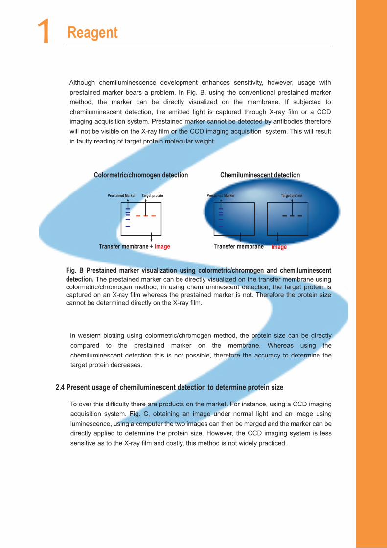
Although chemiluminescence development enhances sensitivity, however, usage with prestained marker bears a problem. In Fig. B, using the conventional prestained marker method, the marker can be directly visualized on the membrane. If subjected to chemiluminescent detection, the emitted light is captured through X-ray film or a CCD imaging acquisition system. Prestained marker cannot be detected by antibodies therefore will not be visible on the X-ray film or the CCD imaging acquisition system. This will result in faulty reading of target protein molecular weight.
In western blotting using colormetric/chromogen method, the protein size can be directly compared to the prestained marker on the membrane. Whereas using the chemiluminescent detection this is not possible, therefore the accuracy to determine the target protein decreases.
2.4 Present usage of chemiluminescent detection to determine protein size
To over this difficulty there are products on the market. For instance, using a CCD imaging acquisition system. Fig. C, obtaining an image under normal light and an image using luminescence, using a computer the two images can then be merged and the marker can be directly applied to determine the protein size. However, the CCD imaging system is less sensitive as to the X-ray film and costly, this method is not widely practiced.
Colormetric/chromogen detection Chemiluminescent detection
Prestained Marker Target protein Target proteinPrestained Marker
Transfer membrane + Image Transfer membrane Image
Fig. B Prestained marker visualization using colormetric/chromogen and chemiluminescent detection. The prestained marker can be directly visualized on the transfer membrane using colormetric/chromogen method; in using chemiluminescent detection, the target protein is captured on an X-ray film whereas the prestained marker is not. Therefore the protein size cannot be determined directly on the X-ray film.

1LuminolPenTM HRP system
Target Protein
Prestained Marker
Capture Normal Image
Merged Image
CCD Imaging acquisition sysytem
Capture luminescence Image
Target proteinPrestained Marker
Transfer Membrane X-ray Film
Overlapping
Difficult
Fig. C Image merges using the CCD imaging acquisition system. Obtaining an image under normal light and an image using luminescence, using a computer the two images can then be merged and the marker can be directly applied to determine the protein size.
Fig. D. Using transfer membrane and X-ray film to estimate the protein molecular weight. Using superimposed image of the chromogen membrane and the X-ray film. This method is relatively inaccurate as it does not have a datum point.
For laboratories not equipped with CCD imaging system or X-ray film development system other method can be applied. Fig. D. An estimation of protein size using superimposed image of the chromogen membrane and the X-ray film. This method is relatively inaccurate as it does not have a datum point.

1 Reagent
Alternatively, users can chose to use the ECLTM DualVue Western Blotting Markers by GE. The western blotting markers are specifically detected by –HRP conjugate and developed using chemiluminescent substrates on X-ray film or CCD imaging acquisition system. Due to the high cost of the kit, it is not widely used.
2.5 Chemiluminescent indirectly affects the outcome results of western blot.
As stated in the previous section, there are ways to overcome molecular weight calibration using chemiluminescent detection. Using CCD imaging acquisition system or ECLTM DualVue Western Blotting Markers can accurately calibrate protein weight; however, the equipments and reagents are costly. In addition, CCD imaging acquisition system is less sensitive compared to X-ray film, therefore the film is more widely used.
The disadvantage of using the X-ray film is that the prestained marker cannot be used for molecular weight calibration. For example, in Fig. E, during western blotting if the antibody specificity is not high, this will leading to non-specific binding and unable to determine which is the target protein. Molecular weight calibration must then be used to determine the target. Prestained marker can be directly visualized on the transfer membrane using colormetric/chromogen method. Chemiluminescent detection where using superimposed image of the transfer membrane and the film will not give an accurate reading due to this method does not have a datum point. Faulty reading can affect the development of a project, needless to say a great deal of time and money wasted and resulting in delay of publications.
97
(KDa)
66 45
30
20
Target Protein
Colormetric/chromogen detection Chemiluminescent detection
Up
Down?
Fig. E Calibration of molecular weight using chemiluminescent detection. Note: western blotting can result in binding of protein other than the target protein. (A) Prestained marker can be directly visualized on the transfer membrane using colormetric/chromogen method. (B) Chemiluminescent detection where using superimposed image of the transfer membrane and the film will not give an accurate reading due to this method does not have a datum point.
(A) (B)
When submitting an article with western blot data, the determinacy of target protein molecular weight is of importance. Reviewers will require authors to address this issue with direct evidence. However, to present there is still not an effective and direct method to demonstrate molecular weight calibration using chemiluminescent detection. Therefore,
1LuminolPenTM HRP system
Characteristics and advantages of LuminolPenTM HPR system recongnizes as follows
█ The world’s first luminescent color development marker penInnovated to provide the best way using luminescent for target(s) indication directly on the X-ray film.
█ Detection of molecular weight using chemiluminescent detection
In chemiluminescent detection, LuminolPenTM HRP system marks the prestained marker position, which can be viewed on the film or the CCD imaging acquisition system. Prevent erroneous molecular weight calibration.
often results are questioned and the articles may be rejected.
2.6 LuminolPenTM HRP system provides the solution
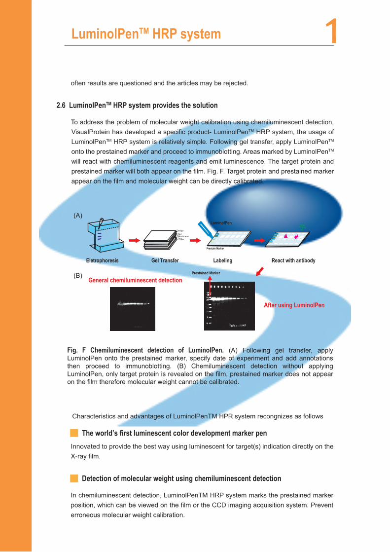
To address the problem of molecular weight calibration using chemiluminescent detection, VisualProtein has developed a specific product- LuminolPenTM HRP system, the usage of LuminolPenTM HRP system is relatively simple. Following gel transfer, apply LuminolPenTM onto the prestained marker and proceed to immunoblotting. Areas marked by LuminolPenTM will react with chemiluminescent reagents and emit luminescence. The target protein and prestained marker will both appear on the film. Fig. F. Target protein and prestained marker appear on the film and molecular weight can be directly calibrated.
General chemiluminescent detection
After using LuminolPen
Prestained Marker
LuminolPen
Prestain Marker
Filter
Filter
GelMembrane
Eletrophoresis Gel Transfer Labeling React with antibody
Fig. F Chemiluminescent detection of LuminolPen. (A) Following gel transfer, apply LuminolPen onto the prestained marker, specify date of experiment and add annotations then proceed to immunoblotting. (B) Chemiluminescent detection without applying LuminolPen, only target protein is revealed on the film, prestained marker does not appear on the film therefore molecular weight cannot be calibrated.
(A)
(B)
1 Reagent
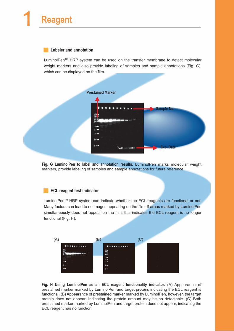
Fig. G LuminolPen to label and annotation results. LuminolPen marks molecular weight markers, provide labeling of samples and sample annotations for future reference.
Exp. Date
Prestained Marker
Sample No.
█ ECL reagent test indicator
LuminolPenTM HRP system can indicate whether the ECL reagents are functional or not. Many factors can lead to no images appearing on the film. If areas marked by LuminolPen simultaneously does not appear on the film, this indicates the ECL reagent is no longer functional (Fig. H).
█ Labeler and annotation
LuminolPenTM HRP system can be used on the transfer membrane to detect molecular weight markers and also provide labeling of samples and sample annotations (Fig. G), which can be displayed on the film.
Fig. H Using LuminolPen as an ECL reagent functionality indicator. (A) Appearance of prestained marker marked by LuminolPen and target protein, indicating the ECL reagent is functional. (B) Appearance of prestained marker marked by LuminolPen, however, the target protein does not appear. Indicating the protein amount may be no detectable. (C) Both prestained marker marked by LuminolPen and target protein does not appear, indicating the ECL reagent has no function.
(A) (B) (C)
1LuminolPenTM HRP system
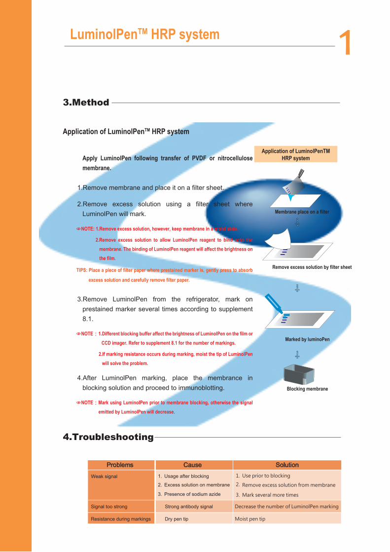
Application of LuminolPenTM HRP system
Apply LuminolPen following transfer of PVDF or nitrocellulose membrane.
1.Remove membrane and place it on a filter sheet.
2.Remove excess solution using a filter sheet where LuminolPen will mark.
NOTE: 1.Remove excess solution, however, keep membrane in a moist state.
2.Remove excess solution to allow LuminolPen reagent to bind onto the membrane. The binding of LuminolPen reagent will affect the brightness on the film.
TIPS: Place a piece of filter paper where prestained marker is, gently press to absorb
excess solution and carefully remove filter paper.
3.Remove LuminolPen from the refrigerator, mark on prestained marker several times according to supplement 8.1.
NOTE:1.Different blocking buffer affect the brightness of LuminolPen on the film or CCD imager. Refer to supplement 8.1 for the number of markings.
2.If marking resistance occurs during marking, moist the tip of LuminolPen will solve the problem.
4.After LuminolPen marking, place the membrance in blocking solution and proceed to immunoblotting.
NOTE:Mark using LuminolPen prior to membrane blocking, otherwise the signal emitted by LuminolPen will decrease.
Membrane place on a filter
Remove excess solution by filter sheet
Marked by luminoPen
Blocking membrane
Problems Cause
Weak signal
1. Use prior to blocking
2. Remove excess solution from membrane
3. Mark several more times
1. Usage after blocking
2. Excess solution on membrane
Presence of sodium azide3.
Solution
Signal too strong Strong antibody signal Decrease the number of LuminolPen marking
Resistance during markings Dry pen tip Moist pen tip
Application of LuminolPenTM HRP system
3.Method
4.Troubleshooting
1 Reagent
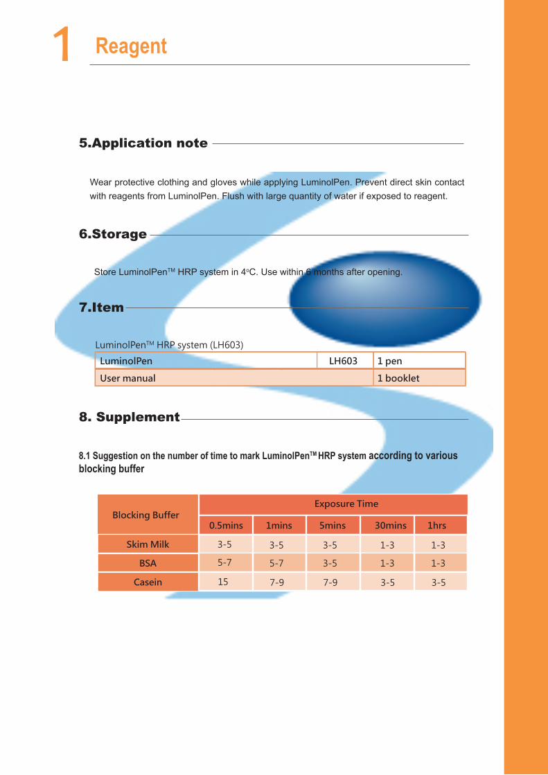
Wear protective clothing and gloves while applying LuminolPen. Prevent direct skin contact with reagents from LuminolPen. Flush with large quantity of water if exposed to reagent.
Store LuminolPenTM HRP system in 4oC. Use within 6 months after opening.
LuminolPenTM HRP system (LH603)
LH603LuminolPen
User manual
1 pen
1 booklet
3-5Skim Milk
BSA
0.5mins 1mins 5mins 30mins 1hrs
Casein
Blocking BufferExposure Time
5-7
3-5
5-7
3-5
3-5
1-3
1-3
1-3
1-3
7-915 7-9 3-5 3-5
8.1 Suggestion on the number of time to mark LuminolPenTM HRP system according to various blocking buffer
5.Application note
6.Storage
7.Item
8. Supplement