2 fingerprinting 3 accepted€¦ · 10 rd., klong 1, klong luang, pathumthani 12120, thailand 11 3...
TRANSCRIPT

1
Investigation of the Lactic Acid Bacteria in Sauerkraut Fermentations by DNA 1
fingerprinting 2
3
Vethachai Plengvidhya1,2., Fredrick Breidt, Jr.1*, Zhongjing Lu1,3, Henry P. Fleming1 4
5
6
1 USDA-ARS, Department of Food Science, 322 Schaub Hall, North Carolina State University, 7
Raleigh, NC 27695-7624, USA, * corresponding author 8
2 Current address: National Center for Genetic Engineering and Biotechnology, 113 Paholyothin 9
Rd., Klong 1, Klong Luang, Pathumthani 12120, Thailand 10
3 Current address: Department of Biology and Physics, Kennesaw State University, 1000 11
Chastain Rd., Kennesaw, GA 30144 12
13
14
Running head: Microbiota of sauerkraut 15
16
17
Paper no. FSR06-22 of the Journal Series of the Department of Food Science, NC State 18
University, Raleigh, NC 27695-7624. Mention of a trademark or proprietary product does not 19
constitute a guarantee or warranty of the product by the U.S. Department of Agriculture or North 20
Carolina Agricultural Research Service, nor does it imply approval to the exclusion of other 21
products that may be suitable. 22
ACCEPTED
Copyright © 2007, American Society for Microbiology and/or the Listed Authors/Institutions. All Rights Reserved.Appl. Environ. Microbiol. doi:10.1128/AEM.01342-07 AEM Accepts, published online ahead of print on 5 October 2007
on June 26, 2020 by guesthttp://aem
.asm.org/
Dow
nloaded from
2
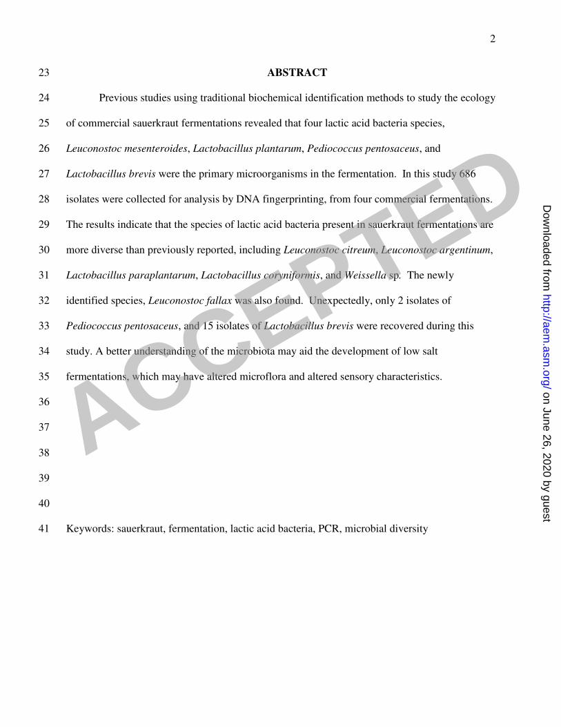
ABSTRACT 23
Previous studies using traditional biochemical identification methods to study the ecology 24
of commercial sauerkraut fermentations revealed that four lactic acid bacteria species, 25
Leuconostoc mesenteroides, Lactobacillus plantarum, Pediococcus pentosaceus, and 26
Lactobacillus brevis were the primary microorganisms in the fermentation. In this study 686 27
isolates were collected for analysis by DNA fingerprinting, from four commercial fermentations. 28
The results indicate that the species of lactic acid bacteria present in sauerkraut fermentations are 29
more diverse than previously reported, including Leuconostoc citreum, Leuconostoc argentinum, 30
Lactobacillus paraplantarum, Lactobacillus coryniformis, and Weissella sp. The newly 31
identified species, Leuconostoc fallax was also found. Unexpectedly, only 2 isolates of 32
Pediococcus pentosaceus, and 15 isolates of Lactobacillus brevis were recovered during this 33
study. A better understanding of the microbiota may aid the development of low salt 34
fermentations, which may have altered microflora and altered sensory characteristics. 35
36
37
38
39
40
Keywords: sauerkraut, fermentation, lactic acid bacteria, PCR, microbial diversity 41
ACCEPTED
on June 26, 2020 by guesthttp://aem
.asm.org/
Dow
nloaded from

3
INTRODUCTION 42
Sauerkraut fermentation involves many physical, chemical and microbiological changes 43
that influence quality and safety of the product. The fermentation can be broadly categorized as 44
having successive stages, including an initial heterofermentative stage, followed by a 45
homofermentative stage (11, 23). Historically, four species of lactic acid bacteria have been 46
identified as present in sauerkraut fermentations: Leuconostoc mesenteroides, Lactobacillus 47
brevis, Pediococcus pentosaceus, Lactobacullus plantarum. The identification of these 48
microorganisms has been based on morphological and biochemical criteria (22). Several species 49
of lactic acid bacteria (LAB), other than the four species mentioned above, have been found in 50
cabbage fermentations, including Lactobacillus curvatus, Lactobacillus sake, Lactococcus lactis 51
subsp. lactis, and Leuconostoc fallax (3, 19, 25). Recently, six L. fallax strains have been isolated 52
from brine samples obtained from sauerkraut fermentations (2). The taxonomic characterization 53
of LAB has been modified, and new species have been described using molecular techniques (1, 54
10, 20, 24) . Improvements in molecular identification techniques for the study of microbial 55
ecology have created new opportunities for the analysis of food fermentations. 56
This study was carried out because of the need to reduce sodium chloride (salt) waste 57
from commercial vegetable fermentations. It is well documented that the concentration of salt 58
has a controlling influence on the microbial succession in a typical sauerkraut fermentation (11, 59
12, 22) . It may be possible to reduce salt waste by fermenting cabbage with 1% instead of the 60
typical 2% salt concentration. The introduction of a L. mesenteroides starter culture to the 61
fermentation could help ensure that the initial stage of the fermentation produces the desirable 62
flavor compounds (11). A method has been developed (23) to determine the ability of an 63
unmarked starter culture predominate over the indigenous microbiota in sauerkraut 64
ACCEPTED
on June 26, 2020 by guesthttp://aem
.asm.org/
Dow
nloaded from

4
fermentations. In that study (23) however, the effect of the starter culture on the indigenous 65
microbiota was not determined. 66
During the sauerkraut fermentation there is a rapid turnover of LAB species. The 67
dominant species present in the fermentation shifts within two to three days from less acid-68
tolerant heterolactic LAB species to more acid-tolerant homolactic fermenting LAB species, with 69
sequential populations each reaching 108 to 109 CFU/g (11). Under normal conditions, the 70
fermentation is essentially complete within two weeks, with the most acid-tolerant species L. 71
plantarum predominating. Our objective was to characterize the dominant LAB species in the 72
successive stages of fermentation. There is strong evidence that the genetic diversity of many 73
ecosystems, as assessed by molecular techniques, exceeds the microbial diversity determined by 74
traditional culture based identification methods (26). However, due to the rapid succession of 75
LAB in sauerkraut, and the potential impact of large numbers of dead cells on culture-free 76
nucleic acid-based methods, we carried out this study using bacterial isolates. In this report we 77
characterized isolates from the microbiota of commercial sauerkraut fermentations using a rDNA 78
intergenic spacer PCR method (ITS-PCR) with a database of known ITS-PCR patterns for LAB 79
(4, and this report), supplemented with 16S rDNA sequence analysis. Several species of LAB 80
not previously found in sauerkraut were observed. Notably, Weissella and Leuconostoc citrium 81
species were found in the heterolactic phase of the fermentations, and two (P. pentosaceus and L. 82
brevis) of the four LAB species expected to be present were apparently minor constituents of the 83
microbiota. A better understanding of the microbial ecology of sauerkraut fermentations may aid 84
in the development of low salt fermentation technology, which may alter normal microflora and 85
sauerkraut flavor (Breidt and coworkers, unpublished). 86
87
ACCEPTED
on June 26, 2020 by guesthttp://aem
.asm.org/
Dow
nloaded from

5
MATERIALS AND METHODS 88
Bacterial strains: Bacterial strains were obtained from the Food Fermentation Culture 89
Collection, US Department of Agriculture, Agriculture Research Service, Raleigh, NC (Suppl. 90
Table 1), or isolated as described below. LAB were maintained at –80°C in MRS broth (Difco 91
Laboratories) supplemented with 16% glycerol, and grown on MRS broth or agar plates (MRS 92
broth supplemented with 1.5% agar, Difco). To inhibit aerobic yeast or mold growth on MRS 93
agar plate with sauerkraut isolates, MRS agar was supplemented with 0.2% sodium azide (Sigma 94
Chemical Co., St. Louis, MO), or plates were incubated in an anaerobic hood (Coy Laboratories, 95
Detroit, MI). 96
Commercial sauerkraut fermentation and sample collection: Sauerkraut 97
fermentations at a commercial processing plant in Wisconsin consisting of approximately 100 98
megagrams of cabbage were sampled for this study, including one tank in the first year of the 99
study (Y1), and three different tanks in the second year (Y2). Only one sauerkraut production 100
facility in Wisconsin was used in this study, because there are currently very few commercial 101
production facilities in the US. The fermentation tanks were of cement construction, with the 102
approximate dimensions: 5 m length by 5 m width by 4 m deep. Each fermentation was carried 103
out with approximately 2.3 % NaCl (final equilibrated concentration), which was added by a dry 104
salting process using shredded cabbage from mixed cultivars. The cabbage was manually spread 105
in the tanks, covered with plastic sheeting, and initially weighted down with water on top of the 106
plastic sheeting. The fermentation temperature was not controlled, but the temperature in these 107
commercial fermentations typically averaged 18˚C. For analysis of cabbage prior to 108
ferementation, shredded cabbage samples (approximately 500 g) were collected in sterile plastic 109
bags prior to salting. Brine samples (100 ml each) from the fermentations were obtained for 110
ACCEPTED
on June 26, 2020 by guesthttp://aem
.asm.org/
Dow
nloaded from

6
microbial and biochemical analysis with a one-cm-diameter, perforated stainless steel tube that 111
was sealed at the bottom, from a depth of approximately 60 cm from the tops of the fermentation 112
tanks, and about 60 cm from the sides. The sampling apparatus was sanitized with dilute (10%) 113
Clorox solution and rinsed with sterile water prior to use. Brine and cabbage samples were 114
obtained on days 1, 3, 7, and 14 after the start of fermentation and were placed in 2 x 50 ml 115
sterile plastic tubes (#430829 Corning Inc., Corning, NY). The cabbage and brine samples were 116
transported to the laboratory by overnight mail in insulated boxes containing wet-ice packs to 117
maintain a temperature between 0 and 5oC. Samples were processed immediately upon arrival. 118
Chemical and microbial analysis of cabbage samples. For chemical and 119
microbiological analysis, 100 g of the shredded cabbage was blended in sterile glass blender jars 120
with 200 g of water for 3 min in a Waring blender (Waring Products, Torrington, CT) and then 121
homogenized in a stomacher (Stomacher 400, Tekmar, Cincinnati, OH) for 3 min at the 122
maximum speed using bags containing an internal filter. Filtrate from the extract, approximately 123
30 mL, was transferred to a 50-mL sterile plastic tube and frozen at -20˚C for subsequent 124
chemical analysis. Prior to freezing, one mL of the cabbage extract was removed for 125
microbiological analysis (see below). Chemical analysis was carried out using high-performance 126
liquid chromatography. Organic acids and ethanol were analyzed using an anion-exchange 127
column (Aminex HPX-87H, Bio-Rad Laboratories) with a 0.8 mL/min flow rate of 0.03 N 128
H2SO4 at 75˚C. A UV detector (UV-6000, Thermo Separation Products Inc., San Jose, CA) and a 129
differential refractometer (Waters 410, Waters, Milford, MA) were connected in series for 130
detection of organic acids (at 210 nm) and ethanol. Sugars and mannitol were separated by a 131
Carbopac PA1 column (Dionex Corp., Sunnyvale, CA) with a 0.8 mL/min flow rate of 0.12 N 132
NaOH at room temperature, and detected by a pulsed amperometric detector (model PAD-2; 133
ACCEPTED
on June 26, 2020 by guesthttp://aem
.asm.org/
Dow
nloaded from

7
Dionex). The salt (NaCl) content in brine was determined by titration with AgNO3 solution using 134
4’5’-dichlorofluorescencein as the indicator (13). 135
For microbial analysis, samples were diluted in sterile saline (0.85% NaCl) and plated on: 136
plate count agar (PCA, Difco Laboratories, Detroit, MI), modified violet red bile agar VRBG 137
(VRB agar, Difco, supplemented with 1% glucose), modified De Man, Rogosa and Sharpe 138
(MMRS) agar (MRS, Difco, supplemented with 0.2% sodium azide), and yeast extract and malt 139
agar (YM, Difco), containing chlortetracycline and chloramphenicol at 250 mg/L each, (Sigma 140
Aldrich, St. Louis, MO), to enumerate total aerobic microbiota, Enterobacteriaceae, LAB, yeasts 141
and molds, respectively. In addition, each brine sample was plated on unmodified MRS agar 142
(without sodium azide) for collection of LAB isolates. Plating and plate counting were 143
performed using a spiral plater (Model 4000, Spiral Biotech, Norwood, MA) with an automated 144
colony counter (Protos Plus, Microbiology International, Frederick, MD). From each of the five 145
sampling times (days 1, 3, 5, 7, and 14 after the start of fermentation) in Y1, 96 isolated colonies 146
from MRS agar were randomly selected and isolated on MRS agar, then cells were frozen in 147
MRS broth containing 16% glycerol at –80°C. For the four sampling times in Y2 (days 1, 3, 7, 148
and 14), 20 isolates were similarly obtained from each of three fermentation tanks, resulting in a 149
combined total for both years of the study of 720 possible isolates, although only 686 isolates 150
were recovered. The isolates were screened for gas production using Durham tubes (6 x 50 mm, 151
Kimble) inverted in 5 ml MRS broth. In addition, cells were cultured on homolactic-heterolactic 152
differentiation (HHD) medium (17). 153
DNA extraction and PCR amplification: MRS broth cultures from each fermentation 154
isolate were incubated at 30°C 12-16 h and then subjected to DNA extraction. Genomic DNA 155
was isolated using a Wizard Genomic DNA Purification kit (Promega Corporation, Madison, 156
ACCEPTED
on June 26, 2020 by guesthttp://aem
.asm.org/
Dow
nloaded from

8
WI) in accordance with the manufacturer’s instructions, with minor modification. Twenty 157
microliters of mutanolysin (2.4 mg/ml, Sigma-Aldrich) was substituted for lysostaphin. The 158
method of Breidt and Fleming (4) was used to amplify the intergenic transcribed spacer (ITS) 159
region between 16S and 23S rRNA genes. Each 100 µl of reaction mixture consisted of 10 µl of 160
10X PCR buffer (500mM KCl and 100mM Tris-Cl, pH 8.0), 10 µl of 25 mM MgCl2, 1 µl of 161
dNTP mixture (25mM each dNTP, Stratagene), 4 µl of DNA preparation as described above, 70 162
µl of water, 1 µl of Taq DNA polymerase (5 U/µl), and 2 µl of each primer. The primers used for 163
PCR amplification (4) were GAAGTCGTAACAAGG and GGGTTTCCCCATTCGGA. All 164
primers in this report were obtained from Sigma-Genosys (Sigma-Aldrich, St. Louis, MO). PCR 165
reactions were carried out using a model GTC-2 Genetic Thermal Cycler with a model LTM-2 166
refrigeration unit (Precision Scientific Inc., Chicago, IL). The temperature program consisted of 167
an initial heat denaturation step at 94°C for 5 min, 25 cycles of 1 min at 94°C, 5 min at 55°C, 168
and 2 min at 72°C followed by a final 5 min interval at 72°C. DNA products from the PCR 169
reaction were treated (without purification) with 1 µl of Rsa I enzyme solution (16 U/µl. No. 170
500890, Stratagene, La Jolla, CA) for 1 h at 37°C. The restriction digest samples were analyzed 171
by electrophoresis in 5% nondenaturing polyacrylamide gels using a vertical gel electrophoresis 172
apparatus (BRL Model V16, Invitrogen, Carlsbad, CA). The DNA-banding profiles were 173
identified by ethidium bromide staining and were subsequently analyzed using the GelCompar II 174
software (Applied Maths, Inc., Austin, TX). For sequencing the 16S rDNA variable regions V1 175
and V2 (21), the primers (2) used for amplification of the 5’ end (approximately 300 bases) of 176
the 16S gene were 5’-AGAGTTTGATCCTGGCTCAG-3’and 5’-177
GTCTCAGTCCCAATGTGGCC-3’. The cycle times and temperatures were: 10 min at 94˚C; 25 178
cycles of 1 min at 94˚C, 2 min at 61˚C, and 2 min at 72˚C; and 5 min at 72˚C. Alternatively, 179
ACCEPTED
on June 26, 2020 by guesthttp://aem
.asm.org/
Dow
nloaded from

9
primers 5’-AGTTTGATCMTGGCTCAG-3’ (M=A or C), and 5’-180
AGGAGGTGATCCARCCGCA-3’ (R=A or G) were used to amplify the entire 16S rDNA gene 181
(7) substituting an annealing temperature of 55oC. PCR products were purified using a 182
QIAquick PCR purification kit (Qiagen Inc., Valencia, CA.) and the DNA fragments were 183
sequenced commercially (Davis Sequencing, Davis, CA). Sequences were analyzed by BLAST 184
(Basic Local Alignment Search Tool) search of the NCBI (National Center for Biotechnology 185
Information) non-redundant DNA sequence database (http://www.ncbi.nlm.nih.gov/). 186
187
RESULTS 188
Fermentation microbiology and chemistry: The shredded cabbage used for all 189
fermentations in this study contained between 4 x 10⁶ and 6 x 10⁶ cfu/g total aerobes, 2 to 3 x 10⁶ 190
cfu/g total Enterobacteriaceae, and less than102 cfu/g yeast and mold. However, the initial LAB 191
populations from the shredded cabbage varied from 104 cfu/g in the Y1 cabbage to 106 cfu/g 192
from two of the Y2 cabbage samples. It is possible some growth of LAB occurred during 193
transport, prior to analysis. As previously reported (11, 22) , during the first week of each 194
fermentation a rapid increase in total and lactic acid bacteria occurred, as well as a rapid decrease 195
in Enterobacteriaceae (data not shown). 196
Glucose and fructose were the primary fermentable sugars in the cabbage (between 1.5% 197
and 2.2%, respectively). Sucrose made up only a small amount of the fermentable sugars, less 198
than 0.2% of the cabbage by weight, and was not detectable in Y2 samples. Overall, the cabbage 199
used in Y1 contained more sugar than the Y2 samples. However, sugar utilization, acid 200
production, and pH profiles were similar in the commercial tanks from Y1 and Y2. The results 201
from chemical analysis indicated that the fermentation in the four commercial sauerkraut 202
ACCEPTED
on June 26, 2020 by guesthttp://aem
.asm.org/
Dow
nloaded from

10
fermentation tanks from Y1 (Fig. 1) and Y2 (not shown) were normal and consistent with those 203
described by Fleming et. al. (14) and Peterson and Albury (22). During the first week of 204
fermentation lactic acid, acetic acid and mannitol were produced. The pH on day 14 for all 205
fermentations was in the range of 3.4 to 3.7 (data not shown). 206
ITS-PCR database and 16S rDNA sequence analysis: A database of ITS-PCR gel 207
banding patterns was generated using 64 LAB strains from 42 different species (Suppl. Table 1 208
and Suppl. Fig. 1), using both the RsaI digested and undigested PCR products. The 686 209
fermentation isolates obtained from brine during the two year study were grouped by ITS-PCR 210
banding patterns using the GenCompar II software, for both the RsaI digested and undigested 211
PCR products. Banding patterns for the isolates were grouped by unique ITS-PCR banding 212
patterns, and 652 isolates tentatively identified using the ITS-PCR database. One or more 213
representative cultures from each group with unique banding patterns were then further 214
characterized by sequencing the 5’ end of the 16S rDNA. The sequence accession numbers, 215
along with the corresponding bacterial strain identification by BLAST analysis, are shown in 216
Suppl. Data Table 2. By this method L. mesenteroides was identified as the predominant species 217
(179 of 686 isolates) in the early heterofermentative stage of fermentation and L. plantarum was 218
the predominant species involved in the late homofermentative stage (280 of 686 isolates). 219
Together, these two species made up 2/3 of all isolates (67%). Weissella sp. and L. curvatus were 220
the next most common species among the isolates, with 57 (8.3%) and 40 (5.8%) total isolates 221
from the two year study. The majority of LAB isolates (90% to 100%) taken from days 1 and 3 222
after the start of fermentation for the Y1 and Y2 samples were heterofermentative species. 223
Homofermentative LAB made up a similar majority of the isolates from day 14 (Figure 2A and 224
2B). 225
ACCEPTED
on June 26, 2020 by guesthttp://aem
.asm.org/
Dow
nloaded from

11
Microbial diversity in commercial sauerkraut fermentations: Three different 226
bacterial species were found in the Y1, day one samples: L. mesenteroides, Weissella sp., and L. 227
citreum (Table 2, Figure 3). As expected, L. mesenteroides was the predominant organism 228
associated with the fermentation, comprising 88% (84 out of 95) of the isolates from the first 229
day. L. mesenteroides was the most frequently isolated species in the brine samples from day 3, 230
but it contributed only 41% (38 out of 93) of total isolates. Other species found on day 3 231
included: L. curvatus (15%), Weissella sp. (14%), L. fallax (9%) and others (Table 2). 232
Homofermentative L. plantarum strains were observed in the fermentation samples from day 3, 233
but only accounted for 9.7% of the isolates. We observed variation in the pattern of hetero- and 234
homofermentative LAB between the Y1 and three Y2 fermentations. Samples from Tank 3 of Y2 235
were unique when compared to the samples from other tanks, because both heterofermentative 236
and homofermentative LAB were present in all samples. 237
As expected, bacterial isolates from Y1 days 7, 9 and 14 contained no heterofermentative 238
L. mesenteroides or Weissella sp. isolates. Overall, the diversity of species was greatest from the 239
day 9 samples from Y1, when 9 different species were isolated, out of a total of 90 isolates. The 240
majority of isolates from the brine samples taken during days 7, 9 and 14, were identified as L. 241
plantarum (68.8%, 56.7%, and 78.1%, respectively). L. argentinum, which has not previously 242
been found in sauerkraut fermentations, accounted for 10% of total isolates recovered from Y1 243
day 9. Interestingly, only one isolate of P. pentosaceus and 14 isolates of L. brevis were found 244
among the 467 total Y1 isolates. Of the 279 Y1 isolates from days 7, 9 and 14 a total of 28 were 245
not identified by ITS-PCR pattern or 16S rDNA sequencing. 246
Isolates from the three Y2 fermentation tanks had bacterial species profiles as described 247
in Table 3. In tank one, three heterofermentative LAB species were recovered from day one, L. 248
ACCEPTED
on June 26, 2020 by guesthttp://aem
.asm.org/
Dow
nloaded from

12
mesenteroides, Weissella sp., and L. citreum. However Weissella sp. represented the majority of 249
isolates recovered (12 of 20 in day 1, and 7 of 19 in day 3). L. curvatus, L. fallax, and L. 250
plantarum were also recovered from day 3 brine samples. L. plantarum was the major LAB 251
species isolated from the Y2 tank one sample from days 7 and 14, representing over 46% and 252
94% of isolates, respectively. The bacterial species from tank two profiles were similar to the 253
tank one fermentation (Table 3). At day 14, L. plantarum was the only LAB isolated from the 254
brine sample, accounted for 100% (18 of 18) of the isolates. In tank three, however, 8 different 255
species were identified from 17 isolates from day one. Only 1 P. pentosaceus and 1 L. brevis 256
isolate was recovered from the 219 total Y2 isolates. 257
258
DISCUSSION 259
We used the ITS-PCR database to identify major bacterial species present in commercial 260
sauerkraut fermentations. It is possible that different bacterial species could be present that have 261
very similar or indistinguishable ITS patterns, or that some bacterial species did not grow on 262
MRS medium. Because we only sequenced 16S DNA from isolates with representative ITS-PCR 263
patterns, we may have failed to identify some species that were present among the isolates. The 264
data presented, therefore, may underreport the microbial diversity in the sauerkraut 265
fermentations. Analysis of ITS-PCR patterns and 16S rDNA sequences from the isolates, 266
however, agreed with the biochemical analysis indicating that heterofermentative LAB species 267
dominated the first week of the commercial sauerkraut fermentations, and homofermentative 268
species were predominate in the second week. Weissella sp. were recovered together with L. 269
mesenteroides both in day one and three. We could not identify species of any Weissella sp. 270
isolates by 16S rDNA sequence comparisons with the GenBank database because of the 271
ACCEPTED
on June 26, 2020 by guesthttp://aem
.asm.org/
Dow
nloaded from

13
sequence similarity of Weissella confusa, Weissella cibraria, and Weissella kimchii. Weissella 272
species have previously been recovered from plant products (5, 8, 18), and have very similar 16S 273
rDNA sequences (3, 5). Complete sequencing of the 16S genes from selected Weissella isolates 274
did not allow conclusive identification with known species (data not shown). Weissella sp. were 275
previously characterized as members of the Leuconostoc genus (6). There was variation between 276
the three Y2 fermentations in microbiota from the heterolactic (days one and three) stage of 277
fermentation, which is believed to be vital to the quality of sauerkraut. This variation may be due 278
in part to the small sampling size (20 isolates from each sampling time, and from each 279
fermentation in Y2) but could represent differences in microbiota. These data indicate that the 280
use of L. mesenteroides starter cultures may help improve consistency in flavor development in 281
commercial sauerkraut. 282
During the transition period between the heterofermentation and homofermentation 283
phases of fermentation (days three to nine), a variety of LAB species including L. curvatus and 284
Leuconostoc argentinum were isolated. L. argentinum was originally isolated from raw milk in 285
Argentina (9). L. plantarum became the dominant microorganism after seven days into the 286
fermentation when the pH decreased to at least 3.9 or lower in all 4 fermentations. However, we 287
recovered relatively few L. brevis (15 total isolates of 686) and only twoisolates of P. 288
pentosaceus. This was surprising because the previous research has shown that both were 289
considered to be major bacterial species involved in sauerkraut fermentation (22). 290
This study and a related study of the bacteriophage ecology of commercial sauerkraut 291
fermentations (15), has significantly revised the classical understanding (11, 22) of the 292
microbiota present in sauerkraut fermentations. Several LABs isolated from the two-year study 293
had never been previously recovered from sauerkraut fermentation including Weissella sp., L. 294
ACCEPTED
on June 26, 2020 by guesthttp://aem
.asm.org/
Dow
nloaded from

14
argentinum, Lactobacillus coryniformis, Leuconostoc citreum, Lactobacillus paraplantaum, and 295
Lactobacillus paracasei. This study, and a recent study of the comparative genomics of lactic 296
acid bacteria, including L. mesenteroides (16), may also aid in the development of starter 297
cultures, and increase our understanding of cabbage fermentation biochemistry and ecology. 298
299
300
ACKNOWLEDGEMENTS 301
This investigation was supported in part by a research grant from Pickle Packers 302
International, Inc. (Washington, D.C.). We thank Bush Brothers and Company and the Great 303
Lakes Kraut Company for their help and support of this research. We thank Ms. Janet Hayes and 304
Dr. Roger McFeeters for aid with experiments and helpful discussions, and Ms. Sandra L. Parker 305
for excellent secretarial assistance. 306
ACCEPTED
on June 26, 2020 by guesthttp://aem
.asm.org/
Dow
nloaded from

15
REFERENCES 307
308
1. Aguirre, M. and M. D. Collins. 1993. Lactic Acid Bacteria and Human Clinical Infection. 309
J. Appl. Bacteriol. 75:95-107. 310
2. Barrangou, R., S.-S. Yoon, F. Breidt, H. P. Fleming, and T. R. Klaenhammer. 2002. 311
Characterization of Six Leuconostoc fallax Bacteriophages Isolated from an Industrial 312
Sauerkraut Fermentation. Appl. Environ. Microbiol. 68:5452-5458. 313
3. Bjorkroth, K. J., U. Schillinger, R. Geisen, N. Weiss, B. Hoste, W. H. Holzapfel, H. J. 314
Korkeala, and P. Vandamme. 2002. Taxonomic study of Weissella confusa and description 315
of Weissella cibaria sp. Nov., detected in food and clinical samples. Int. J. Syst. Evol. 316
Microbiol. 52:141-148. 317
4. Breidt, F. and H. P. Fleming. 1996. Identification of Lactic Acid Bacteria By Ribotyping. 318
Journal of Rapid Methods and Automation in Microbiology 4:219-233. 319
5. Choi, H. K., C. C. Ick, K. S. Bo, L. J. Choul, L. D. Woo, C. S. Won, P. J. Min, and P. Y. 320
Ryang. 2002. Weissella kimchii sp. nov., a novel lactic acid bacterium from kimchi. Int. J. 321
Syst. Evol. Microbiol. 52:507-511. 322
6. Collins, M. D., J. Samelis, J. Metaxopoulos, and S. Wallbanks. 1993. Taxonomics studies 323
on some Leuconostoc-like organisms from fermented sausages: description of a new genus 324
Weissella for the Leuconostoc paramesenteroides group of species. J. Appl. Bacteriol. 325
75:595-603. 326
7. Dasen, G., J. Smutney, M. Teuber, and L. Meile. 1998. Classification and identification of 327
ACCEPTED
on June 26, 2020 by guesthttp://aem
.asm.org/
Dow
nloaded from

16
Propionibacteria based on ribosomal RNA genes and PCR. System Appl. Microbiol. 328
21:251-259. 329
8. De-Vuyst, L., V. Schrijvers, S. Pararmithiotis, B. Hoste, M. Vancanneyt, J. Swings, G. 330
Kalantzopoulos, E. Tsakalidou, and W. Messens. 2002. The biodiversity of lactic acid 331
bacteria in Greek traditional wheat sourdoughs is reflected in both composition and 332
metabolite formation. Appl. Environ. Microbiol. 68:6059-6069. 333
9. Dicks, L. M. T., L. Fantuzzi, F. C. Gonzalez, M. Du Toit, and F. Dellaglio. 1993. 334
Leuconostoc argentinum sp. nov., isolated from Argentine raw milk. Int. J. Syst. Bacteriol. 335
43:347-351. 336
10. Du Plessis, E. M. and L. M. T. Dicks. 1995. Evaluation of Ramdom Amplified 337
Polymorphic DNA (RAPD)-PCR as a Method to Differentiate Lactobacillus acidophilus, 338
Lactobacillus crispatus, Lactobacillus amylovorus, Lactobacillus gallinarum, lactobacillus 339
gasseri, and Lactobacillus johnosnii. Curr. Microbiol. 31:114-118. 340
11. Fleming, H. P., Kyung, K. H., and Breidt, F. 1995. Vegetable Fermentations. 341
Biotechnology. 9, p. 629-661. 95. New York. Rehm, H.-J. and Reed, G. 342
12. Fleming, H. P., R. F. McFeeters, and M. A. Daeschel. 1985. The Lactobacilli, Pediococci, 343
and Leuconostocs: Vegetable Products. CRC Press, Inc., Boca Raton, FL. 344
13. Fleming, H. P., R. F. McFeeters, and M. A. Daeschel. 1992. Fermented and acidified 345
vegetables. In compendium of methods for the microbiological examination of foods, 3rd 346
edition (Vanderzant, C., and D. F. Splittstoesser, eds.), p. 929-952, American public health 347
association, Washington, DC. 348
ACCEPTED
on June 26, 2020 by guesthttp://aem
.asm.org/
Dow
nloaded from

17
14. Fleming, H. P., R. F. McFeeters, J. L. Etchells, and T. A. Bell. 1984. Pickled vegetables. In 349
compendium of methods for the microbiological examination of foods, 2nd edition (M. L. 350
Speck eds.), p. 663-681, American public health association, Washington, DC. 351
15. Lu, Z., F. Breidt, Jr., V. Plengvidhya, and H. P. Fleming. 2003. Bacteriophage ecology in 352
commercial sauerkraut fermentations. Appl. Environ. Microbiol. 69 (6):3192-3202. 353
16. Makarova, K., A. Slesarev, Y. Wolf, A. Sorokin, B. Mirkin, E. Koonin, A. Pavlov, N. 354
Pavlova, V. Karamychev, N. Polouchine, V. Shakhova, I. Grigoriev, Y. Lou, D. Rohksar, 355
S. Lucas, K. Huang, D. M. Goodstein, T. Hawkins, V. Plengvidhya, D. Welker, J. Hughes, 356
Y. Goh, A. Benson, K. Baldwin, J.-H. Lee, I. Díaz-Muñiz, B. Dosti, V. Smeianov, W. 357
Wechter, R. Barabote, G. Lorca, E. Altermann, R. Barrangou, B. Ganesan, Y. Xie, H. 358
Rawsthorne, D. Tamir, C. Parker, F. Breidt, Jr., J. Broadbent, R. Hutkins, D. O' Sullivan, J. 359
Steele, G. Unlu, M. Saier, T. Klaenhammer, P. Richardson, S. Kozyavkin, B. Weimer, D. 360
Mills. 2006. Comparative Genomics of the Lactic Acid Bacteria, 2006. Proceedings of the 361
National Academy of Sciences 103 (42):15611-15616. 362
17. McDonald, L. C., McFeeters, R. F., Daeschel, M. A., and Fleming, H. P. 1987. A 363
differential medium for the enumeration of homofermentative and heterofermentative lactic 364
acid bacteria. Appl. Environ. Microbiol. 53 (6):1382-1384. 365
18. Mugula, J. K., S. Nnko, J. A. Narvhus, and T. Sorhaug. 2003. Microbiological and 366
fermentation characteristics of togwa, a Tanzanian fermented food. Int. J. Food Microbiol. 367
80:187-199. 368
19. Murcia-Martinez, A. J. and M. D. Collins. 1990. A phylogenetic Analysis of the Genus 369
ACCEPTED
on June 26, 2020 by guesthttp://aem
.asm.org/
Dow
nloaded from

18
Leuconostoc Based on Reverse Transcriptase Sequencing of 16 S rRNA. FEMS Microbiol. 370
Lett. 70:73-84. 371
20. Murcia-Martinez, A. J. and M. D. Collins. 1991. A phylogenic analysis of an atypical 372
leuconostoc: description of Leuoconostoc fallax. FEMS Microbiol. Lett. 82:55-60. 373
21. Neefs, J. M., Y. Van de Peer, P. De Rijk, S. Chapelle, and R. De Wachter. 1993. 374
Compilation of small ribosomal subunit RNA sturctures. Nucleic Acids Res. 21:3025-375
3049. 376
22. Pederson, C. S. and Albury, M. N. The Sauerkraut Fermentation. 1969. N.Y. State Agr. 377
Expt. Sta. (Geneva, N.Y.) Tech. Bull. Bulletin 824. 378
23. Plengvidhya, V., F. Breidt, and H. P. Fleming. 2004. Use of RADP-PCR as a method to 379
follow the progress of starter cultures in sauerkraut fermentations. Int. J. Food Microbiol. 380
93:287-296. 381
24. Schleifer, K. H., M. Ehrmann, C. Beimfohr, E. Brockmann, W. Ludwig, and R. Amann. 382
1995. Application of Molecular Methods for the Classification and Identification of Lactic 383
Acid Bacteria. International Dairy Journal 5:1081-1094. 384
25. Vogel, R. F., M. Lohmann, A. N. Weller, M. Hugas, and W. P. Hammer. 1991. Structural 385
similarity and distribution of small cryptic plasmids of Lactobacillus curvatus and L. sake. 386
FEMS Microbiol. Lett. 84:183-190. 387
26. Ward, D. M., R. Weller, and M. M. Bateson. 1990. 16S rRNA Sequences Reveal 388
Numerous Uncultured Microorganisms in a Natural Community. Nature 345:63-65. 389
ACCEPTED
on June 26, 2020 by guesthttp://aem
.asm.org/
Dow
nloaded from

19
FIGURE LEGENDS 390
391
Figure 1. Biochemistry of the Y1 fermentation. Changes in the concentrations of acids and 392
sugars during the first 14 days after the start of the Y1 fermentation are shown: lactic acid 393
(circles); acetic acid (diamonds); mannitol (triangle up); glucose (squares); fructose (triangle 394
down). 395
396
Figure 2. Changes in gas production microrganisms during commercial sauerkraut fermenations. 397
The percent of total isolates that were heterofermentative (A) or homofermentative (B) at each 398
day (1, 3, 7, and 14) for each of the four commercial fermentations sampled (represented by the 399
shaded bars) are shown. 400
401
Figure 3. The microbial diversity of the Y1 fermentation as determined by ITS-PCR and 16S 402
rDNA sequencing. For the total number of isolates on days 1, 3, 7, and 14, (95, 93, 93, and 90, 403
respectively), the percent represented by each species is indicated by the bars. 404
405
Figure 4. The microbial diversity of the Y2 fermentation as determined by ITS-PCR and 16S 406
rDNA sequencing. For the total number of isolates on days 1, 3, 7, and 14, (95, 93, 93, and 90, 407
respectively), the percent represented by each species is indicated by the bars. Results from tank 408
1 (A), tank two (B), and tank three (C) are shown. 409
ACCEPTED
on June 26, 2020 by guesthttp://aem
.asm.org/
Dow
nloaded from
20
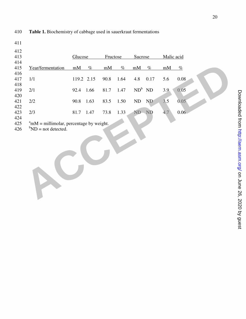
Table 1. Biochemistry of cabbage used in sauerkraut fermentations 410
411
412 Glucose Fructose Sucrose Malic acid 413 414 Year/fermentation mM % mM % mM % mM % 415 416 1/1 119.2 2.15 90.8 1.64 4.8 0.17 5.6 0.08 417 418 2/1 92.4 1.66 81.7 1.47 NDb ND 3.9 0.05 419 420 2/2 90.8 1.63 83.5 1.50 ND ND 3.5 0.05 421 422 2/3 81.7 1.47 73.8 1.33 ND ND 4.7 0.06 423 424 amM = millimolar, percentage by weight. 425 bND = not detected. 426
ACCEPTED
on June 26, 2020 by guesthttp://aem
.asm.org/
Dow
nloaded from

21
427
428 Table 2: Identification of Y1 Isolates
Species Day
1 Day
3 Day 7 Day 9 Day 14
L. mesenteroides 84 38
Weissella sp. 7 20
L. citrium 4 1
L. curvatus 14 14 3
L. fallax 8 1 1
L. plantarum 9 64 51 75
L. brevis 3 3 3 5
L. coryniformis 5 1
L. argentinum 1 11
P. pentosaceus 1
L. paraplantarum 3 6 3
NID* 1 14 13
Samples analyzed** 95 93 93 90 96
* NID = Not identified
**Total number of samples analyzed
429 430
ACCEPTED
on June 26, 2020 by guesthttp://aem
.asm.org/
Dow
nloaded from

22
431 Table 3. Identification of Y2 isolates
Tank 1
Species Day 1 Day 3 Day 7 Day 14
L. mesenteroides 6 2 2
Weissella sp. 12 7
L. citrium 2
L. curvatus 5 3
L. fallax 1 1
L. plantarum 3 7 16
P. pentosaceus 1
L. paraplantarum 1 1
ND* 1
Samples analyzed** 20 19 15 17
Tank 2
Species Day 1 Day 3 Day 7 Day 14
L. mesenteroides 12 16 7
Weissella sp. 2
L. citrium 1
L. fallax 2 1
L. plantarum 1 9 18
L. lactis 1
L. argentinum 4
L. mali 1
Samples analyzed 19 19 19 18
Tank 3
Species Day 1 Day 3 Day 7 Day 14
L. mesenteroides 1 9 2
Weissella sp. 1 7 1
L. citrium 4 1
L. curvatus 1
L. fallax 2 2 4
L. plantarum 1 10 16
L. brevis 1
L. argentinum 1
L. paracasei 4
NID 3 1 1
Samples analyzed 17 20 18 18
* NID = Not identified
** Total number of samples analyzed
432 433
ACCEPTED
on June 26, 2020 by guesthttp://aem
.asm.org/
Dow
nloaded from

Figures for Eddie paper 1
ACCEPTED on June 26, 2020 by guest
http://aem.asm
.org/D
ownloaded from

Time (days)
0 2 4 6 8 10 12 14
Co
nc
en
tra
tio
n (
mM
)
0
50
100
150
200
Figure 1
ACCEPTED on June 26, 2020 by guest
http://aem.asm
.org/D
ownloaded from
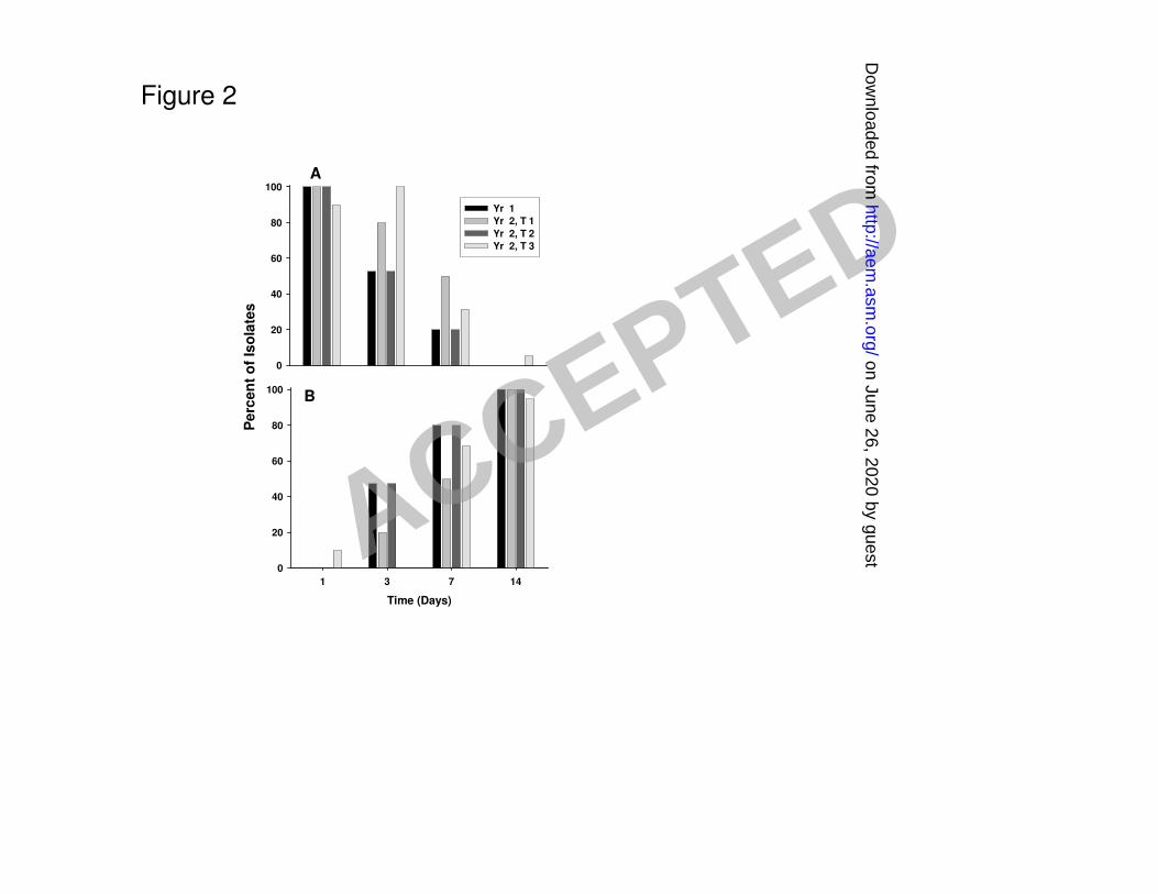
Figure 2
0
20
40
60
80
100
Time (Days)
1 3 7 14
Pe
rcen
t o
f Is
ola
tes
0
20
40
60
80
100
Yr 1
Yr 2, T 1
Yr 2, T 2
Yr 2, T 3
A
B
ACCEPTED on June 26, 2020 by guest
http://aem.asm
.org/D
ownloaded from

Figure 3
L.
me
se
nte
roid
es
We
iss
ella
sp
.
L. c
itre
um
L.
falla
x
P.
pe
nto
sa
ce
us
L. a
rge
nti
nu
m
Un
kn
ow
n
L. c
ory
nif
orm
is
L.
cu
rva
tus
L. p
ara
pla
nta
rum
L.
bre
vis
Un
kn
ow
n
L.
pla
nta
rum
Perc
en
t o
f Is
ola
tes
0
20
40
60
80
100
Day 1
Day 3
Day 7
Day 14
ACCEPTED on June 26, 2020 by guest
http://aem.asm
.org/D
ownloaded from

Figure 4
Weis
sella s
p.
L. m
esen
tero
ides
L. cit
reu
m
P. p
en
tosaceu
s
Un
kn
ow
n
L. fa
llax
L. cu
rvatu
s
L. p
ara
pla
nta
rum
L. p
lan
taru
m
Pe
rce
nt
of
Iso
late
s
0
20
40
60
80
100 Day 1
Day 3
Day 7
Day 14
L.
me
se
nte
roid
es
L.
arg
en
tin
um
We
iss
ella
L.
lac
tis
L.
cit
reu
m
Un
kn
ow
n
L.
falla
x
L.
pla
nta
rum
Perc
en
t o
f Is
ola
tes
0
20
40
60
80
100
L.
me
se
nte
roid
es
We
isse
lla
sp
.
L.
cit
reu
m
L.
pa
rac
ase
i
Un
kn
ow
n
L.
falla
x
L.
arg
en
tin
um
L.
bre
vis
L.
pla
nta
rum
Pe
rce
nt
of
Iso
late
s
0
20
40
60
80
100
A
B
C
G
ACCEPTED on June 26, 2020 by guest
http://aem.asm
.org/D
ownloaded from