2. skeletal system
DESCRIPTION
fgTRANSCRIPT

SKELETAL SYSTEM
The skeletal system is formed of bones and cartilagesThe bones are connected by joints to form the skeleton.
Functions of bones• They form the skeleton which gives the body shape and form
• They provide attachment for muscles & ligaments• They allow movements of the body• They provide protection for vital organs• They provide storage places for calcium salts• Production of blood cells in the red bone marrow

Types of Bone (according to structure)
• The bone is a type of connective tissue• It is hard because of its high content of calcium salts
• There are 2 types of bone tissue:Compact bone: which is dense & hard. It forms the shaft of long bones, and outer shell of other bones.
It consists of cylindrical units of closely packed lamellae (Haversian system)
Cancellous (spongy) bone: a delicate bony meshwork that fills the inside of bones.
Compact

Types of Bones (according to shape)• Long bones: these are longer than wide, and are found in limbs, e.g. humerus.
• Short bones: they are cuboidal in shape, and found in the hand and foot (carpal & tarsal bones)
• Flat bones: thin and flattened, e.g. scapula & skull bones• Irregular bones: they are irregular in shape, e.g. vertebrae• Pneumatic bones: they contain air-filled cavities, e.g. ethmoid bone
• Sesamoid bones: embedded within certain tendons, e.g. patella• Sutural bones: found between the skull sutures
Remember!! Metatarsal & metacarpal bones and phalanges are considered long bones
Flat bone
Long boneShort bones
Long bones

Sutural bones
Pneumatic bone Irregular bonePatella
Sesamoid bone

Features of Long Bones: Long bones consist of a “shaft” and 2 “ends”Shaft (Diaphysis):
• This is the tubular part of the long bone. It is formed of compact bone and contains a central cavity called “medullary” or “bone marrow cavity”.
• The shaft is lined by a membrane called “endosteum” and covered by a vascular membrane called “periosteum”
• The periosteum contains osteoblasts and causes the increase in width of the bones, it is also needed for repair of bone fractures.
Ends (Epiphysis):• These are the expanded ends of the long bone.• They are formed of cancellous bone covered by a thin layer of compact bone.
Note!! the diaphysis is separated from the epiphysis by the “epiphyseal cartilage”The Metaphysis: is the part of the diaphysis adjacent to the epiphyseal line


Shouldergirdle
SKELETON
Axial skeleton:• Skull• Sternum• Ribs• Vertebrae
Appendicular skeleton:• Bones of upper limb & Shoulder girdle• Bones of lower limb & Pelvic girdle

APPENDICULAR SKELETON
Skeleton of the Upper Limb
Shoulder girdle: 2 bones• Clavicle, anteriorly• Scapula, posteriorly
Upper arm, one bone• Humerus
Forearm, 2 bones• Radius, laterally• Ulna, medially
Hand, formed of:• Carpus (8 bones)• Metacarpus (5 bones)• Phalanges (3 in each finger, except the thumb which contains 2 phalanges)

Scapula & HumerusPosterior view
spine acromion
head
Scapula & HumerusAnterior view
Coracoid process
acromion
Greater & lessertuberosities
capitulum trochlea

Rad
ius
Uln
a
head
neck
Styloidprocess
head
Styloidprocess
Radius & Ulna
Wrist & Hand
Carpal bonesMetacarpalbones
P h
a l
a n
g e
s

APPENDICULAR SKELETON
Skeleton of the Lower Limb
Pelvic girdle 1 bone• Hip bone
Thigh 1 bone• Femur
Leg 2 bones• Fibula, laterally• Tibia, medially
Foot formed of:• Tarsus (7 bones)• Metatarsus (5 bones)• Phalanges (3 in each toe, except the big toe which contains 2 phalanges)
Hip bone

I l i u
m
Ischium
Pubis
Acetabulum
Hip bone (lateral view)
Femur (posterior view)
Head
Neck
Greater & lessertrochanters
Femoralcondyles

Tibia & fibulaAnterior view
Foot
Tarsal bones
Metatarsalbones
ph
alang
es
Tibialcondyles
Tib
ia
Fib
ula
Tibialtuberosity

SKULL
The skull is formed of two parts:•Brain box: the upper & posterior part of the skull•Facial skeleton: the anterior part of the skull
The skull is made up of 22 bones• 1 movable bone, the mandible• 21 immovable bones articulating by fibrous joints (sutures)
Single bones of the skull•Frontal bone•Occipital bone•Ethmoid bone•Sphenoid bone•Vomer•Mandible
Paired bones of the skull•Parietal bone•Temporal bone•Maxilla•Zygomatic bone•Nasal bone•Lacrimal bone•Palatine bone•Inferior nasal concha

Norma Frontalis, shows:• Frontal bone• Nasal bones• Maxillae• Zygomatic bones• Mandible
• Orbital openings• Anterior nasal aperture
• Mental foramen
Features of the Skull
Orbit opening
Anterior nasalaperture
Mental foramen

Norma verticalis, shows:• Frontal bone• Parietal bones• Occipital bone• Coronal, sagittal and lambdoid sutures
• Parietal foramen
Frontal bone
Coronal suture
Sagittal suture
Lambdoid suture
Occipital bone
Pariet
al b
one
Parietal foramen

Norma lateralis, shows:• Frontal, Parietal, Temporal, Occipital and Zygomatic bones• Zygomatic arch• Temporal lines & temporal fossa• External auditory meatus
Fron
tal
bone
Parietalbone
Occ
ipital
bone
Temporal bone
Zygomaticbone
Temporal lines
External auditorymeatus
Zygomaticarch

Norma occipitalis, shows:• Occipital bone• Parietal bones• External occipital protuberence & crest• Nuchal lines
Parietal bones
Occipital bone
External occipitalprotuberence
Superior & inferiorNuchal lines

Norma basalis externa, shows:• Alveolar arch• Hard palate• Posterior nasal apertures• Foramina and canals which give passage to structures which enter or leave the skull
• foramen ovale• foramen magnum• carotid canal• jugular foramen
Posterior nasalaperture
Foramenovale
Carotidcanal
Foramenmagnum
Jugularforamen
Hard palate
Alveolar arch

Norma basalis interna, shows• Anterior, middle & posterior cranial fossae• Frontal, ethmoid, sphenoid, temporal & occipital bones• Foramina:
• Foramen rotundum• Foramen ovale• Foramen lacerum• Jugular foramen• Foramen magnum
Frontal bone
Foramen rotundum
Foramen ovale
Foramen lacerum
Temporal bone
Occipital bone
Jugular foramen
Foramenmagnum
Sphenoid bone
Ethmoid bone

Vertebral Column (Spine)
•The vertebral column (spine) consists of:•7 cervical vertebrae•12 thoracic vertebrae•5 lumbar vertebrae•5 sacral vertebrae (fused together to form the sacrum)•4 coccygeal vertebrae (fused together to form the coccyx)
•The vertebral column provides support for the head and trunk•It provides protection for the spinal cord

Basic Structure of a Vertebra• Body: disc shaped and anterior in position• Vertebral arch: a bony ring behind the body. It is formed of 2 pedicles and 2 laminae• Vertebral foramen: the ring bound by the vertebral arch. The vertebral canal is formed by the succession of vertebral foramina
• 7 Processes: They project from the vertebral arch• A Spinous process, projects posteriorly• 2 Transverse processes, one projecting on each side• 2 Superior articular processes projecting up to articulate with the inferior articular processes of the vertebra above.
• 2 Inferior articular processes projecting down to articulate with the superior articular processes of the vertebra below.

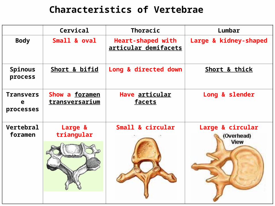
Characteristics of Vertebrae
Cervical Thoracic Lumbar
Body Small & oval Heart-shaped with articular demifacets
Large & kidney-shaped
Spinous process
Short & bifid Long & directed down Short & thick
Transverse processes
Show a foramen transversarium
Have articular facets Long & slender
Vertebral foramen
Large & triangular Small & circular Large & circular

Foramentransversarium
Bifid spine
Cervical Vertebra
Thoracic Vertebra

Lumbar Vertebra

1st & 2nd Cervical Vertebrae

Sacrum: It is formed of 5 vertebrae that are fused together
Intervertebral foramina• These are notches in the upper and lower
borders of each pedicle of the vertebral arch
• Adjacent notches from an intervertebral foramen for the passage of spinal nerves

The sternum is composed of 3 fused pieces•manubrium sterni•body •xiphoid process
Sternal angle: is the junction between the manubrium and the body
Sternum & Ribs

• There are 12 pairs of ribs• All the ribs are attached at their posterior ends to the vertebrae.
• Anteriorly:• The upper 7 pairs (true ribs) are attached directly to the sternum by their costal cartilages
• The 8th, 9th, and 10th ribs (false ribs) are attached to the 7th costal cartilage.
• The 11th and 12th ribs (floating ribs) have no anterior attachment.
Ribs


Basic features of ribs• Head: articulates with the thoracic vertebrae• Neck: the constriction just beyond the head• Tubercle: articulates with the transverse process of its corresponding vertebra• Angle: the sharp turn in the rib• Shaft: thin and flattened, its lower border is sharp and shows a groove for intercostal nerves & vessels.