2005 06-d kopya - kafkas Ünİversİtesİ veterİner...
TRANSCRIPT

Kafkas Univ Vet Fak Derg15 (3): 339-344, 2009DOI:10.9775/kvfd.2008.100-A
RESEARCH ARTICLE
Evaluation of Fractures in Calves due to Forced Extraction during Dystocia:27 Cases (2003-2008) [1]
Özgür AKSOY *�� İsa ÖZAYDIN * Engin KILIÇ * Savaş ÖZTÜRK *Emine GÜNGÖR * Başak KURT * Hasan ORAL**
Makale Kodu (Article Code): 2008/100-A
A part of this study was previously presented in “XIst National Congress of Veterinary Surgery (25-28 July 2008,Kuşadası - Aydın, TURKEY)Department of Surgery, Faculty of Veterinary Medicine, University of Kafkas, Kars - TURKEYDepartment of Obstetrics and Gynaecology, Faculty of Veterinary Medicine, University of Kafkas,Kars - TURKEY
�� İİlleettiişşiimm ((CCoorrrreessppoonnddeennccee))
℡℡ +90 474 2426807/1277�� [email protected]
Summary
This was a retrospective study evaluating fractures occurred in calves within a 6 year period (2003-2008) where dam had dystociaintervened by attending person. A total of 27 calves with fracture due to intervened dystocia were referred to the clinics within thisperiod. The treated Fractures by using PVC bandage were located in corpus mandible in 10 cases, in metacarpus in 7 cases, in radius-ulna in 3 cases, in metatarsus in 2 cases, in tibia in 1 case and, in humerus in 1 case. Bilateral corpus mandible fractures in 8 cases werefixed by transfixation pinning and casting and these cases completely recovered. Two cases of metatarsus fracture were treated byintramedullar pinning. Three cases with metacarpus fracture, one unilateral and one bilateral open fracture cases were treated bytransfixation pinning and casting with PMMA. The first case recovered but later case did not recover due to a severe infection. Two ofradius-ulna fracture of three cases were subjected to PVC bandage. Recovery was obtained in femur fracture after intramedullarpinning. Supra- and inter-condyler fracture in humerus was treated by internal fixation but this case did not recover. The remainingcases (n=8) were either not eligible for treatment or data were not complete for evaluation. In conclusion, the healing based on thetreatment techniques was successfully obtained in terms of the localizations, type, and open or closed condition of fracture.
Keywords: Calf, Dystocia, Fracture
Buzağılarda Güç Doğum Sırasında Hatalı Uygulamalar Sonucu ŞekillenenKırık Olgularının Değerlendirilmesi: 27 Olgu (2003-2008)
Özet
Bu retrospektiv çalışmada, 6 yıllık periyotta (2003-2008) buzağılarda güç doğum sırasındaki hatalı ve/veya aşırı zorlamalar nedeniyleşekillenen kırık olguları değerlendirildi. Bu süre içerisinde kliniğimize getirilen 27 buzağıda güç doğuma bağlı kırık saptandı. Kırıklar, 10olguda corpus mandibula, 7 olguda metacarpus, 3 olguda radius-ulna, 3 olguda femur, 2 olguda metatarsus, 1 olguda tibia ve 1 olgudahumerusta lokalizeydi. Bilateral corpus mandibula kırığı olan olguların 8’inde transfixation pinning ve casting ile fiksasyon yapıldı vetümü problemsiz iyileşti. Metatarsus kırık olgularının 2’sinde de intramedullar pin uygulamasıyla sağaltım gerçekleştirildi. Metacarpustakırık şekillenen olguların 3’ünde sadece PVC’li bandaj ile iyileşme sağlanırken, tek taraflı açık kırık şekillenen 1 olgu ile bilateral açık kırıksaptanan bir olguda transfixation pinning ve PMMA ile casting uygulandı. İlkinden olumlu sonuç alınırken diğerinde yaygın enfeksiyonnedeniyle olumlu sonuç alınamadı. Radius-ulna kırığı bulunan 3 olgunun 2’sinde PVC’li bandaj uygulamasıyla tedavi edildi. Femur kırığıbulunan olguların 1’inde intramedullar pinning uygulanarak iyileşme elde edildi. Humerusta supra ve intercondyler kırık bulunan olgudainternal fixation ile sağaltım uygulandı ancak iyileşme sağlanamadı. Diğer olgularda tedavi ve sonuçları için yeterli veri oluşturulamadıveya herhangi bir sağaltım uygulanmadı. Sonuç olarak, kırığın şekillendiği bölge, kırık tipi ve açık-kapalı oluşuna göre uygulanan sağaltımteknikleriyle yüksek oranlarda iyileşme sağlanmıştır.
Anahtar sözcükler: Buzağı, Güç doğum, Kırık
[1]
***

INTRODUCTION
The current studies of genetic improvement for highmeat and milk production in cattle has apotential tocause a relative discordancy between dam and foetus,and this leads to increase the dystocia problems. Generally,inappropriate manipulation or manual and mechanicalforces by owners and sometimes veterinarians to helpparturation cause traumatic disorders in muscle, boneand joint, and nerve and other soft tissues which mayoften lead to dying of foetus during parturation orpostnatal period. In same cases, dam also could bedamaged in various grades.
It has been reported that the incidence of dystociavaries due to some factors such as age of mother, bredof animal, sex of fetus, nutrition, environmental andclimate conditions, and the presentation or positionof foetus 1
During excessive and forced traction process, thetrauma cased by using material during distocia and/orinappropriate forced extraction lead to fractures anddislocations with various soft tissue lesions (thedestroying of muscle, nerve and vessel) 1 in firstly meta-carpus/metatarsus 2,3, mandible 4-6, femur 3,7, tibia 2,3,radius-ulna 2,3,8, humerus 3 and ribs 9,10.
In order to define treatment choice, it should beconsidered the type, localization and condition offracture (open or closed), the economical value ofanimal, cost of treatment and required care conditionsin healing period 3.
In farm animals, the bandaging technique supportedwith some materials such as Polyvinylchloride (PVC)and aluminum and the splinting such as Thomas Splintalone or combined with bandage have been frequentlyused as a treatment choice for external reduction ofclosed fractures 2,3,11-15.
The transfixation pinning and casting methods arealso used especially in the reduction and fixation offractures of metacarpus/metatarsus, tibia and radius-ulna 2,8,12,13,16. The internal fixation techniques appliedby using some materials such as intramedullar pinning,cerclage wiring, screw, dynamic compression plate(DCP), interlocking pin are recommended particularly inreduction of the dislocated, fragmented and complicatedfractures (with pieces) or in the fractures establishedin bones not suitable for bandaging or splinting 2,13,17,18.
This retrospective study evaluated the types,
localizations and treatment techniques of fractures incalves occurred by the inappropriate forced extractionduring distocia in six-year period (2003-2008).
MATERIAL and METHODS
Twenty seven calves with fractures occurred duringthe inappropriate manipulations of dystocia wereused in the study. These calves with various bred, ageand sex were received to Kafkas University, VeterinaryFaculty Surgery Clinics in the period of 2003-2008.The information based on the anamnesis obtainedfrom the owners about the manipulations duringdistocia was recorded. The types and localizations ofthe fractures were determined by physical and radio-graphic examination.
Seventeen cases of fractures encountered inextremities were treated by using the methods of theclosed reduction with PVC bandage in five cases, theinternal fixation (with intramedullar pin, screw, cerclagewire) in four cases, and the transfixation pinning andcasting in two cases. Eight cases of bilateral mandiblefracture were treated by transfixation pinning andfiberglass casting. The other fractures (six cases withextremities and two cases with mandible) were notevaluated because of unsuitability for treatment and/or obtaining insufficient data about the cases. Whilebandage applications were realized under sedation,the surgery for fractures of mandible and forelimbswere carried out under general anesthesia and thesurgery in the hindlimbs were applied under generalor spinal anesthesia.
The closed reduction with PVC bandage and internalfixations (using by cerclage wire, pin and screw) wereapplied by routine techniques in extremities. Fortransfixation pinning, two pins were replaced to bothproximal and distal fragment of the fractured bonetransversally and the pins were fixed by polymethylmetacrylate (PMMA). In the fracture of corpus mandible,three pins were replaced transversally into oral (twopins) and aboral (one pin) parts of fracture fragmentsand the reduction and stabilization were obtained byfiberglass casting.
The post-treatment period was followed by receivingthe patents to the clinic, calling the owners to get the feed-backs, or visiting to patients based on the stage of cases.
In the cases treated by tranfixation pinning, thepins were removed at the 5th-6th weeks after surgerybased on the radiological findings.
Evaluation of Fractures in Calves...340
RESULTS
Among 511 calves brought the clinic during thestudy period, 45 (%8.8) of fractures were observed byvarious reasons and 27 out of these 45 cases (60%)with fractures of different types and localizations werecaused by inappropriate manipulations for dystocia.These 27 cases correspond to 5.4% of total calvesreceived to the surgery clinic. The ages of calves werevaried in 1-30 days; however, the majority of them were1-3 day-old. The distribution of bred, sex and age ofthe calves are shown in Table 1.
Based on the anamnesis obtained from owners,the fractures were formed depending on the positionand presentation of foetus after the owners forcedthe foetus by tying on metacarpal/metatarsal regionwith a rope or chain by extraction with hand, otheranimal forces, or tractor. It was also recognized thatall of the mandible fractures were formed during theforced extraction by veterinarians during replacing achain to margo interalveolaris of foetus.
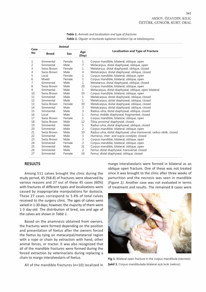
All of the mandible fractures (n=10) localized in
margo interalveolaris were formed in bilateral as anoblique open fracture. One of these was not treatedsince it was brought to the clinic after three weeks ofparturition and the necrosis was seen in mandible(Figure 1). Another case was not evaluated in termsof treatment and results. The remained 8 cases were
AKSOY, ÖZAYDIN, KILIÇÖZTÜRK, GÜNGÖR, KURT, ORAL
341
Table 1. Animals and localization and type of fractures Tablo 1. Olgular ve bunlarda saptanan kırıkların tip ve lokalizasyonu
CaseNo
AnimalLocalization and Type of Fracture
Breed Sex Age(Day)
123456789101112131415161718192021222324252627
SimmentalSimmentalSwiss BrownSwiss BrownLocalMixedSimmentalSwiss BrownSimmentalSwiss BrownSimmentalSimmentalSwiss BrownSimmentalSimmentalLocalSwiss BrownSwiss BrownSimmentalSimmentalSwiss BrownSimmentalSwiss BrownSimmentalSimmentalSimmentalSimmental
FemaleMaleFemaleMaleFemaleFemaleMaleMaleMaleMaleMaleMaleFemaleMaleMaleMaleFemaleMaleMaleMaleMaleMaleMaleFemaleMaleMaleFemale
1714111201201130331112521033221210
Corpus mandible, bilateral, oblique, openMetacarpus, distal diaphyseal, oblique, openMetatarsus, distal diaphyseal, oblique, closedMetatcarpus, distal diaphyseal, oblique, closedCorpus mandible, bilateral, oblique, openCorpus mandible, bilateral, oblique, openMetatarsus, distal diaphyseal, oblique, closedCorpus mandible, bilateral, oblique, openMetacarpus, distal diaphyseal, oblique, open bilateralCorpus mandible, bilateral, oblique, openMetatcarpus, distal diaphyseal, oblique, closedMetatcarpus, distal diaphyseal, oblique, closedMetatcarpus, distal diaphyseal, oblique, closedMetatcarpus, distal diaphyseal, oblique, closedRadius-ulna, distal diaphyseal, oblique, closedFemur, middle diaphyseal, fragmented, closedCorpus mandible, bilateral, oblique, openTibia, proximal diaphyseal, closedRadius-ulna, distal diaphyseal, oblique, closedCorpus mandible, bilateral, oblique, openRadius-ulna, distal diaphyseal, ulna-transversal, radius-oblik, closedHumerus, inter- and supra-condyler, closedCorpus mandible, bilateral, oblique, openCorpus mandible, bilateral, oblique, openCorpus mandible, bilateral, oblique, openFemur, distal diaphyseal, transversal, closedFemur, distal diaphyseal, oblique, closed
Fig 1. Bilateral open fracture in the corpus mandibula (necrosis)
Şekil 1. Corpus mandibulada bilateral açık kırık (nekroz)

recovered by transfixation pinning and fiberglasscasting techniques (Figures 2 and 3) without anycomplication. It was observed that all of these patientsstarted sucking their mothers within 8-12 hours andtaking their food without a help.
Six and one of metacarpus fractures formed asunilaterally and bilaterally in distal diaphyseal regionof the bone, respectively. Five and two of them wereoblique and transversal fracture, respectively. Onebilateral and one unilateral open fractures observedin two cases were operated by transfixation pinningand PMMA casting methods. The unilateral fracturewas recovered; however, the other animal was sent
to slaughterhouse because of a severe infection.While three cases of closed fracture were recovered byPVC bandage, the other two cases was not considered.
While two cases of metatarsus fracture formed indistal diaphyseal oblique fracture and one cases offemur with middle diaphyseal fragmented fracturewere recovered by using intramedullary pinning, oneof cases with supra- and inter-condylar humerusfracture was treated by intramedullar pin and screwfixation; however, it was not recovered due to earlyscrew loosening and pin migration. One case withproximal diaphyseal tibia fracture was not evaluatedbased on the treatment and results.
Evaluation of Fractures in Calves...342
Fig 2. The fracture in the corpusmandibula. Clinical view (A), Radio-graphic image before surgery (B),Post-operative clinical view (C), Post-operative radiographic image (D)
Şekil 2. Corpus mandibula kırığı A-Klinik görünüm, B- Operasyonöncesi radyograf ik görünüm, C-Postoperatif klinik görünüm, D-Postoperatif radyografik görünüm
Fig 3. The fracture in the corpusmandibule. Radiographic imagesbefore (A) and after (B) thesurgery
Şekil 3. Corpus mandibula kırığıA- Operasyon öncesi radyografikgörünüm, B- Postoperatif radyo-grafik görünüm

All of the radius-ulna fractures (n=3) were localizedin distal diaphysis. While the oblique fractures wereobserved in both bones of two cases, the oblique andtransversal fractures were observed in radius andulna in the other case, respectively. Two cases ofthese were treated by PVC bandaging and theremaining case was not evaluated.
DISCUSSION
In Turkey as well as many other countries, themeat and milk consuming has been obtained fromsmall and large ruminants. Various improvementstudies and genetic advances have been performed inorder to get more productivity and yield by a lesser ofmanagement expenses and work. As a result of thiscondition, a remarkable increase of pure and crossbredcattle population compared with local breeds has beenrecently observed in Northeastern Anatolia especiallywithin last 15-20 years. Consequently, this has causedto increase the incidence in losses (foetal death,traumatic conditions due to dystocia, predisposition toinfections of calves and cows in postnatal and post-partal periods, etc.).
The incidence of dystocia is observed related withfetus and dam with higher frequency in beef cattlethan that of in milk cattle. There are many reasons ofdystocia such as inertial uterus, torsio uteri, relativenarrowness of pelvic structure, insufficient wideningin soft and hard structures of birth canal and disordersof presentation - position of foetus. In addition, thedifference of breed between dam and foetus, nutrition,management conditions, climate conditions, thecondition score of dam, sex of foetus (male foetus aremore prone than females), twiness, the disorders ofpresentation and position of foetus also potentiallycause dystocia 1. The most of calves brought to ourclinics were male and Simmental breed. As describedabove, this may be related to the combination of pre-disposition and causative factors (the predispositionof sex and breed, nourishment, nutrition and climateconditions etc.).
Görgül et al.3 reported that the incidence of thedisorders of extremites in surgical disorders were13.8% and 80.6% were caused by inappropriatemanipulations during helping for birth in calvesbrought to the Surgery Clinics of Uludağ University,Veterinary Faculty, within a period of 8 years (1996-2003). In the present study of the calves brought toour clinics within a period of 6 years (2003-2008), the
rate of fractures cases in surgical disease was 8.8%and 60% of these (5.4% of all the calves) were causedby similar inappropriate manipulations during helpingfor birth in dystocia. It may be suggested that thesedata are not enough to reflect the current rates of theKars Province because the most of calves in our clinicswere brought from surrounding villages only. If thenumber cases of died calves during birth or after birthand the calves treated or non-treated by veterinariansare added to these rates, it can be concluded that thementioned problems are significant problem of thisregion.
The rope or chain tied to extremities for tractionof fotetus causes to open or close fractures associatedwith soft tissues damages depending on presentationor position of foetus 2,3,8. In some cases, more proximalbones may also be affected during this force. Inaddition, the excessive tractions also lead to the thoracaltrauma in case of difficulties/impossibilities duringthe passing of fetus in birth canal 9,10. As the materialusing for traction of foetus usually replaces on themetacarpal/metatarsal region, most of fractures duringbirth can be formed especially in these regions.Similarly, it has been reported that most of fractures(n=9) were observed in these regions in the presentstudy. In addition, it was observed that open or closedfractures with accompanied with various soft tissuedamage in the cases of the metacarpal/metatarsalfractures. It is importantly noticed that all of thesecases had distal diaphyseal fractures and these findingsagrees with the other references which explain thatthe fractures are caused by trauma occurred with therope or chain to the region during traction of foetus 2,3.It may be concluded that the other fractures wereformed due to inappropriate biomechanical tractions(the application of traction in unsuitable angles).
It has been shown that the mandible fractures arerarely seen and especially presented as a case report 5,6.In contrast, the rate of bilateral corpus mandibulafractures in our present report were 10 of 27 cases.Unfortunately, it was seen that some veterinarinas hadapplied for mandibular traction in foetus for birth aid.
Various techniques for treatment of extremityfractures are recommended by surgeons as dependingon some factors such as type and localisation of fracture,type and severe of trauma, choice of treatment, geneticalvalue of animal, cost of treatment and the conditionsof management. It was reported that highly successfulresults have been obtained in the calves treated withtechniques of bandage and splint 2,3,11-15, transfixation
AKSOY, ÖZAYDIN, KILIÇÖZTÜRK, GÜNGÖR, KURT, ORAL
343

pinning and PMMA cast 2,8,12,13,16, internal fixation2,13,17,18. In the present study, following the 10 of 17cases with extremity fracture were appropriatelytreated by a suitable techniques mentioned above, allthe patients were recovered except for two patients.Also, the 8 of 10 cases with mandible fracture weresuccessfully treated by transfixation pinning and fiber-glass cast.
In conclusion, it is understood that one of the mostcritical problems among surgical disorders in calvesare fractures associated with other traumatic disordersformed during obstetrical processes. The etiology ofthese is contained by empirical methods by the ownersand inappropriate manipulations of veterinarianswithout caring of biomechanical criteria duringextraction force. It is suggested that the veterinariansshould use appropriate techniques and avoid theowners of animal interfere unless necessary and nottry to treat of fractures by empirical methods to reducethe economical losses. The caring attention to importantcriteria for helping of birth by veterinarians andowners of animal will contribute to decrease ofeconomical losses. It should be remarkably noticedthat the maintenance of profitability of an animalhusbandry and management depend on the obtainingone calve per dam and continuation of regular profitfor every year. Additionally, the appropriate applicationsand false treatments give very strong damage tofoetus beside dam, cause predispositions for obstetricaland gynecological problems, and sometimes lead tocomplete infertility. The treatment of calves withfracture should be considered as a valuable profit totolerate the economical waste in the calves having avalue of high genetics. Because every untreated andlost calve create an additional costing to farm foranother year. It must be considered that if anopportunity intercepts to treat in accurate time, thetreatment methods can be more successful. Further-more, under the light of results of this study, it isnecessary that the informing and education of farmersregularly and continuously by faculties, civilianassociations, ministry and field veterinarians.
REFERENCES
1. Noakes DE: Dystocia and Other Disorders Associated withParturation. In, Noakes DE, Parkinson TJ, England GCW (Eds):Arthur’s Veterinary Reproduction and Obstetrics. 8th ed. WB
Saunders Co, Philadelphia, pp. 205-338, 2002.
2. Steiner A: Management of metacarpal, metatarsal, radialand tibial fractures in calves. Nineth Annual ESVOT Congress,16-19 April, Munich - Germany, 1998.
3. Görgül OS, Seyrek-İntaş D, Çelimli N, Çeçen G, Salc@ H,Ak@n İ: BuzağIlarda kIrIk olgularInIn değerlendirilmesi: 31Olgu (1996-2003). Veteriner Cerrahi Dergisi, 10 (3-4): 16-20,2004.
4. Trent AM, Ferguson JG: Bovine mandibular fractures. CanVet J, 26, 396-399, 1985.
5. Singh KB: Surgical repair of bilateral mandibular fracture ina buffalo calf. Indian Vet J, 74, 63-64, 1997.
6. Yadav GU, Markandeya NM, Bhikane AU, Kale SB:Management of mandibular fracture in a new born calf- Acase report. Indian Vet J, 80, 1287, 2003.
7. Ferguson JG, Dehghani, Petrali EH: Fractures of the femurin newborn calves. Can Vet J, 31, 289-291, 1990.
8. St-Jean G, Debowes RM: Transfixation pinning and castingof radial-ulnar fractures in calves: A review of three cases.Can Vet J, 33, 257-262, 1992.
9. Schuh JoAnn CL: A retrospective study of dystocia-relatedvertebral fractures in neonatal calves. Can Vet J, 29, 830-833,1988.
10. Çeçen G, Görgül OS: Holstein IrkI buzağIda bilateralkosta kIrIğI ve akciğer yaralanmasI. Veteriner Cerrahi Dergisi,12 (1,2,34): 107-108, 2006.
11. Görgül OS, Yan@k K: Koaptasyon ve destek materyaliolarak Polivinylidyn chloride (PVC) atellerin kullanImIüzerine klinik çalIşmalar. Ankara Üniv Vet Fak Derg, 29 (3-4):401-405, 1982.
12. St-Jean G, Debowes RM: Transfixation pinning andcasting of tibial fractures in calves: Five cases (1985-1989).JAVMA, 198 (1): 139-143, 1991.
13. Newman KD, Anderson DE: Fracture management inllamas and alpacas. Small Rum Res, 61, 241-258, 2006. 14. Latrach R, Segard E, Frikha MR: Cas clinique: Traitementd’une fracture du tibia chez un veau par un pansementRobert-jones associe’a des atelles. Revue Med Vet, 158 (7):354-361, 2007.
15. Gangl M, Grulke S, Serteyn D, Touati K: Retrospectivestudy of 99 cases of bone fractures in cattle treated byexternal coaptation or confinement. Vet Rec, 158, 264-268,2006.
16. Anderson DE, St-Jean G: Repair of fractures of the radiusand ulna in an ewe using positive profile transfixation pinsand casting. Can Vet J, 34, 686-688, 1993.
17. Aslanbey D, Sağlam M, Kaya A, Bilgili H: Bir buzağIdadistal diafizer parçalI metacarpus kIrIğInIn DCP plakkullanIlarak sağaltImI. Veteriner Cerrahi Dergisi, 3 (1): 40-43,1997.
18. Thilagar S, Ganesh TN, George RS, Kumaresan A:Management of comminuted fracture of metatarsus usingdynamic compression plate and position screw in a calf.Indian Vet J, 82, 195-197, 2005.
Evaluation of Fractures in Calves...344