2007 mississippi curriculum framework · 2007 mississippi curriculum framework postsecondary...
TRANSCRIPT
2007 Mississippi Curriculum Framework Postsecondary Diagnostic Medical Sonography Technology (Program CIP: 51.0910 – Diagnostic Medical Sonography/Sonographer and Ultrasound Technician) Direct inquiries to Debra West
Director for Career and Technical Education State Board for Community and Junior Colleges 3825 Ridgewood Road Jackson, MS 39211 (601) 432-6518 [email protected] Charlotte Darnell Instructional Design Specialist Research and Curriculum Unit P.O. Drawer DX Mississippi State, MS 39762 (662) 325-2510 [email protected]
Additional copies Research and Curriculum Unit for Workforce Development
Vocational and Technical Education Attention: Reference Room and Media Center Coordinator P.O. Drawer DX Mississippi State, MS 39762 http://cia.rcu.msstate.edu/curriculum/download.asp (662) 325-2510
Published by Office of Vocational Education and Workforce Development
Mississippi Department of Education Jackson, MS 39205 Research and Curriculum Unit for Workforce Development Vocational and Technical Education Mississippi State University Mississippi State, MS 39762
2
Postsecondary Diagnostic Medical Sonography Technology
The Mississippi Department of Education, Office of Vocational Education and Workforce Development does not discriminate on the basis of race, color, religion, national origin, sex, age, or disability in the provision of educational programs and services or employment opportunities and benefits. The following office has been designated to handle inquiries and complaints regarding the non-discrimination policies of the Mississippi Department of Education: Director, Office of Human Resources, Mississippi Department of Education, 359 North West Street, Suite 359, Jackson, Mississippi 39201, (601) 359-3511.
3
Postsecondary Diagnostic Medical Sonography Technology
Acknowledgments
Writing Team Crystal Larimore, RT(R), RDMS, RVS, Hinds Community College, Jackson, MS
Nita Megginson, RT(R)(M), RDMS, Itawamba Community College, Tupelo, MS
Tracy B. Weekley, RT(R), RDMS, RVT, RDCS, Jones County Junior College, Ellisville, MS
RCU Staff Charlotte Darnell – Instructional Design Specialist
Professional Curriculum Advisory Team
Hinds Community College Diagnostic Medical Sonography Advisory Committee
Itawamba Community College Diagnostic Medical Sonography Advisory Committee
Jones County Junior College Diagnostic Medical Sonography Advisory Committee
Standards in this document are based on information from the following organizations: CAAHEP Standards and Guidelines for the Accreditation of Educational Programs in Diagnostic Medical Sonography
Reprinted with permission from the Commission on Accreditation of Allied Health Education Programs (CAAHEP), 1996.
Related Academic Standards
CTB/McGraw-Hill LLC. (1994). Tests of adult basic education, Forms 7 and 8. Monterey, CA: Author. Reproduced with permission of CTB/McGraw-Hill LLC. TABE is a registered trademark of The McGraw-Hill Companies, Inc. Copyright © 1994 by CTB/McGraw-Hill LLC. Reproduction of this material is permitted for educational purposes only.
21st Century Skills
Reproduced with permission of the Partnership for 21st Century Skills. Further information may be found at www.21stcenturyskills.org
4
Postsecondary Diagnostic Medical Sonography Technology
Preface
Postsecondary Diagnostic Medical Sonography Technology Research Synopsis Articles, books, Web sites, and other materials listed at the end of each course were considered during the revision process. The Journal of Diagnostic Medical Sonography was especially useful in providing insight into trends and issues in the field. These references are suggested for use by instructors and students during the study of the topics outlined. Industry advisory team members from colleges throughout the state were asked to give input related to changes to be made to the curriculum framework. Specific comments related to soft skills needed in this program included communication, teamwork, eagerness, perseverance, analytical capability, emotional stability, physical fitness of operator, dedication, appropriate dress, initiative/work ethic, compassion, and flexibility. Occupation-specific skills stated included scanning ability/technical ability, working with computerized equipment, infection control, performing patient assessment and direct patient care, and acquiring and analyzing data. Safety practices emphasized included following OSHA guidelines, using protective gear, and adhering to standard precautions. Instructors from colleges throughout the state were also asked to give input on changes to be made to the curriculum framework. Since this is a relatively new curriculum, the Advisory Committee members and the instructors indicated that no significant changes to the content of the curriculum were needed. Curriculum The following national standards were referenced in each course of the curriculum: • CTB/McGraw-Hill LLC Tests of Adult Basic Education, Forms 7 and 8 Academic Standards • 21st Century Skills • CAAHEP Standards and Guidelines for the Accreditation of Educational Programs in
Diagnostic Medical Sonography Industry and instructor comments, along with current research, were considered by the curriculum revision team during the revision process; changes were made as needed and appropriate. Many of the skills and topics noted in the research were already included in the curriculum framework. Specific changes made to the curriculum at the February 22, 2006, curriculum revision meeting included: • Competencies and objectives were reviewed to ensure accuracy and appropriateness. • In the course description, the words “to apply” were added to the criteria that a student must
meet in order to sit for the American Registry of Diagnostic Medical Sonographers. • Discussion of the principles of 3D/4D imaging was added to the course Ultrasound Physics
and Instrumentation I. • The competency in which students describe images, storage, and display methods used in
diagnostic medical ultrasound was moved from the course Ultrasound Physics and Instrumentation I to Ultrasound Physics and Instrumentation II.
5
Postsecondary Diagnostic Medical Sonography Technology
• The competency in which students describe sonographic appearance of the vascular system in cross-sectional longitudinal and transverse planes was deleted from the course Sectional Anatomy.
• In the course Sonography Seminar, the term “routine” in the competency “Discuss routine sonographic procedures” was changed to the term “general.”
• The Recommended Tools and Equipment list was updated. Assessment Students will be assessed using the American Registry of Diagnostic Medical Sonographers. Professional Learning It is suggested that instructors participate in professional learning related to the following concepts: • Training for prevention of musculoskeletal injuries (msi) specific to sonographers - For PD,
to include AV and online tutorial reference to the following web sites: http://www.mayoclinic.com/health/ultrasound/PR00053; http://www.radiologyinfo.org/en/info.cfm?pg=vascularus&bhcp=1; http://www.radiologyinfo.org/en/info.cfm?pg=abdomus-pdi
• Blackboard® training – To learn more about Blackboard® training, please go to https://cia.rcu.msstate.edu/OnlinePD/.
• Differentiated instruction – To learn more about differentiated instruction, please go to http://www.paec.org/teacher2teacher/additional_subjects.html and click on Differentiated Instruction. Work through this online course and review the additional resources.

6
Postsecondary Diagnostic Medical Sonography Technology
Foreword As the world economy continues to evolve, businesses and industries must adopt new practices and processes in order to survive. Quality and cost control, work teams and participatory management, and an infusion of technology are transforming the way people work and do business. Employees are now expected to read, write, and communicate effectively; think creatively, solve problems, and make decisions; and interact with each other and the technologies in the workplace. Vocational-technical programs must also adopt these practices in order to provide graduates who can enter and advance in the changing work world. The curriculum framework in this document reflects these changes in the workplace and a number of other factors that impact on local vocational-technical programs. Federal and state legislation calls for articulation between high school and community college programs, integration of academic and vocational skills, and the development of sequential courses of study that provide students with the optimum educational path for achieving successful employment. National skills standards, developed by industry groups and sponsored by the U.S. Department of Education and Labor, provide vocational educators with the expectations of employers across the United States. All of these factors are reflected in the framework found in this document. Referenced throughout the courses of the curriculum are the 21st Century Skills, which were developed by the Partnership for 21st Century Skills, a group of business and education organizations concerned about the gap between the knowledge and skills learned in school and those needed in communities and the workplace. A portion of the 21st Century Skills addresses learning skills needed in the 21st century, including information and communication skills, thinking and problem-solving skills, and interpersonal and self-directional skills. The need for these types of skills have been recognized for some time and the 21st Century Skills are adapted in part from the 1991 report from the U.S. Secretary of Labor’s Commission on Achieving Necessary Skills (SCANS). Another important aspect of learning and working in the 21st century involves technology skills, and the International Society for Technology in Education, developers of the National Educational Technology Standards (NETS), were strategic partners in the Partnership for 21st Century Skills. Each postsecondary program of instruction consists of a program description and a suggested sequence of courses which focus on the development of occupational competencies. Each vocational-technical course in this sequence has been written using a common format which includes the following components: • Course Name – A common name that will be used by all community/junior colleges in
reporting students. • Course Abbreviation – A common abbreviation that will be used by all community/junior
colleges in reporting students. • Classification – Courses may be classified as:
o Vocational-technical core – A required vocational-technical course for all students.
7
Postsecondary Diagnostic Medical Sonography Technology
o Area of concentration (AOC) core – A course required in an area of concentration of a cluster of programs.
o Vocational-technical elective – An elective vocational-technical course. o Related academic course – An academic course which provides academic skills
and knowledge directly related to the program area. o Academic core – An academic course which is required as part of the
requirements for an Associate degree. • Description – A short narrative which includes the major purpose(s) of the course and the
recommended number of hours of lecture and laboratory activities to be conducted each week during a regular semester.
• Prerequisites – A listing of any courses that must be taken prior to or on enrollment in the
course. • Corequisites – A listing of courses that may be taken while enrolled in the course. • Competencies and Suggested Objectives – A listing of the competencies (major concepts and
performances) and of the suggested student objectives that will enable students to demonstrate mastery of these competencies.
The following guidelines were used in developing the program(s) in this document and should be considered in compiling and revising course syllabi and daily lesson plans at the local level: • The content of the courses in this document reflects approximately 75 percent of the time
allocated to each course. The remaining 25 percent of each course should be developed at the local district level and may reflect:
o Additional competencies and objectives within the course related to topics not found in the State framework, including activities related to specific needs of industries in the community college district.
o Activities which develop a higher level of mastery on the existing competencies and suggested objectives.
o Activities and instruction related to new technologies and concepts that were not prevalent at the time the current framework was developed/revised.
o Activities which implement components of the Mississippi Tech Prep initiative, including integration of academic and vocational-technical skills and coursework, school-to-work transition activities, and articulation of secondary and postsecondary vocational-technical programs.
o Individualized learning activities, including worksite learning activities, to better prepare individuals in the courses for their chosen occupational area.
• Sequencing of the course within a program is left to the discretion of the local district.
Naturally, foundation courses related to topics such as safety, tool and equipment usage, and other fundamental skills should be taught first. Other courses related to specific skill areas and related academics, however, may be sequenced to take advantage of seasonal and climatic conditions, resources located outside of the school, and other factors.
8
Postsecondary Diagnostic Medical Sonography Technology
• Programs that offer an Associate of Applied Science degree must include a minimum 15 semester credit hour academic core. Specific courses to be taken within this core are to be determined by the local district. Minimum academic core courses are as follows:
o 3 semester credit hours Math/Science Elective o 3 semester credit hours Written Communications Elective o 3 semester credit hours Oral Communications Elective o 3 semester credit hours Humanities/Fine Arts Elective o 3 semester credit hours Social/Behavioral Science Elective
It is recommended that courses in the academic core be spaced out over the entire length of the program, so that students complete some academic and vocational-technical courses each semester. Each community/junior college has the discretion to select the actual courses that are required to meet this academic core requirement.
• In instances where secondary programs are directly related to community and junior college
programs, competencies and suggested objectives from the high school programs are listed as Baseline Competencies. These competencies and objectives reflect skills and knowledge that are directly related to the community and junior college vocational-technical program. In adopting the curriculum framework, each community and junior college is asked to give assurances that:
o Students who can demonstrate mastery of the Baseline Competencies do not receive duplicate instruction, and
o Students who cannot demonstrate mastery of this content will be given the opportunity to do so.
• The roles of the Baseline Competencies are to:
o Assist community/junior college personnel in developing articulation agreements with high schools, and
o Ensure that all community and junior college courses provide a higher level of instruction than their secondary counterparts.
• The Baseline Competencies may be taught as special “Introduction” courses for 3-6 semester
hours of institutional credit which will not count toward Associate degree requirements. Community and junior colleges may choose to integrate the Baseline Competencies into ongoing courses in lieu of offering the “Introduction” courses or may offer the competencies through special projects or individualized instruction methods.
• Technical elective courses have been included to allow community colleges and students to
customize programs to meet the needs of industries and employers in their area. In order to provide flexibility within the districts, individual courses within a framework may be customized by:
• Adding new competencies and suggested objectives. • Revising or extending the suggested objectives for individual competencies. • Integrating baseline competencies from associated high school programs.
9
Postsecondary Diagnostic Medical Sonography Technology
• Adjusting the semester credit hours of a course to be up 1 hour or down 1 hour (after informing the State Board for Community and Junior Colleges [SBCJC] of the change).
In addition, the curriculum framework as a whole may be customized by:
• Resequencing courses within the suggested course sequence. • Developing and adding a new course which meets specific needs of industries and other
clients in the community or junior college district (with SBCJC approval). • Utilizing the technical elective options in many of the curricula to customize programs.
10
Postsecondary Diagnostic Medical Sonography Technology
Table of Contents Acknowledgments............................................................................................................................3 Preface..............................................................................................................................................4 Foreword ..........................................................................................................................................6 Program Description ......................................................................................................................11 Suggested Course Sequences.........................................................................................................12 Diagnostic Medical Sonography Technology Courses..................................................................17
Introduction to Ultrasound.........................................................................................................17 Sectional Anatomy.....................................................................................................................24 Ultrasound Physics and Instrumentation I .................................................................................28 Ultrasound Physics and Instrumentation II................................................................................32 Clinical Experience I..................................................................................................................37 Clinical Experience II ................................................................................................................41 Clinical Experience III...............................................................................................................44 Abdominal Sonography .............................................................................................................48 Obstetrical and Gynecological Sonography...............................................................................53 Advanced Sonographic Procedures ...........................................................................................58 Sonography Seminar..................................................................................................................63 Ultrasound Examination Critique ..............................................................................................67
Recommended Tools and Equipment ............................................................................................70 Assessment.....................................................................................................................................72 Baseline Competencies ..................................................................................................................73 Appendix A: Standards and Guidelines for the Accreditation of Educational Programs in Diagnostic Medical Sonography....................................................................................................78 Appendix B: Related Academic Standards....................................................................................84 Appendix C: 21st Century Skills ....................................................................................................85
11
Postsecondary Diagnostic Medical Sonography Technology
Program Description Diagnostic Medical Sonography uses high frequency sound waves to produce images of organs, masses, fluid collections, and vascular structures within the human body. Sonography is user-dependent, requiring competent and highly skilled professionals to be a part of the integral health care system. Sonographers have extensive, direct patient contact, providing care to a variety of people from healthy to critically ill. The sonographer is responsible for obtaining pertinent patient history, performing the sonographic examination, providing for the needs and comfort of the patient during examination, and recording anatomy and pathology or other data for interpretation by the supervising physician to aid in diagnosis. Sonography is commonly used in the field of obstetrics and gynecology for purposes ranging from confirming and/or dating pregnancies to diagnosing disease processes of the female reproductive system. Sonographers must have knowledge of normal structure and functional anatomy of the human body and use independent judgment in recognizing the need to perform procedures according to sonographic findings. Upon completion of the two-year program of study, the student will be awarded the Associate of Applied Science degree. Until a Diagnostic Medical Sonography program reaches accreditation approval from CAAHEP, the students must meet the following criteria in order to apply to sit for the American Registry of Diagnostic Medical Sonographers: • Be a graduate from a two-year allied health program that is patient care related which
includes but is not limited to Diagnostic Medical Sonography, Radiologic Technology, Respiratory Therapy, Registered Nurse, Occupational Therapy, and Physical Therapy; and have 12 months of full-time clinical ultrasound/vascular experience; or
• Hold a Bachelor’s degree and have 12 months of full-time clinical ultrasound/vascular experience.
Graduates from a CAAHEP accredited Diagnostic Medical Sonography Program may apply to take the ARDMS without further experience. Industry standards referenced are from the CAAHEP Standards and Guidelines for the Accreditation of Educational Programs in Diagnostic Medical Sonography (1996).
12
Postsecondary Diagnostic Medical Sonography Technology
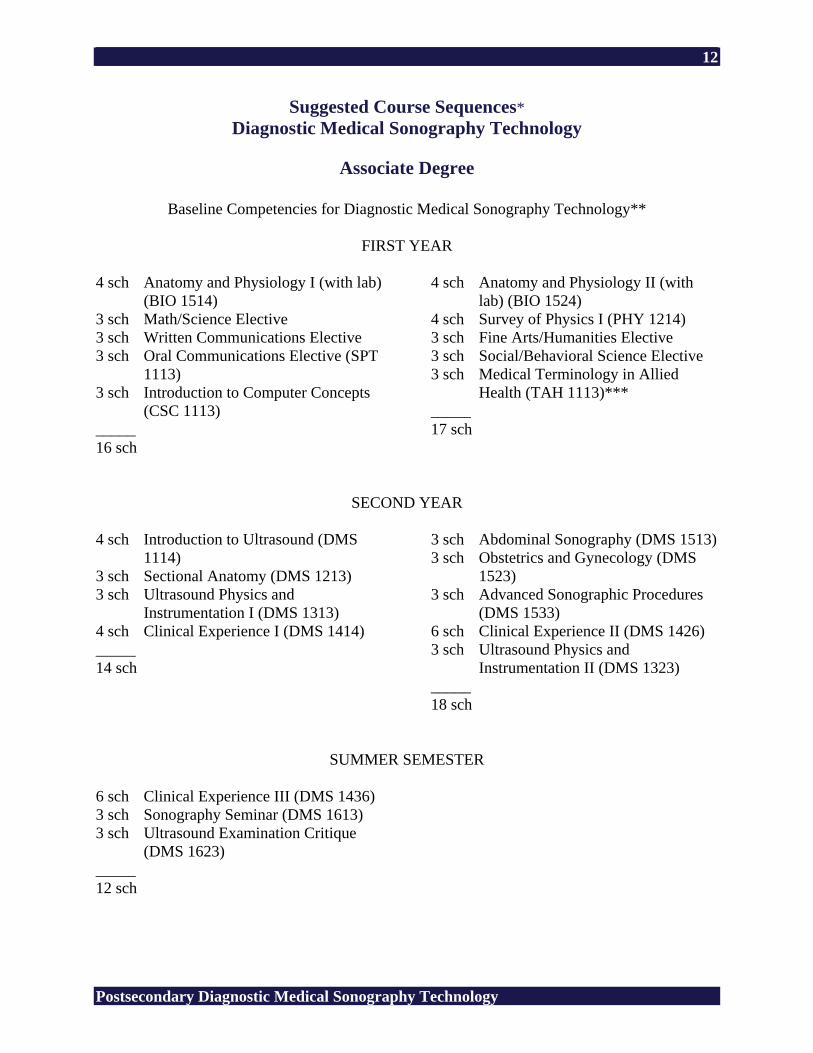
Suggested Course Sequences* Diagnostic Medical Sonography Technology
Associate Degree
Baseline Competencies for Diagnostic Medical Sonography Technology**
FIRST YEAR
4 sch Anatomy and Physiology I (with lab)
(BIO 1514) 3 sch Math/Science Elective 3 sch Written Communications Elective 3 sch Oral Communications Elective (SPT
1113) 3 sch Introduction to Computer Concepts
(CSC 1113) _____ 16 sch
4 sch Anatomy and Physiology II (with lab) (BIO 1524)
4 sch Survey of Physics I (PHY 1214) 3 sch Fine Arts/Humanities Elective 3 sch Social/Behavioral Science Elective 3 sch Medical Terminology in Allied
Health (TAH 1113)*** _____ 17 sch
SECOND YEAR
4 sch Introduction to Ultrasound (DMS 1114)
3 sch Sectional Anatomy (DMS 1213) 3 sch Ultrasound Physics and
Instrumentation I (DMS 1313) 4 sch Clinical Experience I (DMS 1414) _____ 14 sch
3 sch Abdominal Sonography (DMS 1513)3 sch Obstetrics and Gynecology (DMS
1523) 3 sch Advanced Sonographic Procedures
(DMS 1533) 6 sch Clinical Experience II (DMS 1426) 3 sch Ultrasound Physics and
Instrumentation II (DMS 1323) _____ 18 sch
SUMMER SEMESTER 6 sch Clinical Experience III (DMS 1436) 3 sch Sonography Seminar (DMS 1613) 3 sch Ultrasound Examination Critique
(DMS 1623) _____ 12 sch
13
Postsecondary Diagnostic Medical Sonography Technology
Applicants without a two-year allied health patient care related degree must take basic patient care and medical-legal ethics courses. * Students who lack entry level skills in math, English, science, etc. will be provided
related studies. ** Baseline competencies are taken from the high school Allied Health program. Students
who can document mastery of these competencies should not receive duplicate instruction. Students who cannot demonstrate mastery will be required to do so.
*** May be waived for completers of postsecondary allied health programs.
14
Postsecondary Diagnostic Medical Sonography Technology
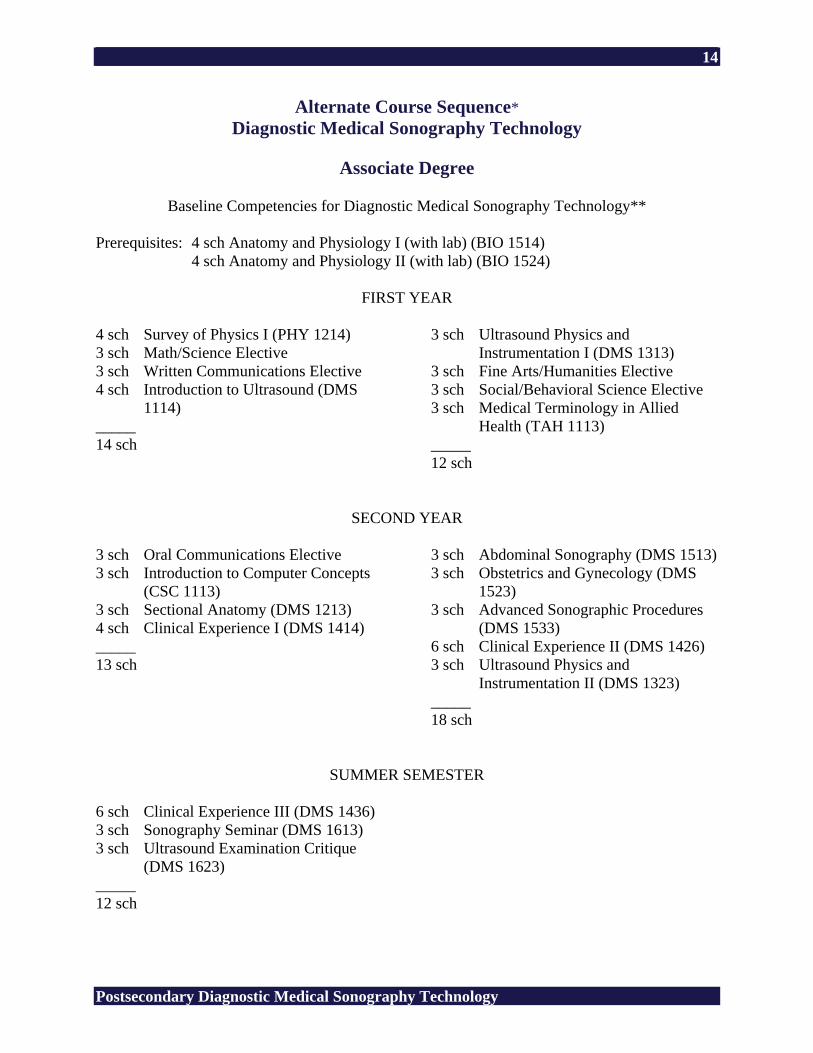
Alternate Course Sequence* Diagnostic Medical Sonography Technology
Associate Degree
Baseline Competencies for Diagnostic Medical Sonography Technology**
Prerequisites: 4 sch Anatomy and Physiology I (with lab) (BIO 1514)
4 sch Anatomy and Physiology II (with lab) (BIO 1524)
FIRST YEAR
4 sch Survey of Physics I (PHY 1214) 3 sch Math/Science Elective 3 sch Written Communications Elective 4 sch Introduction to Ultrasound (DMS
1114) _____ 14 sch
3 sch Ultrasound Physics and Instrumentation I (DMS 1313)
3 sch Fine Arts/Humanities Elective 3 sch Social/Behavioral Science Elective 3 sch Medical Terminology in Allied
Health (TAH 1113) _____ 12 sch
SECOND YEAR
3 sch Oral Communications Elective 3 sch Introduction to Computer Concepts
(CSC 1113) 3 sch Sectional Anatomy (DMS 1213) 4 sch Clinical Experience I (DMS 1414) _____ 13 sch
3 sch Abdominal Sonography (DMS 1513)3 sch Obstetrics and Gynecology (DMS
1523) 3 sch Advanced Sonographic Procedures
(DMS 1533) 6 sch Clinical Experience II (DMS 1426) 3 sch Ultrasound Physics and
Instrumentation II (DMS 1323) _____ 18 sch
SUMMER SEMESTER 6 sch Clinical Experience III (DMS 1436) 3 sch Sonography Seminar (DMS 1613) 3 sch Ultrasound Examination Critique
(DMS 1623) _____ 12 sch
15
Postsecondary Diagnostic Medical Sonography Technology
* For students without a two-year allied health degree. This sequence can only be used with a two-instructor program.
** Baseline competencies are taken from the high school Allied Health program. Students
who can document mastery of these competencies should not receive duplicate instruction. Students who cannot demonstrate mastery will be required to do so.
16
Postsecondary Diagnostic Medical Sonography Technology
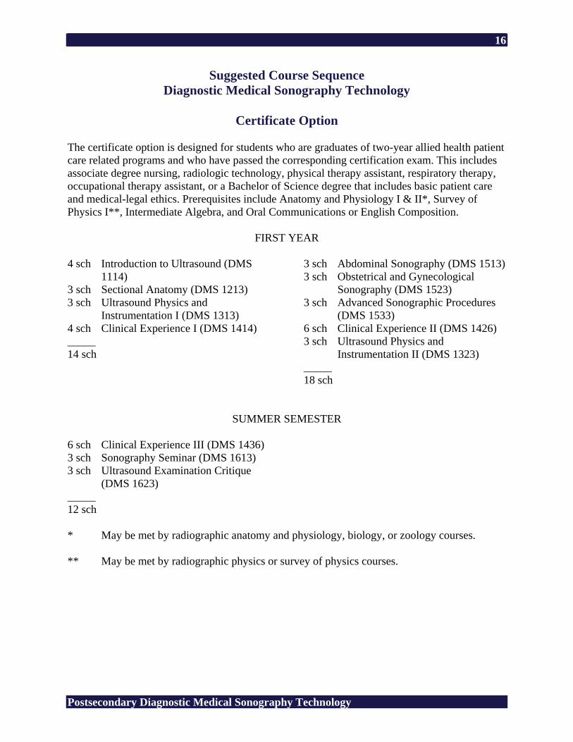
Suggested Course Sequence Diagnostic Medical Sonography Technology
Certificate Option
The certificate option is designed for students who are graduates of two-year allied health patient care related programs and who have passed the corresponding certification exam. This includes associate degree nursing, radiologic technology, physical therapy assistant, respiratory therapy, occupational therapy assistant, or a Bachelor of Science degree that includes basic patient care and medical-legal ethics. Prerequisites include Anatomy and Physiology I & II*, Survey of Physics I**, Intermediate Algebra, and Oral Communications or English Composition.
FIRST YEAR
4 sch Introduction to Ultrasound (DMS 1114)
3 sch Sectional Anatomy (DMS 1213) 3 sch Ultrasound Physics and
Instrumentation I (DMS 1313) 4 sch Clinical Experience I (DMS 1414) _____ 14 sch
3 sch Abdominal Sonography (DMS 1513)3 sch Obstetrical and Gynecological
Sonography (DMS 1523) 3 sch Advanced Sonographic Procedures
(DMS 1533) 6 sch Clinical Experience II (DMS 1426) 3 sch Ultrasound Physics and
Instrumentation II (DMS 1323) _____ 18 sch
SUMMER SEMESTER 6 sch Clinical Experience III (DMS 1436) 3 sch Sonography Seminar (DMS 1613) 3 sch Ultrasound Examination Critique
(DMS 1623) _____ 12 sch
* May be met by radiographic anatomy and physiology, biology, or zoology courses. ** May be met by radiographic physics or survey of physics courses.
17
Postsecondary Diagnostic Medical Sonography Technology
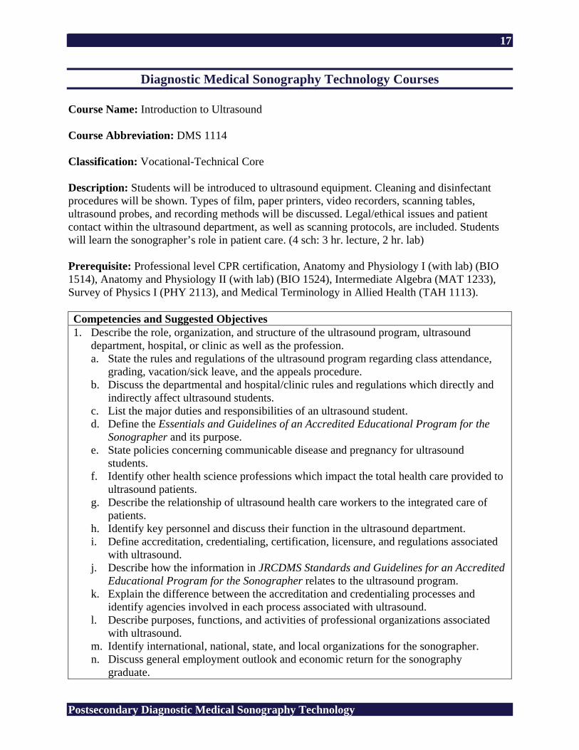
Diagnostic Medical Sonography Technology Courses Course Name: Introduction to Ultrasound Course Abbreviation: DMS 1114 Classification: Vocational-Technical Core Description: Students will be introduced to ultrasound equipment. Cleaning and disinfectant procedures will be shown. Types of film, paper printers, video recorders, scanning tables, ultrasound probes, and recording methods will be discussed. Legal/ethical issues and patient contact within the ultrasound department, as well as scanning protocols, are included. Students will learn the sonographer’s role in patient care. (4 sch: 3 hr. lecture, 2 hr. lab) Prerequisite: Professional level CPR certification, Anatomy and Physiology I (with lab) (BIO 1514), Anatomy and Physiology II (with lab) (BIO 1524), Intermediate Algebra (MAT 1233), Survey of Physics I (PHY 2113), and Medical Terminology in Allied Health (TAH 1113). Competencies and Suggested Objectives 1. Describe the role, organization, and structure of the ultrasound program, ultrasound
department, hospital, or clinic as well as the profession. a. State the rules and regulations of the ultrasound program regarding class attendance,
grading, vacation/sick leave, and the appeals procedure. b. Discuss the departmental and hospital/clinic rules and regulations which directly and
indirectly affect ultrasound students. c. List the major duties and responsibilities of an ultrasound student. d. Define the Essentials and Guidelines of an Accredited Educational Program for the
Sonographer and its purpose. e. State policies concerning communicable disease and pregnancy for ultrasound
students. f. Identify other health science professions which impact the total health care provided to
ultrasound patients. g. Describe the relationship of ultrasound health care workers to the integrated care of
patients. h. Identify key personnel and discuss their function in the ultrasound department. i. Define accreditation, credentialing, certification, licensure, and regulations associated
with ultrasound. j. Describe how the information in JRCDMS Standards and Guidelines for an Accredited
Educational Program for the Sonographer relates to the ultrasound program. k. Explain the difference between the accreditation and credentialing processes and
identify agencies involved in each process associated with ultrasound. l. Describe purposes, functions, and activities of professional organizations associated
with ultrasound. m. Identify international, national, state, and local organizations for the sonographer. n. Discuss general employment outlook and economic return for the sonography
graduate.
18
Postsecondary Diagnostic Medical Sonography Technology
o. Discuss career advancement and opportunities for the sonographer. p. Identify benefits of continuing education of the sonographer.
2. Assess and resolve ethical issues and dilemmas in health care. a. Describe the major milestones in the development of codes of behavior and ethical
standards in the healing arts. b. Identify the significance of health care professions. c. Recognize the moral, social, and cultural basis of the development of an ethic. d. Discuss the role of ethical behavior in health care delivery. e. Differentiate between empathetic and sympathetic involvement in relationships with
patients. f. Identify concepts of personal honesty, integrity, accountability, competence, and
compassion as ethical imperatives in health care. g. Recognize situations and conditions which give rise to ethical dilemmas in health care. h. Discuss the legal implications of professional liability, malpractice, professional
negligence/carelessness, and other legal doctrines applicable to professional practice. i. Discuss the significance of accurate, complete, and correct methods of medical record
keeping as a legal/ethical imperative. j. Articulate responses to theoretical situations and questions relating to the ethics of care
and health care delivery. 3. Identify legal responsibilities related to the scope of practice for sonography.
a. Define the scope of practice for the diagnostic medical sonographer. b. Identify the requirements of the sonographer according to the scope of practice.
4. Describe clinical practice standards in diagnostic ultrasound. a. Identify patient history and correlate with the sonographic procedure requested. b. Determine patient ability to tolerate the sonographic procedure. c. Evaluate any contraindications to the sonographic procedure such as medications,
inappropriate patient preparation, or unwillingness of the patient to tolerate the sonographic procedure.
d. Explain the sonography procedure to the patient and respond to patient questions. e. Refer specific diagnostic, treatment, or prognosis questions to the patient’s physician. f. Develop a procedure plan for the sonographic exam. g. Adapt the sonographic procedure plan to optimize exam results. h. Determine if contrast media will enhance image quality and provide additional
diagnostic information. i. Determine the need for additional accessory equipment or additional personnel. j. Modify sonographic procedure plan according to patient disease process and
circumstances under which the procedure must be performed (i.e., operating room, ultrasound room, patient bedside, or emergency room).
k. Modify sonographic procedure plan according to patient physical and mental status during the exam.
l. Perform basic patient care tasks. m. Analyze sonographic findings throughout the exam and perform measurements to
provide accurate diagnosis for treatment plan. n. Confirm that the sonographic exam complies with applicable protocols and guidelines. o. Document sonographic exam results. p. Notify the appropriate health care provider when immediate medical attention is
19
Postsecondary Diagnostic Medical Sonography Technology
necessary. q. Provide a written summary of preliminary sonographic findings. r. Implement quality assurance within the ultrasound department.
5. Maintain patient care. a. Work in partnership with other health care professionals. b. Maintain appropriate professional credentials. c. Provide a diagnostic sonographic exam for the patient by applying professional
judgment and discretion. d. Maintain continuing medical education on current issues in sonography. e. Identify personal strengths and use them to benefit patients, coworkers, and the
profession. f. Perform diagnostic sonographic procedures in supervised clinical experiences. g. Communicate effectively with all members of the health care team. h. Maintain patient confidentiality. i. Utilize standard precautions.
6. Use ultrasound equipment and accessory items. a. Demonstrate use of ultrasound equipment. b. Scan and document findings in the ultrasound lab setting. c. Produce ultrasound images according to standards of care. d. Identify ultrasound scanning techniques. e. Use proper gain controls to produce diagnostic ultrasound images. f. Document total ultrasound scanning time in each procedure. g. Perform the required images for ultrasound abdominal scanning. h. Perform the required ultrasound images for obstetrical and gynecological scanning.
STANDARDS CAAHEP Standards and Guidelines for the Accreditation of Educational Programs in Diagnostic Medical Sonography DMSC1 Utilize oral and written communication. DMSC2 Provide basic patient care and comfort. DMSC3 Demonstrate knowledge and understanding of human gross and sectional anatomy. DMSC4 Demonstrate knowledge and understanding of physiology, pathology, and
pathophysiology. DMSC5 Demonstrate knowledge and understanding of acoustical physics, Doppler ultrasound
principles, and ultrasound instrumentation. DMSC6 Demonstrate knowledge and understanding of the interaction between ultrasound and
tissue and the probability of biological effects in clinical examinations. DMSC7 Employ professional judgment and discretion. DMSC8 Understand the fundamental elements for implementing a quality assurance and
improvement program, and the policies, protocols, and procedures for the general function of the ultrasound laboratory.
DMSC9 Recognize the importance of continuing education.
20
Postsecondary Diagnostic Medical Sonography Technology
DMSD1 Demonstrate the ability to perform sonographic examinations of the abdomen, superficial structures, non-cardiac chest, and the gravid and nongravid pelvis according to protocol guidelines established by national professional organizations and the protocol of the employing institution utilizing real-time equipment with both transabdominal and endocavitary transducers, Doppler, and color Doppler display modes.
DMSD2 Recognize and identify the sonographic appearance of normal anatomic structures, including anatomic variants and normal Doppler patterns.
DMSD3 Recognize, identify, and appropriately document the abnormal sonographic and Doppler patterns of disease processes, pathology, and pathophysiology of the structures listed above. Modify the scanning protocol based on the sonographic findings and the differential diagnosis.
DMSD4 Recognize and identify the sonographic appearance of normal anatomic structures of the female pelvis, including anatomic variants and normal Doppler patterns.
DMSD5 Recognize and identify the sonographic appearance of normal maternal, embryonic, and fetal anatomic structures during the first, second, and third trimesters.
DMSD6 Recognize, identify, and appropriately document the sonographic appearance of gynecologic disease processes, pathology, and pathophysiology.
DMSD7 Recognize, identify, and appropriately document the sonographic appearance of obstetric abnormalities, disease, pathology, and pathophysiology.
DMSD8 Demonstrate knowledge and understanding of the role of the sonographer in performing interventional/invasive procedures.
Related Academic Standards R1 Interpret Graphic Information (forms, maps, reference sources) R2 Words in Context (same and opposite meaning) R3 Recall Information (details, sequence) R4 Construct Meaning (main idea, summary/paraphrase, compare/contrast, cause/effect) R5 Evaluate/Extend Meaning (fact/opinion, predict outcomes, point of view) M1 Addition of Whole Numbers (no regrouping, regrouping) M2 Subtraction of Whole Numbers (no regrouping, regrouping) M3 Multiplication of Whole Numbers (no regrouping, regrouping) M4 Division of Whole Numbers (no remainder, remainder) M5 Decimals (addition, subtraction, multiplication, division) A1 Numeration (ordering, place value, scientific notation) A2 Number Theory (ratio, proportion) A3 Data Interpretation (graph, table, chart, diagram) A5 Measurement (money, time, temperature, length, area, volume) A6 Geometry (angles, Pythagorean theory) A7 Computation in Context (whole numbers, decimals, fractions, algebraic operations) A8 Estimation (rounding, estimation) L1 Usage (pronoun, tense, subject/verb agreement, adjective, adverb) L2 Sentence Formation (fragments, run-on, clarity) L3 Paragraph Development (topic sentence, supporting sentence, sequence) L4 Capitalization (proper noun, titles)
21
Postsecondary Diagnostic Medical Sonography Technology
L5 Punctuation (comma, semicolon) L6 Writing Conventions (quotation marks, apostrophe, parts of a letter) S1 Vowel (short, long) S2 Consonant (variant spelling, silent letter) S3 Structural Unit (root, suffix) Copyright © 1994 by CTB/McGraw-Hill LLC 21st Century Skills CS1 Global Awareness CS2 Financial, Economic, and Business Literacy CS3 Civic Literacy CS4 Information and Communication Skills CS5 Thinking and Problem-Solving Skills CS6 Interpersonal and Self-Directional Skills SUGGESTED REFERENCES American Registry for Diagnostic Medical Sonography. (n.d.). Retrieved February 24, 2006,
from http://www.ardms.org/ American Institute of Ultrasound in Medicine. (n.d.). Retrieved February 24, 2006, from
http://www.aium.org/ Callen, P. W. (2000). Ultrasonography in obstetrics and gynecology (4th ed.). Philadelphia: W.
B. Saunders. Commission on Accreditation of Allied Health Education Programs. (n.d.). Retrieved February
17, 2006, from http://www.caahep.org/ Curry, R. A., & Tempkin, B. B. (2004). Sonography introduction to normal structure and
function (2nd ed.). St. Louis, MO: Elsevier Science. Dirckx, J. H. (Ed.). (2001). Stedman’s concise medical dictionary for the health professions (4th
ed.). Baltimore: Lippincott Williams & Wilkins. Edelman, S. K. (2004). Understanding ultrasound physics (3rd ed.). Woodlands, TX: ESP. Green, L. (2002). Doppler physics and color fundamentals [DVD]. (Available from Gulfcoast
Ultrasound Institute, Inc., 4615 Gulf Blvd., Suite 205, St. Pete Beach, FL 33706) Green, L., & Sens, L. (1998). Introduction to abdominal sonography [Videotape]. (Available
from Gulfcoast Ultrasound Institute, Inc., 4615 Gulf Blvd., Suite 205, St. Pete Beach, FL 33706)
22
Postsecondary Diagnostic Medical Sonography Technology
Green, L., & Sens, L. (1998). Introduction to gynecological sonography [Videotape]. (Available from Gulfcoast Ultrasound Institute, Inc., 4615 Gulf Blvd., Suite 205, St. Pete Beach, FL 33706)
Green, L., & Sens, L. (1998). Introduction to obstetrical sonography [Videotape]. (Available
from Gulfcoast Ultrasound Institute, Inc., 4615 Gulf Blvd., Suite 205, St. Pete Beach, FL 33706)
Green, L., & Sens, L. (1998). Introduction to ultrasound physics [Videotape]. (Available from
Gulfcoast Ultrasound Institute, Inc., 4615 Gulf Blvd., Suite 205, St. Pete Beach, FL 33706)
Hagen-Ansert, S. L. (2006). Textbook of diagnostic ultrasonography (6th ed.). St. Louis, MO:
Mosby. Hecht, L. C., Jr. (2004). The registry review CD quiz series [CD]. (Available from Gulfcoast
Ultrasound Institute, Inc., 4615 Gulf Blvd., Suite 205, St. Pete Beach, FL 33706) Hecht, L. C., Jr. (2004). Testicular sonography [CD]. (Available from Gulfcoast Ultrasound
Institute, Inc., 4615 Gulf Blvd., Suite 205, St. Pete Beach, FL 33706) Journal of Diagnostic Medical Sonography. Thousand Oaks, CA: SAGE. Retrieved February 17,
2006, from http://www.sagepub.com/journal.aspx?pid=259 Krebs, C. A., Odwin, C. S., & Fleischer, A. C. (2004). Appleton & Lange review for the
ultrasonography examination (3rd ed.). Singapore: McGraw-Hill. Lee, W. (1998). Fetal ultrasound simulator [CD]. (Available from The American College of
Obstetricians and Gynecologists Distribution Center, P. O. Box 933104, Atlanta, GA 31193)
Miele, F. R. (2002). Unifying physics review [Videotape]. (Available from Pegasus Lectures,
Inc., P. O. Box 157, Forney, TX 75126) Miele, F. R. (2006). Ultrasound physics and instrumentation (4th ed., Vols. 1-2). Forney, TX:
LLC. Neil, C. (2002). Endovaginal scanning [Videotape]. (Available from Gulfcoast Ultrasound
Institute, Inc., 4615 Gulf Blvd., Suite 205, St. Pete Beach, FL 33706) Ott, W. J. (2001). Clinical obstetrical case studies (Vols. 1-3) [Interactive CD]. (Available from
Gulfcoast Ultrasound Institute, Inc., 4615 Gulf Blvd., Suite 205, St. Pete Beach, FL 33706)
23
Postsecondary Diagnostic Medical Sonography Technology
Owen, C. A. (2005). Endovaginal scanning (Vols. 1-2) [Interactive CD]. (Available from Gulfcoast Ultrasound Institute, Inc., 4615 Gulf Blvd., Suite 205, St. Pete Beach, FL 33706)
Rumack, C. M., Wilson, S. R., Charboneau, J. W., & Johnson, J. M. (2005). Diagnostic
ultrasound (3rd ed., Vols. 1-2). St. Louis, MO: Mosby. Society of Diagnostic Medical Sonography. (n.d.). Retrieved February 24, 2006, from
http://www.sdms.org/ Stephenson, S. R., & Layton, T. (2004). The registry review CD quiz series: Abdominal series
[CD]. (Available from Gulfcoast Ultrasound Institute, Inc., 4615 Gulf Blvd., Suite 205, St. Pete Beach, FL 33706)
Stephenson, S. R., & Layton, T. (2004). The registry review CD quiz series: OB/GYN
sonography [CD]. (Available from Gulfcoast Ultrasound Institute, Inc., 4615 Gulf Blvd., Suite 205, St. Pete Beach, FL 33706)
Tempkin, B. B. (1999). Ultrasound scanning principles and protocols (2nd ed.). Philadelphia: W.
B. Saunders. Ultrasound case studies. (n.d.). Retrieved February 24, 2006, from http://www.sonoworld.com/ Women’s health resources for medical industry professionals. (n.d.). Retrieved February 24,
2006, from http://www.obgyn.net/ Zagzebski, J. A. (1996). Essentials of ultrasound physics. St. Louis, MO: Mosby.
24
Postsecondary Diagnostic Medical Sonography Technology
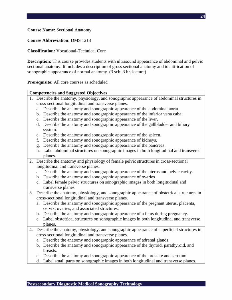
Course Name: Sectional Anatomy Course Abbreviation: DMS 1213 Classification: Vocational-Technical Core Description: This course provides students with ultrasound appearance of abdominal and pelvic sectional anatomy. It includes a description of gross sectional anatomy and identification of sonographic appearance of normal anatomy. (3 sch: 3 hr. lecture) Prerequisite: All core courses as scheduled Competencies and Suggested Objectives 1. Describe the anatomy, physiology, and sonographic appearance of abdominal structures in
cross-sectional longitudinal and transverse planes. a. Describe the anatomy and sonographic appearance of the abdominal aorta. b. Describe the anatomy and sonographic appearance of the inferior vena caba. c. Describe the anatomy and sonographic appearance of the liver. d. Describe the anatomy and sonographic appearance of the gallbladder and biliary
system. e. Describe the anatomy and sonographic appearance of the spleen. f. Describe the anatomy and sonographic appearance of kidneys. g. Describe the anatomy and sonographic appearance of the pancreas. h. Label abdominal structures on sonographic images in both longitudinal and transverse
planes. 2. Describe the anatomy and physiology of female pelvic structures in cross-sectional
longitudinal and transverse planes. a. Describe the anatomy and sonographic appearance of the uterus and pelvic cavity. b. Describe the anatomy and sonographic appearance of ovaries. c. Label female pelvic structures on sonographic images in both longitudinal and
transverse planes. 3. Describe the anatomy, physiology, and sonographic appearance of obstetrical structures in
cross-sectional longitudinal and transverse planes. a. Describe the anatomy and sonographic appearance of the pregnant uterus, placenta,
cervix, ovaries, and associated structures. b. Describe the anatomy and sonographic appearance of a fetus during pregnancy. c. Label obstetrical structures on sonographic images in both longitudinal and transverse
planes. 4. Describe the anatomy, physiology, and sonographic appearance of superficial structures in
cross-sectional longitudinal and transverse planes. a. Describe the anatomy and sonographic appearance of adrenal glands. b. Describe the anatomy and sonographic appearance of the thyroid, parathyroid, and
breasts. c. Describe the anatomy and sonographic appearance of the prostate and scrotum. d. Label small parts on sonographic images in both longitudinal and transverse planes.
25
Postsecondary Diagnostic Medical Sonography Technology
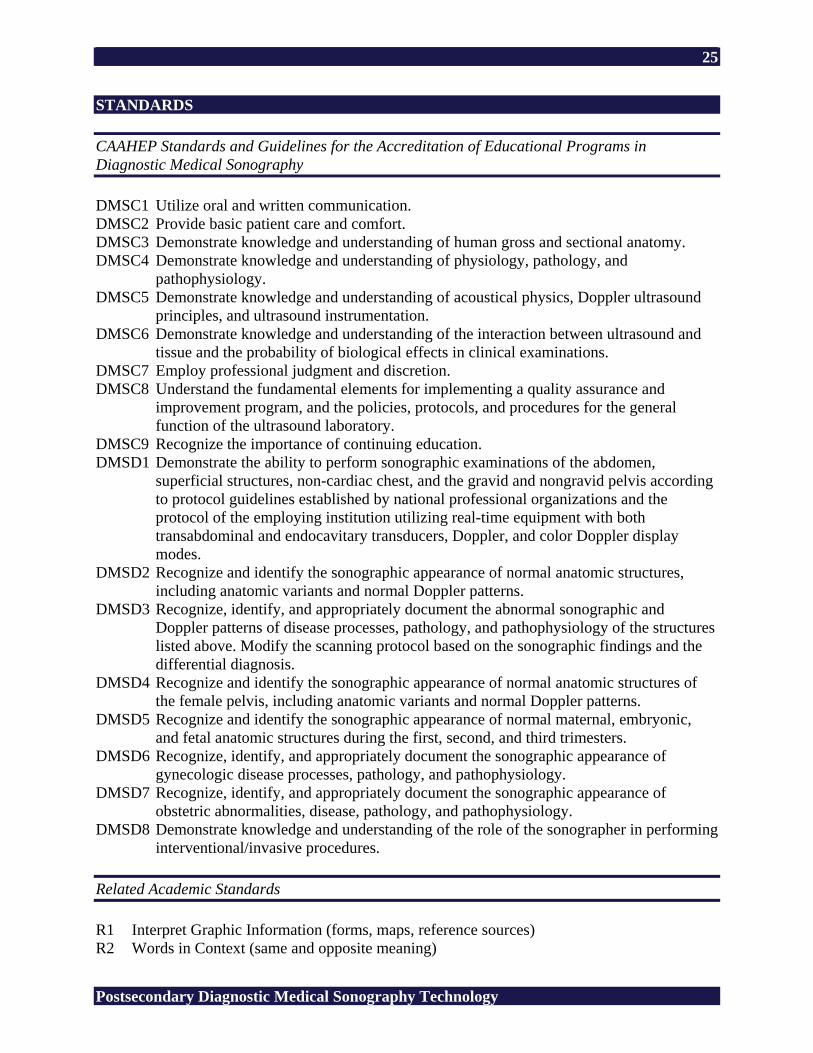
STANDARDS CAAHEP Standards and Guidelines for the Accreditation of Educational Programs in Diagnostic Medical Sonography DMSC1 Utilize oral and written communication. DMSC2 Provide basic patient care and comfort. DMSC3 Demonstrate knowledge and understanding of human gross and sectional anatomy. DMSC4 Demonstrate knowledge and understanding of physiology, pathology, and
pathophysiology. DMSC5 Demonstrate knowledge and understanding of acoustical physics, Doppler ultrasound
principles, and ultrasound instrumentation. DMSC6 Demonstrate knowledge and understanding of the interaction between ultrasound and
tissue and the probability of biological effects in clinical examinations. DMSC7 Employ professional judgment and discretion. DMSC8 Understand the fundamental elements for implementing a quality assurance and
improvement program, and the policies, protocols, and procedures for the general function of the ultrasound laboratory.
DMSC9 Recognize the importance of continuing education. DMSD1 Demonstrate the ability to perform sonographic examinations of the abdomen,
superficial structures, non-cardiac chest, and the gravid and nongravid pelvis according to protocol guidelines established by national professional organizations and the protocol of the employing institution utilizing real-time equipment with both transabdominal and endocavitary transducers, Doppler, and color Doppler display modes.
DMSD2 Recognize and identify the sonographic appearance of normal anatomic structures, including anatomic variants and normal Doppler patterns.
DMSD3 Recognize, identify, and appropriately document the abnormal sonographic and Doppler patterns of disease processes, pathology, and pathophysiology of the structures listed above. Modify the scanning protocol based on the sonographic findings and the differential diagnosis.
DMSD4 Recognize and identify the sonographic appearance of normal anatomic structures of the female pelvis, including anatomic variants and normal Doppler patterns.
DMSD5 Recognize and identify the sonographic appearance of normal maternal, embryonic, and fetal anatomic structures during the first, second, and third trimesters.
DMSD6 Recognize, identify, and appropriately document the sonographic appearance of gynecologic disease processes, pathology, and pathophysiology.
DMSD7 Recognize, identify, and appropriately document the sonographic appearance of obstetric abnormalities, disease, pathology, and pathophysiology.
DMSD8 Demonstrate knowledge and understanding of the role of the sonographer in performing interventional/invasive procedures.
Related Academic Standards R1 Interpret Graphic Information (forms, maps, reference sources) R2 Words in Context (same and opposite meaning)
26
Postsecondary Diagnostic Medical Sonography Technology
R3 Recall Information (details, sequence) R4 Construct Meaning (main idea, summary/paraphrase, compare/contrast, cause/effect) R5 Evaluate/Extend Meaning (fact/opinion, predict outcomes, point of view) M1 Addition of Whole Numbers (no regrouping, regrouping) M2 Subtraction of Whole Numbers (no regrouping, regrouping) M3 Multiplication of Whole Numbers (no regrouping, regrouping) M4 Division of Whole Numbers (no remainder, remainder) M5 Decimals (addition, subtraction, multiplication, division) M6 Fractions (addition, subtraction, multiplication, division) M8 Percents A1 Numeration (ordering, place value, scientific notation) A2 Number Theory (ratio, proportion) A3 Data Interpretation (graph, table, chart, diagram) A5 Measurement (money, time, temperature, length, area, volume) A6 Geometry (angles, Pythagorean theory) A7 Computation in Context (whole numbers, decimals, fractions, algebraic operations) A8 Estimation (rounding, estimation) L1 Usage (pronoun, tense, subject/verb agreement, adjective, adverb) L2 Sentence Formation (fragments, run-on, clarity) L3 Paragraph Development (topic sentence, supporting sentence, sequence) L4 Capitalization (proper noun, titles) L5 Punctuation (comma, semicolon) L6 Writing Conventions (quotation marks, apostrophe, parts of a letter) S1 Vowel (short, long) S2 Consonant (variant spelling, silent letter) S3 Structural Unit (root, suffix) Copyright © 1994 by CTB/McGraw-Hill LLC 21st Century Skills CS1 Global Awareness CS2 Financial, Economic, and Business Literacy CS3 Civic Literacy CS4 Information and Communication Skills CS5 Thinking and Problem-Solving Skills CS6 Interpersonal and Self-Directional Skills SUGGESTED REFERENCES American Registry for Diagnostic Medical Sonography. (n.d.). Retrieved February 24, 2006,
from http://www.ardms.org/ American Institute of Ultrasound in Medicine. (n.d.). Retrieved February 24, 2006, from
http://www.aium.org/
27
Postsecondary Diagnostic Medical Sonography Technology
Callen, P. W. (2000). Ultrasonography in obstetrics and gynecology (4th ed.). Philadelphia: W. B. Saunders.
Commission on Accreditation of Allied Health Education Programs. (n.d.). Retrieved February
17, 2006, from http://www.caahep.org/ Curry, R. A., & Tempkin, B. B. (2004). Sonography introduction to normal structure and
function (2nd ed.). St. Louis, MO: Elsevier Science. Dirckx, J. H. (Ed.). (2001). Stedman’s concise medical dictionary for the health professions (4th
ed.). Baltimore: Lippincott Williams & Wilkins. Journal of Diagnostic Medical Sonography. Thousand Oaks, CA: SAGE. Retrieved February 17,
2006, from http://www.sagepub.com/journal.aspx?pid=259 Society of Diagnostic Medical Sonography. (n.d.). Retrieved February 24, 2006, from
http://www.sdms.org/ Ultrasound case studies. (n.d.). Retrieved February 24, 2006, from http://www.sonoworld.com/ Women’s health resources for medical industry professionals. (n.d.). Retrieved February 24,
2006, from http://www.obgyn.net/
28
Postsecondary Diagnostic Medical Sonography Technology
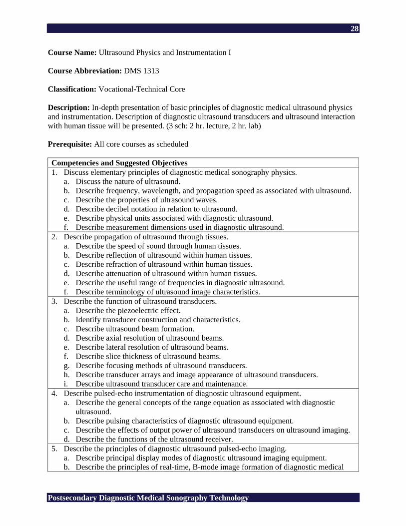
Course Name: Ultrasound Physics and Instrumentation I Course Abbreviation: DMS 1313 Classification: Vocational-Technical Core Description: In-depth presentation of basic principles of diagnostic medical ultrasound physics and instrumentation. Description of diagnostic ultrasound transducers and ultrasound interaction with human tissue will be presented. (3 sch: 2 hr. lecture, 2 hr. lab) Prerequisite: All core courses as scheduled Competencies and Suggested Objectives 1. Discuss elementary principles of diagnostic medical sonography physics.
a. Discuss the nature of ultrasound. b. Describe frequency, wavelength, and propagation speed as associated with ultrasound. c. Describe the properties of ultrasound waves. d. Describe decibel notation in relation to ultrasound. e. Describe physical units associated with diagnostic ultrasound. f. Describe measurement dimensions used in diagnostic ultrasound.
2. Describe propagation of ultrasound through tissues. a. Describe the speed of sound through human tissues. b. Describe reflection of ultrasound within human tissues. c. Describe refraction of ultrasound within human tissues. d. Describe attenuation of ultrasound within human tissues. e. Describe the useful range of frequencies in diagnostic ultrasound. f. Describe terminology of ultrasound image characteristics.
3. Describe the function of ultrasound transducers. a. Describe the piezoelectric effect. b. Identify transducer construction and characteristics. c. Describe ultrasound beam formation. d. Describe axial resolution of ultrasound beams. e. Describe lateral resolution of ultrasound beams. f. Describe slice thickness of ultrasound beams. g. Describe focusing methods of ultrasound transducers. h. Describe transducer arrays and image appearance of ultrasound transducers. i. Describe ultrasound transducer care and maintenance.
4. Describe pulsed-echo instrumentation of diagnostic ultrasound equipment. a. Describe the general concepts of the range equation as associated with diagnostic
ultrasound. b. Describe pulsing characteristics of diagnostic ultrasound equipment. c. Describe the effects of output power of ultrasound transducers on ultrasound imaging. d. Describe the functions of the ultrasound receiver.
5. Describe the principles of diagnostic ultrasound pulsed-echo imaging. a. Describe principal display modes of diagnostic ultrasound imaging equipment. b. Describe the principles of real-time, B-mode image formation of diagnostic medical
29
Postsecondary Diagnostic Medical Sonography Technology
ultrasound equipment. c. Describe limitations of scanning speed of diagnostic medical ultrasound equipment. d. Describe the principles of 3D/4D imaging.
STANDARDS CAAHEP Standards and Guidelines for the Accreditation of Educational Programs in Diagnostic Medical Sonography DMSC1 Utilize oral and written communication. DMSC2 Provide basic patient care and comfort. DMSC3 Demonstrate knowledge and understanding of human gross and sectional anatomy. DMSC4 Demonstrate knowledge and understanding of physiology, pathology, and
pathophysiology. DMSC5 Demonstrate knowledge and understanding of acoustical physics, Doppler ultrasound
principles, and ultrasound instrumentation. DMSC6 Demonstrate knowledge and understanding of the interaction between ultrasound and
tissue and the probability of biological effects in clinical examinations. DMSC7 Employ professional judgment and discretion. DMSC8 Understand the fundamental elements for implementing a quality assurance and
improvement program, and the policies, protocols, and procedures for the general function of the ultrasound laboratory.
DMSC9 Recognize the importance of continuing education. DMSD1 Demonstrate the ability to perform sonographic examinations of the abdomen,
superficial structures, non-cardiac chest, and the gravid and nongravid pelvis according to protocol guidelines established by national professional organizations and the protocol of the employing institution utilizing real-time equipment with both transabdominal and endocavitary transducers, Doppler, and color Doppler display modes.
DMSD2 Recognize and identify the sonographic appearance of normal anatomic structures, including anatomic variants and normal Doppler patterns.
DMSD3 Recognize, identify, and appropriately document the abnormal sonographic and Doppler patterns of disease processes, pathology, and pathophysiology of the structures listed above. Modify the scanning protocol based on the sonographic findings and the differential diagnosis.
DMSD4 Recognize and identify the sonographic appearance of normal anatomic structures of the female pelvis, including anatomic variants and normal Doppler patterns.
DMSD5 Recognize and identify the sonographic appearance of normal maternal, embryonic, and fetal anatomic structures during the first, second, and third trimesters.
DMSD6 Recognize, identify, and appropriately document the sonographic appearance of gynecologic disease processes, pathology, and pathophysiology.
DMSD7 Recognize, identify, and appropriately document the sonographic appearance of obstetric abnormalities, disease, pathology, and pathophysiology.
DMSD8 Demonstrate knowledge and understanding of the role of the sonographer in performing interventional/invasive procedures.

30
Postsecondary Diagnostic Medical Sonography Technology
Related Academic Standards R1 Interpret Graphic Information (forms, maps, reference sources) R2 Words in Context (same and opposite meaning) R3 Recall Information (details, sequence) R4 Construct Meaning (main idea, summary/paraphrase, compare/contrast, cause/effect) R5 Evaluate/Extend Meaning (fact/opinion, predict outcomes, point of view) M1 Addition of Whole Numbers (no regrouping, regrouping) M2 Subtraction of Whole Numbers (no regrouping, regrouping) M3 Multiplication of Whole Numbers (no regrouping, regrouping) M4 Division of Whole Numbers (no remainder, remainder) M5 Decimals (addition, subtraction, multiplication, division) M6 Fractions (addition, subtraction, multiplication, division) M7 Integers (addition, subtraction, multiplication, division) M8 Percents M9 Algebraic Operations A1 Numeration (ordering, place value, scientific notation) A2 Number Theory (ratio, proportion) A3 Data Interpretation (graph, table, chart, diagram) A4 Pre-Algebra and Algebra (equations, inequality) A5 Measurement (money, time, temperature, length, area, volume) A6 Geometry (angles, Pythagorean theory) A7 Computation in Context (whole numbers, decimals, fractions, algebraic operations) A8 Estimation (rounding, estimation) L1 Usage (pronoun, tense, subject/verb agreement, adjective, adverb) L2 Sentence Formation (fragments, run-on, clarity) L3 Paragraph Development (topic sentence, supporting sentence, sequence) L4 Capitalization (proper noun, titles) L5 Punctuation (comma, semicolon) L6 Writing Conventions (quotation marks, apostrophe, parts of a letter) S1 Vowel (short, long) S2 Consonant (variant spelling, silent letter) S3 Structural Unit (root, suffix) Copyright © 1994 by CTB/McGraw-Hill LLC 21st Century Skills CS1 Global Awareness CS2 Financial, Economic, and Business Literacy CS3 Civic Literacy CS4 Information and Communication Skills CS5 Thinking and Problem-Solving Skills CS6 Interpersonal and Self-Directional Skills
31
Postsecondary Diagnostic Medical Sonography Technology
SUGGESTED REFERENCES American Registry for Diagnostic Medical Sonography. (n.d.). Retrieved February 24, 2006,
from http://www.ardms.org/ American Institute of Ultrasound in Medicine. (n.d.). Retrieved February 24, 2006, from
http://www.aium.org/ Commission on Accreditation of Allied Health Education Programs. (n.d.). Retrieved February
17, 2006, from http://www.caahep.org/ Dirckx, J. H. (Ed.). (2001). Stedman’s concise medical dictionary for the health professions (4th
ed.). Baltimore: Lippincott Williams & Wilkins. Edelman, S. K. (2004). Understanding ultrasound physics (3rd ed.). Woodlands, TX: ESP. Green, L., & Sens, L. (1998). Introduction to ultrasound physics [Videotape]. (Available from
Gulfcoast Ultrasound Institute, Inc., 4615 Gulf Blvd., Suite 205, St. Pete Beach, FL 33706)
Journal of Diagnostic Medical Sonography. Thousand Oaks, CA: SAGE. Retrieved February 17,
2006, from http://www.sagepub.com/journal.aspx?pid=259 Krebs, C. A., Odwin, C. S., & Fleischer, A. C. (2004). Appleton & Lange review for the
ultrasonography examination (3rd ed.). Singapore: McGraw-Hill. Miele, F. R. (2006). Ultrasound physics and instrumentation (4th ed., Vols. 1-2). Forney, TX:
LLC. Society of Diagnostic Medical Sonography. (n.d.). Retrieved February 24, 2006, from
http://www.sdms.org/ Zagzebski, J. A. (1996). Essentials of ultrasound physics. St. Louis, MO: Mosby.
32
Postsecondary Diagnostic Medical Sonography Technology
Course Name: Ultrasound Physics and Instrumentation II Course Abbreviation: DMS 1323 Classification: Vocational-Technical Core Description: A continuation of Ultrasound Physics and Instrumentation I (DMS 1313). This class includes an in-depth presentation of image display modes, Doppler, color, and hemodynamics of diagnostic ultrasound. The causes of artifacts and how to scan safely, conduct instrument performance measurements, and prepare for registry examinations. (3 sch: 2 hr. lecture, 2 hr. lab) Prerequisite: All core courses as scheduled Competencies and Suggested Objectives 1. Describe images, storage, and display methods used in diagnostic medical ultrasound.
a. Describe the role of the scan converter in diagnostic ultrasound imaging. b. Describe digital devices used in diagnostic ultrasound equipment. c. Describe pre- and post-processing functions of diagnostic medical ultrasound
equipment. d. Describe the display devices used with diagnostic medical ultrasound equipment. e. Describe recording and archiving techniques employed in diagnostic medical
ultrasound. 2. Describe Doppler instrumentation of diagnostic medical ultrasound.
a. Describe hemodynamics. b. Describe the physical principles of Doppler ultrasound imaging. c. Describe continuous and pulsed wave Doppler instrumentation in diagnostic medical
ultrasound. d. Describe color flow imaging in diagnostic ultrasound. e. Describe color power mode imaging in diagnostic ultrasound.
3. Discuss ultrasound artifacts. a. Define artifacts in ultrasound imaging. b. Describe artifacts associated with resolution of ultrasound waves in human tissues. c. Describe ultrasound artifacts associated with propagation of ultrasound waves in
human tissues. d. Describe ultrasound artifacts associated with attenuation of ultrasound waves in human
tissues. e. Describe artifacts associated with Doppler and color flow instrumentation in diagnostic
ultrasound. f. Describe artifacts caused by electronic noise and equipment malfunction in diagnostic
ultrasound. g. Describe the effects of artifacts on measurements in diagnostic ultrasound.
4. Perform performance and safety standards for ultrasound equipment. a. Discuss general concepts regarding the need for quality assurance in diagnostic
ultrasound. b. Discuss methods for evaluating ultrasound instrument performance.
33
Postsecondary Diagnostic Medical Sonography Technology
c. Identify parameters to be evaluated in quality assurance of diagnostic medical ultrasound equipment.
d. Describe preventative maintenance of diagnostic ultrasound equipment. e. Describe record keeping techniques involved with quality assurance in diagnostic
ultrasound. f. Discuss statistical indices associated with diagnostic ultrasound.
5. Describe bioeffects and safety of diagnostic ultrasound. a. Describe acoustic output quantities of diagnostic ultrasound. b. Describe acoustic labeling standards for diagnostic ultrasound equipment. c. Describe acoustic exposure of diagnostic ultrasound. d. Describe primary mechanisms of biological effects of diagnostic ultrasound. e. Describe experimental biological effect studies of diagnostic ultrasound. f. Describe guidelines and regulations of diagnostic ultrasound equipment use. g. Describe electrical and mechanical hazards associated with diagnostic ultrasound
equipment. STANDARDS CAAHEP Standards and Guidelines for the Accreditation of Educational Programs in Diagnostic Medical Sonography DMSC1 Utilize oral and written communication. DMSC2 Provide basic patient care and comfort. DMSC3 Demonstrate knowledge and understanding of human gross and sectional anatomy. DMSC4 Demonstrate knowledge and understanding of physiology, pathology, and
pathophysiology. DMSC5 Demonstrate knowledge and understanding of acoustical physics, Doppler ultrasound
principles, and ultrasound instrumentation. DMSC6 Demonstrate knowledge and understanding of the interaction between ultrasound and
tissue and the probability of biological effects in clinical examinations. DMSC7 Employ professional judgment and discretion. DMSC8 Understand the fundamental elements for implementing a quality assurance and
improvement program, and the policies, protocols, and procedures for the general function of the ultrasound laboratory.
DMSC9 Recognize the importance of continuing education. DMSD1 Demonstrate the ability to perform sonographic examinations of the abdomen,
superficial structures, non-cardiac chest, and the gravid and nongravid pelvis according to protocol guidelines established by national professional organizations and the protocol of the employing institution utilizing real-time equipment with both transabdominal and endocavitary transducers, Doppler, and color Doppler display modes.
DMSD2 Recognize and identify the sonographic appearance of normal anatomic structures, including anatomic variants and normal Doppler patterns.
34
Postsecondary Diagnostic Medical Sonography Technology
DMSD3 Recognize, identify, and appropriately document the abnormal sonographic and Doppler patterns of disease processes, pathology, and pathophysiology of the structures listed above. Modify the scanning protocol based on the sonographic findings and the differential diagnosis.
DMSD4 Recognize and identify the sonographic appearance of normal anatomic structures of the female pelvis, including anatomic variants and normal Doppler patterns.
DMSD5 Recognize and identify the sonographic appearance of normal maternal, embryonic, and fetal anatomic structures during the first, second, and third trimesters.
DMSD6 Recognize, identify, and appropriately document the sonographic appearance of gynecologic disease processes, pathology, and pathophysiology.
DMSD7 Recognize, identify, and appropriately document the sonographic appearance of obstetric abnormalities, disease, pathology, and pathophysiology.
DMSD8 Demonstrate knowledge and understanding of the role of the sonographer in performing interventional/invasive procedures.
Related Academic Standards R1 Interpret Graphic Information (forms, maps, reference sources) R2 Words in Context (same and opposite meaning) R3 Recall Information (details, sequence) R4 Construct Meaning (main idea, summary/paraphrase, compare/contrast, cause/effect) R5 Evaluate/Extend Meaning (fact/opinion, predict outcomes, point of view) M1 Addition of Whole Numbers (no regrouping, regrouping) M2 Subtraction of Whole Numbers (no regrouping, regrouping) M3 Multiplication of Whole Numbers (no regrouping, regrouping) M4 Division of Whole Numbers (no remainder, remainder) M5 Decimals (addition, subtraction, multiplication, division) M6 Fractions (addition, subtraction, multiplication, division) M7 Integers (addition, subtraction, multiplication, division) M8 Percents M9 Algebraic Operations A1 Numeration (ordering, place value, scientific notation) A2 Number Theory (ratio, proportion) A3 Data Interpretation (graph, table, chart, diagram) A4 Pre-Algebra and Algebra (equations, inequality) A5 Measurement (money, time, temperature, length, area, volume) A6 Geometry (angles, Pythagorean theory) A7 Computation in Context (whole numbers, decimals, fractions, algebraic operations) A8 Estimation (rounding, estimation) L1 Usage (pronoun, tense, subject/verb agreement, adjective, adverb) L2 Sentence Formation (fragments, run-on, clarity) L3 Paragraph Development (topic sentence, supporting sentence, sequence) L4 Capitalization (proper noun, titles) L5 Punctuation (comma, semicolon) L6 Writing Conventions (quotation marks, apostrophe, parts of a letter) S1 Vowel (short, long)
35
Postsecondary Diagnostic Medical Sonography Technology
S2 Consonant (variant spelling, silent letter) S3 Structural Unit (root, suffix) Copyright © 1994 by CTB/McGraw-Hill LLC 21st Century Skills CS1 Global Awareness CS2 Financial, Economic, and Business Literacy CS3 Civic Literacy CS4 Information and Communication Skills CS5 Thinking and Problem-Solving Skills CS6 Interpersonal and Self-Directional Skills SUGGESTED REFERENCES American Registry for Diagnostic Medical Sonography. (n.d.). Retrieved February 24, 2006,
from http://www.ardms.org/ American Institute of Ultrasound in Medicine. (n.d.). Retrieved February 24, 2006, from
http://www.aium.org/ Commission on Accreditation of Allied Health Education Programs. (n.d.). Retrieved February
17, 2006, from http://www.caahep.org/ Dirckx, J. H. (Ed.). (2001). Stedman’s concise medical dictionary for the health professions (4th
ed.). Baltimore: Lippincott Williams & Wilkins. Edelman, S. K. (2004). Understanding ultrasound physics (3rd ed.). Woodlands, TX: ESP. Green, L. (2002). Doppler physics and color fundamentals [DVD]. (Available from Gulfcoast
Ultrasound Institute, Inc., 4615 Gulf Blvd., Suite 205, St. Pete Beach, FL 33706) Green, L., & Sens, L. (1998). Introduction to ultrasound physics [Videotape]. (Available from
Gulfcoast Ultrasound Institute, Inc., 4615 Gulf Blvd., Suite 205, St. Pete Beach, FL 33706)
Hecht, L. C., Jr. (2004). The registry review CD quiz series [CD]. (Available from Gulfcoast
Ultrasound Institute, Inc., 4615 Gulf Blvd., Suite 205, St. Pete Beach, FL 33706) Journal of Diagnostic Medical Sonography. Thousand Oaks, CA: SAGE. Retrieved February 17,
2006, from http://www.sagepub.com/journal.aspx?pid=259 Krebs, C. A., Odwin, C. S., & Fleischer, A. C. (2004). Appleton & Lange review for the
ultrasonography examination (3rd ed.). Singapore: McGraw-Hill.
36
Postsecondary Diagnostic Medical Sonography Technology
Miele, F. R. (2002). Unifying physics review [Videotape]. (Available from Pegasus Lectures, Inc., P. O. Box 157, Forney, TX 75126)
Miele, F. R. (2006). Ultrasound physics and instrumentation (4th ed., Vols. 1-2). Forney, TX:
LLC. Society of Diagnostic Medical Sonography. (n.d.). Retrieved February 24, 2006, from
http://www.sdms.org/ Zagzebski, J. A. (1996). Essentials of ultrasound physics. St. Louis, MO: Mosby.
37
Postsecondary Diagnostic Medical Sonography Technology
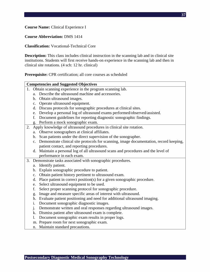
Course Name: Clinical Experience I Course Abbreviation: DMS 1414 Classification: Vocational-Technical Core Description: This class includes clinical instruction in the scanning lab and in clinical site institutions. Students will first receive hands-on experience in the scanning lab and then in clinical site rotations. (4 sch: 12 hr. clinical) Prerequisite: CPR certification; all core courses as scheduled Competencies and Suggested Objectives 1. Obtain scanning experience in the program scanning lab.
a. Describe the ultrasound machine and accessories. b. Obtain ultrasound images. c. Operate ultrasound equipment. d. Discuss protocols for sonographic procedures at clinical sites. e. Develop a personal log of ultrasound exams performed/observed/assisted. f. Document guidelines for reporting diagnostic sonographic findings. g. Perform a mock sonographic exam.
2. Apply knowledge of ultrasound procedures in clinical site rotation. a. Observe sonographers at clinical affiliates. b. Scan patients under the direct supervision of the sonographer. c. Demonstrate clinical site protocols for scanning, image documentation, record keeping,
patient contact, and reporting procedures. d. Maintain a personal log of all ultrasound scans and procedures and the level of
performance in each exam. 3. Demonstrate tasks associated with sonographic procedures.
a. Identify patient. b. Explain sonographic procedure to patient. c. Obtain patient history pertinent to ultrasound exam. d. Place patient in correct position(s) for a given sonographic procedure. e. Select ultrasound equipment to be used. f. Select proper scanning protocol for sonographic procedure. g. Image and measure specific areas of interest with ultrasound. h. Evaluate patient positioning and need for additional ultrasound imaging. i. Document sonographic diagnostic images. j. Demonstrate written and oral responses regarding ultrasound images. k. Dismiss patient after ultrasound exam is complete. l. Document sonographic exam results in proper logs. m. Prepare room for next sonographic exam. n. Maintain standard precautions.
38
Postsecondary Diagnostic Medical Sonography Technology
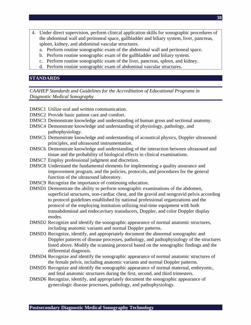
4. Under direct supervision, perform clinical application skills for sonographic procedures of the abdominal wall and peritoneal space, gallbladder and biliary system, liver, pancreas, spleen, kidney, and abdominal vascular structures. a. Perform routine sonographic exam of the abdominal wall and peritoneal space. b. Perform routine sonographic exam of the gallbladder and biliary system. c. Perform routine sonographic exam of the liver, pancreas, spleen, and kidney. d. Perform routine sonographic exam of abdominal vascular structures.
STANDARDS CAAHEP Standards and Guidelines for the Accreditation of Educational Programs in Diagnostic Medical Sonography DMSC1 Utilize oral and written communication. DMSC2 Provide basic patient care and comfort. DMSC3 Demonstrate knowledge and understanding of human gross and sectional anatomy. DMSC4 Demonstrate knowledge and understanding of physiology, pathology, and
pathophysiology. DMSC5 Demonstrate knowledge and understanding of acoustical physics, Doppler ultrasound
principles, and ultrasound instrumentation. DMSC6 Demonstrate knowledge and understanding of the interaction between ultrasound and
tissue and the probability of biological effects in clinical examinations. DMSC7 Employ professional judgment and discretion. DMSC8 Understand the fundamental elements for implementing a quality assurance and
improvement program, and the policies, protocols, and procedures for the general function of the ultrasound laboratory.
DMSC9 Recognize the importance of continuing education. DMSD1 Demonstrate the ability to perform sonographic examinations of the abdomen,
superficial structures, non-cardiac chest, and the gravid and nongravid pelvis according to protocol guidelines established by national professional organizations and the protocol of the employing institution utilizing real-time equipment with both transabdominal and endocavitary transducers, Doppler, and color Doppler display modes.
DMSD2 Recognize and identify the sonographic appearance of normal anatomic structures, including anatomic variants and normal Doppler patterns.
DMSD3 Recognize, identify, and appropriately document the abnormal sonographic and Doppler patterns of disease processes, pathology, and pathophysiology of the structures listed above. Modify the scanning protocol based on the sonographic findings and the differential diagnosis.
DMSD4 Recognize and identify the sonographic appearance of normal anatomic structures of the female pelvis, including anatomic variants and normal Doppler patterns.
DMSD5 Recognize and identify the sonographic appearance of normal maternal, embryonic, and fetal anatomic structures during the first, second, and third trimesters.
DMSD6 Recognize, identify, and appropriately document the sonographic appearance of gynecologic disease processes, pathology, and pathophysiology.
39
Postsecondary Diagnostic Medical Sonography Technology
DMSD7 Recognize, identify, and appropriately document the sonographic appearance of obstetric abnormalities, disease, pathology, and pathophysiology.
DMSD8 Demonstrate knowledge and understanding of the role of the sonographer in performing interventional/invasive procedures.
Related Academic Standards R1 Interpret Graphic Information (forms, maps, reference sources) R2 Words in Context (same and opposite meaning) R3 Recall Information (details, sequence) R4 Construct Meaning (main idea, summary/paraphrase, compare/contrast, cause/effect) R5 Evaluate/Extend Meaning (fact/opinion, predict outcomes, point of view) M1 Addition of Whole Numbers (no regrouping, regrouping) M2 Subtraction of Whole Numbers (no regrouping, regrouping) M3 Multiplication of Whole Numbers (no regrouping, regrouping) M4 Division of Whole Numbers (no remainder, remainder) M5 Decimals (addition, subtraction, multiplication, division) M6 Fractions (addition, subtraction, multiplication, division) M7 Integers (addition, subtraction, multiplication, division) M8 Percents A1 Numeration (ordering, place value, scientific notation) A2 Number Theory (ratio, proportion) A3 Data Interpretation (graph, table, chart, diagram) A5 Measurement (money, time, temperature, length, area, volume) A6 Geometry (angles, Pythagorean theory) A7 Computation in Context (whole numbers, decimals, fractions, algebraic operations) A8 Estimation (rounding, estimation) L1 Usage (pronoun, tense, subject/verb agreement, adjective, adverb) L2 Sentence Formation (fragments, run-on, clarity) L4 Capitalization (proper noun, titles) L5 Punctuation (comma, semicolon) L6 Writing Conventions (quotation marks, apostrophe, parts of a letter) S1 Vowel (short, long) S2 Consonant (variant spelling, silent letter) S3 Structural Unit (root, suffix) Copyright © 1994 by CTB/McGraw-Hill LLC 21st Century Skills CS1 Global Awareness CS2 Financial, Economic, and Business Literacy CS3 Civic Literacy CS4 Information and Communication Skills CS5 Thinking and Problem-Solving Skills CS6 Interpersonal and Self-Directional Skills
40
Postsecondary Diagnostic Medical Sonography Technology
SUGGESTED REFERENCES American Registry for Diagnostic Medical Sonography. (n.d.). Retrieved February 24, 2006,
from http://www.ardms.org/ American Institute of Ultrasound in Medicine. (n.d.). Retrieved February 24, 2006, from
http://www.aium.org/ Commission on Accreditation of Allied Health Education Programs. (n.d.). Retrieved February
17, 2006, from http://www.caahep.org/ Dirckx, J. H. (Ed.). (2001). Stedman’s concise medical dictionary for the health professions (4th
ed.). Baltimore: Lippincott Williams & Wilkins. Journal of Diagnostic Medical Sonography. Thousand Oaks, CA: SAGE. Retrieved February 17,
2006, from http://www.sagepub.com/journal.aspx?pid=259 Society of Diagnostic Medical Sonography. (n.d.). Retrieved February 24, 2006, from
http://www.sdms.org/ Ultrasound case studies. (n.d.). Retrieved February 24, 2006, from http://www.sonoworld.com/ Women’s health resources for medical industry professionals. (n.d.). Retrieved February 24,
2006, from http://www.obgyn.net/
41
Postsecondary Diagnostic Medical Sonography Technology
Course Name: Clinical Experience II Course Abbreviation: DMS 1426 Classification: Vocational-Technical Core Description: This course includes clinical practice and instruction in a clinical rotation site. (6 sch: 18 hr. clinical) Prerequisite: All core courses as scheduled Competencies and Suggested Objectives 1. Under direct supervision, perform clinical application skills for sonographic procedures for
first, second, and third trimester pregnancy; female pelvis; breast; male pelvis; and thyroid. a. Perform routine sonographic exam for first trimester pregnancy. b. Perform routine sonographic exam for second trimester pregnancy. c. Perform routine sonographic exam for third trimester pregnancy. d. Perform routine sonographic exam of the female pelvis. e. Perform routine sonographic exam of the breast. f. Perform routine sonographic exam of the male pelvis. g. Perform routine sonographic exam of the thyroid.
2. Under direct supervision, perform routine abdomen sonographic procedures. a. Perform routine sonographic exam of the gallbladder and biliary system. b. Perform routine sonographic exam of the liver, pancreas, and spleen. c. Perform routine sonographic exam of abdominal vascular structures. d. Perform routine sonographic exam of the abdominal wall and peritoneal space.
STANDARDS CAAHEP Standards and Guidelines for the Accreditation of Educational Programs in Diagnostic Medical Sonography DMSC1 Utilize oral and written communication. DMSC2 Provide basic patient care and comfort. DMSC3 Demonstrate knowledge and understanding of human gross and sectional anatomy. DMSC4 Demonstrate knowledge and understanding of physiology, pathology, and
pathophysiology. DMSC5 Demonstrate knowledge and understanding of acoustical physics, Doppler ultrasound
principles, and ultrasound instrumentation. DMSC6 Demonstrate knowledge and understanding of the interaction between ultrasound and
tissue and the probability of biological effects in clinical examinations. DMSC7 Employ professional judgment and discretion. DMSC8 Understand the fundamental elements for implementing a quality assurance and
improvement program, and the policies, protocols, and procedures for the general function of the ultrasound laboratory.
DMSC9 Recognize the importance of continuing education.
42
Postsecondary Diagnostic Medical Sonography Technology
DMSD1 Demonstrate the ability to perform sonographic examinations of the abdomen, superficial structures, non-cardiac chest, and the gravid and nongravid pelvis according to protocol guidelines established by national professional organizations and the protocol of the employing institution utilizing real-time equipment with both transabdominal and endocavitary transducers, Doppler, and color Doppler display modes.
DMSD2 Recognize and identify the sonographic appearance of normal anatomic structures, including anatomic variants and normal Doppler patterns.
DMSD3 Recognize, identify, and appropriately document the abnormal sonographic and Doppler patterns of disease processes, pathology, and pathophysiology of the structures listed above. Modify the scanning protocol based on the sonographic findings and the differential diagnosis.
DMSD4 Recognize and identify the sonographic appearance of normal anatomic structures of the female pelvis, including anatomic variants and normal Doppler patterns.
DMSD5 Recognize and identify the sonographic appearance of normal maternal, embryonic, and fetal anatomic structures during the first, second, and third trimesters.
DMSD6 Recognize, identify, and appropriately document the sonographic appearance of gynecologic disease processes, pathology, and pathophysiology.
DMSD7 Recognize, identify, and appropriately document the sonographic appearance of obstetric abnormalities, disease, pathology, and pathophysiology.
DMSD8 Demonstrate knowledge and understanding of the role of the sonographer in performing interventional/invasive procedures.
Related Academic Standards R1 Interpret Graphic Information (forms, maps, reference sources) R2 Words in Context (same and opposite meaning) R3 Recall Information (details, sequence) R4 Construct Meaning (main idea, summary/paraphrase, compare/contrast, cause/effect) R5 Evaluate/Extend Meaning (fact/opinion, predict outcomes, point of view) M1 Addition of Whole Numbers (no regrouping, regrouping) M2 Subtraction of Whole Numbers (no regrouping, regrouping) M3 Multiplication of Whole Numbers (no regrouping, regrouping) M4 Division of Whole Numbers (no remainder, remainder) M5 Decimals (addition, subtraction, multiplication, division) M6 Fractions (addition, subtraction, multiplication, division) M7 Integers (addition, subtraction, multiplication, division) M8 Percents A1 Numeration (ordering, place value, scientific notation) A2 Number Theory (ratio, proportion) A3 Data Interpretation (graph, table, chart, diagram) A5 Measurement (money, time, temperature, length, area, volume) A6 Geometry (angles, Pythagorean theory) A7 Computation in Context (whole numbers, decimals, fractions, algebraic operations) A8 Estimation (rounding, estimation) L1 Usage (pronoun, tense, subject/verb agreement, adjective, adverb)
43
Postsecondary Diagnostic Medical Sonography Technology
L2 Sentence Formation (fragments, run-on, clarity) L4 Capitalization (proper noun, titles) L5 Punctuation (comma, semicolon) L6 Writing Conventions (quotation marks, apostrophe, parts of a letter) S1 Vowel (short, long) S2 Consonant (variant spelling, silent letter) S3 Structural Unit (root, suffix) Copyright © 1994 by CTB/McGraw-Hill LLC 21st Century Skills CS1 Global Awareness CS2 Financial, Economic, and Business Literacy CS3 Civic Literacy CS4 Information and Communication Skills CS5 Thinking and Problem-Solving Skills CS6 Interpersonal and Self-Directional Skills SUGGESTED REFERENCES American Registry for Diagnostic Medical Sonography. (n.d.). Retrieved February 24, 2006,
from http://www.ardms.org/ American Institute of Ultrasound in Medicine. (n.d.). Retrieved February 24, 2006, from
http://www.aium.org/ Commission on Accreditation of Allied Health Education Programs. (n.d.). Retrieved February
17, 2006, from http://www.caahep.org/ Dirckx, J. H. (Ed.). (2001). Stedman’s concise medical dictionary for the health professions (4th
ed.). Baltimore: Lippincott Williams & Wilkins. Journal of Diagnostic Medical Sonography. Thousand Oaks, CA: SAGE. Retrieved February 17,
2006, from http://www.sagepub.com/journal.aspx?pid=259 Society of Diagnostic Medical Sonography. (n.d.). Retrieved February 24, 2006, from
http://www.sdms.org/ Ultrasound case studies. (n.d.). Retrieved February 24, 2006, from http://www.sonoworld.com/ Women’s health resources for medical industry professionals. (n.d.). Retrieved February 24,
2006, from http://www.obgyn.net/
44
Postsecondary Diagnostic Medical Sonography Technology
Course Name: Clinical Experience III Course Abbreviation: DMS 1436 Classification: Vocational-Technical Core Description: This course is a clinical practice and instruction in a clinical affiliate. Areas included are patient care and management, operation of equipment, and sonographic procedures. All procedures will be performed under direct supervision. (6 sch: 18 hr. clinical) Prerequisite: All core courses as scheduled Competencies and Suggested Objectives 1. Perform clinical application skills for sonographic procedures for the gallbladder and
biliary system, liver, pancreas, spleen, kidney, abdominal vascular structures, abdominal wall, and peritoneal space. a. Perform routine sonographic exam of the gallbladder and biliary system. b. Perform routine sonographic exam of the liver, pancreas, spleen, and kidney. c. Perform routine sonographic exam of abdominal vascular structures. d. Perform routine sonographic exam of the abdominal wall and peritoneal space.
2. Perform clinical application skills for sonographic procedures for first, second, and third trimester pregnancy. a. Perform routine sonographic exam for first trimester pregnancy. b. Perform routine sonographic exam for second trimester pregnancy. c. Perform routine sonographic exam for third trimester pregnancy.
3. Perform clinical application skills for sonographic procedures for the female pelvis, breast, male pelvis, and thyroid. a. Perform routine sonographic exam of the female pelvis. b. Perform routine sonographic exam of the breast. c. Perform routine sonographic exam of the male pelvis. d. Perform routine sonographic exam of the thyroid.
STANDARDS CAAHEP Standards and Guidelines for the Accreditation of Educational Programs in Diagnostic Medical Sonography DMSC1 Utilize oral and written communication. DMSC2 Provide basic patient care and comfort. DMSC3 Demonstrate knowledge and understanding of human gross and sectional anatomy. DMSC4 Demonstrate knowledge and understanding of physiology, pathology, and
pathophysiology. DMSC5 Demonstrate knowledge and understanding of acoustical physics, Doppler ultrasound
principles, and ultrasound instrumentation. DMSC6 Demonstrate knowledge and understanding of the interaction between ultrasound and
tissue and the probability of biological effects in clinical examinations.
45
Postsecondary Diagnostic Medical Sonography Technology
DMSC7 Employ professional judgment and discretion. DMSC8 Understand the fundamental elements for implementing a quality assurance and
improvement program, and the policies, protocols, and procedures for the general function of the ultrasound laboratory.
DMSC9 Recognize the importance of continuing education. DMSD1 Demonstrate the ability to perform sonographic examinations of the abdomen,
superficial structures, non-cardiac chest, and the gravid and nongravid pelvis according to protocol guidelines established by national professional organizations and the protocol of the employing institution utilizing real-time equipment with both transabdominal and endocavitary transducers, Doppler, and color Doppler display modes.
DMSD2 Recognize and identify the sonographic appearance of normal anatomic structures, including anatomic variants and normal Doppler patterns.
DMSD3 Recognize, identify, and appropriately document the abnormal sonographic and Doppler patterns of disease processes, pathology, and pathophysiology of the structures listed above. Modify the scanning protocol based on the sonographic findings and the differential diagnosis.
DMSD4 Recognize and identify the sonographic appearance of normal anatomic structures of the female pelvis, including anatomic variants and normal Doppler patterns.
DMSD5 Recognize and identify the sonographic appearance of normal maternal, embryonic, and fetal anatomic structures during the first, second, and third trimesters.
DMSD6 Recognize, identify, and appropriately document the sonographic appearance of gynecologic disease processes, pathology, and pathophysiology.
DMSD7 Recognize, identify, and appropriately document the sonographic appearance of obstetric abnormalities, disease, pathology, and pathophysiology.
DMSD8 Demonstrate knowledge and understanding of the role of the sonographer in performing interventional/invasive procedures.
Related Academic Standards R1 Interpret Graphic Information (forms, maps, reference sources) R2 Words in Context (same and opposite meaning) R3 Recall Information (details, sequence) R4 Construct Meaning (main idea, summary/paraphrase, compare/contrast, cause/effect) R5 Evaluate/Extend Meaning (fact/opinion, predict outcomes, point of view) M1 Addition of Whole Numbers (no regrouping, regrouping) M2 Subtraction of Whole Numbers (no regrouping, regrouping) M3 Multiplication of Whole Numbers (no regrouping, regrouping) M4 Division of Whole Numbers (no remainder, remainder) M5 Decimals (addition, subtraction, multiplication, division) M6 Fractions (addition, subtraction, multiplication, division) M7 Integers (addition, subtraction, multiplication, division) M8 Percents A1 Numeration (ordering, place value, scientific notation) A2 Number Theory (ratio, proportion) A3 Data Interpretation (graph, table, chart, diagram)
46
Postsecondary Diagnostic Medical Sonography Technology
A5 Measurement (money, time, temperature, length, area, volume) A6 Geometry (angles, Pythagorean theory) A7 Computation in Context (whole numbers, decimals, fractions, algebraic operations) A8 Estimation (rounding, estimation) L1 Usage (pronoun, tense, subject/verb agreement, adjective, adverb) L2 Sentence Formation (fragments, run-on, clarity) L4 Capitalization (proper noun, titles) L5 Punctuation (comma, semicolon) L6 Writing Conventions (quotation marks, apostrophe, parts of a letter) S1 Vowel (short, long) S2 Consonant (variant spelling, silent letter) S3 Structural Unit (root, suffix) Copyright © 1994 by CTB/McGraw-Hill LLC 21st Century Skills CS1 Global Awareness CS2 Financial, Economic, and Business Literacy CS3 Civic Literacy CS4 Information and Communication Skills CS5 Thinking and Problem-Solving Skills CS6 Interpersonal and Self-Directional Skills SUGGESTED REFERENCES American Registry for Diagnostic Medical Sonography. (n.d.). Retrieved February 24, 2006,
from http://www.ardms.org/ American Institute of Ultrasound in Medicine. (n.d.). Retrieved February 24, 2006, from
http://www.aium.org/ Commission on Accreditation of Allied Health Education Programs. (n.d.). Retrieved February
17, 2006, from http://www.caahep.org/ Dirckx, J. H. (Ed.). (2001). Stedman’s concise medical dictionary for the health professions (4th
ed.). Baltimore: Lippincott Williams & Wilkins. Journal of Diagnostic Medical Sonography. Thousand Oaks, CA: SAGE. Retrieved February 17,
2006, from http://www.sagepub.com/journal.aspx?pid=259 Society of Diagnostic Medical Sonography. (n.d.). Retrieved February 24, 2006, from
http://www.sdms.org/ Ultrasound case studies. (n.d.). Retrieved February 24, 2006, from http://www.sonoworld.com/

47
Postsecondary Diagnostic Medical Sonography Technology
Women’s health resources for medical industry professionals. (n.d.). Retrieved February 24, 2006, from http://www.obgyn.net/
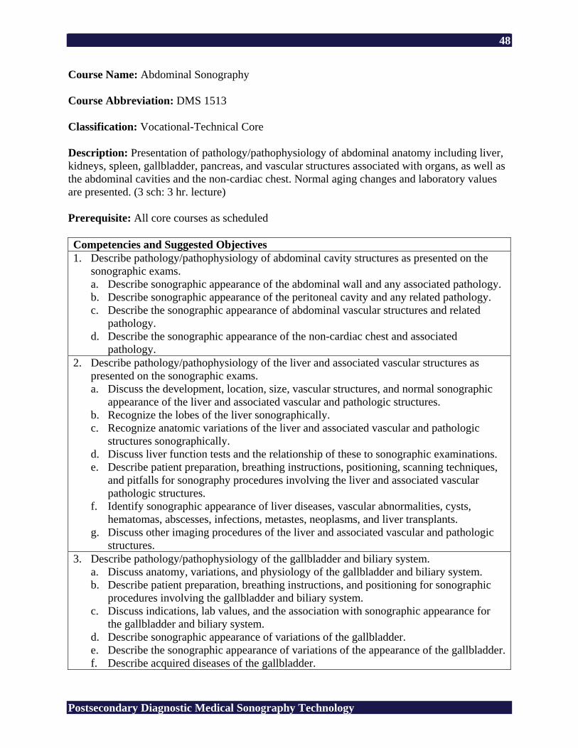
48
Postsecondary Diagnostic Medical Sonography Technology
Course Name: Abdominal Sonography Course Abbreviation: DMS 1513 Classification: Vocational-Technical Core Description: Presentation of pathology/pathophysiology of abdominal anatomy including liver, kidneys, spleen, gallbladder, pancreas, and vascular structures associated with organs, as well as the abdominal cavities and the non-cardiac chest. Normal aging changes and laboratory values are presented. (3 sch: 3 hr. lecture) Prerequisite: All core courses as scheduled Competencies and Suggested Objectives 1. Describe pathology/pathophysiology of abdominal cavity structures as presented on the
sonographic exams. a. Describe sonographic appearance of the abdominal wall and any associated pathology. b. Describe sonographic appearance of the peritoneal cavity and any related pathology. c. Describe the sonographic appearance of abdominal vascular structures and related
pathology. d. Describe the sonographic appearance of the non-cardiac chest and associated
pathology. 2. Describe pathology/pathophysiology of the liver and associated vascular structures as
presented on the sonographic exams. a. Discuss the development, location, size, vascular structures, and normal sonographic
appearance of the liver and associated vascular and pathologic structures. b. Recognize the lobes of the liver sonographically. c. Recognize anatomic variations of the liver and associated vascular and pathologic
structures sonographically. d. Discuss liver function tests and the relationship of these to sonographic examinations. e. Describe patient preparation, breathing instructions, positioning, scanning techniques,
and pitfalls for sonography procedures involving the liver and associated vascular pathologic structures.
f. Identify sonographic appearance of liver diseases, vascular abnormalities, cysts, hematomas, abscesses, infections, metastes, neoplasms, and liver transplants.
g. Discuss other imaging procedures of the liver and associated vascular and pathologic structures.
3. Describe pathology/pathophysiology of the gallbladder and biliary system. a. Discuss anatomy, variations, and physiology of the gallbladder and biliary system. b. Describe patient preparation, breathing instructions, and positioning for sonographic
procedures involving the gallbladder and biliary system. c. Discuss indications, lab values, and the association with sonographic appearance for
the gallbladder and biliary system. d. Describe sonographic appearance of variations of the gallbladder. e. Describe the sonographic appearance of variations of the appearance of the gallbladder.f. Describe acquired diseases of the gallbladder.
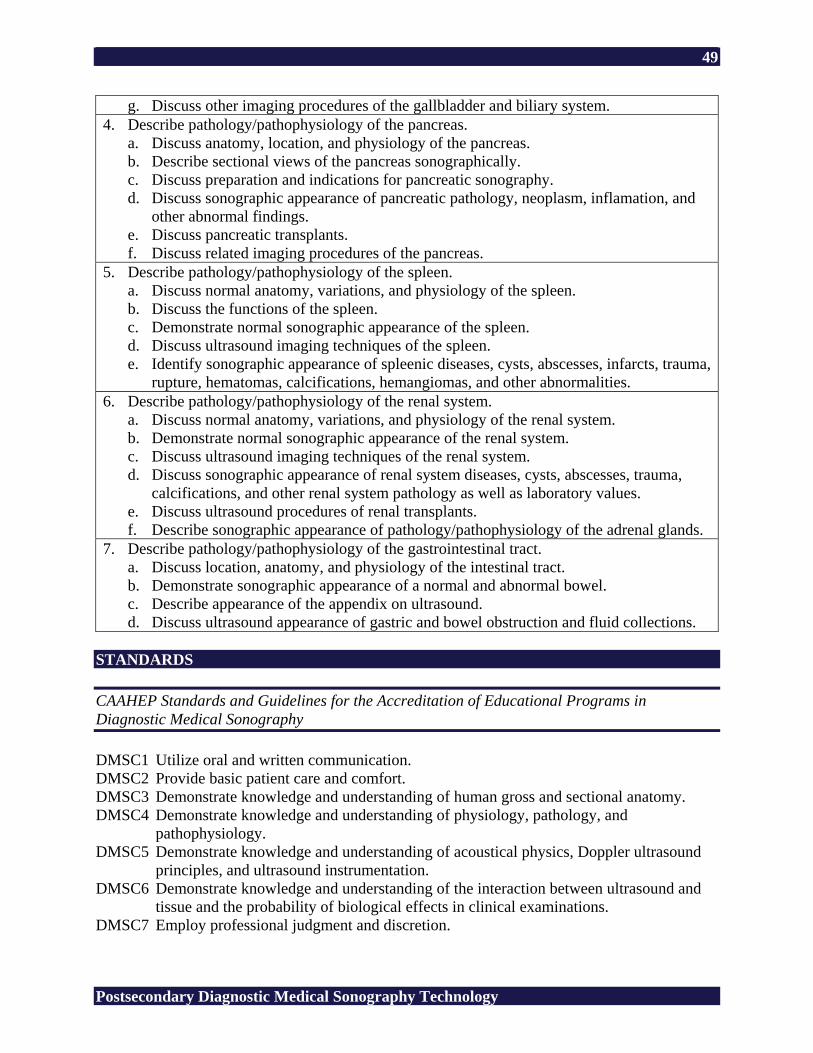
49
Postsecondary Diagnostic Medical Sonography Technology
g. Discuss other imaging procedures of the gallbladder and biliary system. 4. Describe pathology/pathophysiology of the pancreas.
a. Discuss anatomy, location, and physiology of the pancreas. b. Describe sectional views of the pancreas sonographically. c. Discuss preparation and indications for pancreatic sonography. d. Discuss sonographic appearance of pancreatic pathology, neoplasm, inflamation, and
other abnormal findings. e. Discuss pancreatic transplants. f. Discuss related imaging procedures of the pancreas.
5. Describe pathology/pathophysiology of the spleen. a. Discuss normal anatomy, variations, and physiology of the spleen. b. Discuss the functions of the spleen. c. Demonstrate normal sonographic appearance of the spleen. d. Discuss ultrasound imaging techniques of the spleen. e. Identify sonographic appearance of spleenic diseases, cysts, abscesses, infarcts, trauma,
rupture, hematomas, calcifications, hemangiomas, and other abnormalities. 6. Describe pathology/pathophysiology of the renal system.
a. Discuss normal anatomy, variations, and physiology of the renal system. b. Demonstrate normal sonographic appearance of the renal system. c. Discuss ultrasound imaging techniques of the renal system. d. Discuss sonographic appearance of renal system diseases, cysts, abscesses, trauma,
calcifications, and other renal system pathology as well as laboratory values. e. Discuss ultrasound procedures of renal transplants. f. Describe sonographic appearance of pathology/pathophysiology of the adrenal glands.
7. Describe pathology/pathophysiology of the gastrointestinal tract. a. Discuss location, anatomy, and physiology of the intestinal tract. b. Demonstrate sonographic appearance of a normal and abnormal bowel. c. Describe appearance of the appendix on ultrasound. d. Discuss ultrasound appearance of gastric and bowel obstruction and fluid collections.
STANDARDS CAAHEP Standards and Guidelines for the Accreditation of Educational Programs in Diagnostic Medical Sonography DMSC1 Utilize oral and written communication. DMSC2 Provide basic patient care and comfort. DMSC3 Demonstrate knowledge and understanding of human gross and sectional anatomy. DMSC4 Demonstrate knowledge and understanding of physiology, pathology, and
pathophysiology. DMSC5 Demonstrate knowledge and understanding of acoustical physics, Doppler ultrasound
principles, and ultrasound instrumentation. DMSC6 Demonstrate knowledge and understanding of the interaction between ultrasound and
tissue and the probability of biological effects in clinical examinations. DMSC7 Employ professional judgment and discretion.
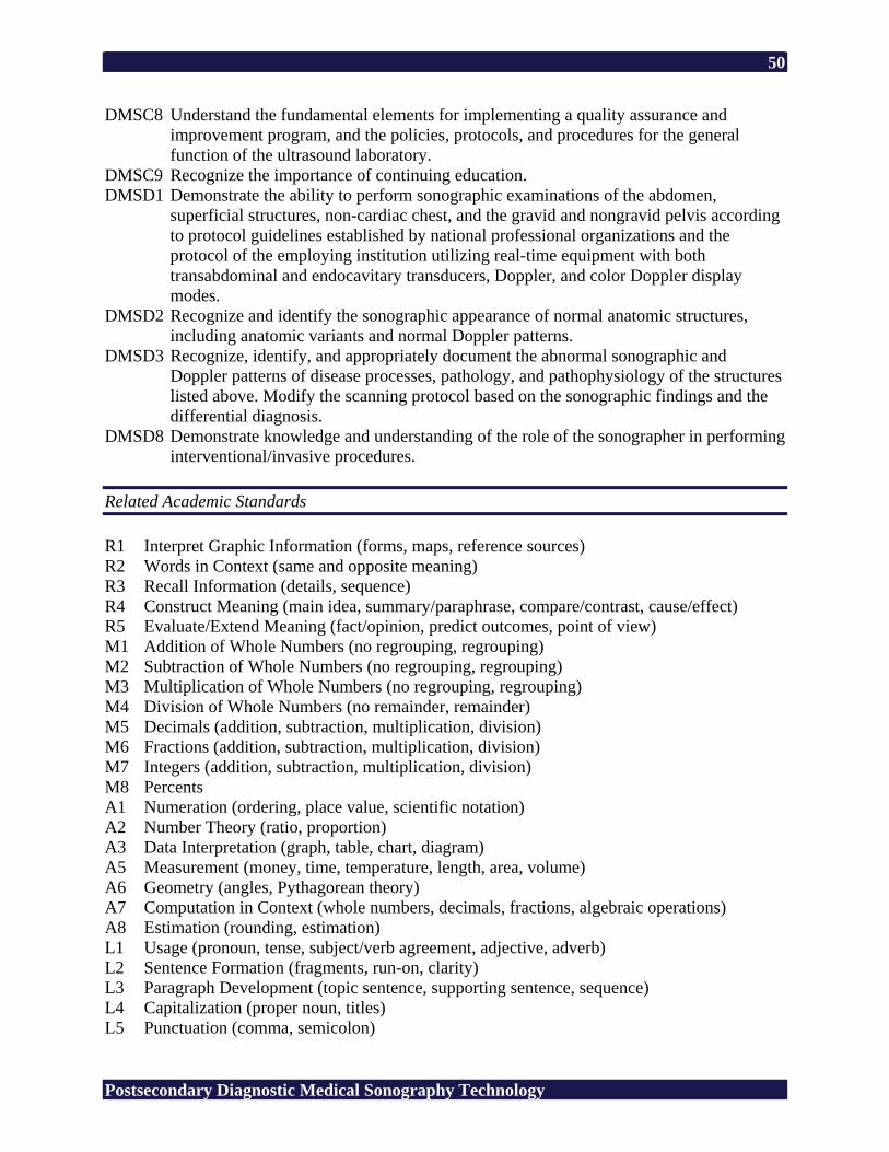
50
Postsecondary Diagnostic Medical Sonography Technology
DMSC8 Understand the fundamental elements for implementing a quality assurance and improvement program, and the policies, protocols, and procedures for the general function of the ultrasound laboratory.
DMSC9 Recognize the importance of continuing education. DMSD1 Demonstrate the ability to perform sonographic examinations of the abdomen,
superficial structures, non-cardiac chest, and the gravid and nongravid pelvis according to protocol guidelines established by national professional organizations and the protocol of the employing institution utilizing real-time equipment with both transabdominal and endocavitary transducers, Doppler, and color Doppler display modes.
DMSD2 Recognize and identify the sonographic appearance of normal anatomic structures, including anatomic variants and normal Doppler patterns.
DMSD3 Recognize, identify, and appropriately document the abnormal sonographic and Doppler patterns of disease processes, pathology, and pathophysiology of the structures listed above. Modify the scanning protocol based on the sonographic findings and the differential diagnosis.
DMSD8 Demonstrate knowledge and understanding of the role of the sonographer in performing interventional/invasive procedures.
Related Academic Standards R1 Interpret Graphic Information (forms, maps, reference sources) R2 Words in Context (same and opposite meaning) R3 Recall Information (details, sequence) R4 Construct Meaning (main idea, summary/paraphrase, compare/contrast, cause/effect) R5 Evaluate/Extend Meaning (fact/opinion, predict outcomes, point of view) M1 Addition of Whole Numbers (no regrouping, regrouping) M2 Subtraction of Whole Numbers (no regrouping, regrouping) M3 Multiplication of Whole Numbers (no regrouping, regrouping) M4 Division of Whole Numbers (no remainder, remainder) M5 Decimals (addition, subtraction, multiplication, division) M6 Fractions (addition, subtraction, multiplication, division) M7 Integers (addition, subtraction, multiplication, division) M8 Percents A1 Numeration (ordering, place value, scientific notation) A2 Number Theory (ratio, proportion) A3 Data Interpretation (graph, table, chart, diagram) A5 Measurement (money, time, temperature, length, area, volume) A6 Geometry (angles, Pythagorean theory) A7 Computation in Context (whole numbers, decimals, fractions, algebraic operations) A8 Estimation (rounding, estimation) L1 Usage (pronoun, tense, subject/verb agreement, adjective, adverb) L2 Sentence Formation (fragments, run-on, clarity) L3 Paragraph Development (topic sentence, supporting sentence, sequence) L4 Capitalization (proper noun, titles) L5 Punctuation (comma, semicolon)
51
Postsecondary Diagnostic Medical Sonography Technology
L6 Writing Conventions (quotation marks, apostrophe, parts of a letter) S1 Vowel (short, long) S2 Consonant (variant spelling, silent letter) S3 Structural Unit (root, suffix) Copyright © 1994 by CTB/McGraw-Hill LLC 21st Century Skills CS1 Global Awareness CS2 Financial, Economic, and Business Literacy CS3 Civic Literacy CS4 Information and Communication Skills CS5 Thinking and Problem-Solving Skills CS6 Interpersonal and Self-Directional Skills SUGGESTED REFERENCES American Registry for Diagnostic Medical Sonography. (n.d.). Retrieved February 24, 2006,
from http://www.ardms.org/ American Institute of Ultrasound in Medicine. (n.d.). Retrieved February 24, 2006, from
http://www.aium.org/ Commission on Accreditation of Allied Health Education Programs. (n.d.). Retrieved February
17, 2006, from http://www.caahep.org/ Dirckx, J. H. (Ed.). (2001). Stedman’s concise medical dictionary for the health professions (4th
ed.). Baltimore: Lippincott Williams & Wilkins. Green, L., & Sens, L. (1998). Introduction to abdominal sonography [Videotape]. (Available
from Gulfcoast Ultrasound Institute, Inc., 4615 Gulf Blvd., Suite 205, St. Pete Beach, FL 33706)
Hagen-Ansert, S. L. (2006). Textbook of diagnostic ultrasonography (6th ed.). St. Louis, MO:
Mosby. Journal of Diagnostic Medical Sonography. Thousand Oaks, CA: SAGE. Retrieved February 17,
2006, from http://www.sagepub.com/journal.aspx?pid=259 Krebs, C. A., Odwin, C. S., & Fleischer, A. C. (2004). Appleton & Lange review for the
ultrasonography examination (3rd ed.). Singapore: McGraw-Hill. Owen, C. A. (2005). Endovaginal scanning (Vols. 1-2) [Interactive CD]. (Available from
Gulfcoast Ultrasound Institute, Inc., 4615 Gulf Blvd., Suite 205, St. Pete Beach, FL 33706)

52
Postsecondary Diagnostic Medical Sonography Technology
Rumack, C. M., Wilson, S. R., Charboneau, J. W., & Johnson, J. M. (2005). Diagnostic ultrasound (3rd ed., Vols. 1-2). St. Louis, MO: Mosby.
Society of Diagnostic Medical Sonography. (n.d.). Retrieved February 24, 2006, from
http://www.sdms.org/ Ultrasound case studies. (n.d.). Retrieved February 24, 2006, from http://www.sonoworld.com/
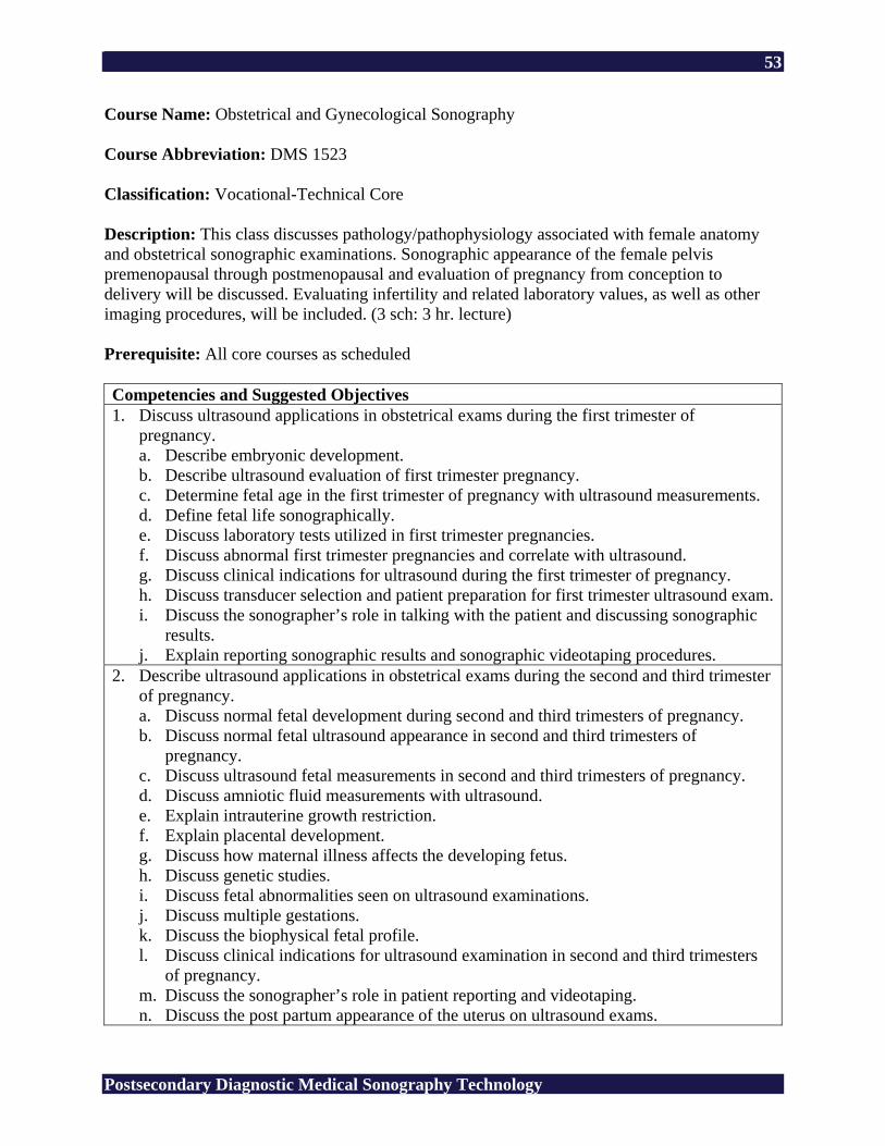
53
Postsecondary Diagnostic Medical Sonography Technology
Course Name: Obstetrical and Gynecological Sonography Course Abbreviation: DMS 1523 Classification: Vocational-Technical Core Description: This class discusses pathology/pathophysiology associated with female anatomy and obstetrical sonographic examinations. Sonographic appearance of the female pelvis premenopausal through postmenopausal and evaluation of pregnancy from conception to delivery will be discussed. Evaluating infertility and related laboratory values, as well as other imaging procedures, will be included. (3 sch: 3 hr. lecture) Prerequisite: All core courses as scheduled Competencies and Suggested Objectives 1. Discuss ultrasound applications in obstetrical exams during the first trimester of
pregnancy. a. Describe embryonic development. b. Describe ultrasound evaluation of first trimester pregnancy. c. Determine fetal age in the first trimester of pregnancy with ultrasound measurements. d. Define fetal life sonographically. e. Discuss laboratory tests utilized in first trimester pregnancies. f. Discuss abnormal first trimester pregnancies and correlate with ultrasound. g. Discuss clinical indications for ultrasound during the first trimester of pregnancy. h. Discuss transducer selection and patient preparation for first trimester ultrasound exam. i. Discuss the sonographer’s role in talking with the patient and discussing sonographic
results. j. Explain reporting sonographic results and sonographic videotaping procedures.
2. Describe ultrasound applications in obstetrical exams during the second and third trimester of pregnancy. a. Discuss normal fetal development during second and third trimesters of pregnancy. b. Discuss normal fetal ultrasound appearance in second and third trimesters of
pregnancy. c. Discuss ultrasound fetal measurements in second and third trimesters of pregnancy. d. Discuss amniotic fluid measurements with ultrasound. e. Explain intrauterine growth restriction. f. Explain placental development. g. Discuss how maternal illness affects the developing fetus. h. Discuss genetic studies. i. Discuss fetal abnormalities seen on ultrasound examinations. j. Discuss multiple gestations. k. Discuss the biophysical fetal profile. l. Discuss clinical indications for ultrasound examination in second and third trimesters
of pregnancy. m. Discuss the sonographer’s role in patient reporting and videotaping. n. Discuss the post partum appearance of the uterus on ultrasound exams.
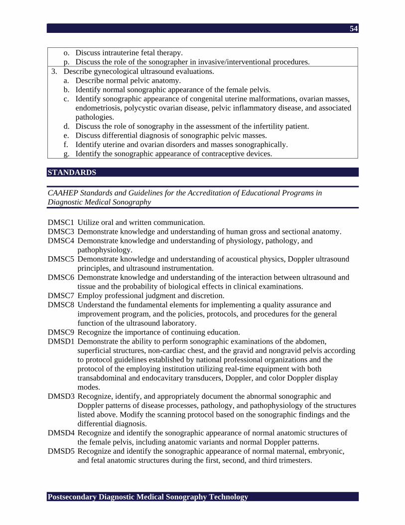
54
Postsecondary Diagnostic Medical Sonography Technology
o. Discuss intrauterine fetal therapy. p. Discuss the role of the sonographer in invasive/interventional procedures.
3. Describe gynecological ultrasound evaluations. a. Describe normal pelvic anatomy. b. Identify normal sonographic appearance of the female pelvis. c. Identify sonographic appearance of congenital uterine malformations, ovarian masses,
endometriosis, polycystic ovarian disease, pelvic inflammatory disease, and associated pathologies.
d. Discuss the role of sonography in the assessment of the infertility patient. e. Discuss differential diagnosis of sonographic pelvic masses. f. Identify uterine and ovarian disorders and masses sonographically. g. Identify the sonographic appearance of contraceptive devices.
STANDARDS CAAHEP Standards and Guidelines for the Accreditation of Educational Programs in Diagnostic Medical Sonography DMSC1 Utilize oral and written communication. DMSC3 Demonstrate knowledge and understanding of human gross and sectional anatomy. DMSC4 Demonstrate knowledge and understanding of physiology, pathology, and
pathophysiology. DMSC5 Demonstrate knowledge and understanding of acoustical physics, Doppler ultrasound
principles, and ultrasound instrumentation. DMSC6 Demonstrate knowledge and understanding of the interaction between ultrasound and
tissue and the probability of biological effects in clinical examinations. DMSC7 Employ professional judgment and discretion. DMSC8 Understand the fundamental elements for implementing a quality assurance and
improvement program, and the policies, protocols, and procedures for the general function of the ultrasound laboratory.
DMSC9 Recognize the importance of continuing education. DMSD1 Demonstrate the ability to perform sonographic examinations of the abdomen,
superficial structures, non-cardiac chest, and the gravid and nongravid pelvis according to protocol guidelines established by national professional organizations and the protocol of the employing institution utilizing real-time equipment with both transabdominal and endocavitary transducers, Doppler, and color Doppler display modes.
DMSD3 Recognize, identify, and appropriately document the abnormal sonographic and Doppler patterns of disease processes, pathology, and pathophysiology of the structures listed above. Modify the scanning protocol based on the sonographic findings and the differential diagnosis.
DMSD4 Recognize and identify the sonographic appearance of normal anatomic structures of the female pelvis, including anatomic variants and normal Doppler patterns.
DMSD5 Recognize and identify the sonographic appearance of normal maternal, embryonic, and fetal anatomic structures during the first, second, and third trimesters.
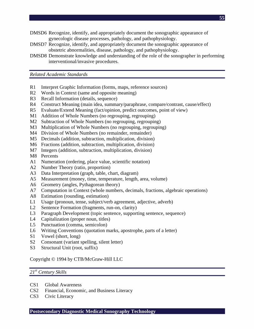
55
Postsecondary Diagnostic Medical Sonography Technology
DMSD6 Recognize, identify, and appropriately document the sonographic appearance of gynecologic disease processes, pathology, and pathophysiology.
DMSD7 Recognize, identify, and appropriately document the sonographic appearance of obstetric abnormalities, disease, pathology, and pathophysiology.
DMSD8 Demonstrate knowledge and understanding of the role of the sonographer in performing interventional/invasive procedures.
Related Academic Standards R1 Interpret Graphic Information (forms, maps, reference sources) R2 Words in Context (same and opposite meaning) R3 Recall Information (details, sequence) R4 Construct Meaning (main idea, summary/paraphrase, compare/contrast, cause/effect) R5 Evaluate/Extend Meaning (fact/opinion, predict outcomes, point of view) M1 Addition of Whole Numbers (no regrouping, regrouping) M2 Subtraction of Whole Numbers (no regrouping, regrouping) M3 Multiplication of Whole Numbers (no regrouping, regrouping) M4 Division of Whole Numbers (no remainder, remainder) M5 Decimals (addition, subtraction, multiplication, division) M6 Fractions (addition, subtraction, multiplication, division) M7 Integers (addition, subtraction, multiplication, division) M8 Percents A1 Numeration (ordering, place value, scientific notation) A2 Number Theory (ratio, proportion) A3 Data Interpretation (graph, table, chart, diagram) A5 Measurement (money, time, temperature, length, area, volume) A6 Geometry (angles, Pythagorean theory) A7 Computation in Context (whole numbers, decimals, fractions, algebraic operations) A8 Estimation (rounding, estimation) L1 Usage (pronoun, tense, subject/verb agreement, adjective, adverb) L2 Sentence Formation (fragments, run-on, clarity) L3 Paragraph Development (topic sentence, supporting sentence, sequence) L4 Capitalization (proper noun, titles) L5 Punctuation (comma, semicolon) L6 Writing Conventions (quotation marks, apostrophe, parts of a letter) S1 Vowel (short, long) S2 Consonant (variant spelling, silent letter) S3 Structural Unit (root, suffix) Copyright © 1994 by CTB/McGraw-Hill LLC 21st Century Skills CS1 Global Awareness CS2 Financial, Economic, and Business Literacy CS3 Civic Literacy
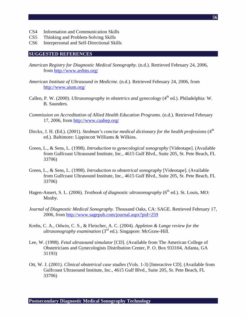
56
Postsecondary Diagnostic Medical Sonography Technology
CS4 Information and Communication Skills CS5 Thinking and Problem-Solving Skills CS6 Interpersonal and Self-Directional Skills SUGGESTED REFERENCES American Registry for Diagnostic Medical Sonography. (n.d.). Retrieved February 24, 2006,
from http://www.ardms.org/ American Institute of Ultrasound in Medicine. (n.d.). Retrieved February 24, 2006, from
http://www.aium.org/ Callen, P. W. (2000). Ultrasonography in obstetrics and gynecology (4th ed.). Philadelphia: W.
B. Saunders. Commission on Accreditation of Allied Health Education Programs. (n.d.). Retrieved February
17, 2006, from http://www.caahep.org/ Dirckx, J. H. (Ed.). (2001). Stedman’s concise medical dictionary for the health professions (4th
ed.). Baltimore: Lippincott Williams & Wilkins. Green, L., & Sens, L. (1998). Introduction to gynecological sonography [Videotape]. (Available
from Gulfcoast Ultrasound Institute, Inc., 4615 Gulf Blvd., Suite 205, St. Pete Beach, FL 33706)
Green, L., & Sens, L. (1998). Introduction to obstetrical sonography [Videotape]. (Available
from Gulfcoast Ultrasound Institute, Inc., 4615 Gulf Blvd., Suite 205, St. Pete Beach, FL 33706)
Hagen-Ansert, S. L. (2006). Textbook of diagnostic ultrasonography (6th ed.). St. Louis, MO:
Mosby. Journal of Diagnostic Medical Sonography. Thousand Oaks, CA: SAGE. Retrieved February 17,
2006, from http://www.sagepub.com/journal.aspx?pid=259 Krebs, C. A., Odwin, C. S., & Fleischer, A. C. (2004). Appleton & Lange review for the
ultrasonography examination (3rd ed.). Singapore: McGraw-Hill. Lee, W. (1998). Fetal ultrasound simulator [CD]. (Available from The American College of
Obstetricians and Gynecologists Distribution Center, P. O. Box 933104, Atlanta, GA 31193)
Ott, W. J. (2001). Clinical obstetrical case studies (Vols. 1-3) [Interactive CD]. (Available from
Gulfcoast Ultrasound Institute, Inc., 4615 Gulf Blvd., Suite 205, St. Pete Beach, FL 33706)
57
Postsecondary Diagnostic Medical Sonography Technology
Rumack, C. M., Wilson, S. R., Charboneau, J. W., & Johnson, J. M. (2005). Diagnostic ultrasound (3rd ed., Vols. 1-2). St. Louis, MO: Mosby.
Society of Diagnostic Medical Sonography. (n.d.). Retrieved February 24, 2006, from
http://www.sdms.org/ Ultrasound case studies. (n.d.). Retrieved February 24, 2006, from http://www.sonoworld.com/ Women’s health resources for medical industry professionals. (n.d.). Retrieved February 24,
2006, from http://www.obgyn.net/
58
Postsecondary Diagnostic Medical Sonography Technology
Course Name: Advanced Sonographic Procedures Course Abbreviation: DMS 1533 Classification: Vocational-Technical Core Description: Neurosonology, ophthalmology, adult cardiac, pediatric cardiac, and vascular technology will be discussed. Superficial structures scanning including prostate, thyroid, scrotum and breast will be included. (3 sch: 3 hr. lecture) Prerequisite: All core courses as scheduled Competencies and Suggested Objectives 1. Describe pathology/pathophysiology of the thyroid, parathyroid, and associated vascular
structures as presented on the sonographic exam. a. Discuss sonographic indications and laboratory values associated with the thyroid. b. Discuss related imaging procedures for thyroid, parathyroid, and vascular structures of
the neck. c. Describe sonographic appearance of cysts, masses, hematomas, and other pathology
associated with the thyroid/parathyroid. d. Discuss sonographic protocols and procedures for thyroid, parathyroid, and vascular
structures of the neck. e. Document patient history and physical findings. f. Communicate sonographic findings and verbal history given by the patient to
physician. g. Identify vascular structures in the neck sonographically. h. Document blood flow, intravasculature structures, and pathology sonographically.
2. Describe pathology/pathophysiology appearance of the prostate as presented on the sonographic exam. a. Discuss indications for prostate sonography. b. List prostate laboratory values and patient history. c. Identify prostate scanning techniques and protocols. d. Identify sonographic images of masses, cysts, abscesses, parenchymal disease, and
benign hypertrophy. e. Document patient history and physical findings pertinent to ultrasound exam of
prostate. 3. Describe sonographic appearance of pathology/pathophysiology of the scrotum.
a. Discuss scrotal scanning techniques and protocols. b. List scrotal laboratory values and patient history. c. Discuss clinical indications for ultrasound exam of scrotum. d. Identify sonographic images of masses, inflamation, cysts, fluid collections,
hematomas, and parenchymal disease of scrotum. e. List all protocols for scrotal ultrasound imaging. f. Document patient history and physical findings pertinent of ultrasound exam of
scrotum.
59
Postsecondary Diagnostic Medical Sonography Technology
4. Describe vascular sonography. a. Discuss vascular anatomy and indications for ultrasound examination. b. Discuss educational requirements for the vascular sonography registry. c. Describe sonographic appearance of normal vascular anatomy. d. Describe sonographic appearance of pathology of vascular structures. e. Identify protocols for vascular scanning techniques. f. Identify sonographic appearance of vascular pathology/pathophysiology.
5. Describe adult cardiovascular, pediatric cardiovascular, opthalmic, and neurological sonography. a. Discuss adult cardiac sonography. b. Discuss pediatric cardiac sonography. c. Discuss opthalmic sonography. d. Discuss neurosonography. e. Discuss educational requirements for adult cardiac, pediatric cardiac, opthalmic, and
neurological sonography registries. 6. Describe normal and pathology/pathophysiology of the breast as presented on the
sonographic exam. a. Discuss anatomy of the breast. b. Identify sonographic appearance of normal breast structures. c. Discuss sonographic appearance of breast masses. d. Discuss related breast imaging. e. Differentiate between whole breast imaging and imaging a palpable mass with
ultrasound. f. Document patient positioning and measurements of masses for ultrasound imaging of
the breast. g. Discuss patient history and clinical indications pertinent to ultrasound exam of breast. h. Discuss differential diagnosis of sonographic masses. i. Identify appearance of breast implants sonographically.
STANDARDS CAAHEP Standards and Guidelines for the Accreditation of Educational Programs in Diagnostic Medical Sonography DMSC1 Utilize oral and written communication. DMSC3 Demonstrate knowledge and understanding of human gross and sectional anatomy. DMSC4 Demonstrate knowledge and understanding of physiology, pathology, and
pathophysiology. DMSC5 Demonstrate knowledge and understanding of acoustical physics, Doppler ultrasound
principles, and ultrasound instrumentation. DMSC6 Demonstrate knowledge and understanding of the interaction between ultrasound and
tissue and the probability of biological effects in clinical examinations. DMSC7 Employ professional judgment and discretion. DMSC8 Understand the fundamental elements for implementing a quality assurance and
improvement program, and the policies, protocols, and procedures for the general function of the ultrasound laboratory.
60
Postsecondary Diagnostic Medical Sonography Technology
DMSC9 Recognize the importance of continuing education. DMSD1 Demonstrate the ability to perform sonographic examinations of the abdomen,
superficial structures, non-cardiac chest, and the gravid and nongravid pelvis according to protocol guidelines established by national professional organizations and the protocol of the employing institution utilizing real-time equipment with both transabdominal and endocavitary transducers, Doppler, and color Doppler display modes.
DMSD2 Recognize and identify the sonographic appearance of normal anatomic structures, including anatomic variants and normal Doppler patterns.
DMSD3 Recognize, identify, and appropriately document the abnormal sonographic and Doppler patterns of disease processes, pathology, and pathophysiology of the structures listed above. Modify the scanning protocol based on the sonographic findings and the differential diagnosis.
DMSD8 Demonstrate knowledge and understanding of the role of the sonographer in performing interventional/invasive procedures.
Related Academic Standards R1 Interpret Graphic Information (forms, maps, reference sources) R2 Words in Context (same and opposite meaning) R3 Recall Information (details, sequence) R4 Construct Meaning (main idea, summary/paraphrase, compare/contrast, cause/effect) R5 Evaluate/Extend Meaning (fact/opinion, predict outcomes, point of view) M1 Addition of Whole Numbers (no regrouping, regrouping) M2 Subtraction of Whole Numbers (no regrouping, regrouping) M3 Multiplication of Whole Numbers (no regrouping, regrouping) M4 Division of Whole Numbers (no remainder, remainder) M5 Decimals (addition, subtraction, multiplication, division) M6 Fractions (addition, subtraction, multiplication, division) M7 Integers (addition, subtraction, multiplication, division) M8 Percents A1 Numeration (ordering, place value, scientific notation) A2 Number Theory (ratio, proportion) A3 Data Interpretation (graph, table, chart, diagram) A5 Measurement (money, time, temperature, length, area, volume) A6 Geometry (angles, Pythagorean theory) A7 Computation in Context (whole numbers, decimals, fractions, algebraic operations) A8 Estimation (rounding, estimation) L1 Usage (pronoun, tense, subject/verb agreement, adjective, adverb) L2 Sentence Formation (fragments, run-on, clarity) L3 Paragraph Development (topic sentence, supporting sentence, sequence) L4 Capitalization (proper noun, titles) L5 Punctuation (comma, semicolon) L6 Writing Conventions (quotation marks, apostrophe, parts of a letter) S1 Vowel (short, long) S2 Consonant (variant spelling, silent letter)
61
Postsecondary Diagnostic Medical Sonography Technology
S3 Structural Unit (root, suffix) Copyright © 1994 by CTB/McGraw-Hill LLC 21st Century Skills CS1 Global Awareness CS2 Financial, Economic, and Business Literacy CS3 Civic Literacy CS4 Information and Communication Skills CS5 Thinking and Problem-Solving Skills CS6 Interpersonal and Self-Directional Skills SUGGESTED REFERENCES American Registry for Diagnostic Medical Sonography. (n.d.). Retrieved February 24, 2006,
from http://www.ardms.org/ American Institute of Ultrasound in Medicine. (n.d.). Retrieved February 24, 2006, from
http://www.aium.org/ Commission on Accreditation of Allied Health Education Programs. (n.d.). Retrieved February
17, 2006, from http://www.caahep.org/ Dirckx, J. H. (Ed.). (2001). Stedman’s concise medical dictionary for the health professions (4th
ed.). Baltimore: Lippincott Williams & Wilkins. Hagen-Ansert, S. L. (2006). Textbook of diagnostic ultrasonography (6th ed.). St. Louis, MO:
Mosby. Hecht, L. C., Jr. (2004). Testicular sonography [CD]. (Available from Gulfcoast Ultrasound
Institute, Inc., 4615 Gulf Blvd., Suite 205, St. Pete Beach, FL 33706) Journal of Diagnostic Medical Sonography. Thousand Oaks, CA: SAGE. Retrieved February 17,
2006, from http://www.sagepub.com/journal.aspx?pid=259 Krebs, C. A., Odwin, C. S., & Fleischer, A. C. (2004). Appleton & Lange review for the
ultrasonography examination (3rd ed.). Singapore: McGraw-Hill. Rumack, C. M., Wilson, S. R., Charboneau, J. W., & Johnson, J. M. (2005). Diagnostic
ultrasound (3rd ed., Vols. 1-2). St. Louis, MO: Mosby. Society of Diagnostic Medical Sonography. (n.d.). Retrieved February 24, 2006, from
http://www.sdms.org/ Ultrasound case studies. (n.d.). Retrieved February 24, 2006, from http://www.sonoworld.com/

62
Postsecondary Diagnostic Medical Sonography Technology
Women’s health resources for medical industry professionals. (n.d.). Retrieved February 24, 2006, from http://www.obgyn.net/
63
Postsecondary Diagnostic Medical Sonography Technology
Course Name: Sonography Seminar Course Abbreviation: DMS 1613 Classification: Vocational-Technical Core Description: This course will prepare students for ARDMS/ARRT certification examinations. (3 sch: 3 hr. lecture) Prerequisite: All core courses as scheduled Competencies and Suggested Objectives 1. Review ultrasound physics and principles.
a. Define diagnostic ultrasound. b. Discuss how ultrasound works to produce an image. c. Explain how ultrasound equipment turns sound into diagnostic images. d. Describe how Doppler ultrasound works. e. Describe ultrasound artifacts. f. Discuss performance and safety standards for ultrasound equipment. g. Perform simulated registries for ultrasound physics and instrumentation.
2. Examine all aspects of patient care. a. Describe patient care legal and professional responsibilities. b. Discuss patient education and safety. c. Explain patient care and standard precautions.
3. Discuss general sonographic procedures. a. Identify the sonographic anatomy, pathology, and physiology of the abdomen. b. Identify the sonographic anatomy, pathology, and physiology of obstetrics. c. Identify the sonographic anatomy, pathology, and physiology of gynecology. d. Identify general procedural considerations for abdominal sonography. e. Identify general procedural considerations for obstetrical sonography. f. Identify general procedural considerations for gynecological sonography. g. Perform simulated registries of abdomen, superficial structures, and obstetrical and
gynecological sonography. STANDARDS CAAHEP Standards and Guidelines for the Accreditation of Educational Programs in Diagnostic Medical Sonography DMSC1 Utilize oral and written communication. DMSC2 Provide basic patient care and comfort. DMSC3 Demonstrate knowledge and understanding of human gross and sectional anatomy. DMSC4 Demonstrate knowledge and understanding of physiology, pathology, and
pathophysiology. DMSC5 Demonstrate knowledge and understanding of acoustical physics, Doppler ultrasound
principles, and ultrasound instrumentation.
64
Postsecondary Diagnostic Medical Sonography Technology
DMSC6 Demonstrate knowledge and understanding of the interaction between ultrasound and tissue and the probability of biological effects in clinical examinations.
DMSC7 Employ professional judgment and discretion. DMSC8 Understand the fundamental elements for implementing a quality assurance and
improvement program, and the policies, protocols, and procedures for the general function of the ultrasound laboratory.
DMSC9 Recognize the importance of continuing education. DMSD1 Demonstrate the ability to perform sonographic examinations of the abdomen,
superficial structures, non-cardiac chest, and the gravid and nongravid pelvis according to protocol guidelines established by national professional organizations and the protocol of the employing institution utilizing real-time equipment with both transabdominal and endocavitary transducers, Doppler, and color Doppler display modes.
DMSD2 Recognize and identify the sonographic appearance of normal anatomic structures, including anatomic variants and normal Doppler patterns.
DMSD3 Recognize, identify, and appropriately document the abnormal sonographic and Doppler patterns of disease processes, pathology, and pathophysiology of the structures listed above. Modify the scanning protocol based on the sonographic findings and the differential diagnosis.
DMSD4 Recognize and identify the sonographic appearance of normal anatomic structures of the female pelvis, including anatomic variants and normal Doppler patterns.
DMSD5 Recognize and identify the sonographic appearance of normal maternal, embryonic, and fetal anatomic structures during the first, second, and third trimesters.
DMSD6 Recognize, identify, and appropriately document the sonographic appearance of gynecologic disease processes, pathology, and pathophysiology.
DMSD7 Recognize, identify, and appropriately document the sonographic appearance of obstetric abnormalities, disease, pathology, and pathophysiology.
DMSD8 Demonstrate knowledge and understanding of the role of the sonographer in performing interventional/invasive procedures.
Related Academic Standards R1 Interpret Graphic Information (forms, maps, reference sources) R2 Words in Context (same and opposite meaning) R3 Recall Information (details, sequence) R4 Construct Meaning (main idea, summary/paraphrase, compare/contrast, cause/effect) R5 Evaluate/Extend Meaning (fact/opinion, predict outcomes, point of view) M1 Addition of Whole Numbers (no regrouping, regrouping) M2 Subtraction of Whole Numbers (no regrouping, regrouping) M3 Multiplication of Whole Numbers (no regrouping, regrouping) M4 Division of Whole Numbers (no remainder, remainder) M5 Decimals (addition, subtraction, multiplication, division) M6 Fractions (addition, subtraction, multiplication, division) M7 Integers (addition, subtraction, multiplication, division) M8 Percents M9 Algebraic Operations
65
Postsecondary Diagnostic Medical Sonography Technology
A1 Numeration (ordering, place value, scientific notation) A2 Number Theory (ratio, proportion) A3 Data Interpretation (graph, table, chart, diagram) A4 Pre-Algebra and Algebra (equations, inequality) A5 Measurement (money, time, temperature, length, area, volume) A6 Geometry (angles, Pythagorean theory) A7 Computation in Context (whole numbers, decimals, fractions, algebraic operations) A8 Estimation (rounding, estimation) L1 Usage (pronoun, tense, subject/verb agreement, adjective, adverb) L2 Sentence Formation (fragments, run-on, clarity) L3 Paragraph Development (topic sentence, supporting sentence, sequence) L4 Capitalization (proper noun, titles) L5 Punctuation (comma, semicolon) L6 Writing Conventions (quotation marks, apostrophe, parts of a letter) S1 Vowel (short, long) S2 Consonant (variant spelling, silent letter) S3 Structural Unit (root, suffix) Copyright © 1994 by CTB/McGraw-Hill LLC 21st Century Skills CS1 Global Awareness CS2 Financial, Economic, and Business Literacy CS3 Civic Literacy CS4 Information and Communication Skills CS5 Thinking and Problem-Solving Skills CS6 Interpersonal and Self-Directional Skills SUGGESTED REFERENCES American Registry for Diagnostic Medical Sonography. (n.d.). Retrieved February 24, 2006,
from http://www.ardms.org/ American Institute of Ultrasound in Medicine. (n.d.). Retrieved February 24, 2006, from
http://www.aium.org/ Commission on Accreditation of Allied Health Education Programs. (n.d.). Retrieved February
17, 2006, from http://www.caahep.org/ Dirckx, J. H. (Ed.). (2001). Stedman’s concise medical dictionary for the health professions (4th
ed.). Baltimore: Lippincott Williams & Wilkins. Edelman, S. K. (2004). Understanding ultrasound physics (3rd ed.). Woodlands, TX: ESP.
66
Postsecondary Diagnostic Medical Sonography Technology
Hecht, L. C., Jr. (2004). The registry review CD quiz series [CD]. (Available from Gulfcoast Ultrasound Institute, Inc., 4615 Gulf Blvd., Suite 205, St. Pete Beach, FL 33706)
Journal of Diagnostic Medical Sonography. Thousand Oaks, CA: SAGE. Retrieved February 17,
2006, from http://www.sagepub.com/journal.aspx?pid=259 Krebs, C. A., Odwin, C. S., & Fleischer, A. C. (2004). Appleton & Lange review for the
ultrasonography examination (3rd ed.). Singapore: McGraw-Hill. Miele, F. R. (2006). Ultrasound physics and instrumentation (4th ed., Vols. 1-2). Forney, TX:
LLC. Society of Diagnostic Medical Sonography. (n.d.). Retrieved February 24, 2006, from
http://www.sdms.org/ Stephenson, S. R., & Layton, T. (2004). The registry review CD quiz series: Abdominal series
[CD]. (Available from Gulfcoast Ultrasound Institute, Inc., 4615 Gulf Blvd., Suite 205, St. Pete Beach, FL 33706)
Stephenson, S. R., & Layton, T. (2004). The registry review CD quiz series: OB/GYN
sonography [CD]. (Available from Gulfcoast Ultrasound Institute, Inc., 4615 Gulf Blvd., Suite 205, St. Pete Beach, FL 33706)
Ultrasound case studies. (n.d.). Retrieved February 24, 2006, from http://www.sonoworld.com/ Women’s health resources for medical industry professionals. (n.d.). Retrieved February 24,
2006, from http://www.obgyn.net/
67
Postsecondary Diagnostic Medical Sonography Technology
Course Name: Ultrasound Examination Critique Course Abbreviation: DMS 1623 Classification: Vocational-Technical Core Description: This course will present case studies of normal and abnormal sonographic exams. Students will attend presentations of guest lecturers. (3 sch: 3 hr. lecture) Prerequisite: All core courses as scheduled Competencies and Suggested Objectives 1. Discuss obstetrical and gynecological case studies obtained in clinical site rotations.
a. Discuss case studies pertaining to obstetrical sonography. b. Discuss case studies pertaining to gynecological sonography.
2. Discuss case studies pertaining to abdominal sonography. a. Discuss sonographic case studies of the abdominal wall. b. Discuss sonographic case studies of the liver. c. Discuss sonographic case studies of the gallbladder and biliary system. d. Discuss sonographic case studies of the pancreas. e. Discuss sonographic case studies of the spleen. f. Discuss sonographic case studies of the renal system. g. Discuss sonographic case studies of the gastrointestinal system.
3. Review abdominal, obstetrical, and gynecological sonography case studies from clinical site rotations. a. Discuss abdominal sonographic case studies. b. Discuss obstetrical sonographic case studies. c. Discuss gynecologic sonographic case studies. d. Critique case studies associated with abdominal, obstetrical, and gynecological
sonography. STANDARDS CAAHEP Standards and Guidelines for the Accreditation of Educational Programs in Diagnostic Medical Sonography DMSC1 Utilize oral and written communication. DMSC3 Demonstrate knowledge and understanding of human gross and sectional anatomy. DMSC4 Demonstrate knowledge and understanding of physiology, pathology, and
pathophysiology. DMSC5 Demonstrate knowledge and understanding of acoustical physics, Doppler ultrasound
principles, and ultrasound instrumentation. DMSC7 Employ professional judgment and discretion. DMSD2 Recognize and identify the sonographic appearance of normal anatomic structures,
including anatomic variants and normal Doppler patterns.
68
Postsecondary Diagnostic Medical Sonography Technology
DMSD3 Recognize, identify, and appropriately document the abnormal sonographic and Doppler patterns of disease processes, pathology, and pathophysiology of the structures listed above. Modify the scanning protocol based on the sonographic findings and the differential diagnosis.
DMSD4 Recognize and identify the sonographic appearance of normal anatomic structures of the female pelvis, including anatomic variants and normal Doppler patterns.
DMSD5 Recognize and identify the sonographic appearance of normal maternal, embryonic, and fetal anatomic structures during the first, second, and third trimesters.
DMSD6 Recognize, identify, and appropriately document the sonographic appearance of gynecologic disease processes, pathology, and pathophysiology.
DMSD7 Recognize, identify, and appropriately document the sonographic appearance of obstetric abnormalities, disease, pathology, and pathophysiology.
Related Academic Standards R1 Interpret Graphic Information (forms, maps, reference sources) R2 Words in Context (same and opposite meaning) R3 Recall Information (details, sequence) R4 Construct Meaning (main idea, summary/paraphrase, compare/contrast, cause/effect) R5 Evaluate/Extend Meaning (fact/opinion, predict outcomes, point of view) M1 Addition of Whole Numbers (no regrouping, regrouping) M2 Subtraction of Whole Numbers (no regrouping, regrouping) M3 Multiplication of Whole Numbers (no regrouping, regrouping) M4 Division of Whole Numbers (no remainder, remainder) M5 Decimals (addition, subtraction, multiplication, division) M6 Fractions (addition, subtraction, multiplication, division) M7 Integers (addition, subtraction, multiplication, division) M8 Percents A1 Numeration (ordering, place value, scientific notation) A2 Number Theory (ratio, proportion) A3 Data Interpretation (graph, table, chart, diagram) A5 Measurement (money, time, temperature, length, area, volume) A6 Geometry (angles, Pythagorean theory) A7 Computation in Context (whole numbers, decimals, fractions, algebraic operations) A8 Estimation (rounding, estimation) L1 Usage (pronoun, tense, subject/verb agreement, adjective, adverb) L2 Sentence Formation (fragments, run-on, clarity) L3 Paragraph Development (topic sentence, supporting sentence, sequence) L4 Capitalization (proper noun, titles) L5 Punctuation (comma, semicolon) L6 Writing Conventions (quotation marks, apostrophe, parts of a letter) S1 Vowel (short, long) S2 Consonant (variant spelling, silent letter) S3 Structural Unit (root, suffix) Copyright © 1994 by CTB/McGraw-Hill LLC
69
Postsecondary Diagnostic Medical Sonography Technology
21st Century Skills CS1 Global Awareness CS2 Financial, Economic, and Business Literacy CS3 Civic Literacy CS4 Information and Communication Skills CS5 Thinking and Problem-Solving Skills CS6 Interpersonal and Self-Directional Skills SUGGESTED REFERENCES American Registry for Diagnostic Medical Sonography. (n.d.). Retrieved February 24, 2006,
from http://www.ardms.org/ American Institute of Ultrasound in Medicine. (n.d.). Retrieved February 24, 2006, from
http://www.aium.org/ Commission on Accreditation of Allied Health Education Programs. (n.d.). Retrieved February
17, 2006, from http://www.caahep.org/ Dirckx, J. H. (Ed.). (2001). Stedman’s concise medical dictionary for the health professions (4th
ed.). Baltimore: Lippincott Williams & Wilkins. Journal of Diagnostic Medical Sonography. Thousand Oaks, CA: SAGE. Retrieved February 17,
2006, from http://www.sagepub.com/journal.aspx?pid=259 Rumack, C. M., Wilson, S. R., Charboneau, J. W., & Johnson, J. M. (2005). Diagnostic
ultrasound (3rd ed., Vols. 1-2). St. Louis, MO: Mosby. Society of Diagnostic Medical Sonography. (n.d.). Retrieved February 24, 2006, from
http://www.sdms.org/ Ultrasound case studies. (n.d.). Retrieved February 24, 2006, from http://www.sonoworld.com/ Women’s health resources for medical industry professionals. (n.d.). Retrieved February 24,
2006, from http://www.obgyn.net/
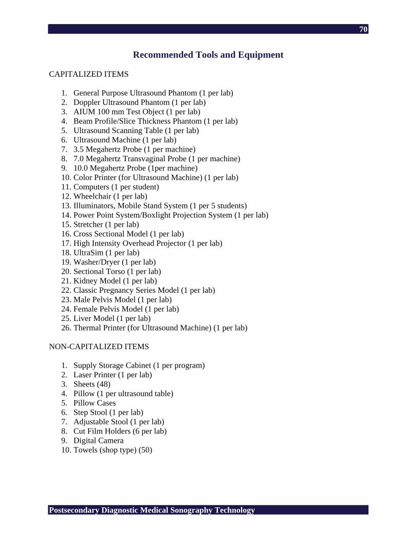
70
Postsecondary Diagnostic Medical Sonography Technology
Recommended Tools and Equipment CAPITALIZED ITEMS
1. General Purpose Ultrasound Phantom (1 per lab) 2. Doppler Ultrasound Phantom (1 per lab) 3. AIUM 100 mm Test Object (1 per lab) 4. Beam Profile/Slice Thickness Phantom (1 per lab) 5. Ultrasound Scanning Table (1 per lab) 6. Ultrasound Machine (1 per lab) 7. 3.5 Megahertz Probe (1 per machine) 8. 7.0 Megahertz Transvaginal Probe (1 per machine) 9. 10.0 Megahertz Probe (1per machine) 10. Color Printer (for Ultrasound Machine) (1 per lab) 11. Computers (1 per student) 12. Wheelchair (1 per lab) 13. Illuminators, Mobile Stand System (1 per 5 students) 14. Power Point System/Boxlight Projection System (1 per lab) 15. Stretcher (1 per lab) 16. Cross Sectional Model (1 per lab) 17. High Intensity Overhead Projector (1 per lab) 18. UltraSim (1 per lab) 19. Washer/Dryer (1 per lab) 20. Sectional Torso (1 per lab) 21. Kidney Model (1 per lab) 22. Classic Pregnancy Series Model (1 per lab) 23. Male Pelvis Model (1 per lab) 24. Female Pelvis Model (1 per lab) 25. Liver Model (1 per lab) 26. Thermal Printer (for Ultrasound Machine) (1 per lab)
NON-CAPITALIZED ITEMS
1. Supply Storage Cabinet (1 per program) 2. Laser Printer (1 per lab) 3. Sheets (48) 4. Pillow (1 per ultrasound table) 5. Pillow Cases 6. Step Stool (1 per lab) 7. Adjustable Stool (1 per lab) 8. Cut Film Holders (6 per lab) 9. Digital Camera 10. Towels (shop type) (50)
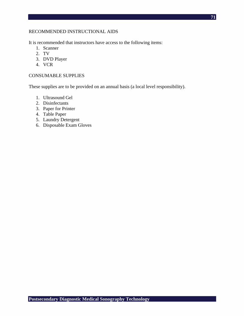
71
Postsecondary Diagnostic Medical Sonography Technology
RECOMMENDED INSTRUCTIONAL AIDS It is recommended that instructors have access to the following items:
1. Scanner 2. TV 3. DVD Player 4. VCR
CONSUMABLE SUPPLIES These supplies are to be provided on an annual basis (a local level responsibility).
1. Ultrasound Gel 2. Disinfectants 3. Paper for Printer 4. Table Paper 5. Laundry Detergent 6. Disposable Exam Gloves
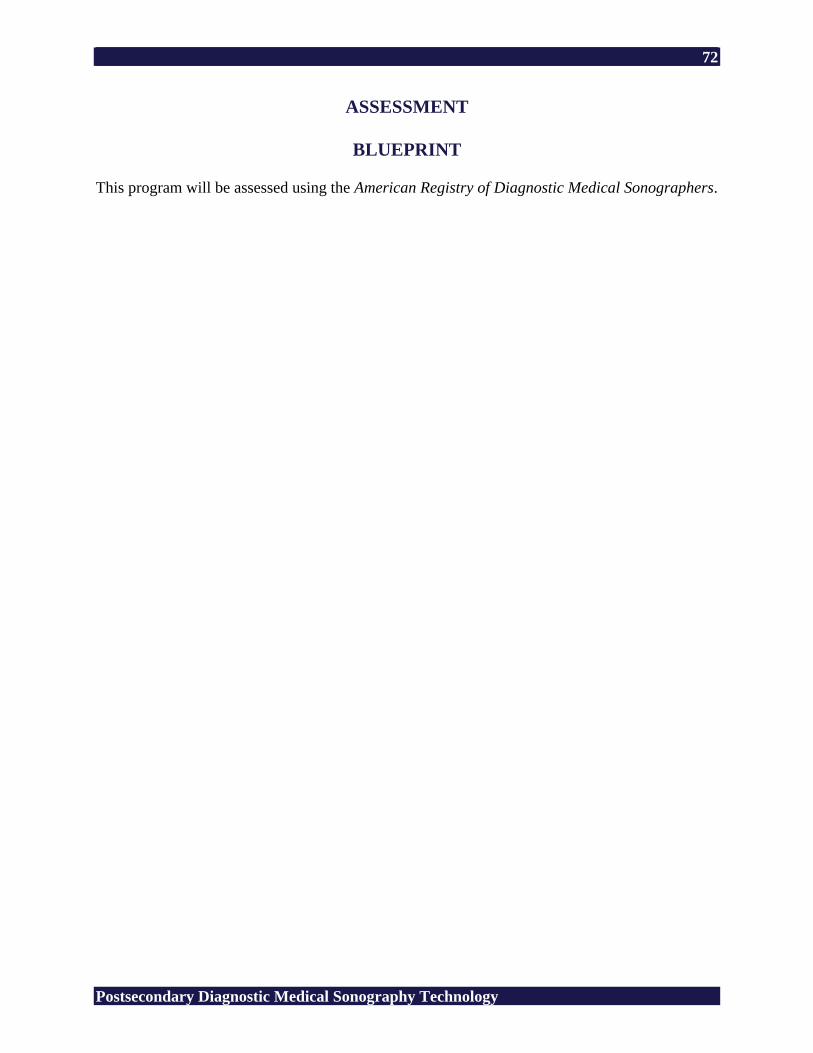
72
Postsecondary Diagnostic Medical Sonography Technology
ASSESSMENT
BLUEPRINT This program will be assessed using the American Registry of Diagnostic Medical Sonographers.
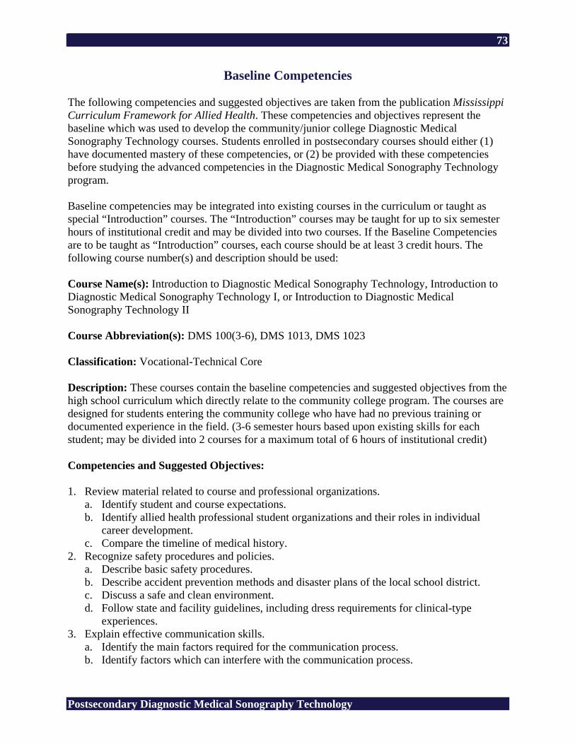
73
Postsecondary Diagnostic Medical Sonography Technology
Baseline Competencies The following competencies and suggested objectives are taken from the publication Mississippi Curriculum Framework for Allied Health. These competencies and objectives represent the baseline which was used to develop the community/junior college Diagnostic Medical Sonography Technology courses. Students enrolled in postsecondary courses should either (1) have documented mastery of these competencies, or (2) be provided with these competencies before studying the advanced competencies in the Diagnostic Medical Sonography Technology program. Baseline competencies may be integrated into existing courses in the curriculum or taught as special “Introduction” courses. The “Introduction” courses may be taught for up to six semester hours of institutional credit and may be divided into two courses. If the Baseline Competencies are to be taught as “Introduction” courses, each course should be at least 3 credit hours. The following course number(s) and description should be used: Course Name(s): Introduction to Diagnostic Medical Sonography Technology, Introduction to Diagnostic Medical Sonography Technology I, or Introduction to Diagnostic Medical Sonography Technology II Course Abbreviation(s): DMS 100(3-6), DMS 1013, DMS 1023 Classification: Vocational-Technical Core Description: These courses contain the baseline competencies and suggested objectives from the high school curriculum which directly relate to the community college program. The courses are designed for students entering the community college who have had no previous training or documented experience in the field. (3-6 semester hours based upon existing skills for each student; may be divided into 2 courses for a maximum total of 6 hours of institutional credit) Competencies and Suggested Objectives: 1. Review material related to course and professional organizations.
a. Identify student and course expectations. b. Identify allied health professional student organizations and their roles in individual
career development. c. Compare the timeline of medical history.
2. Recognize safety procedures and policies. a. Describe basic safety procedures. b. Describe accident prevention methods and disaster plans of the local school district. c. Discuss a safe and clean environment. d. Follow state and facility guidelines, including dress requirements for clinical-type
experiences. 3. Explain effective communication skills.
a. Identify the main factors required for the communication process. b. Identify factors which can interfere with the communication process.
74
Postsecondary Diagnostic Medical Sonography Technology
c. Demonstrate effective teamwork skills. d. Explore professional literature and medical references.
4. Introduce careers in the health care industry. a. Introduce careers in health care information and administration. b. Introduce careers in direct health care. c. Introduce careers in medical therapy. d. Introduce careers in diagnostic health care.
5. Discuss education and credentials required for health care careers. a. Discuss educational levels for health careers, including certification, associate degree,
bachelor’s degree, master’s degree, and doctoral degree. b. Compare the credentials needed for careers in health care, including certification,
registration, and licensure. 6. Discuss professional ethics.
a. Explain professional ethics. b. Discuss confidentiality. c. Discuss HIPAA, the Health Insurance Portability and Accountability Act of 1996.
7. Discuss legal responsibility and client’s rights. a. Explain torts and legal responsibility. b. Identify ways to promote clients’ rights and privacy. c. Discuss the requirement for health care workers to undergo a background check.
8. Explain standard precautions. a. Explain importance of standard precautions in life practices and health care. b. Explain the state and federal government’s role in standard precautions. c. Relate standard precautions to the transmission of infectious diseases including HIV,
AIDS, HBV, and TB. 9. Utilize standard precautions.
a. Demonstrate hand-washing technique. b. Demonstrate donning and removing clean gloves.
10. Perform basic emergency procedures. a. Explain first aid procedures for sudden illness. b. Explain first aid procedures for accidents.
11. Perform advanced emergency procedures. a. Perform CPR. b. Demonstrate first aid for an obstructed airway.
12. Explain medical terminology. a. Spell designated medical terms correctly. b. Demonstrate the use of medical references to spell medical terms correctly. c. Define and divide medical terms into root words, prefixes, and suffixes.
13. Recognize and use medical terminology. a. Interpret the common medical abbreviations and symbols including meanings and uses. b. Demonstrate the use of medical terms and abbreviations in reading, speaking,
interpreting, and writing simulated medical records. 14. Review the relationship among cells, tissues, organs, and systems.
a. Review the main parts of a cell. b. Review the functions of the main parts of a cell. c. Compare types of tissues and their relationships to body organs and systems.
75
Postsecondary Diagnostic Medical Sonography Technology
15. Identify the body planes, directions, and cavities. a. Identify the names of the planes and the directional terms. b. Locate the body cavities. c. Identify the body organs in each cavity. d. Describe the abdominal regions.
16. Interpret the basic structures and functions of the integumentary system. a. Identify the parts of the integumentary system. b. Explain the functions of the integumentary system. c. Discuss related diseases and disorders.
17. Interpret the basic structures and functions of the muscular system. a. Identify major muscles. b. Explain the function of the muscles. c. Discuss related diseases and disorders. d. Demonstrate active range of motion exercises and indications for use.
18. Interpret the basic structure and function of the skeletal system. a. Identify the bones of the body. b. Explain functions of the skeletal system. c. Discuss related diseases and disorders. d. Demonstrate procedures for patient transfer using a stretcher, wheelchair, or a pneumatic
lift. 19. Interpret the basic structures and functions of the circulatory system.
a. Identify components of blood and their function. b. Identify the types of blood vessels and the action of each. c. Identify the anatomy of the heart. d. Explain the flow of blood through the heart. e. Discuss related diseases and disorders.
20. Measure vital signs. a. Measure oral temperature. b. Explain procedures for measuring axillary, rectal, and tympanic temperatures. c. Identify the body’s pulse points. d. Demonstrate radial pulse measurement. e. Measure blood pressure.
21. Interpret the basic structures of the respiratory system. a. Identify the structures of the respiratory system. b. Discuss related diseases and disorders. c. Auscultate lung sounds.
22. Interpret the basic functions of the respiratory system. a. Discuss how gas exchange occurs in the lungs. b. Recognize factors that cause respiratory disorders. c. Count respirations.
23. Interpret the basic structures and functions of the digestive system. a. Identify organs of the digestive system. b. Discuss the functions of organs of the digestive system. c. Discuss related diseases and disorders.
24. Interpret the basic structures and functions of the urinary system. a. Identify structures of the urinary system.
76
Postsecondary Diagnostic Medical Sonography Technology
b. State the functions of each structure of the urinary system. c. Discuss related diseases and disorders.
25. Interpret the basic structures and functions of the nervous system. a. Identify the major structures and functions of the nervous system. b. Recognize procedures for neurological exam. c. Perform neurological exams. d. Discuss related diseases and disorders.
26. Interpret basic structure and functions of the sensory systems. a. Label the basic structures of the sensory organs. b. Identify the functions of the sensory organs.
27. Interpret the basic structures and functions of the female reproductive system. a. Identify the major structures and functions of the female reproductive system. b. Discuss diseases and disorders of the female reproductive system. c. Discuss the procedures of a breast exam. d. Perform breast exam on model in lab.
28. Interpret the basic structures and functions of the male reproductive system. a. Identify major structures and functions of the male reproductive system. b. Discuss diseases and disorders of the male reproductive system. c. Discuss procedures of a testicular exam. d. Perform testicular exam on model in lab.
29. Interpret the basic structures of the endocrine system. a. Define key terms related to the endocrine system. b. Label structures of the endocrine system.
30. Identify ways pathogenic microorganisms are spread in relation to the infection cycle. a. Review the integumentary system. b. Define terms related to infection control and asepsis. c. Define general principles, purposes, and types of isolation. d. Demonstrate how to don and remove isolation garments and equipment. e. Describe basic methods of sterilization and disinfection. f. Discuss concurrent and terminal cleaning of a patient unit.
31. Explain procedures related to infection control. a. Demonstrate a sterile procedure maintaining a sterile field. b. Describe basic techniques to prepare, wrap, and sterilize instruments. c. Observe a surgical scrub. d. Discuss repair of medical equipment by biomedical personnel.
32. Discuss stages of growth and development. a. Review the reproductive system. b. Identify physical, mental, emotional, and social development characteristics of each of
Erikson’s stages of development from infancy through late adulthood. c. Identify Maslow’s Hierarchy of Human Needs. d. Discuss cultural practices that affect needs.
33. Describe careers in medical imaging. a. Compare job descriptions in the field. b. Differentiate among educational levels and credentials required.
77
Postsecondary Diagnostic Medical Sonography Technology
34. Explore medical imaging procedures and circumstances requiring use. a. Discuss echocardiography, fluoroscopy, magnetic resonance imagery, mammography,
nuclear medicine, radiography, sonography, and tomography. b. Discuss advancements in medical imagery. c. Discuss hazards and safety measures associated with medical imaging. d. Demonstrate basic radiological positioning.
35. Demonstrate job seeking skills. a. Prepare a resume containing essential information utilizing word processing software. b. Complete a job application form on paper or online. c. Discuss procedures for job interviews. d. Demonstrate the role of an applicant in a job interview. e. Describe job interview etiquette.
36. Explain job keeping skills. a. Discuss positive relations with clients and peers. b. Write a letter of resignation.
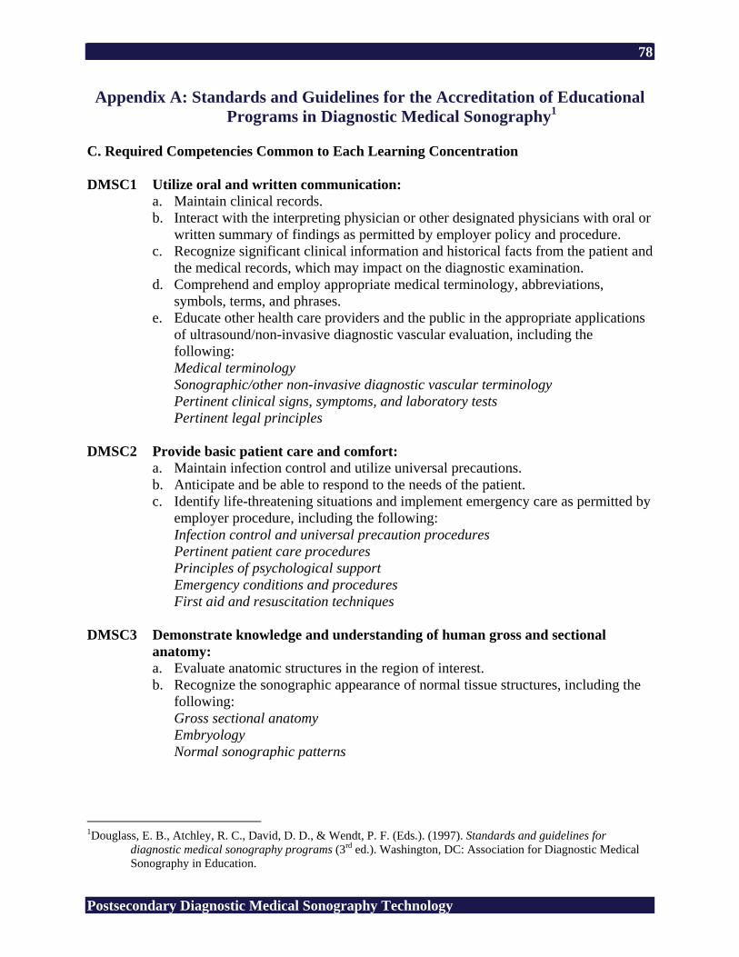
78
Postsecondary Diagnostic Medical Sonography Technology
Appendix A: Standards and Guidelines for the Accreditation of Educational Programs in Diagnostic Medical Sonography1
C. Required Competencies Common to Each Learning Concentration DMSC1 Utilize oral and written communication:
a. Maintain clinical records. b. Interact with the interpreting physician or other designated physicians with oral or
written summary of findings as permitted by employer policy and procedure. c. Recognize significant clinical information and historical facts from the patient and
the medical records, which may impact on the diagnostic examination. d. Comprehend and employ appropriate medical terminology, abbreviations,
symbols, terms, and phrases. e. Educate other health care providers and the public in the appropriate applications
of ultrasound/non-invasive diagnostic vascular evaluation, including the following: Medical terminology Sonographic/other non-invasive diagnostic vascular terminology Pertinent clinical signs, symptoms, and laboratory tests Pertinent legal principles
DMSC2 Provide basic patient care and comfort:
a. Maintain infection control and utilize universal precautions. b. Anticipate and be able to respond to the needs of the patient. c. Identify life-threatening situations and implement emergency care as permitted by
employer procedure, including the following: Infection control and universal precaution procedures Pertinent patient care procedures Principles of psychological support Emergency conditions and procedures First aid and resuscitation techniques
DMSC3 Demonstrate knowledge and understanding of human gross and sectional
anatomy: a. Evaluate anatomic structures in the region of interest. b. Recognize the sonographic appearance of normal tissue structures, including the
following: Gross sectional anatomy Embryology Normal sonographic patterns
1Douglass, E. B., Atchley, R. C., David, D. D., & Wendt, P. F. (Eds.). (1997). Standards and guidelines for
diagnostic medical sonography programs (3rd ed.). Washington, DC: Association for Diagnostic Medical Sonography in Education.
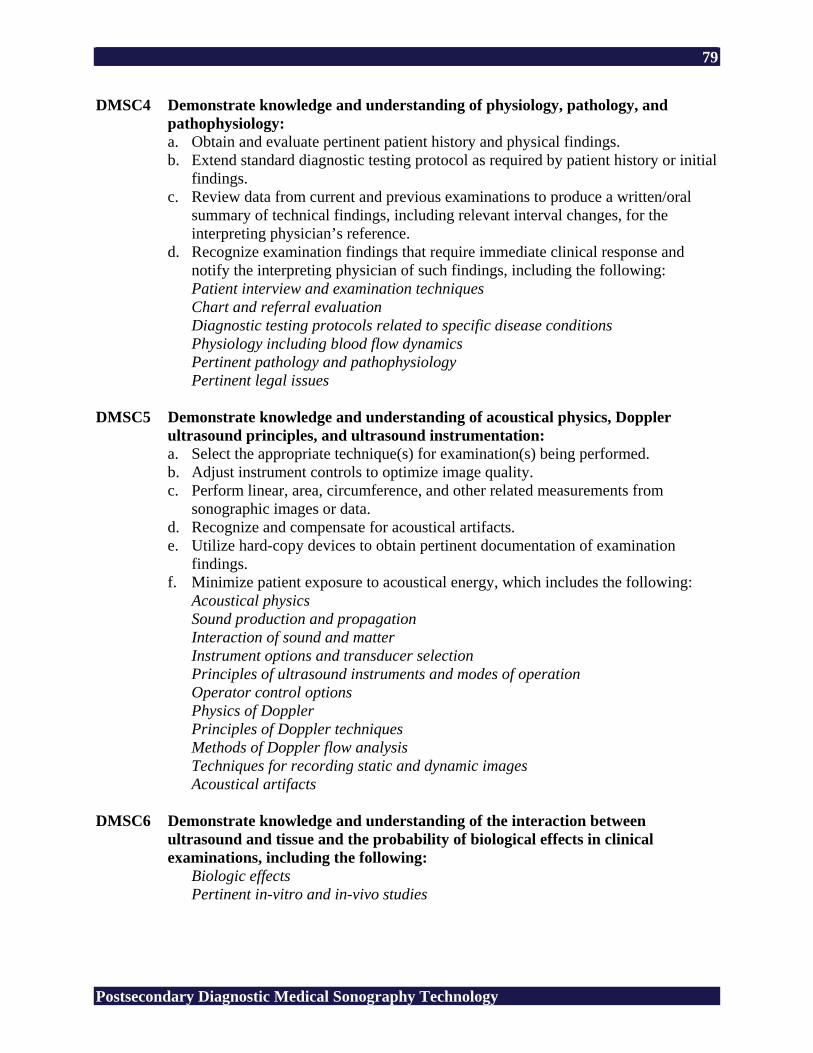
79
Postsecondary Diagnostic Medical Sonography Technology
DMSC4 Demonstrate knowledge and understanding of physiology, pathology, and pathophysiology: a. Obtain and evaluate pertinent patient history and physical findings. b. Extend standard diagnostic testing protocol as required by patient history or initial
findings. c. Review data from current and previous examinations to produce a written/oral
summary of technical findings, including relevant interval changes, for the interpreting physician’s reference.
d. Recognize examination findings that require immediate clinical response and notify the interpreting physician of such findings, including the following: Patient interview and examination techniques Chart and referral evaluation Diagnostic testing protocols related to specific disease conditions Physiology including blood flow dynamics Pertinent pathology and pathophysiology Pertinent legal issues
DMSC5 Demonstrate knowledge and understanding of acoustical physics, Doppler
ultrasound principles, and ultrasound instrumentation: a. Select the appropriate technique(s) for examination(s) being performed. b. Adjust instrument controls to optimize image quality. c. Perform linear, area, circumference, and other related measurements from
sonographic images or data. d. Recognize and compensate for acoustical artifacts. e. Utilize hard-copy devices to obtain pertinent documentation of examination
findings. f. Minimize patient exposure to acoustical energy, which includes the following:
Acoustical physics Sound production and propagation Interaction of sound and matter Instrument options and transducer selection Principles of ultrasound instruments and modes of operation Operator control options Physics of Doppler Principles of Doppler techniques Methods of Doppler flow analysis Techniques for recording static and dynamic images Acoustical artifacts
DMSC6 Demonstrate knowledge and understanding of the interaction between
ultrasound and tissue and the probability of biological effects in clinical examinations, including the following:
Biologic effects Pertinent in-vitro and in-vivo studies
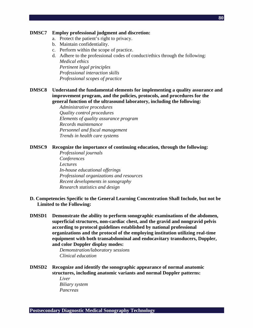
80
Postsecondary Diagnostic Medical Sonography Technology
DMSC7 Employ professional judgment and discretion: a. Protect the patient’s right to privacy. b. Maintain confidentiality. c. Perform within the scope of practice. d. Adhere to the professional codes of conduct/ethics through the following:
Medical ethics Pertinent legal principles Professional interaction skills Professional scopes of practice
DMSC8 Understand the fundamental elements for implementing a quality assurance and
improvement program, and the policies, protocols, and procedures for the general function of the ultrasound laboratory, including the following:
Administrative procedures Quality control procedures Elements of quality assurance program Records maintenance Personnel and fiscal management Trends in health care systems
DMSC9 Recognize the importance of continuing education, through the following:
Professional journals Conferences Lectures In-house educational offerings Professional organizations and resources Recent developments in sonography Research statistics and design
D. Competencies Specific to the General Learning Concentration Shall Include, but not be
Limited to the Following: DMSD1 Demonstrate the ability to perform sonographic examinations of the abdomen,
superficial structures, non-cardiac chest, and the gravid and nongravid pelvis according to protocol guidelines established by national professional organizations and the protocol of the employing institution utilizing real-time equipment with both transabdominal and endocavitary transducers, Doppler, and color Doppler display modes:
Demonstration/laboratory sessions Clinical education
DMSD2 Recognize and identify the sonographic appearance of normal anatomic
structures, including anatomic variants and normal Doppler patterns: Liver Biliary system Pancreas
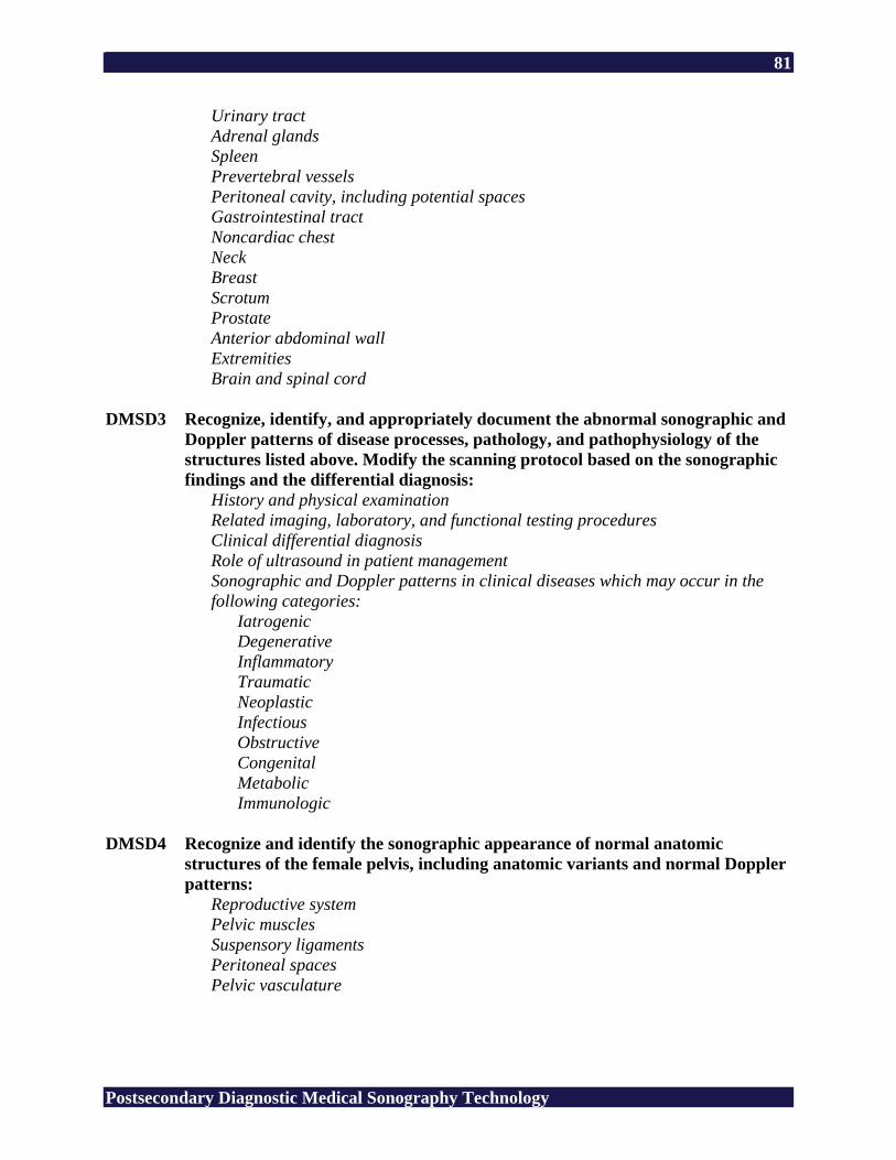
81
Postsecondary Diagnostic Medical Sonography Technology
Urinary tract Adrenal glands Spleen Prevertebral vessels Peritoneal cavity, including potential spaces Gastrointestinal tract Noncardiac chest Neck Breast Scrotum Prostate Anterior abdominal wall Extremities Brain and spinal cord
DMSD3 Recognize, identify, and appropriately document the abnormal sonographic and
Doppler patterns of disease processes, pathology, and pathophysiology of the structures listed above. Modify the scanning protocol based on the sonographic findings and the differential diagnosis:
History and physical examination Related imaging, laboratory, and functional testing procedures Clinical differential diagnosis Role of ultrasound in patient management Sonographic and Doppler patterns in clinical diseases which may occur in the following categories:
Iatrogenic Degenerative Inflammatory Traumatic Neoplastic Infectious Obstructive Congenital Metabolic Immunologic
DMSD4 Recognize and identify the sonographic appearance of normal anatomic
structures of the female pelvis, including anatomic variants and normal Doppler patterns:
Reproductive system Pelvic muscles Suspensory ligaments Peritoneal spaces Pelvic vasculature
82
Postsecondary Diagnostic Medical Sonography Technology
DMSD5 Recognize and identify the sonographic appearance of normal maternal, embryonic, and fetal anatomic structures during the first, second, and third trimesters:
Sonographic sectional anatomy Pertinent measurement techniques Doppler applications Normal sonographic appearance of fetal and maternal structures
DMSD6 Recognize, identify, and appropriately document the sonographic appearance of
gynecologic disease processes, pathology, and pathophysiology: History and physical examination Related imaging, laboratory, and functional testing procedures Differential diagnosis Role of ultrasound in patient management Abnormal sonographic patterns in pregnancy:
Iatrogenic Degenerative Inflammatory Traumatic Neoplastic Infectious Obstructive Congenital Metabolic Immunologic
Contraceptive devices Infertility procedures Doppler applications
DMSD7 Recognize, identify, and appropriately document the sonographic appearance of
obstetric abnormalities, disease, pathology, and pathophysiology: History and physical examination Related imaging, laboratory, and functional testing procedures Differential diagnosis Role of ultrasound in patient management Abnormal sonographic patterns in pregnancy:
Placenta Congenital/genetic anomalies Growth abnormalities Amniotic fluid Viability Multiple gestation Fetal monitoring Maternal factors Postpartum Fetal therapy
83
Postsecondary Diagnostic Medical Sonography Technology
DMSD8 Demonstrate knowledge and understanding of the role of the sonographer in performing interventional/invasive procedures.
84
Postsecondary Diagnostic Medical Sonography Technology
Appendix B: Related Academic Standards2
Reading R1 Interpret Graphic Information (forms, maps, reference sources) R2 Words in Context (same and opposite meaning) R3 Recall Information (details, sequence) R4 Construct Meaning (main idea, summary/paraphrase, compare/contrast, cause/effect) R5 Evaluate/Extend Meaning (fact/opinion, predict outcomes, point of view) Mathematics Computation M1 Addition of Whole Numbers (no regrouping, regrouping) M2 Subtraction of Whole Numbers (no regrouping, regrouping) M3 Multiplication of Whole Numbers (no regrouping, regrouping) M4 Division of Whole Numbers (no remainder, remainder) M5 Decimals (addition, subtraction, multiplication, division) M6 Fractions (addition, subtraction, multiplication, division) M7 Integers (addition, subtraction, multiplication, division) M8 Percents M9 Algebraic Operations Applied Mathematics A1 Numeration (ordering, place value, scientific notation) A2 Number Theory (ratio, proportion) A3 Data Interpretation (graph, table, chart, diagram) A4 Pre-Algebra and Algebra (equations, inequality) A5 Measurement (money, time, temperature, length, area, volume) A6 Geometry (angles, Pythagorean theory) A7 Computation in Context (whole numbers, decimals, fractions, algebraic operations) A8 Estimation (rounding, estimation) Language L1 Usage (pronoun, tense, subject/verb agreement, adjective, adverb) L2 Sentence Formation (fragments, run-on, clarity) L3 Paragraph Development (topic sentence, supporting sentence, sequence) L4 Capitalization (proper noun, titles) L5 Punctuation (comma, semicolon) L6 Writing Conventions (quotation marks, apostrophe, parts of a letter) Spelling S1 Vowel (short, long) S2 Consonant (variant spelling, silent letter) S3 Structural Unit (root, suffix)
2 CTB/McGraw-Hill LLC. (1994). Tests of adult basic education, Forms 7 and 8. Monterey, CA: Author.
Reproduced with permission of CTB/McGraw-Hill LLC. TABE is a registered trademark of The McGraw-Hill Companies, Inc. Copyright © 1994 by CTB/McGraw-Hill LLC. Reproduction of this material is permitted for educational purposes only.
85
Postsecondary Diagnostic Medical Sonography Technology
Appendix C: 21st Century Skills3 CS1 Global Awareness
• Using 21st century skills to understand and address global issues • Learning from and working collaboratively with individuals representing diverse
cultures, religions, and lifestyles in a spirit of mutual respect and open dialogue in personal, work, and community contexts
• Promoting the study of non-English language as a tool for understanding other nations and cultures
CS2 Financial, Economic, and Business Literacy • Knowing how to make appropriate personal economic choices • Understanding the role of the economy and the role of business in the economy • Applying appropriate 21st century skills to function as a productive contributor within
an organizational setting • Integrating oneself within and adapting continually to our nation’s evolving economic
and business environment CS3 Civic Literacy
• Being an informed citizen to participate effectively in government • Exercising the rights and obligations of citizenship at local, state, national, and global
levels • Understanding the local and global implications of civic decisions • Applying 21st century skills to make intelligent choices as a citizen
CS4 Information and Communication Skills • Information and media literacy skills: Analyzing, accessing, managing, integrating,
evaluating, and creating information in a variety of forms and media; understanding the role of media in society
• Communication skills: Understanding, managing, and creating effective oral, written, and multimedia communication in a variety of forms and contexts
CS5 Thinking and Problem-Solving Skills • Critical thinking and systems thinking: Exercising sound reasoning in understanding
and making complex choices, understanding the interconnections among systems • Problem identification, formulation, and solution: Ability to frame, analyze, and solve
problems • Creativity and intellectual curiosity: Developing, implementing, and communicating
new ideas to others, staying open and responsive to new and diverse perspectives CS6 Interpersonal and Self-Directional Skills
• Interpersonal and collaborative skills: Demonstrating teamwork and leadership, adapting to varied roles and responsibilities, working productively with others, exercising empathy, respecting diverse perspectives
• Self-direction: Monitoring one’s own understanding and learning needs, locating appropriate resources, transferring learning from one domain to another
• Accountability and adaptability: Exercising personal responsibility and flexibility in personal, workplace, and community contexts; setting and meeting high standards and goals for one’s self and others; tolerating ambiguity
3 21st century skills. (n.d.). Washington, DC: Partnership for 21st Century Skills.

86
Postsecondary Diagnostic Medical Sonography Technology
• Social responsibility: Acting responsibly with the interests of the larger community in mind; demonstrating ethical behavior in personal, workplace, and community contexts