200pm imaging of coronary artery bypass grafts and … - imaging of...saphenous vein grafts ( svg )...
TRANSCRIPT
Imaging of Coronary artery Bypass grafts and Stents
Dharshan R Vummidi MRCP, FRCR
University of Michigan

• No relevant disclosures

Objectives• CT technique in the evaluation of
bypass grafts and stents• Post processing and image analysis• Post surgical anatomy• Complications

Background• Cardiovascular disease is the leading
cause of death in the US with over 450000 deaths.
• Approximately 469000 coronary artery bypass grafts ( CABGs ) are performed annually in the US.
• CT coronary angiography has now established itself as an equally accurate modality for the assessment of grafts .

Accuracy AnalysisType and (n= No of studies)
Number of grafts Sensitivity (%) Specificity (%)
Graft occlusion (16)
2023 97.6 96.7
16 section (9) 1047 96.9 96.4
64 section (6) 976 98.1 96.9
Occlusion (10) 1308 99.3 98.7
> 50 % Stenosis (9)
871 94.1 98.0
Hamon et al. Radiology 2007

CT Technique• Beta blockers, if HR > 65/ min• 64 MDCT : Peak + 6 seconds• Sublingual nitroglycerin• Triphasic technique
– Timing bolus 15 ml @ 5ml/sec– Contrast bolus of 70 ml @ 5 ml/sec– Contrast/ Saline mix of 50 ml (60/40
mix) @ 5mls/ sec– Saline chase of 50 ml

• Craniocaudal coverage : 2cm above aortic arch to 2cm below base of the heart
• 0.5 - 0.7 mm collimation• 0.16 - 0.24, based on heart rate for
prospective triggering• kVp: based on a BMI chart• mAs : based on a BMI chart• Acquisition time : 6 – 8 seconds


Image analysis• Start with axial images
– Reformats ( curved planar, linear lumen and cross sectional )
– Maximum intensity projection ( MIPs ) – 3D volume rendering ( VR )
• Assess proximal anastomosis, graft proper and distal anastomosis.
• Sequential or jump graft• Distal runoff and native coronary arteries• Position of the graft in relation to the sternum• Incidental findings with larger FOV

Knowledge of surgical anatomy• Saphenous vein • Internal mammary artery ( LIMA and
RIMA )
• Radial artery• Right Gastroepiploic artery


SVG
LIMA
RV
LV
PAAORTA

Saphenous Vein grafts ( SVG )• Advantages
– Ease of harvest– Larger caliber than arteries– Fewer surgical clips
• Disadvantages– Atherosclerosis and intimal hyperplasia– 73% and 41% patency rates at 5 and 10
years

SVG

OM1OM2
SVG

Internal Mammary Artery Grafts ( LIMA or RIMA )
• Advantages– In situ graft– Graft of choice for revascularization of
LAD and diagonals– Superior patency rates, > 90% at 10
years– Relatively resistant to atherosclerosis
• Disadvantages– Smaller caliber– Use of more surgical clips

LIMA
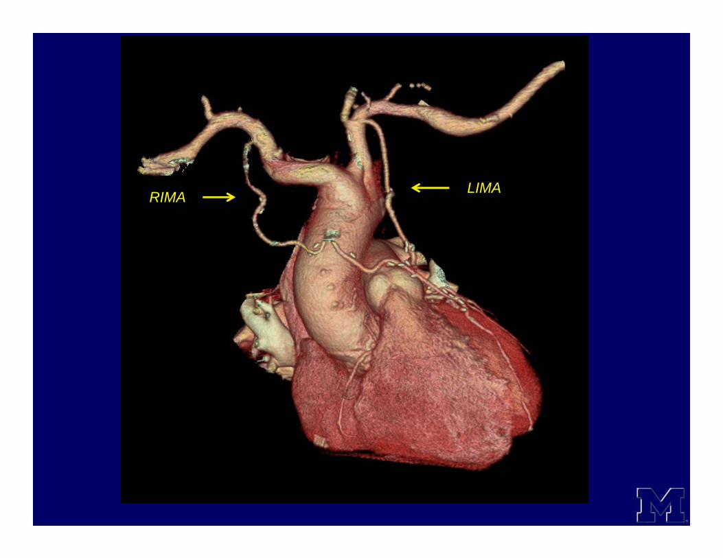
LIMARIMA
Complications• Graft thrombosis
– Acute : typically SVG• < 1month post operatively• Typically 3 -12 %• Endothelial injury during graft harvest
– Late graft thrombosis and stenosis• Atherosclerotic plaque• IMA is relatively resistant
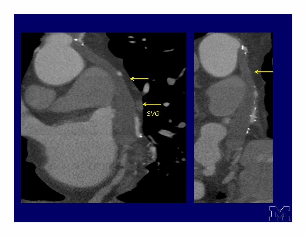
SVG
SVG
SVG

SVG

SVG

Nubbin sign• Acute graft thrombosis manifesting as
a small outpouching on the anterior aorta.

LVRV
AORTA PA
SVG

Graft Aneurysm• True
– 5 – 7 years post surgery– Atherosclerotic– Almost half of them may be
asymptomatic• Pseudoaneurysms
– Acute complication ( < 6 months )– Infection– Tension at the graft site or suture
dehiscence

SVG

SVG

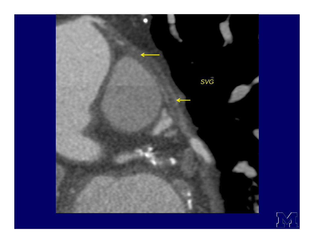
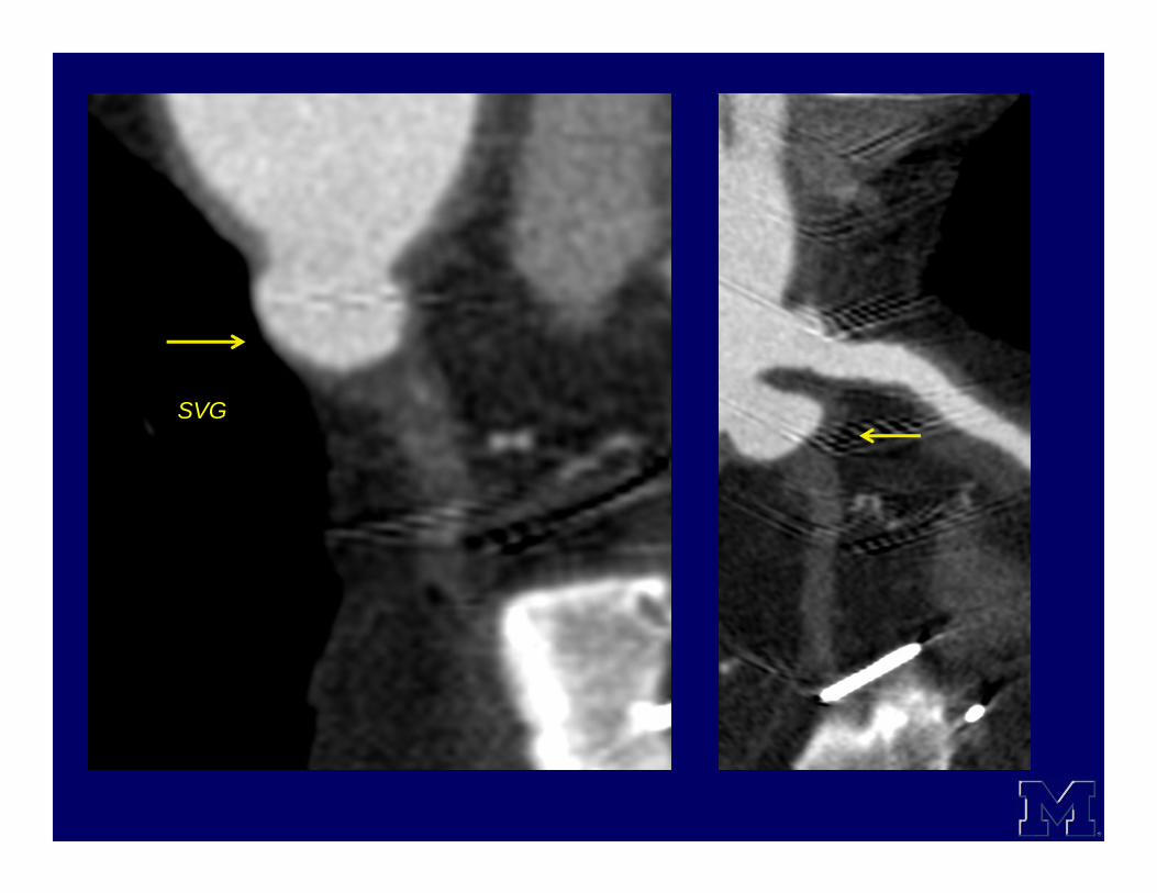
• Graft malposition• Graft spasm• Pleural and pericardial effusions• Wound and sternal infections

Relationship to sternum
• In a survey of 2,046 catastrophic bleeding events in reoperations reported by 1,116 surgeons, the most common cardiac structures injured were the right ventricle (39%),SVG (20%), aorta (15%), IMA (12%), and innominate vein (6%).
Aviram et al. Ann Thorac Surg 2005;79:589 –95


Stents• Over 6,00,000 coronary artery stent
placements annually in the US • Bare metal stents ( Steel, Titanium,
Nitinol ) and drug eluting stents• Lower rate of stenosis than
angioplasty• In stent restenosis ( ISR ) 25-30% for
bare metal and 5-10 % for drug eluting stents
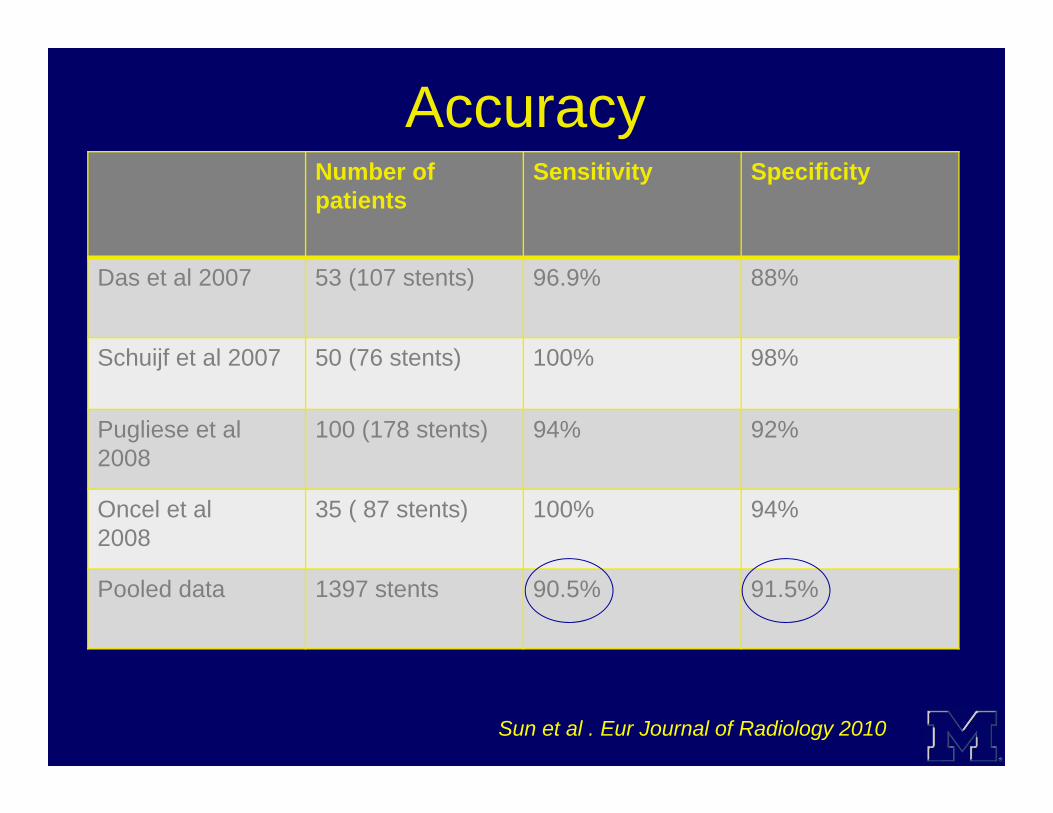
Accuracy Number of patients
Sensitivity Specificity
Das et al 2007 53 (107 stents) 96.9% 88%
Schuijf et al 2007 50 (76 stents) 100% 98%
Pugliese et al 2008
100 (178 stents) 94% 92%
Oncel et al2008
35 ( 87 stents) 100% 94%
Pooled data 1397 stents 90.5% 91.5%
Sun et al . Eur Journal of Radiology 2010

Image analysis• Sharp kernel• Loss of contrast enhancement within
the stent implies occlusion• Reduced contrast enhancement
distally implies occlusion or retrograde perfusion
• Look for stent fracture

RCA
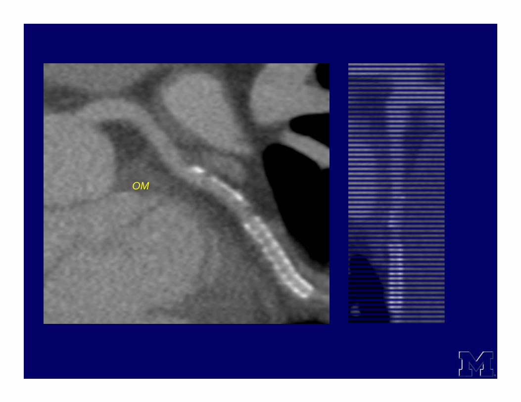
OM

SVG
Conclusion• MDCT coronary angiography is an
invaluable tool for the assessment of coronary artery bypass grafts and stents.– Accurate– Relatively non invasive– Information about vessel wall– Incidental findings