2014 honors cells prelim ppt - pkwy.k12.mo.us honors cells prelim...• eukaryotes are complex in...
TRANSCRIPT
Cells
Variation and Function of Cells

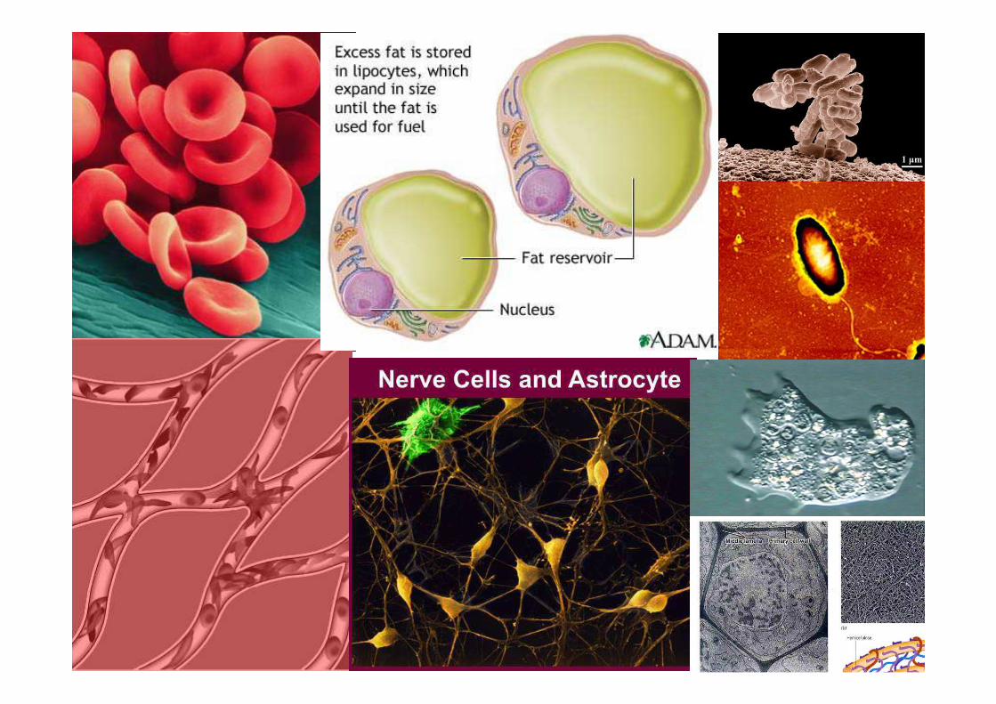
• Cell Theory states that:1. All living things are made of cells2. Cells are the basic unit of structure and functio n
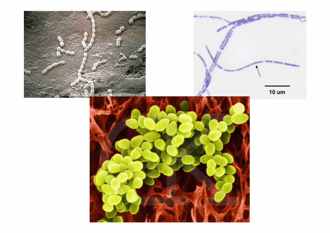
in living things3. New cells are produced from existing cellsTwo major types of CellsProkaryotic cells are very small and have no membrane
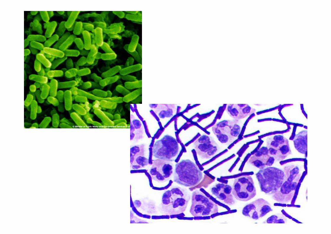
bound organelles or a nucleus. All Bacteria are prokaryotes and have circular DNA.
Eukaryotic Cells are more organized and complex than prokaryotes, they have membrane bound organelles, linear chromosomes and tend to be very large in comparison. P169-173
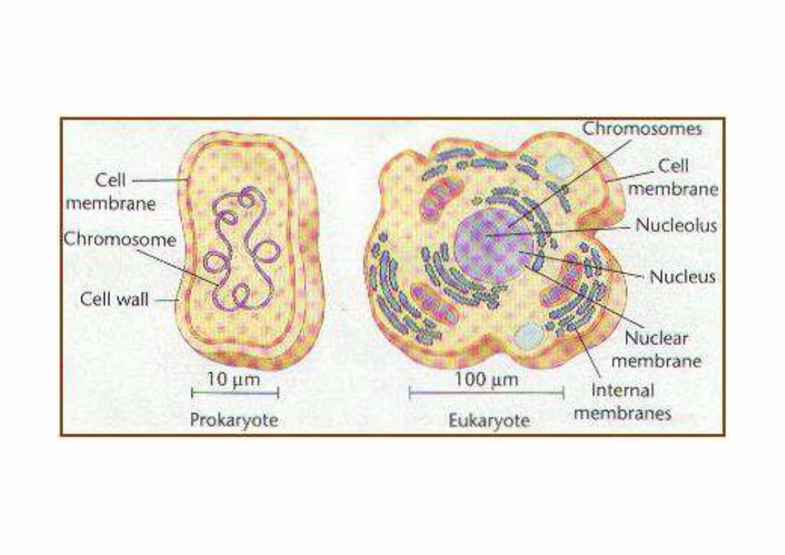

Comparison: Prokaryotes and Eukaryotes• Prokaryotic cells are limited in their structure.
They have ribosomes, circular DNA, rely on the exterior membrane to complete any membrane related function and rely on diffusion for transport.
• Prokaryotic Kingdoms/Domains1. Eubacteria-are very diverse often have a cell
wall that contains peptidoglycan2. Archaebacteria- lack peptidoglycan in cell
walls, have different membrane lipids, and have some genes that are more similar to eukaryotes than eubacteria.

• Eukaryotes are complex in comparison to prokaryotes with several unique organelles that maximize their efficiency : they do or can
1. Grow much larger as they developed organization and distribution abilities2. Compartmentalize delicate and destructive processes within separate microenvironments.3. Specialize function of individual cells to work with other cells (multi-cellular)Plasma Membrane= the “skin” of a cell, it
protects, nourishes, and communicates with other cells. Organelle membranes do the same
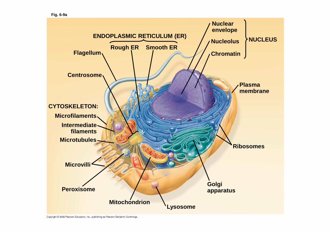
Fig. 6-9a
ENDOPLASMIC RETICULUM (ER)
Smooth ERRough ERFlagellum
Centrosome
CYTOSKELETON:
Microfilaments
Intermediatefilaments
Microtubules
Microvilli
Peroxisome
MitochondrionLysosome
Golgiapparatus
Ribosomes
Plasma membrane
Nuclearenvelope
Nucleolus
Chromatin
NUCLEUS

Fig. 6-9b
NUCLEUS
Nuclear envelopeNucleolusChromatin
Rough endoplasmic reticulum
Smooth endoplasmic reticulum
Ribosomes
Central vacuole
Microfilaments
Intermediate filamentsMicrotubules
CYTO-SKELETON
Chloroplast
PlasmodesmataWall of adjacent cell
Cell wall
Plasma membrane
Peroxisome
Mitochondrion
Golgiapparatus
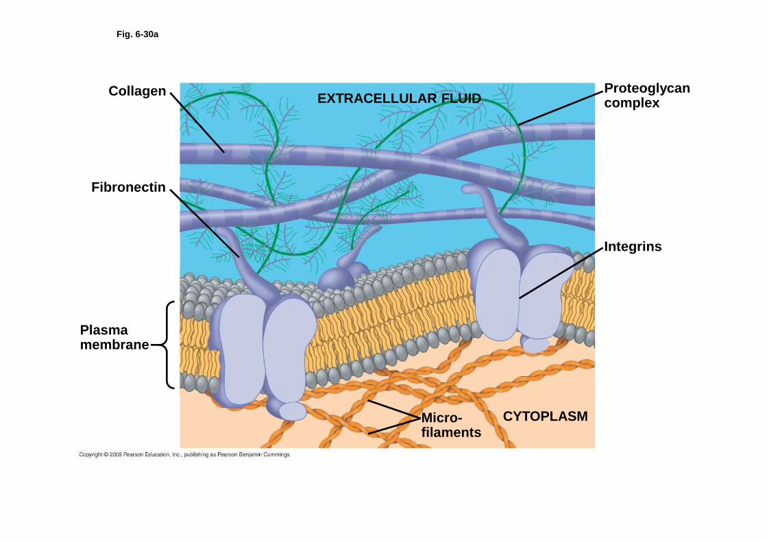
Fig. 6-30a
Collagen
Fibronectin
Plasma membrane
Proteoglycan complex
Integrins
CYTOPLASMMicro-filaments
EXTRACELLULAR FLUID

Fig. 6-7
TEM of a plasmamembrane
(a)
(b) Structure of the plasma membrane
Outside of cell
Inside ofcell 0.1 µm
Hydrophilicregion
Hydrophobicregion
Hydrophilicregion Phospholipid Proteins
Carbohydrate side chain
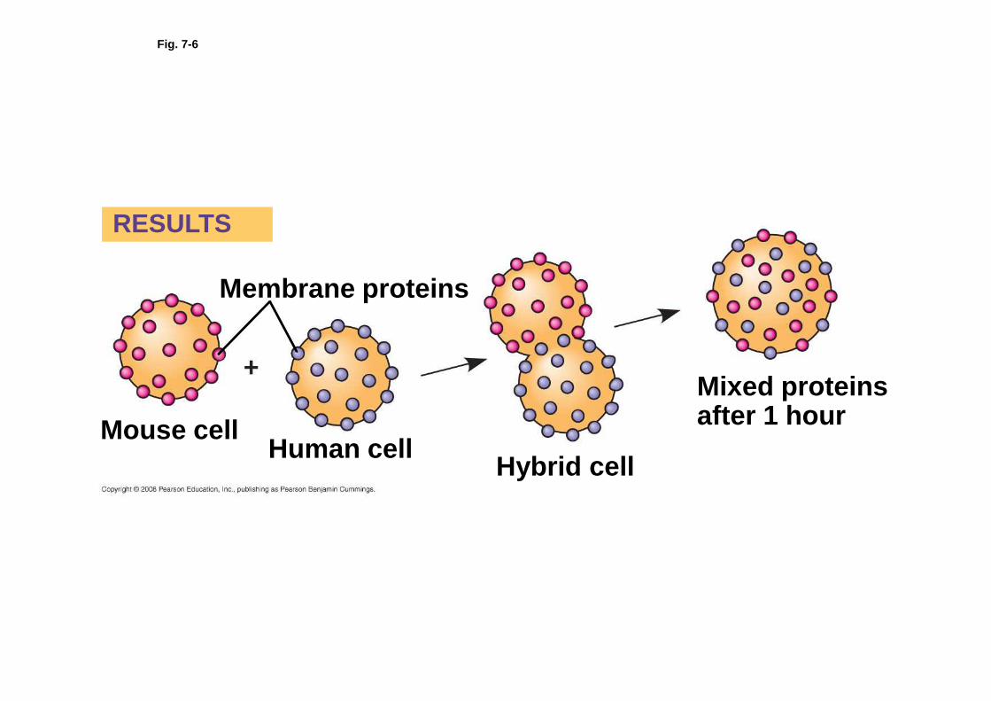
Fig. 7-6
RESULTS
Membrane proteins
Mouse cellHuman cell
Hybrid cell
Mixed proteinsafter 1 hour

Fig. 7-7
Fibers ofextracellularmatrix (ECM)
Glyco-protein
Microfilamentsof cytoskeleton
Cholesterol
Peripheralproteins
Integralprotein
CYTOPLASMIC SIDEOF MEMBRANE
GlycolipidEXTRACELLULARSIDE OFMEMBRANE
Carbohydrate

Fig. 6-30
EXTRACELLULAR FLUIDCollagen
Fibronectin
Plasmamembrane
Micro-filaments
CYTOPLASM
Integrins
Proteoglycancomplex
Polysaccharidemolecule
Carbo-hydrates
Coreprotein
Proteoglycanmolecule
Proteoglycan complex
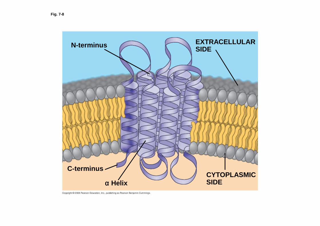
Fig. 7-8
N-terminus
C-terminus
αααα HelixCYTOPLASMICSIDE
EXTRACELLULARSIDE

Fig. 7-15
EXTRACELLULAR FLUID
Channel protein
(a) A channel protein
Solute CYTOPLASM
Solute Carrier protein
(b) A carrier protein
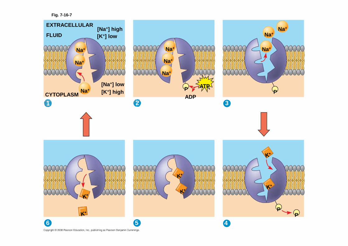
2
EXTRACELLULAR
FLUID[Na+] high[K+] low
[Na+] low [K+] high
Na+
Na+
Na+
Na+
Na+
Na+
CYTOPLASM
ATP
ADPP
Na+
Na+
Na+
P
3
6 5 4
PP
1
Fig. 7-16-7
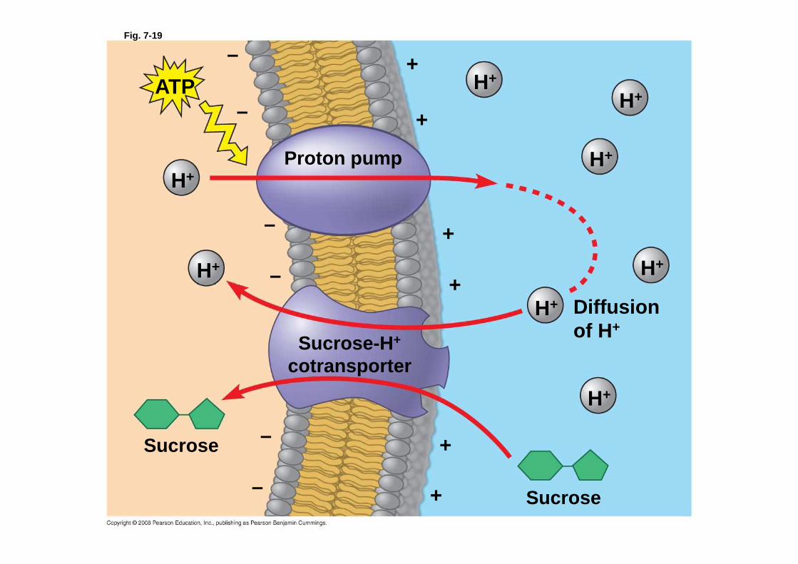
Fig. 7-19
Proton pump
–
–
–
–
–
–
+
+
+
+
+
+
ATP
H+
H+
H+
H+
H+
H+
H+
H+
Diffusionof H+
Sucrose-H +
cotransporter
Sucrose
Sucrose

Fig. 6-14a
Nucleus 1 µm
Lysosome
Lysosome
Digestive enzymes
Plasma membrane
Food vacuole
Digestion
(a) Phagocytosis
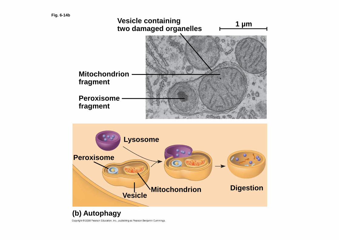
Fig. 6-14b
Vesicle containingtwo damaged organelles
Mitochondrion fragment
Peroxisome fragment
Peroxisome
Lysosome
DigestionMitochondrionVesicle
(b) Autophagy
1 µm

Fig. 7-13
Hypotonic solution
(a) Animalcell
(b) Plantcell
H2O
Lysed
H2O
Turgid (normal)
H2O
H2O
H2O
H2O
Normal
Isotonic solution
Flaccid
H2O
H2O
Shriveled
Plasmolyzed
Hypertonic solution
• All Eukaryotes have the following organelles and structures.
Nucleus• Nuclear envelope- a membrane that contains the DNA
and the proteins necessary to organize and maintain the DNA
• Chromatin-DNA and Protein that is found unwound in a cell between divisions.
• Chromosomes-condensed form of chromatin these are linear and found during mitosis.
• Nucleolus- area of the nucleus thought to be used to assemble ribosomes.
Cytosol• Ribosomes- RNA and protein complex that work
together to read mRNA and build a protein from its code.
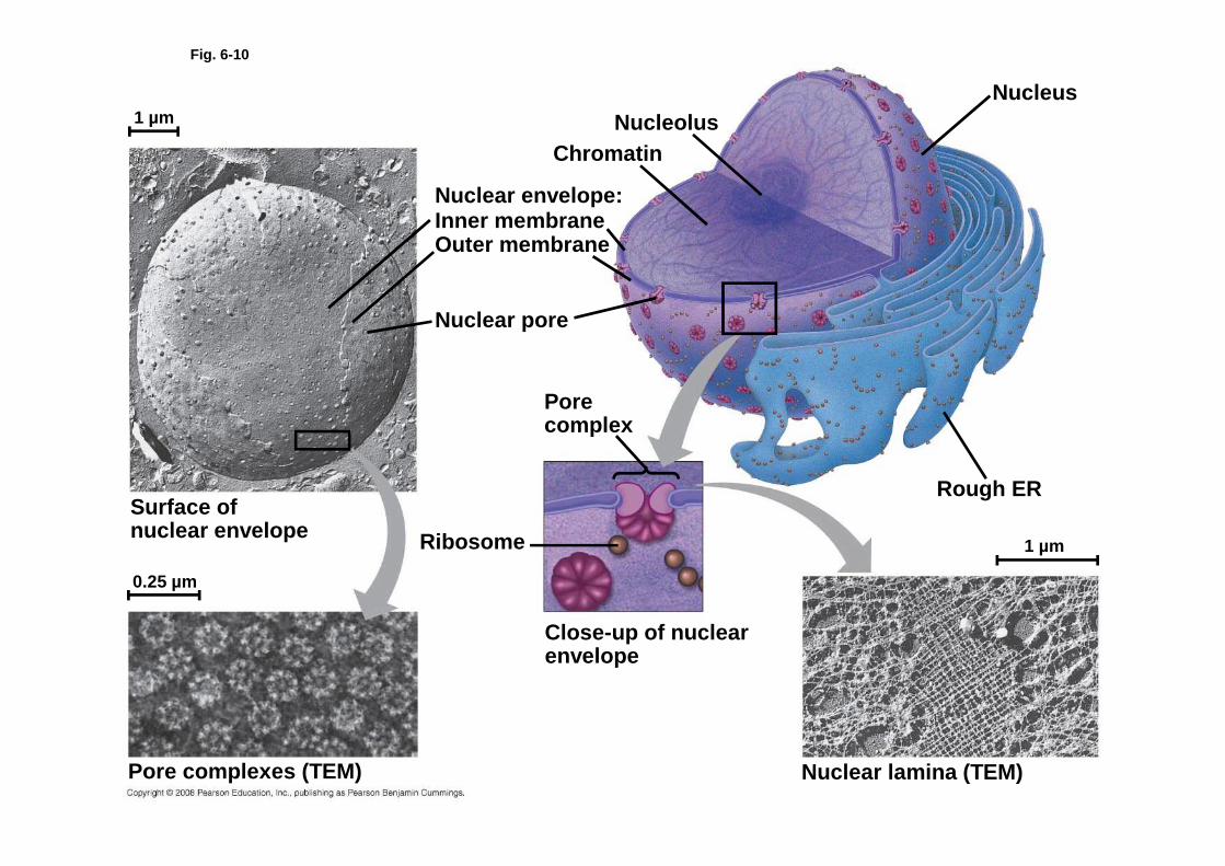
Fig. 6-10
NucleolusNucleus
Rough ER
Nuclear lamina (TEM)
Close-up of nuclear envelope
1 µm
1 µm
0.25 µm
Ribosome
Pore complex
Nuclear pore
Outer membraneInner membraneNuclear envelope:
Chromatin
Surface ofnuclear envelope
Pore complexes (TEM)
• Organelles are membrane bound, specially designed and tasked parts of cells
• Endoplasmic Reticulum- network of membrane found in the cell along side the nucleus, makes lipid and protein components of the membrane and materials for export from the cell.
• Rough ER looks grainy because it has ribosomes embedded in its membrane. Produces membrane bound proteins and proteins for export.
• Smooth ER is the side of the ER away from the nucleus this is the ER responsible for lipid synthesis and detoxification often refines or modifies products from rough ER
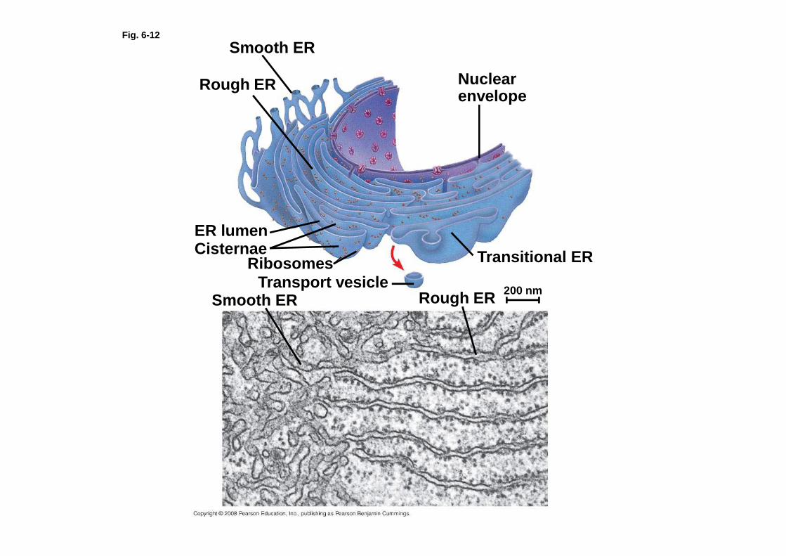
Fig. 6-12
Smooth ER
Rough ER Nuclear envelope
Transitional ER
Rough ERSmooth ERTransport vesicle
RibosomesCisternaeER lumen
200 nm

• Golgi Apparatus- Acts as a processing center for products from the ER. May modify some chemicals, while just sorting and packaging others for storage or release.
• Lysosomes are membrane bags of hydrolytic enzymes. Lysosomes keep these dangerous chemicals separate from the rest of the cell’s chemicals and concentrated. They bind with food vacuoles to start the digestions of materials that have been consumed.
• Vacuoles-mean membrane bag and is used to refer to contractile vacuoles that pump out extra water, food vacuoles, and central vacuoles.
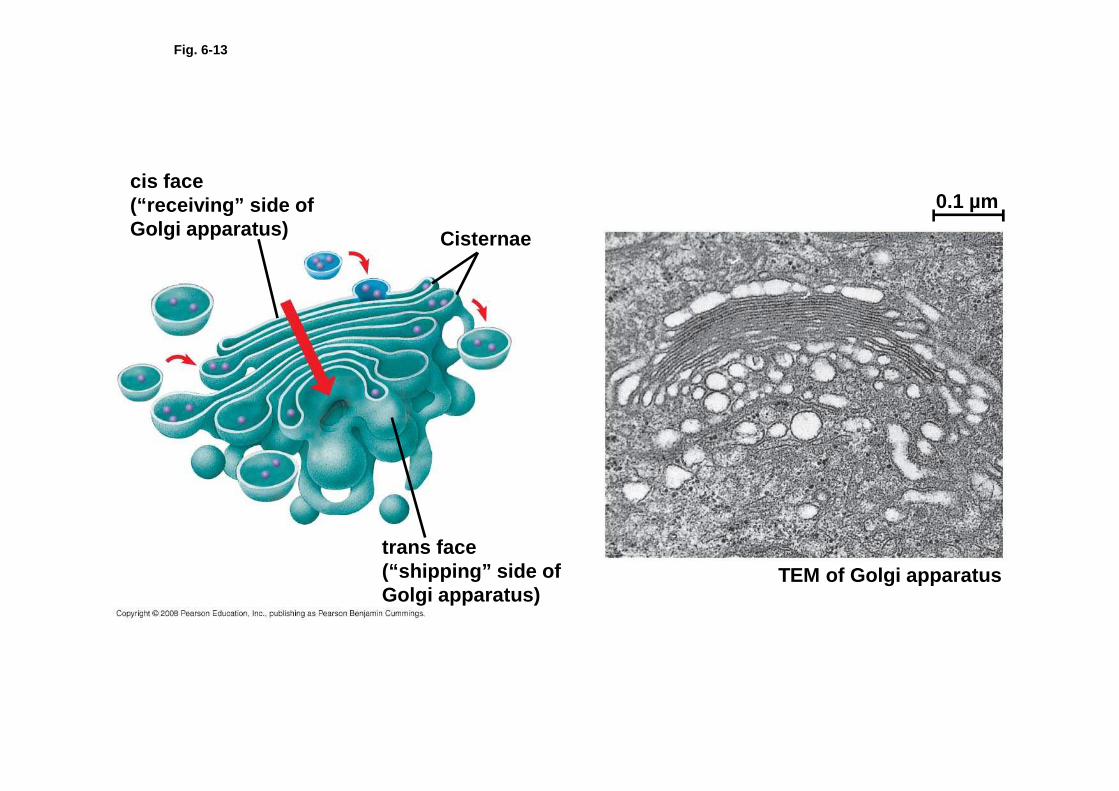
Fig. 6-13
cis face(“receiving” side of Golgi apparatus) Cisternae
trans face(“shipping” side of Golgi apparatus)
TEM of Golgi apparatus
0.1 µm
• Mitochondria-the “power house” of the cell. This is the organelle that takes glucose or other chemical energy sources and converts them into ATP. Mitochondria have their own circular DNA, a lot of membrane folds, and their own ribosomes.
• Chloroplasts are the organelles that house chlorophyll allowing them to capture sunlight and convert the energy it carries into the chemical energy (glucose). These also have their own circular DNA, large amounts of internal membrane, and their own ribosomes.

Fig. 6-17
Free ribosomesin the mitochondrial matrix
Intermembrane spaceOuter membrane
Inner membraneCristae
Matrix
0.1 µm

Fig. 9-19
Glucose
Glycolysis
Pyruvate
CYTOSOL
No O2 present:Fermentation
O2 present:Aerobic cellular
respiration
MITOCHONDRION
Acetyl CoAEthanolor
lactateCitricacidcycle
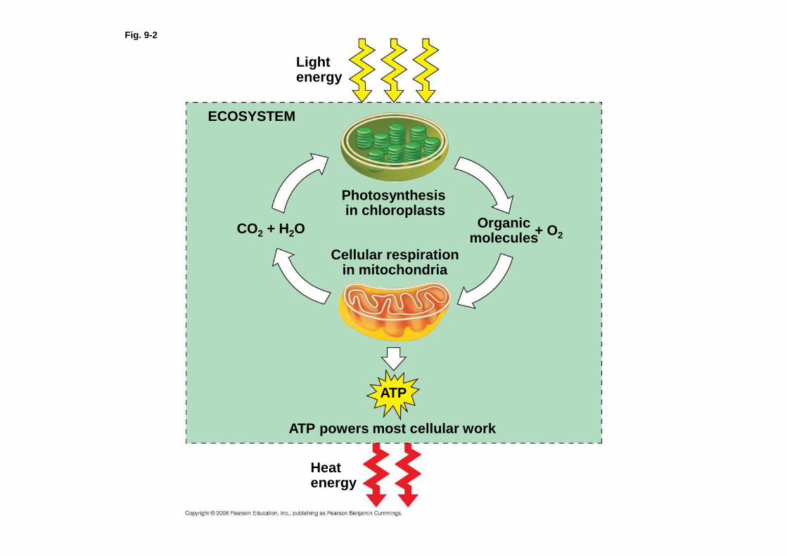
Fig. 9-2
Lightenergy
ECOSYSTEM
Photosynthesisin chloroplasts
CO2 + H2O
Cellular respirationin mitochondria
Organicmolecules + O2
ATP powers most cellular work
Heatenergy
ATP

Fig. 10-3b
1 µm
Thylakoidspace
Chloroplast
GranumIntermembranespace
Innermembrane
Outermembrane
Stroma
Thylakoid

Fig. 10-7
Reflectedlight
Absorbedlight
Light
Chloroplast
Transmittedlight
Granum

• Structure unique to Plants• Cell Wall- in plants these are made up of
Cellulose and lignin creating a rigid outer shell that is made stronger when the plant has a hypotonic environment
• Plastids- several different membrane sacs that hold various pigments, metabolites, etc.
• Chloroplast- the light converting plastid (membrane sac) it absorbs light energy and converts it to chemical energy.
• Central Vacuole- a huge vacuole found in the center of the cell that tends to hold water and keeps the organelles nearer the edge of the cell

Fig. 6-9b
NUCLEUS
Nuclear envelopeNucleolusChromatin
Rough endoplasmic reticulum
Smooth endoplasmic reticulum
Ribosomes
Central vacuole
Microfilaments
Intermediate filamentsMicrotubules
CYTO-SKELETON
Chloroplast
PlasmodesmataWall of adjacent cell
Cell wall
Plasma membrane
Peroxisome
Mitochondrion
Golgiapparatus

• Cytoskeleton is the structural framework found inside of cells.
• Microfilaments are the smallest form of cytoskeleton fibers and are made of actin These resist tension very well. These are the fibers that are pulled on in muscles to create a contraction.
• Microtubules are the largest form of cytoskeleton fibers made of tubulin and they resist compression very well. These provide support to a cell against being crushed and act as a rail along which vacuoles and lysosomes can be transported. They are also the basis for the movement of cilia and flagella

Fig. 6-20
Microtubule
Microfilaments0.25 µm
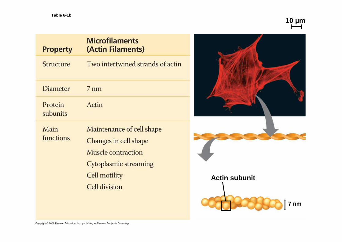
Table 6-1b
Actin subunit
10 µm
7 nm
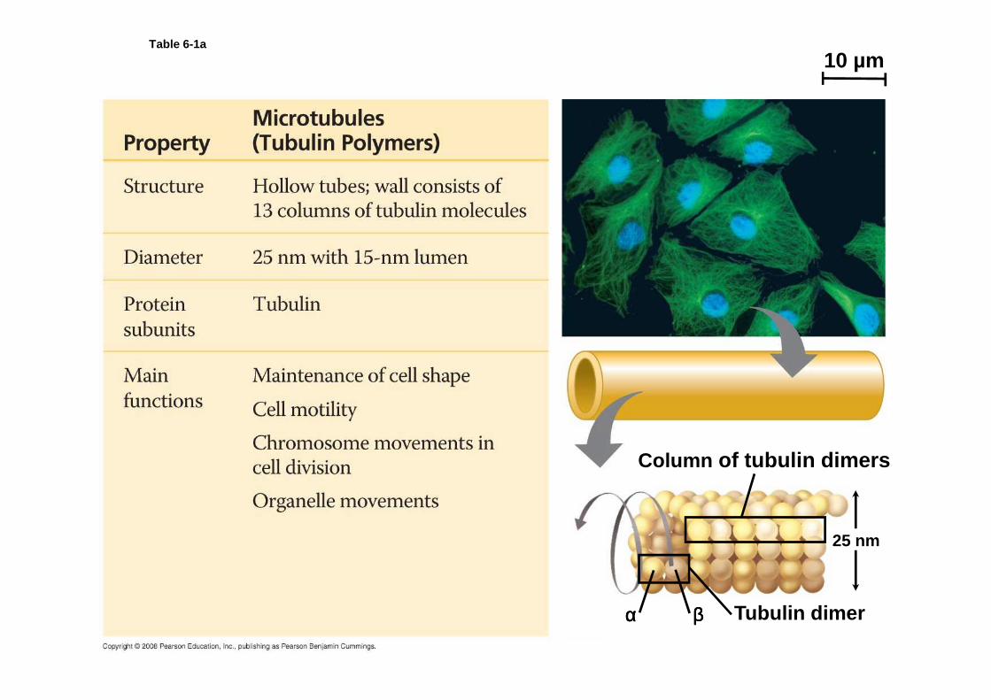
Table 6-1a
10 µm
Column of tubulin dimers
Tubulin dimerαααα ββββ
25 nm
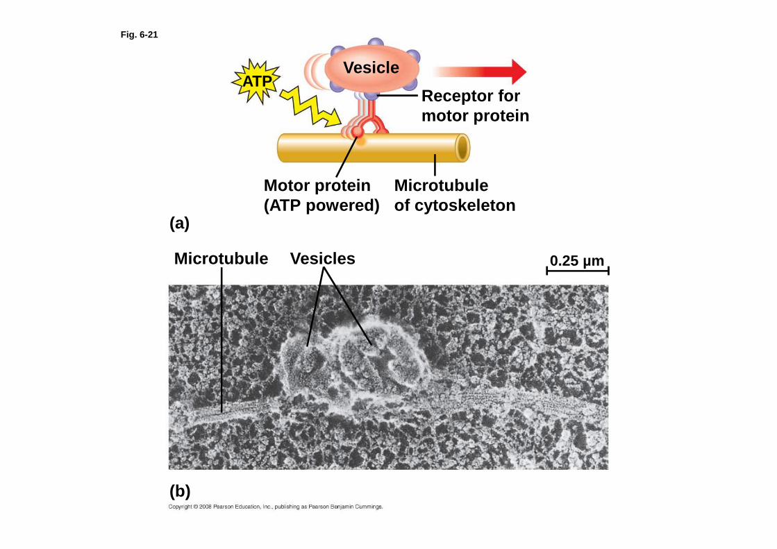
Fig. 6-21
VesicleATP
Receptor for motor protein
Microtubuleof cytoskeleton
Motor protein (ATP powered)
(a)
Microtubule Vesicles
(b)
0.25 µm