2014 respiratory block practical. streptococcus pyogenes = group a strep carried by 10-25% of many...
TRANSCRIPT

2014
RESPIRATORY BLOCK Practical

Streptococcus pyogenes = Group A Strep
Carried by 10-25% of many in throatoften no symptomsit is Cause of
• strep throat• impetigo• Necrotizing
fasciitis

Left. Gram stain of Streptococcus pyogenes in a clinical specimen. Right. Colonies of Streptococcus pyogenes on blood agar exhibiting beta (clear) hemolysis
Streptococcus pyogenes. = Group A Strept

• Principle:– Bacitracin test is used for
presumptive identification of group A
– To distinguish between S. pyogenes (susceptible to B) & non group A such as S. agalactiae (Resistant to B)
– Bacitracin will inhibit the growth of gp A Strep. pyogenes giving zone of inhibition around the disk
• Procedure:– Inoculate BAP with heavy
suspension of tested organism
– Bacitracin disk (0.04 U) is applied to inoculated BAP
– After incubation, any zone of inhibition around the disk is considered as susceptible

Bacitracin sensitivity

A 5 year boy was brought to king Khalid University hospital, outpatient department complaining of fever and sore throat. He had regular vaccination history. On examination his temperature was 38.5° c, the tonsil area and pharynx were obviously inflamed with some foci of pus.
Case 1

1. What is the differential diagnosis?
2. What investigation should be done?

• Lab tests
• The full blood count showed a total white cell count of 15000ml.Throat swab culture showed colonies with clear haemolysis on blood agar. They were catalase negative .The gram stain of these colonies showed gram positive cocci in chains

1. What is the likely identity of the organism?
2. What is the best antibiotic therapy for this child?
3. If not treated what complication may this child have after 6 weeks period?

Streptococcus pneumoniae(Pneumococci)

Alpha-hemolysis
• Alpha-hemolytic Streptococcus species "Viridans group" streptococci, including species such as the Streptococcus mutans, mitis, and salivarius groups display alpha hemolysis.

Optochin Susceptibility Test
Optochin susceptibleS. pneumoniae
Optochin resistantS. viridans

Optochin Sensitive

A 28 Year Old Female presented to the accident and emergency of KKUH with a sudden onset of fever, right sided chest pain and productive cough of purulent sputum. On examination her temperature was 39 °C. There were Rhonci and dullness on the right side of the chest. X-ray showed massive consolidation on the right side of the chest.
CASE 2

1. What is the most likely diagnosis?
2. What investigation should be done?
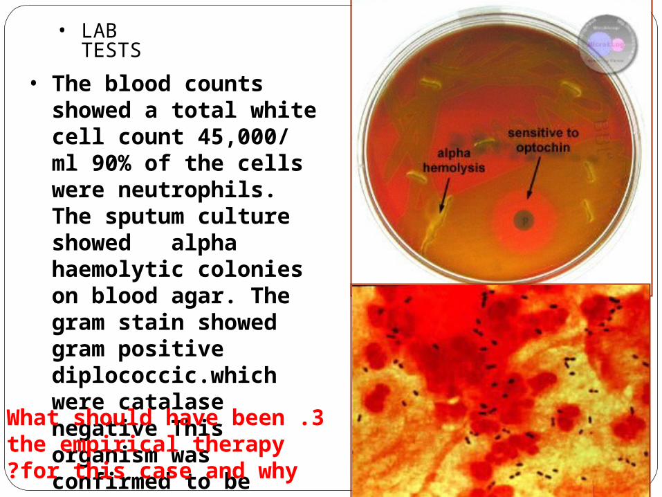
• LAB TESTS
• The blood counts showed a total white cell count 45,000/ ml 90% of the cells were neutrophils. The sputum culture showed alpha haemolytic colonies on blood agar. The gram stain showed gram positive diplococcic.which were catalase negative This organism was confirmed to be optician susceptible.
3 .What should have been the empirical therapy for this case and why?

Mycobacterium tuberculosis: Ziehl-Neelsen stain
Sputum Microscopy

Mycobacterium tuberculosis
Growth on L.J medium( selective for mycobacteria

Abdul Karim is a 45 year old Saudi man who was admitted to King Khalid University Hospital because of 2-3 month history of loss of appetite, weight loss, and on and off fever with attacks of cough. Two days before admission .he coughed blood (haemoptysis) Abdul karim is diabetic for the last 5 years. His father died of tuberculosis at the age of 45 yrs.
CASE 3

• On examination Abdul Karim looked weak with a temperature 38.6 °C, CVS and Respiratory system examinnation was unremarkable.
• The chest X- ray done showed multiple opacities and cavities
• The ESR was increased (85 m /hour)
• What further tests should be done?

• Sputum AFB smear
• Sputum smear showed AFB
• What is the probable diagnosis?
• How can the diagnosis be confirmed?

Gram positive, cocci, in clusters
Gram positive, cocci, in clusters

StaphylococciStained in Pus

Vaginal Smear of a Person with Candida Vaginitis
Note epithelial cells, rod-shaped bacteria, and Candida albicans in its hyphal form

Candida albicans ProducingGerm tube
Dimorphic Candida albicans switching from a yeast form to a filamentous form

Chlamydospore Oral Candida or oral thrush
Gram stain of candida: ovoid budingYeast

Growth on Sabouraud's Dextrose Media

Gram stain of Candida albicans Showing budding yeast celols

Aspergillus niger
Culture of Aspergillus niger.
Conidial head of A. niger

Aspergillus niger

Methenamine silver (GMS) stained tissue section of lung showing dichotomously branched
Aspergillosis