2014 validity of measuring distal vastus medialis muscle using rehabilitative ultrasound imaging...
TRANSCRIPT
8/11/2019 2014 Validity of measuring distal vastus medialis muscle using rehabilitative ultrasound imaging versus magnetic r…
http://slidepdf.com/reader/full/2014-validity-of-measuring-distal-vastus-medialis-muscle-using-rehabilitative 1/5
Technical and measurement report
Validity of measuring distal vastus medialis muscle usingrehabilitative ultrasound imaging versus magnetic resonance imaging
Peter R. Worsley * , Fleur Kitsell, Dinesh Samuel, Maria StokesFaculty of Health Sciences, University of Southampton, UK
a r t i c l e i n f o
Article history:Received 15 July 2013Received in revised form22 January 2014Accepted 3 February 2014
Keywords:Ultrasound imagingVastus medialisReliabilityValidity
a b s t r a c t
Objective quanti cation of muscle size can aid clinical assessment when treating musculoskeletal con-ditions. To date the gold standard of measuring muscle morphology is magnetic resonance imaging(MRI). However, there ’ s a growing body of evidence validating rehabilitative ultrasound imaging (RUSI)against MRI.Objective: This study aimed to validate RUSI against MRI for the linear measurements of the distal bresof vastus medialis muscle in the thigh.Twelve healthy male participants were recruited from a local university population. The distal portion of their right vastus medialis was imaged with the participant in long-sitting, using MRI and RUSI whilst theleg was in extension and neutral hip rotation. Cross sectional area (CSA) and three linear measures weretaken from the MRI and these were compared with the same linear measures from RUSI. Statisticalanalysis included comparison of MRI and RUSI measures using the paired t -test and intra-class corre-lation coef cients (ICC 3,1).Mean differences between the linear measures taken from the MRI and RUSI were 0.5 mm to 2.9 mm(95% con dence intervals 0.6 e 8.3 mm), which were not statistically different ( p > 0.05) and werehighly correlated (ICCs 3,1 0.84 e 0.94). Correlations between the three linear measurements and muscleCSA ranged from r ¼ 0.23 to 0.87, the greatest being muscle thickness. Multiplying the linear measures
did not improve the correlation of 0.87 found for muscle thickness.Linear measures of vastus medialis depth made using RUSI were shown to be as valid as using MRI.Muscle thickness measures using RUSI could be used within an objective assessment of this muscle.
2014 Elsevier Ltd. All rights reserved.
1. Introduction
The current gold-standard for measuring the size of soft-tissuesin humans is magnetic resonance imaging (MRI) ( Reeves et al.,2004 ). However, MRI scans can be dif cult to access, are expen-sive and requires skilled clinicians to interpret the images. Alter-
native valid, accurate and reliable techniques are needed tomeasure muscle size for research and clinical purposes. Rehabili-tative ultrasound imaging (RUSI) can offer a safe, objective andrelatively inexpensive means of evaluating muscle and related softtissue morphology, and can provide visual feedback to aid in-terventions in research and clinical practice ( Whittaker et al., 2007;Whittaker and Stokes, 2011 ).
Several studies have reported RUSI as a valid method formeasuring the size of muscles when compared to MRI, including;the lumbar multi dus ( Hides et al., 1995 ), cervical multi dus ( Leeet al., 2007 ), abdominals ( Hides et al., 2006 ), trapezius ( O’ Sullivanet al., 2009 ), quadriceps ( Walton et al., 1997; Reeves, Maganaris,2004; Thomaes et al., 2012 ) and anterior hip muscles ( Mendis
et al., 2010 ). A systematic review of 13 studies on the validity of using RUSI compared with MRI or computed tomography (CT)concluded that RUSI can provide valid measurements of skeletalmuscles ( Pretorius and Keating, 2008 ). They hypothesised that, dueto the varied nature of the populations in the reviewed studies, itwas possible to generalise this statement, rather than restrict it to aparticular muscle.
Several muscles have yet to be evaluated for validity using RUSI.An example is vastus medialis, where substantial alterations in
bre alignment are seen between proximal and distal muscle por-tions ( Smith et al., 2009 ). The distal oblique bres are thought toplay a pivotal role in the kinematics and alignment at the
* Corresponding author. Faculty of Health Sciences, Building 45, High eldCampus, University of Southampton, Southampton, SO17 1BJ, UK. Tel.: þ 44 0 2380798287.
E-mail address: [email protected] (P.R. Worsley).
Contents lists available at ScienceDirect
Manual Therapy
j ou rna l homepage : www.e l sev i er. com/ma th
http://dx.doi.org/10.1016/j.math.2014.02.002
1356-689X/ 2014 Elsevier Ltd. All rights reserved.
Manual Therapy 19 (2014) 259 e 263

8/11/2019 2014 Validity of measuring distal vastus medialis muscle using rehabilitative ultrasound imaging versus magnetic r…
http://slidepdf.com/reader/full/2014-validity-of-measuring-distal-vastus-medialis-muscle-using-rehabilitative 2/5
8/11/2019 2014 Validity of measuring distal vastus medialis muscle using rehabilitative ultrasound imaging versus magnetic r…
http://slidepdf.com/reader/full/2014-validity-of-measuring-distal-vastus-medialis-muscle-using-rehabilitative 3/5
were calculated (mean and standard deviations) and compared
between MRI and RUSI using ICCs (3,1) and Bland and Altmananalysis. A threshold ICC of 0.9 or above was used as this is anappropriate level of validity for measures used for decision-makingor diagnosis ( Portney and Watkins, 2000 ). Paired t -test was used toexamine signi cant differences between measures using the twoimaging techniques. The relationship between linear and CSAmeasures of MRI data were examined using Pearson ’ s correlationcoef cients (r).
3. Results
3. 1RUSI versus MRI
There were no signi cant differences in the linear measures
using the two imaging techniques ( p>
0.05), with mean differencesranging from 0.5 to 2.9 mm (95% con dence intervals 0.6 e
8.3 mm, Table 1 ). Comparison of measures from RUSI and MRI usingICC analysis showed good agreement for Line A, exceeding thethreshold of ICC ¼ 0.9. Lower agreement between modalities wasfound for Line B and C, which showed moderate agreement of ICC0.84 ( Table 2 ). The Bland and Altman results support the ICC nd-ings, showing small mean differences ( < 1 mm) and limits of agreement for linear measures A & B, with greater differences(mean 2.9 mm) between the measures for Line C ( Table 2 , Fig. 5).
3.2. Correlation between linear measures and CSA
Pearson ’ s correlation coef cients ranged between 0.23 and 0.87when comparing single and combined linear measurementsagainst CSA from the MRI scans. Line C had the highest level of correlation of r ¼ 0.87 ( Fig. 6), with line B the poorest correlation of r ¼ 0.23 ( Table 3 ). The linear measures were then multiplied andexamined for correlation with CSA, which gave correlations rangingfrom r ¼ 0.60 to 0.86.
4. Discussion
Linear measures taken from RUSI had good to excellent levels of
agreement when compared to MRI images of the same muscle,making it a viable option for clinical evaluation of vastus medialis
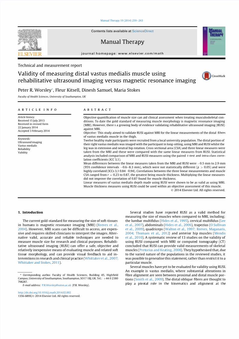
Fig. 3. Participant having an ultrasound scan taken of the right vastus medialis musclein long-sitting.
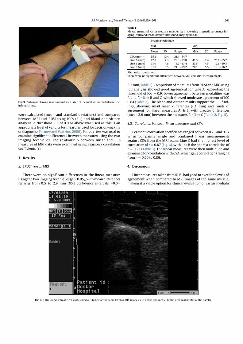
Fig. 4. Ultrasound scan of right vastus medialis taking at the same level as MRI images, just above and medial to the proximal border of the patella.
Table 1
Measurements of vastus medialis muscle size made using magnetic resonance im-aging (MRI) and rehabilitative ultrasound imaging (RUSI).
Imaging technique
MRI RUSI
Mean SD Range Mean SD Range
CSA (mm 2) 35.2 10.4 21.1 e 54.7 e e e
Line A (mm) 42.0 7.3 29.8 e 57.8 41.3 7.4 25.1 e 55.2Line B (mm) 23.0 4.6 15.2 e 33.4 23.5 4.5 17.5 e 30.3Line C (mm) 31.0 5.5 21.8 e 39.2 28.1 5.5 19.3 e 36.2
SD standard deviation.There were no signi cant differences between MRI and RUSI measurements.
P.R. Worsley et al. / Manual Therapy 19 (2014) 259 e 263 261

8/11/2019 2014 Validity of measuring distal vastus medialis muscle using rehabilitative ultrasound imaging versus magnetic r…
http://slidepdf.com/reader/full/2014-validity-of-measuring-distal-vastus-medialis-muscle-using-rehabilitative 4/5
muscle size. The CSA of the muscle belly assessed using MRI washighly correlated with the single linear measurement (line C) and acombination of linear measures.
Three previous studies have compared measures taken from USand MRI images of the quadriceps muscle group ( Walton, Roberts,1997; Reeves, Maganaris, 2004; Thomaes, Thomis, 2012 ). Directcomparison to the present study is limited for two of these studies(Walton, Roberts, 1997; Reeves, Maganaris, 2004 ), as theycompared CSA rather than linear measurements using a compoundscanning technique (Cavalieri technique). Walton et al (1997)
compared the agreement of CSA and volume measures takenfrom MRI and US images of the quadriceps femoris muscle group in10 healthy participants. They found no signi cant difference be-tween data sets, with a mean difference of 0.49 cm 2 between CSAmeasures with each imaging modality. Reeves et al. (2004)compared the agreement of CSA measures, taken from MRI andRUSI images of the vastus lateralis muscle in six healthy partici-pants. Their ICC ’ s ranged between 0.997 and 0.999 with a meanerror of 2.6%. They also showed very high correlation coef cientsbetween MRI and RUSI CSA measures ( r ¼ 0.99). This level of agreement was higher than that of our VM data, although thelandmark identi cation for vastus lateralis may have offered abetter opportunity for more accurate and repeatable measures.Finally, Thomaes et al. (2012) assessed rectus femoris CSA using
RUSI and CT in a cohort of patients with coronary artery disease.Reliability of measures was achieved (ICCs of 0.97) and the ICCcomputed between US and CT was 0.92 (95%CI: 0.81 e 0.97). Theabsolute difference between both techniques was 0.01 0.12 cm( p ¼ 0.66) resulting in a typical percentage error of 4.4%.
Other studies comparing MRI and RUSI measures of differentmuscles have shown good results but differing analytical tech-niques limits the comparison to the present study. The CSA of lumbar multi dus was compared using MRI and RUSI at vertebrallevels from L2 to S1 and no signi cant differences were found,despite different imaging positions ( Hides et al., 1995 ). Measures of cervical multi dus muscle from C4 to C6 were also shown to bevalid for thickness ( R2 ¼ 0.42 e 0.64) but not for CSA ( R2 ¼ 0.11 e0.39) or width ( R2 ¼ 0.16 e 0.69) ( Lee, Tseng, 2007 ). The small CSA of the muscle (approximately 1 cm 2) may have ampli ed errors, thusin uence the accuracy of measurements. Finally, the abdominalmuscles were examined at rest and contraction with good agree-ment found between RUSI and MRI for transversus abdominis andinternal oblique muscles (ICC ¼ 0.84 e 0.95) ( Hides et al., 2006 ).
The present study showed Line C was most highly correlatedwith VM CSA. However, this was the measurement that showed thelowest agreement between MRI and RUSI (ICC 0.84). Correlationvalues for lines A ( r ¼ 0.66) and B ( r ¼ 0.23) were too low to be of use clinically ( Kline, 1986 ) and their combinations with line C didnot increase the predictive value. The opposite was shown inlumbar multi dus, where the greatest correlations with CSA werefor combined linear measures ( r < 0.9) rather than any singlemeasure ( r ¼ 0.7 e 0.8) ( Hides et al., 1992 ). This difference betweenstudies may be explained by the contrasting shape between themuscles. Linear measures of muscle size could be a clinically usefuldimension when CSA is unobtainable. Although CSA of vastusmedialis can be estimated from a single linear measure, Line C, itrequires line A to be drawn (but not measured) in order to locate it.
Table 2
Comparison of linear measurements of vastus medialis from MRI and RUSI scans byintra-class correlation coef cients (ICC) and Bland and Altman analysis.
ICC Bland and Altman
ICCCoef cient
95%Con dencelimits
Meandifference(mm)
SD of differences(mm)
95% Limits of agreement(mm)
Line A 0.94 0.79 e 0.98 0.7 2.6 4.9 to 4.7Line B 0.84 0.53 e 0.95 0.5 2.6 6.2 to 3.1Line C 0.84 0.54 e 0.95 2.9 3.1 0.6 to 8.3
Fig. 5. Bland and Altman Plot showing difference between measurements of linearmeasurement C taken from MRI and ultrasound scans. The dashed line represents themean difference, solid lines are 95% upper and lower limits of agreement, representing
two standard deviations.
Fig. 6. Regression plot of cross-sectional area (CSA) against linear measurement C fromMRI images of the vastus medialis muscle ( r ¼ 0.87).
Table 3
Pearson correlation coef cients for each line measure and multiplication of linemeasures with the CSA. Measures were taken from the MRI images of the VMOmuscle.
Linear measure Correlation with CSA measure ( r ) P -value
A 0.66 0.019B 0.23 0.477C 0.87 0.001
A B 0.60 0.041A C 0.86 0.001B C 0.66 0.019
P.R. Worsley et al. / Manual Therapy 19 (2014) 259 e 263262

8/11/2019 2014 Validity of measuring distal vastus medialis muscle using rehabilitative ultrasound imaging versus magnetic r…
http://slidepdf.com/reader/full/2014-validity-of-measuring-distal-vastus-medialis-muscle-using-rehabilitative 5/5
It would be worth exploring other ways to measure VM musclethickness, so that only one line needs to be drawn, thus saving time.
4.1. Limitations
Only a small sample size was used for this validation study,which limited the statistical power of the results and ability forconclusion to be generalised across differing populations. However,it included more participants than most previous studies. A formalreliability study was not conducted to test the investigator ’ s RUSItechnique and obtaining measurements on different days. Theability to measure the same scans on different days was, however,shown to be highly reliable for both MRI and RUSI (all ICCs above0.90). Further examination of between-day and between observer(inter-rater) reliability is needed for VM, as has been demonstratedfor several other muscles ( Whittaker et al., 2007 ). Finally, since thelargest difference between modalities was found for line C, whichwas also found to have the best correlation with CSA, a moreclinically meaningful measurement of vastus medialis size could besought in future studies.
4.2. Recommendations
Linear measures of distal vastus medialis muscle can bemeasured using both MRI and RUSI techniques. A single linearmeasurement of VM thickness, line C, was highly correlated withCSA in the MRI images.
5. Conclusions
Linear measures made on RUSI scans were comparable to thoseon MRI scans, with small mean differences, indicating that RUSI is avalid technique for measuring vastus medialis muscle depth. Thestrong correlations found between a muscle thickness measure andCSA using MRI images, indicated that the linear measure have po-tential to estimate CSA. The validity of measuring muscle thicknessmight be improved by further research to nd an even more robustlinear measurement of vastus medialis in cross section.
References
Berry PA, Teichtahl AJ, Galevska-Dimitrovska A, Hanna F, Wluka AE, Wang YC, et al.Vastus medialis cross-sectional area is positively associated with patella carti-
lage and bone volumes in a pain-free community-based population. ArthritisRes Ther 2008;10:R143 .
Engelina S, Robertson C, Moggridge J, Killingback A, Adds P. Using ultrasound tomeasure the bre angle of vastus medialis oblique: a cardaveric validationstudy. Knee 2014;21:107 e 11.
Fagan V, Delahunt E. Patellofemoral pain syndrome: a review on the associatedneuromuscular de cits and current treatment options. Br J Sports Med2008;42:489 e 95 .
Hides J, Wilson SJ, Stanton WR, McMahon S, Keto H, McMahon K, et al. An MRIinvestigation into the function of the transversus abdominis muscle during
"drawing-in" of the abdominal wall. Spine 2006;31:E175 e 8.Hides JA, Cooper DH, Stokes MJ. Diagnostic ultrasound imaging for measurement of
the lumbar multi dus muscle in Normal Young adults. Physiotherapy TheoryPract 1992;8:19 e 26 .
Hides JA, Richardson C, Jull G. Magnetic resonance imaging and ultrasonography of the lumbar multi dus muscle: comparison of two different modalities. Spine1995;20:54 e 8.
Jan M-H, Lin D-H, Lin J-J, Lin C-HJ, Cheng C-K, Lin Y-F. Differences in sonographiccharacteristics of the vastus medialis obliquus between patients with patello-femoral pain syndrome and healthy adults. Am J Sports Med 2009;37:1743 e 9.
Kline PA. Handbook of test construction. London, UK: Methuen &Co Ltd; 1986 .Lankhorst NE, Bierm-Zeinstra SMA, van Middelkoop M. Risk factors for patellofe-
moral pain syndrome: a systematic review. J Orthop Sports Phys Ther 2012;42:81 e 94 .
Lee J-P, Tseng W-YI, Shau Y-W, Wang C-L, Wang H-K, Wang S-F. Measurement of segmental cervical multi dus contraction by ultrasonography in asymptomaticadults. Man Ther 2007;12:286 e 94 .
Lin Y-F, Lin J-J, Cheng C-K, Lin D-H, Jan M-H. Association between sonographicmorphology of vastus medialis obliquus and patellar alignment in patients withpatellofemoral pain syndrome. J Orthop Sports Phys Ther 2008;38:196 e 202 .
Mendis MD, Wilson SJ, Stanton WR, Hides J. Validity of real-time ultrasound im-aging to measure anterior hip muscle size: a comparison with magnetic reso-nance imaging. J Orthop Sports Phys Ther 2010;40:577 e 81.
O’ Sullivan C, Meaney J, Boyle G, Gormley J, Stokes M. The validity of rehabilitativeultrasound imaging for measurement of trapezius muscle thickness. Man Ther2009;14:572 e 8.
Portney L, Watkins M. Statistical measures of reliabilityIn Foundations of clinicialresearch: Applications to practice. 2nd ed. New Jersey: Prentice Hall; 2000.pp. 557 e 84 .
Pretorius A, Keating JL. Validity of real time ultrasound for measuring skeletalmuscle size. Phys Ther Rev 2008;13:415 e 26 .
Reeves N, Maganaris C, Narici M. Ultrasonographic assessment of human skeletalmuscle size. Eur J Appl Physiology 2004;91:116 e 8.
Smith TO, Nichols R, Harle D, Donell ST. Do the vastus medialis obliquus and vastusmedialis longus really exist? A systematic review. Clin Anat 2009;22:183 e 99 .
Thomaes T, Thomis M, Onkelinx S, Coudyzer W, Cornelissen V, Vanhees L. Reliabilityand validity of the ultrasound technique to measure the rectus femoris muscle
diameter in older CAD-patients. BMC Med Imaging 2012;12 .Walton JM, Roberts N, Whitehouse GH. Measurement of the quadriceps femorismuscle using magnetic resonance and ultrasound imaging. Br J Sports Med1997;31:59 e 64 .
Whittaker JL, Stokes M. Ultrasound imaging and muscle function (Review). J OrthopSports Phys Ther 2011;41:572 e 80 .
Whittaker VJ, Teyhen D, Elliot J, Cook K, Langevin H, Dahl H, et al. Rehabilitativeultrasound imaging: understanding the technology and its applications. J Orthop Sports Phys Ther 2007;37:434 e 49 .
P.R. Worsley et al. / Manual Therapy 19 (2014) 259 e 263 263