2015 data summary report - washington state … environmental radiation oversight program - 2015...
TRANSCRIPT

Hanford Environmental Radiation Oversight Program 2015 Data Summary Report
DOH 320-115 December 2016

Hanford Environmental Radiation Oversight Program 2015 Data Summary Report
December 2016
For more information or additional copies of this report, contact:
Environmental Sciences Section Office of Radiation Protection Washington State Department of Health 309 Bradley Blvd., Suite 201 Richland, WA 99357 509-946-0564FAX: 509-946-0876
If you need this publication in an alternate format, call 800-525-0127. For TTY/TDD call 800-833-6388
David B. Jansen, P.E., LEED AP Director, Office of Radiation Protection

Acknowledgements
Activities in the Hanford Environmental Radiation Oversight Program include sample schedule development, sample collection, radiochemical sample analyses, data entry, data quality assurance, laboratory reporting, contextual analysis of the data, writing and completing a technical review of the annual report, document preparation, database maintenance, and overall program management.
Office of Radiation Protection
David B. Jansen, Director, Office of Radiation Protection Mike Priddy, Manager, Environmental Sciences Section
Written and Prepared by:
Scott Van Verst, PhD
Contributors:
Office of Radiation Protection
Lynn Albin Megan Babcock Mike Brennan Eileen Kramer Scott McDonald Thomas Rogers Bob Ruben Scott Van Verst
Public Health Laboratory
Romesh Gautom, Director, Public Health Laboratories Blaine Rhodes, Director, Public Health Laboratories Environmental Laboratory Sciences Bud Taylor, Supervisor, Environmental and Radiation Sciences
Catherine Franklin Stephanie Haase Richard Hinderer Karin Kerr Paul Marbourg Josephine Pompey David Robbins Lynn Skidmore Hung Tran

Contents
Acronyms and Abbreviations ............................................................................................. 1
Background ......................................................................................................................... 2
Summary ............................................................................................................................. 2
1. Introduction ............................................................................................................. 4
2. The Hanford Environmental Radiation Oversight Program Description ............... 5
2.1 Laboratory Qualifications ........................................................................ 5 2.2 Interpretation of Results ........................................................................... 6 2.2.1 Uncertainty in Radioactivity Measurements ............................................ 6 2.2.2 Detection Limits....................................................................................... 7 2.2.3 Laboratory Background and Negative Results ........................................ 8 2.2.4 Techniques for Comparison of Health and Energy Contractor Data ....... 8 2.2.4.1 Qualitative Comparisons .......................................................................... 9 2.2.4.2 Regression Analysis and Scatter Plots ..................................................... 9 2.2.5 Comparison of Current Health Results to Historical Results ................ 10 2.2.6 Gamma Analysis .................................................................................... 10
3. Environmental Monitoring Results ....................................................................... 11
3.1 Ambient Air Monitoring ........................................................................ 12 3.1.1 Purpose and General Discussion ............................................................ 12 3.1.2 Sample Types and Monitoring Locations .............................................. 12 3.1.3 Monitoring Procedures........................................................................... 13 3.1.4 Comparison of Health and Energy Contractor Data .............................. 14 3.1.5 Other Discussion .................................................................................... 18 3.2 Groundwater, Riverbank Seep, and Surface Water Monitoring ............ 29 3.2.1 Purpose and General Discussion ............................................................ 29 3.2.2 Sample Types and Monitoring Locations .............................................. 29 3.2.3 Monitoring Procedures........................................................................... 31 3.2.4 Comparison of Health and Energy Contractor Data .............................. 32 3.2.5 Other Discussion .................................................................................... 34 3.3 External Radiation Monitoring .............................................................. 42 3.3.1 Purpose and General Discussion ............................................................ 42 3.3.2 Sample Types and Monitoring Locations .............................................. 42 3.3.3 Monitoring Procedures........................................................................... 43 3.3.4 Comparison of Health and Energy Contractor Data .............................. 43 3.3.5 Other Discussion .................................................................................... 44 3.4 Soil and Sediment Monitoring ............................................................... 50 3.4.1 Purpose and General Discussion ............................................................ 50 3.4.2 Sample Types and Monitoring Locations .............................................. 50 3.4.3 Monitoring Procedures........................................................................... 51 3.4.4 Comparison of Health and Energy Contractor Data .............................. 51

3.4.5 Other Discussion .................................................................................... 52 3.5 Biota Monitoring .................................................................................... 55 3.5.1 Purpose and General Discussion ............................................................ 55 3.5.2 Sample Types and Monitoring Locations .............................................. 55 3.5.3 Monitoring Procedures........................................................................... 56 3.5.4 Comparison of Health and Energy Contractor Data .............................. 57 3.5.5 Other Discussion .................................................................................... 58
4. Summary of Evaluation of Health and Energy Contractor Results ...................... 60
Appendix A - Radiation Tutorial ...................................................................................... 62 Appendix B - Laboratory a priori Lower Limits of Detection.......................................... 65 Appendix C - Glossary of Terms ...................................................................................... 73 Appendix D - List of Analytes .......................................................................................... 77

Hanford Environmental Radiation Oversight Program - 2015 Data Summary Report 1
Acronyms and Abbreviations
DOH Department of Health (Washington State) ERDF Environmental Restoration Disposal Facility LIGO Laser Interferometer Gravitational-wave Observatory LLD Lower Limit of Detection MAPEP Mixed Analyte Proficiency Evaluation Program MDA Minimum Detectable Activity MSA Mission Support Alliance OSL Optically Stimulated Luminescence PFP Plutonium Finishing Plant TLD Thermoluminescent Dosimeters

Hanford Environmental Radiation Oversight Program - 2015 Data Summary Report 2
Background
The Washington State Department of Health (Health) began monitoring environmental radiation in 1961. The focus of the early program was fallout from atmospheric testing of nuclear weapons. Health now monitors radiation at several state-licensed and federal facilities throughout the state, including the Hanford Site in eastern Washington. Health’s purpose is to provide oversight to the environmental monitoring programs run by these facilities. Environmental samples are first divided into two parts: one going to Health, the other to the facility (split sampling). Each program assesses their split sample and Health compares the two results.
In 1985, Health began working with the U.S. Department of Energy (Energy) to collect environmental samples in and around the Hanford Site. Health’s Hanford Environmental Radiation Oversight Program independently verifies the quality of Energy’s environmental monitoring programs at Hanford. The program assesses the potential for public health risk, and addresses public concerns about environmental radiation at Hanford. Health typically monitors air, groundwater, surface water, riverbank seep water, drinking water, sediment, food and farm products, fish and wildlife, vegetation, and radiation levels in the surrounding area.
Summary
In this report, Health uses the categories of good, fair, and poor to describe how closely radioactivity measurements by Health and Energy agree. These data are not expected to be in exact agreement because of the random nature of radioactive decay, the fact that split samples collected from the field are not identical, and analytical methods may differ between programs. Health investigates and reports all unexpected discrepancies in split sample results.
Sections 3 and 4 of the report discuss the analytical results. Many environmental samples analyzed by Health have radioactivity concentrations either below detection limits or consistent with naturally occurring (background) radiation. Some samples have concentrations elevated above background. In most cases, however, the concentrations are consistent with historical trends. Generally, there is good to fair agreement between analytical results from samples split between Health and Energy. The Hanford Environmental Radiation Oversight Program met the program objectives and made the following conclusions:
• Health independently evaluated and verified Energy’s monitoring program byconducting split sampling, and comparing the results. Health investigated anydifferences in results. The general good to fair agreement between the limitedsplit data provides confidence that the remainder of the Energy’s environmentaldata is valid.

Hanford Environmental Radiation Oversight Program - 2015 Data Summary Report 3
• Health’s oversight program finds Hanford-related radioactivity in theenvironment. However, Health’s assessment of the data shows that publicexposure to radioactivity from Hanford is far below regulatory limits.
• Health responds to any concerns the public has over radiation issues at Hanford.Health also participates in the Hanford Advisory Board, where the public canraise issues or express concerns. Health follows up on the issues raised in thisforum.

Hanford Environmental Radiation Oversight Program - 2015 Data Summary Report 4
1. Introduction
Chapter 70.98 of the Revised Code of Washington designates the Washington State Department of Health (Health) as the state agency with the responsibility to protect human health and the environment from the effects of ionizing radiation. To meet this legislative mandate, Health conducts radiological monitoring throughout the state, placing emphasis on major nuclear facilities with known or potential radiological impacts associated with the facility operations, decommissioning, or cleanup. This report summarizes environmental radiation sampling results from the Department of Health’s Hanford Environmental Radiation Oversight Program.
From 1943 until the mid-1980s, the primary mission of the U.S. Department of Energy’s (Energy) Hanford Site was the production of plutonium for nuclear weapons. Operations resulted in releases of radioactivity to the environment. Today, weapons production operations have ceased, and the current mission of the Site includes cleanup of radioactive waste originating from the plutonium production era. Energy has extensive monitoring programs to characterize and track this contamination as it moves through the environment. The primary purpose of Health’s Hanford Environmental Radiation Oversight Program is to provide oversight of Energy’s monitoring programs.
The primary objectives of the oversight program are:
• To independently verify the quality of the U.S. Department of Energy monitoringprograms at the Hanford Site by conducting split, collocated, and independentsampling at locations having the potential to release radionuclides to the environmentor locations which may be impacted by such releases.
• To independently assess impacts to the public, using Health’s oversight data, tocompare radionuclide concentrations in samples potentially impacted by Hanford withconcentrations in background samples. With the primary role of oversight, Health’smonitoring program is not intended to completely characterize environmentalradiation from the Hanford Site, nor is it intended to find and report the highestenvironmental contaminant concentrations.
• To address public concerns related to environmental radiation at Hanford.
This report presents the annual results of environmental radiation measurements made by the Washington State Department of Health’s Hanford Environmental Radiation Oversight Program.
Section 2 describes the Hanford Environmental Radiation Oversight Program, including a discussion of laboratory qualifications and how to interpret the results presented in this report. Environmental results are presented in Section 3. Tutorial information on radiation is found in Appendix A. The laboratory lower limits of detection are listed in Appendix B. Appendix C lists a glossary of radiation terms. Appendix D lists the full element names of the radionuclides discussed in this report.

Hanford Environmental Radiation Oversight Program - 2015 Data Summary Report 5
2. The Hanford Environmental Radiation Oversight Program Description
The objectives of the Oversight Program (see Section 1, Introduction) are met through collection and analysis of environmental samples and interpretation of results. Samples are either split or collocated with Energy contractors.
Split samples are prepared by dividing a sample into two parts. Collocated samples are those samples that are collected adjacent to the Energy contractor sample. In each case, Health’s sample is sent to the Washington State Public Health Laboratory in Shoreline, Washington for radiochemical analysis. Results of Health’s analyses are compared to the Energy contractor results to assess the quality of the federal monitoring program at the Hanford Site. In addition, the results are compared to historical data to identify trends, and are used to identify impacts to public health and the environment.
2.1 Laboratory Qualifications
Analytical techniques are based on laboratory standard operating procedures (Appendix B). The state laboratory serves as a regional reference laboratory and, as such, operates under a rigorous quality assurance program. This program contains quality control elements, which help ensure the laboratory's high analytical proficiency and accuracy. Laboratory quality control includes analysis of samples distributed by the federal government's quality assurance programs; split samples distributed on a smaller scale between cooperating federal, state, and private laboratories; and internal procedures related to the counting facilities and analytical techniques. Collectively, the state laboratory’s quality assurance program encompasses:
• Personnel requirements and qualifications• Quality control• Sample handling and custody requirements• Analytical methods• Equipment calibration and maintenance• Data reporting• Records management and archiving• Corrective action
In 2015, the laboratory participated in three intercomparison programs: 1) The Department of Energy's Mixed Analyte Performance Evaluation Program (MAPEP) tests the laboratory's ability to correctly analyze multiple radionuclides covering four matrices: soil, air filter, vegetation, and water. This is a National Institute of Standards and Technology traceable proficiency-testing program. 2) The FDA/USDA Food Emergency Response Network proficiency testing program tests the laboratory’s ability to correctly analyze for gamma emitters in foodstuffs. The samples provided meet the requirements for NIST traceability. This work was conducted under contract with the US Food and Drug Administration. 3)Lastly, the laboratory designed and conducted an exercise with performance testingmaterials provided by a certified reference laboratory for testing food under emergency

Hanford Environmental Radiation Oversight Program - 2015 Data Summary Report 6
conditions. This work was conducted under contract with the USDA-Food Safety Inspection Service.
These programs provide an independent check of laboratory proficiency for analyzing environmental samples. The laboratory quality assurance plan also includes analysis of standard reference samples as part of analysis of a batch of samples. Reference material is generally any environmental media containing known quantities of radioactive material in a solution or homogenous matrix.
2.2 Interpretation of Results
Environmental radiation data are reported as the number of radiation decays per unit time period per unit quantity of sample material. Most results are reported in units of picocuries. A picocurie equals 2.22 decays per minute. Airborne radioactivity is expressed as picocuries per cubic meter (pCi/m3); radioactivity in liquids such as water and milk is expressed as picocuries per liter (pCi/l); and radioactivity in solid material such as soil, vegetation, and food is expressed as picocuries per gram (pCi/g). Ambient gamma radiation is expressed as radiation exposure, measured in milliroentgens per day (mR/day). Radiation exposure is discussed in Appendix A, and the units used to quantify radioactivity and exposure are defined in Appendix C.
2.2.1 Uncertainty in Radioactivity Measurements
All radioactivity measurements (i.e., counting the number of decays per unit time) have an associated uncertainty, which originates from random and systematic effects. Counting uncertainty is the dominant source of laboratory random measurement uncertainty. It is an estimate of the possible range of radioactivity results because radioactive decay is a random process. If a sample was measured many times, each result would vary randomly around the mean of all measurements. Systematic uncertainty comes from the measurement process itself and is observed as a bias, or tendency, for the results to be higher or lower than the true value.
The uncertainties reported in this report are primarily counting uncertainties, although for gamma emitting radionuclides, the systematic uncertainty associated with calibrating the detector is included. A limited effort is made to estimate other sources of uncertainty, however, the laboratory does not attempt to completely identify and quantify all sources of uncertainty.
The uncertainties are reported as a 2-sigma (two-standard deviation) confidence interval. A 2-sigma uncertainty means there is 95-percent confidence that the true concentration in the sample lies somewhere between the measured concentration minus the uncertainty, and the measured concentration plus the uncertainty.

Hanford Environmental Radiation Oversight Program - 2015 Data Summary Report 7
2.2.2 Detection Limits
The laboratory is capable of measuring very small amounts of radioactivity in environmental samples, but there is a limit below which a sample’s radiation cannot be distinguished from background radiation. This limit is called the lower limit of detection and depends on several factors, including the sample size, analytical method, counting time, and background radiation. Appendix B lists the typical lower limits of detection that are achievable by the state laboratory.
For samples with very low radionuclide concentration, it is often difficult to determine if the radionuclide is actually detected. This also may lead to difficulty in comparing Health and Energy results. This situation often arises with semiannual air and soil/sediment samples.
When concentrations are very low, it is a challenge to compare Health and Energy results. 1) In some cases, both Health and Energy report concentrations below laboratory detectionlimits. In this situation, a comparison only determines if both parties agree that theconcentrations are too small to detect. 2) In other cases, concentrations are reported as“detected”; however, the concentrations are usually very small and similar in value to thedetection limit. In this situation, the comparison attempts to determine if one or both partiesdetects the contaminant. However, since Health and Energy contractor detection limits maydiffer, and since the concentrations are very near to the detection limit, it is often difficult todefinitively make this determination. 3) Finally, in yet other cases, concentrations are“definitively detected” above the detection limit. In this situation, the number of detectedresults is typically too small for a meaningful quantitative comparison by a statistical analysis.
Health intends to measure to the lowest concentration practical and minimize the error of reporting a non-detectable contaminant concentration when the contaminant is actually present. Detection limits are set low to ensure that measurements can verify protection of public health and the environment.
Health has traditionally used the measured concentration, uncertainty, and minimum detectable activity (MDA) values to determine if a contaminant is present. The MDA represents the balance point between the probability functions that describe the likelihood of false-detection and false-rejection; it is not the point above which calculated activity can be considered to be positively detected. During the last few years, consensus among the environmental radioactivity measurements community has been building to move away from the use of the MDA to determine whether an analyte has been detected, precisely because of this ambiguity.
One additional statistical term that applies to data interpretation where the results are at or very near the limit of detection, is the critical level. This key concept describes the minimum significant concentration that can be discriminated from the concentration observed for a blank sample, thus allowing a decision to be made that the radionuclide was detected or not. Health is revising data interpretation procedures to include evaluation of the critical level when samples are at the edge of detection capabilities, such as with plutonium in air composite samples.

Hanford Environmental Radiation Oversight Program - 2015 Data Summary Report 8
2.2.3 Laboratory Background and Negative Results
The environmental results are reported as net sample activity, which is defined as gross sample activity minus detector background activity. Gross sample activity and detector background activity are measured separately. Gross sample activity results from the sum of radioactivity in the environmental sample and the background radiation originating from sources outside of the sample. Background activity is measured by counting the radioactivity in a blank sample.
A negative net sample activity is occasionally reported for environmental samples. When the amount of radioactivity in the sample is very small, the random nature of radioactive decay may result in a gross sample activity that is less than the background activity. In this case, the net result will be negative. In most cases, negative results have an associated uncertainty range that includes zero activity. A negative result indicates that radioactivity in the sample was not detected at concentrations above the detection limit.
The net sample activity represents the best estimate of the true value of the sample activity. Therefore, to prevent biased reporting, Health reports the net sample activity even when the result is negative (as opposed to reporting a value of “zero” or “not detected”). The negative results are included in statistical analyses of data to look for systematic bias in laboratory procedures and to provide a more accurate measure of analytical detection limits.
2.2.4 Techniques for Comparison of Health and Energy Contractor Data
Since the primary purpose of the Department of Health Hanford Environmental Radiation Oversight Program is to verify the quality of Energy environmental monitoring programs, Health either splits samples or collects collocated samples with Energy contractors. Health and Energy samples are independently analyzed and the results compared. At the very least, qualitative data comparisons are made (see Section 2.2.4.1). When sufficient data are available, the analysis is supplemented by a quantitative linear regression analysis (see Section 2.2.4.2).
Currently, the oversight program uses a qualitative approach as the primary method to compare Health and Energy contractor data. Several arguments support this approach.
1) A goal of the oversight program is to validate as many different types ofenvironmental samples and test for as many different radioactive contaminants aspossible. Since the total number of samples is fixed by the budget, this goal oftenlimits the number of samples for any given type. There are often too few samples ortoo few detectable results of a given sample type for a rigorous quantitativeevaluation.
2) Samples are often collocated, not split, and the radioactivity results are not expectedto be identical because they represent distinctly different samples.

Hanford Environmental Radiation Oversight Program - 2015 Data Summary Report 9
3) For split samples, the non-homogeneous nature of environmental samples may resultin the two splits containing different amounts of radioactivity, and the results are notexpected to be identical.
4) The evaluation of uncertainty in Health and Energy contractor data is limited, whereasa rigorous quantitative approach requires a more complete characterization ofuncertainty.
2.2.4.1 Qualitative Comparisons
All of the collocated or split data are sorted by sample type and radionuclide. Then, for each sample type and radionuclide, all of the Health and Energy contractor data for each sample location are plotted on a graph and visually inspected to qualitatively assess the agreement of the data. In addition, graphs of historical data are inspected to ascertain temporal trends.
The qualitative agreement is categorized as either good, fair, or poor. Good agreement indicates that the uncertainty range (see Section 2.2.1) of the split or collocated concentrations overlaps for a majority of the samples. Fair agreement indicates that the split or collocated concentrations are similar, but the uncertainty range does not overlap for a significant number of samples. This is often indicative of a systematic bias in a laboratory procedure, and often shows up as the contractor and Health results differing by a consistent percentage. Poor agreement indicates that the uncertainty range of the split or collocated concentrations does not overlap for a majority of the samples, and there is no apparent systematic bias.
The results of regression analysis and visual inspection of scatter plots (discussed in Section 2.2.4.2 below) are assessed and incorporated into the qualitative assessment when appropriate.
The results of the assessment are discussed in the text of the report. Figures of the graphical representation of the data are included in the report to better explain the more complicated comparison data.
2.2.4.2 Regression Analysis and Scatter Plots
In addition to qualitative assessment, linear regression analysis is used to compare Health and Energy data when appropriate. In this report, regression analysis is carried out when: (a) there are a sufficient amount of data to analyze; (b) the data are consistently greater thanthe detection limit; and (c) the data are sufficiently correlated.
Assuming there is a sufficient amount of data above the detection limit for a meaningful regression analysis, each of the split or collocated Health and Energy results for a given sample type and radionuclide are formed into an (x, y) pair. The x-value represents the Health result and the y-value represents the Energy result for a particular sample. The paired data for all samples of a given sample type and radionuclide are plotted on a two-dimensional scatter plot. The correlation coefficient R is then calculated for the set of (x, y) pairs. R can vary from
Hanford Environmental Radiation Oversight Program - 2015 Data Summary Report 10
-1 to +1. A value near ±1 implies a strong correlation, while a value near 0 implies a weak ornon-correlation.
If the two data sets are sufficiently correlated (in this report, the criterion is R > 0.75), the best-fit straight line that describes the relationship between the two monitoring programs is determined. The parameters that describe the straight line are the slope and y-intercept. The functional form of the straight line is y = ax + b, where a is the slope and b is the y-intercept.
If the results between Health and Energy monitoring programs were in perfect agreement, the slope of the best-fit line would be 1, and the y-intercept would be 0. A zero value for the y-intercept means that if Health measures zero activity, then Energy also measures zero forthe same sample. A non-zero y-intercept indicates an overall offset between Health andEnergy results. The slope is simply the ratio of Health and Energy results.
If a regression analysis is carried out, a scatter plot (x, y paired data) of the Health and Energy split or collocated sample data may be presented in this report. Along with the data, these plots also show the straight line representing the ideal case where the data sets are in perfect agreement and the best-fit straight line. The slope and y-intercept of the best-fit straight line are shown in the plot legend.
If the two data sets are not sufficiently correlated (R < 0.75), it is not meaningful to find a best-fit straight line describing the relationship between the two data sets. In this case, the comparison is limited in this report to a qualitative assessment.
2.2.5 Comparison of Current Health Results to Historical Results
The range of Health concentrations for the current year is compared to the range of historical concentrations for the same analyte and sample type. If current year data are similar to historical results, then there are no anomalous data. If current year data differ from historical results, then there are anomalous data, and these data are discussed in the text.
2.2.6 Gamma Analysis
Concentrations of the gamma emitting radionuclides Co-60 and Cs-137 are reported, regardless of whether the concentrations are above or below a detection limit. Concentrations of other gamma emitting radionuclides are reported if they are detected.
Gamma spectroscopy is the method used to determine concentrations of Co-60 and Cs-137, and this method has the capability to measure concentrations of any other gamma emitting radionuclides. Health will report concentrations of all radionuclides found above detection limits in the gamma spectroscopy analysis. The absence of a reported concentration for a gamma emitting radionuclide indicates that it was not detected.
Other possible gamma emitting radionuclides at Hanford include, but are not limited to, Eu-152, Eu-154, Eu-155, Ru-106, and Sb-125.

Hanford Environmental Radiation Oversight Program - 2015 Data Summary Report 11
3. Environmental Monitoring Results
This section presents Health and Energy contractor results for the Hanford Environmental Radiation Oversight Program. The types of samples collected are intended to encompass all of the potential public exposure pathways. These samples include air (Section 3.1); groundwater, riverbank seep water, surface water, and drinking water (Section 3.2); dosimeters measuring external gamma radiation (Section 3.3); soil and sediment (Section 3.4); food and farm products, fish and wildlife, and vegetation (Section 3.5).
The sub-sections which follow discuss each of these sample types. Note that the figures for each sub-section are located at the end of the sub-section.

Hanford Environmental Radiation Oversight Program - 2015 Data Summary Report 12
3.1 Ambient Air Monitoring
3.1.1 Purpose and General Discussion
Atmospheric releases of radioactive material from the Hanford Site are a potential source of human exposure. Energy contractors monitor radioactivity in air to determine if the Hanford Site is contributing to airborne contamination. Health collects air samples that are collocated with samples collected by Energy contractors.
Sources of Hanford-specific airborne emissions include resuspension of contaminated soil (caused by wind or cleanup activities, for example) and escape of radioactive particulates and gasses from facilities and operations. Sources of natural airborne radioactivity include natural radon gas and its decay products; resuspension of soil containing natural radionuclides such as U-234, U-238, and K-40; and radioactive atoms such as Be-7 and H-3 (tritium) that aregenerated in the atmosphere by interactions with cosmic radiation. Other sources ofman-made airborne radioactivity include resuspension of fallout from historical atmospherictesting of nuclear weapons, including Sr-90, Cs-137 and Pu-239/240.
3.1.2 Sample Types and Monitoring Locations
Ambient air monitoring locations fall into two categories: (1) Near Facilities and Operations, and (2) Site-Wide and Offsite. For the Near Facilities program, most air samplers are located within 500 meters from, and in the prevailing downwind direction from sites having the potential for environmental releases. For the Site-Wide and Offsite program, samplers are located throughout the Hanford Site, along the Hanford perimeter, in nearby communities, and in distant communities. Mission Support Alliance (MSA) is the Energy contractor for both of these programs.
Health collected air samples collocated with the Near Facilities and Operations program at six locations, five of which are near facilities that have the potential to emit radionuclides to the atmosphere. These locations include the Liquid Effluent Retention Facility (N499 LERF), the Environmental Restoration Disposal Facility (ERDF-SE), and the Plutonium Finishing Plant
Major Findings:
• Health and Energy biweekly air concentrations are in fair agreement for gross alphaand gross beta activity. The concentrations are similar and follow the same trends overtime, but there is a systematic discrepancy between the Health and Energy data.
• Health and Energy semiannual composite air sample results are in fair agreement forPu-239/240, U-234, and U-238, and in good agreement for all other radionuclides.
• Most Health air concentrations are consistent with historical results. A few anomalousresults were found for Am-241, Pu-238, and U-238 concentrations in semiannual airsamples.
Hanford Environmental Radiation Oversight Program - 2015 Data Summary Report 13
(PFP-N165), all in the 200 Area; the 100K East Area near the fuel storage basins (100K N576); and a burial ground in the 600 Area (618-10 BG N548). The sixth collocated site, which is not near any facility, is at the Wye Barricade.
Historically, the site called 100K East Basin has been the air monitoring location at the 100K East Area. However, cleanup activities resulted in the dismantling of this equipment in January 2013. A new sampling site, located nearby and called 100K N576, became active in April 2013.
Health historically collected air samples at a site located near the 200 Area’s C Tank Farm; however, sampling at this site was discontinued at the end of 2014 and replaced in 2015 by the location at LERF.
Health collected air samples collocated with the Site-Wide and Offsite program at six locations. These locations include the 300 Area Water Intake, Wye Barricade, Prosser Barricade, and Yakima Barricade, which are located throughout the Hanford Site; Station 8, which is located across the Columbia River from the Hanford perimeter; and Battelle Complex, which is located in the nearby community of Richland. The Yakima Barricade is in the prevailing upwind direction of potential sources of airborne radioactivity. The Near Facilities and Operations program and the Site-Wide and Offsite program both use the results at Wye Barricade.
Health also independently collects biweekly air samples at the LIGO facility in the 600 Area. This sampling location is not collocated with Energy.
Figure 3.1.1 shows some of Health’s historical air sampling sites, indicating the general areas on the Hanford site targeted for sampling. Note that the map does not show all of the current sampling locations.
3.1.3 Monitoring Procedures
The air samplers work by continuously drawing air through a filter that traps airborne particulates. The filters are collected at each sample location every other week (biweekly), are stored for three days, and then analyzed for gross beta and gross alpha activity. The storage period allows naturally occurring short-lived radionuclides to decay that would otherwise obscure detection of radionuclides potentially present from Hanford Site emissions.
The amount of radioactive material collected on a filter in a two-week period is typically too small to accurately detect concentrations of individual radionuclides. In order to increase the sensitivity and accuracy, so that individual radionuclide concentrations can be determined, the biweekly filter samples for a three or six-month period are dissolved and combined into quarterly or semiannual composite samples.
Energy requested to discontinue the analysis of quarterly composite air samples because that time period is too short to accurately detect individual radionuclides. The semiannual

Hanford Environmental Radiation Oversight Program - 2015 Data Summary Report 14
composite samples are analyzed for gamma emitting radionuclides, and isotopes of strontium, americium, uranium, and plutonium. Note that the laboratories do not carry out analysis for all radionuclides at every sample location.
The Site-Wide and Offsite program also collects monthly atmospheric water vapor for tritium (H-3) analysis by continuously drawing air through samplers containing adsorbent silica gel. Collocated samples are collected from only two locations for this analysis, the 300 Water Intake and Battelle Complex. The collected water is distilled from the silica gel and analyzed for its tritium content.
3.1.4 Comparison of Health and Energy Contractor Data
Table 3.1.1 summarizes the comparison of Health and Energy data (see Section 2.2). The first columns in the table list the analytes assessed in the laboratory sample analyses and the sample collection period. Then, for each analyte, the table lists the number of results, the quality of agreement between the Health and Energy results (see Section 2.2.4.1), and the range of concentrations measured by Health. A concentration value prefaced by the “less than” symbol (<) indicates that the value is the detection limit and that some or all Health results are less than this value. Finally, the “Anomalous Data ?” column denotes whether any of the measured Health concentrations for the current year are anomalous compared to historical results (see Section 2.2.5).
In some cases, the number of scheduled results for a given analyte differs from the number of actual results reported. This situation typically occurs because either the Energy contractor or Health’s laboratory does not provide a scheduled result. When this occurs, the table lists the number of reported results, followed by the number of scheduled results in parentheses.
The text following the table discusses cases in which 1) the agreement between Health and Energy data is not good (i.e. is fair or poor), or 2) some of the Health data are anomalous compared to historical results.

Hanford Environmental Radiation Oversight Program - 2015 Data Summary Report 15
Analyte Collection Period
Number of Results
Quality of Agreement
Health’s Data Range (pCi/m3)
Anomalous Data ?
Gross Alpha biweekly 247 fair 0.0005 to 0.006 no Gross Beta biweekly 247 fair 0.003 to 0.07 no H-3(a) monthly 3 (24) 1.4 to 3.2 Am-241(b) semiannual 11 good < 0.00001 to 0.0007 yes Co-60(b) semiannual 38 good < 0.0007 no Cs-134(b) semiannual 38 good < 0.0005 no Cs-137(b) semiannual 38 good < 0.0003 no Eu-152(b) semiannual 38 good < 0.001 no Eu-154(b) semiannual 38 good < 0.001 no Eu-155(b) semiannual 38 good < 0.001 no Pu-238(b) semiannual 30 good < 0.00001 to 0.00004 yes Pu-239/240(b) semiannual 33 fair < 0.000005 to 0.003 no Pu-241(b) semiannual 5 good < 0.0004 no Sr-90(b) semiannual 30 good < 0.00006 to 0.0003 no U-234(b) semiannual 30 fair < 0.00002 to 0.00013 no U-235(b) semiannual 30 good < 0.00001 to 0.00002 no U-238(b) semiannual 30 fair < 0.00002 to 0.0014 yes
(a) Health did not provide some of the scheduled results.(b) The semiannual data correspond to samples collected in 2014 and 2015.
Table 3.1.1 Summary of Samples Collocated with Energy
The semiannual composite air samples collected in 2014 had not finished analysis at Health’s laboratory in time for the 2014 Annual Report. Those 2014 results are now complete and are included along with the current 2015 results in this report.
Health and Energy gross alpha concentrations are in fair agreement. Figure 3.1.2 shows the collocated data at Prosser Barricade. The concentrations are similar and follow the same temporal trend, but the concentrations reported by Energy are systematically less than those reported by Health. The data at some locations do not show the same level of disagreement. For example, the Health and Energy concentrations at the 618-10 Burial Ground, shown in Figure 3.1.3, do not show the same systematic bias.
Figure 3.1.4 shows the scatter plot for gross alpha concentrations at all monitoring locations. There is significant scatter about the theoretical line in which Health and Energy concentrations are identical, with differences up to a factor of two being common. Regression analysis indicates that, on average, the concentrations reported by Energy are approximately 65% of the values reported by Health. These data are similar to historical results.
Health and Energy gross beta concentrations are in fair agreement. Figure 3.1.5 shows the collocated data at Station 8. The concentrations are similar and follow the same temporal trend, but the concentrations reported by Energy are systematically greater than those reported

Hanford Environmental Radiation Oversight Program - 2015 Data Summary Report 16
by Health. The data at some locations do not show the same level of disagreement. For example, the Health and Energy concentrations at ERDF SE, shown in Figure 3.1.6, do not show the same systematic bias.
Figure 3.1.7 shows the scatter plot for gross beta concentrations at all monitoring locations. There is significant scatter about the theoretical line in which Health and Energy concentrations are identical, with differences up to a factor of two being common. Data analysis indicates that, on average, the concentrations reported by Energy are approximately 0.005 pCi/m3 greater than the values reported by Health. These data are similar to historical results.
Typically, Health and Energy collect 24 collocated atmospheric water vapor samples each year for tritium (H-3) analysis. Twelve samples are collected monthly from each of the 300 Water Intake and Battelle Complex locations. For 2015 however, only three collocated samples were collected from the 300 Water Intake site. Technical difficulties with the air samplers, along with site access issues, prevented collection of the full set of samples. Because most of Health’s data has not been reported, an analysis of the agreement between Health and Energy has not been conducted.
Historically, the agreement between Health and Energy tritium concentrations is poor. Figure 3.1.8 shows the scatter plot of historical data from all sites. As can be seen in the figures, Energy typically reports concentrations higher than those reported by Health.
Heath and Energy Am-241 concentrations in semiannual air samples are in good agreement. Both Health and Energy detected Am-241 in a few of the samples, at concentrations ranging from the detection limit of 0.00001 pCi/m3 to 0.0007 pCi/m3. The value of 0.0007 pCi/m3 measured by Health at the 618-10 Burial Ground is several times greater than Am-241 concentrations that are typically detected in air. Energy’s corresponding result for the collocated sample of 0.00009 pCi/m3 is the only result that does not agree with Health. Figure 3.1.9 shows the Am-241 data.
Most Health and Energy Co-60, Cs-134, Cs-137, Eu-152, Eu-154, and Eu-155 concentrations in semiannual air samples are in good agreement, and most of the concentrations are below detection limits. The single exception is a collocated sample from the 618-10 Burial Ground, where Health detected Cs-137 and Energy did not.
Historically, Cs-137 is occasionally detected in air samples. When it is detected, the concentrations reported by Energy are typically one-half the concentration value reported by Health. This can be seen in Figures 3.1.10 and 3.1.11, which show scatter plots for historical Cs-137 concentrations in air for lower concentration and higher concentration data, respectively.
Most Health and Energy Pu-238 concentrations in semiannual air samples are in good agreement, and most of the concentrations are below detection limits (see Figure 3.1.12). Plutonium-238, which was produced in historical reactor operations in small quantities and has a relatively short half-life of 88 years, is generally not detected in Hanford environmental

Hanford Environmental Radiation Oversight Program - 2015 Data Summary Report 17
air samples. However, Health occasionally detects Pu-238 at small concentrations just a few times greater than the detection limit. This is the case for two of the samples collected at the 618-10 Burial Ground. The highest concentration reported by Health is 0.00004 pCi/m3. Thecorresponding Energy result is a non-detect; however, Energy’s detection limit isapproximately 0.00004 pCi/m3 and therefore the Health and Energy results are considered inagreement.
The Health and Energy Pu-239 concentrations in semiannual air samples are in fair agreement. Both Health and Energy did not detect Pu-239/240 in the majority of air samples. However, Pu-239 was detected in several of the samples at the 618-10 Burial Ground, the Plutonium Finishing Plant, and the Environmental Restoration Disposal Facility. Figure 3.1.13 shows all the data from these locations, while Figure 3.1.14 shows the data for concentrations below 0.0005 pCi/m3. When Pu-239/240 is detected, Health systematically reports higher concentrations than Energy. Plutonium-239/240 is often detected at small concentrations in environmental samples, as it was produced from historical atmospheric testing of nuclear weapons, as well as from Hanford operations.
The Health and Energy Sr-90 concentrations in semiannual air samples are in good agreement. Figure 3.1.15 shows the results for locations where Sr-90 was detected. Both Health and Energy detect Sr-90, at small concentrations less than 0.0007 pCi/m3. Energy’s detection limit is larger than Health’s (see Figure 3.1.15), so there are a few instances where Health detects Sr-90 at concentrations less than Energy’s detection limits. Similar to Pu-239, Sr-90 is often detected at small concentrations in environmental samples, as it was produced from historical atmospheric testing of nuclear weapons, as well as from Hanford operations.
The Health and Energy U-235 concentrations are in good agreement, and most concentrations are below detection limits. Health detected U-235 at small concentrations in two samples at the 618-10 Burial Ground, with a maximum concentration of 0.00002 pCi/m3.
The U-234 and U-238 concentrations are in fair agreement. Figure 3.1.16 shows the U-234 results. The Health and Energy results follow the same trend, as seen in the figure, but in several cases, the results display a non-systematic disagreement. The U-238 data show a similar level of disagreement. Historically, there is a systematic discrepancy between Health’s and Energy’s isotopic uranium results, with Health typically reporting concentrations significantly greater than Energy, as can be seen in the scatter plot for Figure 3.1.17.
The isotopic uranium results for the 2nd half of 2015 at the 618-10 Burial Ground appear to be anomalous. Health’s U-234 result is 0.00013 pCi/m3, and Energy’s result is within reasonable agreement. This is a typical result for the higher concentrations observed in air samples. Typically for samples that contain natural uranium, the U-238 concentrations are similar to those reported for U-234. However, for this particular sample, the U-238 concentration measured by Health is 0.0014 pCi/m3, which is ten times greater than the U-234 result. Both Health and Energy report an anomalous U-234/U-238 ratio.
Hanford Environmental Radiation Oversight Program - 2015 Data Summary Report 18
3.1.5 Other Discussion
Radioactivity in air data shows a trend of higher concentration during the winter months, typically October through February. The gross beta data clearly show this trend. Higher concentrations are attributed to increased concentrations of radon decay products due to decreased atmospheric mixing during the winter months when there is decreased atmospheric heating. The annual cycle of increased gross beta activity in the winter months is seen in Figure 3.1.18, which shows gross beta activity at Wye Barricade for the last ten-year period.
In addition to the collocated samples, Health also independently collects biweekly air samples at the LIGO facility in the 600 Area. The gross alpha, gross beta, and isotopic uranium concentrations in 2015 at this site are consistent with historical data. All other radionuclides were not detected.

Hanford Environmental Radiation Oversight Program - 2015 Data Summary Report 19
Figure 3.1.1 Hanford Area Air Monitoring Locations

Hanford Environmental Radiation Oversight Program - 2015 Data Summary Report 20
Figure 3.1.2 Health and Energy Gross Alpha Concentrations in Air at Prosser Barricade
Figure 3.1.3 Health and Energy Gross Alpha Concentrations in Air at 618-10 Burial Ground

Hanford Environmental Radiation Oversight Program - 2015 Data Summary Report 21
Figure 3.1.4 Health and Energy Scatter Plot for Gross Alpha Concentrations in Air
Figure 3.1.5 Health and Energy Gross Beta Concentrations in Air at Station 8

Hanford Environmental Radiation Oversight Program - 2015 Data Summary Report 22
Figure 3.1.6 Health and Energy Gross Beta Concentrations in Air at ERDF SE
Figure 3.1.7 Health and Energy Scatter Plot for Gross Beta Concentrations in Air

Hanford Environmental Radiation Oversight Program - 2015 Data Summary Report 23
Figure 3.1.8 Health and Energy Scatter Plot for Historical H-3 Concentrations in Air
Figure 3.1.9 Health and Energy Am-241 Concentrations in Air, 2014 - 2015

Hanford Environmental Radiation Oversight Program - 2015 Data Summary Report 24
Figure 3.1.10 Health and Energy Scatter Plot for Historical Cs-137 Low Concentration Data in Air
Figure 3.1.11 Health and Energy Scatter Plot for Historical Cs-137 High Concentration Data in Air

Hanford Environmental Radiation Oversight Program - 2015 Data Summary Report 25
Figure 3.1.12 Health and Energy Pu-238 Concentrations in Air
Figure 3.1.13 Health and Energy Pu-239/240 Concentrations in Air

Hanford Environmental Radiation Oversight Program - 2015 Data Summary Report 26
Figure 3.1.14 Health and Energy Low Concentration Pu-239/240 Data in Air
Figure 3.1.15 Health and Energy Sr-90 Concentrations in Air

Hanford Environmental Radiation Oversight Program - 2015 Data Summary Report 27
Figure 3.1.16 Health and Energy U-234 Concentrations in Air
Figure 3.1.17 Health and Energy Scatter Plot for Historical U-238 Concentrations in Air

Hanford Environmental Radiation Oversight Program - 2015 Data Summary Report 28
Figure 3.1.18 Health’s Historical Gross Beta Concentrations in Air at Wye Barricade
Hanford Environmental Radiation Oversight Program - 2015 Data Summary Report 29
3.2 Groundwater, Riverbank Seep, Drinking Water and Surface Water Monitoring
3.2.1 Purpose and General Discussion
Operations at the Hanford Site have resulted in contaminated groundwater and surface water. Radioactive contaminants have leached from waste sites in the soil to groundwater beneath the Site, and then have migrated with groundwater towards the Columbia River. Groundwater may also enter the Columbia River through riverbank seeps.
Human exposure to contaminants can occur directly through ingestion of, or swimming in, contaminated water, or indirectly through ingestion of plants, animals, or fish that have been exposed to contaminated water. Radioactive contaminants are monitored by collecting samples from inland groundwater wells, riverbank seeps, and Columbia River water.
Health splits groundwater, surface water, riverbank seep water, and drinking water samples with various Energy contractors. Monitoring is carried out to track contaminant plumes and to evaluate impacts to the public and environment.
3.2.2 Sample Types and Monitoring Locations
Figure 3.2.1 shows some of Health’s historical water sampling sites, indicating the general areas on the Hanford site targeted for sampling. Note that the map does not show all of the current sampling locations. Locations may vary from year to year.
Major Findings:
• Health and Energy split water concentrations are in poor agreement for C-14; fairagreement for gross alpha, gross beta, and I-129; and good agreement for all otherreported radionuclides.
• Radionuclides were detected in groundwater near known groundwater plumes, and inriverbank seep water and Columbia River surface water near plumes known to beentering the Columbia River.
• Health detected C-14, Cs-137, H-3, I-129, Pu-238, Pu-239/240, Sr-90, Tc-99, andisotopes of uranium in some Hanford groundwater, seep water, or surface watersamples. Most concentrations are consistent with historical trends. In addition, Co-60was detected in a 200 Area groundwater well.
• Drinking water samples met federal standards.

Hanford Environmental Radiation Oversight Program - 2015 Data Summary Report 30
Groundwater
Health split 17 groundwater samples from 17 groundwater wells with the Energy contractor (CH2MHILL). Well locations are on the Hanford Site, either within contaminated plumes, near waste sites, or along the Columbia River shoreline.
Groundwater sampling is conducted in the 100, 200, 300, 400, and 600 Areas of the Hanford Site. The 100 Area consists of nine retired reactors and support facilities located along the Columbia River. Tritium (H-3), C-14, and Sr-90 are contaminants commonly found in groundwater beneath the reactor facilities. A primary objective of the groundwater collection in the 100 Area is to monitor contaminants that may enter the Columbia River. At the 100K Area, groundwater is sampled to evaluate potential changes in radioactivity as spent nuclear fuel, shield water, and sludge are removed from the 100K East Fuel Storage Basin.
The 200 Area consists of retired reactor fuel processing facilities located in the center of the Hanford Site on the central plateau. Common groundwater contaminants include H-3, I-129, Sr-90, Tc-99, and isotopes of uranium. A primary objective of groundwater collection in the 200 Area is to track radioactive plume movement and monitor potential leaks from waste storage tanks.
The 300 Area consists of retired reactor fuel fabrication facilities located adjacent to the Columbia River. Groundwater contains tritium originating from the 200 Area and uranium originating from past 300 Area fuel fabrication activities. A primary objective of the groundwater collection in the 300 Area is to monitor contaminants at the southern boundary of the Hanford Site, which is close to the City of Richland’s drinking water wells.
The 400 Area is the location of the Fast Flux Test Facility, a liquid sodium cooled test reactor that ceased operation in 1993. Tritium originating from the 200 Area is a common contaminant found in 400 Area groundwater. The primary objective of groundwater monitoring in this area is to assess impacts to the primary drinking water source for this part of Hanford. Note that the 400 Area is not shown on the map in Figure 3.2.1. It is located approximately four miles south and slightly west of the Columbia Generating Station (CGS).
The 600 Area includes all the land outside the operational areas of the Hanford Site (not specifically labeled on the map in Figure 3.2.1). The Old Hanford Town Site is within this region. Tritium originating from the 200 Area is a common contaminant found in 600 Area groundwater. The major objective of sampling 600 Area groundwater is to assess the nature and extent of radioactive plumes originating in the 200 Area that may be moving off-site.
Riverbank Seeps
Health and the Energy contractor (MSA) split six Columbia River riverbank seep samples. Groundwater enters the Columbia River through riverbank seeps. Split samples are collected from the historically predominant areas for discharge of riverbank seep water to the Columbia River, which include the 100 Area (three samples), the Old Hanford Town Site (one split sample), and the 300 Area (two split samples).

Hanford Environmental Radiation Oversight Program - 2015 Data Summary Report 31
Surface Water
Health and the Energy contractor (MSA) split six surface water samples from five different locations. Three samples were collected from the Columbia River upstream of Hanford, two from Priest Rapids Dam and one from Vernita Bridge. Two samples were collected from irrigation canals, one located across the Columbia River at Riverview and the other at the southern boundary of the Hanford Site at the Horn Rapids Yakima River irrigation pumping station. One sample was collected from the Richland Pumphouse, where water is pumped out of the Columbia River.
The Priest Rapids Dam and Vernita Bridge locations are upstream of the Hanford Site, while the remaining surface water sites are downstream of areas that may be impacted by Hanford. A comparison of contaminant concentrations at these sites gives an indication of Hanford’s impact on the Columbia River.
Drinking Water
Drinking water is supplied to Energy facilities on the Hanford Site by numerous water systems, most of which use water from the Columbia River. One of these systems, in the 400 Area at the Fast Flux Test Facility (FFTF), uses groundwater from the unconfined aquifer beneath the site. One composite drinking water sample, from a drinking water storage tank in the 400 Area, was split with the Energy contractor. One groundwater sample, from a well that is used to make up part of the drinking water used in the 400 Area, was also split with the Energy contractor. In addition to the split 400 Area samples, Health independently collected three drinking water samples, one from the LIGO Facility on the Hanford Site and two from the Edwin Markham elementary school in Pasco. Note that the drinking water sample locations are not shown on the map in Figure 3.2.1.
3.2.3 Monitoring Procedures
Groundwater
Energy contractors, who follow standard operating procedures that call for purging the well prior to sampling, collect the groundwater samples from the upper, unconfined aquifer. The samples are analyzed for those radionuclides that are most likely present in the area, based on previous sampling and review of radiological contaminants present nearby. Most samples are analyzed for gross alpha, gross beta, tritium, and gamma emitting radionuclides. Specific analyses for C-14, I-129, Sr-90, Tc-99, and isotopes of uranium and plutonium are added where appropriate.
Riverbank Seeps
Columbia River riverbank seep samples are collected when the river flow is lowest, typically in the fall. This ensures that riverbank seep water contains primarily groundwater, instead of Columbia River water stored in the riverbank during high flow rates. The seeps have a very

Hanford Environmental Radiation Oversight Program - 2015 Data Summary Report 32
small flow rate and are collected with the aid of a small pump. All seep samples are split in the field and analyzed as unfiltered samples. Most samples are analyzed for gross alpha, gross beta, gamma emitting radionuclides, and H-3. Specific analyses for Sr-90, Tc-99, and isotopes of uranium are added where appropriate.
Surface Water
Columbia River surface water is monitored by collecting samples at several points spanning the width of the river. This technique is known as transect sampling. Columbia River samples are also collected from near the Hanford shoreline at locations where known groundwater plumes are near the river. Finally, surface water samples are collected from irrigation pumping stations located at Horn Rapids (Yakima River water) and Riverview (Columbia River water).
Samples are split in the field and analyzed unfiltered. Most samples are analyzed for isotopes of uranium, H-3, and Sr-90. Analyses for gross alpha, gross beta, gamma emitting radionuclides, and Tc-99 are added where appropriate.
Drinking Water
Drinking water is monitored by sampling either tap water, water from storage tanks, or groundwater wells that supply drinking water. The samples are typically analyzed for gross alpha, gross beta, gamma emitting radionuclides, Sr-90, and H-3.
3.2.4 Comparison of Health and Energy Contractor Data
Table 3.2.1 summarizes the comparison of Health and Energy data (see Section 2.2). The first columns in the table list the analytes assessed in the laboratory sample analyses and the sample collection period. Then, for each analyte, the table lists the number of results, the quality of agreement between the Health and Energy results (see Section 2.2.4.1), and the range of concentrations measured by Health. A concentration value prefaced by the “less than” symbol (<) indicates that the value is the detection limit and that some or all Health results are less than this value. Finally, the “Anomalous Data ?” column denotes whether any of the measured Health concentrations for the current year are anomalous compared to historical results (see Section 2.2.5).
In some cases, the number of scheduled results for a given analyte differs from the number of actual results reported. This situation typically occurs because either the Energy contractor or Health’s laboratory does not provide a scheduled result. When this occurs, the table lists the number of reported results, followed by the number of scheduled results in parentheses.
The text following the table discusses cases in which 1) the agreement between Health and Energy data is not good (i.e. is fair or poor), or 2) some of the Health data are anomalous compared to historical results.

Hanford Environmental Radiation Oversight Program - 2015 Data Summary Report 33
Analyte Collection Period
Number of Results
Quality of Agreement
Health’s Data Range (pCi/l)
Anomalous Data ?
C-14(a) annual 6 (7) poor < 50 to 8,200 no Co-60 annual 7 good < 2 to 13 yes Cs-134 annual 7 good < 2 no Cs-137 annual 7 good < 2 to 840 no Eu-152 annual 8 good < 5 no Eu-154 annual 7 good < 5 no Eu-155 annual 7 good < 8 no Gross Alpha annual 21 fair <5 to 56,000 no Gross Beta annual 21 fair < 2 to 52,000 no H-3 annual 26 good < 75 to 450,000 no I-129(b) annual 8 fair < 1 to 9 no Pu-238(b) annual 7 good < 0.1 to 0.14 no Pu-239/240 annual 3 good 0.06 to 13 no Sr-90 annual 15 good < 1 to 10,000 no Tc-99 annual 12 good < 4 to 42,000 no U-234 annual 9 good 0.15 to 25,000 no U-235 annual 9 good < 0.06 to 1,300 no U-236(c) annual 0 < 0.05 to 480 no U-238 annual 9 good 0.13 to 26,000 no
(a) Health did not provide some of the scheduled results.(b) Data includes results from both 2014 and 2015.(c) Energy did not report U-236 results for any samples.
Table 3.2.1 Summary of Water Samples Split with Energy Contractors.
Health and Energy C-14 concentrations in water samples are in poor agreement. Of the six split results, two agree, while four results have a significant discrepancy, with Energy reporting higher concentrations than Health. Figure 3.2.2 shows all the C-14 split data. Because the large range of concentrations in Figure 3.2.2 obscures the results at lower concentrations, the lower concentration data are shown in Figure 3.2.3. Historically, C-14 results for water samples have been in poor agreement.
Health’s laboratory did not provide one of the scheduled C-14 results. In this case, the C-14 analysis is still underway due to difficulties with interference from low energy beta particles emitted by other radionuclides, and the result will not be available in time for this report.
Health and Energy Co-60 concentrations in water samples are in good agreement, and most data are below detection limits. Cobalt-60 is not typically detected in water samples. However, since 2013, both Health and Energy have detected Co-60 at groundwater well 299-E28-24 within Hanford’s 200 Area. Health and Energy have split a sample at this well since 2011 (historical results shown in Figure 3.2.4), and in the first two years, Co-60 was not detected. A Co-60 plume may be moving through the groundwater table near this well.

Hanford Environmental Radiation Oversight Program - 2015 Data Summary Report 34
Health and Energy concentrations for the gamma emitting radionuclides Cs-134, Cs-137, Eu-152, Eu-154, and Eu-155 are all in good agreement. Both Health and Energy commonly detect Cs-137 from groundwater wells at Hanford’s 200 Area, with concentrations ranging up to 1,000 pCi/L.
Health and Energy gross alpha concentrations in water samples are in fair agreement. Figure 3.2.5 shows the split gross alpha concentrations at all sites except for groundwater well 299-E33-344 (this well’s data was not included in the graph because the high concentration of56,000 pCi/L reported by Health and 52,000 pCi/L reported by Energy would have obscuredall the other data). Figure 3.2.6 shows the scatter plot for the same data. These graphs indicatethat Health typically reports higher concentrations than Energy. The regression analysisindicates that on average, Energy’s reported concentrations are approximately one-half thosereported by Health.
Health and Energy gross beta concentrations in water samples are in fair agreement. Because of the large range in concentration values, the high, mid, and low concentration data are shown separately in Figures 3.2.7, 3.2.8, and 3.2.9, respectively. In many cases the Health and Energy data are in good agreement, but in other cases Health reports significantly higher concentrations than Energy.
Health and Energy I-129 concentrations in water samples are in fair agreement. This year’s report includes results from both 2014 and 2015, as Health had not reported out the 2014 results at the time of last year’s report. The data are shown in Figure 3.2.10, where it can be seen that the split results are similar but there are up to 40% random relative differences (difference between Health and Energy concentration divided by the their average).
The majority of the Health and Energy U-234, U-235, and U-238 concentrations in water samples are in good agreement. However, the sample from groundwater well 299-E33-344 has significantly higher concentrations than any of the other water samples, and the Health and Energy results do not agree. Figure 3.2.11 shows the isotopic uranium concentrations at groundwater well 299-E33-344, where the Health and Energy results clearly do not agree.
Health reported U-236 in two riverbank seep samples and two groundwater samples. Energy did not report any corresponding results for U-236. Protocol calls for reporting U-236 anytime it is detected. Health’s U-236 results are discussed in Section 3.2.5.
3.2.5 Other Discussion
Isotopic uranium results are typically reported for U-234, U-235, and U-238. These isotopes occur in nature as well as in Hanford byproducts. Uranium-236 is an isotope that does not occur in nature, but rather is a byproduct of reactor operations. Detection of U-236 indicates a Hanford contaminant, rather than a naturally occurring radioactivity. Uranium-236 is occasionally detected in Columbia River sediments and in groundwater or river water samples.

Hanford Environmental Radiation Oversight Program - 2015 Data Summary Report 35
Health reported U-236 from two riverbank seep samples adjacent to the 300 Area (0.14 and 0.6 pCi/L) and from two groundwater wells in the 200 Area (0.14 [299-E28-24] and 480 [299-E33-344] pCi/L). Consistent with historical results, Health detected high concentrations of all uranium isotopes in groundwater well 299-E33-344.
Health analyzed drinking water samples from the 400 Area Drinking Water Tank and the LIGO Facility, both on the Hanford Site, and from the Edwin Markam elementary school in Pasco. Tritium (H-3) was detected in the 400 Area sample at 1,100 pCi/L. Gamma emitting radionuclides, tritium, gross alpha, and gross beta concentrations were below detection limits at the LIGO Facility. Health detected gross beta (14 pCi/L) and total uranium (1.3 pCi/L) at Edwin Markam. Gross alpha and tritium were not detected at Edwin Markam. The U.S. Environmental Protection Agency’s drinking water standards are 15 pCi/L for gross alpha, 50 pCi/L for gross beta, 20,000 pCi/L for H-3, and 21 pCi/L for total uranium.

Hanford Environmental Radiation Oversight Program - 2015 Data Summary Report 36
Figure 3.2.1 Historical Locations for Split Water Samples

Hanford Environmental Radiation Oversight Program - 2015 Data Summary Report 37
Figure 3.2.2 Health and Energy C-14 Concentrations in Water Samples
Figure 3.2.3 Health and Energy Low Concentration C-14 Results in Water Samples

Hanford Environmental Radiation Oversight Program - 2015 Data Summary Report 38
Figure 3.2.4 Health and Energy Co-60 Concentrations at Groundwater Well 299-E28-24
Figure 3.2.5 Health and Energy Gross Alpha Concentrations in Water Samples

Hanford Environmental Radiation Oversight Program - 2015 Data Summary Report 39
Figure 3.2.6 Health and Energy Scatter Plot for Gross Alpha Concentrations in Water
Figure 3.2.7 Health and Energy High Concentration Gross Beta Concentrations in Water

Hanford Environmental Radiation Oversight Program - 2015 Data Summary Report 40
Figure 3.2.8 Health and Energy Mid Concentration Gross Beta Concentrations in Water
Figure 3.2.9 Health and Energy Low Concentration Gross Beta Concentrations in Water

Hanford Environmental Radiation Oversight Program - 2015 Data Summary Report 41
Figure 3.2.10 Health and Energy I-129 Concentrations in Water Samples
Figure 3.2.11 Health and Energy Isotopic Uranium Concentrations at 299-E33-344

Hanford Environmental Radiation Oversight Program - 2015 Data Summary Report 42
3.3 External Radiation Monitoring 3.3.1 Purpose and General Discussion It is possible to receive radiation exposure from a radioactive source outside the body at a distance. External radiation is the name of radiation emitted from a source external to the human body or other living organisms. This radiation travels through space and may interact with a living organism, resulting in radiation exposure. Sources of background external radiation include natural cosmic and terrestrial radiation, as well as fallout from historical atmospheric testing of nuclear weapons. Contamination from the Hanford Site may contribute to man-made sources of external radiation. In addition to oversight of Energy’s monitoring program, Health compares on-site and off-site radiation rates to determine if Hanford impacts workers or the public. External radiation levels can vary by up to 25 percent over the course of a year at any one location. This variation is primarily due to changes in soil moisture and snow cover, both of which affect shielding of natural radiation from the earth’s crust. Health has historically maintained external radiation monitoring sites collocated with Energy. In 2006, Energy terminated its Site-Wide and Offsite external radiation monitoring program. In response, Health added 26 new monitoring sites along the Columbia River to independently monitor locations that were previously monitored by Energy. In addition, Health will continue to maintain its original monitoring sites that were collocated with Energy. Therefore, from 2006 forward, this report will cover the sites collocated with Energy’s Near-Facilities and Operations program, as well as the sites operated independently by Health. 3.3.2 Sample Types and Monitoring Locations Historically, Health has used thermoluminescent dosimeters (TLDs) to measure external radiation. Starting in 2012, Health switched to using optically stimulated luminescence dosimeters (OSLs), while Energy continues to use TLDs. Both OSLs and TLDs, referred to as dosimeters, measure the time-integrated exposure to external radiation at their location.
Major Findings:
• Health and Energy external radiation exposure rates are in good agreement. • Exposure rates on the Hanford Site are consistent with historical results, and are
similar to rates at locations along the Hanford perimeter and offsite locations. • Exposure rates along the Columbia River are consistent with background.

Hanford Environmental Radiation Oversight Program - 2015 Data Summary Report 43
Health operates 48 external radiation monitoring sites that are relevant to the Hanford Site. Health’s Hanford Environmental Radiation Oversight Program operates 39 of these sites, in which dosimeters from five sites are collocated with Energy’s Near-Facilities and Operations program currently run by Mission Support Alliance (MSA), and 34 sites are independently monitored by Health. The remaining nine sites are part of Health’s Columbia Generating Station Oversight Program, and they are included in this report because the sites are located along the Hanford perimeter.
Figure 3.3.1 shows Health’s external radiation monitoring locations. Eight of the sites are near Hanford facilities with known, suspected, or potential radiation sources. Three sites (Yakima and Wye Barricades, and LIGO Facility) are located on the Hanford Site, but away from radiation sources. Twenty-five sites are along the Columbia River shoreline from the Vernita Bridge to downstream of Bateman Island at the mouth of the Yakima River. Nine sites are located around the Hanford Site perimeter. The remaining three sites (Othello, Yakima Airport, and Benton County Shops) are significantly distant from the Hanford Site. Many of these dosimeter sites are collocated with the air monitoring sites discussed in Section 3.1.
3.3.3 Monitoring Procedures
Most collocated dosimeters are deployed on a quarterly basis at each monitoring location, with the dosimeters retrieved at the end of each calendar quarter. Columbia River dosimeters are deployed semi-annually. Starting in 2012, Health sends its dosimeters to a contracted laboratory (Landauer); while prior to 2012, Health sent its dosimeters to Health’s Public Health Laboratory. In both cases, the time-integrated external radiation exposure is determined for the deployment period. The results are converted to an average daily radiation rate reported in units of milliroentgen per day (mR/day). At the same time the dosimeters are retrieved, new dosimeters are placed at each site.
3.3.4 Comparison of Health and Energy Contractor Data
Table 3.3.1 summarizes the comparison of Health and Energy data (see Section 2.2). The first columns in the table list the analyte assessed in the laboratory sample analysis and the sample collection period. Then the table lists the number of results, the quality of agreement between the Health and Energy results (see Section 2.2.4.1), and the range of concentrations measured by Health. Finally, the “Anomalous Data ?” column denotes whether any of the measured Health concentrations for the current year are anomalous compared to historical results (see Section 2.2.5).
Analyte Collection Period
Number of Results
Quality of Agreement
Health’s Data Range (mR/day)
Anomalous Data ?
External Rad quarterly 20 good 0.18 to 0.30 no
Table 3.3.1 Summary of External Radiation Dosimeters Collocated with MSA

Hanford Environmental Radiation Oversight Program - 2015 Data Summary Report 44
Historically, the agreement between Health and Energy external radiation rates has been fair. The Energy contractors have systematically reported slightly higher exposure rates (approximately 10 percent averaged over all data) than Health.
For 2015, however, the Health and Energy quarterly collocated external radiation rate data are in good agreement. Figure 3.3.2 shows the collocated data for dosimeters collected in 2015. At each location, the graph first shows the fourth quarter data from 2014, followed by the first, second, and third quarter data for 2015. The prior year’s fourth quarter results are included because the collection date for these dosimeters was in early January of 2015.
Figure 3.3.3 shows the scatter plot for the Health/Energy collocated external radiation rate data. All of the data are closely scattered about the line where Health and Energy results are theoretically equal, indicating good agreement.
3.3.5 Other Discussion
In addition to the five sites collocated with the Energy contractor discussed above, Health independently monitors 34 sites, and monitors nine sites collocated with the Columbia Generating Station that are associated with the Hanford Site. Table 3.3.2 summarizes the data from these additional 43 sites.
The table lists the analyte, the collection period, and the number of samples. The table also lists the range of external radiation rates measured by Health. Finally, the “Anomalous Data ?” column denotes whether any of the Health exposure rates for the current year are anomalous compared to historical results (see Section 2.2.5). The exposure rates reported by Health are consistent with historical results, and Health did not encounter anomalous data.
Analyte Collection Period
Number of Results
Health’s Data Range (mR/day)
Anomalous Data ?
External Rad Quarterly / Semiannual 122 0.18 to 0.3 no
Table 3.3.2 Summary of Independent Department of Health External Radiation Dosimeters
Health categorizes its external radiation monitoring sites by their location type, as described in Section 3.3.2. Figure 3.3.4 shows the average, minimum, and maximum radiation rates for all of the sites in each location category. This graph includes data from the 43 sites discussed in this section and the five sites collocated with Energy discussed in Section 3.3.4 (all 48 Health sites). As can be seen, the average radiation rates are similar for all location categories, except for the distant sites where the average is lower. The maximum radiation rates are slightly higher for the sites that are onsite and near to contaminated or operational facilities, or along the Hanford perimeter.

Hanford Environmental Radiation Oversight Program - 2015 Data Summary Report 45
The average radiation rates are slightly lower at sites distant from Hanford, most likely because these sites are located in areas covered by concrete, which has a greater shielding factor than the soil cover for most other sites.
Historically, external radiation rates were elevated compared to background at site 100N Spring, which is within Hanford’s 100N Area. The exposure rate at this site has steadily been decreasing with time, due to the natural decay of Co-60 surface contamination. With the recent cleanup of contaminated surface soil, exposure rates over the past several years now are constant, and are consistent with exposure rates from locations away from contaminated areas. Historical radiation rates at 100N Spring are shown in Figure 3.3.5.
External radiation rates were elevated compared to background at the 100K East Basin from 2005 through 2011. This site is near a fuel storage basin within Hanford’s 100K East Area. Radioactive material had been temporarily stored outside of the facility, resulting in increased radiation rates. In addition, cleanup activities during that time resulted in temporary increased radiation rates. The storage area was properly posted and access restricted. Since 2011, radiation rates have returned to pre-2005 values. Measurements along the Columbia River at the 100K Area (site location 100K Boat Ramp), the closest public access point, did not indicate elevated exposure rates. Historical radiation rates at 100K East Basin are shown in Figure 3.3.6.
Figures 3.3.5 and 3.3.6 show a slight increase in radiation rates going from the year 2011 to 2012, and then a slight decrease going from 2012 to 2013. As discussed in Sections 3.3.2 and 3.3.3, in the year 2012 Health switched dosimeter types from TLD to OSL and switched the dosimeter analysis laboratory from Health’s Public Health Laboratory to the outside contract laboratory Landauer. Health observed a slight increase in radiation rates at all of their monitoring locations in 2012.

Hanford Environmental Radiation Oversight Program - 2015 Data Summary Report 46
Figure 3.3.1 DOH External Radiation Monitoring (TLD) Locations

Hanford Environmental Radiation Oversight Program - 2015 Data Summary Report 47
Figure 3.3.2 Health and Energy Quarterly External Radiation
Figure 3.3.3 Health and Energy Scatter Plot for Quarterly External Radiation

Hanford Environmental Radiation Oversight Program - 2015 Data Summary Report 48
Figure 3.3.4 External Radiation Rate Statistics by Location Type
Figure 3.3.5 Historical External Radiation Rates at 100N Spring

Hanford Environmental Radiation Oversight Program - 2015 Data Summary Report 49
Figure 3.3.6 Historical External Radiation Rates at 100K East Basin

Hanford Environmental Radiation Oversight Program - 2015 Data Summary Report 50
3.4 Soil and Sediment Monitoring
3.4.1 Purpose and General Discussion
Contaminated soil and river sediments are a potential source of radiation exposure for people and biota in the environment. Human exposure may result from direct exposure to contaminated soil/sediment, ingestion of contaminated soil/sediment, ingestion of water contaminated by sediment resuspension, inhalation of contaminants resuspended in air, or ingestion of fish, animals, plants, or farm products exposed to contaminated soil and sediments.
Radionuclides in soil and sediment originate from many sources, including natural terrestrial sources, atmospheric fallout from nuclear weapons tests, and contaminated liquid and gaseous effluents. In addition, contaminants can reach Columbia River sediments from erosion of contaminated soil and flow of contaminated groundwater. Cesium-137, Sr-90, and plutonium isotopes are radionuclides consistently seen in soil or sediments because they exist in worldwide fallout, as well as potentially in effluents from the Hanford Site. Uranium isotopes, also consistently seen in soil and sediment, occur naturally in the environment in addition to being present from Hanford operations.
3.4.2 Sample Types and Monitoring Locations
Health and Energy (contractor MSA) split five sediment samples from the Columbia River. Two sediment samples were collected upriver from Hanford at Priest Rapids Dam, one along the Hanford Site perimeter at the White Bluff Slough, and two downriver from Hanford at McNary Dam. Figure 3.4.1 shows these locations, along with other historical sediment sample sites.
Priest Rapids Dam, being upstream from Hanford, is a background location. McNary Dam is the first dam downstream from Hanford, and therefore should have the highest radionuclide concentrations. Sediment locations within the Hanford boundary change from year to year. The locations are chosen to monitor areas where contaminants may be discharged into the river, areas where deposits could accumulate, or areas where the public may gain access to the shoreline.
Major Findings:
• Health and Energy sediment data are in good agreement.• Most radionuclide concentrations either are below detection limits or are consistent
with expected historical values.

Hanford Environmental Radiation Oversight Program - 2015 Data Summary Report 51
Health and Energy also split three soil samples from locations off the Hanford Site near the towns of George, Othello, and Wanapum. These locations, although considered background sites, may receive Hanford related contaminants through atmospheric transport.
3.4.3 Monitoring Procedures
Soil samples are collected by compositing four one-square foot areas, each excavated to a depth of one inch. The composited samples are split, and then dried prior to radiochemical analysis. Samples are analyzed for radionuclides that are most likely present in the area sampled, which at Hanford typically include gamma emitting radionuclides, Sr-90, isotopic uranium, and isotopic plutonium.
Sediment samples represent surface sediments and are collected with either a clam-shell style sediment dredge or, in the case of shoreline sediments, a plastic spoon. The samples are split, and then dried prior to radiochemical analysis. Samples are analyzed for gamma emitting radionuclides, strontium-90, isotopic uranium, and isotopic plutonium.
Radiochemical analysis methods for soil and sediment are identical. Soil and sediment concentrations are reported in units of pCi/g dry weight.
3.4.4 Comparison of Health and Energy Contractor Data
Table 3.4.1 summarizes the comparison of Health and Energy data (see Section 2.2). The first columns in the table list the analytes assessed in the laboratory sample analyses and the sample collection period. Then, for each analyte, the table lists the number of results, the quality of agreement between the Health and Energy results (see Section 2.2.4.1), and the range of concentrations measured by Health. A concentration value prefaced by the “less than” symbol (<) indicates that the value is the detection limit and that some or all Health results are less than this value. Finally, the “Anomalous Data ?” column denotes whether any of the measured Health concentrations for the current year are anomalous compared to historical results (see Section 2.2.5).
The text following the table discusses cases in which 1) the agreement between Health and Energy data is not good (i.e. is fair or poor), or 2) some of the Health data are anomalous compared to historical results.
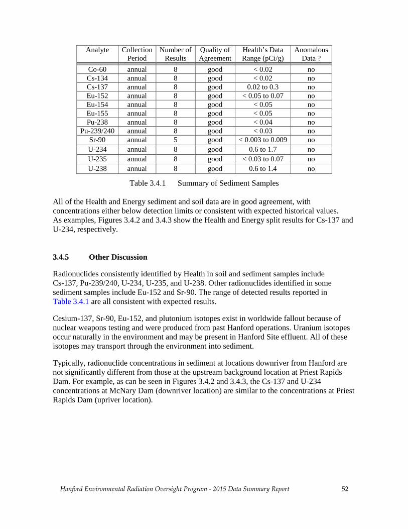
Hanford Environmental Radiation Oversight Program - 2015 Data Summary Report 52
Analyte Collection Period
Number of Results
Quality of Agreement
Health’s Data Range (pCi/g)
Anomalous Data ?
Co-60 annual 8 good < 0.02 no Cs-134 annual 8 good < 0.02 no Cs-137 annual 8 good 0.02 to 0.3 no Eu-152 annual 8 good < 0.05 to 0.07 no Eu-154 annual 8 good < 0.05 no Eu-155 annual 8 good < 0.05 no Pu-238 annual 8 good < 0.04 no
Pu-239/240 annual 8 good < 0.03 no Sr-90 annual 5 good < 0.003 to 0.009 no U-234 annual 8 good 0.6 to 1.7 no U-235 annual 8 good < 0.03 to 0.07 no U-238 annual 8 good 0.6 to 1.4 no
Table 3.4.1 Summary of Sediment Samples
All of the Health and Energy sediment and soil data are in good agreement, with concentrations either below detection limits or consistent with expected historical values. As examples, Figures 3.4.2 and 3.4.3 show the Health and Energy split results for Cs-137 and U-234, respectively.
3.4.5 Other Discussion
Radionuclides consistently identified by Health in soil and sediment samples include Cs-137, Pu-239/240, U-234, U-235, and U-238. Other radionuclides identified in some sediment samples include Eu-152 and Sr-90. The range of detected results reported in Table 3.4.1 are all consistent with expected results.
Cesium-137, Sr-90, Eu-152, and plutonium isotopes exist in worldwide fallout because of nuclear weapons testing and were produced from past Hanford operations. Uranium isotopes occur naturally in the environment and may be present in Hanford Site effluent. All of these isotopes may transport through the environment into sediment.
Typically, radionuclide concentrations in sediment at locations downriver from Hanford are not significantly different from those at the upstream background location at Priest Rapids Dam. For example, as can be seen in Figures 3.4.2 and 3.4.3, the Cs-137 and U-234 concentrations at McNary Dam (downriver location) are similar to the concentrations at Priest Rapids Dam (upriver location).

Hanford Environmental Radiation Oversight Program - 2015 Data Summary Report 53
Figure 3.4.1 Typical Sediment Monitoring Locations

Hanford Environmental Radiation Oversight Program - 2015 Data Summary Report 54
Figure 3.4.2 Health and Energy Cs-137 Concentrations in Sediment and Soil
Figure 3.4.3 Health and Energy U-234 Concentrations in Sediment and Soil

Hanford Environmental Radiation Oversight Program - 2015 Data Summary Report 55
3.5 Biota Monitoring 3.5.1 Purpose and General Discussion Health and Energy contractors monitor farm products, fish and wildlife, and vegetation to determine if contamination has migrated into the food chain, potentially exposing people or other biota. Farm product sampling includes food, milk, and wine. Sample locations include farms near the Hanford Reservation. Contaminants in farm products may arise from deposition of contaminated air and irrigation with or consumption of contaminated Columbia River water. Fish and wildlife sampling includes fish, shellfish, small and large mammals, and game birds. Sample locations include the Hanford Site, adjacent to the Hanford boundary, and nearby to the Hanford Site. Contaminants in fish may arise from exposure to contaminated water, sediment, and aquatic biota. Contaminants in wildlife may arise from ingestion of contaminated soil, vegetation, and water. Vegetation sampling includes various grass, brush, and leaves and twigs from tress. Sample locations include the Hanford Site, adjacent to Hanford along the Columbia River, and nearby to the Hanford Site. Contaminants in vegetation may arise from airborne deposition, soil to plant transfer, and water to plant transfer. Strontium-90 and isotopes of uranium are often detected in biota samples. In addition to the possibility that these radionuclides originate from Hanford-related contamination, Sr-90 is a product of fallout from atmospheric weapons testing, and uranium exists naturally in soil. 3.5.2 Sample Types and Monitoring Locations For this year’s oversight program, farm products include one apple sample, two cherry samples, and three samples each of corn, leafy vegetables, melon, potato, red wine, and white wine. All farm products are collected from farms which are nearby, but off-site of the Hanford Reservation. These farms are generally located in the areas of Riverview, Sagemoor, Horn Rapids, East Wahluke, Ringold, Mattawa, Sunnyside, Yakima Valley, and the Columbia Valley.
Major Findings:
• All Health and Energy radionuclide concentrations are in good agreement. • Most Health radionuclide concentrations are below detection limits. Strontium-90 and
isotopes of uranium were detected in a few biota samples, with concentrations consistent with the range of concentrations typically detected in these media.

Hanford Environmental Radiation Oversight Program - 2015 Data Summary Report 56
Fish and wildlife samples include one whitefish carcass sample from the Columbia River collected near the 100 Area, one whitefish meat sample collected upriver of the Hanford Site near Priest Rapids Dam, and a rabbit sample from the 300 Area. Vegetation samples include one alfalfa sample from a farm in Riverview, one hay sample from a farm in Horn Rapids, and two rabbit brush samples from the towns of George and Wanapum. 3.5.3 Monitoring Procedures Farm Products The Energy contractor (currently MSA) collects farm product samples and then splits the samples with Health. Energy collects the samples once a year, typically in the fall at harvest. Fruit and vegetable samples are analyzed for Sr-90 and gamma emitting radionuclides, which include Co-60, Cs-134, Cs-137, Eu-152, Eu-154, Eu-155, Ru-106, and Sb-125. Concentration units are pCi/g (wet weight). Some samples are also analyzed for C-14. Wine is analyzed for gamma emitting radionuclides and tritium (H-3). Concentration units are pCi/L. Fish and Wildlife For fish sampling, the Energy contractor (currently MSA) collects multiple samples at each location, one or more of which are analyzed by the contractor, and one analyzed by Health. As such, fish results are from collocated samples, as opposed to split samples of the same fish. Since there is no control over the life history of the collocated fish, including their exposure to contaminants, differences in Health and Energy results are expected. The Energy contractor collects most wildlife samples. Some of the samples are split with Health. In other cases, multiple collocated samples are collected, with Health taking one of the samples and the Energy contractor taking one or more of the remaining samples. Health occasionally collects deer or elk from western Washington for background samples, although none were collected for this reporting period. Carcass and bone samples are analyzed for Sr-90, as strontium accumulates in the bone, not the meat. Liver samples, when collected, are analyzed for isotopes of plutonium, as plutonium accumulates in the liver. Meat samples are analyzed for gamma emitting radionuclides, which include Co-60, Cs-134, Cs-137, Eu-152, Eu-154, Eu-155, Ru-106, and Sb-125. Concentration units are pCi/g (dry weight).

Hanford Environmental Radiation Oversight Program - 2015 Data Summary Report 57
Vegetation
The Energy contractor (currently MSA) collects vegetation samples and then splits the samples with Health. Energy typically collects the samples in the spring, when the plants are starting to grow and have a high probability to absorb contaminants.
Vegetation samples are analyzed for Sr-90 and gamma emitting radionuclides, which include Co-60, Cs-134, Cs-137, Eu-152, Eu-154, Eu-155, Ru-106, and Sb-125. Some samples are also analyzed for isotopes of uranium and C-14. Concentration units are pCi/g (dry weight).
3.5.4 Comparison of Health and Energy Contractor Data
Tables 3.5.1 and 3.5.2 summarize the comparison of Health and Energy data (see Section 2.2). The first columns list the analytes assessed in the laboratory sample analyses and the sample collection period. Then, for each analyte, the tables list the number of results, the quality of agreement between the Health and Energy results (see Section 2.2.4.1), and the range of concentrations measured by Health. A concentration value prefaced by the “less than” symbol (<) indicates that the value is the detection limit and that some or all Health results are less than this value. Finally, the “Anomalous Data ?” column denotes whether any of the measured Health concentrations for the current year are anomalous compared to historical results (see Section 2.2.5).
The text following the tables discusses cases in which 1) the agreement between Health and Energy data is not good (i.e. is fair or poor), or 2) some of the Health data are anomalous compared to historical results.
Analyte Collection Period
Number of Results
Quality of Agreement
Health’s Data Range (pCi/g)
Anomalous Data ?
C-14 Annual 9 good < 0.9 no Co-60 annual 21 good < 0.02 no Cs-134 annual 21 good < 0.02 no Cs-137 annual 21 good < 0.02 no Eu-152 annual 21 good < 0.05 no Eu-154 annual 21 good < 0.05 no Eu-155 annual 21 good < 0.05 no Pu-238 annual 3 good < 0.004 no
Pu-239/240 annual 3 good < 0.001 no Sr-90 annual 20 good < 0.02 to 0.06 no U-234 annual 3 good < 0.001 to 0.009 no U-235 annual 3 good < 0.003 no U-238 annual 3 good 0.001 to 0.008 no
Table 3.5.1 Summary of Split Biota Samples

Hanford Environmental Radiation Oversight Program - 2015 Data Summary Report 58
Analyte Collection Period
Number of Results
Quality of Agreement
Health’s Data Range (pCi/g)
Anomalous Data ?
Co-60 annual 6 good < 8 no Cs-134 annual 6 good < 8 no Cs-137 annual 6 good < 8 no Eu-152 annual 6 good < 20 no Eu-154 annual 6 good < 20 no Eu-155 annual 6 good < 20 no
H-3 annual 6 good < 80 no
Table 3.5.2 Summary of Split Wine Samples
All of the reported Health and Energy concentrations in split biota samples are in good agreement, and most concentrations are below detection limits. All of the Health and Energy concentrations in split wine samples are in good agreement, and all concentrations are below detection limits. The Health and Energy split/collocated data for U-238 are shown in Figure 3.5.1. These data include the whitefish sample from the Priest Rapids Dam background location and the rabbit brush samples from George and Wanapum.
3.5.5 Other Discussion
Health occasionally detects small concentrations of Sr-90 in biota, with historical concentrations typically ranging from below the detection limit to 0.3 pCi/g. Health occasionally detects small concentrations of isotopes of uranium, with historical U-234 and U-238 concentrations typically ranging from 0.002 to 0.1 pCi/g. Health does not typicallydetect gamma emitting radionuclides in biota or wine samples.
Based on analysis of samples from background locations, detected concentrations of Sr-90 and isotopic uranium are most likely due to fallout from historical atmospheric testing of nuclear weapons.

Hanford Environmental Radiation Oversight Program - 2015 Data Summary Report 59
Figure 3.5.1 Health and Energy U-238 Concentrations in Biota

Hanford Environmental Radiation Oversight Program - 2015 Data Summary Report 60
4. Summary of Evaluation of Health and Energy Contractor Results
The agreement between Health and Energy contractor results is described qualitatively with the categories of good, fair, and poor. This section summarizes all data described as fair or poor.
Health and Energy gross alpha concentrations in biweekly air samples are in fair agreement. The data are similar and follow the same temporal trend, but the concentrations reported by Energy are systematically less than those reported by Health. This discrepancy is seen throughout historical data.
Health and Energy gross beta concentrations in biweekly air samples are in fair agreement. The data are similar and follow the same temporal trend, but the concentrations reported by Energy are systematically greater than those reported by Health. This discrepancy is seen throughout historical data, although it was not seen in 2013.
Historically, Health and Energy tritium (H-3) concentrations in monthly air samples are in poor agreement. Significant differences between the Health and Energy concentrations occur, and Energy often reports concentrations higher than those reported by Health. This discrepancy is seen throughout historical data. For 2015, technical difficulties with Health’s air samplers, along with site access issues, prevented Health from collecting samples.
Based on the historical poor agreement for tritium in air samples, Health conducted a review of their analysis method for tritium in air samples. Based on the findings, Health’s laboratory added a second distillation step to assure all of the tritium captured in the collection process is measured. Additionally, Health contracted with a vendor to provide spiked samples for a qualitative validation of the analysis protocol. The validation study will take place in 2016. Future annual reports will discuss how these steps affect the split data analysis.
Health and Energy Pu-239/240 concentrations in semiannual composite air samples are in fair agreement. When concentrations are below or only slightly above the detection limit, the Health and Energy data are in good agreement. However, in cases where the concentration is significantly above the detection limit, Health systematically reports higher concentrations, up to four times greater than those reported by Energy.
Similar to Pu-239/240, the historical Health and Energy Cs-137 concentrations in semiannual composite air samples are in fair agreement when concentrations are significantly above the detection limit. However, all Cs-137 concentrations for 2015 samples are below detection limits. Health’s laboratory has found cause to believe their counting efficiency for gamma radiation analysis (which includes Cs-137) needs to be updated. A future report will discuss this effort.
Health and Energy U-234 and U-238 concentrations in semiannual composite air samples are in fair agreement, as the data display a non-systematic random discrepancy.

Hanford Environmental Radiation Oversight Program - 2015 Data Summary Report 61
Health and Energy C-14 concentrations in water samples are in poor agreement, as the concentrations reported by Energy are within 25% of those reported by Health. However, historically the agreement has been poor, as much greater differences have been observed. Health’s laboratory is reviewing published methods to quantify C-14 in water samples. Three viable methods have been identified, each with their own challenges. The laboratory intends to a) identify a procedure to better concentrate the carbon contained in the sample, and b) develop a procedure to address the case where the presence of other radionuclides interferes with the ability to quantify concentrations of C-14. A future report will discuss this effort. Health and Energy gross alpha and gross beta concentrations in water samples are in fair agreement. Health systematically reports higher concentrations than Energy, on average a factor of two times higher. Health and Energy I-129 results in water samples are in fair agreement. The data follow the same general trend; however there is a non-systematic discrepancy for some of the samples. All discrepancies are under investigation, and findings will be discussed in future annual reports as issues are resolved.

Hanford Environmental Radiation Oversight Program - 2015 Data Summary Report 62
Appendix A - Radiation Tutorial
A.1 Radiation and Radioactivity
Radioactivity from natural sources is found throughout nature, including in air, water, soil, within the human body, and animals. Naturally occurring radioactivity originates from the decay of primordial terrestrial sources such as uranium and thorium. Other sources are continually produced in the upper atmosphere through interactions of atoms with cosmic rays. These naturally occurring sources of radiation produce the background levels of radiation to which humans are unavoidably exposed.
Radioactivity is the name given to the phenomenon of matter emitting ionizing radiation. Radiation emitted from the nucleus of an atom is termed nuclear radiation. Atoms that emit radiation are termed radioactive. The three most common types of radiation are:
• Alpha – A particle consisting of two protons and two neutrons emitted from thenucleus of an atom. These charged particles lose their energy very rapidly inmatter and are easily shielded by small amounts of material, such as a sheet ofpaper or the surface layer of skin. Alpha particles are only hazardous when theyare internally deposited.
• Beta – An electron emitted from the nucleus of an atom. These charged particleslose their energy rapidly in matter, although less so than alpha radiation. Betaradiation is easily shielded by thin layers of metal or plastic. Beta particles aregenerally only hazardous when they are internally deposited.
• Gamma – Electromagnetic radiation, or photons, emitted from the nucleus of anatom. Gamma radiation is best shielded by thick layers of lead or steel. Gammaenergy may cause an external or internal radiation hazard. (X-rays are similar togamma radiation but originate from the outer shell of the atom instead of thenucleus.)
In the past century, exposure of people to radiation has been influenced by the use and manufacture of radioactive materials. Such uses of radioactive materials include the healing arts, uranium mining and milling operations, nuclear power generation, nuclear weapons manufacturing and testing, and storage and disposal of nuclear wastes. Radiation levels were most altered by residual fallout from nuclear weapons testing. The United States ceased atmospheric testing following adoption of the 1963 Nuclear Test Ban Treaty, and exposure has been decreasing since then.
Radioisotope and radionuclide are interchangeable terms used to refer to radioactive isotopes of an element. An element is delineated by its chemical name followed by its atomic number, which is the sum of its number of protons and neutrons. For example, carbon-12, which is the most naturally abundant form of carbon, consists of six protons and six neutrons for a total of twelve. Carbon-13 and carbon-14, which consist of six protons and seven and eight neutrons respectively, are also found in nature. These forms of carbon are called isotopes of carbon.

Hanford Environmental Radiation Oversight Program - 2015 Data Summary Report 63
If an isotope is radioactive it is called a radioisotope. In the example given, carbon-12 and carbon-13 are non-radioactive isotopes of carbon. Carbon-14 is radioactive, and is therefore a radioisotope of carbon.
All radioisotopes will eventually decay, by emitting radiation, and will become non-radioactive isotopes. For example, carbon-14 decays to nitrogen-14. An important property of any radioisotope is the half-life. Half-life is the amount of time it takes for a quantity of any radioisotope to decay to one-half of its original quantity.
In the example above, carbon-14 has a half-life of 5,730 years. Thus, one gram of pure carbon-14 would transform into 1/2 gram of carbon-14 and 1/2 gram of nitrogen-14 after 5,730 years. After another 5,730 years, for a total of 11,460 years, 1/4 gram of carbon-14 and 3/4 grams of nitrogen-14 would remain. This decay process would continue indefinitely until all of the carbon-14 had decayed to nitrogen-14.
Heavier radioisotopes often decay to another radioisotope, which decays to another radioisotope, and so on until the decay process culminates in a non-radioactive isotope. This sequence of decays is called a decay chain. Each of the isotopes produced by these decays is called a decay product. For example, uranium-238 decays to thorium-234, which decays to protactinium-234, and so on, until the decay chain ends with non-radioactive lead-206.
A.2 Radiological Units and Measurement
From the perspective of human health, exposure to radiation is quantified in terms of radiation dose. Radiation dose measures the amount of energy deposited in biological tissues. Commonly, units of the roentgen, rad, and rem are used interchangeably to quantify the radiation energy absorbed by the body. The international scientific units (SI) for rad and rem are gray and sievert, respectively. There is no SI unit for roentgen.
The roentgen is a measure of radiation exposure in air, rad is a measure of energy absorbed per mass of material, and rem is a unit that relates radiation exposure to biological effects in humans. See the glossary (Appendix D) for more complete definitions of these terms.
The quantity of radioactivity in material is measured in curies. A curie (Ci) is a quantity of any radionuclide that undergoes an average transformation rate of 37 billion transformations per second. One curie is the approximate activity of 1 gram of radium. The SI unit for activity is the becquerel which is equal to one disintegration per second.
Human radiation doses are expressed in units of rems or seiverts. Since radiation doses are often small, units of millirem (mrem) or milliseivert (mSv) are commonly used. A mrem is one-thousandth of a rem. Table A.1 shows the average annual dose for the United States from both natural and artificial sources. Natural sources account for 82 percent of the annual dose to the U.S. population, with radon being the dominant natural dose contributor at 55 percent.

Hanford Environmental Radiation Oversight Program - 2015 Data Summary Report 64
Source Dose (mrem/yr)
Dose (mSv/yr)
Percent of Total
Natural Radon 200 2.0 55 Cosmic 27 0.27 8 Terrestrial 28 0.28 8 Internal 39 0.39 11 Total Natural 300 3 82%
Artificial Medical X-Ray 39 0.39 11 Nuclear Medicine 14 0.14 4 Consumer Products 10 0.1 3 Total Artificial 63 0.63 18%
Other Occupational 0.9 < 0.01 < 0.3 Nuclear Fuel Cycle < 1 < 0.01 < 0.03 Fallout < 1 < 0.01 < 0.03 Miscellaneous < 1 < 0.01 < 0.03 Grand Total 363 3.63 100%
Table A.1 Annual Effective Dose Equivalent (National Council on Radiation Protection and Measurements Report No. 93, Ionizing Radiation Exposure of the Population of the United States, 1987)
It is well established that very high radiation doses, in the neighborhood of 300,000 to 500,000 mrem, are fatal. At lower, but still high doses (above approximately 20,000 mrem), the primary biological impact is an increased risk of cancer.
The Health effects of radiation are substantially better known than those of most other carcinogens because, in addition to animal data, there is a wealth of human data. However, virtually all the evidence on the harmful effects of radiation comes from observations of the effects from high doses or high dose rates. The primary source of information on the Health effects of radiation comes from studies of the survivors of the Japanese atomic bombings. Other sources include radiation accidents, occupational exposures, and medical exposures.
Most exposures to radiation workers and the general public, however, involve low doses; i.e., lifetime doses of less than approximately 20,000 mrem above natural background. The Health effects of exposure to low doses of radiation are too small to unambiguously measure. In the absence of direct evidence of the harmful effects of radiation at low doses, estimates of health effects are made by extrapolation from observations at high doses. There is much controversy and disagreement about the procedure for such an extrapolation. The conventional procedure traditionally has hypothesized a linear extrapolation of the high dose health effects data to a point of zero dose, zero risk.
Typically, radiation doses associated with exposure to environmental contamination are very small, and the Health effects from these exposures are not known with a reasonable degree of certainty.

Hanford Environmental Radiation Oversight Program - 2015 Data Summary Report 65
Appendix B - Laboratory a priori Lower Limits of Detection
Air Cartridge (pCi/m3)
Nuclide Volume (m3) Method* Standard LLD (100 min.)
Gamma I-131* 450 INGe 2.00E-02
Air Filter (pCi/m3)
Nuclide Volume (m3) Method Standard LLD (100 min.)
Beta Gross 450 αβ Cntr 1.00E-03
Quarterly Composite Air Filter (pCi/m3)
Nuclide Volume (m3) Method Standard LLD (400 min.)
Gamma Be-7 5200 INGe 8.00E-02 Co-60 5200 INGe 1.00E-03 Cs-134 5200 INGe 2.00E-03 Cs-137 5200 INGe 1.00E-03
Standard LLD (1000 min.)
Alpha Nat U 5200 Alpha Spec 2.50E-05 U-234 5200 Alpha Spec 2.50E-05 U-235 5200 Alpha Spec 1.00E-05 U-238 5200 Alpha Spec 2.50E-05
Semi-Annual Composite Air Filter (pCi/m3)
Nuclide Volume (m3) Method Standard LLD (400 min.)
Gamma Be-7 10400 INGe 4.00E-02 Co-60 10400 INGe 5.00E-04 Cs-134 10400 INGe 1.00E-03 Cs-137 10400 INGe 5.00E-04
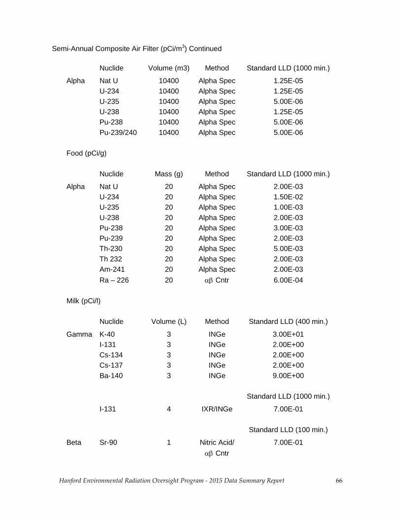
Hanford Environmental Radiation Oversight Program - 2015 Data Summary Report 66
Semi-Annual Composite Air Filter (pCi/m3) Continued
Nuclide Volume (m3) Method Standard LLD (1000 min.) Alpha Nat U 10400 Alpha Spec 1.25E-05 U-234 10400 Alpha Spec 1.25E-05 U-235 10400 Alpha Spec 5.00E-06 U-238 10400 Alpha Spec 1.25E-05 Pu-238 10400 Alpha Spec 5.00E-06 Pu-239/240 10400 Alpha Spec 5.00E-06 Food (pCi/g)
Nuclide Mass (g) Method Standard LLD (1000 min.) Alpha Nat U 20 Alpha Spec 2.00E-03 U-234 20 Alpha Spec 1.50E-02 U-235 20 Alpha Spec 1.00E-03 U-238 20 Alpha Spec 2.00E-03 Pu-238 20 Alpha Spec 3.00E-03 Pu-239 20 Alpha Spec 2.00E-03 Th-230 20 Alpha Spec 5.00E-03 Th 232 20 Alpha Spec 2.00E-03 Am-241 20 Alpha Spec 2.00E-03 Ra – 226 20 αβ Cntr 6.00E-04 Milk (pCi/l)
Nuclide Volume (L) Method Standard LLD (400 min.) Gamma K-40 3 INGe 3.00E+01 I-131 3 INGe 2.00E+00 Cs-134 3 INGe 2.00E+00 Cs-137 3 INGe 2.00E+00 Ba-140 3 INGe 9.00E+00 Standard LLD (1000 min.) I-131 4 IXR/INGe 7.00E-01 Standard LLD (100 min.) Beta Sr-90 1 Nitric Acid/ 7.00E-01 αβ Cntr

Hanford Environmental Radiation Oversight Program - 2015 Data Summary Report 67
Meat (pCi/g)
Nuclide Mass (g) Method Standard LLD (1000 min.) Gamma K-40 400 INGe 1.40E-01 Mn-54 400 INGe 7.00E-03 Co-58 400 INGe 7.00E-03 Co-60 400 INGe 8.00E-03 Cs-137 400 INGe 6.00E-03 I-131 400 INGe 2.00E-02 Ra-226(DA) 400 INGe 2.50E-01 Am-241(GA) 400 INGe 2.00E-02 Alpha Nat U 10 Alpha Spec 4.00E-03 U-234 10 Alpha Spec 3.00E-03 U-235 10 Alpha Spec 2.00E-03 U-238 10 Alpha Spec 3.00E-03 Pu-238 10 Alpha Spec 5.00E-03 Pu-239 10 Alpha Spec 4.00E-03 Am-241 10 Alpha Spec 4.00E-03 Beta Sr-90 (bone) 5 Nitric Acid/ 2.00E-01 αβ Cntr Shellfish (pCi/g)
Nuclide Mass (g) Method Standard LLD (400 min.) Gamma I-131 400 INGe 6.00E-03 Co-60 400 INGe 6.00E-03 K-40 400 INGe 1.00E-01

Hanford Environmental Radiation Oversight Program - 2015 Data Summary Report 68
Soil/Sediment (pCi/g)
Nuclide Mass (g) Method Standard LLD (1000 min.) Alpha Nat U 1 Alpha Spec 4.00E-02 U-234 1 Alpha Spec 3.00E-02 U-235 1 Alpha Spec 2.00E-02 U-238 1 Alpha Spec 3.00E-02 Pu-238 10 Alpha Spec 5.00E-03 Pu-239 10 Alpha Spec 4.00E-03 Th-230 1 Alpha Spec 4.00E-02 Th 232 1 Alpha Spec 4.00E-02 Am-241 10 Alpha Spec 4.00E-03 Ra - 226 1 αβ Cntr 1.00E-01 Ra-226(DA) 600 INGe 2.00E-02
Standard (100 min.) Alpha Gross 0.1 αβ Cntr 4.00E+01 Standard LLD (1000 min.)
Gamma K-40 600 INGe 1.50E-01 Mn-54 600 INGe 1.00E-02 Co-60 600 INGe 1.00E-02 Zn-65 600 INGe 2.00E-02 Zr-95 600 INGe 1.00E-02 Ru-103 600 INGe 1.50E-02 Ru-106 600 INGe 1.00E-02 Sb-125 600 INGe 2.00E-02 Cs-134 600 INGe 1.20E-02 Cs-137 600 INGe 1.50E-02 Ce-144 600 INGe 5.00E-02 Eu-152 600 INGe 1.50E-02 Eu-154 600 INGe 1.50E-02 Eu-155 600 INGe 2.00E-02 Ra-226(DA) 600 INGe 1.00E-01 Am-241(GA) 600 INGe 2.00E-02 Tot U(GA) 600 INGe 2.00E-01 Standard (100 min.) Beta Sr-90 150 Nitric Acid/ 1.80E-03 Tc-99 10 3M/LS 2.00E-01 Gross beta 0.4 αβ Cntr 1.50E+00

Hanford Environmental Radiation Oversight Program - 2015 Data Summary Report 69
Vegetation (pCi/g except H-3 which is expressed as pCi/l)
Nuclide Mass (g) Method Standard LLD (1000 min.) Alpha Nat U 10 Alpha Spec. 8.00E-03 U-234 10 Alpha Spec. 6.00E-03 U-238 10 Alpha Spec. 6.00E-03 Pu-238 10 Alpha Spec. 5.00E-03 Pu-239 10 Alpha Spec. 4.00E-03 Am-241 10 Alpha Spec. 4.00E-03 Gamma K-40 100 INGe 3.00E-01 Mn-54 100 INGe 4.00E-02 Co-60 100 INGe 4.00E-02 Zn-65 100 INGe 1.50E-01 Zr-95 100 INGe 2.00E-01 Ru-106 100 INGe 4.00E-01 Cs-137 100 INGe 4.00E-02 I-131 100 INGe 4.00E-02 Am-241(GA) 100 INGe 2.00E-01 Standard LLD (100 min.) Beta Gross 0.4 αβ Cntr 1.50E+00 Sr-90 20 Nitric Acid/ 5.00E-02 αβ Cntr Tc-99 5 3M/LS 1.50E+00 Nuclide Volume (L) Method Standard LLD (200 min.) C-14 0.0002 Oxid/LS 3.00E+02 H-3 0.002 LS 5.00E+02

Hanford Environmental Radiation Oversight Program - 2015 Data Summary Report 70
Water (pCi/l) Standard LLD Standard LLD Nuclide Volume (L) Method (1000 min.) (100 min.)
Alpha Nat U 0.5 Alpha Spec 1.30E-01 U-234 0.5 Alpha Spec 8.00E-02 U-235 0.5 Alpha Spec 6.00E-02 U-238 0.5 Alpha Spec 8.00E-02 Ra-226 0.5 αβ Cntr 2.00E-01 Pu-238 0.5 Alpha Spec 8.00E-02 Pu-239 0.5 Alpha Spec 6.10E-02 Th-230 0.5 Alpha Spec 1.00E-01 Th 232 0.5 Alpha Spec 1.00E-01 Am-241 0.5 Alpha Spec 8.00E-02 Standard LLD (1000 min.) Gamma Am-241 3 INGe 1.00E+01 Ba-140 3 INGe 9.00E+00 Ce-144 3 INGe 1.30E+01 C0-58 3 INGe 1.50E+00 Co-60 3 INGe 2.00E+00 Cr-51 3 INGe 1.60E+01 Cs-134 3 INGe 2.00E+00 Cs-137 3 INGe 2.00E+00 Eu-152 3 INGe 5.00E+00 Eu-154 3 INGe 5.00E+00 Eu-155 3 INGe 8.00E+00 Fe-59 3 INGe 3.00E+00 I-129 3 IXR/LEP 8.00E-01 I-131 3 INGe 2.00E+00 K-40 3 INGe 3.00E+01 Mn-54 3 INGe 1.50E+00 Nb-95 3 INGe 2.00E+00 Ru-103 3 INGe 2.00E+00 Ru-106 3 INGe 1.50E+01 Sb-125 3 INGe 5.00E+00 Sn-113 3 INGe 2.00E+00 Zn-65 3 INGe 3.00E+00 Zr-95 3 INGe 2.00E+00

Hanford Environmental Radiation Oversight Program - 2015 Data Summary Report 71
Water (pCi/l) Continued Standard LLD Standard LLD Nuclide Volume (L) Method (200 min.) (100 min.) Beta H-3 0.010 Dist/LS 6.00E+01 C-14 0.010 LS 1.50E+02 Sr-90 1 Nitric Acid/ 7.00E-01 αβ Cntr Tc-99 0.5 3M/LS 4.00E+00 Gross Alpha 0.1 αβ Cntr 4.00E+00 Beta 0.5 αβ Cntr 1.00E+00
*LLD for Air Cartridge is 3 days METHOD Preparation Methods
IXR = Ion Exchange Resin Nitric Acid 3M = 3M Ion Exchange Disks Oxid = Oxidation Counting Methods
INGe = Intrinsic Germanium Detector αβ Cntr = Alpha, Beta Counter Alpha Spec = Alpha Spectrometry LS = Liquid Scintillation LEP = Low Energy Photon Detector

Hanford Environmental Radiation Oversight Program - 2015 Data Summary Report 72
Formulas A. Random Uncertainty RU = 1.96((gross sample cpm/T1) + (BKGCPM/T2))1/2/((E)(2.22)(V)(Y)(D)) B. Uncertainty (standard error) of the sample mean (U) U = s/(n)1/2 C. Lower Limit of Detection (LLD) LLD = 4.66S/((2.22)(E)(V)(Y)(D)) D. Definitions 2.22 = conversion factor from dpm to picocuries BKGCPM = background counts per minute D = decay factor = e-(ln2/T1/2)(t) E = counting efficiency: counts per disintegration LLD = the a priori determination of the smallest concentration of radioactive material sampled that has a 95 percent probability of being detected, with only five percent probability that a blank sample will yield a response interpreted to mean that radioactivity is present above the system background n = number of samples analyzed (number of data points) RU = random uncertainty at the 95 percent confidence level (sometimes referred to as counting error) s = sample standard deviation S = one standard deviation of the background count rate (which equals (BKG/T2)1/2) sample cpm = counts per minute of sample t = elapsed time between sample collection and counting T1 = sample count time T2 = background count time T1/2 = half-life of radionuclide counted U = uncertainty (standard error) of the sample mean V = volume in liters (or mass in grams) of sample Y = fractional radiochemical yield (when applicable)

Hanford Environmental Radiation Oversight Program - 2015 Data Summary Report 73
Appendix C - Glossary of Terms
Alpha Particle A heavy particle emitted from the nucleus of an atom. It consists of two protons and two neutrons, which is identical to the nucleus of a helium atom without orbital electrons. These heavy charged particles lose their energy very rapidly in matter. Thus, they are easily shielded by paper or the surface layer of skin. Alpha particles are only hazardous when they are internally deposited.
Analyte The specific radioisotope measured in a radiochemical analysis. For example, tritium, Sr-90, and U-238 are analytes.
Background (Background Radiation)
Radiation that occurs naturally in the environment. Background radiation consists of cosmic radiation from outer space, radiation from the radioactive elements in rocks and soil, and radiation from radon and its decay products in the air we breathe.
Baseline Samples Environmental samples taken in areas unlikely to be affected by any facilities handling radioactive materials.
Becquerel A unit, in the International System of Units (SI), of measurement of radioactivity equal to one transformation per second.
Beta Particle A high-speed particle emitted from the nucleus, which is identical to an electron. They can have a -1 or +1 charge and are effectively shielded by thin layers of metal or plastic. Beta particles are generally only hazardous when they are internally deposited.
Curie The basic unit of activity. A quantity of any radionuclide that undergoes an average transformation rate of 37 billion transformations per second. One curie is the approximate activity of 1 gram of radium. Named for Marie and Pierre Curie, who discovered radium in 1898.

Hanford Environmental Radiation Oversight Program - 2015 Data Summary Report 74
Decay, Radioactive The decrease in the amount of any radioactive material with the passage of time, due to the spontaneous emission from the atomic nuclei of either alpha or beta particles, often accompanied by gamma radiation.
Detection Level The minimum amount of a substance that can be measured with a 95-percent confidence that the analytical result is greater than zero.
Dose A generic term that means absorbed dose, equivalent dose, effective dose, committed equivalent dose, committed effective dose, or total effective dose.
Fallout Radioactive materials that are released into the earth’s atmosphere following a nuclear explosion or atmospheric release and eventually fall to earth.
Gamma Ray Electromagnetic waves or photons emitted from the nucleus of an atom. They have no charge and are best shielded by thick layers of lead or steel. Gamma energy may cause an external or internal radiation hazard. (X-rays are similar to gamma radiation but originate from the outer shell of the atom instead of the nucleus.)
Gross Alpha / Gross Beta A screening test that reports alpha particle activity in a sample. The test is not intended to identify specific radioisotopes. The tests are primarily used to evaluate trends. In addition, screening tests are used to determine if further radioisotope specific analysis is necessary; and if radioisotope analysis has been carried out, to determine if the activities from specific radioisotopes account for all of the activity found in the screening test.
Half-life The time in which half the atoms of a particular radioactive substance disintegrate to another nuclear form. Measured half-lives vary from millionths of a second to billions of years. Also called physical half-life.
Ionizing Radiation Any radiation capable of displacing electrons from atoms or molecules, thereby producing ions. Examples: alpha, beta, gamma, x-rays, and neutrons.

Hanford Environmental Radiation Oversight Program - 2015 Data Summary Report 75
Isotope One of two or more atoms with the same number of protons, but different numbers of neutrons, in the nuclei.
Lower Limit of Detection (LLD) The smallest amount or concentration of a radioactive element that can be reliably detected in a sample.
Optically Stimulated Luminescence (OSL)
A radiation monitoring device used to measure accumulated ambient radiation dose OSLs are similar to the thermoluminescence dosimeters, TLDs, but use light rather than heat to release the stored energy and measure the dose of ionizing radiation received.
pCi (picocurie) 10-12 curies (one trillionth of a curie)
Quality Assurance All those planned and systematic actions necessary to provide adequate confidence that a facility, structure, system, or component will perform satisfactorily and safely in service.
Quality Control A component of Quality Assurance; comprises all those actions necessary to control and verify that a material, process, or product meets specified requirements.
Quality Factor (Q) A numerical factor assigned to describe the average effectiveness of a particular kind (and sometimes energy) of radiation in producing biological effects on humans.
Rad The special unit of absorbed dose. It is a measure of the energy absorbed per mass of material. One rad is equal to an absorbed dose of 0.01 J kg-1 (1 rad = 0.01 gray).
Radioactivity The process of undergoing spontaneous transformation of the nucleus, generally with the emission of alpha or beta particles, often accompanied by gamma rays. The term is also used to designate radioactive materials.
Radioisotope A radioactive isotope; i.e., an unstable isotope that undergoes spontaneous transformation, emitting radiation. Approximately 2500 natural and artificial radioisotopes have been identified.
Radionuclide A radioactive nuclide.

Hanford Environmental Radiation Oversight Program - 2015 Data Summary Report 76
Rem The special unit of dose equivalent. The dose equivalent in rem is equal to the absorbed dose in rad multiplied by a quality factor that accounts for the biological effect of the radiation (1 rem = 0.01 sievert).
Replicate Sample Two or more samples from one location that are analyzed by the same laboratory.
Roentgen A unit of exposure to ionizing radiation. It is that amount of gamma or x-rays required to produce ions carrying 1 electrostatic unit of electrical charge in 1 cubic centimeter of dry air under standard conditions. Named after Wilhelm Roentgen, German scientist who discovered x-rays in 1895.
Split Sample A sample from one location that is divided into two samples and analyzed by different laboratories.
TLD Thermoluminescent Dosimeters
X-Ray Electromagnetic waves or photons emitted from the outer shell of the atom instead of the nucleus. They have no charge and are best shielded by thick layers of lead or steel. X-Ray energy may cause an external or internal radiation hazard.

Hanford Environmental Radiation Oversight Program - 2015 Data Summary Report 77
Appendix D - List of Analytes
Am-241 Americium-241 Be-7 Beryllium-7 C-14 Carbon-14 Cm-244 Curium-244 Co-60 Cobalt-60 Cs-137 Cesium-137 Eu-152 Europium-152 Eu-154 Europium-154 Eu-155 Europium-155 H-3 Hydrogen-3 I-129 Iodine-129 K-40 Potassium NO2+NO3 Nitrite + Nitrate Pu-238 Plutonium-238 Pu-239/240 Plutonium-239/240 Ru-106 Ruthenium-106 Sb-125 Antimony-125 Sr-90 Strontium-90 Tc-99 Technetium-99 Total U Total Uranium U-234 Uranium-234 U-235 Uranium-235 U-236 Uranium-236 U-238 Uranium-238