2015 mohd fadli asmanilibir.josai.ac.jp/il/user_contents/02/g0000284repository/...2015 mohd fadli...
TRANSCRIPT

BASIC RESEARCH AND PERSPECTIVE TO
DEVELOP TOPICAL FORMULATIONS FOR
ACNE THERAPY
2015
MOHD FADLI ASMANI

BASIC RESEARCH AND PERSPECTIVE TO
DEVELOP TOPICAL FORMULATIONS FOR
ACNEE THERAPY
2015
MOHD FADLI ASMANI

- i -
Contents
Abstract ………………………………………………………………………………...….1
General introduction ……………………………………………………………...………3
Chapter 1 Contribution of hair follicular pathway of topically applied and
exposed chemicals for the total skin permeation
1.1. Introduction………………………………………………………………….……….7
1.2. Method……………………………………………………………………………….10
1.2.1. Materials
1.2.2. Determination of n-octanol /buffer partition coefficient
1.2.3. Animals
1.2.4. Preparation of skin membrane
1.2.5. Hair follicle-plugging process
1.2.6. Preparation of applied solution
1.2.7. In vitro skin permeation experiments
1.2.8. Determination of FD-4 and FL using a spectrofluorophotometer
1.2.9. Determination of drugs using an HPLC system
1.2.10. Analysis of permeation parameters
1.2.11. Statistical analysis
1.3. Results……………………………………………………………………….........19
1.3.1 Effect of pH on the skin permeation of ISDN
1.3.2 Effect of hair follicle plugging on the skin permeation of drugs

- ii -
1.3.3 Factors of skin permeation reduction by hair follicle plugging
1.4. Discussion…………………………………………………………………………..25
1.5. Chapter conclusion………………………………………………………………..29
Chapter 2 Evaluation of drug disposition in hair follicles after topical application
2.1. Introduction………………………………………………………………….……..30
2.2. Methods………………………………………………………………………….....31
2.2.1. Materials
2.2.2. Determination of n-octanol / buffer partition coefficient
2.2.3. Animals
2.2.4. Preparation of skin membrane
2.2.5. Preparation of applied solution
2.2.6. In vitro skin permeation study
2.2.7. Estimation drug concentration in hair follicles
2.2.8. Determination of Cal using a spectrofluorophotometer
2.2.9. Determination of drugs using an HPLC system
2.2.10. Calculation of skin permeation parameters
2.2.11. Confocal laser scanning microscope observation
2.3. Results……………………………………………………………………………38
2.3.1. In vitro skin permeation of drugs
2.3.2. Changes in flux of skin permeation and hair follicle concentration
Of lipophilic drugs

- iii -
2.3.3 Changes in flux of skin permeation and hair follicle concentration of
hydrophilic drugs
2.3.4. Skin disposition of drugs
2.3.5. Relationship between logKo/w and log (Steady-state drug concentration in
hair follicles)
2.4. Discussion and chapter conclusion…………………………….…………………..44
General conclusions……………………………………………………………………..46
Appendix Using nano-emulsion formulation approach to enhance the skin
permeation of clindamycin and tetracycline as a new strategy for acne
therapy.
A.1. Introduction……………………………………………………………………….48
A.2. Method……………………………………………………………………………50
A.2.1. Materials
A.2.2. Apparatus
A.2.3. Pre-formulation studies of pseudo-ternary phase diagram
A.3. Result………………………………………………………………………..........54
A.3.1 Formulation.
A.3.2. Mean droplet size of clindamycin and tetracycline nano-emulsions
A.4. Discussion…………………………………………………………………………56
A.5. Appendix conclusion……………………………………………………………..56
Acknowledgments……………………………………………………………………..57
References…………………………………………………………………………......58

- iv -
Abbreviation
AMP aminopyrine
BA benzoic acid
BP butyl paraben
Cal calcein
CBS carbonate buffer saline
CP clindamycin phosphate
DFP diisopropyl fluorophosphate
DL-2
diffusion parameter
EIP emulsion inversion point
F127 Pluronic 127
FD-4 fluorescein isothiocyanate-dextran 4 kDa
FL fluorescein
HF hair follicle
IP ibuprofen
ISDN isosorbide dinitrate
ISMN isosorbide mononitrate
Jss steady-state flux
KL partition parameter
LC lidocaine hydrochloride
P permeability coefficient
PBS phosphate buffered saline
PIT phase inversion temperature
SC stratum corneum
TEWL transepidermal water loss
tlag lag time

- 1 -
Abstract
Acne vulgaris is a very common skin disease, which causes a high degree of
psychosocial suffering and has a detrimental effect on the quality of life of the patients
irrespective of age or gender. Treatment of acne is principally directed towards these
known pathogenic factors. Clindamycin, tetracycline and erythromycin are commonly
prescribed topical antibiotics for acne vulgaris with anti-inflammatory properties.
However, the effectiveness of acne treatments has been limited by their relative inability to
penetrate into the pilosebaceous unit, the site of acne formation.
It is very important to evaluate concentration of the therapeutic and
cosmeceutical chemicals in skin because their pharmacodynamics and toxicodynamics can
be expressed by a function of these concentrations. Their permeation pathway of topically
applied chemical compounds, i.e., stratum corneum and hair follicle (HF) is closely related
to their skin concentration. Then, we aimed to investigate the contribution of skin
permeation of drugs through HF as well as stratum corneum to enable their selective
delivery to HF.
The contribution of HF pathway on the skin permeation of chemicals was
calculated from a difference between their permeability coefficients through skin with and
without HF plugging using in vitro skin permeation experiment. The obtained result
revealed that the contribution of HF pathway could be predicted by their lipophilicities. In
a hydrophilic region of chemicals (logKo/w < 0), a higher reduction ratio was observed by
HF plugging compared with lipophilic chemicals (logKo/w ≥ 0). In addition, the reduction
ratio was decreased with an increase in the logKo/w. This consideration on the HF pathway

- 2 -
would be helpful to investigate usefulness and safety of chemicals after their topical
application and exposure, because skin permeation and disposition must be changed at
different sites of skin due to different sites and densities of HF. Furthermore, another study
was conducted to evaluate the drug disposition in HF. HF concentration of drugs with
different lipophilicities was investigated to evaluate the effect of physicochemical
properties on their HF disposition, where drugs having logKo/w < 0 and logKo/w ≥ 0 were
assumed to be lipophilic and hydrophilic, respectively. Results showed that the lag time
observed in the skin permeation before obtaining a steady-state profile for
hydrophilic drugs was delayed compared with that for lipophilic drugs. Hydrophilic drugs
were found to be distributed through the HF as well as into the shallow part of stratum
corneum, whereas lipophilic drugs distributed both into the stratum corneum and HF from
a histological observation using fluorescent makers. These results suggest that lipophilic
drugs could be easily delivered both into the stratum corneum and HF, whereas hydrophilic
drugs were mainly delivered through HF, but not for deep layer of the stratum corneum.
Thus, in a future study, aiming to formulate the clindamycin and tetracycline
nano-emulsion by using emulsion phase inversion method to increase the effectiveness of
acne treatment through the increased the penetration of the hydrophilic active compounds
into the pilosebaceous unit. However, only a few studies have been focused on selective
drug delivery to HF.

- 3 -
General introduction
Skin is the outermost and largest organ of the body; i.e., it contributes 10% of
body weight and 1.7 m2 of surface area. Skin consists of three structural layers known as,
the epidermis (outer surface layer), dermis and subcutaneous tissues (deepest layer).
Epidermis comprises of two different layers of epithelium, which are the viable epidermis
and the stratum corneum. The viable epidermis serves as a hydrophilic layer with
composition of 70% water. On the other hands, the stratum corneum composed of only
13% water and serves as a hydrophobic layer. Hydrophilic compound is not able to
penetrate easily across the hydrophobic stratum corneum. Hydrophobic compounds can
penetrate the stratum corneum but cannot enter the next layer of hydrophilic viable
epidermis layer1)
. Dermis gives a mechanical strength to the skin as it composed of
collagen fibrils embedded in mucopolysaccharide gel. In addition, the dermis contains few
embedded structures including blood and lymphatic vessels, hair follicles (HF), sebaceous
glands and sweat glands2)
.
A topically therapeutic agent is able to penetrate through the skin via the stratum
corneum3)
and skin appendages including HF4)
. The stratum corneum route can be divided
into transcellular and intercellular routes. The transcellular route gives a direct penetration
of drugs to cross through the stratum corneum. However, this route gives a significant
resistance against drug permeation as the compound has to pass through both lipophilic
and hydrophilic layers. Intercellular route is a more common pathway for drug entrance in
which the compound moving between the corneocytes5)
. Recently, follicular penetration
route has shown to be one of the efficient pathways for topically applied compounds6)
.

- 4 -
Pig skin has been a well-known experimental animal skin for many years. It
serves as a good representative for human model skin due to similarities in terms of
physiological and anatomical points between human and pig. The decision of choosing pig
ear skin in the experiments was because it represents a suitable model for human skin
according to Jacobi studied in 20057)
. Besides that, human skin has a disadvantage of
becoming contracted during excision. This causes the HF to be permanently blocked the
contracted elastic fibers8)
. Thus, pig ear skin can be described as a superior model to
excised human skin in follicular penetration studies.
Acne vulgaris is one of the most common skin disorders in 80% of most
adolescents, though it can continue to occur in adulthood especially in women due to
hormonal imbalance during menstrual cycle. Comedones, inflamed papules, pustules and
nodules can be observed in the lesion of acne. Acne is mostly present in the highest
number of pilosebaceous glands area such as the face, chest and back9)
.
The use of antimicrobial therapy or antibiotics in the treatment of acne has
started since 1930s and 1940s. Although antibiotics have been a part of mainstay treatment
for a long time, acne experts recommend it to act as an adjunctive therapy instead of
primary in the role of acne treatment10)
. The mechanism on how topical antibiotics help to
improve acne has not been clearly defined. They probably act on Propionibacterium acne
(P. acne) colonization and thus produce pro-inflammatory actions on the comedogenesis.
The most commonly used topical antibiotics on the acne are clindamycin, tetracycline and
erythromycin11)
. However, topical gentamicin sulfate can also be used to secondary treat
skin infections including pustular acne12)
.

- 5 -
General concepts of this research are to quantitatively analyze the HF
contribution pathway on the skin permeation of topically applied hydrophilic and lipophilic
chemical compounds. Objective of this research is to investigate the permeation pathway
of hydrophilic and lipophilic chemicals in the stratum corneum and HF for each chemical
in details. Understanding the kinetic parameters in HF as well as stratum corneum will be
essential for development of topical dosage forms for acne therapy
Many reports have published that skin appendages such as HF and sweat glands
becomes an important permeation/penetration pathway especially of hydrophilic
compounds and macromolecules. Blood and skin concentrations of topically applied or
exposed chemicals can be calculated from their in vitro skin permeation profiles by taking
into consideration of skin thickness and applying chemical concentrations. It is very
important to evaluate usefulness and safety of topically applied or exposed chemicals,
because their pharmacodynamics and toxicodynamics can be expressed by a function of
these concentrations. Thus, permeation pathways of hydrophilic chemicals, i.e., stratum
corneum and HF, should be discussed for each chemical in details. Most of the experiments
were performed to identify the diffusion pathway and distribution of topically applied or
exposed chemicals through and in skin by an imaging analysis using a confocal
microscope
In Chapter 1, contribution to HF pathway of topically applied or exposed
chemicals was determined from a difference between permeability coefficient of chemicals
through skin with and without HF plugging. By concerning the topical routes of drug
administration, the common skin permeation for drugs is mainly by passing through the

- 6 -
stratum corneum. The stratum corneum which located at the outermost layer of skin
became a major barrier for drug permeation into the skin. Hence, several chemical and
physical approaches have been researched to encounter the skin barrier.
In Chapter 2, drug disposition in HF is a key issue after topical
application. Although dermatopharmacokinetic parameters of topically applied drugs in
the stratum corneum have been evaluated using in vitro skin permeation experiment, in
vivo tape-stripping technique, and so on, few studies have reported on the pharmacokinetic
parameters in HFs after topical application. Understanding the dermatopharmacokinetic
parameters on the HF penetration as well as stratum corneum would be essential for
development of topical application forms. In the present study, HF concentration of
topically applied drugs with different lipophilicities was investigated to evaluate the effect
of physicochemical properties on their HF disposition.
In Appendix, development of the nano-emulsion formulations to enhance skin
permeation and HF concentration after topical application was described. Nano-emulsions
can be used to deliver drugs to patients via several routes. Currently the administration of
drugs as in nano-emulsions via topical application has gained increasing interest. Nano-
emulsions can increase the rate of absorption and eliminate variability in absorption.
Hydrophilic and lipophilic drugs can also be delivered by nano-emulsions to increase their
bioavailability, since the skin permeability of drugs may be affected by nano-emulsions.

- 7 -
Chapter 1
Contribution of hair follicular pathway of topically applied and exposed chemicals for
the total skin permeation
1.1. Introduction
The body is exposed to many chemical compounds in daily life. These
chemicals are mainly absorbed via oral, pulmonary and dermal routes, as well as through
other mucosa. Among these, the dermal pathway is more easily accessed by chemicals
than the other pathways because skin is the outermost tissue covering the whole body and
has a large surface area13)
. The skin is also focused on as the application site of drugs and
cosmetics. The pathway for the permeation of therapeutic and cosmeceutical chemicals
through the skin is thus very important to evaluate their effects. In case of either skin
application or skin exposure, skin permeation and the concentration of chemicals should be
investigated to evaluate their effects and/or toxicities.
Skin can be histologically divided into three different layers from the surface to
deeper tissues: stratum corneum, viable epidermis and dermis. The superficial layer, the
stratum corneum, is composed of dead corneocytes embedded in intercellular lipid
matrices consisting of ceramides, free fatty acids, cholesterol and cholesteryl esters14)
.
These lipids are organized into lamella structures in the intracellular region of the stratum
corneum and form the primary barrier against the elimination of endogenous compounds
and the penetration of exogenous chemicals through the skin15)
. The permeation profile of
chemicals through skin is theoretically expressed by Fick’s 2nd law of diffusion, which

- 8 -
expresses the behavior of chemicals passing through the stratum corneum9, 16)
. On the
other hand, many reports17, 18, 19)
have been published describing that skin appendages such
as HF and sweat glands are an important permeation/penetration pathway, especially for
hydrophilic compounds and macromolecules.
Many researchers have already investigated the transfollicular permeation of
topically applied chemicals. Feldman et al.12)
and Maibach et al.20)
have reported that
regional variation in percutaneous absorption was obtained due to different hair densities.
Hueber et al.21)
investigated the role of HF as a skin permeation route with burn scar tissue.
Grice et al. 22)
studied the effect of drug uptake into HF on the skin permeation by a
cyanoacrylate casting method. Hairy and non-hairy guinea pig skins were used for in vivo
and in vitro studies to check the transfollicular absorption. Furthermore, pharmacokinetic
modeling was also applied to define the relative contribution of the HF route. In addition
to these skin permeation studies, the diffusion pathway and the distribution of topically
applied or exposed chemicals through and in skin were identified by imaging analysis
using a confocal microscope23, 24, 1)
. However, all of these studies did not involve
quantitative analyses, and few studies evaluated the contribution of the HF pathway of
topically applied or exposed chemicals by in vivo or in vitro skin permeation experiments25,
11, 26).
Horita et al. 27)
have already established a HF plugging method using
cyanoacrylate-grease mixture, and then reported that the permeation of hydrophilic
chemicals through HF -plugged skin was dramatically decreased, whereas lipophilic
chemical permeation through HF-plugged skin was seldom changed compared with that

- 9 -
through non-HF-plugged skin. Otberg et al.28)
applied caffeine in a mixed solution of
ethanol : polyethylene glycol (30 : 70 v/v) to volunteers before and after blocking all HF
with a varnish solution at the site of its application. From this study, caffeine was observed
in blood 20 min after application on the HF-blocked skin, but 5 min after topical
application to normal skin. A possible reason for the more rapid appearance of caffeine in
blood is the rapid absorption of the substance by penetrating through HF to blood
capillaries. In addition, Trauer et al.29)
reported that the in vivo absorption of caffeine from
whole skin was much more rapid and substantial than the in vitro absorption. Furthermore,
a sandwich technique has been used to clarify the contribution of the HF pathway to the
whole absorption of topically applied chemicals26)
. The sandwich technique revealed that
the HF pathway could contribute 34% to 60% to the whole skin permeation of chemicals
with different lipophilicities (logKo/w range: -1.05 to 2.29) and molecular weights (M.W.
range: 251 to 362). However, no clear relationship has been reported between
physicochemical properties and the contribution of the HF pathway to whole skin
permeation.
Blood and skin concentrations of topically applied or exposed chemicals can be
calculated from the in vitro skin permeation profile by taking into consideration of the skin
thickness30) and the applied chemical concentrations. This is very important to evaluate
the usefulness and safety of topically applied or exposed chemicals because their
pharmacodynamics and toxicodynamics can be expressed as functions of these
concentrations. Thus, the permeation pathway of hydrophilic chemicals, namely, the
stratum corneum and HF, should be discussed in detail for each chemical.

- 10 -
The hair follicular openings occupy only about 0.1% of the whole skin area, but
this can be as high as about 10% for the face around the mouth and scalp18)
. Thus, we have
to take this into account in terms of the applied chemicals and the skin sites in order to
evaluate the usefulness and safety of chemicals. In the present study, the contribution of
the HF pathway against the whole permeation of chemicals was determined from the
difference between the permeation coefficients of chemicals through skin with and without
HF plugging.
1.2. Method
1.2.1. Materials
Lidocaine hydrochloride (LC), fluorescein isothiocyanate-dextran 4 kDa (FD-4),
sodium calcein (Cal-Na) and ibuprofen (IP) were obtained from Sigma-Aldrich Co., Ltd.
(St. Louis, MO, U.S.A.). Isosorbide dinitrate (ISDN) was kindly donated by Toko
Pharmaceutical Industrial Co., Ltd. (Tokyo, Japan). Butyl paraben (BP) and isosorbide
mononitrate (ISMN) were obtained from Tokyo Kasei Kogyo Co., Ltd. (Tokyo, Japan).
Nile red was obtained from Kanto Chemical Co., Inc. (Tokyo, Japan). Aminopyrine
(AMP) and diisopropyl fluorophosphate (DFP) were obtained from Wako Pure Chemical
Ind., Ltd. (Osaka, Japan). All other reagents and solvents were of reagent grade or HPLC
grade, and were used without further purification. Table 1.1 shows the physicochemical
properties of the model chemicals used in the present skin permeation experiment.

- 11 -
Table 1.1 Physicochemical properties of drugs used in this experiment
Model drug M.W. LogKo/w (pH) pKa
FD-4 ca. 4000 -0.77 (7.4) e) 6.7 h)
Cal-Na 644.5 -3.50 (7.4) f) 5.5 i)
CP 505 -2.27 (10) 6.9
FL-Na 376.3 -0.61 (7.4) e) 6.4 e)
ISDN 236.1 1.23 (7.4) g) ─
LC 234.3 -0.90 (5.0)b)
1.40 (10.0) d) 7.9 j)
AMP 231.3 -1.00 (3.0) a)
5.0 k) 0.98 (7.4) c)
IP 206.3 1.93 (3.0) a)
4.9 l) 1.25 (7.4) c)
BP 194.2 3.50 (7.4) c) 8.3 m)
ISMN 191.1 -0.15 (7.4) g) ─
a) n-octanol/pH3.0 citrate buffer logKo/w at 32 ºC.
b) n-octanol/pH5.0 citrate buffer logKo/w at 32 ºC.
c) n-octanol/pH7.4 phosphate buffer logKo/w at 32 ºC.
d) n-octanol/pH10.0 carbonate buffer logKo/w at 32 ºC.
e) n-octanol/pH7.4 phosphate buffer logKo/w at 32 ºC19)
.
f) n-octanol/pH7.4 phosphate buffer logKo/w at 32 ºC31)
.
g) n-octanol/pH7.4 phosphate buffer logKo/w at 32 ºC32)
.
h) pKa at 25 ºC8)
.
i) pKa at 25 ºC33)
.
j) pKa at 25 ºC6)
.
k) pKa at 25 ºC34)
.
l) pKa at 25 ºC10)
.
m) pKa at 25 ºC4)
.

- 12 -
1.2.2. Determination of n-octanol /buffer partition coefficient
n-Octanol was saturated with pH3.0 citrate buffer, pH5.0 citrate buffer, pH7.4
phosphate buffer or pH10.0 carbonate buffer for at least 24 h before the partition
experiment at 37°C. Drug was dissolved in n-octanol-saturated buffered solution. The
obtained solution was mixed with an equal volume of buffer solution-saturated n-octanol at
37°C for 24 h. Drug concentration in the aqueous phase was then analyzed by HPLC.
Apparent n-octanol/buffer solution partition coefficients (Ko/w) were determined.
1.2.3. Animals
Frozen pig ear skins were purchased from the National Federation of
Agricultural Cooperative Associations (Tokyo, Japan). These skins were stored at -30°C
until the skin permeation experiments. All animal studies were carried out with the
recommendations of the Institutional Board for Animal Studies, Josai University (Sakado,
Saitama, Japan).
1.2.4. Preparation of skin membrane
Purchased skin was frozen at -80°C prior to use. The skin was thawed at room
temperature and excised from the outer surface of pig ear after being cleaned with pH7.4
phosphate buffered saline (PBS). The excised skin was mounted on a Franz-type diffusion
cell to conduct the skin permeation study. In the case of skin permeation study with HF-
plugged skin, the plugging procedure was carried out after skin excision. The integrity of
the excised skin was measured using a skin impedance meter 1 h after hydration with 1 mL

- 13 -
of PBS and all of the skin samples used in the present study showed resistance of above 20
kohm/cm2.
1.2.5. Hair follicle-plugging process
The HF-plugging procedure was as previously reported in detail (Horita et al.,
2014). This procedure can be briefly summarized as follows. HF in the designated area
(effective skin permeation area: 1.77 cm2) were plugged with silicone grease-cyanoacrylate
adhesive mixture paste to block chemical penetration through the HF. The mixture paste
consisted of equal parts of silicone grease (Super Lube® Silicone Dielectric Grease; Synco
Chemical Corp., Bohemia, NY, U.S.A.) and α-cyanoacrylate adhesives (Aron Alpha Jelly;
Konishi Co., Ltd., Osaka, Japan) with small amounts of Nile red. Nile red was used to
visualize the area to which the mixture paste had been applied. Thus, the HF were plugged
with the mixture paste to prevent chemical penetration through the follicular pathway. A
mean of 56.1 2.5 HF (n=15) were found in the effective pig ear skin permeation area
(1.77 cm2) in the preliminary experiment, and half of them were plugged with the mixture
paste in the present experiment. According to Horita et al.27)
, an almost linear decrease in
the skin permeation ratio was observed with an increase in the number of HF plugged with
the mixture paste. The mixture paste-treated area was measured using imaging software
(cellSens, Olympus Corp., Tokyo, Japan), equipped with a stereoscopic microscope (SZ61,
Olympus Corp., Tokyo, Japan). The effective skin permeation area was about 1.62 cm2
after treatment with the mixture paste. Figure 1.1 shows the skin surface with or without
the HF plugging.

- 14 -
Fig.1.1. Pictures of the pig ear skin surface with (a) or without (b) HF plugging.
1.2.6. Preparation of applied solution
First, 5.0 mM FD-4, 1.0 mM FL-Na, 5.0 mM ISDN, 10 mM non-ionized AMP,
5.0 mM ionized IP, 0.6 mM BP and 500 mM ISMN were prepared with 1/30 mM PBS
(pH7.4). Then, 1.0 mM Cal-Na was prepared with pH7.4 PBS containing 1.0 mM EDTA-
2Na. In addition, 10 mM non-ionized LC was prepared with 100 mM carbonate-
bicarbonate buffer solution (pH10.0) and 100 mM ionized LC was prepared with 100 mM
citrate buffer solution (pH5.0). Furthermore, 100 mM ionized AMP and 0.5 mM non-
ionized IP were prepared with pH3.0 citrate buffer. The pH levels for the fluorescent
compounds such as FD-4, Cal-Na and FL-Na and weak electrolytes except for BP were
adjusted to ensure that about 99% were in non-ionized or ionized form.
1.2.7. In vitro skin permeation experiments
Excised pig ear skin membrane was mounted on vertical-type diffusion cells
5 mm
5 mm
a) b)

- 15 -
(effective diffusion area: 1.77 cm2). The stratum corneum was hydrated for 1 h with pH-
adjusted solution or PBS containing 2.7 mol/mL DFP. The latter was used to
prevent the metabolism of ester compounds during the skin permeation experiment. It has
already been confirmed that the hydration procedures with DFP did not affect the skin
permeation of the esters and their metabolites 35-37)
.
After the pre-hydration process, solution applied to the skin was completely
removed from the diffusion cell and 1.0 mL of test chemical solution and 6 mL of pH-
adjusted solution were applied to the donor and receiver cells, respectively. The total of
0.54 mol/mL DFP was added on the dermis side when ester compounds were applied on
the stratum corneum. The permeation experiments were performed at 32°C, while the
receiver solution was continuously stirred with a star-head-type magnetic stirrer. At
predetermined times, an aliquot (0.5 mL) was withdrawn from the receiver solution and an
identical volume of fresh solution was added to keep the volume constant. Each
experiment was performed in 3 to 4 replicates.
1.2.8. Determination of FD-4 and FL using a spectrofluorophotometer
The concentrations of FL, Cal and FD-4 in the samples were analyzed using a
spectrofluorophotometer (RF 5300PC; Shimadzu) at excitation wavelengths of 480 nm,
488 nm and 490 nm, and at fluorescent emission wavelengths of 535 nm, 515 nm and 520
nm, respectively.
1.2.9. Determination of drugs using an HPLC system

- 16 -
Concentrations of drugs (LC, ISDN, AMP, IP, BP and ISMN) in the samples
were determined using an HPLC system (Prominence; Shimadzu, Kyoto, Japan) equipped
with a UV detector (SPD-M20A; Shimadzu). The drug samples (0.2 mL) were added to
the same volume of acetonitrile for ISMN or acetonitrile containing internal standard
(methyl paraben for LC, butyl paraben for ISDN, AMP and IP, and propyl paraben for BP),
and mixed with a vortex mixer. After centrifugation at 3600×g and 4°C for 5 min, 20 µL
of the supernatant was injected into the HPLC system. Chromatographic separation was
performed using an Inertsil-ODS-3 (5 µm, 150×4.6 mm i.d.; GL Science, Kyoto, Japan) at
40°C. The mobile phase was 0.1% phosphoric acid containing 5 mM sodium 1-
heptanesulfate/acetonitrile (70/30, v/v) for LC, water/acetonitrile (55/45, v/v) for ISDN,
0.1% phosphoric acid containing 5 mM sodium dodecyl sulfate/acetonitrile (30/70, v/v) for
AMP, 0.1% phosphoric acid/acetonitrile (55/45, v/v) for ISMN and water/acetonitrile
(90/10, v/v) for ISMN. The flow rate was adjusted to 1.0 mL/min and detection was
performed at UV 220 nm (ISMN and ISDN), 230 nm (LC), 245 nm (AMP), 260 nm (BP)
or 263 nm (IP).
1.2.10. Analysis of permeation parameters
Skin permeation parameters were calculated using time courses of the
cumulative amounts of chemicals that permeated through a unit area of skin with or
without HF plugging. The steady-state flux (Jss) (a steady state was reached 6–10 h after
starting the experiment) was estimated from the slope of the linear portion of the profile of
the cumulative amount of chemical that permeated through a unit area of skin versus time,

- 17 -
and the lag time (tlag) was calculated from the intercept on the time axis by extrapolation
from the steady-state skin permeation profile. From Jss and the initial donor concentration
(Cv), the permeability coefficient (P) was calculated using eq. 1. The partition parameter
(KL) and the diffusion parameter (DL-2
) were then obtained from eqs. 2 and 3 38)
:
where L, D and K are the thickness of the barrier membrane, the diffusion coefficient of
chemicals in the membrane and the partition coefficient of chemicals into the membrane,
respectively. DL-2
was obtained from eq. 2 and KL was calculated from P and DL-2
values
according to eq. 2.
In addition, the reduction ratio of P of topically applied chemicals due to HF
plugging was calculated using eq. 4.
Then, the contribution of the HF pathway to the whole skin permeation of
chemicals was obtained by doubling the reduction ratio shown in the figure, since plugging
of 30 of the total of 60 HF in the effective skin area had been carried out.
1.2.11. Statistical analysis
Statistical analysis was performed using unpaired Student’s t-test and ANOVA.

- 18 -
p value of less than 0.05 was considered significant.
1.3. Results
1.3.1 Effect of pH on the skin permeation of ISDN
Figure 1.2 shows the time course of the cumulative amount of ISDN that
permeated through pig ear skin from its solution with different pH levels (pH3.0, 7.4 and
10.0) in order to investigate the effect of pH on the skin permeation of the neutral drug. As
expected, almost the same ISDN permeation profiles were obtained independently of the
pH under these experimental conditions. In addition, skin permeation parameters of tlag,
DL-2
and KL obtained from the skin permeation profiles of ISDN for pH3.0 and pH10
solutions were very similar to those for the pH7.4 solution (Table 1.2).

- 19 -
0
0.05
0.1
0.15
0.2
0 2 4 6
Cu
mul
ativ
e am
ount
of
ISD
N p
erm
eate
d
(μm
ol/c
m2)
Time (h)
Fig.1. 2. Time course of the cumulative amount of ISDN that permeated through pig ear
skin from its solution. pH3.0 (), pH7.4 () and pH10.0 (). Each point represents the
mean S.D. (n = 3-4).
1.3.2 Effect of hair follicle plugging on the skin permeation of drugs
Figures 1.3a and b show the effect of HF plugging on the skin permeation of
ionized and non-ionized AMP, respectively. The cumulative amount of ionized AMP (at
pH3.0) that permeated through the plugged skin was about half that through the non-
Table 1.2 Skin permeation parameters of ISDN from solutions with different pHs
pH3.0 pH7.4 pH10.0
Q6
(mol/cm2)# 1.3010-1 1.6410-2 1.1910-1 2.1810-2 1.2210-1 1.1110-2
P (cm/s) 2.4610-6 4.6710-7 2.0110-6 2.9310-7 1.8610-6 2.3410-7
tlag (h) 1.91 0.23 2.22 0.16 2.02 0.21
DL-2 (cm-1) 1.0210-1 1.2510-2 7.5610-2 5.3610-3 8.4210-2 8.2710-3
KL (cm) 2.3510-5 2.7410-6 2.6210-5 2.2410-6 2.3010-5 5.0310-6
#Q6: Cumulative amount of ISDN that permeated through the skin over 6 h.

- 20 -
plugged skin (Fig. 1.3a). On the other hand, the cumulative amount of non-ionized AMP
(at pH7.4) that permeated through the plugged skin was about 0.7-fold that through the
non-plugged skin (Fig. 1.3b). The sizes of these decreases in the cumulative amount of
AMP that permeated were greatly affected by the ratio of ionized to non-ionized forms.
Table 3 shows permeation parameters calculated from the skin permeation profiles shown
in Fig. 1.3. These parameters can be utilized to evaluate the changes in distribution and/or
diffusion of topically applied or exposed chemicals to and across the skin 19, 39)
. The KL
values obtained through the HF plugging were decreased at both pH3.0 and pH7.4
compared with those of non-plugged skin, although the DL-2
values were almost the same.
These decreased KL values at both pH3.0 and pH7.4 corresponded to the sizes of the
decreases in the cumulative amount of AMP that permeated through the plugged skin
compared with the non-plugged skin.
Table 1.3 Skin permeation parameters and reduction ratio of AMP with or
without HF-plugged skin
Non-HF plugging HF plugging Ratio#
pH3.0
Q10
(mol/cm2) 2.4010-2 7.5510-3 1.1710-2 4.4110-3 0.49
P (cm/s) 8.7010-9 2.3810-9 4.7310-9 2.0210-9 0.54
tlag (h) 2.53 0.5 2.56 0.4 1.01
DL-2 (h-1) 1.1710-4 2.3210-5 1.1910-4 1.8610-5 1.02
KL (cm) 7.5810-5 2.0010-5 3.9910-5 1.7010-6 0.52
pH7.4
Q10
(mol/cm2) 3.7810-2 1.4110-3 2.7310-2 2.1110-3 0.72
P (cm/s) 1.6110-7 8.1010-9 1.4310-7 1.6110-8 0.89
tlag (h) 3.85 0.14 4.18 0.24 1.09

- 21 -
0
0.01
0.02
0.03
0.04
0 2 4 6 8 10
Cu
mu
lati
ve
amo
un
t o
f A
MP
per
mea
ted
(μm
ol/
cm
2)
Time (h)
DL-2 (h-1) 1.7810-4 6.3410-6 1.9410-4 1.1110-5 1.09
KL (cm) 9.0610-4 5.0510-5 7.5110-4 1.0510-4 0.83
Ratio#: HF plugging/non-HF plugging
(a)
(b)
Fig.1.3. Time course of the cumulative amount of AMP that permeated through pig ear
skin at pH3.0 (a) and pH7.4 (b). Non-HF-plugged skin (), HF-plugged skin ().
Each point represents the mean S.D. (n = 4).
Next, a skin permeation experiment for several chemicals was carried out with or
without HF plugging to determine their P values as listed in Table 1.4. This table also
shows the reduction ratio of P values calculated using eq.4. Reductions of the P value of
around 50% were observed for FD-4, Cal-Na, FL-Na, ionized LC, ionized AMP and ISMN,
0
0.01
0.02
0.03
0.04
0 2 4 6 8 10
Cu
mu
lati
ve
amo
un
t o
f A
MP
per
mea
ted
(μm
ol/
cm2)
Time (h)

- 22 -
whereas no or only slight reductions (less than 20%) in P value were observed for non-
ionized BP, non-ionized IP and non-ionized LC. Furthermore, reduction ratios of P values
of 20% to 40% were shown for non-ionized AMP, ionized IP and ISDN.

- 23 -
Table 1.4 Reduction in permeability coefficient of chemicals through the HF-
plugged skin. Values are the mean S.E. (n = 3-4).
pH of
applied
solution
P values through
HF-non-plugged
skin (×10-8 cm/s)
P values
through HF-
plugged
skin(×10-8 cm/s)
Reduction
ratio (%)
FD-4 7.4 0.064 0.032 0.026 0.012 59
Cal-Na 7.4 2.8 1.2 1.4 1.7 50
CP 10.0 3.2 0.68 1.2 0.12 61
FL-Na 7.4 1.4 0.57 0.62 0.089 56
ISDN 7.4 371 122 242 65 35
Ionized LC 5.0 2.3 0.61 1.1 0.31 52
Non-ionized LC 10.0 255 109 210 49 18
Ionized AMP 3.0 0.88 0.48 0.46 0.42 48
Non-ionized AMP 7.4 17 0.74 13 0.50 24
Non-ionized IP 3.0 1520 270 1300 81 14
Ionized IP 7.4 87 16 59 5.4 32
Non-ionized BP 7.4 132 50 132 143 0
ISMN 7.4 8.9 1.2 4.9 3.6 45
1.3.3 Factors of skin permeation reduction by hair follicle plugging
Figure 1.4 shows the relationship between the molecular weight of topically
applied chemicals and the reduction ratio of P values by HF plugging. A reduction ratio of
P values of around 50% was observed for chemicals with a molecular weight greater than
400 Da. The reduction ratio of P values seems to increase with an increase in the

- 24 -
molecular weight in the range from 200 to 400 Da, but the relationship between the
reduction ratio of the P value of chemicals and their molecular weights was inconsistent,
especially from 200 to 350 Da.
Fig. 1. 4. Relationship between the reduction ratio of skin permeation by HF
plugging and the molecular weight of the chemicals. : ionized form (acidic or
basic chemicals), : non-ionized form (acidic or basic chemicals), : neutral
chemicals.
M.W.
Sk
in p
erm
ea
tion
-dec
rea
sin
g r
ati
o
(%)
0
10
20
30
40
50
60
0 100 0 4000500 600 700
〜~〜~〜~〜~
200 400150 250
〜~〜~
〜~〜~
Red
uct
ion r
atio
(%
)
FD-4
Cal-Na FL-Na
LC (pH5.0)
IP (pH3.0)
IP (pH7.4)
AMP (pH3.0)
AMP (pH7.4)
BP
ISDN
ISMN
LC (pH10.0)
CP
○ ; ionized form (acidic or basic) drugs
○ ; non-ionized form (acidic or basic) drugs
○ ; neutral drugs.○○

- 25 -
Next, the effect of the lipophilicity of chemicals, logKo/w, on the reduction ratio
of the P value was investigated. Figure 1.5 shows the relationship between the reduction
ratio of the P value of chemicals and their log Ko/w values. For hydrophilic chemicals
(logKo/w < 0), a higher reduction ratio was observed than for lipophilic ones (logKo/w ≥ 0).
In addition, the reduction ratio decreased with an increase in logKo/w. Furthermore, about a
10% reduction of chemical permeation was evenly observed for every 1.0 logKo/w increase
of chemicals (i.e., every 10-fold increase in Ko/w value) within the range of logKo/w values
from 0 to 4.
Fig .1.5. Relationship between the reduction ratio of skin permeation by HF
plugging and the partition coefficient of the chemicals. : ionized form (acidic
or basic chemicals), : non-ionized form (acidic or basic chemicals), : neutral
chemicals.
- 26 -
1.4. Discussion
Generally, blood and skin concentration profiles of topically applied or exposed
chemicals or drugs can be calculated from the in vitro skin permeation profile40), 41), 42)
.
Presently, the calculation method is especially applicable for chemicals for which the main
permeation route is the stratum corneum. If the contribution of HF to the total skin
permeation of chemicals can be obtained, blood and skin concentrations of drug would be
more precisely predicted. Since excised skin used in an in vitro study might show a
histological reduction in the follicular penetration pathway via contraction of the elastic
fibers surrounding HF, Patzelt et al.43)
speculated that an in vitro skin permeation
experiment may not be suitable for determining the contribution of the HF pathway to the
total skin permeation of chemicals. On the other hand, Raber et al.44)
reported an excellent
in vivo/in vitro correlation in terms of the nanoparticles level recovered from HF after their
topical application. In vitro skin permeation experiments have already been broadly used
to evaluate and develop transdermal drug delivery systems (TDDS). Furthermore, there
are ethical issues associated with in vivo experiments, especially regarding the
development of cosmeceutical products. Therefore, in the present study, an in vitro
experiment was carried out to determine the contribution of the HF pathway.
According to Horita et al.27)
, a good linear regression was obtained between the
decrease in the skin permeation of a high-molecular-weight chemical, FD-4, and the
number of HF plugged. About 58 visible HF were confirmed in an effective skin
permeation area of 1.77 cm2. The point at which the regression line crossed the x-axis
showed that plugging of 28 and 58 HF led to decreases in the skin permeation of FD-4 of
- 27 -
50 and 100 percent, respectively. Furthermore, no permeation of FD-4 or FL-Na was
observed through three-dimensional cultured human skin models19)
. This is because the
cultured human skin models have no HF. Thus, the present results strongly suggest that
the HF pathway is the primary pathway of FD-4 and FL-Na through the skin. Thirty HF in
the effective skin permeation area were plugged in this study to calculate the contribution
of the HF pathway to the whole skin permeation of topically applied or exposed chemicals.
Among the present results, only KL parameters were decreased by HF plugging,
while the values of DL-2
were almost constant after the application of AMP solution at
different pH levels, suggesting that the plugging method could enable evaluation of the
contribution of the HF pathway to the total skin permeation of chemicals. The fraction of
HF in a diffusion area should be related to the partition parameter of chemicals when HF
are its main permeation route19)
. Therefore, the present plugging method should be useful
for calculating the contribution of HF to the permeation of topically applied or exposed
chemicals.
Figure 1.6 shows the relationship between the contribution of the HF pathway
and the partition coefficient of chemicals. The contribution of the HF pathway was also
compared with other published data to verify our results. Frum et al.26)
reported the
influence of logKo/w on HF penetration by an in vitro sandwich method. The percentage
contribution by follicles decreased with an increase in the lipophilicity of topically applied
or exposed compounds, and the levels of contribution (4%, 2%, 46%, 60%, 58%, 46% and
34% for estradiol [logKo/w: 2.29], corticosterone [logKo/w: 1.94], hydrocortisone [logKo/w:
1.60] and aldosterone [logKo/w: 1.08], respectively) were almost the same as our calculated

- 28 -
values. On the other hand, a downward parabolic relationship was observed for the
hydrophilic compounds (58, 48 and 34% for cimetidine [logKo/w: 0.40], deoxyadenosine
[logKo/w: 0.40] and adenosine [logKo/w: -1.05], respectively).
These data are very different from my calculated values. Frum et al.26)
calculated the HF contribution from a 28 or 48 h-skin permeation experiment. Excess
hydration of the stratum corneum may change the skin characteristics by causing swelling
and the development of water pools in the intercellular lamellar region45)
. This might be a
reason for the low contribution of the HF pathway for hydrophilic drugs compared with the
present results. Furthermore, the formulation of topically applied drugs would also affect
the contribution of the HF pathway due to variation in the partitioning of chemicals in skin.
Liu et al. 25)
reported that caffeine (logKo/w: –0.01) was absorbed through HF within 30 min
after topical application in a binary solvent of 30% ethanol and 70% propylene glycol, and
that 10.5% to 33.8% of the total amount was absorbed through the HF.
This binary solvent could increase the skin permeation of chemicals by causing
variation in the fluidity of the stratum corneum lipids. However, a much higher
contribution of HF (over 90%) was obtained from aqueous solution in the present study.
Thus, differences in the formulation should be an important issue to determine the
contribution of HF to the permeation of topically applied or exposed chemicals.

- 29 -
0
20
40
60
80
100
120
-4 -3 -2 -1 0 1 2 3 4 Sk
in p
erm
eati
on
-decr
easi
ng r
ati
o (
%)
Log Ko/w
IP (pH3.0)
IP (pH7.4)
AMP (pH3.0)
AMP (pH7.4)
BP
ISDN
ISMN
Cal-Na FL-Na
LC (pH10.0)
LC (pH5.0)
CP
FD-4
Fig.1.6. Relationship between the contribution of the HF pathway and the partition
coefficient of the chemicals. : ionized form (acidic or basic) chemicals, : non-ionized
form (acidic or basic) chemicals, : neutral chemicals.
This report reveals that the contribution of the HF pathway to the total skin
permeation of topically applied or exposed chemicals could be calculated by using their
lipophilicity. Molecular weight, lipophilicity, number of hydrogen donors or acceptors,
electron affinity, charge densities, and atomic and molecular orbits (LUMO/HOMO) are
also very important for the skin permeation of chemical compounds3, 5, 7, 44, 45)
. More
precise estimation might thus be possible by considering these parameters.

- 30 -
1.5. Chapter conclusion
This chaptershows that the contribution of the HF pathway to the total skin
permeation of chemicals can be determined by using their lipophilicity. Consideration of
the HF pathway would be helpful to investigate the usefulness and safety of chemicals
after their topical application because skin permeation and disposition would vary among
different sites of skin due to the differences in the character and/or density of HF.

- 31 -
Chapter 2
Evaluation of drug disposition in hair follicles after topical application
2.1. Introduction
Drug concentration at target sites is very important to expect the
pharmacological effect. Therefore, optimization of drug formula and formulation design
should be conducted to effectively deliver an adequate amount of drug to the target site. In
the 1st chapter, HF route showed higher contribution for hydrophilic drugs against the total
skin permeation compared for lipophilic drugs. Hydrophilic acne treatment agents such as
clindamycin, gentamycin sulfate and tetracycline hydrochloride, therefore, could be
delivered to HF. The total skin permeation of hydrophilic drugs is normally lower than that
of lipophilic ones, and the cumulative amount of drug permeated through skin was
increased with an increase of lipophilicity. In addition, drug concentration has a very close
relation to its skin permeation. Sugibayashi et al.32)
, reported that skin concentration of
topically applied drugs could be calculated with permeation parameters such as partition
coefficient and permeability coefficient obtained from drug permeation profiles thorough
full-thickness and stripped skins.
To develop the formulation and structure for topically applied drugs such as
local anesthesia, painless and atopic dermatitis treatments, several method like suction
blister46-48)
, punch and shave biopsies47,48)
, tape-stripping49-51)
, heat-separation52,53)
,
autoradiography54)
methods have been used to determine drug concentration at target site in
the skin. On the other hand, few studies have published on the drug concentration profiles
- 32 -
in the HF after topical application, although pharmacokinetics at the target site is important
for drug discovery and development,
Thus, in the 2nd chapter, HF concentration after topical application of drugs with
different physicochemical properties was considered to estimate the effect of lipophilicity
of topically applied drugs on its steady-state concentration in HF and related
pharmacokinetics.
2.2. Methods
2.2.1. Materials and methods
Isosorbide-5-mononitrate (ISMN), butyl paraben (BP), clindamycin phosphate
(CP) and sodium calcein (Cal-Na) were purchased from Tokyo Chemical Industry Co., Ltd.
(Tokyo, Japan). Benzoic acid (BA) and diisopropyl fluorophosphates (DFP) were
purchased from Wako Pure Chemical Industries, Ltd (Osaka, Japan). Lidocaine
hydrochloride monohydrate (LC) was purchased from Sigma-Aldrich (St. Louis, MO). The
other solvents and reagents were of HPLC grade and regent grade-purity. All reagents
were commercially available and used without further purification. Table 2.1 shows
chemical structures, molecular weights (M.W.), n-octanol/water coefficients (logKo/w) and
acid dissociation constant (pKa). In the present study, chemicals were classified into
lipophilic and hydrophilic chemicals according to their logKo/w values; chemicals with
logKo/w values greater than 0 are regarded as lipophilic drugs and those with logKo/w values
less than 0 are considered hydrophilic drugs.

- 33 -
Table 2.1. Chemical structures and physicochemical properties of model permeants.
Drug Chemical structure M.W. LogKo/w pKa
Benzoic acid
(BA)
122.1 1.90 (pH 3.0)
a)
-0.41 (pH 7.4) c)
4.2
f)
Isosorbide
mononitrate
(ISMN)
191.1 -0.20 (pH 7.4) c)
-
Butyl paraben
(BP)
194.2 3.50 (pH 7.4) c)
8.3 g)
Lidocaine
(LC)
234.3 -0.90 (pH 5.0)
b)
1.40 (pH 10.0) d)
7.9
h)
Clindamycin
phosphate
(CP)
505.0 -2.27 (pH 10.0) d)
6.9 i)
Sodium
calcein
(Cal-Na)
644.5 -3.51 (pH 7.4) e)
5.5 j)
a) n-octanol/pH 3.0 citrate buffer logKo/w at 32ºC.
b) n-octanol/pH 5.0 citrate buffer logKo/w at 32ºC.
c) n-octanol/pH 7.4 phosphate buffer logKo/w at 32ºC.
d) n-octanol/pH 10.0 carbonate buffer logKo/w at 32ºC.
e) from Ref. 55)
.
f) from Ref. 56)
.
g) from Ref. 57)
.
h) from Ref. 58)
.
i ) from Ref. 59)
.

- 34 -
j ) from Ref. 60)
.

- 35 -
2.2.2. Determination of n-octanol / buffer partition coefficient
n-Octanol was saturated with various pH buffer solutions as follow: pH3.0
citrate buffer, pH 5.0 citrate buffer, pH 7.4 phosphate buffered saline (PBS) or pH 10.0
carbonate-bicarbonate buffer for at least 24 h before starting the experiment at 37 °C. Each
drug was dissolved in n-octanol-saturated buffered solution. The obtained solution was
mixed with an equal volume of buffer solution-saturated n-octanol at 37 °C for 24 h. Drug
concentration in the aqueous phase was then analyzed with HPLC. Apparent n-octanol /
buffer solution partition coefficients (Ko/w) were then determined.
2.2.3. Animals
Frozen pig ear skin samples were purchased from the National Federation of
Agricultural Cooperative Associations (Tokyo, Japan). These skin samples were stored at -
30°C until the skin permeation experiments. All animal studies were carried out with the
recommendations of the Institutional Board for Animal Studies, Josai University (Sakado,
Saitama, Japan).
2.2.4. Preparation of skin membrane
Purchased skin was maintained as frozen at -30°C prior to use. The skin was
thawed at room temperature and excised from the outer surface of pig ear skin after being
cleaned with the same buffered solution to be applied in skin permeation study. The
excised skin was mounted on a Franz-type diffusion cell to conduct skin permeation study.
The same buffered solutions used in the skin permeation study were applied for 1 h to the

- 36 -
hydrate stratum corneum. To avoid hydrolysis by esterase in skin, 2.7 mM diisopropyl
fluorophosphates (DFP), a serine protease inhibitor, was included in the buffer solution for
1 h. No skin permeation changes were appeared for 1 h treatment with DFP 61-63)
.
2.2.5. Preparation of applied solution
30 mM ionized BA, 50 mM ISMN, 0.5 mM BP and 1 mM Cal-Na were prepared
with 1/30 mM PBS (pH 7.4). In addition, 10 mM non-ionized LC and 100 mM CP were
prepared with 100 mM carbonate-bicarbonate buffer (pH10.0) and 100 mM ionized LC
and non-ionized BA were prepared with 100 mM citrate buffer (pH5.0).
2.2.6. In vitro skin permeation study
After the pre-hydration process, applied solution to the skin was completely
removed from the diffusion cell and 1.0 mL of test chemical solution and 6 mL of pH-
adjusted solution were applied to the donor and receiver cells, respectively. A total of 0.54
mol/mL DFP was added on the dermis side when ester compounds were applied on the
stratum corneum. The permeation experiments were performed at 32°C, while the receiver
solution was continuously stirred with a star-head-type magnetic stirrer. At predetermined
times, an aliquot (0.5 mL) was withdrawn from the receiver solution and an identical
volume of fresh solution was added to keep the volume constant. Each experiment was
performed in 3 to 4 replicates.

- 37 -
2.2.7. Estimationdrug concentration in hair follicles
After in vitro skin permeation study, the skin was removed carefully from the
diffusion cell after the applied solution containing drug was recovered. Then, the stratum
corneum and dermis sides were washed 3 times, respectively, with 1.0 mL of the same pH-
adjusted solution used to skin permeation study. Thirty hairs were removed from the
effective skin permeation area with forceps under a stereoscopic microscope observation.
The removed hairs with follicle tissue were measured by weight and vortexed for 20 min in
400 µL of pH-adjusted buffer solution. The solution was centrifuged (CT15RE®, Hitachi
Koki Co., Ltd., Tokyo, Japan) at 15,000 rpm for 4 min at 4ºC, drug concentration in the
obtained supernatant was measured with HPLC or spectrofluorophotometer. Apparent drug
concentration inHF was calculated from the obtained drug amount with the hair removed
from the drug-treated skin. This apparent drug concentration (drug amount/hair weight%)
was assumed to “that in HF” (see discussion in detail).
2.2.8. Determination of Cal using a spectrofluorophotometer
The concentrations of Cal in the samples were analyzed using a
spectrofluorophotometer (RF 5300PC; Shimadzu) at an excitation wavelength of 488 nm
and at a fluorescent emission wavelength of 515 nm.
2.2.9. Determination of drugs using an HPLC system
Concentrations of drugs (LC, ISDN, BA, CP, BP and ISMN) in the samples were
determined using an HPLC system (Prominence; Shimadzu, Kyoto, Japan) equipped with a

- 38 -
UV detector (SPD-M20A; Shimadzu). The drug samples (0.2 mL) were added to the same
volume of acetonitrile for ISMN and CP or acetonitrile containing internal standard
(methyl paraben for LC, butyl paraben for ISDN, BA for ethyl paraben, and propyl paraben
for BP), and mixed with a vortex mixer. After centrifugation at 3600×g and 4°C for 5 min,
20 µL of the supernatant was injected into the HPLC system. Chromatographic separation
was performed using an Inertsil-ODS-3 (5 µm, 150×4.6 mm i.d.; GL Science, Kyoto,
Japan) at 40°C. The mobile phase was 0.1% phosphoric acid containing 5 mM sodium 1-
heptanesulfate/acetonitrile (70/30, v/v) for LC, water/acetonitrile (55/45, v/v) for ISDN,
0.1% phosphoric acid/acetonitrile (55/45, v/v) for BA and BP and water/acetonitrile (90/10,
v/v) for ISMN, 100 mM KH2PO4 (pH 2.5)/acetonitrile (80/20, v/v) for CP. The flow rate
was adjusted to 1.0 mL/min and detection was performed at UV 220 nm (ISMN and ISDN),
230 nm (LC), 210 nm (CP), 260 nm (BP) or 254 nm (BA).
2.2.10. Calculation of skin permeation parameters
The flux of skin permeation experiment was calculated from drug permeation
profile through the skin. Then, permeability coefficient of drug was obtained from division
of flux by applied drug concentration, Cv (equation 4).
…..(4)
Drug concentration in HF was supposed to be values calculated by eq. 5 as follows:

- 39 -
Drug concentration in HF (µmol/g)
rug hair )
……..(5)
2.2.11. Confocal Laser Scanning Microscope Observation
After topical application of Cal-Na and rhodamin over 8 h, the donor solution
was immediately removed from the skin surface and the skin section was prepared with a
cryotome (CM3050S; Leica Microsystems Inc., Tokyo, Japan). The cryostat skin section
with the stratum corneum side up was examined using a confocal laser scanning
microscope (CLSM, Fluoview FV1000; Olympus Corp., Tokyo, Japan). Cal was excited
using an argon laser at an excitation wavelength of 488 nm, whereas rhodamine was
excited by 543 nm helium–neon laser.

- 40 -
2.3. Results
2.3.1. In vitro skin permeation of drugs
Table 2.2 shows relationships P values of topically applied drugs through intact
and stripped pig ear skin, respectively. P values of lipophilic chemicals (logKo/w ≥ 0), BP
(pH7.4), BA (pH3.0) and LC (pH10.0), through intact skin were 21.8×10⁻⁷
, 35.7×10⁻⁷
and
13.6 ×10⁻⁷
cm/s, respectively. On the other hand, P values of those compounds through
stripped skin were 16.1×10⁻⁷
, 93.1×10⁻⁷
and 64.2 ×10⁻⁷
cm/s, respectively. P values
through stripped skin were not so higher than those through intact skin. Especially, P value
of BP permeations through intact skin were a little higher than that through stripped skin.
Table 2.2. Permeability coefficient of drugs through pig ear full-thickness skin and
stripped skin. Values are the mean ± S.D. (n = 3-5).
Drug LogKo/w Applied dose/1.77 cm2
P values in full-
thickness skin
(×10-7
cm/s)
P values in
stripped skin
(×10-6
cm/s)
Cal-Na
(pH 7.4) -3.50 5 nmol 0.18 ± 0.04 2.51 ± 0.49
CP
(pH 10.0) -2.27 100 nmol 0.30 ± 0.14 3.61 ± 0.27
LC
(pH 5.0) -0.90 100 nmol 0.34 ± 0.09 4.67 ± 1.17
BA
(pH 7.4) -0.41 30 nmol 1.53 ± 0.35 11.8 ±2.38
ISMN
(pH 7.4) -0.20 50 nmol 1.19 ± 0.35 5.10 ± 0.35
LC
(pH 10.0) 1.40 100 nmol 13.6 ± 4.66 6.42 ± 1.63
BA
(pH 3.0) 1.90 10 nmol 35.7 ± 4.67 9.31 ± 1.07
BP
(pH 7.4) 3.50 0.5 nmol 21.8 ± 8.94 1.61 ± 0.37

- 41 -
In case of skin permeation of hydrophilic compounds (logKo/w < 0), on the other
hand, P values of hydrophilic compounds, ISMN (pH 7.4), BA (pH 7.4), LC (pH5.0), CP
(pH10.0) and Cal-Na (pH7.4), were 1.19×10⁻⁷
, 1.53 ×10⁻⁷
, 0.34×10⁻⁷
, 0.30×10⁻⁷
and
0.18×10⁻⁷
cm/s, respectively. The P values of these hydrophilic chemicals through stripped
skin was dramatically improved compared with full-thickness skin [ISMN (pH7.4);
51.0×10⁻⁷
, BA (pH 7.4); 118×10-7
, LC (pH5.0); 46.7 ×10⁻⁷
, CP (pH10.0); 36.7×10⁻⁷
and Ca-
Na (pH7.4); 25.1 ×10⁻⁷
cm/s, respectively].
2.3.2. Changes in flux of skin permeation and hair follicle concentration of lipophilic
drugs
Figure 2.1 shows changes in flux of skin permeation of lipophilic drugs through
intact skin (closed circle) and drug concentrations in HF (open square). Flux of lipophilic
drugs (ISDN and BA) became that steady-state 4 h after topical application (Fig 1a, b).
The supposed concentration profiles of drugs in HF were increased with an increase of
time, and then each drug concentration showed the constant value as steady-state fluxes
were observed for skin permeation (Fig 1a, b). On the other hand, BP concentration of in
HF markedly increased immediately after topical application (1c), but the steady-state flux
of BP was not obtained in 10 h after application.
2.3.3 Changes in flux of skin permeation and hair follicle concentration of hydrophilic
drugs
Figure 2.2 shows changes in flux of skin permeation of hydrophilic drugs

- 42 -
through intact skin and drug concentrations in HF. Steady-state of fluxes were observed
for ISMN, BA and LC, whereas they were not observed for CP and Cal-Na. On the other
hand, drug concentrations in HF were rapidly increased after topical application, and then
steady-state concentrations in HF were achieved.
Fig. 2.1. Time courses of HF concentration and flux through pig ear skin of a) ISDN (pH
7.4), b) BA (pH 3.0) and c) BP (pH 7.4) after topical application. Symbols: flux ●), drug
concentration in HF □). Each point shows the means ± S.D. (n = 3-6).
a) b)
c)

- 43 -
Fig. 2.2. Time courses of HF concentration and flux through pig ear skin of a) ISMN (pH
7.4), b) BA (pH 7.4), c) LC (pH 5.0), d) CP (pH 10.0) and e) Cal-Na (pH 7.4) after topical
application. Symbols: the same in Fig. 2.1. Each point shows the means ± S.D. (n = 3-6).
a) b)
c) d)
e)

- 44 -
2.3.4. Skin disposition of drugs
Rhodamin or Cal-Na, a lipophilic or hydrophilic fluorescent maker, respectively,
was topically applied to skin and the drug distribution was observed with a confocal laser
scanning microscopy immediately after preparation of skin section. Rhodamin was
observed both into the stratum corneum and HF, whereas Cal was slightly distributed in the
stratum corneum and was much observed in the shallower part of HF and the HF shaft, as
shown in Fig. 2.3.
c) d)
Fig. 2. 3. Conformal microscopic observation of Cal (a and b) and rhodamin
(c and d) in the skin. a and c were merged with transmitted light and
fluorescent images (b and d). Abbreviation of HF is HF.
Stratum corneum
HF
Stratum corneum
HF
a) b)
c) d)
- 45 -
Lo
g (
stea
dy
-sta
te d
rug
am
ou
nt
in H
F)/
do
se
LogKo/w
Ca-Na
LC (pH5.0)
ISMN LC (pH10.0)
ISDN
BP BA (pH3.0)
BA (pH7.4) CP
-2.5
-1.5
-0.5
0.5
-4 -2 0 2 4
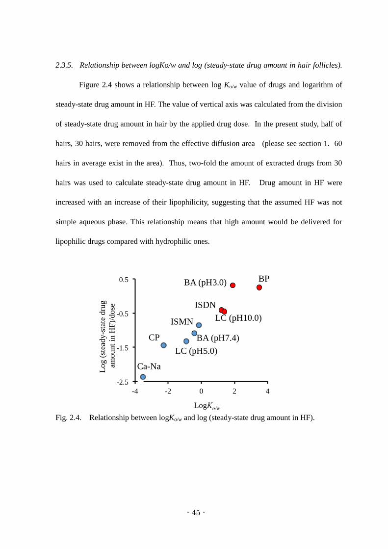
2.3.5. Relationship between logKo/w and log (steady-state drug amount in hair follicles).
Figure 2.4 shows a relationship between log Ko/w value of drugs and logarithm of
steady-state drug amount in HF. The value of vertical axis was calculated from the division
of steady-state drug amount in hair by the applied drug dose. In the present study, half of
hairs, 30 hairs, were removed from the effective diffusion area (please see section 1. 60
hairs in average exist in the area). Thus, two-fold the amount of extracted drugs from 30
hairs was used to calculate steady-state drug amount in HF. Drug amount in HF were
increased with an increase of their lipophilicity, suggesting that the assumed HF was not
simple aqueous phase. This relationship means that high amount would be delivered for
lipophilic drugs compared with hydrophilic ones.
Fig. 2.4. Relationship between logKo/w and log (steady-state drug amount in HF).

- 46 -
2.4. Discussion and chapter conclusion
Dermatopharmacokinetics is used to evaluate bioequivalence of topically
applied drugs by a tape-stripping method49, 64)
. Furthermore, a few reports have recently
published that in silico approach to estimate skin concentration of drug with different
physicochemical properties in viable epidermis and dermis65, 66)
. Considering skin as a
homogeneous membrane, these researches supposed a direct relation between chemicals
concentration and its effect. It is an evidence that Hill equation could be applied to reveal
the relationship between cell viability of topically applied chemicals and its concentration
in skin 67)
.
Scheuplein reported that distribution of drugs into the HF would be occurred at
the beginning process of skin permeation, although HF accounted only approximately
0.1 % of are against the total skin surface68)
. In addition, cyanoacrylate biopsy method was
used to evaluate effect and safety of topically applied or exposed chemicals by
investigating their concentrations in HF. However, in spite of the present measurements of
drug concentration in stratum corneum and viable epidermis/dermis, few studies have been
published on the drug concentration in the HF.
In this chapter, drug amount extracted from a removal hair was supposed to be
that in the HF. Although this assumption is not fully right, we do not have any better or
best determination method or tool for assessment of HF concentration of topically applied
or exposed chemicals. Development of the technique or tool for assessing the follicle
concentration of chemicals must be a future issue in this or related topics.
Delayed lag time of HF concentration for lipophilic drugs was observed

- 47 -
compared with those for hydrophilic ones. The time to become steady-state concentration
of hydrophilic drugs in HF was much faster than those for fluxes. HF would be assumed to
water-filled pore route 19)
. Therefore, comparatively rapid distribution of hydrophilic drugs
into HF might be occurred compared with lipophilic ones. On other hand, lipophilic drugs
except for BP showed almost the same profiles between concentration in HF and skin
permeation flux. It might be related to high distribution of lipophilic drugs into the HF
from viable epidermis and dermis.
In the present experiment, drug distribution in HF was not observed after topical
application of rhodamin and Cal-Na to HF-plugged skin. Thus, distribution profile of
hydrophilic and lipophilic drugs could not be confirmed. Concentration profile of
lipophilic drugs in HF should be the same to hydrophilic ones, because partitioning is a
physical phenomenon. To reveal a relationship between physicochemical properties and
HF concentration of drug in more detail, further experiments should be conducted.
However, this chapter would markedly provide a new strategy for development of drug
formulations having HF targeting ability.

- 48 -
General conclusions
The contribution of HF pathway on the skin permeation of chemicals was
calculated from a difference between their permeability coefficients through skin with and
without HF plugging using in vitro skin permeation experiment. The obtained results
revealed that the contribution of HF pathway could be predicted by their lipophilicities. In
a hydrophilic region of chemicals (logKo/w < 0), a higher reduction ratio was observed by
HF plugging compared with lipophilic chemicals (logKo/w ≥ 0). In addition, the reduction
ratio was decreased with an increase in the logKo/w. This consideration on the HF pathway
would be helpful to investigate usefulness and safety of chemicals after their topical
application and exposure, because skin permeation and disposition must be changed at
different sites of skin due to different sites and densities of HF.
Furthermore, another study was conducted to evaluate the drug disposition in HF.
HF concentration of drugs with different lipophilicities was investigated to evaluate the
effect of physicochemical properties on their HF disposition, where drugs having logKo/w <
0 and logKo/w ≥ 0 were assumed to be lipophilic and hydrophilic, respectively. Results
showed that the lag time observed in the skin permeation before obtaining a steady-state
profile for hydrophilic drugs was delayed compared with that for lipophilic
drugs. Hydrophilic drugs were found to be distributed through the HF as well as into the
shallow part of stratum corneum, whereas lipophilic drugs distributed both into the stratum
corneum and HF from a histological observation using fluorescent makers. These results
suggest that lipophilic drugs could be easily delivered both into the stratum corneum and
HF, whereas hydrophilic drugs were mainly delivered through HF, but not for deep layer of
the stratum corneum.

- 49 -
Generally, stratum corneum route is the main permeation pathway for lipophilic
drugs and HF route is the main permeation pathway for hydrophilic drugs. However
lipophilic drugs will a give higher amount of drugs deposited at HF. The amount of
hydrophilic drugs was not so high in HF. Therefore suitable drug delivery systems to treat
acne need to be considered in detail from now on. Acne vulgaris is a very common skin
disease, which causes a high degree of psychosocial suffering and has a detrimental effect
on the quality of life for the patients irrespective of their age or gender.
Treatment of acne is principally directed towards these known pathogenic
factors. Clindamycin and erythromycin are of commonly prescribed topical antibiotics for
acne vulgaris with anti-inflammatory properties, among which the efficacy of clindamycin
has remained better over a period of time. However, the effectiveness of acne treatments
has been limited by their relative inability to penetrate into the pilosebaceous unit, the site
of acne formation. By understanding characteristics of drugs and vehicles through this
research, an efficient delivery to HF would be feasible in the near feature. In Appendix, I
would like to show a possible formulation design for acne treatment.

- 50 -
Appendix
Using nano-emulsion formulation approach to enhance the skin permeation of
clindamycin and tetracycline as a new strategy for acne therapy.
A.1. Introduction
Selective HF delivery of topically applied drugs has been investigated with size-
controlled particles. Many reports have been published that size-controlled particles (less
than 10 m) accumulated in the HF and the number of particles delivered to deeper area of
HF was increased with a decrease of their particle size. The same principal has been used
for adapalene gel, one of the acne care drug in the market, by containing size-controlled
tretinoin particles (3 to 10 m) in the formulation. Formulation design has been performed
for selective HF delivery for topical application drugs with nanoparticles and lipid nano-
vehicles.
Contribution of hair follicular pathway of topically applied medication can be
useful to formulate the topically applied medication and to treat the acne. Development of
nano-emulsion formulation to enhance skin permeation and HF concentration after topical
application was investigated by Allec et al.69)
. In their study, the desirable particles (3 to
10 μm in diameter) were fabricated for HF delivery as shown in Fig. A..1.

- 51 -
Fig. A..1. Desirable particle size 3 to 10 μm in diameter) for HF delivery usig Differin
(adapalene) Gel containing tretinoin.
Anti-acne hydrophilic drugs, clindamycin phosphate, tetracycline HCl and
gentamicin sulfate are not preferable for acne therapy due to difficult to deposition into HF,
as shown in the 2nd chapter. Efficient acne therapy could be obtained by development of
novel lipophilic drugs for acne therapy or by design of formulations with lipophilic
vehicles such as nano-emulsions.
Nano-emulsions consist of fine oil-in-water or water-in-oil dispersions, having
droplets covering the size range of 10-600 nm. Nano-emulsion can be used for
pharmaceuticals and biomedical aids. These vehicles especially show great promise for the
future of cosmetics, diagnostics, drug therapies and biotechnologies.
The aim of this work was to formulate the clindamycin and tetracycline nano-
emulsions by emulsion phase inversion method and olive oil as an oil phase to increase the
effectiveness of acne treatment through the increase the penetration of the active

- 52 -
compounds inside the lipophilic environment of the pilosebaceous unit.
A..2 Method
A..2.1. Materials
Clindamycin phosphate and tetracycline were provided by Y.S.P. Industries (M)
Sdn. Bhd., CIRIO® olive oil from local supermarket, and polyethylene glycol sorbitan
monooleate (Tween®80), sorbitan monolaurate (Span
®20), methyl paraben and distilled
water were obtained from the Management and Science University, Pharmacy Laboratory.
A..2.2. Apparatus
Laser diffractometer Mastersizer 2000 with the Hydro 2000SM module
(Malvern Instruments, UK) and Brookfield RS portable rheometer with Coxial CC3-14
Spindle were used in this experiment.
A..2.3. Pre-formulation studies pseudo-ternary phase diagram
All emulsions were prepared by according the emulsion phase inversion method,
where the water and oil phases were separately heated at 75°C, the water phase was added
into the oil phase (olive oil, clindamycin phosphate and mixture of surfactants) while
stirred at 600 rpm, and the mixture was then cooled to 25°C while stirring. Figure A..2
shows flow diagram of this preparation method.
Surfactant (Tween®) and co-surfactant (Span
®20) were mixed at fixed mass ratio
(1:1) which was then labeled as mixture of surfactant (Smix). For the phase diagram, oil,
- 53 -
distilled water and Smix at a specific ratio was mixed thoroughly at different mass ratios.
Twenty one different combinations of oil, distilled water and Smix were made, so that the
maximum ratios can be covered for this study.
The physical state of the nano-emulsion was marked on a pseudo-three-
component phase diagram with one axis representing the aqueous phase, the second one
representing oil and the third representing a mixture of surfactant and co-surfactant at a
fixed mass ratio. Figures A..3 and A..4 are the pseudo-ternary phase diagram for
clindamycin and tetracycline to develop nano-emulsion formulations.

- 54 -
Fig. A..2. Preparation flow of nano-emulsion using emulsion phase inversion method.

- 55 -
Fig. A..3. Pseudo ternary phase diagram of clindamycin emulsion with olive oil, water
and mixture of surfactant (Tween® 80 and Span® 20). Red region shows non-transparent
emulsion, yellow region is transparent gel, and blue region shows viscous region.
Fig. A..4. Pseudo ternary phase diagram of tetracycline emulsion. Four different areas
were observed. The pink area represents a clear one homogenous preparation, the blue area
represents cloudy creamy preparation, the yellow represents cream preparation and the red
area shows the phase separation area.

- 56 -
A..3. Result
A..3.1 Formulation.
Three best formulations were selected to be candidates to proceed further
experiments. Table A..1 shows the best 3 formulations with suitable of physical
characteristics as nano-emulsions.
Table A..1. Selected formula of nano-emulsions to be used to determine particle size.
Ingredients F8 (%) F17 (%)
F18 (%)
Clindamycin phosphate or tetracycline 1 1
1
Methyl paraben 1 1
1
Olive oil 8 28
8
Mixture of surfactant (1:1) 20 60
60
Distilled water 70 10
30
These 3 formulations of clindamycin and tetracycline emulsions were selected to
be the best formulations which produce the nano-sized droplets which can easily penetrate
into HF after application on the skin surface.

- 57 -
A..3.2. Mean droplet size of clindamycin and tetracycline nano-emulsions
In the three selected nano-emulsions shown in Table A..2, the particle size was
analyzed by a laser diffractometer Mastersizer 2000 with the Hydro 2000SM module
(Malvern Instruments, UK).
Table A..2. Particle size of the selected emulsions
From the results, all the three formulations (F8, F17 and F18) showed the mean
droplet size below 45 µm. The mean droplet size of the micro-emulsions was between
0.920 µm to 44.695 µm. The average size of F8, F17 and F18 were 0.920 µm, 41.647 µm
and 44.695 µm, respectively.
The droplet size increased with increases in oil concentration and Smix
concentration in the formulations. The droplet size of formulation F8, containing 10% of
oil and 20% on Smix was 0.920 µm, which was lower as compared to other formulations.
Only a marginal difference was observed in the mean droplet size of formulations F17 and
F18, which may be due to the equal concentration of Smix (60%) in both the formulations.
Formula Mean droplet size of
clindamycin emulsion
Mean droplet size of
tetracycline emulsion
Formula 8 0.9 micron 0.5 micron
Formula 17 41.6 micron 5 micron
Formula 18 44.6 micron 60 micron

- 58 -
A.4. Discussion
Clindamycin and tetracycline nano-emulsions were not obtained using the
emulsion phase inversion method in this study. The emulsions that produced in this study
were all in the micro-range. This result is in accordance with a report that the addition of
surfactant to micro-emulsion systems caused the interfacial film to condense and stabilize,
while the co-surfactant caused the film to expand 22
. All the formulations in the present
study had droplets in the micro range.
A.5. Appendix conclusion
Clindamycin emulsion and tetracycline emulsion with suitable particle sizes
were obtained using the emulsion phase inversion method for HF route delivery. Further
studies to evaluate efficiency of nano-emulsion formulation as a vehicle to deliver
hydrophilic drugs to HF, especially as drugs foracne treatment, or looking for the best
formulation to deliver both hydrophilic and lipophilic drugs to treat acne will be needed.

- 59 -
Acknowledgments
The author isgratefully acknowledged to the School of Pharmacy, Management
& Science University (MSU), Shah Alam, Selangor Darul Ehsan, Malaysia and the Faculty
of Pharmaceutical Sciences, Josai University, Japan especially to Professor Dr. Kenji
Sugibayashi, Dr. Hiroaki Todo members of Faculty of Pharmaceutical Sciences Josai
University Japan for providing the necessary facilities to carry out the research project
successfully. Furthermore the author would like to extend their special thanks to
Pharmaniaga Manufacturing Berhad and Pharmaniaga Lifescience Sdn. Bhd. Malaysia for
their supports of supplying the raw materials during the research work.

- 60 -
References
1. Alvarez-Roman R., Naik A., Kalia Y. N., Guy R. H., Fessi H., Skin penetration and
distribution of polymeric nanoparticles, J. Control. Release, 99, 53–62 (2004).
2. Gawkrodger, D. J., Dermatology: an illustrated color text, 3rd
ed. (2002).
3. Anderson B. D., Raykar P. V., Solute structure-permeability relationships in human
stratum corneum, J. Invest. Dermatol., 93, 280–286 (1989).
4. Błędzka D., Gryglik D., Miller J. S., Photodegradation of butylparaben in aqueous
solutions by 254 nm irradiation, J. Photochem. Photobiol., A 203, 131–136 (2009).
5. Buchwald P., Bodor N., A simple, predictive, structure-based skin permeability model,
J. Pharm. Pharmacol., 53, 1087–1098 (2001).
6. Cazares-Delgadillo J., Naik A., Kalia Y. N., Quintanar-Guerrero D., Ganem-Quintanar
A., Skin permeation enhancement by sucrose esters: a pH dependent phenomenon, Int.
J. Pharm., 297, 204–212 (2005).
7. Jacobi U., Tassopoulos T., Surber C., Lademann J., Cutaneous distribution and
localization of dyes affected by vehicles all with different lipophilicity, Arch Dermatol
Res., 297, 303–310 (2006).
8. Cole L., Coleman J., Evans D., Hawes C., Internalisation of fluorescein isothiocyanate
and fluorescein isothiocyanatedextran by suspension-cultured plant cells, Journal of
Cell Sci., 96, 721–730 (1990).
9. Crank J., The Mathematics of Diffusion, Oxford University Press (1979).
10. Domanska U., Pobudkowska A., pKa and solubility of drugs in water, ethanol, and 1-
octanol, J. Phys. Chem. B., 113, 8941–8947 (2009).

- 61 -
11. Essa E. A., Bonner M. C., Barry B. W., Human skin sandwich for assessing shunt route
penetration during passive and iontophoretic drug and liposome delivery, J. Pharm.
Pharmacol., 54, 1481–1490 (2002).
12. Frldman R. J., Maibach H. I., Regional variation in percutaneous penetration of 14 C
cortisol in man, Regional variation in percutaneous penetration of 14 C cortisol in man,
J. Invest. Dermatol., 48, 181–183 (1967).
13. Naik A., Kalia Y., Guy R., Transdermal drug delivery: overcoming the skin's barrier
function, Pharm. Sci. Technol. Today., 3, 318–326 (2000).
14. Pappas A., Epidermal surface lipids, Dermatoendocrinol, 1, 72–76 (2009).
15. Mizutani Y., Mitsutake S., Tsuji K., Kihara A., Igarashi Y., Ceramide biosynthesis in
keratinocyte and its role in skin function, Biochimie, 91, 784–790 (2009).
16. Scheuplein R.J., Mechanism of percutaneous absorption. II. Transient diffusion and the
relative importance of various routes of skin penetration, J. Invest. Dermatol., 48, 79–
88. (1967).
17. Mitragotri S., Modeling skin permeability to hydrophilic and hydrophobic solutes
based on four permeation pathways, J. Control. Release, 86, 69–92 (2003).
18. Wosicka H., Cal K., Targeting to the hair follicles: Current status and potential, J.
Dermatol. Sci., 57, 83–89 (2010).
19. Todo H., Kimura E., Yasuno H., Tokudome Y., Hashimoto F., Ikarashi Y., Sugibayashi
K., Permeation pathway of macromolecules and nanospheres through skin, Biol. Pharm.
Bull., 33, 1394–1399 (2010).
20. Maibach H. I., Feldman R. J., Milby T. H., Serat W. F., Regional variation in

- 62 -
percutaneous penetration in man. Pesticides, Arch. Environ. Health, 23, 208–211
(1971).
21. Hueber F., Wepierre J., Schaefer H., Role of transepidermal and transfollicular routes
in percutaneous absorption of hydrocortisone and testosterone: in vivo study in the
hairless rat, Skin Pharmacol., 5, 99–107 (1992).
22. J. E. Grice, S. Ciotti, N. Weiner, P. lockwood, S. E. Cross, M. S. Roberts, Relative
uptake of minoxidil into appendages and stratum corneum and permeation through
human skin in vitro, J. Pharm. Sci., 99, 712-8 (2010).
23. Ogiso T., Shiraki T., Okajima K., Tanino T., Iwaki M., Wada T., Transfollicular drug
delivery: penetration of drugs through human scalp skin and comparison of penetration
between scalp and abdominal skins in vitro, J. Drug Target., 10, 369–378 (2002).
24. Lieb L. M., Liimatta A. P., Bryan R. N., Brown B. D., Krueger G. G., Description of
the intrafollicular delivery of large molecular weight molecules to follicles of human
scalp skin in vitro, J. Pharm. Sci., 86, 1022–1029 (1997).
25. Liu X., Grice J. E., Lademann J., Otberg N., Trauer S., Patzelt A., Roberts M. S., Hair
follicles contribute significantly to penetration through human skin only at times soon
after application as a solvent deposited solid in man, B. J. Clin. Pharmacol., 72, 768–
774 (2011).
26. Frum Y., Bonner M. C., Eccleston G. M., Meidan V. M., The influence of drug
partition coefficient on follicular penetration: in vitro human skin studies, Eur. J.
Pharm. Sci., 30, 280–287 (2007).
27. Horita D., Yoshimoto M., Todo H., Sugibayashi K., Analysis of hair follicle

- 63 -
penetration of lidocaine and fluorescein isothiocyanate-dextran 4 kDa using hair
follicle-plugging method, Drug Dev. Ind. Pharm., 40, 345–351 (2014).
28. Otberg N., Patzelt A., Rasulev U., Hagemeister T., Linscheid M., Sinkgraven R., Sterry
W., Lademann J., The role of hair follicles in the percutaneous absorption of caffeine,
B. J. C. Pharmacol., 65, 488–492 (2008).
29. Trauer S., Patzelt A., Otberg N., Knorr F., Rozycki C., Balizs G., Buttemeyer R.,
Linscheid M., Liebsch M., Lademann J., Permeation of topically applied caffeine
through human skin; a comparison of in vivo and in vitro data, Br. J. Clin. Pharmacol.,
68, 181–186 (2009).
30. Takeuchi H., Takeuchi H., Ishida M., Ishida M., Furuya A., Furuya A., Todo H., Urano
H., Sugibayashi K., Influence of skin thickness on the in vitro permeabilities of drugs
through Sprague-Dawley rat or Yucatan micropig skin, Biol. Pharm. Bull., 35, 192–
202 (2011).
31. Yamada K., Yamashita J., Todo H., Miyamoto K., Hashimoto S., Tokudome Y.,
Hashimoto F., Sugibayashi K., Preparation and evaluation of liquid-crystal
formulations with skin-permeation-enhancing abilities for entrapped drugs, J. Oleo.
Sci., 60, 31–40 (2010).
32. Sugibayashi K., Todo H., Oshizaka T., Owada Y., Mathematical model to predict skin
concentration of drugs: toward utilization of silicone membrane to predict skin
concentration of drugs as an animal testing alternative, Pharm. Res., 27, 134-142
(2010).
33. Heger M., Salles I. I., van Vuure W., Deckmyn H., Beek J. F., Fluorescent labeling of

- 64 -
platelets with polyanionic fluorescein derivatives, Anal. Quant. Cytol. Histol., 31, 227–
232 (2009).
34. Kolthoff I. M., Stenger V. A., Volumetric Analysis, vol. 1, Interscience Publishers,
New York (1942).
35. Tamura M., Sueishi T., Sugibayashi K., Morimoto Y., Juni K., Hasegawa T.,
Kawaguchi T., Metabolism of testosterone and its ester derivatives in organotypic
coculture of human dermal fibroblasts with differentiated epidermis, Int. J. Pharm.,
131, 263–271. (1995).
36. Ahmed S., Imai T., Otagiri M., Evaluation of stereo selective transdermal transport and
concurrent cutaneous hydrolysis of several ester prodrugs of propranolol: Mechanism
of stereoselective permeation. Pharm. Res., 13, 1524–1529 (1996).
37. Sugibayashi K., Hayashi T., Matsumoto K., Hasegawa T., Utility of a three
dimensional cultured human skin model as a tool to evaluate the simultaneous diffusion
and metabolism of ethyl nicotinate in skin, Drug Metab. Pharmacokinet., 19, 352–362
(2004).
38. Hashida M., Okamoto H., Sezaki H., Analysis of drug penetration through skin
considering donor concentration decrease, J. Pharmacobio.-Dyn., 11, 636–644 (1988).
39. Okamoto, H., Hashida, M., Sezaki, H., Structure-activity relationship of 1-alkyl- or 1-
alkenylazacycloalkanone derivatives as percutaneous penetration enhancers, J. Pharm.
Sci., 77, 418–424. (1988).
40. Sato K., Oda T., Sugibayashi K., Morimoto Y., Estimation of blood concentration of
drugs after topical application from in vitro skin permeation data. I. Prediction by
- 65 -
convolution and confirmation by deconvolution, Chem. Pharm. Bull., 36, 2232–2238.
(1988).
41. Guy R. H., Hadgraft J., Pharmacokinetic interpretation of the plasma levels of
clonidine following transdermal delivery, J. Pharm. Sci., 74, 1016–1018 (1985).
42. Yamashita F., Bando H., Koyama Y., Kitagawa S., Takakura Y., Hashida M., In vivo
and in vitro analysis of skin penetration enhancement based on a two-layer diffusion
model with polar and nonpolar routes in the stratum corneum, Pharm. Res., 11, 185–
191 (1994).
43. Patzelt A., Richter H., Buettemeyer R., Huber H. J. R., Blume-Peytavi U., Sterry W.,
Lademann J., Differential stripping demonstrates a significant reduction of the hair
follicle reservoir in vitro compared to in vivo, Eur. J. Pharm. Biopharm., 70, 234–238
(2008).
44. Raber A.S., Mittal A., Schafer, J., Bakowsky U., Reichrath J., Vogt T., Schaefer U. F.,
Hansen S., Lehr C. M., Quantification of nanoparticle uptake into hair follicles in pig
ear and human forearm, J. Control. Release, 179, 25–32. (2014).
45. Warner R. R., Stone K. J., Boissy Y. L., Hydration disrupts human stratum corneum
ultrastructure, J. Invest. Dermatol., 120, 275–284 (2003).
46. Kiistala U., Suction blister device for separation of viable epidermis from dermis, J.
Invest. Dermatol., 50, 129-137 (1968).
47. Surber C., Wilhelm K. P., Bermann D., Maibach H. I., In vivo skin penetration of
acitretin in volunteers using three sampling techniques, Pharm. Res., 10, 1291-1294
(1993).

- 66 -
48. Laugier J. P., Surber C., Bun H., Geiger J. M., Wilhelm K. P., Durand A., Maibach
H.I., Determination of acitretin in the skin, in the suction blister, and in plasma of
human volunteers after multiple oral dosing, J. Pharm. Sci., 83, 623-628 (1994).
49. N'Dri-Stempfer B., Navidi W. C., Guy R. H., Bunge A. L., Improved bioequivalence
assessment of topical dermatological drug products using dermatopharmacokinetics,
Pharm. Res., 26, 316-328 (2009).
50. Kalia Y. N., Pirot F., Guy R. H., Homogeneous transport in a heterogeneous
membrane: water diffusion across human stratum corneum, in vivo, Biophys. J., 71,
2692-2700 (1996).
51. Rougier A., Dupuis D., Lotte C., Roguet R., Schaefer H., In vivo correlation between
stratum corneum reservoir function and percutaneous absorption, J. Invest. Dermatol.,
81, 275-278 (1983).
52. Surber C., Wilhelm K. P., Hori M., Maibach H. I., Guy R. H., Optimization of topical
therapy: partitioning of drugs into stratum corneum, Pharm. Res., 7, 1320-1324 (1990).
53. Kassis V., Sondergaard J., Heat-separation of normal human skin for epidermal and
dermal prostaglandin analysis, Arch. Dermatol. Res., 273, 301-306 (1982).
54. Bidmon H. J., Pitts J. D., Solomon H. F., Bondi J. V., Stumpf W. E., Estradiol
distribution and penetration in rat skin after topical application, studied by high
resolution autoradiography, Histochem., 95, 43-54 (1990).
55. Yamada K., Yamashita J., Todo H., Miyamoto K., Hashimoto S., Tokudome Y.,
Hashimoto F., Sugibayashi K., Preparation and evaluation of liquid-crystal
formulations with skin permeation-enhancing abilities for entrapped drugs, J. Oleo Sci.,

- 67 -
60, 31-40 (2010).
56. Mori K., Hasegawa T., Sato S., Sugibayashi K., Effect of electric field on the enhanced
skin permeation of drugs by electroporation, J. Control Release, 90, 171-179 (2003).
57. Dorota B., Dorota G., Jacek S. M., Photodegradation of butylparaben in aqueous
solutions by 254 nm irradiation, J. Photochem. Photobiol A: Chemistry, 203, 131-136
(2009).
58. Cazares-Delgadillo J., Naik A., Kalia Y. N., Quintanar-Guerrero D., Ganem-Quintanar
A., Skin permeation enhancement by sucrose esters: a pH-dependent phenomenon, Int.
J. Pharm., 297, 204-212 (2005).
59. Connors K. A., Connors K. A., Amidon G. L., Stella V. J., Chemical Stability of
Pharmaceuticals: A Handbook for Pharmacists, John Wiley & Sons, 1986.
60. Heger M., Salles I. I., Van V. W., Deckmyn H., Beek J. F., Fluorescent labeling of
platelets with polyanionic fluorescein derivatives, Anal. Quant. Cytol. Histol., 31, 227-
232 (2009).
61. Tamura M., Sueishi T., Sugibayashi K., Morimoto Y., Juni K., Hasegawa T.,
Kawaguchi T., Metabolism of testosterone and its ester derivatives in organotypic
coculture of human dermal fibroblasts with differentiated epidermis, Int. J. Pharm.,
131, 263–271 (1996).
62. Ahmed S., Imai T., Otagiri M., Evaluation of stereoselective transdermal transport and
concurrent cutaneous hydrolysis of several ester prodrugs of propranolol: mechanism
of stereoselective permeation, Pharm Res., 13, 1524-1529 (1996).
63. Sugibayashi K., Hayashi T., Matsumoto K., Hasegawa T., Utility of a three

- 68 -
dimensional cultured human skin model as a tool to evaluate the simultaneous diffusion
and metabolism of ethyl nicotinate in skin, Drug Metabol. Phamacokin., 19, 352-362
(2004).
64. Herkenne C., Naik A., Kalia Y. N., Hadgraft J., Guy R. H., Pig ear skin ex vivo as a
model for in vivo dermatopharmacokinetic studies in man, Pharm. Res., 23, 1850-1856
(2006).
65. Polak S., Ghobadi C., Mishra H., Ahamadi M., Patel N., Jamei M., Rostami-Hodjegan
A., Prediction of concentration-time profile and its inter-individual variability
following the dermal drug absorption, J. Pharm. Sci. 101, 2584–2595 (2012).
66. Chen L., Han L., Saib O., Lian G., In silico prediction of percutaneous absorption and
disposition kinetics of chemicals, Pharm. Res., 32, 1779–1793 (2015).
67. Kano S., Sugibayashi K., Kinetic analysis on the skin disposition of cytotoxicity as an
index of skin irritation produced by cetylpyridinium chloride: comparison of in vitro
data using a three-dimensional cultured human skin model with in vivo results in
hairless mice, Pharm. Res., 23, 329–335 (2006).
68. Scheuplein R. J., Mechanism of percutaneous absorption. II. Transient diffusion and
the relative importance of various routes of skin penetration, J. Invest. Dermatol., 48,
79–88 (1967).
69. Allec J., Chatelus A., Wagner N., Skin distribution and pharmaceutical aspects of
adapalene gel, J. Am. Acad. Dermatol., 36,119-25, (1997).