2019 cvaa occlusion management guideline for central
TRANSCRIPT


cvaa.info Vascular Access��t��7PMVNF��� �4VQQMFNFOU�����t���1BHF��
2019 CVAA Occlusion Management Guideline for Central Venous Access Devices (CVADs)
CVAA Occlusion Management Guideline for Central Venous
Access Devices (CVADs) 2019 Second Edition (First Edition Published 2013)
AuthorsDaphne Broadhurst, RN, MN, CVAA(c)Carmen Cernusca, RN, BScN, MScN, CVAA(c), CPN(C)Cheryl Cook, RN, CVAA(c)Jocelyn Hill, MN, RN, CVAA(c), VA-BC™Kristie Naayer, RN, BScN, CVAA(c), VA-BC™France Paquet, RN, MSN, CVAA(c)Andrea Raynak, RN, MPH(N), CVAA(c), VA-BC™
External ReviewersKelly Bellamon, RN, BScN, CNCC(c) Melanie Cates, RN, MSN, ENC(C,) CVAA(c) Jennifer Cham, BA, BSN, ND, CVAA(c) Nancy Friesen, RN, CVAA(c), VA-BC™ Brenda Gray, PharmD, CNSC, VA-BC™, CVAA(c), BCNSP, BCSCP Diane Jack, RN, BN, CVAA(c) Karen Laforet, RN, BA, MCISc, VA-BC™, CVAA(c) Tracey Lang, BN, MN Janny Proba, RN, BScN, MEd, CON(C), CHPCN(C), CVAA(c) Kevin Telfer, BScN, RN, CVAA(c)
Disclaimer!e Canadian Vascular Access Association and the publisher shall not be held responsible for any liability incurred as a consequence of the use or application of any of the contents of this guideline. !is document serves only as a guide to practice. Readers must make an independent assessment of the appropriateness and applicability of the guideline’s content and should also consider the applicable federal, provincial, and professional laws and regulations, as these take precedence.
PUBLICATIONS AGREEMENT NUMBER 43332020
ISSN 1913-6692
CVAA EXECUTIVE DIRECTOR AND PROJECT MANAGER
Melissa Stark, BA, MLIS, CAE
MANAGING EDITOR )FBUIFS�$PVHIMJO�
ART DIRECTOR 4IFSSJ�,FMMFS
______________
5IJT� QVCMJDBUJPO� BDUT� BT� UIF� $7"%� 0DDMVTJPO�
DIBQUFS�PG�UIF������$BOBEJBO�7BTDVMBS�"DDFTT�BOE�
*OGVTJPO�5IFSBQZ�(VJEFMJOFT �XIJDI�DBO�CF�GPVOE�BU�
XXX�DWBB�JOGP
Vascular Access�JT�QVCMJTIFE�UISFF�UJNFT�QFS�ZFBS�
8F�XFMDPNF�FEJUPSJBM�TVCNJTTJPOT�CVU�DBOOPU�BTTVNF�
SFTQPOTJCJMJUZ�PS�DPNNJUNFOU�GPS�VOTPMJDJUFE�NBUFSJBM��
"OZ�FEJUPSJBM�NBUFSJBM � JODMVEJOH�QIPUPHSBQIT�UIBU�
BSF�BDDFQUFE� GSPN�BO�VOTPMJDJUFE�DPOUSJCVUPS �XJMM�
CFDPNF�UIF�QSPQFSUZ�PG�5IF�$BOBEJBO�7BTDVMBS�"DDFTT�
"TTPDJBUJPO�
5IF� $BOBEJBO�7BTDVMBS�"DDFTT�"TTPDJBUJPO� TIBMM�
OPU�CF�MJBCMF�GPS�BOZ�PG�UIF�WJFXT�FYQSFTTFE�CZ�UIF�
BVUIPST�QVCMJTIFE�JO�Vascular Access �OPS�TIBMM�UIFTF�
PQJOJPOT�OFDFTTBSJMZ�SFnFDU�UIPTF�PG�UIF�QVCMJTIFS��
"VUIPST�BSF�SFTQPOTJCMF�GPS�FOTVSJOH�UIF�PSJHJOBMJUZ�PG�
UIFJS�BSUJDMFT�

1BHF�����t���Vascular Access��t��7PMVNF��� �4VQQMFNFOU��� DWBB�JOGP
2019 CVAA Occlusion Management Guideline for Central Venous Access Devices (CVADs)
Table of Contents
6 Introduction
6 Purpose
7 Scope
7 Guideline Methodology
9 Disclosures
9 Acknowledgements
9 Background: CVAD Occlusion
11 1.0 Assessment of CVAD Patency
12 2.0 Assessment and Management of Mechanical Occlusion
13 3.0 Assessment and Management of !rombotic Occlusion
17 Methods and Techniques for Instillation
20 Pediatric Implications
20 Caution
21 Other Interventions
21 4.0 Assessment and Management of Chemical Occlusion
23 5.0 Prevention, Monitoring, and Auditing Criteria for CVAD Occlusion
24 Monitoring and Auditing Criteria for CVAD Occlusion
24 Implementation Strategies
25 Glossary
26 Appendix 1
27 Appendix 2
28 References

cvaa.info Vascular Access��t��7PMVNF��� �4VQQMFNFOU�����t���1BHF��
2019 CVAA Occlusion Management Guideline for Central Venous Access Devices (CVADs)
AbstractCentral venous access devices (CVADs) are an essential part of patient therapy and provide a route for the delivery of intravenous medications, solutions, and blood sampling. Complications such as CVAD occlusions can have a signi"cant impact on the patient and healthcare system, causing suboptimal treatment, yet there is a lack of standard practices for CVAD occlusion management outside of hemodialysis. A national Occlusion Management Guideline (OMG) Revision Group (ORG) of Canadian clinicians was formed to review the published literature and develop a clinical guideline for the management of catheter occlusions – for CVADs not used specifically for hemodialysis. The recommendations were originally published in 2013 and revised in 2019; the revised recommendations are presented here. Clinical practice tools and templates that support the application of this guideline are available to ensure safe and effective management of CVAD occlusions. Find them at www.cvaa.info.
Summary of Key RecommendationsFor full recommendations, please refer to the corresponding section.
1.0 Assessment of CVAD Patencyr� Assess catheter patency and identify type of catheter
occlusion (i.e., partial, withdrawal, or complete) if present. [IB]*
r� Flush each lumen with sterile preservative-free 0.9% sodium chloride and a#empt to aspirate blood from each lumen to determine ease of $ush and aspiration. [IB]
r� Document catheter patency assessment and signs and symptoms of catheter occlusion. [IB]
2.0 Assessment and Management of Mechanical Occlusionr� Assess for signs of mechanical occlusion of CVAD.
[IB]r� Resolve the mechanical obstruction accordingly. [IB]r� Consider changing the dressing, ensuring no
twisting/kinking of the catheter. [IB]r� Consider chest x-ray to rule out internal kinking,
malposition, or pinch-o% syndrome. [IB]3.0 Assessment and Management of !rombotic Occlusion
r� Assess for signs and symptoms of thrombotic occlusion of CVAD. [IB]
r� Manage as thrombotic occlusion if unable to determine type of occlusion. [IB]
r� Promptly administer thrombolytic agents approved for restoring CVAD patency in catheter with partial, withdrawal, or complete occlusion suspected to be caused by blood/"brin. [IB]
r� Treat all catheter lumens with partial, withdrawal, or complete occlusion. Do not leave an occluded lumen untreated because another lumen is functional. [IB]
r� Let thrombolytic dwell for 30–120 minutes. [IB] Consider extending dwell to 24–72 hours (to permit longer contact time of thrombolytic with the "brin in the catheter or around the catheter tip in the case of a mural thrombus or "brin sheath). [IC]
r� Consider use of thrombolytic for CVAD occlusions in the community and long-term care se#ings. [IB]
4.0 Assessment and Management of Chemical Occlusionr� Assess catheter occlusion to identify if the occlusion is
caused by a chemical obstruction of the CVAD. [IB]r� Promptly attempt to restore patency of CVAD
occluded by chemical precipitate by instillation of clearing agent(s) recognized to dissolve precipitate. [IIB]
5.0 Prevention, Monitoring, and Auditing Criteria for CVAD Occlusionr� Ensure ongoing education and competency
validation of the healthcare professional responsible for CVAD care and management in (1) principles of catheter patency; (2) assessment, prevention, and management of catheter occlusions; and (3) CVAD type and add-on device features. [IC]
*Please see Table 1 on page 8 for grading scale for recommendations.
1BHF�����t���Vascular Access��t��7PMVNF��� �4VQQMFNFOU��� DWBB�JOGP
2019 CVAA Occlusion Management Guideline for Central Venous Access Devices (CVADs)
IntroductionCentral venous access devices (CVADs) are catheters inserted into the venous system that terminate in the central vasculature. !e ideal tip position for a CVAD is in the lower one-third of the superior vena cava (SVC) near the junction of the right atrium (commonly referred to as the atrial caval or cavo-atrial junction) or in the inferior vena cava (IVC) above the level of the diaphragm (for CVADs with femoral, saphenous, or translumber access) (CVAA, 2013). CVADs with ideal tip position will have less risk for complications such as thrombosis and catheter-related occlusion (CVAA, 2013).
CVADs facilitate the administration of intravenous (IV) medications, solutions, blood products, and parenteral nutrition to patients. The blood flow in the SVC is rapid and allows for immediate hemodilution of solution and/or medication (CVAA, 2013). IV infusions flow directly through the CVAD into the SVC/IVC and are delivered more e&ciently and in larger volumes than would be possible via a peripheral vascular access device (PVAD). !ese $uids are diluted rapidly as they emerge from the catheter lumen. !is allows for simultaneous administration of incompatible medications and/or solutions through multi-lumen catheters. It also allows for the safe and efficient administration of concentrated medications and/or solutions, vesicants, or irritants without pain or damage to the vessel wall and with minimal risk of extravasation and chemical phlebitis (CVAA, 2013). CVADs also provide an access for blood sampling (CVAA, 2013). The four main types of CVADs are peripherally inserted central catheters (PICCs), non-tunneled CVADs, tunneled CVADs, and implanted vascular access devices (IVADs). !e types and features of CVADs are described in Appendix 1. CVADs such as PICCs, tunneled CVADs, and IVADs provide a convenient access for infusion therapy in se#ings such as community, home, and long-term care (CVAA, 2013).
CVAD occlusions are a common complication and can have a signi"cant impact on healthcare (CVAA, 2013). A study of outcomes in 50,000 patients undergoing home infusion demonstrated that occlusions led to therapy interruption caused by loss of patency (43%), device replacement (29%), device removal (14%), emergency room visits (9%), and unscheduled hospital visits (6%) (CVAA, 2013).
PurposeIn 2012, the Canadian Vascular Access Association (CVAA) recognized a lack of standardized practice across the country for managing occlusions of CVADs not speci"cally used for hemodialysis. !e 2013 CVAA Occlusion Management Guideline (OMG) were developed to provide direction to various healthcare professionals (HCP) who were involved in CVAD insertion, care, and management outside of hemodialysis. !e OMG (2013) was predicated by an extensive literature review. To ensure relevancy of the guideline and in keeping with guideline methodology, CVAA presents this 2019 updated version based on current evidence.
A national OMG Revision Group (ORG) of clinicians in Canada was convened to create a guideline, based on current evidence and clinical expert recommendations, for HCPs who are responsible for the management of occluded CVADs outside of hemodialysis. The ORG composition provided pan-Canadian representation of experienced clinicians from di%erent provinces and covers a large scope of practice settings and specialties for this topic (acute, community, oncology, vascular access, and infusion therapy). !e purpose of this guideline is to de"ne the recommended strategies for safely and effectively managing CVAD occlusions in patients in Canada. The goal of this guideline is to standardize care and minimize variation of clinical practice to obtain positive outcomes with CVADs. !e intent of this guideline is to supplement and guide clinical practice and decision-making; it is not meant to replace critical thinking and judgment based on professional training and education. Specifically, a standardized approach to the effective management of CVAD occlusions will help achieve and maintain catheter patency and ensure optimal and appropriate delivery of therapy. The target audience includes HCPs who are involved with CVADs outside of hemodialysis and who are trained and competent in CVAD management in clinical se#ings such as acute, community, and long-term care.

cvaa.info Vascular Access��t��7PMVNF��� �4VQQMFNFOU�����t���1BHF��
2019 CVAA Occlusion Management Guideline for Central Venous Access Devices (CVADs)
ScopeCVAD occlusion assessment, management, and prevention shall be performed by HCPs caring for patients with CVADs as permi#ed by relevant provincial legislation, organizational policies, procedures, and practice guidelines, and scope of practice. HCPs include, but are not limited to, the following:r� Nursesr� Physiciansr� Radiology technicians and technologistsr� Respiratory therapistsr� Pharmacists.
Applicable healthcare settings in Canada include acute and alternate care se#ings, such as ambulatory, long-term, complex continuing, and community care. Applicable populations include adult and pediatric patients with a CVAD occlusion. Also, the scope of this guideline does not include CVADs used specifically for hemodialysis. For specific information on occlusion management for hemodialysis catheters, refer to Recommendations for Management of Vascular Access in Hemodialysis Patients by the Canadian Association of Nephrology Nurses and Technologists (CANNT, 2015). The scope of this guideline also does not include the neonatal population. For information on CVAD care and maintenance for the neonatal population, refer to Best Practice Guidelines in the Care and Maintenance of Pediatric Central Venous Catheters by the Association for Vascular Access/PediSIG (PediSIG, 2015). The use of catheter clearance agents described in this document does not apply to midline catheters, as this document refers to CVADs only.
!is guideline does not address practice recommendations speci"c to the assessment, prevention, and management of catheter-related infection (CRI). Although the relationship between thrombosis and CRI is described in the literature, the la#er requires a separate focus and is, therefore, beyond the scope of the guideline presented here. For specific recommendations on CRI, refer to 2019 Canadian Vascular Access and Infusion !erapy Guidelines (CVAA, 2019).
Guideline Methodology!e ORG was composed of seven experienced clinicians in Canada who work in acute and community se#ings in the "elds of infusion therapy, vascular access, and oncology. Two CVAA Board of Directors liaisons were part of the revision group. The revision group was responsible for reviewing
current literature, evaluating the quality of the evidence, developing and grading the recommendations, and creating the manuscript.
Literature ReviewTo ensure currency, the consulted literature publication dates included 2012 to 2019. Two professional medical librarians (one from Vancouver Coastal Health, Vancouver, British Columbia and one from Montreal General Hospital, Montreal, Quebec) conducted the literature searches. !e Cumulative Index to Nursing and Allied Health, Medline, PubMed, and Embase were searched for publications between 2012 and 2018 and again in 2019 for newly released literature using the terms: “central venous,” “central venous catheter,” “central venous access device,” “central venous line,” and “catheter,” associated with “clearance,” “patency,” “occlusion,” “obstruction,” “dysfunction,” and “catheter-related thrombosis,” “thrombolytics,” “"brinolytic,”, “t-PA,” “alteplase,” “rt-PA,” “fluid lock(s),” “locking solutions,” “Heparin,” “EDTA,” “ethanol,” and “sodium citrate.” The search terms were used in different combinations. With CVADs being de"ned speci"cally by tip location, literature about hemodialysis CVADs was included on the basis of their close relationship with, and relevance to, other CVADs. Reference lists were also examined for any additional relevant literature not identi"ed through the searches.
In total, 280 articles were retrieved. Articles were divided amongst the ORG and each article title and abstract was reviewed by two members. Articles were deemed pertinent if their focus included: (1) mechanical occlusion in CVADs, (2) chemical occlusion in CVADs, (3) thrombotic occlusion in CVADs, (4) use of thrombolytics for CVAD occlusion, (5) use of agents for chemical occlusion in CVADs, (6) other fluid lock for CVAD management, (7) adult patient population, and/or (8) pediatric patient population.
These screening strategies generated 92 publications appropriate for inclusion, as selected by the ORG. After full text reading, 88 articles contained new and appropriate information that was included in this revision. An additional 5 articles were found using the snowball method and these were also included in this review, for a total of 93 articles. The ORG also identified and reviewed English-language national and international vascular access and infusion therapy guidelines published by professional organizations (CANNT, 2015; epic3, 2014; INS, 2016; PediSIG, 2015; and RCN, 2016).
1BHF�����t���Vascular Access��t��7PMVNF��� �4VQQMFNFOU��� DWBB�JOGP
2019 CVAA Occlusion Management Guideline for Central Venous Access Devices (CVADs)
Exclusion criteria included literature discussing heparin for deep vein thrombosis, other catheters that are not CVADs (chest tube, urinary), high-dose anticoagulants, and thrombolytics for other clinical indications such as myocardial infarction and stroke.
Recommendations for practice are based on published evidence with references cited. Practice strategies and supporting evidence to support the implementation of the recommendation are discussed a(er each recommendation, where applicable. The grading of recommendations is described in Table 1.
The strength of each recommendation is categorized as strong (I) or weak (II). !e quality of evidence is classed as high to moderately high (A), low to very low (B), or evidence obtained by consensus (C). Consensus was defined as 100% agreement of ORG members with a
recommendation. Consensus statements by the ORG are presented as a separate level of evidence when the quality of evidence is minimal or poor, but supported by accepted practice and clinical experience, expertise, with minimal or no scienti"c evidence. Clinical expertise is a core component of evidence-informed practice, particularly in the absence of clinically relevant research. !e overall body of evidence for each recommendation was graded.
External reviewExternal review of this guideline was performed by a group of multidisciplinary clinicians selected from different regions across the country. !e dra( guideline was revised, incorporating external review feedback, edited by the ORG, and subsequently approved by the CVAA Board of Directors. The process for feedback from reviewers was a systematic online questionnaire survey (Survey
Table 1. Grading Scale for Recommendations
IA: Strong recommendation with high to moderate quality evidence
Strongly recommended for implementation. Strongly supported by evidence obtained from well-designed experimental, clinical, or epidemiologic studies (e.g., randomized control trial [RCT], meta analysis, systematic literature reviews, guidelines based on RCT).
IB: Strong recommendation with low to very low-quality evidence
Strongly recommended for implementation. Supported by limited evidence in experimental, clinical, or epidemiologic studies (e.g., clinical trials without randomization, cohort studies, narrative literature review, systematic literature review of descriptive and qualitative studies). Rationale and theoretical bene"ts are clear, and the risks are marginal.
IC: Strong recommendation with published consensus
Strongly recommended for implementation. Supported by accepted practice in publications based on opinions and clinical experience, expertise, with minimal or no scienti"c evidence (e.g., clinical article/book, consensus report/guideline, case report, descriptive study, well-designed quality improvement project). Rationale and theoretical bene"ts are clear, and the risks are marginal.
ICVAA: Strong recommendation with consensus
Strongly recommended for implementation. Strongly supported by evidence based on opinions and clinical experience and expertise of consensus panel, with minimal or no scienti"c evidence.
IIA: Low recommendation with high to moderate quality evidence
Suggested for implementation when deemed appropriate. Strongly supported by evidence obtained from well-designed experimental, clinical, or epidemiologic studies (e.g., RCT, meta analysis, systematic literature reviews, guidelines based on RCT).
IIB: Low recommendation with low to very low-quality evidence
Suggested for implementation when deemed appropriate. Supported by some experimental, clinical, or epidemiologic studies (e.g., clinical trials without randomization, cohort studies, narrative literature review, systematic literature review of descriptive and qualitative studies). Rationale and theoretical bene"ts are clear, and the risks are marginal.
IIC: Low recommendation with minimal or no scienti"c evidence
Suggested for implementation when deemed appropriate. Supported by accepted practice based on opinions and clinical experience, expertise, and clinical practice of consensus panel, with minimal or no scienti"c evidence (e.g., clinical article/book, consensus report/guideline, case report, descriptive study, well-designed quality improvement project). Rationale and theoretical bene"ts are clear, and the risks are marginal.
IICVAA: Low recommendation with ORG consensus
Suggested for implementation when deemed appropriate. Supported by evidence based on opinions and clinical experience and expertise of consensus panel, with minimal or no scienti"c evidence.
UCVAA: Unable to make recommendation
Unresolved issue due to lack of evidence and/or consensus.

cvaa.info Vascular Access��t��7PMVNF��� �4VQQMFNFOU�����t���1BHF��
2019 CVAA Occlusion Management Guideline for Central Venous Access Devices (CVADs)
Monkey ®). External review feedback was incorporated into the guideline with subsequent consensus by the ORG. !is revised CVAA Occlusion Management Guideline is available on the CVAA website (www.cvaa.info). CVAA will be responsible for reviewing, revising, and updating the guideline every five years under the direction of the Board of Directors. The revision and update may be in full, partial, or none (based on new evidence and considering the impact on the guideline’s content) and will be communicated and distributed through CVAA on the CVAA website and in the CVAA journal.
Disclosures !is revision project was funded wholly by the CVAA. !e content of this guideline was entirely within the control of the authors. ORG member disclosures within the last 36 months, all unrelated to this publication, include: Daphne Broadhurst – personal fees (i.e., consultancy, travel and honouraria) and non-"nancial support from 3M Canada, AngioDynamics, and Fresenius Kabi; Carmen Cernusca – speaker bureau for 3M Canada and Baxter; Jocelyn Hill – consultant, key opinion leader, speaker, and honouraria from AngioDynamics, Adhezion Medical, BD – Canada, Cook Medical, Fresenius Kabi, and Interrad Medical; Kristie Naayer – speaker bureau for BD – Canada; France Paquet – speaker bureau for BD – Canada and CHS and consultant for BD – Canada, Smiths Medical, and OIIAQ. No disclosures to report for Cheryl Cook and Andrea Raynak.
AcknowledgementsThe CVAA and the ORG would like to thank and acknowledge the contributions, time, and efforts of the external reviewers in the development of this guideline, as well Nancy Friesen and Amera Taylor for their contributions.
In addition, we appreciate and acknowledge the help and guidance of librarians during the development process: Chantalle Jack, Vancouver Coastal Health Library Service and Tara Landry, Montreal General Hospital.
Background: CVAD OcclusionCVAD occlusions can be categorized as mechanical, chemical, or thrombotic. Mechanical occlusions are related to internal or external problems with the catheter. They can be the result of issues such as catheter or tubing kinks, CVAD dislodgement or tip migration, a clogged cap/needle-free connector or "lter, or incorrect placement of a non-coring needle in an implanted vascular access device
(IVAD). Chemical occlusions are related to medication or medication precipitate and can speci"cally be the result of precipitate from the mixing of incompatible solutions and/or medications or lipid residue (CVAA, 2013). It is estimated that mechanical and chemical occlusions account for 42% of CVAD occlusions (CVAA, 2013).
Thrombotic occlusions account for the remaining 58% of CVAD occlusions and are related to the formation of thrombus within or around the CVAD or in a surrounding vessel (CVAA, 2013). !e degree of CVAD occlusion can be categorized as partial, withdrawal, or complete, as shown in Table 2 (CVAA, 2013).
The types of thrombotic occlusions that are associated with CVADs are intraluminal thrombus, "brin tail or $ap, "brin sheath or sleeve, and mural thrombus. !ese types are described in Table 3 (CVAA, 2013).
Immediately after a CVAD is inserted into a vessel, the coagulation cascade begins. !rombus formation may occur within 24 hours of the insertion of a device (CVAA, 2013). Platelets and white blood cells a#ach to the catheter surface. As the platelets begin to aggregate, "brin strands form to cover the foreign object, resulting in catheter dysfunction due to partial or complete occlusion of the catheter lumen (CVAA, 2013).
In addition to causing catheter dysfunction, CVAD thrombotic occlusions can lead to catheter-related thrombosis
Table 2. Degrees/Types of Occlusion
Degree/Type of Occlusion
Symptoms/Signs Causes
Partial Decreased ability to infuse $uids into the CVAD; resistance with $ushing and aspiration
Sluggish $ow through catheter
Increased pressure during infusion (as displayed on the infusion device)
Mechanical, chemical, or thrombotic occlusion
Withdrawal Inability to aspirate blood but ability to infuse without any resistance
Lack of free-$owing blood return
Mechanical or thrombotic occlusion
Complete Inability to infuse or withdraw blood or $uid through CVAD
Mechanical, chemical, or thrombotic occlusion
Source: CVAA, 2013

1BHF������t���Vascular Access��t��7PMVNF��� �4VQQMFNFOU��� DWBB�JOGP
2019 CVAA Occlusion Management Guideline for Central Venous Access Devices (CVADs)
(CRT). This refers to a thrombus that has attached to the catheter and has also adhered to the vessel wall. CRT is associated with catheter-related infection (CRI), a serious and potentially life-threatening complication. A broad body of literature demonstrates that CRT increases the risk and incidence of CRI and (conversely) that the presence of CRI can increase the risk and incidence of CRT (CVAA, 2013).
CVAD salvage is preferred over CVAD removal (INS, 2016). [IC] However, consider if CVAD removal or replacement is warranted for the patient (e.g., contraindication for thrombolytic agent, patient with CVAD-associated sepsis such as candidemia or staphylococcus aureus) (Bolton,
2013; Schiffer et al., 2013). [IIA] Restoring patency to the CVAD reduces trauma and psychological stress to the patient, reduces the risk of complications, is less time consuming, is more convenient, ensures limited interruption of therapy, and decreases costs (CVAA, 2013). A CVAD remains in situ as long as the device is functional and required. Restoration of catheter patency supports the longevity of the device’s lifespan, as many CVADs can have a lifespan of multiple years (CVAA, 2013). !e cost of device replacement can be an estimated $200 to $1,500 and far exceeds the cost of thrombolysis (approximate medication cost $65), as well as the costs of supplies, nursing time, and clinic time (CVAA, 2013).
Table 3. Types of !rombotic Occlusions
Intraluminal An intraluminal thrombus o(en causes complete catheter obstruction. Intraluminal thrombi account for 5–25% of catheter occlusions (CVAA, 2013).r�'PSNT�XJUIJO�UIF�MVNFO�PG�UIF�DBUIFUFS�BOE�NBZ�SFTVMU�JO�B�QBSUJBM�PS�DPNQMFUF�PDDMVTJPO�$7"" ������r�%FWFMPQT�GSPN�CMPPE�CVJMEVQ�XJUIJO�UIF�MVNFO�PG�B�DBUIFUFS�BT�UIF�SFTVMU�PG�JOTVđDJFOU�ĚVTIJOH �JOBEFRVBUF�ĚPX�through the lumen of the catheter, or frequent withdrawals of blood via the catheter (CVAA, 2013).r�.BZ�BMTP�CF�EVF�UP�CMPPE�SFĚVY�DBVTFE�CZ�DPVHI �DIBOHF�JO�JOUSBUIPSBDJD�QSFTTVSF �BOE�JNQSPQFS�EJTDPOOFDUJPO�XJUI�negative displacement devices (CVAA, 2013).
Fibrin Tail A "brin tail occurs when "brin adheres to the end of the catheter. As the tail a#aches to the catheter and “sticks out” or extends into the bloodstream, more cells and other blood products become deposited onto the tail. r�"DUT�BT�B�POF�XBZ�WBMWF�UIBU�QFSNJUT�JOGVTJPO�CVU�OPU�XJUIESBXBM�PG�ĚVJE�GSPN�UIF�DBUIFUFS�$7"" ������r�(FUT�iTVDLFE�CBDLu�PWFS�UIF�PQFOJOH�XIFO�CMPPE�BTQJSBUJPO�JT�BĨFNQUFE��ĉF�ėCSJO�UBJM�HFUT�QVTIFE�BTJEF�CZ�UIF�positive pressure of injecting or infusing through the device (CVAA, 2013).
Fibrin Sheath A "brin sheath forms when "brin adheres to the external surface of the catheter, creating a “sock” over the end of the catheter or its whole length (CVAA, 2013). Fibrin sheaths can cover a catheter within one week or sooner a(er placement (CVAA, 2013).r�0DDBTJPOBMMZ�UIF�TIFBUI�PS�TMFFWF�DPWFST�UIF�FOE�IPMF�PG�UIF�DBUIFUFS�BOE�DBVTFT�PDDMVTJPO��'MVJE�DBO�VTVBMMZ�CF�injected, but blood cannot be aspirated (CVAA, 2013).r�4FSJPVT�JOėMUSBUJPO�FYUSBWBTBUJPO�DPNQMJDBUJPOT�DBO�SFTVMU�XIFO�NFEJDBUJPOT�BSF�QSFWFOUFE�GSPN�FOUFSJOH�UIF�bloodstream by the "brin sheath. As a result, medications will infuse “up” the "brin sheath back to the insertion site (CVAA, 2013).r�.BZ�DBVTF�NJYJOH�PG�JODPNQBUJCMF�TPMVUJPOT�$7"" ������
Mural A mural thrombus forms when "brin from a vessel wall injury binds to "brin covering the catheter surface (CVAA, 2013). Vessel wall injury may be due to the catheter rubbing in the vessel with motion, a traumatic insertion, poor blood $ow, aberrant vasculature, or a high catheter-to-vein ratio (CVAA, 2013).r�.BZ�PDDMVEF�UIF�UJQ�PG�UIF�DBUIFUFS�BOE�DBVTF�QBSUJBM�WFOPVT�PCTUSVDUJPO�PS�QSPHSFTT�JOUP�B�WFOPVT�UISPNCPTJT�UIBU�leads to complete occlusion of the vein (CVAA, 2013).
Source: CVAA, 2013. Images courtesy of A. Questell. Used with permission.

cvaa.info Vascular Access��t��7PMVNF��� �4VQQMFNFOU�����t���1BHF���
2019 CVAA Occlusion Management Guideline for Central Venous Access Devices (CVADs)
The use of an algorithm to guide clinical practice is recommended, as it may lead to improved patient outcomes and resource use (CVAA, 2013). Key recommendations in this guideline are summarized in Appendix 2, “Algorithm for Management of CVAD Occlusions.” !is tool is designed to facilitate prompt assessment and interventions related to occlusions because early assessment and management are crucial to the successful restoration of catheter patency (CVAA, 2013).
1.0 Assessment of CVAD Patency 1.0 RecommendationsSigns and symptoms of CVAD occlusion may include, but are not limited to, the following:
Upon Infusion or Flushing:r� Frequent occlusion alarm on infusion pump and/or
delayed completion of infusion r� Inability to infuse $uidsr� Infiltration or extravasation or swelling or leaking at
insertion siter� Resistance when $ushingr� Sluggish $ow.
Upon Aspiration of Blood:r� Inability to withdraw bloodr� Sluggish blood return.
1. Assess patency of all CVAD lumens (flushes without resistance and able to obtain brisk blood return) (CVAA, 2013): [IC]a) At established intervals:
i) Immediately prior to starting infusion, prior to administration of solution and/or medication, and with needle-free connector/administration set/non-coring IVAD access needle change (INS, 2016; RCN, 2016) [IC]
ii) CVAD (not in use): at least every seven days; IVAD (non-accessed/not in use): no more frequently than monthly and consider extending frequency to three months (CVAA, 2019). [IC]
b) Flush each lumen with sterile preservative-free 0.9% sodium chloride and a#empt to obtain blood return that is the colour and consistency of whole blood from each lumen to determine ease of flush and
aspiration (Ast & Ast, 2014; Bolton, 2013; Buchini et al., 2014; CANNT, 2015; CVAA, 2013; Dal Molin et al., 2014; Ferreira dos Santos et al., 2015; INS, 2016; PediSIG, 2015; Pollo et al., 2016; RCN, 2016; Rykov et al., 2018; Sona et al., 2012). [IA]
2. Identify type of occlusion (i.e., partial, withdrawal, or complete) if present (Ast & Ast, 2014; Barrier et al., 2012; Bolton, 2013; Buchini et al., 2014; CVAA, 2013; Gabriel, 2013; Giordano et al., 2015; Jafari et al., 2018; Kumwenda et al., 2018; PediSIG, 2015; RCN, 2016). [IA]
3. Promptly investigate dysfunctional lumen to identify cause of occlusion (i.e., mechanical, thrombotic, chemical) (Ast & Ast, 2014; Bolton, 2013; CANNT, 2015; CVAA, 2013; Gabriel, 2013; INS, 2016; PediSIG, 2015; Stammers et al., 2017). [IB]
4. Do not leave lumen with a partial, withdrawal, or complete occlusion untreated (CVAA, 2013; INS, 2016). [IC]
5. Document patency and signs and symptoms of occlusion (CANNT, 2015; Dal Molin et al., 2014; Ferreira dos Santos et al., 2015; Gabriel, 2013; Giordano et al., 2015; INS, 2016; Jafari et al., 2018; Kumwenda et al., 2018; MacLean et al., 2018; PediSIG, 2015; Pollo et al., 2016; RCN, 2016; Smith et al., 2017; Stammers et al., 2017). [IA]
BackgroundCatheter patency refers to the ability to easily aspirate blood from a catheter lumen and to easily infuse or $ush $uid through a lumen (CVAA, 2013). Catheter patency can be compromised by any one type of occlusion: partial, withdrawal, or complete. This compromise leads to catheter dysfunction and can put patients at risk for delayed treatment, suboptimal therapy, thrombosis, and infection. An assessment of catheter patency must be carried out by an HCP with competency in CVAD use and maintenance, identi"cation of potential complications, and appropriate nursing interventions (CVAA, 2013). !e HCP must know the factors contributing to catheter occlusion to ensure catheter patency for the duration of the therapy (CVAA, 2013).

1BHF������t���Vascular Access��t��7PMVNF��� �4VQQMFNFOU��� DWBB�JOGP
2019 CVAA Occlusion Management Guideline for Central Venous Access Devices (CVADs)
2.0 Assessment and Management of Mechanical Occlusion 2.0 RecommendationsSigns and symptoms of mechanical occlusion may include, but are not limited to, the following:r� Administration set kinkr� Change in external lengthr� Clogged "lter r� Clogged or visible blood in needle-free connector r� Closed clampr� Report by patient of gurgling or rushing sound in ear on
side of CVADr� Tight suturesr� Visible kink of catheter on chest x-ray (CXR).
1. Assess for signs and symptoms of mechanical occlusion (Ast & Ast, 2014; CANNT, 2015; CVAA, 2013; INS, 2016; PediSIG, 2015; RCN, 2016). [IC]
2. Assess for positional CVAD (internal or external) (CVAA, 2013; INS, 2016). [IC]a) Reposition patient or extremity where CVAD is located;
turn patient onto side; raise ipsilateral arm; roll ipsilateral shoulder backward (CVAA, 2013; Wall et al., 2016). [IB]
b) Have patient sit, stand, or lie with foot of bed tipped up (Trendelenburg position).
c) Ask patient to cough, deep-breathe, or perform Valsalva manoeuvre (to a#empt to move catheter tip that may be blocked by blood vessel wall).
d) Assess for pinch-o% syndrome (for CVAD inserted in subclavian vein only) (Ast & Ast, 2014; Bolton, 2013; CVAA, 2013; PediSIG, 2015; RCN, 2016). [IC]
3. Assess for damage by visual inspection and palpation as evidenced by the following (CVAA, 2013): [IC]a) Swelling along CVAD pathway b) CVAD material bulgingc) Leaking from CVAD.
4. Investigate any report by patient that may indicate tip malposition (e.g., gurgling or rushing sound in ear on side of CVAD, pain during infusion or $ushing, altered sensation during infusion) (CVAA, 2013). [IB]
5. Resolve mechanical obstruction accordingly (to restore CVAD function and avoid unnecessary intervention, expense, and patient exposure to inappropriate clearance agents) (Ast & Ast, 2014; INS, 2016). [IC]a) Remove any add-on device(s), such as needle-free
connector, and perform aseptic no-touch technique to aspirate and flush CVAD directly at the hub with sterile preservative-free 0.9% sodium chloride (CVAA, 2013). [IB]
b) Consider changing dressing, ensuring no twisting or kinking of CVAD (Ast & Ast, 2014; Bolton, 2013; Gabriel, 2013; INS, 2016; PediSIG, 2015; RCN 2016; Wall et al., 2016). [IIB]
c) Verify correct placement of non-coring IVAD needle and replace if malpositioned or if occluded needle suspected (CVAA, 2013). [IB]
d) Replace a clogged "lter (CVAA, 2013). [IB] e) If suture is tight, consider removing and replacing with
sutureless securement device (CVAA, 2019). [IB] f) Attempt non-invasive techniques, such as elevating
patient’s head, walking, and/or changing patient po-sition to correct radiographically-con"rmed malposi-tion (CVAA, 2019; INS, 2016). [IC]
g) Vascular access specialist may consider using power flushing to correct malposition in accordance with organizational policies, procedures, and practice guidelines (CVAA, 2019; Natividad & Rowe, 2015; Spencer, 2017). [IIB]
h) Repair or replace damaged CVAD (CVAA, 2013; INS, 2016; RCN, 2016). [IC]
i) Follow organizational policies, procedures, and practice guidelines.
6. Obtain radiographic study (such as CXR) to check proper tip location if change in external catheter length, for internal kinking of catheter, and for possible pinch-o% syndrome (CVAA, 2013; RCN, 2016). [IB] If recent CXR is available, review for tip location position (Bolton, 2013; Giordano et al., 2015; INS, 2016; Mendes et al., 2014; Pollo et al., 2016; Ponce et al., 2015). [IA]a) Consider stopping infusion if tip malposition is
suspected until tip placement is con"rmed (CVAA, 2013). [IIB]
b) If pinch-o% syndrome is con"rmed by CXR, remove CVAD; if replacement is required, advocate for insertion site in jugular vein or lateral to midclavicular line (Ast & Ast, 2014; CVAA, 2013). [IC]
7. Amend plan of care to re$ect any occlusion-preventative strategies to ensure CVAD patency, considering causative factors of CVAD occlusion (CVAA, 2013; INS, 2016). [IC]
8. Document assessment findings, related interventions, and response to interventions (CVAA, 2013; INS, 2016; RCN, 2016). [IC]

cvaa.info Vascular Access��t��7PMVNF��� �4VQQMFNFOU�����t���1BHF���
2019 CVAA Occlusion Management Guideline for Central Venous Access Devices (CVADs)
BackgroundMechanical obstruction of the CVAD can be either internal or external. External occlusions can be caused by issues such as clamped or kinked tubing, obstructed needle-free connector, tight sutures, or a clogged filter. Internal occlusions can be caused by improper catheter tip placement, kinking or compression of the catheter inside the vein or body, including the implanted port body and the catheter tip's abuttting or adhering to the vessel wall (CVAA, 2013). Additional etiologies of catheter mechanical occlusion include implanted port reservoir detachment (for IVAD) and defective catheter material. Patient may report gurgling or rushing sound in ear on side of CVAD (CVAA, 2013). Chest radiography (CXR) can show malpositioned catheters and catheters of incorrect length, tip placement, looping, kinking, or an implanted port reservoir detached from its catheter (CVAA, 2013).
A rare cause of mechanical obstruction is pinch-o% syndrome (CVAA, 2013). CVADs (excluding PICCs) can become compressed between the first rib and clavicle (associated only with a subclavian insertion approach). In 2011, Health Canada released a safety notice reminding HCPs to remain vigilant for early identi"cation of catheter pinch-o% (CVAA, 2013). Up to 40% of these cases may develop catheter fragmentation and embolization of catheter fragments in the pulmonary artery or the heart (CVAA, 2013).
Catheter pinch-off can present as the intermittent or constant inability to aspirate blood from a catheter line. It can occasionally present as chest pain or cardiac arrhythmias during infusion procedures, or if the patient has to maintain an unnatural position (e.g., a raised arm or shoulder rolled forward) to infuse solution (CVAA, 2013). !e signs and symptoms of problems related to catheter pinch-off are variable, and some patients with a fractured catheter can remain asymptomatic (CVAA, 2013). Pinch-o% syndrome may be suspected if the requirement of repositioning the upper extremity on the catheter side (such as raising the arm or pulling the shoulder backward) is required to enable the $ushing or aspiration of blood through the CVAD. !e periclavicular site should be assessed for redness, swelling, or crepitus (CVAA, 2013). To identify pinch-o% syndrome, speci"c positioning for the CXR is required (patient arms kept down at sides) (CVAA, 2013).
If pinch-o% syndrome is causing catheter compression, the CVAD should be removed and replaced with insertion from the jugular vein or lateral to the midclavicular line to prevent extravasation injury and catheter embolus (CVAA, 2013).
3.0 Assessment and Management of Thrombotic Occlusion 3.0 RecommendationsSigns and symptoms of thrombotic occlusion may include, but are not limited to, the following:r� Presence of visible blood or streak in catheter, add-on
device(s), or needle-free connector
Upon Infusion or Flushing:r� Frequent occlusion alarm on infusion pump and/or
delayed completion of infusion r� Inability to infuse $uidsr� Infiltration or extravasation or swelling or leaking at
insertion siter� Resistance when $ushingr� Sluggish $ow.
Upon Aspiration of Blood:r� Inability to withdraw bloodr� Sluggish blood return.
1. Assess for signs and symptoms of thrombotic occlusion (CVAA, 2013). [IC]a) If no blood return on aspiration, may alternate gently
drawing back and infusing small amounts of sterile preservative-free 0.9% sodium chloride (Steere et al., 2018). [IIC] Consider flushing briskly with 10 mL and having patient take deep breaths (Kumwenda et al., 2018). [IIB]
b) Consider using a small-barrel syringe to aspirate blood if no blood return obtained, but able to $ush CVAD. A smaller-barrel syringe exerts less negative pressure when withdrawing blood and may result in more success. Do not $ush with small barrel syringe (i.e., 1 mL or 3 mL syringe) because of high pressures generated (CVAA, 2013). [IIB]
c) Consider dye study for persistent or recurring unresolved CVAD occlusion. [IICVAA]
2. CVAD salvage is preferred over CVAD removal (INS, 2016). [IC] However, consider if CVAD removal or replacement is warranted for patient (e.g., contraindication for thrombolytic agent, patient with CVAD-associated sepsis such as candidemia or staphylococcus aureaus (Bolton, 2013; Schiffer et al., 2013). [IIA]
3. Promptly administer recombinant tissue plasminogen activator (e.g., t-PA, Cath$o® [alteplase, recombinant]) (thrombolytic agent approved for restoring CVAD
1BHF������t���Vascular Access��t��7PMVNF��� �4VQQMFNFOU��� DWBB�JOGP
2019 CVAA Occlusion Management Guideline for Central Venous Access Devices (CVADs)
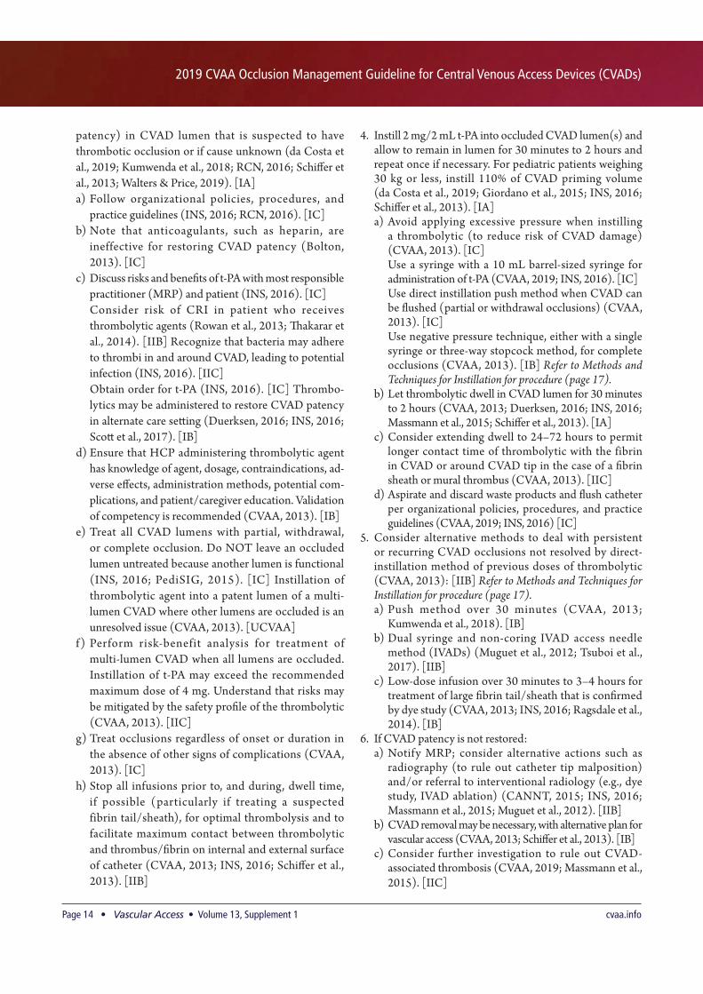
patency) in CVAD lumen that is suspected to have thrombotic occlusion or if cause unknown (da Costa et al., 2019; Kumwenda et al., 2018; RCN, 2016; Schi%er et al., 2013; Walters & Price, 2019). [IA] a) Follow organizational policies, procedures, and
practice guidelines (INS, 2016; RCN, 2016). [IC]b) Note that anticoagulants, such as heparin, are
ineffective for restoring CVAD patency (Bolton, 2013). [IC]
c) Discuss risks and bene"ts of t-PA with most responsible practitioner (MRP) and patient (INS, 2016). [IC]Consider risk of CRI in patient who receives thrombolytic agents (Rowan et al., 2013; !akarar et al., 2014). [IIB] Recognize that bacteria may adhere to thrombi in and around CVAD, leading to potential infection (INS, 2016). [IIC]Obtain order for t-PA (INS, 2016). [IC] Thrombo-lytics may be administered to restore CVAD patency in alternate care se#ing (Duerksen, 2016; INS, 2016; Sco# et al., 2017). [IB]
d) Ensure that HCP administering thrombolytic agent has knowledge of agent, dosage, contraindications, ad-verse e%ects, administration methods, potential com-plications, and patient/caregiver education. Validation of competency is recommended (CVAA, 2013). [IB]
e) Treat all CVAD lumens with partial, withdrawal, or complete occlusion. Do NOT leave an occluded lumen untreated because another lumen is functional (INS, 2016; PediSIG, 2015). [IC] Instillation of thrombolytic agent into a patent lumen of a multi-lumen CVAD where other lumens are occluded is an unresolved issue (CVAA, 2013). [UCVAA]
f) Perform risk-benefit analysis for treatment of multi-lumen CVAD when all lumens are occluded. Instillation of t-PA may exceed the recommended maximum dose of 4 mg. Understand that risks may be mitigated by the safety pro"le of the thrombolytic (CVAA, 2013). [IIC]
g) Treat occlusions regardless of onset or duration in the absence of other signs of complications (CVAA, 2013). [IC]
h) Stop all infusions prior to, and during, dwell time, if possible (particularly if treating a suspected fibrin tail/sheath), for optimal thrombolysis and to facilitate maximum contact between thrombolytic and thrombus/"brin on internal and external surface of catheter (CVAA, 2013; INS, 2016; Schi%er et al., 2013). [IIB]
4. Instill 2 mg/2 mL t-PA into occluded CVAD lumen(s) and allow to remain in lumen for 30 minutes to 2 hours and repeat once if necessary. For pediatric patients weighing 30 kg or less, instill 110% of CVAD priming volume (da Costa et al., 2019; Giordano et al., 2015; INS, 2016; Schi%er et al., 2013). [IA]a) Avoid applying excessive pressure when instilling
a thrombolytic (to reduce risk of CVAD damage) (CVAA, 2013). [IC]Use a syringe with a 10 mL barrel-sized syringe for administration of t-PA (CVAA, 2019; INS, 2016). [IC]Use direct instillation push method when CVAD can be $ushed (partial or withdrawal occlusions) (CVAA, 2013). [IC]Use negative pressure technique, either with a single syringe or three-way stopcock method, for complete occlusions (CVAA, 2013). [IB] Refer to Methods and Techniques for Instillation for procedure (page 17).
b) Let thrombolytic dwell in CVAD lumen for 30 minutes to 2 hours (CVAA, 2013; Duerksen, 2016; INS, 2016; Massmann et al., 2015; Schi%er et al., 2013). [IA]
c) Consider extending dwell to 24–72 hours to permit longer contact time of thrombolytic with the fibrin in CVAD or around CVAD tip in the case of a "brin sheath or mural thrombus (CVAA, 2013). [IIC]
d) Aspirate and discard waste products and $ush catheter per organizational policies, procedures, and practice guidelines (CVAA, 2019; INS, 2016) [IC]
5. Consider alternative methods to deal with persistent or recurring CVAD occlusions not resolved by direct-instillation method of previous doses of thrombolytic (CVAA, 2013): [IIB] Refer to Methods and Techniques for Instillation for procedure (page 17).a) Push method over 30 minutes (CVAA , 2013;
Kumwenda et al., 2018). [IB] b) Dual syringe and non-coring IVAD access needle
method (IVADs) (Muguet et al., 2012; Tsuboi et al., 2017). [IIB]
c) Low-dose infusion over 30 minutes to 3–4 hours for treatment of large "brin tail/sheath that is con"rmed by dye study (CVAA, 2013; INS, 2016; Ragsdale et al., 2014). [IB]
6. If CVAD patency is not restored:a) Notify MRP; consider alternative actions such as
radiography (to rule out catheter tip malposition) and/or referral to interventional radiology (e.g., dye study, IVAD ablation) (CANNT, 2015; INS, 2016; Massmann et al., 2015; Muguet et al., 2012). [IIB]
b) CVAD removal may be necessary, with alternative plan for vascular access (CVAA, 2013; Schi%er et al., 2013). [IB]
c) Consider further investigation to rule out CVAD-associated thrombosis (CVAA, 2019; Massmann et al., 2015). [IIC]
cvaa.info Vascular Access��t��7PMVNF��� �4VQQMFNFOU�����t���1BHF���
2019 CVAA Occlusion Management Guideline for Central Venous Access Devices (CVADs)
7. Monitor patient who has received thrombolytic agent for signs of CRI or CVAD-related thrombosis (Giordano et al., 2015; Westergaard et al., 2013). [IA]
8. Amend plan of care to re$ect any occlusion preventative strategies to ensure CVAD patency, including, but not limited to, (CVAA, 2013): [IC]a) Considering causative factors of CVAD occlusion b) Reviewing $ushing and locking practice.
9. Document assessment findings, related interventions, response to interventions, and follow-up actions (CVAA, 2013). [IC]
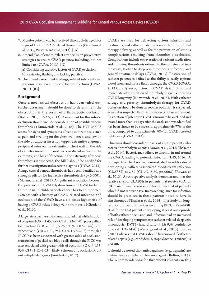
BackgroundOnce a mechanical obstruction has been ruled out, further assessment should be done to determine if the obstruction is the result of a thrombotic occlusion (Bolton, 2013; CVAA, 2013). Assessment for thrombotic occlusion should include consideration of possible venous thrombosis (Kumwenda et al., 2018). The HCP should assess for signs and symptoms of venous thrombosis such as pain and swelling on the chest wall, neck, and jaw on the side of catheter insertion/upper extremity; engorged peripheral veins on the extremity or chest wall on the side of catheter insertion; paresthesia or discoloration of the extremity; and loss of function in the extremity. If venous thrombosis is suspected, the MRP should be noti"ed for further investigation and accurate diagnosis (CVAA, 2013).
A large central venous thrombosis has been identi"ed as a strong predictor for ine%ective thrombolysis (p<0.0001) (Massmann et al., 2015). A signi"cant association between the presence of CVAD dysfunction and CVAD-related thrombosis in children with cancer has been reported. Patients with a history of CVAD-related infection and occlusion of the CVAD have a 6.4 times higher risk of having a CVAD-related deep vein thrombosis (Giordano et al., 2015).
A large retrospective study demonstrated that while infusion of cefepime (OR = 1.45; 95% CI ¼ 1.23–1.70), piperacillin/tazobactam (OR = 1.21; 95% CI ¼ 1.02–1.44), and vancomycin (OR = 1.81; 95% CI ¼ 1.57–2.07) through a PICC has been associated with greater odds of occlusion, transfusion of packed red blood cells through the PICC was also associated with greater odds of occlusion (OR ¼ 1.35; 95% CI ¼ 1.12–1.63) (likely a thrombotic occlusion), but not anti-platelet agents (Smith et al., 2017).
CVADs are used for delivering various infusions and treatments, and catheter patency is important for optimal therapy delivery, as well as for the prevention of serious complications resulting from thrombotic occlusion. Complications include extravasation of vesicant medication and infusates; thrombosis external to the catheter and into the vessel, leading to deep vein thrombosis; infection; and general treatment delays (CVAA, 2013). Restoration of catheter patency is de"ned as the ability to easily aspirate blood from, and infuse $uids through, the CVAD (CVAA, 2013). Early recognition of CVAD dysfunction and immediate administration of thrombolytic agents improves CVAD longevity (Kumwenda et al., 2018). With catheter salvage as a priority, thrombolytic therapy for CVAD occlusion should be done as soon as occlusion is suspected, even if it is suspected that the occlusion is not new or recent. Restoration of patency to CVADs known to be occluded and treated more than 14 days a(er the occlusion was identi"ed has been shown to be successful approximately 77% of the time, compared to approximately 90% for CVADs treated right away (CVAA, 2013).
Clinicians should consider the risk of CRI in patients who receive thrombolytic agents (Rowan et al., 2013; !akarar et al., 2014). Bacteria may adhere to thrombi in and around the CVAD, leading to potential infection (INS, 2016). A retrospective chart review demonstrated an odds ratio of developing a catheter-associated bloodstream infection (CLABSI) as 2.87 (CI1.42–5,80, p=.0002) (Rowan et al., 2013). A retrospective analysis demonstrated that the relative risk for CLABSIs in patients that receive t-PA for PICC maintenance was over three times that of patients who did not require t-PA. Increased vigilance for infection should be practiced in those patients noted to have in situ thrombus (!akarar et al., 2014). In a study on long-term central venous devices including PICCs, Revel-Vilk et al. found that patients developing at least one episode of both catheter occlusion and infection had an increased risk of developing symptomatic catheter-related deep vein thrombosis (DVT) (hazard ratio: 4.15; 95% con"dence interval: 1.2–14.4) (Westergaard et al., 2013). Bolton (2013) advises that CVADs should be removed if catheter-related sepsis (e.g., candidemia, staphylococcus aureus) is present.
It should be noted that anticoagulants (e.g., heparin) are ineffective as a catheter clearance agent (Bolton, 2013). The recommendations for thrombolytic agents in this
1BHF������t���Vascular Access��t��7PMVNF��� �4VQQMFNFOU��� DWBB�JOGP
2019 CVAA Occlusion Management Guideline for Central Venous Access Devices (CVADs)
guideline section refer to the use of Cathflo® (alteplase, recombinant). At the time of this guideline’s publication, Cathflo® (alteplase, recombinant) is the only Health Canada–approved thrombolytic agent proven to be safe, e%ective, and appropriate for restoring catheter patency in the adult and pediatric (older than two years) population (da Costa et al., 2019; CVAA, 2013; Giordano et al., 2015; INS, 2016; Schi%er et al., 2013). !e Cath$o® (alteplase, recombinant) Pediatric Study demonstrated the safety and efficacy of Cathflo® (alteplase, recombinant) in the pediatric population, including children younger than two years (CVAA, 2013). Recommendations that are outside the Cath$o® (alteplase, recombinant) product monograph are rated as level C recommendations, as they are obtained through ORG consensus (CVAA, 2013).
As per the Cathflo® (alteplase, recombinant) product monograph, the recommended dose for persons weighing more than 30 kg is 2 mg with a dose volume of 2 mL (CVAA, 2013). A large multi-centre prospective medical record review of 1,684 patients who experienced PICC occlusion found that the mean doses of t-PA provided per catheter occlusion event was 1.45 (median, 1), while some PICCs received as many as nine doses of t-PA during follow-up (Smith et al., 2017).
The ORG recognizes that the priming volumes of some manufacturer recommended CVADs are less than 2 mL and that the recommended Cath$o® (alteplase, recombinant) dose volume of 2 mL may, therefore, lead to over"ll. ORG consensus suggests that this over"ll allows the interface of the thrombolytic agent with any external fibrin that may extend beyond the distal end of the CVAD (e.g., a fibrin sheath along the external portion of the catheter); thus, common practice is to instill the full recommended dose of 2 mg/2 mL (CVAA, 2013).
Limited evidence suggests that lower doses of Cathflo® (alteplase, recombinant) (e.g., 1 mg/mL) in lumens requiring less than, or equal to, 1 mL volume are e%ective and may provide cost savings. However, randomized controlled trials are required to determine the e&cacy of alternate dosing (CVAA, 2013; Giordano et al., 2015; Jafari et al., 2018; Massmann et al., 2015; Sapienza & Ciaschini, 2015).
A quasi-experimental non-randomized study of 270 CVAD occlusions (predominantly PICCs) in a long-term acute facility demonstrated that there is no di%erence in e&cacy between 1 mg/mL intraluminal dose of Cath$o® (alteplase, recombinant) and standard 2 mg/2mL with the intraluminal volume dose signi"cantly more cost-e%ective (Sapienza & Ciaschini, 2015). A literature review reported Cathflo® (alteplase, recombinant) doses of 0.5–2 mg instilled into the lumen of pediatric patients with dwell times ranging from 30 to more than 240 minutes, with greater efficacy generally reported with larger doses and longer dwell times (Anderson et al., 2013).
There is ongoing controversy and debate over standard management for multi-lumen CVADs, especially CVADs with two or more lumens that are occluded. Treating all lumens in a multi-lumen CVAD will exceed the maximum dose tested in clinical studies that used a total dose of 4 mg/4 mL (CVAA, 2013). Consideration should be made as to the type of CVAD, whether it is for short-term or long-term duration, and the type of tip (staggered [distal, medial, or proximal] or non-staggered), keeping in mind that catheter salvage is a priority. Performing a risk-bene"t analysis is recommended when the medication product monograph is not followed and when more than one lumen in a multi-lumen CVAD requires thrombolytic (CVAA, 2013).
Radiographic studies (i.e., chest radiograph) should be considered if catheter patency is not restored, if occlusion recurs or if catheter migration is suspected and not necessarily prior to thrombolytic instillation (CVAA, 2013). Radiographic studies were not required prior to Cath$o® (alteplase, recombinant) instillation in the pivotal clinical study protocols for Cath$o® (alteplase, recombinant) (CVAA, 2013). A specialty team and the use of a CVAD patency assessment algorithm can be e%ective to determine the root cause of occlusion prior to thrombolytic use (Steere et al., 2018).
cvaa.info Vascular Access��t��7PMVNF��� �4VQQMFNFOU�����t���1BHF���
2019 CVAA Occlusion Management Guideline for Central Venous Access Devices (CVADs)
Methods and Techniques for InstillationProcedures to restore catheter patency should be performed as soon as signs of occlusion (partial, withdrawal, or complete) are identi"ed. !is will increase the e&cacy of thrombolysis and, thereby, avoid, or at least delay, the need for catheter replacement (CVAA, 2013). !ere are several methods used for instillation of the catheter clearance agent. It is recommended to stop all infusions, if possible, in a multi-lumen CVAD during thrombolytic dwell time to optimize thrombolysis (INS, 2016).
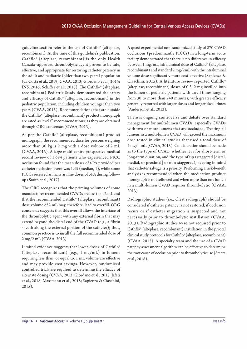
For partial and withdrawal occlusions, direct instillation of the thrombolytic can be performed with a single 10 mL syringe with thrombolytic (Figure 1). Instillation should be done slowly versus quickly “injecting” into the CVAD lumen; the goal is for the thrombolytic to come into contact with the thrombus or clot burden and be “soaked up” (CVAA, 2013).
Instillation of a thrombolytic in a completely occluded catheter requires the use of negative pressure to create a vacuum by aspirating air or dead space from within the catheter, thus allowing the thrombolytic to be drawn forward into the catheter to the clot interface (Bolton, 2013; CVAA, 2013). !ere are two techniques for achieving negative pressure (single syringe or stopcock methods).
Method 1: Single Syringe!e single syringe technique uses a single 10 mL syringe (with reconstituted thrombolytic) a#ached directly to the occluded CVAD lumen hub. When the plunger is pulled back, a vacuum is created. The plunger is then slowly released; this allows the thrombolytic to be “pulled” into the lumen toward the thrombus/clot burden causing the occlusion (Figure 1) (Bolton, 2013; CVAA, 2013).
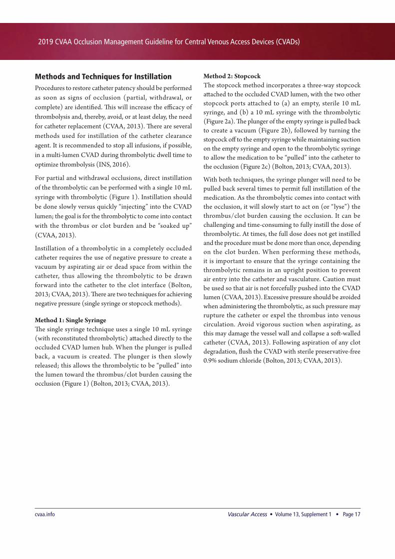
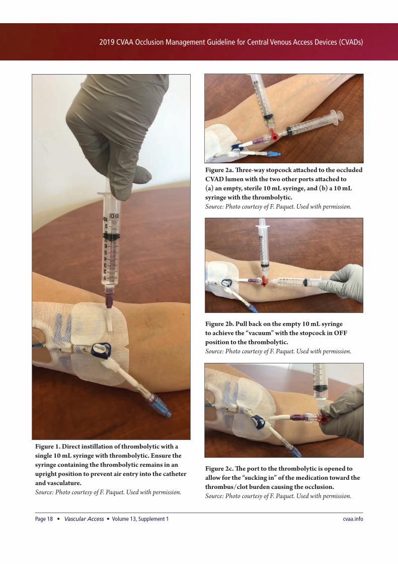
Method 2: StopcockThe stopcock method incorporates a three-way stopcock a#ached to the occluded CVAD lumen, with the two other stopcock ports attached to (a) an empty, sterile 10 mL syringe, and (b) a 10 mL syringe with the thrombolytic (Figure 2a). !e plunger of the empty syringe is pulled back to create a vacuum (Figure 2b), followed by turning the stopcock o% to the empty syringe while maintaining suction on the empty syringe and open to the thrombolytic syringe to allow the medication to be “pulled” into the catheter to the occlusion (Figure 2c) (Bolton, 2013; CVAA, 2013).
With both techniques, the syringe plunger will need to be pulled back several times to permit full instillation of the medication. As the thrombolytic comes into contact with the occlusion, it will slowly start to act on (or “lyse”) the thrombus/clot burden causing the occlusion. It can be challenging and time-consuming to fully instill the dose of thrombolytic. At times, the full dose does not get instilled and the procedure must be done more than once, depending on the clot burden. When performing these methods, it is important to ensure that the syringe containing the thrombolytic remains in an upright position to prevent air entry into the catheter and vasculature. Caution must be used so that air is not forcefully pushed into the CVAD lumen (CVAA, 2013). Excessive pressure should be avoided when administering the thrombolytic, as such pressure may rupture the catheter or expel the thrombus into venous circulation. Avoid vigorous suction when aspirating, as this may damage the vessel wall and collapse a so(-walled catheter (CVAA, 2013). Following aspiration of any clot degradation, $ush the CVAD with sterile preservative-free 0.9% sodium chloride (Bolton, 2013; CVAA, 2013).
1BHF������t���Vascular Access��t��7PMVNF��� �4VQQMFNFOU��� DWBB�JOGP
2019 CVAA Occlusion Management Guideline for Central Venous Access Devices (CVADs)
Figure 1. Direct instillation of thrombolytic with a single 10 mL syringe with thrombolytic. Ensure the syringe containing the thrombolytic remains in an upright position to prevent air entry into the catheter and vasculature.Source: Photo courtesy of F. Paquet. Used with permission.
Figure 2a. !ree-way stopcock a#ached to the occluded CVAD lumen with the two other ports a#ached to (a) an empty, sterile 10 mL syringe, and (b) a 10 mL syringe with the thrombolytic. Source: Photo courtesy of F. Paquet. Used with permission.
Figure 2b. Pull back on the empty 10 mL syringe to achieve the “vacuum” with the stopcock in OFF position to the thrombolytic. Source: Photo courtesy of F. Paquet. Used with permission.
Figure 2c. !e port to the thrombolytic is opened to allow for the “sucking in” of the medication toward the thrombus/clot burden causing the occlusion. Source: Photo courtesy of F. Paquet. Used with permission.

cvaa.info Vascular Access��t��7PMVNF��� �4VQQMFNFOU�����t���1BHF���
2019 CVAA Occlusion Management Guideline for Central Venous Access Devices (CVADs)
Method 3: Push The push method for administration of thrombolytics has been successfully used with hemodialysis catheters with recurrent occlusions or pump speeds of less than 200 mL/min (CVAA, 2013). For other CVADs such as PICCs, IVADs, and tunneled devices that are smaller in lumen size than hemodialysis CVADs, the push method may be considered when there is a recurrence of partial and withdrawal occlusions a(er multiple direct instillations of thrombolytic. In the push method, Cath$o® (alteplase, recombinant) is administered by direct instillation. A total amount of 2 mg/2 mL is instilled, and 0.3 mL of sterile preservative-free 0.9% sodium chloride is “pushed in” every 10 minutes for 30 minutes (CVAA, 2013). !e theory behind this method is that the thrombolytic will slowly be pushed into the CVAD lumen to interface with the thrombus or clot burden over 30 minutes and act on (or “lyse”) the thrombus causing the occlusion (CVAA, 2013).
Method 4: Dual syringe and non-coring IVAD access needle (IVADs) Two small retrospective studies suggest the safety and e&cacy of a two-syringe mechanical $ushing approach or dual-needle pumping technique to resolve IVAD occlusions. !is method uses an empty syringe and pre-"lled sterile preservative-free 0.9% sodium chloride syringe (Muguet et al., 2012)/distilled water or urokinase solutions (Tsuboi et al., 2017) each a#ached to a non-coring needle (Figure 3a). !ese needles are then inserted close to each other within the septum of the IVAD (Figure 3b). !e pumping e%ect is obtained by aspirating with one syringe while infusing with the other syringe and repeating these actions (Figure 3a). !ese small studies reported an 88% success rate (Tsuboi et al., 2017) and a 92% success rate (Muguet et al., 2012). It should be noted that in one study, Tsuboi et al., 2017, $uoroscopy or venography was completed to examine a system break in the IVAD, prior to a#empting the dual-needle pumping technique. !is technique may be considered a safe and e%ective alternative when other methods of occlusion treatment are not successful.
Method 5: Low-Dose Infusion !is method of administration of thrombolytic has also been used successfully for thrombotic occlusion management of hemodialysis catheters in both the adult and pediatric patient populations (CVAA, 2013; Ragsdale et al., 2014). For other CVADs such as PICCs, IVADs, and tunneled devices that are smaller in lumen size than hemodialysis CVADs, low-dose infusion of thrombolytic may be considered when there is a recurrence of partial and withdrawal occlusions a(er multiple direct instillations of thrombolytic, including administration
Figure 3b. Use di$erent needle lengths to access port if needle footprint is too large to be side by side. Source: Photo courtesy of F. Paquet. Used with permission.
Figure 3a. Dual syringe method to alternate %ushing and aspiration of IVAD.Source: Photo courtesy of K. Naayer. Used with permission.
by the push method. Consider the use of a low-dose infusion over 30 minutes to 3–4 hours for treatment of large "brin tail/sheath that is con"rmed by dye study (CVAA, 2013; INS, 2016; Ragsdale et al., 2014). Low-dose infusion of Cath$o® (alteplase, recombinant) has been demonstrated to be e%ective in studies with protocols ranging from 1–4 mg of Cath$o® (alteplase, recombinant) (one study used 10 mg) in sterile preservative-free 0.9% sodium chloride over 30–60 minutes (CVAA, 2013). !ere is also literature to support low-dose infusion over 3 hours (CVAA, 2013; Ragsdale et al., 2014).
Sample dosing includes the following (CVAA, 2013; Ragsdale et al., 2014): r� 1–2 mg reconstituted Cath$o® (alteplase, recombinant) in
50 mL mini-bag of sterile preservative-free 0.9% sodium chloride over 30 minutes
r� 2–4 mg reconstituted Cath$o® (alteplase, recombinant) in 100 mL mini-bag of sterile preservative-free 0.9% sodium chloride over 60 minutes

1BHF������t���Vascular Access��t��7PMVNF��� �4VQQMFNFOU��� DWBB�JOGP
2019 CVAA Occlusion Management Guideline for Central Venous Access Devices (CVADs)
r� 3–5 mg reconstituted Cath$o® (alteplase, recombinant) in 50–100 mL mini-bag of sterile preservative-free 0.9% sodium chloride over 3 hours
r� Max. 2 mg Cathflo® (alteplase, recombinant) in 25 mL of sterile preservative-free 0.9% sodium chloride over 3 hours (reported in critically ill children).
The theory behind this method is that the thrombolytic will slowly and continuously reach and act on the thrombus causing occlusions that can be located along the length of the catheter’s external surface (CVAA, 2013).
Pediatric ImplicationsThe recommendations listed in this guideline apply to the pediatric population. Patients with CVADs who are between the ages of 12 months and 18 years are included. There is evidence for the use and effectiveness of thrombolytics for CVAD occlusion management in this population (Anderson et al., 2013; CVAA, 2013; de Lorenzo-Pinto et al., 2014; Giordano et al., 2015; Ragsdale et al., 2014). !e dosing of thrombolytic is based on the patient’s weight and the priming volume of the catheter if the patient weighs less than 30 kg, as shown in Table 4 (CVAA, 2013). Table 5 presents estimated catheter priming volume and thrombolytic dose ranges (CVAA, 2013).
Caution!e medication product monograph for Cath$o® (alteplase, recombinant) that outlines precautions, contraindications, and side e%ects to be aware of when using this thrombolytic for CVAD occlusion management should be used as the main reference for all precautions. Caution should be exercised with patients who have active internal bleeding, have thrombocytopenia or other hemostatic defects, are pregnant, or have a known or suspected CRI (CVAA, 2013).
Other Interventions Additional thrombolytic agents are being investigated (i.e., reteplase, alfimeprase, tenecteplase, urokinase), and the results vary in terms of e&cacy, dwell time, number of doses required, adverse events, and cost (Bolton, 2013; CVAA, 2013; de Lorenzo-Pinto et al., 2014; Gallieni et al., 2016; Giordano et al., 2015; Kennard et al., 2017; Kumwenda et al., 2018; Mendes et al., 2014; Muguet et al., 2012; Pollo et al., 2016; Schi%er et al., 2013; Westergaard et al., 2013).
At the time of publication, these agents are not available in Canada for management of CVAD occlusion. More studies are needed to show the e&cacy and safety of other agents for thrombolysis as well as for direct comparison with Cath$o® (alteplase, recombinant) (CVAA, 2013).
Table 5. CVAD Priming Volumes and !rombolytic Dose Ranges
Types of CVAD
Priming Volume Ranges (Estimated Volumes)
!rombolytic Dose Ranges* (Estimated Doses)
PICC SL 1.9F/2F 0.08–0.1 mL 0.09–0.11 mL
3F 0.22–0.38 mL 0.24–0.42 mL
4F 0.38–1.5 mL 0.42–1.7 mL
5F 0.82 mL 0.9 mL
DL 4F 0.33–0.45 mL 0.36–0.5 mL
5F 0.41–1 mL 0.45–1.1 mL
Non-tunneled CVAD
SL 2.5F 0.05 mL 0.06 mL
3F 0.1 mL 0.11 mL
4F 0.1 mL 0.11 mL
DL 4F 0.1–0.2 mL 0.11–0.22 mL
5F 0.2 mL 0.22 mL
TL 5F 0.2–0.3 mL 0.22–0.33 mL
7F 0.3–0.5 mL 0.33–0.55 mL
Tunneled CVAD
SL 2.7F 0.15 mL 0.17 mL
4.2F 0.3 mL 0.33 mL
6.6F 0.7 mL 0.77 mL
DL 7F 0.6–0.9 mL 0.66–1 mL
9F 1.5–2 mL 1.7–2 mL
Port SL 6F 1.6 mL 1.8 mL
6.6F 0.9–1.2 mL 1.0–1.3 mL
8F 1.8 mL 2 mL
DL 10F 1.8 mL 2 mL
DL = double lumen; PICC = peripherally inserted central catheter; SL = single lumen; TL = triple lumen. *Maximum dose is 2 mL.
Source: Used with permission "om the Association for Vascular Access/PediSIG (CVAA, 2013)
Table 4. Pediatric Dosing of Cath%o® (alteplase, recombinant)
Patient Weight Cath%o® (alteplase, recombinant) (!rombolytic) Dose
Less than 30 kg 110% of "ll volume
30 kg or more 2 mg/2 mL
Source: Ho#man-La Roche Limited. (2018). Product Monograph Cath$o. Retrieved "om: h%ps://www.rochecanada.com/PMs/Cath$o/Cath$o_PM_E.pdf

cvaa.info Vascular Access��t��7PMVNF��� �4VQQMFNFOU�����t���1BHF���
2019 CVAA Occlusion Management Guideline for Central Venous Access Devices (CVADs)
Surgical/interventional radiologic interventions (such as endoluminal snare, sheath stripping, stenting with radiofrequency guidewire, catheter removal with balloon disruption of sheath, or guidewire exchange) are described in the literature as interventions to be performed if the thrombolytic agent is unsuccessful (CVAA, 2013; Gallieni et al., 2016; Massmann et al., 2015; Muguet et al., 2012; Niyyar & Chan, 2013). However, no strong studies were found to support the e&cacy and safety of these measures (CVAA, 2013). A risk-bene"t analysis is recommended, as well as a full consultation with MRP (CVAA, 2013).
4.0 Assessment and Management of Chemical Occlusion 4.0 RecommendationsSigns and symptoms of chemical occlusion may include, but are not limited to, the following:r� Presence of visible precipitate in catheter or tubingr� Previous administration of certain solutions and/or medica-
tions or interaction of certain solutions and/or medications.
1. Assess CVAD occlusion to identify if occlusion is caused by chemical obstruction (Ast & Ast, 2014; Bolton, 2013; CVAA, 2013; Gabriel, 2013; INS, 2016; Mokha et al., 2017; PediSIG, 2015; RCN, 2016). [IB]a) Observe CVAD or tubing for presence of visible precipitate. b) Assess infusion plan to identify which solutions and/or
medications were administered. c) Verify solution and/or medication dilution properties,
check solution and/or medication incompatibilities, and assess types of solutions and/or medications instilled (e.g., rule out lipid-related occlusion or lipid residue).
d) Obtain history of current and past infusion rate and $ushing frequency.
2. Promptly attempt to restore patency occluded by chemical precipitate by instillation of CVAD clearance agent recognized to dissolve precipitate (Ast & Ast, 2014; Bolton, 2013; CVAA, 2013; Giordano et al., 2015; INS, 2016; Pai & Plogsted, 2014; PediSIG, 2015; RCN, 2016; Schilcher et al., 2013; Wall et al., 2016). [IIA] a) Follow organizational policies, procedures, and
practice guidelines. [IC]b) HCP administering CVAD clearance agent must
have knowledge of agent, dosage, contraindications, adverse effects, administration methods, potential complications, and patient/caregiver education. Validation of competency is recommended (CVAA, 2013). [IB] Only HCP with specialized knowledge and extensive experience with CVAD occlusion management should perform this procedure. [IC]
c) Discuss risks and benefits of CVAD clearance agent with MRP and patient and obtain order from licensed prescriber.
d) Instill by direct-syringe instillation method if partial or withdrawal occlusion (CVAA, 2013). [IB]
e) Instill by negative pressure using single syringe or three-way stopcock method if complete occlusion (CVAA, 2013). [IB]
f) Instillation volume of CVAD clearance agent should be "ll-volume of CVAD lumen only.
3. Notify MRP if CVAD clearance procedure does not result in patency of CVAD (CVAA, 2013). [IIB]
4. Consider use of thrombolytic agent if patency is not restored with chemical clearance agent (CVAA, 2013; Wall et al., 2016). [IIB]
5. Amend plan of care to re$ect any occlusion-preventative strategies to ensure CVAD patency, considering causative factors of CVAD occlusion (CVAA, 2013; INS, 2016). [IC]
6. Document assessment findings, related interventions, and response to interventions (CVAA, 2013; INS, 2016; RCN, 2016). [IC]
BackgroundOcclusions can be caused by the infusion of crystallized medication, by preformed precipitates, or by the formation of precipitates within the CVAD lumen (CVAA, 2013). Alteration in the pH of solutions and/or medications exposed to other solutions and/or medications that have an opposing pH is associated with precipitation. Solutions and/or medications such as phenytoin, lipids and parenteral nutrition, and mannitol are commonly affected, and lipid solutions can produce a waxy residue that can cause CVAD occlusion (CVAA, 2013). !e ORG identi"ed cloxacillin as another medication that is known to precipitate and occlude CVADs.
Instillation of catheter clearance agents recognized to dissolve precipitate may be indicated to restore catheter patency. It is worth noting that phenytoin occlusion can be permanent and requires catheter replacement. A#empts to clear CVAD lumens occluded by chemical precipitate or lipid residue continue to be made despite a 2012 Cochrane Systematic Review that concluded there are no strong studies investigating the e&cacy and safety of chemical interventions for the management of chemical occlusions in CVADs (CVAA, 2013). Catheter salvage is still a priority, and every e%ort should be made to clear occlusions appropriately.

1BHF������t���Vascular Access��t��7PMVNF��� �4VQQMFNFOU��� DWBB�JOGP
2019 CVAA Occlusion Management Guideline for Central Venous Access Devices (CVADs)
!e use of hydrochloric acid (HCl), L-cysteine hydrochloride (L-cysteine), sodium bicarbonate (NaHCO3), and sodium hydroxide (NaOH) to clear medication precipitates in CVADs is noted in the literature to be effective (CVAA, 2013). HCl and L-cysteine are e%ective in treating CVAD occlusions from precipitates of acidic (low pH) medications, parenteral nutrition, amino acids, and calcium phosphorous precipitants (Ast & Ast, 2014; Pai & Plogsted, 2014; Zheng et al., 2019). NaHCO3 is e%ective in treating CVAD occlusion from precipitates of alkaline (high pH) medications such as ganciclovir, acyclovir, ampicillin, imipenem, and heparin. NaOH has been demonstrated in a few studies to be e%ective in clearing partially occluded CVADs due to parenteral nutrition (with or without lipids) (CVAA, 2013). In one study, the protocol for NaOH administration involved a long infusion (more than 10 hours) followed by sterile preservative-free 0.9% sodium chloride infusion and $ushing through the CVAD lumen (CVAA, 2013).
HCPs should not follow NaHCO3 with HCl as the combination could generate damage or further precipitation material—these two agents should not be mixed (CVAA, 2013). !ere is also concern with the use of HCl because of risk of damage to the wall of the catheter (CVAA, 2013). HCPs should be aware that direct infusion of HCl or NaHCO3 into the venous system may cause reactions such as fever, phlebitis, and sepsis (CVAA, 2013). !ese reactions may be avoided by aspirating the solution in full, rather than $ushing it through the catheter and into the central venous system (CVAA, 2013). Infusion of NaOH is not hazardous if administered slowly and was not seen to contribute to catheter material degradation as noted with HCl and NaHCO3 (CVAA, 2013). Check the compatibility of other clearing agents such as HCl and NaHCO3 with the manufacturer's instructions for use of the catheter (CVAA, 2013).
The compounding and preparation of HCl, L-cysteine, NaHCO3, and NaOH should be done in pharmacy by HCPs who have extensive knowledge of the precautions required during compounding and preparation. !e following should be noted for each agent:r� HCl: Chemical grade HCl 0.1 N is diluted down with
sterile preservative-free 0.9% sodium chloride to get a 0.1 N solution (CVAA, 2013).
r� NaHCO3: Sterile injectable preparations are commercially available in an 8.4% concentration (CVAA, 2013).
r� NaOH: Commercially manufactured/prepared pellets (powder) can be dissolved with sterile water and prepared
with an appropriate filter to make up approximately a 0.1 N NaOH solution.
!ere is literature to support the use of 70% sterile ethanol to treat occlusions caused by lipid residue (CVAA, 2013). !e safety of sterile ethanol in the laboratory se#ing has been demonstrated and side effects of sterile ethanol administration include dizziness, headaches, nausea, fatigue, and light-headedness (CVAA, 2013). Precautions for the use of 70% sterile ethanol for CVAD occlusion management include ensuring there is no patient sensitivity to sterile ethanol and informing the patient (or caregiver for pediatric patients) that the instillation of sterile ethanol may a%ect blood levels. !is agent must be used with caution with polyurethane CVADs, as sterile ethanol may damage catheter materials. Check the compatibility of sterile ethanol with the manufacturer's instructions for use of the catheter (CVAA, 2013). !e compounding and preparation of ethyl alcohol 70% should be done by the pharmacy by diluting concentrated sterile ethyl alcohol with sterile water to make a 70% concentration (CVAA, 2013).There i s l i terature ident i f y ing that L -c ysteine hydrochloride has been effective in reducing the pH of parenteral nutrition solutions and increasing the solubility of calcium phosphate in the neonate population. !e use of L-cysteine in this manner would be considered an “o%-label” use and is not approved by the US Food and Drug Administration (FDA) or Health Canada. Due to the risks of HCl and the cost of preparation, L-cysteine may yet become an appropriate option (Ast & Ast, 2014). Usage of these products as catheter clearance agents is considered off-label use; the HCP should follow organizational policies, procedures, and practice guidelines regarding o%-label use of products.
The procedure for the instillation of a catheter clearance agent is similar to that for the instillation of a thrombolytic by using the direct instillation method for partial occlusions and using negative pressure either with the single syringe or three-way stopcock method for complete occlusions (CVAA, 2013). !ere is no evidence to support the strategy of “over"ll” of the CVAD lumen for chemical occlusions, and the recommendation in this guideline is to instill the clearance agent to the "ll volume of the lumen only. Table 6 outlines precipitates and treatments. Refer to Table 5 for estimated CVAD volumes and instill amount to "ll the catheter lumen.

cvaa.info Vascular Access��t��7PMVNF��� �4VQQMFNFOU�����t���1BHF���
2019 CVAA Occlusion Management Guideline for Central Venous Access Devices (CVADs)
Table 6. Types of Chemical Occlusion and Treatment
Cause of Occlusion Treatment (Clearance Agent)
Lipid residue Sterile ethanol 70% (CVAA, 2013)
Acidic medication precipitate (low pH: less than 6.0) (e.g., vancomycin, parenteral nutrition)
Hydrochloric acid (HCl) 0.1 N (CVAA, 2013) or L-cysteine* (Pai & Plogsted, 2014; Zheng et al., 2019)
Calcium Phosphate Hydrochloric acid (HCl) 0.1 N or L-cysteine* Hydrochloride (Ast & Ast, 2014)
Alkaline (basic) medication precipitate (high pH: greater than 7.0) (e.g., imipenem, heparin)
Sodium bicarbonate (NaHCO3) 8.4% (CVAA, 2013)Sodium hydroxide (NaOH) 0.1 mmol/L (CVAA, 2013)
* Not a Food and Drug Administration (FDA)/Health Canada approved indication
!e following basic procedure is recommended (Refer to Methods and Techniques for Instillation on page 17):1. Instill sufficient volume to fill the catheter lumen,
using negative pressure if the CVAD is completely occluded.
2. Allow to dwell for 20–60 minutes.3. A(er dwell time, a#empt to aspirate 3–5 mL of blood and
contents.4. Instillation may be repeated once if patency is not
restored a(er the "rst a#empt or dose.
For pediatric patients, a dose of 0.55 mL/kg of 70% sterile ethanol (to a maximum of 3 mL) may be used to treat occlusions related to lipid administration (CVAA, 2013). Successful clearance with HCl has been reported in the pediatric population; one study protocol used up to three doses of HCl 0.1 N (0.2–0.5 mL) with a 20 minute dwell and up to 1 mL in patients between 1 and 3 kg and up to 3 mL in patients greater than 3 kg (CVAA, 2013).
5.0 Prevention, Monitoring, and Auditing Criteria for CVAD Occlusion 5.0 Recommendations1. Ensure ongoing education and competency validation of
HCP responsible for CVAD care and management in the following (CVAA, 2013; CVAA, 2019): [IB]a) Principles of CVAD patencyb) Assessment, prevention, and management of catheter
occlusions (PediSIG, 2015; RCN, 2016) [IC]c) CVAD type and add-on device(s) features (CVAA,
2013; INS, 2016; Westergaard et al., 2013) [IC]d) Documentation and continued surveillance (Nailon &
Rupp, 2015; RCN, 2016). [IC]2. Ensure outcomes are measured and include information
on catheter type, patient information, therapy details and methods of occlusion management (CVAA, 2013). [IB]
3. Consider using products designed speci"cally to prevent CVAD occlusion (e.g., neutral or anti-re$ux connector, anti-thrombogenic catheter or coating, alternate locking solutions) (de Lorenzo-Pinto et al., 2014; McDiarmid et al., 2017). [IIB]
Refer to Canadian Vascular Access and Infusion !erapy Guidelines – 2019 for recommendations on Patency, Flushing, and Locking.
BackgroundStrategies to prevent CVAD occlusion should occur routinely. HCPs should consider $ushing, using pulsatile or “push-pause” technique, with an appropriate amount of flush solution and minimizing the number of times the CVAD is being accessed. It is important to $ush with sterile preservative-free 0.9% sodium chloride in between the administration of incompatible medications and/or solutions to prevent CVAD occlusions due to precipitation or residue. For solution and/or medication that is not compatible with sterile preservative-free 0.9% sodium chloride, use a compatible solution (e.g., sterile dextrose in water infusion) before and a(er solution and/or medication administration. Follow with sterile preservative-free 0.9% sodium chloride $ush to clear solution from CVAD (CVAA, 2019). At a minimum, ensure CVAD $ushing and locking is performed to maintain patency (CVAA, 2019).
Sterile preservative-free 0.9% sodium chloride is commonly used for CVAD $ushing and locking to maintain patency (CVAA, 2019). The use of alternate lock solutions such

1BHF������t���Vascular Access��t��7PMVNF��� �4VQQMFNFOU��� DWBB�JOGP
2019 CVAA Occlusion Management Guideline for Central Venous Access Devices (CVADs)
as heparin, sterile ethanol, taurolidine, sodium citrate, or ethylenediaminetetra-acetic acid (EDTA) after sterile preservative-free 0.9% sodium chloride flushing is also common practice for fluid locking CVADs to maintain patency in certain clinical situations. If heparin is required, the concentration should be the lowest available (i.e., 10 units/mL or 100 units/mL) to maintain CVAD catheter patency (CVAA, 2013). The risk for heparin-induced thrombocytopenia is associated with any heparin exposure. The use of thrombolytic prophylaxis has been studied in various clinical settings (CVAA, 2013). Based on the observation that catheter-related thrombosis and CRI are closely associated, several of these studies reported that the use of a thrombolytic catheter locking solution (urokinase or Cath$o® [alteplase recombinant]) resulted in a reduced incidence of both catheter-related thrombosis and CRI (CVAA, 2013). Collaborate with MRP and healthcare team when considering use of antibiotic lock only as catheter salvage strategy (CVAA, 2019). Use lock solution that is aligned with systemic antimicrobial therapy and organizational antibiotic stewardship (CVAA, 2019).
Monitoring and Auditing Criteria for CVAD OcclusionThere is no data addressing acceptable benchmarks for CVAD occlusion rates in Canada. It is important to consider the complexity of CVADs in terms of their varied types and features, as well as the di%erent types of occlusions. Applying the recommendations in this guideline can help manage, treat, and prevent occlusions, but it is important for the HCP to measure outcomes in addition to using these strategies, as a way to validate work and efforts toward positive patient outcomes. Outcome data should be used to guide quality improvement measures to reduce CVAD occlusion rates and related complications. Recommendations for outcome measurement related to CVAD occlusions include monitoring the following:r� CVAD type, location, number of lumens, insertion date,
tip locationr� Patient gender, age, diagnosisr� Patient location in healthcare se#ing (acute, community,
long-term care)r� !erapies administered through CVADr� Date occlusion identi"ed; which lumenr� Type of occlusion identified (partial, withdrawal, or
complete)r� Number of doses of thrombolytic instilled
r� Method of instillation for administration of thrombolyticr� Outcomes:
■■ Success of catheter clearance■■ Lack of success of catheter clearance (leading to
catheter replacement or catheter removal)■■ Upper-extremity deep vein thrombosis■■ Catheter-related bloodstream infection.
Implementation StrategiesA goal for the ORG was to identify barriers to, and strategies for, implementing the recommendations outlined in this guideline. Barriers identi"ed include the following:r� Advances in technologyr� Change fatiguer� Complexity and cumbersomeness of change processesr� Diverse practice se#ingsr� Diversity in current practices; lack of standardizationr� Financial and human resource shortagesr� Knowledge gapsr� Labour-intensive nature of updating education, policies,
and procedures.
Strategies identi"ed to overcome such barriers may include (CVAA, 2013):r� Identify and secure the required resources at facility to
assist with the implementation.r� Ensure HCPs are oriented to technology, equipment,
and materials, and that there is ongoing assessment of knowledge to allow for consistency and sustainability of practice standards.
r� Enlist a dedicated occlusion management resource person who can provide clinical expertise, leadership, and support.
r� Provide educational sessions and ongoing support for the implementation of the occlusion management guideline. !is might include the following:■■ Direct in-servicing and sessions with sta% (consider the
use of technologies such as videos, mobile applications, etc.)
■■ Case studies – presentation and discussion for problem solving
■■ Handouts – “cheat sheets”■■ Pocket cards
r� Identify, develop, and support occlusion management “champions” on designated clinical units to promote and support the guideline implementation. Champions can train and mentor other HCPs within the facility to ensure knowledge transfer and sustainability.

cvaa.info Vascular Access��t��7PMVNF��� �4VQQMFNFOU�����t���1BHF���
2019 CVAA Occlusion Management Guideline for Central Venous Access Devices (CVADs)
r� Ensure that sufficient numbers of staff are trained to perform CVAD clearance procedures to facilitate prompt management of thrombotic or chemical occlusions.
r� Provide ongoing support and resources such as policies, procedures, algorithms, competency assessment checklists, and documentation tools.
r� Develop and provide a range of self-learning, group learning, mentorship, and reinforcement strategies that will build the knowledge and confidence of HCPs in implementing this guideline.
r� Use the clinical practice tools that complement this guideline for procedures, tools, and resources designed to assist with the implementation of the recommendations outlined in this guideline.
Refer to the Canadian Vascular Access Association website (www.cvaa.info) for clinical practice tools, strategies, and templates to facilitate catheter occlusion management:r� Pre-printed ordersr� Procedure templatesr� Medical directivesr� Competency assessmentsr� Patient education and informationr� Outcome measurement tools (data collection tools).
GlossaryCavo-atrial (atrial caval) junction: The point at which the superior vena cava meets and melds into the superior wall of the cardiac right atrium. Both the superior vena cava and inferior vena cava enter the right atrium, but only the superior entry is called the cavo-atrial junction (or atrial caval junction). This junction marks the inferior end of the superior vena cava, the continuation below that point being considered part of the heart.
Chemical occlusion: CVAD occlusion resulting from the mixing of two incompatible medications and/or solutions or from the buildup of lipid or medication within the lumen.
Complete occlusion: Inability to infuse fluid into the CVAD lumen or withdraw blood or fluid from the CVAD lumen.
CVAD patency: Ability to easily aspirate blood from and easily instill/infuse $uid through the CVAD lumen(s).
Mechanical occlusion: Occlusion of a CVAD involving a component of the infusion system. An external occlusion may include a "lter, a needle-free connector, a malpositioned or blocked non-coring needle, or a closed clamp. An internal occlusion results from pinch-o% syndrome or from a kinked or malpositioned CVAD.
Occlusion: Obstruction of a CVAD lumen, preventing or limiting the ability to flush, withdraw blood, and/or administer solutions or medications.
Partial occlusion: Decreased ability to infuse fluid into the CVAD lumen or withdraw blood from the CVAD lumen.
Persistent occlusion: Catheter patency is not achieved a(er two to three doses of catheter clearance agent.
Recurrent occlusion: Catheter patency is not maintained, requiring repeated doses of catheter clearance agent.
!rombotic occlusion: CVAD occlusion resulting from "brin buildup (i.e., "brin sheath or "brin tail) or a blood clot within the catheter lumen or vessel.
!rombolytic: A medication that dissolves or lyses blood clots; activates plasminogen and breaks down "brin.
Withdrawal occlusion: Blood return is sluggish or absent, yet the CVAD $ushes or infuses without di&culty.

1BHF������t���Vascular Access��t��7PMVNF��� �4VQQMFNFOU��� DWBB�JOGP
2019 CVAA Occlusion Management Guideline for Central Venous Access Devices (CVADs)
Appendix 1. Types and Features of CVADs
Type of Central Venous Access Device (CVAD)
Features
Non-tunneled CVAD r� Percutaneous insertion of catheter into the internal jugular, subclavian, or femoral veins.r� Associated with high risk of catheter-related infections due to skin exit point of catheter in close proximity to the
entry point of the vein used.r� Temporary or short-term devices; generally not used in community or long-term care se#ings.r� Available with single, double, triple, or quadruple lumen.
Peripherally inserted central catheter (PICC)
r� Can be used in community and long-term care se#ings.r� Inserted into peripheral vein (e.g., basilic, brachial, cephalic, saphenous, temporal scalp) and advanced to the
superior or inferior vena cava.r� Less risk of complications such as pneumothorax on insertion than other CVADs due to peripheral vein access
on upper arm (compared to accessing of jugular or subclavian vein).r� For short to intermediate duration of therapy.r� Available with single, double, or triple lumen.
Tunneled CVAD r� Can be used in community and long-term care se#ings.r� Catheters are “tunneled” through a subcutaneous tract a(er accessing vein on insertion (i.e., subclavian or
jugular) and before exiting the skin.r� Cu%s are on the catheter to adhere to the subcutaneous tissue within 10–14 days. !ey function to stabilize the
catheter under the skin or can have antimicrobial properties and can reduce the risk of infection by creating an antimicrobial barrier from the exit site to the vein.
r� For long-term access and long duration of therapy.r� Available with single, double or triple lumen.
Implanted vascular access device (IVAD) (also known as a port or dome)
r� Can be used in community and long-term care se#ings.r� Tunneled CVADs with proximal end terminating in a subcutaneous pocket with a self-sealing reservoir
implanted under the skin.r� Associated with a low risk of infection because the device is a closed system until accessed.r� Ideal for long-term, intermi#ent infusion.r� Must be accessed with a non-coring needle inserted into the port septum.r� Available with single or double port.
A CVAD can be valved or non-valved. A non-valved CVAD is a catheter that is open at the tip and at the lumen hub, with a clamp on the external portion of the catheter to stop blood from coming into the catheter and out of the hub. In regard to an IVAD, the term refers to the catheter or device not having an integrated valve.A valved CVAD is a catheter with an integrated valve that can be located at the catheter tip (distal) or in the catheter hub (proximal). !e valve will open with infusion and $ushing into the catheter and also when pressure is exerted for aspiration, such as for blood sampling or when checking for blood return. !e valve is neutral or remains closed when no pressure is applied and will prevent blood from coming into the catheter.
Source: CVAA, 2013

cvaa.info Vascular Access��t��7PMVNF��� �4VQQMFNFOU�����t���1BHF���
2019 CVAA Occlusion Management Guideline for Central Venous Access Devices (CVADs)
Appendix 2. Algorithm for Management of CVAD Occlusion

1BHF������t���Vascular Access��t��7PMVNF��� �4VQQMFNFOU��� DWBB�JOGP
2019 CVAA Occlusion Management Guideline for Central Venous Access Devices (CVADs)
ReferencesAnderson, D. M., Pesaturo, K. A., Casavant, J., & Ramsey, E. Z.
(2013). Alteplase for the treatment of catheter occlusion in pediatric patients. Annals of Pharmacotherapy, 47(3), 405–409.
Ast, D., & Ast, T. (2014). Nonthrombotic complications related to central vascular access devices. Journal Of Infusion Nursing, 37(5), 349–358.
Barrier, A., Williams, D. J., Connelly, M., & Creech, C. B. (2012). Frequency of peripherally inserted central catheter complications in children. Pediatric Infectious Disease Journal, 31(5), 519–521.
Bolton, D. (2013). Preventing occlusion and restoring patency to central venous catheters. British Journal of Community Nursing, 18(11), 539–544.
Buchini, S., Scarsini, S., Montico, M., Buzze#i, R., Ronfani, L., Decorti, C. (2014). Management of central venous catheters in pediatric onco-hematology using 0.9% sodium chloride and positive-pressure-valve needleless connector. European Journal of Oncology Nursing, 18(4), 393–396.
Canadian Association of Nephrology Nurses and Technologists. (2015). Nursing recommendations for the management of vascular access in adult hemodialysis patients: 2015 update. Kingston, ON: Author. [cited as CANNT, 2015]
Canadian Vascular Access Association. (2013). Occlusion Management Guideline for Central Venous Access Devices (CVADs). Dundas, ON: Andrew John Publishing Inc. [cited as CVAA, 2013]
Canadian Vascular Access Association. (2019). Canadian Vascular Access and Infusion Therapy Guidelines. Pembroke, ON: Pappin Communications. [cited as CVAA, 2019]
da Costa, A. C. C., Ribeiro, J. M., Vasques, C. I., De Luca Canto, G., Porpora#i, A. L., & Dos Reis, P. E. D. (2019). Interventions to obstructive long-term central venous catheter in cancer patients: A meta-analysis. Supportive Care in Cancer, 27, 407–421.
Dal Molin, A., Allara, E., Montani, D., Milani, S., Frassati, C., Cossu, S., Tonella, S., Brioschi, D., & Rasero, L. (2014). Flushing the central venous catheter: Is heparin necessary? Journal of Vascular Access, 15(4), 241–248.
de Lorenzo-Pinto, A., Sanchez-Galindo, A. C., Manrique-Rodriguez, S., Fernandez-Llamazares, C. M., Fernandez-Lafever, S. N., San-Prudencio, M. G., Cortejoso, L., & Sanjurjo-Saez, M. (2014). Prevention and treatment of intraluminal catheter thrombosis in children hospitalised in a paediatric intensive care unit. Journal of Paediatrics & Child Health, 50(1), 40–46.
Doellman, D., Buckner, J. K., Garre#, J. H., Jr., Catudal, J. P., Frey, A. M., Lamagna, P., Runde, D. A., Schears, D. A., Whitehead, M., AVA Pediatric Special Interest Group. (2015). Best Practice Guidelines in the Care and Maintenance of Pediatric Central Venous Catheters (2nd ed.). Herriman, UT: AVA Pediatric Special Interest Group. Association for Vascular Access. [cited as PediSIG, 2015]
Duerksen, C. (2016). UNBLOCKED: Introducing Alteplase to Community Care. Vascular Access, 10(3), 15–22.
Ferreira dos Santos, E. J., Cunha Nunes, M. M. J., Batista Cardoso, D. F., Alves Apóstolo, J. L., Pina Queirós, J. P., & Alves Rodrigues, M. (2015). E%ectiveness of heparin versus 0.9% saline solution in maintaining the permeability of central venous catheters: A systematic review. Revista da Escola de Enfermagem da USP, 49(6), 995–103.
Gabriel, J. (2013). Preventing and managing complications of CVADs. Nursing Times, 109(40), 20–23.
Gallieni, M., Giordano, A., Rossi, U., & Cariati, M. (2016). Optimization of dialysis catheter function. Journal of Vascular Access, 17(suppl 1). S42–S46.
Giordano, P., Saracco, P., Grassi, M., Luciani, M., Banov, L., Carraro, F., Crocoli, A., Cesaro, S., Zanazzo, G. A., & Molinari, A. C. (2015). Recommendations for the use of long-term central venous catheter (CVC) in children with hemato-oncological disorders: Management of CVC-related occlusion and CVC-related thrombosis. On behalf of the coagulation defects working group and the supportive therapy working group of the Italian Association of Pediatric Hematology and Oncology (AIEOP). Annals of Hematology, 94(11), 1765–1776.
Gorski, L., Hadaway, L., Hagle, M. E., McGoldrick, M., Orr, M., & Doellman, D. (2016). Infusion therapy standards of practice. Journal of Infusion Nursing, 39(1S). [cited as INS, 2016]
Ho%man-La Roche Limited. (2018). Product Monograph Cath$o. Retrieved from: https://www.rochecanada.com/PMs/Cath$o/Cath$o_PM_E.pdf
Jafari, N., Seidl, E., & Dancsecs, K. (2018). Evaluation of alteplase 1 mg for the restoration of occluded central venous access devices in a tertiary care hospital. Journal of the Association for Vascular Access, 23(1), 51–55.
Kennard, A.L., Walters, G.D., Jiang, S.H., & Talaulikar, G.S. (2017). Interventions for treating central venous haemodialysis catheter malfunction. Cochrane Database of Systematic Reviews, 10 (CD011953).
Kumwenda, M., Dougherty, L., Spooner, H., Jackson, V., Mitra, S., & Inston, N. (2018). Managing dysfunctional central venous access devices: A practical approach to urokinase thrombolysis. British Journal of Nursing, 27(2), S4–S10.
MacLean, J., MacDonald, T., Digout, C., Smith, N., Rigby, K., & Kulkarni, K. (2018). Need for tissue plasminogen activator for central venous catheter dysfunction is significantly associated with thrombosis in pediatric cancer patients. Pediatric Blood and Cancer, 65(6), e27015.
Massmann, A., Jagoda, P., Kranzhoefer, N., & Buecker, A. (2015). Local low-dose thrombolysis for safe and e%ective treatment of venous port-catheter thrombosis. Annals of Surgical Oncology, 22(5), 1593–1597.
McDiarmid, S., Scrivens, N., Carrier, M., Sabri, E., Baldwin, T., Huebsch, L., Fergusson, D. (2017). Outcomes in a nurse-led peripherally inserted central catheter program: A retrospective cohort study. CMAJ Open, 5(3), E535-539.

cvaa.info Vascular Access��t��7PMVNF��� �4VQQMFNFOU�����t���1BHF���
2019 CVAA Occlusion Management Guideline for Central Venous Access Devices (CVADs)
Mendes, M. L., Castro, J. H., Silva, T. N., Barre#i, P., & Ponce, D. (2014). E%ective use of alteplase for occluded tunneled venous catheter in hemodialysis patients. Arti&cial Organs, 38(5), 399–403.
Mokha, J. S., Davidovics, Z. H., Samela, K., & Emerick, K. (2017). Effects of ethanol lock therapy on central line infections and mechanical problems in children with intestinal failure. Journal of Parenteral and Enteral Nutrition, 41(4), 625–631.
Muguet, S., Couraud, S., Perrot, E., Claer, I., & Souquet, P. J. (2012). Clearing obstructed totally implantable central venous access ports: An efficient protocol using a second needle. Supportive Care in Cancer, 20(11), 2859–2864.
Nailon, R., & Rupp, M. E. (2015). A community collaborative to develop consensus guidelines to standardize out-of-hospital maintenance care of central venous catheters. Journal of Infusion Nursing, 38(2), 115–121.
Natividad, E., & Rowe, T. (2015). Simultaneous rapid saline $ush to correct catheter malposition: A clinical overview. Journal of Vascular Access, 20(3), 159–166.
Niyyar, V. D., & Chan, M. R. (2013). Interventional nephrology: Catheter dysfunction-prevention and troubleshooting. Clinical Journal of the American Society of Nephrology, 8(7), 1234–1243.
Pai, V.B., & Plogsted, S. (2014). Efficacy and safety of using L -c ysteine as a catheter-clearing agent for nonthrombotic occlusions of central venous catheters in children. Nutrition in Clinical Practice, 29(5), 636-638. doi:10.1177/0884533614539177
Pollo, V., Dionizio, D., Bucuvic, E. M., Castro, J. H., & Ponce, D. (2016). Alteplase vs. urokinase for occluded hemodialysis catheter: A randomized trial. Hemodialysis International, 2016, 20(3), 378–384.
Ponce, D., Mendes, M., Silva, T., & Oliveira, R. (2015). Occluded Tunneled Venous Catheter in Hemodialysis Patients: Risk Factors and E'cacy of Alteplase. Arti&cial Organs, 39(9), 741–747.
Ragsdale, C. E., Oliver, M. R., !ompson, A. J., & Evans, M. C. (2014). Alteplase infusion versus dwell for clearance of partially occluded central venous catheters in critically ill pediatric patients. Pediatric Critical Care Medicine, 15(6), e253–e260.
Rowan, C. M., Miller, K. E., Beardsley, A. L., Ahmed, S. S., Rojas, L. A., Hedlund, T. L., Speicher, R. H., & Nitu, M. E. (2013). Alteplase use for malfunctioning central venous catheters correlates with catheter-associated bloodstream infections. Pediatric Critical Care Medicine, 14(3), 306–309.
Royal College of Nursing. (2016). Standards for infusion therapy. (4th ed.). London, UK: Author. [cited as RCN, 2016]
Rykov, M. Y., Zaborovskij, S. V., Shvecov, A. N., & Shukin, V. V. (2018). Peripherally inserted central catheters in the treatment of children with cancer: Results of a multicenter study. Journal of Vascular Access, 19(4), 378–381.
Sapienza, S. P., & Ciaschini, D. R. (2015). Intraluminal volume dose alteplase for the clearance of occluded peripherally inserted central catheter lines at a long-term acute care hospital: E&cacy and economic impact. Hospital Pharmacy, 50(3), 202–207.
Schi%er, C. A., Mangu, P. B., Wade, J. C., Camp-Sorrell, D., Cope, D. G., El-Rayes, B. F., Gorman, M., Ligibel, J., Mans"eld, P., & Levine, M. (2013). Central venous catheter care for the patient with cancer: American Society of Clinical Oncology clinical practice guideline. Journal of Clinical Oncology, 31(10), 1357–1370.
Schilcher, G., Schlagenhauf, A., Schneditz, D., Scharnagl, H., Ribitsch, W., Krause, R., Rosenkranz, A.R., Stojakovic, T., & Horina, J.H. (2013). Ethanol causes protein precipitation--new safety issues for catheter locking techniques. PLoS ONE [Electronic Resource], 8(12), e84869
Sco#, D. M., Ling, C. Y., MacQueen, B. C., Baer, V. L., Gerday, E., & Christensen, R . D. (2017). Recombinant tissue plasminogen activator to restore catheter patency: E&cacy and safety analysis from a multihospital NICU system. Journal of Perinatology, 37(3), 291–295.
Smith, S. N., Moureau, N., Vaughn, V. M., Boldenow, T., Kaatz, S., Grant, P. J., Bernstein, S. J., Flanders, S. A., & Chopra, V. (2017). Pa#erns and predictors of peripherally inserted central catheter occlusion: !e 3P-O study. Journal of Vascular and Interventional Radiology, 28(5), 749–756.e2.
Sona, C., Prentice, D., & Schallom, L. (2012). National survey of central venous catheter $ushing in the intensive care. Crit Care Nurse, 32(1), e12–9.
Spencer, T. (2017). Repositioning of central venous access devices using a high-flow flush technique – A clinical practice and cost review. Journal of Vascular Access, 18(5): 419–425.
Stammers, D., Connolly, B., Brandao, L. R., Zupanec, S., & Gupta, S. (2017). Evaluation of the need for chest X-rays in the management of asymptomatic, intraluminal vascular access device occlusion in childhood cancer. Pediatric Blood & Cancer, 64, e26378. doi:10.1111/pbc.26378
Steere, L., Rousseau, M., & Durland, L. (2018). Lean Six Sigma for intravenous therapy optimization: A hospital use of Lean thinking to improve occlusion management. Journal of the Association for Vascular Access, 23(1), 42–50.
Thakarar, K., Collins, M., Kwong, L., Sulis, C., Korn, C., & Bhadelia, N. (2014). !e role of tissue plasminogen activator use and systemic hypercoagulability in central line-associated bloodstream infections. American Journal of Infection Control, 42(4), 417–420.
Tsuboi, Y., Yamagami, T., & Tsuboi, N. (2017). A dual-needle pumping technique for unblocking an obstructed central venous access port. Interventional Radiology, 2, 59–63.

1BHF������t���Vascular Access��t��7PMVNF��� �4VQQMFNFOU��� DWBB�JOGP
2019 CVAA Occlusion Management Guideline for Central Venous Access Devices (CVADs)
Wall, C., Moore, J., & Thachil, J. (2016). Catheter-related thrombosis: A practical approach. Journal Of !e Intensive Care Society, 17(2).
Walters, B., & Price, C. (2019). Quality improvement initiative reduces the occurrence of complications in peripherally inserted central catheters. Journal of Infusion Nursing , 42(1), 29–36.
Westergaard, B., Classen, V., & Walther-Larsen, S. (2013). Peripherally inserted central catheters in infants and children - Indications, techniques, complications and clinical recommendations. Acta Anaesthesiologica Scandinavica, 57(3), 278–287.
Zheng, L., Xue, H., Yuan, H., Liu, S., Zhang, X.(2019). E&cacy of management for obstruction caused by precipitated medication or lipids in central venous access devices: A systematic review and meta-analysis. !e Journal of Vascular Access, 20(6), 583–591. doi:10.1177/1129729819836846