20.201 mechanisms of drug action uptake and … mechanisms of drug action uptake and distribution...
TRANSCRIPT

20.201 Mechanisms of Drug Action
Uptake and Distribution
Pharmacokinetics
October 9, 2013
1

Review and Agenda
• Covered significant portions of ADMET
A ~ Uptake = absorption D ~ Distribution Transporters - M ~ Metabolism - Tannenbaum Hoffmaster E ~ Elimination T ~ Toxicology - Wright, Tannenbaum
• Pharmacokinetics was defined as 1/2 of pharmacology: ~ “Pharmacokinetics” - getting to the target ~ “Pharmacodynamics” - action at the target
• Now look at pharmacokinetics in a more practical, quantitative sense
2

Things to learn today
• Volume of distribution • Portal circulation/Hepatic extraction • Fluid compartments • Protein binding concepts and constants • Drug-drug interactions due to protein binding • Routes of administration • Bioavailability/bioequivalence • Area under the plasma concentration-time curve • Zero-, first-, second-order kinetics • Plasma half-life • Clearance
• Pharmacokinetic models – one-, two-, multi-compartment• Dosing calculations
3
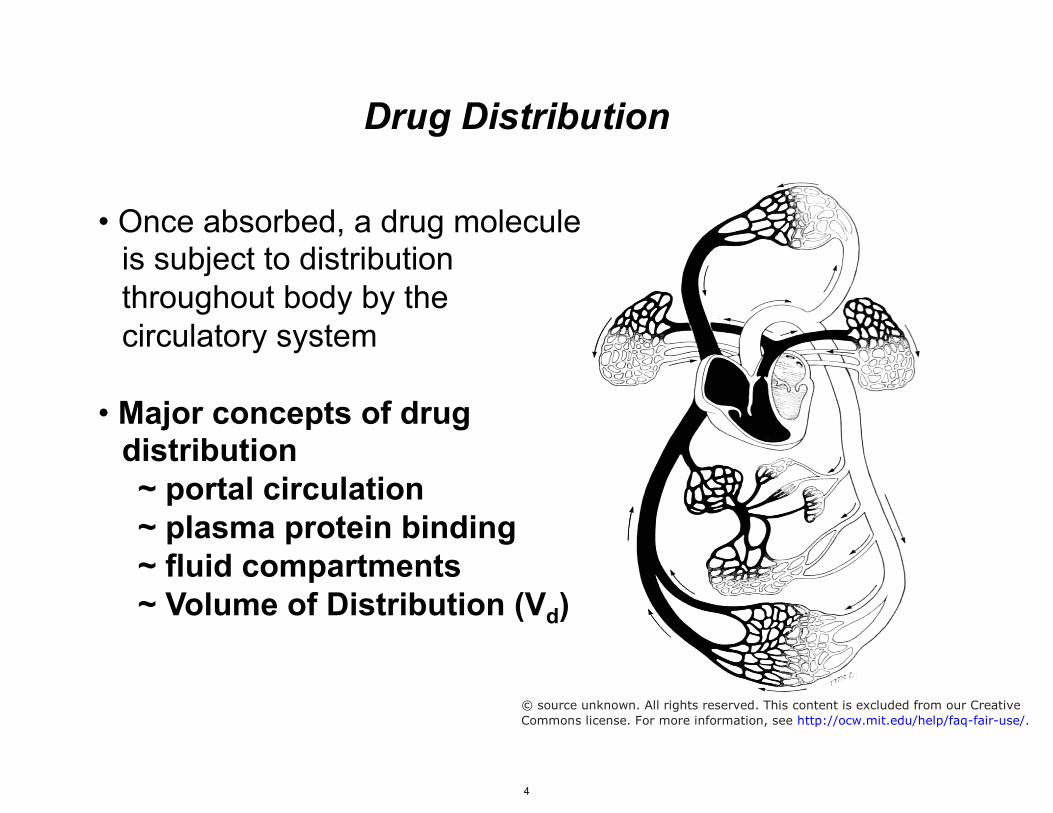
• Once absorbed, a drug molecule is subject to distribution throughout body by the circulatory system
• Major concepts of drug
distribution ~ portal circulation ~ plasma protein binding ~ fluid compartments ~ Volume of Distribution (Vd)
Drug Distribution
© source unknown. All rights reserved. This content is excluded from our Creative
Commons license. For more information, see http://ocw.mit.edu/help/faq-fair-use/.
4
Cardiac output arterial blood
Venous return
from lower extremities
Portal circulation
Drug Distribution
Liver
Intestines
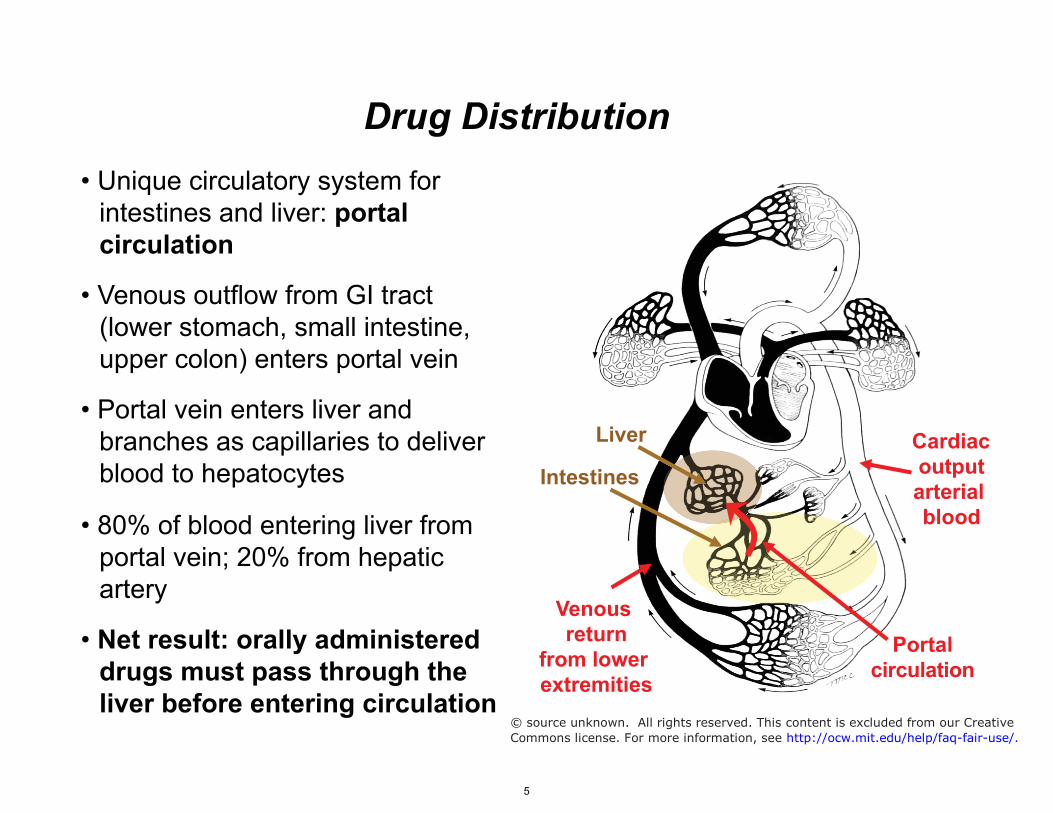
• Unique circulatory system for intestines and liver: portal circulation
• Venous outflow from GI tract (lower stomach, small intestine, upper colon) enters portal vein
• Portal vein enters liver and branches as capillaries to deliver blood to hepatocytes
• 80% of blood entering liver from portal vein; 20% from hepatic artery
• Net result: orally administered drugs must pass through the liver before entering circulation
© source unknown. All rights reserved. This content is excluded from our Creative
Commons license. For more information, see http://ocw.mit.edu/help/faq-fair-use/.
5

Drug Distribution
Nitroglycerin ADME • Vd ~200 L
• t1/2 ~1-4 min • Metabolism: 1,3- & 1,2-
dinitroglycerol (active, t1/2 3-4 hr); 2 inactive mets.
• 60% protein bound • Renal excretion of parent, metabolites
• Hepatic extraction: degree to which drug is removed from blood on each pass through the liver
• Example: 63% of rosuvastatin is "captured" by liver on each pass
• First-pass metabolism: degree to which a drug is metabolized on first pass through liver in portal circulation
• Example: nitroglycerin for angina
• >90% first-pass metabolism demands alternate route for administration
• Sublingual and rectal routes: venous absorption leads to systemic circulation and bypasses liver
6
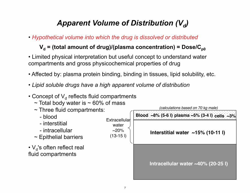
• Hypothetical volume into which the drug is dissolved or distributed
Vd = (total amount of drug)/(plasma concentration) = Dose/Cp0
• Limited physical interpretation but useful concept to understand water compartments and gross physicochemical properties of drug
• Affected by: plasma protein binding, binding in tissues, lipid solubility, etc.
• Lipid soluble drugs have a high apparent volume of distribution
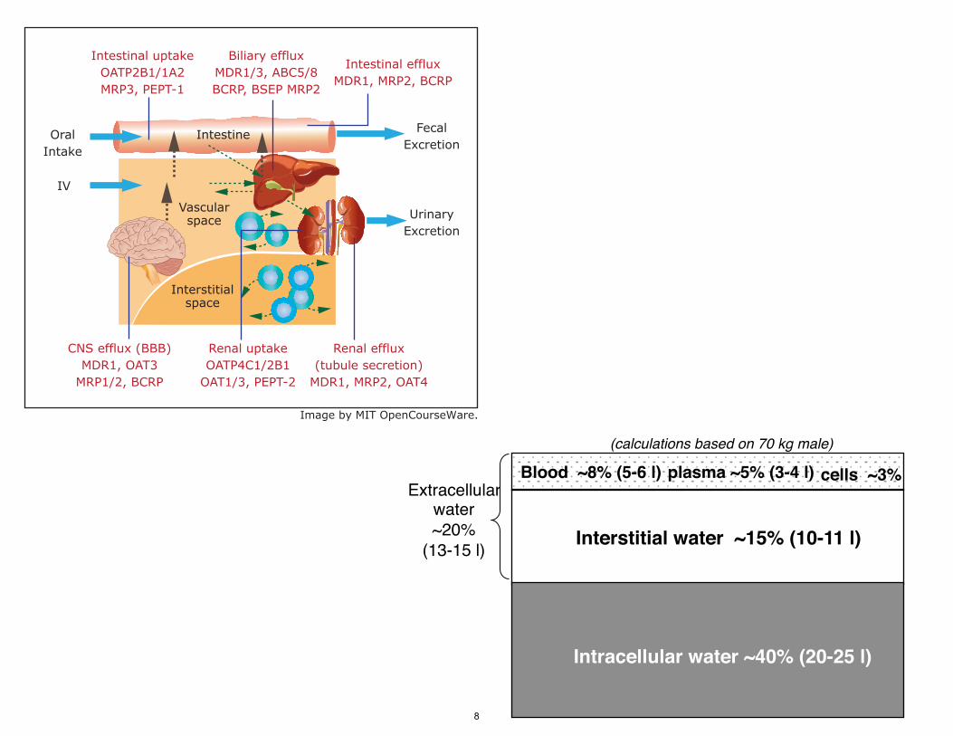
• Concept of Vd reflects fluid compartments ~ Total body water is ~ 60% of mass ~ Three fluid compartments: - blood - interstitial - intracellular ~ Epithelial barriers
• Vd's often reflect real fluid compartments
Blood ~8% (5-6 l) plasma ~5% (3-4 l) cells ~3%
Interstitial water ~15% (10-11 l)
Apparent Volume of Distribution (Vd)
(calculations based on 70 kg male) Extracellular �
water�~20% �
(13-15 l) �
Intracellular water ~40% (20-25 l)
7
Blood ~8% (5-6 l) plasma ~5% (3-4 l) cells ~3%
Interstitial water ~15% (10-11 l)
(calculations based on 70 kg male) Extracellular �
water�~20% �
(13-15 l) �
Intracellular water ~40% (20-25 l)
8
Vascularspace
Intestine
Interstitialspace
Intestinal uptakeOATP2B1/1A2MRP3, PEPT-1
Biliary effluxMDR1/3, ABC5/8BCRP, BSEP MRP2
Intestinal effluxMDR1, MRP2, BCRP
CNS efflux (BBB)MDR1, OAT3
MRP1/2, BCRP
OralIntake
IV
FecalExcretion
UrinaryExcretion
Renal uptakeOATP4C1/2B1
OAT1/3, PEPT-2
Renal efflux(tubule secretion)
MDR1, MRP2, OAT4
Image by MIT OpenCourseWare.

NCl
HN
N
H3CH2C
CH2CH3
Chloroquine Vd ~ >104L
Partition Coefficient (Octanol/H2O) = 52,000
Drug V (L/Kg) V (L, 70 Kg) Sulfisoxazole 0.16 11 Amoxicillin 0.3 20 Phenobarbital 0.55 38 Phenytoin 0.63 44 Diazepam 2.4 168 Digoxin 7 490 Chloroquine >100 >104
Digoxin Vd ~ 490 L
Partition Coefficient (Octanol/H2O) = 18.4
AmoxicillinVd ~ 20 L
Partition Coefficient (Octanol/H2O) = 0.03
Blood and interstitial fluids
Fat depot!
9

Concepts of distribution: Protein binding
• Binding of drugs to proteins in blood is a major determinant of PKs and a source of toxic drug-drug interaction
• Binding generally depends on charge and water solubility: hydrophobic drugs bind to hydrophobic pockets in serum proteins
• Importance of protein binding: ~ "active" drug = unbound drug = can bind to target ~ binding affects concentration of "active" drug at the site of action ~ wide variation in serum protein concentrations in different diseases ~ drug-drug interactions can involve competition for protein binding ~ "bumping" a drug off of protein increases its unbound concentration
10
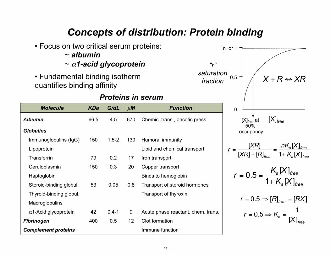
Proteins in serum Molecule KDa G/dL μ
• Focus on two critical serum proteins: ~ albumin ~
M Function
Albumin 66.5 4.5 670 Chemic. trans., oncotic press.
Globulins
Immunoglobulins (IgG) 150 1.5-2 130 Humoral immunity
Lipoprotein Lipid and chemical transport
Transferrin 79 0.2 17 Iron transport
Ceruloplasmin 150 0.3 20 Copper transport
Haptoglobin Binds to hemoglobin
Steroid-binding globul. 53 0.05 0.8 Transport of steroid hormones
Thyroid-binding globul. Transport of thyroxin
Macroglobulins
α1-Acid glycoprotein 42 0.4-1 9 Acute phase reactant, chem. trans.
Fibrinogen 400 0.5 12 Clot formation
Complement proteins Immune function
α1-acid glycoprotein
• Fundamental binding isotherm quantifies binding affinity
"r" saturation
fraction
n or 1
0.5
0
[X]free [X]free at 50%
occupancy
r =
[XR][XR] + [R]free
=nKa [X ]free
1+ Ka [X ]free
X + R ↔ XR
r = 0.5 =
Ka [X ]free
1+ Ka [X ]free
r = 0.5 ⇒ [R]free = [RX ]
r = 0.5 ⇒ Ka =
1[X ]free
Concepts of distribution: Protein binding
11
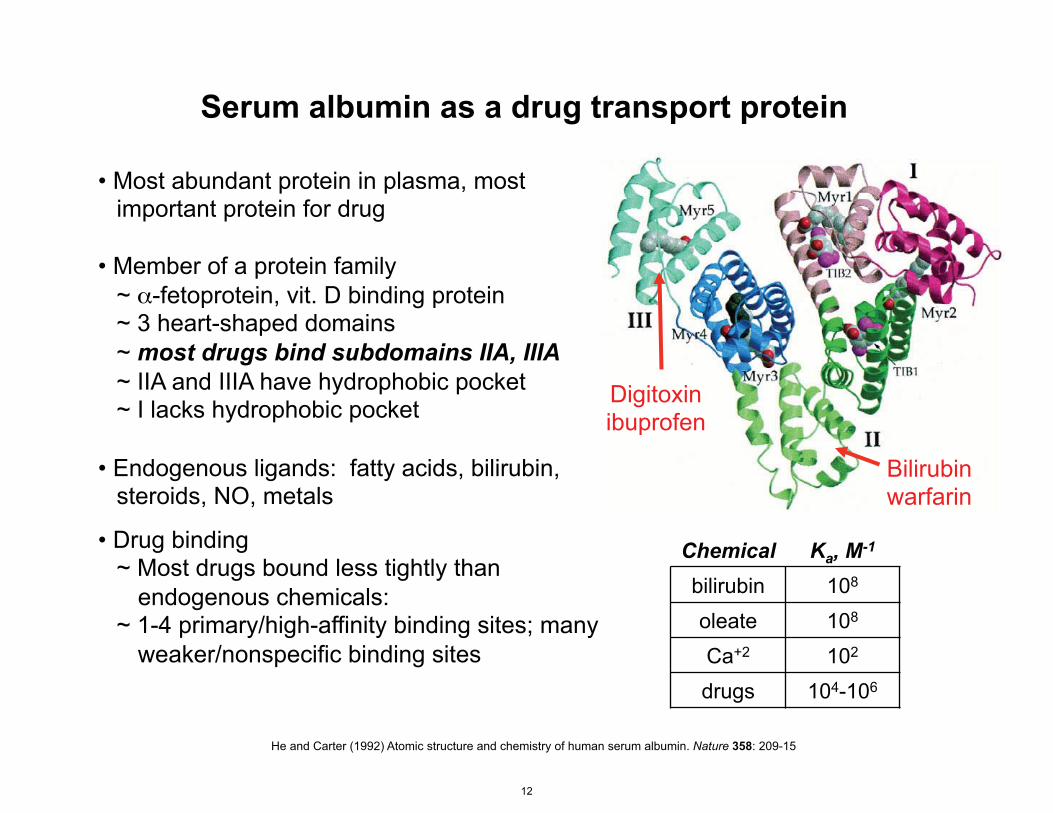
Serum albumin as a drug transport protein
• Most abundant protein in plasma, most important protein for drug
• Member of a protein family ~ α-fetoprotein, vit. D binding protein ~ 3 heart-shaped domains ~ most drugs bind subdomains IIA, IIIA ~ IIA and IIIA have hydrophobic pocket ~ I lacks hydrophobic pocket
• Endogenous ligands: fatty acids, bilirubin, steroids, NO, metals
• Drug binding ~ Most drugs bound less tightly than
endogenous chemicals: ~ 1-4 primary/high-affinity binding sites; many
weaker/nonspecific binding sites
Chemical bilirubin
Ka, M-1 108
oleate 108 Ca+2 102 drugs 104-106
Bilirubin warfarin
Digitoxin ibuprofen
He and Carter (1992) Atomic structure and chemistry of human serum albumin. Nature 358 : 209-15
12

% Unbound (fu x 100) Drug
Digitoxin ~95% bound
Digoxin ~20% bound
Amoxicillin
Concepts of distribution: Protein binding
• Bound drugs can be displaced by competition
• Competition by endogenous ligands or other drugs
• Net result: increase in the unbound/free concentration of a drug
• Danger for drugs with narrow TI! ~ Digitoxin (compare to digoxin) ~ Warfarin
Danger!
13

Consequences of altered protein binding in disease
• Propranolol: β-adrenergic receptor antagonist used to treat hypertension, tachyarrythmias, migraine
• Bound extensively to αα-acid glycoprotein: cationic charge
• What happens to the level of drug binding when the protein level is altered by disease?
Propranolol
[X]Ka = b
[X] f • [P]f
From: Clinical Pharmacokinetics: Concepts and Applications (1989) ed. Rowland and Tozer, Lea and Febiger, Philadelphila.
fu =
[X]f
[X]f + [X]b
=[X]f
[X]t
fu • [X]t = [X]f
(1− fu)• [X]t = [X]b
Ka =
(1− fu)• [X]t
fu • [X]t • [P]f
fu =
11+Ka • fPf
• [P]t
fPf
=[P]f
[P]t
Binding of drug X to protein P Xf = free or unbound drug
Xb = bound drug Pf = free or unoccupied protein
fu = Fraction of drug unbound
fPf = Fraction of protein
unoccupied
Substitute
Solve for fu
• Free concentration of drug depends on binding constant, concentration of unoccupied binding sites on protein, and protein concentration
• In general, fpf ~ 1: most sites are unoccupied
• Thus, concentration of free drug depends on protein concentration and is relatively constant at different drug concentrations (steep part of binding isotherm)
14

Consequences of altered protein binding in disease
• Propranolol: β-adrenergic receptor antagonist used to treat hypertension, tachyarrythmias, migraine
• Bound extensively to αα-acid glycoprotein: cationic charge
• The level of α-acid glycoprotein changes as a function of inflammation and disease (acute phase reactant)
• A reduction in the level of the protein leads to an increase in the proportion of unbound drug
onsequences of a
Propranolol
© Lea & Febiger. All rights reserved. This content is excluded from our Creative Commonslicense. For more information, see http://ocw.mit.edu/help/faq-fair-use/.
From: Clinical Pharmacokinetics: Concepts and Applications (1989) ed. Rowland and Tozer, Lea and Febiger, Philadelphila. 15

BREAK • Two drugs bind to albumin with the following dissociation constants:
Drug A Drug B Kd ~ 1 pM Kd ~ 1 μM
• Which drug has a higher affinity for albumin? • Which drug would be displaced by bilirubin, which has a Kd ~ 10 nM
16
Pharmacokinetics and the Fate of Drugs in the Body
• Definition of Pharmacokinetics/Toxicokinetics: quantitative temporal analysis of the processes of ADME; how much of and how fast the drug reaches its target
• Compare to pharmacodynamics: mechanism by which a
chemical or agent exerts its effects (e.g., binding to receptor, interfering with cell wall formation)
• Applications in pharmacology: determine how often to
administer a drug to maintain therapeutic concentration • Applications in toxicology: define the association between
exposure and the progression of disease • Approaches to pharmacokinetic analysis: ~ Simple compartment models ~ Physiologically-based pharmacokinetic models (PBPK)
17

Route of Administration
Blood/Plasma
Absorption
Tissues Distribution
Paradigm for Pharmacokinetics Concepts
Route of Elimination
Target
Liver
kabs kmet
kdist kelim
Bile
18

Routes of administration and absorption
• Already looked at mechanisms of absorption
• Now look at quantifying the kinetics of absorption
• Rates of absorption dictated by route of administration: ~ Enteral vs parenteral ~ Vascular vs extravascular
• Enteral routes • Parenteral routes ~ Oral - portal! ~ Intravenous (iv) ~ Sublingual - bypass portal ~ Intramuscular (im) ~ Rectal - bypass portal ~ Subcutaneous (sc)
T ~ opical/transdermal ~ Inhalation/nasal ~ Ocular
19
Factors affecting absorption from site of administration
• Quantitative aspects of absorption are important for GI, lung and topical routes
• Transport ~ diffusion - not saturable ~ active, facilitated; saturable
• pH effects ~ charge affects transport/diffusion ~ pH stomach ~ 2; tissue pH ~6.5-8
• Physical factors at the site of absorption ~ blood flow ~ surface area - lungs 140 m2 - skin 1.5-2 m2 - GI tract 300 m2 (small intestine) ~ contact time
20

Quantifying absorption: Bioavailability
• Concept of AUC: ~ area under plasma concentration vs time curve ~ measure of the total quantity of drug entering the general circulation
• Bioavailability ~ defined as fraction (F) of administered drug entering general circulation ~ calculate as plasma AUCoral /AUCIV
• Determinants of bioavailability ~ Formulation (salt form, particle size, excipients) affects rate of dissolution ~ Chemical stability - E.g. penicillin unstable at acid pH of stomach ~ Hepatic extraction - E.g. nitroglycerin has >90% 1st pass metabolism
• Bioequivalence - relative bioavailability of two drugs
F =
AUCev
AUCiv
From: Clinical Pharmacokinetics: Concepts and Applications (1989) ed. Rowland and Tozer, Lea and Febiger, Philadelphila.
• 500 mg of a drug administered IM and orally to same subject • Quantify [drug] in plasma vs time
IM ≈ IV
Oral
© Lea & Febiger. All rights reserved. This content is excludedfrom our Creative Commons license. For more information, see http://ocw.mit.edu/help/faq-fair-use/.
Plasma Data Urine Data
AUC t1/2 decay Cumul. Route (mg•hr/ phase Excret.
L) (min) (mg)
IV 7.6 190 152
IM 7.4 185 147
Oral 3.5 193 70 21

EXERCISE
22
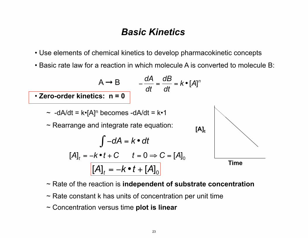
Basic Kinetics
• Use elements of chemical kinetics to develop pharmacokinetic concepts
• Basic rate law for a reaction in which molecule A is converted to molecule B:
• Zero-order kinetics: n = 0
~ -dA/dt = k•[A]n becomes -dA/dt = k•1
~ Rearrange and integrate rate equation:
~ Rate of the reaction is independent of substrate concentration
~ Rate constant k has units of concentration per unit time
~ Concentration versus time plot is linear
[A]t
Time
−
dAdt
=dBdt
= k • [A]n
−dA = k •dt∫ [A]t = −k •t + C t = 0 ⇒ C = [A]0
[A]t = −k •t + [A]0
A � B
23
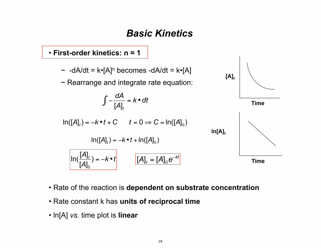
• First-order kinetics: n = 1
~ -dA/dt = k•[A]n becomes -dA/dt = k•[A]
~ Rearrange and integrate rate equation: • Rate of the reaction is dependent on substrate concentration
• Rate constant k has units of reciprocal time
• ln[A] vs. time plot is linear
−
dA[A]t
= k •dt∫
ln([A]t ) = −k •t + C t = 0 ⇒ C = ln([A]0)
ln([A]t ) = −k •t + ln([A]0)
ln( [A]t
[A]0
) = −k •t [A]t = [A]0e−kt
[A]t
Time
ln[A]t
Time
Basic Kinetics
24

[A]ln( t
[A] 0
) = ln(0.5) = −0.693 = −k •t
0.693t1/ 2 = k
• Half-life - fundamental pharmacokinetic concept and parameter
• Definition: time to decrease concentration by one-half
• Define mathematically by setting [A]t = [A]0/2
Time
[A]ln( t
[A] 0
t1/2
) = −0.693
Basic Kinetics
25

• “Saturable” processes: ligand molecules completely occupy available binding sites
• Metabolic enzymes ~ Aspirin - glycine conjugation and phenolic glucuronidation ~ Ethanol - alcohol/aldehyde dehydrogenase ~ Phenytoin - CYP2C9; Km~ 5 mg/L; therapeutic range 10-20 mg/L
• Transporters: glucose transporter in renal tubule (filtered [glucose] > 320 ng/min)
• Mathematical basis for zero-order kinetics ~ Michaelis-Menten rate equation considerations:
dP V =
dt ~ When [S] >> Km, all substrate binding sites occupied and enzyme
operates at Vmax
=Vmax • [S]Km + [S]
V =dPdt
=Vmax • [S]Km + [S]
~ Vmax • [S][S]
Basic Kinetics: Processes subject to zero-order kinetics
= Vmax
26

Basic Kinetics: Processes subject to first-order kinetics
• Definition of a first-order process: a reaction or activity, the rate of which depends on the concentration of reactants or the chemical of interest
• Most processes of absorption, distribution, metabolism, elimination are first-order
• Diffusion: Rate of diffusion depends on the concentration gradient (i.e., the concentration of the "reactant") dQ − dt• Metabolism and transport proteins: Enzyme kinetics generally first-order, except under conditions of substrate saturation:
when Km>>[S], then
= P • A •ΔC
d[Product]dt
= V =Vmax • [S]Km + [S]
d[Product]dt
= V =Vmax
Km
• [S] = kmet • [S]
27
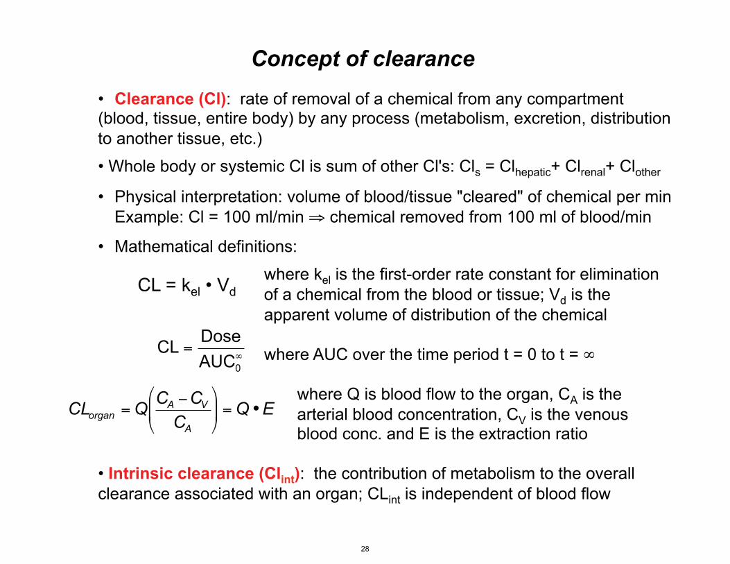
Concept of clearance • Clearance (Cl): rate of removal of a chemical from any compartment (blood, tissue, entire body) by any process (metabolism, excretion, distribution to another tissue, etc.)
• Whole body or systemic Cl is sum of other Cl's: Cls = Clhepatic+ Clrenal+ Clother
• Physical interpretation: volume of blood/tissue "cleared" of chemical per min Example: Cl = 100 ml/min ⇒ chemical removed from 100 ml of blood/min
• Mathematical definitions:
kwhere el is the first-order rate constant for elimination of a chemical from the blood or tissue; Vd is the apparent volume of distribution of the chemical
DoseCL = ∞ where AUC over the time period t = 0 to t = ∞ AUC 0
where Q is blood flow to the organ, CA is the arterial blood concentration, CV is the venous blood conc. and E is the extraction ratio
• Intrinsic clearance (Clint): the contribution of metabolism to the overall clearance associated with an organ; CLint is independent of blood flow
CL = kel • Vd
CLorgan = Q CA −CV
CA
⎛
⎝ ⎜
⎞
⎠ ⎟ = Q •E
28
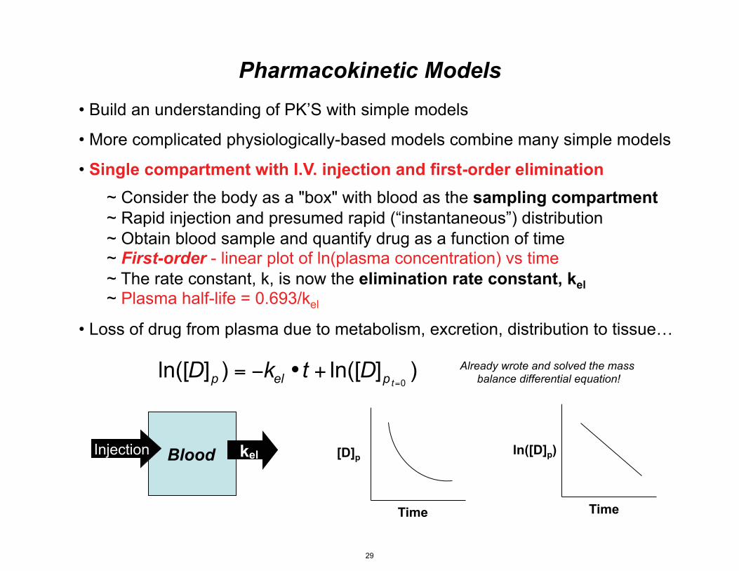
Pharmacokinetic Models • Build an understanding of PK’S with simple models
• More complicated physiologically-based models combine many simple models
• Single compartment with I.V. injection and first-order elimination
~ Consider the body as a "box" with blood as the sampling compartment ~ Rapid injection and presumed rapid (“instantaneous”) distribution ~ Obtain blood sample and quantify drug as a function of time ~ First-order - linear plot of ln(plasma concentration) vs time ~ The rate constant, k, is now the elimination rate constant, kel ~ Plasma half-life = 0.693/kel
• Loss of drug from plasma due to metabolism, excretion, distribution to tissue�
[D]p
Time
ln([D]p)
Time
Blood Injection
ln([D] p ) = −kel •t + ln([D]pt=0) Already wrote and solved the mass
balance differential equation!
kel
29

• Single compartment with absorption from gut and first-order elimination
~ Factor in kinetics of absorption with kinetics of elimination from blood ~ Distribution is no longer instantaneous ~ Assume first-order absorption from gut (why?) ~ Write rate equation that accounts for 1° absorption and 1° elimination
~ As drug absorbed from gut, e-kabst goes to zero and [D]p dominated by kel
Blood kel kabs Gut [D]t
Time
First-order absorption
First-order elimination
d
[D]p
dt= kabs [D]gut − kel [D]p
d
[D]p
dt= kabs [D]gut − kel [D]p = kabs [D]gut0e−k abst( ) − kel [D]p
Integrate ⇒ [D]pt
= [D]gut0
kabs
kabs − kel
Pharmacokinetic Models
⎛ ⎞ ⎜ ⎟ e−k elt − e−k abst
⎝ ⎠ ( )
30

• Two compartments with I.V. injection and first-order elimination
~ Rate equation now has 3 terms ~ Injected drug distributes in blood compartment “instantaneously” ~ Observe two "phases" ~ Rapid movement of drug out of blood into tissue compartment ~ Slower phase: as plasma concentration falls below tissue concentration, drug moves into blood
[D]d p
dt= k21[D]t is − k12 [D]p − kel [D]p
Integrate ⇒ [D]pt= Ae−αt + Be−βt
Blood kel injection
Tissue
k21 k12 ln([D]t)
Time
Rapid 1° distribution
Slower 1° elimination
α + β = k12 + k21 + kel
α •β = k21 •kel
A = [D]p0
α − k21
α − β
⎛
⎝ ⎜
⎞
⎠ ⎟
B = [D]p0
k21 − βα − β
⎛
⎝ ⎜
⎞
⎠ ⎟
ln([D]t)
Time
Blood
Tissue [D]t
Time
Absorption from blood
Elimination from tissue
Pharmacokinetic Models
Linear plot
looks familiar?
31
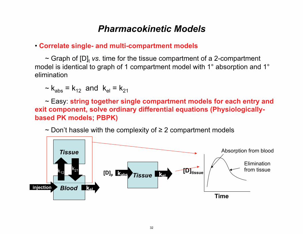
• Correlate single- and multi-compartment models
~ Graph of [D]t vs. time for the tissue compartment of a 2-compartment model is identical to graph of 1 compartment model with 1° absorption and 1° elimination
~ kabs = k12 and kel = k21
~ Easy: string together single compartment models for each entry and exit component, solve ordinary differential equations (Physiologically-based PK models; PBPK)
~ Don’t hassle with the complexity of ≥ 2 compartment models
[D]tissue
Time
Absorption from blood
Elimination from tissue
Tissue kel kabs [D]p
Blood kel injection
Tissue
k21
Pharmacokinetic Models
k12
32
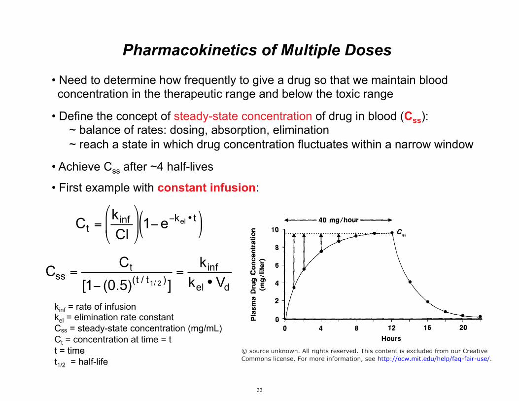
• Need to determine how frequently to give a drug so that we maintain blood concentration in the therapeutic range and below the toxic range
• Define the concept of steady-state concentration of drug in blood (Css): ~ balance of rates: dosing, absorption, elimination ~ reach a state in which drug concentration fluctuates within a narrow window
• Achieve Css after ~4 half-lives
• First example with constant infusion:
CCss = t
[1− (0.5)(t / t1/ 2 ) ]=
kinfkel • Vd
Pharmacokinetics of Multiple Doses
kinf = rate of infusion kel = elimination rate constant Css = steady-state concentration (mg/mL) Ct = concentration at time = t t = time t1/2 = half-life
Ct =kinfCl
⎛
⎝ ⎜
⎞
⎠ ⎟ 1− e−kel • t( )
© source unknown. All rights reserved. This content is excluded from our Creative
Commons license. For more information, see http://ocw.mit.edu/help/faq-fair-use/.
33
[D]p
Time (multiples of t1/2)
Css
Fluctuations about Css • proportional to t and t1/2 • amplitude dampened by slow absorption
0 1 2 3 4 5 6
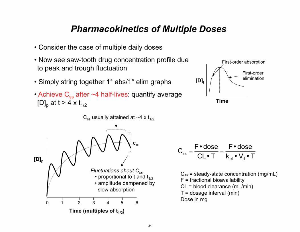
• Consider the case of multiple daily doses
• Now see saw-tooth drug concentration profile due to peak and trough fluctuation
• Simply string together 1° abs/1° elim graphs
• Achieve Css after ~4 half-lives: quantify average [D]p at t > 4 x t 1/2
Css usually attained at ~4 x t1/2
F• doseCss =CL • T
=F• dose
kel • Vd • T
Pharmacokinetics of Multiple Doses
Css = steady-state concentration (mg/mL) F = fractional bioavailability CL = blood clearance (mL/min) T = dosage interval (min) Dose in mg
[D]t
Time
First-order absorption
First-order elimination
34

Pharmacokinetics Web Sites
• Excellent web site for pharmacokinetics: http://www.boomer.org/c/p1/index.html • JAVA calculator for plotting blood concentrations approaching steady-state: http://www.boomer.org/c/p1/Ch15/Fig57/Fig57.html
35
MIT OpenCourseWarehttp://ocw.mit.edu
20.201 Mechanisms of Drug ActionsFall 2013
For information about citing these materials or our Terms of Use, visit: http://ocw.mit.edu/terms.