224 week 3 lecture 22005
TRANSCRIPT
8/4/2019 224 Week 3 Lecture 22005
http://slidepdf.com/reader/full/224-week-3-lecture-22005 1/55
Biology 224 Human Anatomy and Physiology II Week 3; Lecture 2; Monday Dr. Stuart S. Sumida
Structure of the Lung
Biomechanics of Breathing
8/4/2019 224 Week 3 Lecture 22005
http://slidepdf.com/reader/full/224-week-3-lecture-22005 2/55
Diaphragm:
•Derived from hypaxial musculature of cervical
segments.
•So motor innervation is from cervical
segmental nerves: right and left phrenic nerves
(C3,4,5).
•Diaphragm is a muscular dome-shaped
structure.

8/4/2019 224 Week 3 Lecture 22005
http://slidepdf.com/reader/full/224-week-3-lecture-22005 3/55
•Derived from
hypaxial
musculature of cervical
segments.
•So motor
innervation is
from cervical
segmental
nerves: right andleft phrenic
nerves (C3,4,5).
•Diaphragm is a muscular dome-shaped structure.
8/4/2019 224 Week 3 Lecture 22005
http://slidepdf.com/reader/full/224-week-3-lecture-22005 4/55
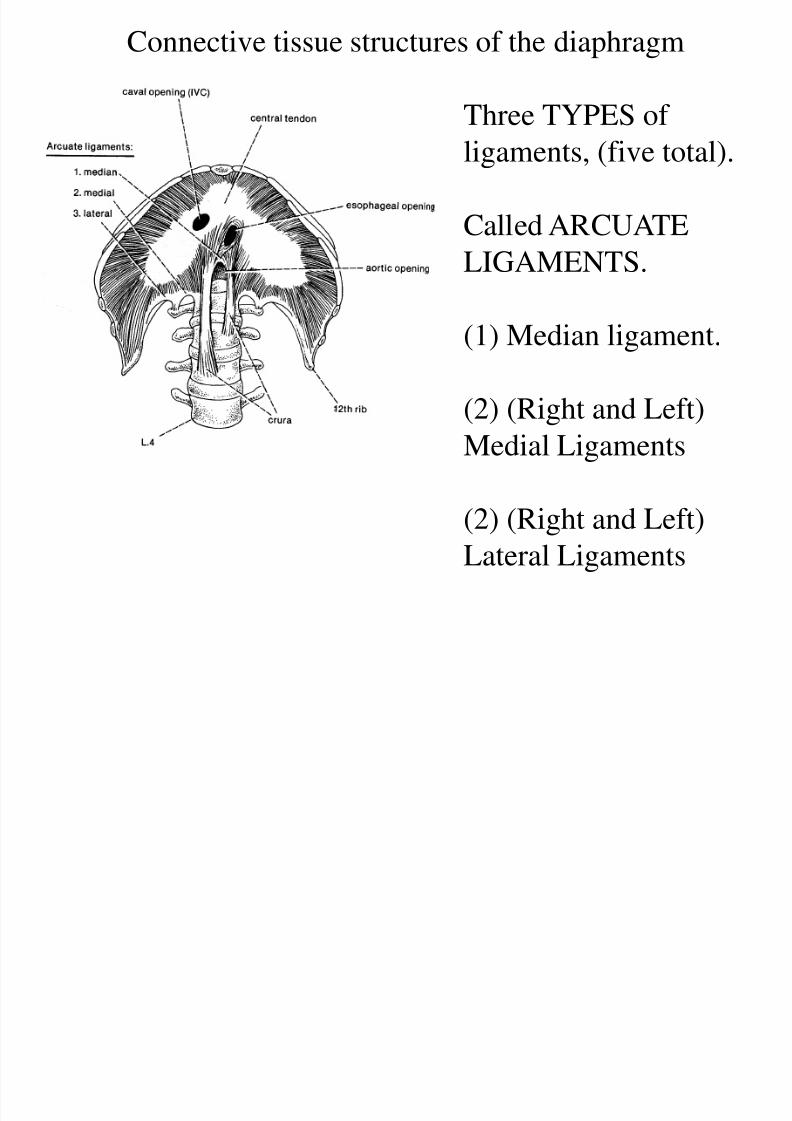
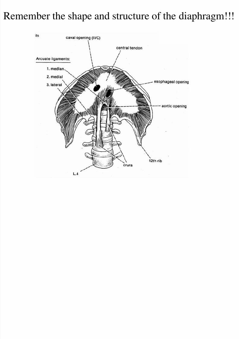
Connective tissue structures of the diaphragm
Three TYPES of
ligaments, (five total).
Called ARCUATE
LIGAMENTS.
(1) Median ligament.
(2) (Right and Left)Medial Ligaments
(2) (Right and Left)
Lateral Ligaments
8/4/2019 224 Week 3 Lecture 22005
http://slidepdf.com/reader/full/224-week-3-lecture-22005 5/55
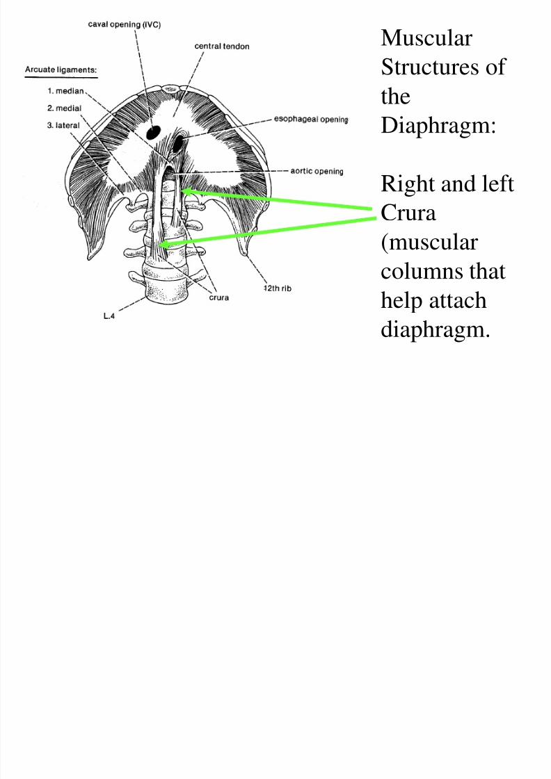
Muscular
Structures of
theDiaphragm:
Right and leftCrura
(muscular
columns that
help attach
diaphragm.
8/4/2019 224 Week 3 Lecture 22005
http://slidepdf.com/reader/full/224-week-3-lecture-22005 6/55
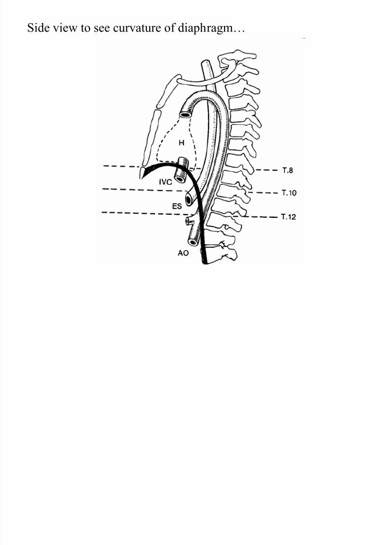
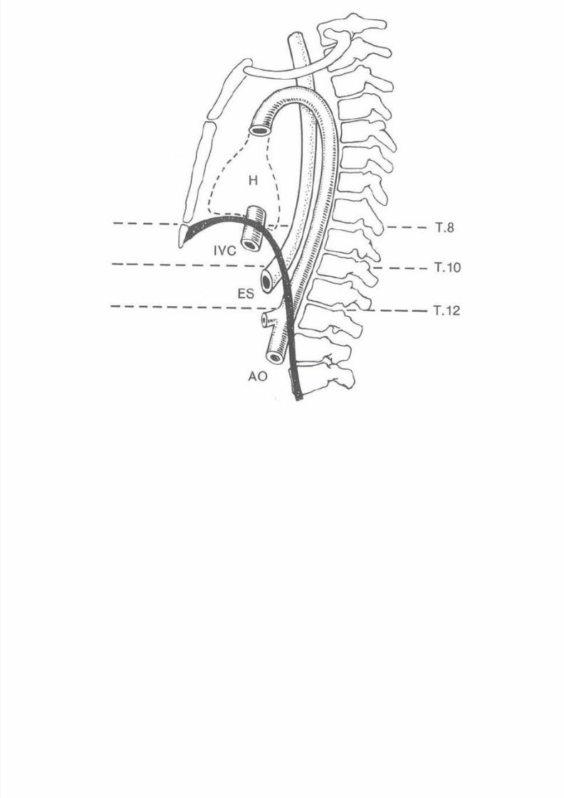
Side view to see curvature of diaphragm…

8/4/2019 224 Week 3 Lecture 22005
http://slidepdf.com/reader/full/224-week-3-lecture-22005 7/55
RESPIRATORY TREE
Trachea 2 Primary Bronchi (right and left)
Each Primary Bronchus to many Secondary
Bronchi
Each Secondary Bronchus to many Tertiary
Bronchi
Tertiary bronchi to many Bronchioles
Bronchioles to ―Alveoli‖

8/4/2019 224 Week 3 Lecture 22005
http://slidepdf.com/reader/full/224-week-3-lecture-22005 8/55
RESPIRATORY TREE
Trachea 2 PrimaryBronchi (right and left)
Each Primary Bronchus
to many Secondary
Bronchi
Each Secondary
Bronchus to many
Tertiary Bronchi
8/4/2019 224 Week 3 Lecture 22005
http://slidepdf.com/reader/full/224-week-3-lecture-22005 9/55
BLOOD VESSELS
Lung highly vascularized.
Vessels from mesoderm.
Arteries tend to run ventral to branches of
bronchial tree.
Veins more variable in pattern.
Wheer bronchi and vessels disappear into tissue
of lung: called ROOT OF THE LUNG.

8/4/2019 224 Week 3 Lecture 22005
http://slidepdf.com/reader/full/224-week-3-lecture-22005 10/55
LUNG STRUCTURE
8/4/2019 224 Week 3 Lecture 22005
http://slidepdf.com/reader/full/224-week-3-lecture-22005 11/55
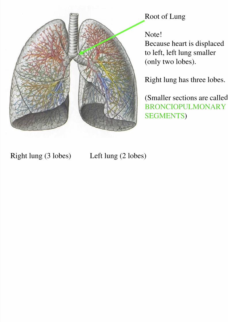
Root of Lung
Note!
Because heart is displaced
to left, left lung smaller
(only two lobes).
Right lung has three lobes.
(Smaller sections are calle
BRONCIOPULMONARY
SEGMENTS)
Right lung (3 lobes) Left lung (2 lobes)
8/4/2019 224 Week 3 Lecture 22005
http://slidepdf.com/reader/full/224-week-3-lecture-22005 12/55
8/4/2019 224 Week 3 Lecture 22005
http://slidepdf.com/reader/full/224-week-3-lecture-22005 13/55
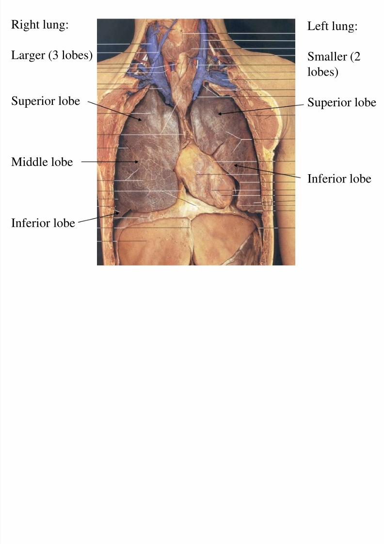
Left lung:
Smaller (2
lobes)
Superior lobe
Inferior lobe
Right lung:
Larger (3 lobes)
Superior lobe
Middle lobe
Inferior lobe
8/4/2019 224 Week 3 Lecture 22005
http://slidepdf.com/reader/full/224-week-3-lecture-22005 14/55
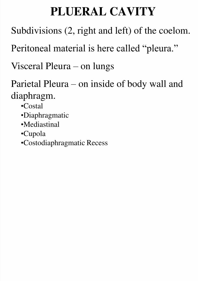
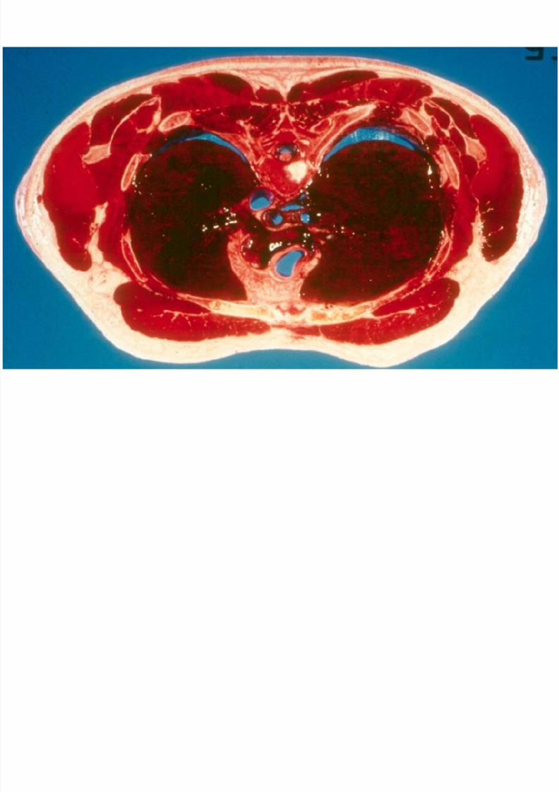
PLUERAL CAVITY
Subdivisions (2, right and left) of the coelom.Peritoneal material is here called ―pleura.‖
Visceral Pleura – on lungsParietal Pleura – on inside of body wall and
diaphragm.•Costal•Diaphragmatic
•Mediastinal
•Cupola
•Costodiaphragmatic Recess
8/4/2019 224 Week 3 Lecture 22005
http://slidepdf.com/reader/full/224-week-3-lecture-22005 15/55
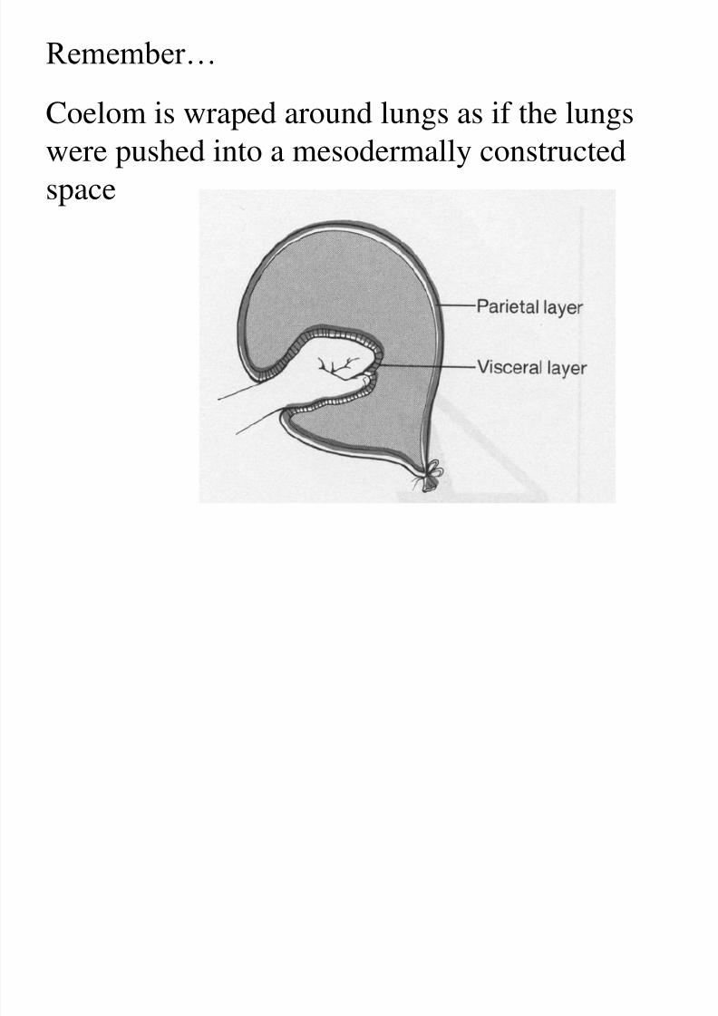
Remember…
Coelom is wraped around lungs as if the lungswere pushed into a mesodermally constructed
space
8/4/2019 224 Week 3 Lecture 22005
http://slidepdf.com/reader/full/224-week-3-lecture-22005 16/55
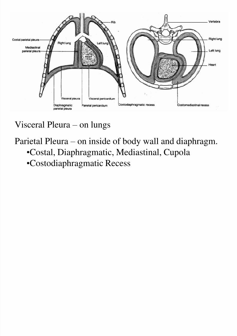
Visceral Pleura – on lungs
Parietal Pleura – on inside of body wall and diaphragm.
•Costal, Diaphragmatic, Mediastinal, Cupola
•Costodiaphragmatic Recess
8/4/2019 224 Week 3 Lecture 22005
http://slidepdf.com/reader/full/224-week-3-lecture-22005 17/55
8/4/2019 224 Week 3 Lecture 22005
http://slidepdf.com/reader/full/224-week-3-lecture-22005 18/55
Functional Considerations for the Pleura…
Lung does not expand up into cupola.
Expands downward toward pleural recess (the
inferior space between ribs and diaphragm.
Pleura secretes coelomic fluid (for lubrication and to
pull lungs when body wall moves).
―Pleurisy‖ is the painful chaffing between visceral
and parietal pleura.
8/4/2019 224 Week 3 Lecture 22005
http://slidepdf.com/reader/full/224-week-3-lecture-22005 19/55
The ―MEDIASTINUM‖ is
the partition between the
right and left pleura and theenclosed lungs.
Exercise: What is in the mediastinum? (Look at the
pictures in your lab manual and in the Cartmill text.
8/4/2019 224 Week 3 Lecture 22005
http://slidepdf.com/reader/full/224-week-3-lecture-22005 20/55
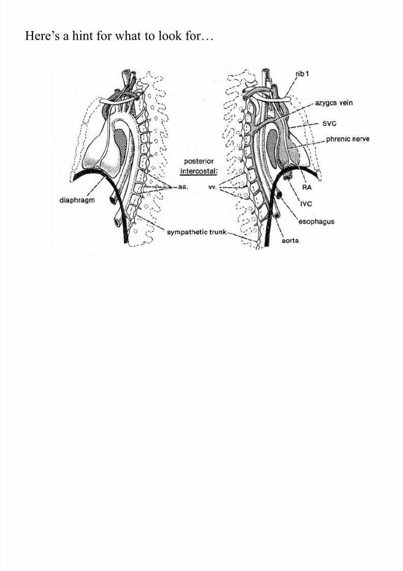
Here’s a hint for what to look for…

8/4/2019 224 Week 3 Lecture 22005
http://slidepdf.com/reader/full/224-week-3-lecture-22005 21/55
LUNGFUNCTION
AND
BREATHING

8/4/2019 224 Week 3 Lecture 22005
http://slidepdf.com/reader/full/224-week-3-lecture-22005 22/55
Smooth Muscle and Nervous
Supply of Lung• Smooth muscle can constrict or open
respiratory tree.
• CONSTICTION: Parasympathetic nervouscontrol is by VAGUS NERVE (X).
• Ganglia between pre- and post-ganglionic
neurons right on target organ.
8/4/2019 224 Week 3 Lecture 22005
http://slidepdf.com/reader/full/224-week-3-lecture-22005 23/55
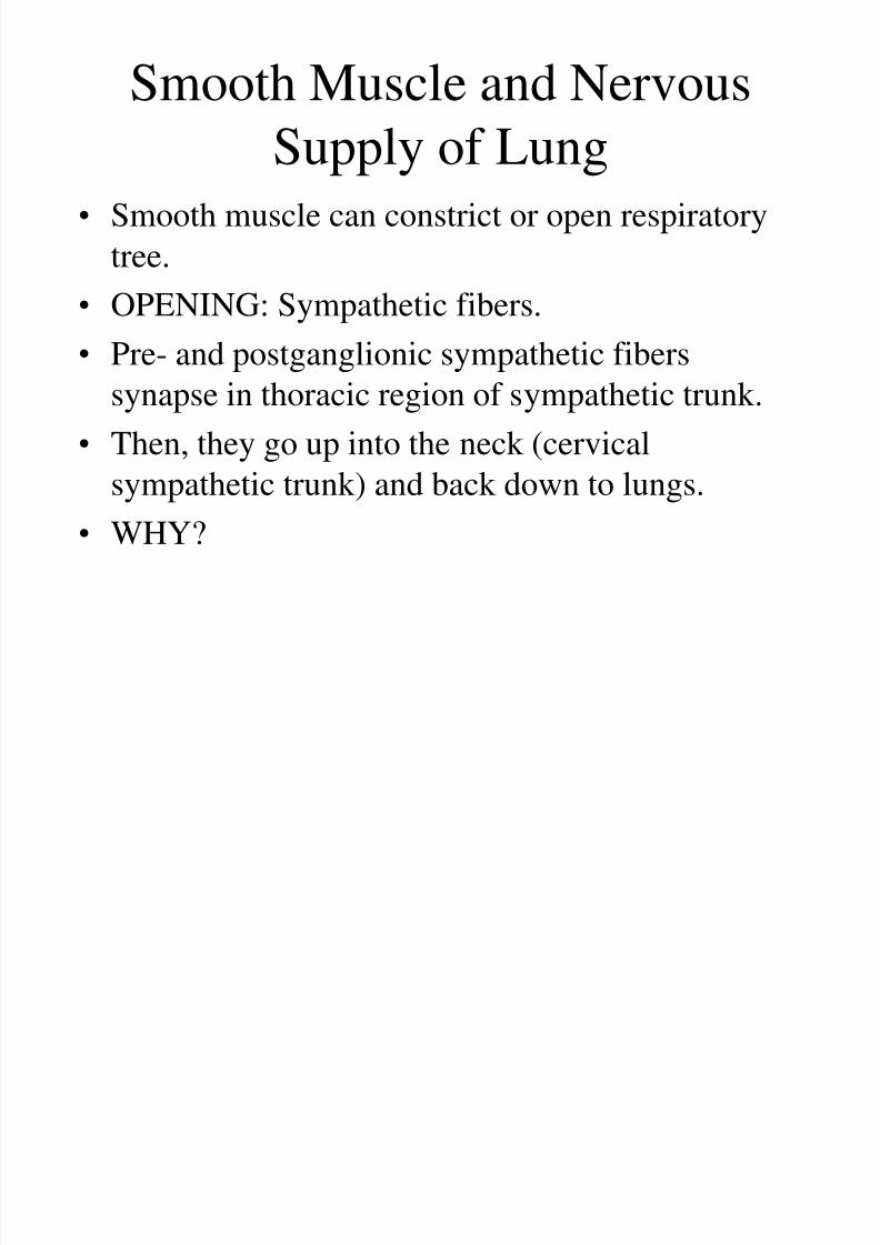
Smooth Muscle and Nervous
Supply of Lung• Smooth muscle can constrict or open respiratory
tree.
• OPENING: Sympathetic fibers.• Pre- and postganglionic sympathetic fibers
synapse in thoracic region of sympathetic trunk.
• Then, they go up into the neck (cervical
sympathetic trunk) and back down to lungs.
• WHY?

8/4/2019 224 Week 3 Lecture 22005
http://slidepdf.com/reader/full/224-week-3-lecture-22005 24/55
Pre- and
postganglionic
sympathetic fiberssynapse in thoracic
region of sympathetic
trunk.
Then, they go up intothe neck (cervical
sympathetic trunk) and
back down to lungs.
WHY?
BECAUSE! Remember: Lungs started out in the neck, and
then moved down. The nerves were simply following!
8/4/2019 224 Week 3 Lecture 22005
http://slidepdf.com/reader/full/224-week-3-lecture-22005 25/55
BIOMECHANICSAND NERVOUS
CONTROL OF
BREATHING

8/4/2019 224 Week 3 Lecture 22005
http://slidepdf.com/reader/full/224-week-3-lecture-22005 26/55
THORACIC BREATHING
Based on RIB MOVEMENTS:• Scalene muscles pull cranially (up) on 1st and 2nd
ribs.
• Ribs move like bucket handles.
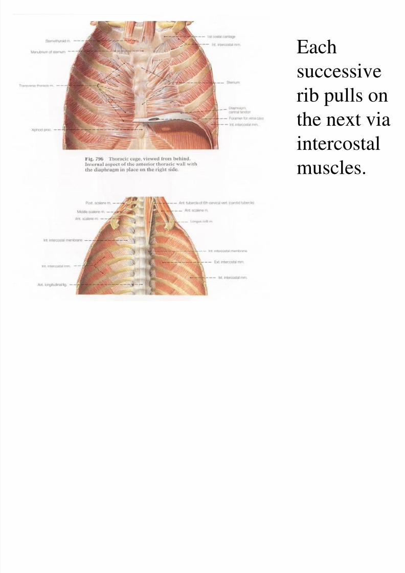
• Each successive rib pulls on the next viaintercostal muscles.
• When ribs/bucket handles move up and out,
VOLUME OF THORACIC CAVITYINCREASES.

8/4/2019 224 Week 3 Lecture 22005
http://slidepdf.com/reader/full/224-week-3-lecture-22005 27/55
Scalene
muscles pull
cranially (up)
on 1st and 2nd
ribs.
(Scalenes are
segmentally
innervated:
C2-7.)

8/4/2019 224 Week 3 Lecture 22005
http://slidepdf.com/reader/full/224-week-3-lecture-22005 28/55
Ribs move like bucket handles.
8/4/2019 224 Week 3 Lecture 22005
http://slidepdf.com/reader/full/224-week-3-lecture-22005 29/55
Each
successiverib pulls on
the next via
intercostalmuscles.

8/4/2019 224 Week 3 Lecture 22005
http://slidepdf.com/reader/full/224-week-3-lecture-22005 30/55
When ribs/bucket handles move up
and out, VOLUME OF THORACICCAVITY INCREASES.
So what happens when volume
increases?
PRESSURE DECREASES...
8/4/2019 224 Week 3 Lecture 22005
http://slidepdf.com/reader/full/224-week-3-lecture-22005 31/55
When PRESSURE
DECREASES…
Air gets SUCKED IN.
(All amniotes do this. In other
words, amniotes (includinghumans as mammals)...
…SUCK.
8/4/2019 224 Week 3 Lecture 22005
http://slidepdf.com/reader/full/224-week-3-lecture-22005 32/55
ABDOMINAL
BREATHING
(Use of theDiaphragm)
8/4/2019 224 Week 3 Lecture 22005
http://slidepdf.com/reader/full/224-week-3-lecture-22005 33/55
8/4/2019 224 Week 3 Lecture 22005
http://slidepdf.com/reader/full/224-week-3-lecture-22005 34/55
ABDOMINAL BREATHING
• Diaphragm is ―dome-shaped.‖
• When it contracts, the dome flattens out.
• This INCREASES THORACIC VOLUME.
• Where have you heard this before…?
8/4/2019 224 Week 3 Lecture 22005
http://slidepdf.com/reader/full/224-week-3-lecture-22005 35/55
So, when diaphragm contracts,
VOLUME OF THORACICCAVITY INCREASES.
So what happens when volume
increases?
PRESSURE DECREASES...
8/4/2019 224 Week 3 Lecture 22005
http://slidepdf.com/reader/full/224-week-3-lecture-22005 36/55
When PRESSURE
DECREASES…
Air gets SUCKED IN.
Only mammals (including
humans) have a diaphragm.
So, humans SUCK really well.
8/4/2019 224 Week 3 Lecture 22005
http://slidepdf.com/reader/full/224-week-3-lecture-22005 37/55
8/4/2019 224 Week 3 Lecture 22005
http://slidepdf.com/reader/full/224-week-3-lecture-22005 38/55
FORCED BREATHING
• Inhalation can be increased by increasingthe amplitude of the movements we just
discussed.
• Forced Exhalation -- facilitated by all themuscles of the ribcage, pressurizing coelom,and contracting limb muscles around theaxial body wall.

8/4/2019 224 Week 3 Lecture 22005
http://slidepdf.com/reader/full/224-week-3-lecture-22005 39/55
Forced Exhalation
Muscles of the ribcage (bucket handles move
down).
Pressurizing coelom (pushes diaphragm back
up into dome-shape)** -- decreases thoracic
volume to push air out.
Contracting limb muscles around the axial body
wall can help compress thoracic cavity.
8/4/2019 224 Week 3 Lecture 22005
http://slidepdf.com/reader/full/224-week-3-lecture-22005 40/55
NOTE:
Pressurizing coelom (pushes diaphragm back up
into dome-shape)** -- decreases thoracic volume
to push air out.
In other words, mammals (including humans)
also BLOW**.
HUMANS BOTH SUCK AND BLOW.
(**I’m quoting Bart Simpson here.)

8/4/2019 224 Week 3 Lecture 22005
http://slidepdf.com/reader/full/224-week-3-lecture-22005 41/55
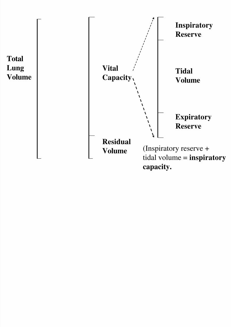
VOLUMES OF AIR IN
LUNGS
• Normal Breathing: about half a liter per
breath.
• This is known as “TIDAL VOLUME.”
8/4/2019 224 Week 3 Lecture 22005
http://slidepdf.com/reader/full/224-week-3-lecture-22005 42/55
Total
Lung
Volume
Vital
Capacity
Residual
Volume
Inspiratory
Reserve
Tidal
Volume
Expiratory
Reserve
(Inspiratory reserve +
tidal volume = inspiratory
capacity.
8/4/2019 224 Week 3 Lecture 22005
http://slidepdf.com/reader/full/224-week-3-lecture-22005 43/55
INNERVATIONS
• Diaphragm: PHRENIC NERVES (right and
left)
• Scalenes: C2-7.
• Breathing is ―involuntary behavior powered
by voluntary muscles.‖

8/4/2019 224 Week 3 Lecture 22005
http://slidepdf.com/reader/full/224-week-3-lecture-22005 44/55
Diaphragm: PHRENIC NERVES (right and left)
Phrenic
nerves pierce
diaphragm
near apex;
sendbranches
across
inferior
(abdominal)surface of
diaphragm.
8/4/2019 224 Week 3 Lecture 22005
http://slidepdf.com/reader/full/224-week-3-lecture-22005 45/55
CENTRAL NERVOUS
CONTROL OF BREATHING
• Normal Breathing: known as ―EUPNEA‖
• Main controls in pons and medulla oblongata.
• In Pons:
• APNEUSTIC AREA - causes strong inhalation,
weak exhalation.
• PNEUMOTAXIC AREA - causes stronginhalation, weak exhalation.
8/4/2019 224 Week 3 Lecture 22005
http://slidepdf.com/reader/full/224-week-3-lecture-22005 46/55
CHEMICAL CONTROLS OF
BREATHING• CO2 in blood dissociates into CARBONIC ACID.
• More carbonic acid means lower pH.
• CAROTID BODIES (at junction of internal and
external carotid): Sense pH and communicate with
medulla.
• AORTIC BODY (on arch of aorta): Sense pH and
communicate with medulla.

8/4/2019 224 Week 3 Lecture 22005
http://slidepdf.com/reader/full/224-week-3-lecture-22005 47/55
RHYMICITY CENTERS OF
MEDULLA OBLONGATA• Increased CO2 (in form of carbonic acid) or
increased blood pressure signals from
carotid and aortic bodies.• Carotid bodies and arotic body tell
medullary rhymicity centers.
• Medullary rhymicity centers can thenincrease activity of apneustic area (deeperbreathing.)
8/4/2019 224 Week 3 Lecture 22005
http://slidepdf.com/reader/full/224-week-3-lecture-22005 48/55
RHYMICITY CENTERS OF
MEDULLA OBLONGATA• Decreased CO2 is called RESPIRATORY
ALKALOSIS (higher pH).
• Carotid bodies and aortic body tellmedullary rhymicity centers.
• Medullary rhymicity centers can then
increase activity of pneumotaxic area(shallower breathing.)

8/4/2019 224 Week 3 Lecture 22005
http://slidepdf.com/reader/full/224-week-3-lecture-22005 49/55
MICROSCOPIC DETAILOF RESPIRATORY TREE
8/4/2019 224 Week 3 Lecture 22005
http://slidepdf.com/reader/full/224-week-3-lecture-22005 50/55
ALVEOLI:
Terminal ―Grape-like Lobes of Respiratory Tree.
Microscopic airsacs, thin enough for gas to pass
across.
Each alveolus is surrounded by capillary plexus
(deoxygenated blood from pulmonary artery,oxygenated blood returned via pulmonary vein).
Note! Only at this microscopic level is lung ―hollow.‖

8/4/2019 224 Week 3 Lecture 22005
http://slidepdf.com/reader/full/224-week-3-lecture-22005 51/55
Terminal ―Grape-like Lobes of Respiratory Tree.
Microscopic airsacs, thin enough for gas to pass across.
Each alveolus is surrounded by capillary plexus (deoxygenated blood
from pulmonary artery, oxygenated blood returned via pulmonary
vein).

8/4/2019 224 Week 3 Lecture 22005
http://slidepdf.com/reader/full/224-week-3-lecture-22005 52/55
GAS EXCHANGE
Alveolar and capillary membranes: extremely thin.(Capillaries only one red blood cell wide.)
Thus, hemoglobin in RBCs maximally exposed to
fresh oxygen.
Remember, oxygen BINDS TO HEMOGLOBIN in
regions of high oxygen concentration.Carbon dioxide dumped.
8/4/2019 224 Week 3 Lecture 22005
http://slidepdf.com/reader/full/224-week-3-lecture-22005 53/55
SURFACTANTS
Specialized cells of alveolar lining secrete thse
chemicals.
They reduce ―surface tension‖ – prevents fluid
from beading up on alveolar surface.
Prevents collapse of alveoli due to concentrated
fluid weight.
Thinner layer of fluid makes gas diffusion easier.

8/4/2019 224 Week 3 Lecture 22005
http://slidepdf.com/reader/full/224-week-3-lecture-22005 54/55
OTHER DEFENSES
Alveoli contain lots of phagocytic cells:ALVEOLAR MACROPHAGES.
•Ingest and destroy microorganisms and other
foreign substances (from breathing them in…)
Cilia can transport small bits of foreign material and
mucous back up.
Coughing
Foreign material can be carried into lymphatic
system.
Smooth Muscle and Nervous Supply:
8/4/2019 224 Week 3 Lecture 22005
http://slidepdf.com/reader/full/224-week-3-lecture-22005 55/55
Smooth Muscle and Nervous Supply:
Bronchial segments include smooth muscle — can expand or
constrict tree.
PARASYMPATHETIC:
•Vagus Nerve – signals cause smooth muscle to contract and
constrict bronchioles.
•Ganglia between pre- and postganglionic neurons right on
target organ (on bronchioles themselves).
SYMPATHETIC:
•Pre- and post-ganglionic neurons synapse in thoracic part of
sympathetic chain.•Go up to cervical region, then go back down sympathetic chain
to lungs.
•Why? Because lungs started out in neck.
•Cause dilation of bronchi.