2,3,7,8-tetrachlorodibenzo-p-dioxin treatment alters eicosanoid levels in several organs of the...
TRANSCRIPT

Toxicology and Applied Pharmacology 259 (2012) 143–151
Contents lists available at SciVerse ScienceDirect
Toxicology and Applied Pharmacology
j ourna l homepage: www.e lsev ie r .com/ locate /ytaap
2,3,7,8-Tetrachlorodibenzo-p-dioxin treatment alters eicosanoid levels in severalorgans of the mouse in an aryl hydrocarbon receptor-dependent fashion
Peter Bui a,b,c, Parrisa Solaimani a,b,c, Xiaomeng Wu b,c, Oliver Hankinson a,b,c,d,⁎a Molecular Toxicology Program, University of California, Los Angeles, California 90095, USAb Dept of Pathology and Laboratory Medicine, University of California, Los Angeles, California 90095, USAc Jonsson Comprehensive Cancer Center, University of California, Los Angeles, California 90095, USAd Molecular Biology Institute, University of California, Los Angeles, California 90095, USA
⁎ Corresponding author at: Dept of Pathology and LabCenter for Health Sciences, University of California, LosFax: +1 310 794 9272.
E-mail address: [email protected] (O. Hankin
0041-008X/$ – see front matter © 2011 Elsevier Inc. Alldoi:10.1016/j.taap.2011.12.009
a b s t r a c t
a r t i c l e i n f oArticle history:Received 3 October 2011Revised 22 November 2011Accepted 5 December 2011Available online 20 December 2011
Keywords:2,3,7,8-tetrachlorodibenzo-p-dioxinAryl hydrocarbon receptorEicosanoidsArachidonic acidLinoleic acidDocosahexaenoic acidCytochrome P450
2,3,7,8-Tetrachlorodibenzo-p-dioxin (TCDD) adversely affects many mammalian organs and tissues. Theseeffects are mediated by the aryl hydrocarbon receptor (AHR). CYP1A1, CYP1A2 and CYP1B1 are upregulatedby the liganded AHR. These (and other) cytochromes P450 can metabolize arachidonic acid into a variety ofbioactive eicosanoids. Towards investigating a potential role of eicosanoids in TCDD toxicity, arachidonic acid,two other unsaturated long-chain fatty acids, and up to twenty-five eicosanoids were measured in five organs/tissues of male and female wild-type and Ahr null mice treated or untreated with TCDD. TCDD generallyincreased the levels of the four dihydroxyeicosatrienoic acids (DHETs) and (where measured) 5,6-epoxyeicosatrienoic acid and 18-, 19- and 20-hydroxyeicosatrienoic acids (HETEs) in the serum, liver, spleenand lungs, but not the heart, of both sexes, and increased the levels in the serum, liver and spleen of severalmetabolites that are usually considered products of lipoxygenase activity, but which may also be generatedby cytochromes P450. TCDD also increased the levels of the esterified forms of these eicosanoids in the liverin parallel with the corresponding free forms. The levels of prostanoids were generally not affected by TCDD.The above changes did not occur in Ahr null mice, and are therefore mediated by the AHR. TCDD increased themRNA levels of Cyp1a1, Cyp1a2, Cyp1b1 and the Pla2g12a form of phospholipase A2 to varying degrees in thedifferent organs, and these increases correlated with some but not all the changes in eicosanoids levels in theorgans, suggesting that other enzymes may also be involved.
© 2011 Elsevier Inc. All rights reserved.
Introduction
Eicosanoids are derivatives of twenty carbon polyunsaturatedfatty acids, particularly arachidonic acid (AA). Equivalent derivativesof eighteen and twenty-two carbon unsaturated fatty acids, such aslinoleic (LA) acid and docosahexaenoic acid (DHA), may have similarbiological activities, although less is known about the biologicaleffects of these latter compounds. Eicosanoids are generally short-lived in vivo. They have effects on many organs and tissues (Nebertand Karp, 2008). Drugs targeting the arachidonate cascade representover 25% of the world's pharmaceutical sales (Li et al., 2011).
Arachidonic acid (and the other aforementioned polyunsaturatedfatty acids) are metabolized via three pathways: the cyclooxygenase,lipoxygenase and cytochrome P450 epoxidation/hydroxylation
oratory Medicine, Box 951732,Angeles, CA 90095–1732, USA.
son).
rights reserved.
pathways (although cytochromes P450 are or can be involved in allthree pathways). The least is known about the last pathway. Mamma-lian cytochromes P450 frommany different subfamilies, including theCYP1A, CYP1B, CYP2B, CYP2C, CYP2D, CYP2E, CYP2F, CYP2G, CYP2J,CYP2P, CYP2U, CYP3A, CYP4A, CYP4B and CYP4F subfamilies exhibitarachidonic acid epoxidation and/or hydroxylation activities. The im-mediate products of the epoxidation/hydroxylation pathway of ara-chidonic acid metabolism include four cis-epoxyeicosatrienoic acids(EETs), 5,6-EET, 8,9-EET, 11,12-EET and 14,15-EET, the “terminal” hy-droxides, 16-HETE, 17-HETE, 18-HETE, 19-HETE and 20-HETE, andcertain “midchain” hydroxides, including 5-HETE, 8-HETE, 12-HETEand 15-HETE. The midchain hydroxides are also products of the lipox-ygenase catalyzed metabolism of arachidonic acid (Zeldin, 2001;Rifkind, 2006). Several of the cis-epoxyeicosantrienoic acids and hy-droxides, including some of the terminal hydroxides (particularly20-HETE), exhibit potent biological activities. For example, EETs canaffect a number of functions, including angiogenesis, apoptosis, fibri-nolysis, mitogenesis, hormone action, vasodilation, bronchodilation,inflammation, and neuropathic pain (Norwood et al., 2010). The ep-oxides can be further metabolized, particularly by soluble epoxide
144 P. Bui et al. / Toxicology and Applied Pharmacology 259 (2012) 143–151
hydrolase, which converts them to the dihydroxyeicosatrienoic acids(DHETs). DHETs are generally less biologically active, although someactivities have been ascribed to them (Buczynski et al., 2009; Nebertand Karp, 2008).
2,3,7,8- tetrachlorodibenzo-p-dioxin (TCDD) elicits a wide varietyof toxic, teratogenic and carcinogenic responses in animals and inhumans. Most if not all the effects of dioxin are mediated by thearyl hydrocarbon receptor (AHR), which after binding TCDD, transo-locates to the nucleus, dimerizes with the aryl hydrocarbon nucleartranslocator protein (ARNT) and activates transcription of a largenumber of genes, including those for CYP1A1, CYP1A2 and CYP1B1,and represses others (White and Birnbaum, 2009; Ma et al., 2009;Denison et al., 2011). A wide variety of other compounds, includinghalogenated aromatic hydrocarbons and polycyclic aromatic hydro-carbons (PAH) can bind to, and activate the AHR. Many if not mostof the biological effects of the AHR are probably mediated by its effecton these transcriptional responses (Bunger et al., 2008; Fujii-Kuriyama and Kawajiri, 2010). Nebert and Karp pointed out that“the myriad AHR-mediated processes mirror the vast universe of ac-tion of the eicosanoids” and they proposed that many of the biologicaleffects of AHR are mediated by synthesis of eicosanoids by the CYP1subfamily (Nebert and Karp, 2008). Compatible with this notion, sev-eral investigators have shown that microsomes from organs of TCDD-or PAH-treated mammals catalyze the conversion of arachidonic acidto certain eicosanoids at different rates compared with microsomesfrom non-treated mammals (Capdevilla et al., 1990; Lee et al., 1998;Aboutabl et al., 2009). Furthermore, Dalton and coworkers showedthat TCDD exposure increased the levels of three cyclooxygenase-derived arachidonic metabolism in the urine of mice (Dalton et al.,2001). However, to directly address the potential role of eicosanoids
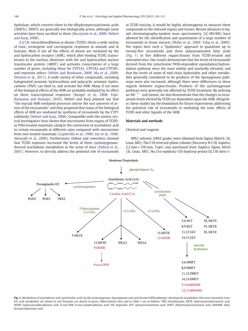
Fig. 1.Metabolism of arachadonic acid (and linoleic acid) by the cyclooxygenase, lipoxygenaleic acid metabolites are shown in red. Enzymes are shown in green. Abbreviations here aHODE, hydroxyoctadecadienoic acid; 9-oxo-ODE, 9-oxo-octadecadienoic acid; HX, hepoxildroxyoctadecenoic acid.
in TCDD toxicity, it would be highly advantageous to measure thesecompounds in the relevant organs and tissues. Recent advances in liq-uid chromatography-tandem mass spectrometry (LC-MS/MS) haveallowed for the identification and quantitation of a large number ofeicosanoids in tissue extracts (Blaho et al., 2009; Yang et al., 2009).We report here such a “lipidomics” approach to quantitate up totwenty-five eicosanoids and three polyunsaturated fatty acids(Fig. 1) in five different organs/tissues from TCDD-treated anduntreatedmice. Our results demonstrate that the levels of eicosanoidsderived from the cytochrome P450-dependent epoxidation/hydrox-ylation pathway were the most widely and markedly elevated, andthat the levels of some of mid-chain hydroxides and other metabo-lites generally considered to be products of the lipoxygenase path-ways were also increased, although there were differences in theseregards between organs/tissues. Products of the cyclooxygenasepathway were generally not affected by TCDD treatment. By utilizingan Ahr−/− null mouse, we also demonstrate that the changes in eicos-anoids levels elicited by TCDD are dependent upon the AHR. Altogeth-er, these studies lay the foundation for future experiments addressingthe potential role of eicosanoids in mediating the toxic effects ofTCDD and other ligands of the AHR.
Materials and methods
Chemical and reagents
HPLC solvents (HPLC grade) were obtained from Sigma Aldrich (St.Louis, MO). The C18 reversed-phase column (Discovery ® C18, Supelco,2.2 mm×150 mm, 5 μm) was purchased from Supelco Sigma Alrich(St. Louis, MO). 9α,11α-epidioxy-15S-hydroxy-prosta-5Z,13E-dien-1-
se and cytochrome P450 pathways, showing the matabolites that were measured. Lino-nd in Table 1 are as follows: TBX, thromboxane; HETE, hydroxyeicosatetraenoic acid;in; EET, epoxyeicosatrienoic acid; DHET, dihydroxyeicosatrienoic acid; diHOME, dihy-

145P. Bui et al. / Toxicology and Applied Pharmacology 259 (2012) 143–151
oic acid (PGH2), 9-oxo-11α,15S-dihydroxy-prosta-5Z,13E-dien-1-oicacid (pGE2), 9α,15S-dihydroxy-11-oxo-prosta-5Z,13E-dien-1-oic acid(PGD2), 9α,11,15S-trihydroxythromba-5Z,13E-dien-1-oic acid (TXB2),6-oxo-9α,11α,15S-trihydroxy-prost-13E-en-1-oic acid (6-k-PGF1α),9α,15S-dihydroxy-11-oxo-prosta-5Z,13E-dien-1-oic-3,34,4-d4 acid(PGD2-d4), 9,15-dioxo-11α-hydroxy-prosta-5Z,13E-dien-1-oic acid(15-keto-PGE2), 5S,12R-dihydroxy-6Z,8E,10E,14Z-eicosatetrae-noic acid (LTB4), 7S,8R,17S-trihydroxy-4Z,9E,11E,13Z,15E,19Z-docosahexaenoic acid (Resolvin D1), 10(S),17(S)-dihydroxy-4Z,7Z,11E,13Z,15E,19Z-docosahexaenoic acid (Protectin D1),12,13-dihydroxy-9Z-octadecenoic acid (12,13-DiHOME), (±)5,6-dihydroxy-8Z,11Z,14Z-eicosatrienoic acid (5,6-DHET), (±)8,9-dihydroxy-5Z,11Z,14Z-eicosatrienoic acid (8,9-DHET), (±)11,12-dihydroxy-5Z,8Z,14Z-eicosatrienoic acid (11,12-DHET), (±)14,15-dihydroxy-5Z,8Z,11Z-eicosatrienoic acid (14,15-DHET),(±)5(6)-epoxy-8Z,11Z,14Z-eicosatrienoic acid (5(6)-EET), (±)8(9)-epoxy-5Z,11Z,14Z-eicosatrienoic acid (8(9)-EET), (±)11(12)-epoxy-5Z,8Z,14Z-eicosatrienoic acid (11(12)-EET), (±)14(15)-epoxy-5Z,8Z,11Z-eicosatrienoic acid (14(15)EET), 5-oxo-6E,8Z,11Z,14Z-eicosatetraenoic acid (5-oxo-ETE), 12-oxo-5Z,8Z,10E,14Z-eico-satetraenoic acid (12-oxoETE), 15-oxo-5Z,8Z,10E,14Z-eicosatetrae-noic acid (15-oxoETE), 13-oxo-9Z,11E-octadecadienoic acid(13-oxoODE), 5S-hydroxy-6E,8Z,11Z,14Z-eicosatetraenoic acid(5-HETE), 12S-hydroxy-5Z,8Z,10E,14Z-eicosatetraenoic acid(12-HETE), 15S-hydroxy-5Z,8Z,10E,14Z-eicosatetraenoic acid (15-HETE), 13S-hydroxy-9Z,11E-octadecadienoic acid (13-HODE), (±)18-hy-droxy-5Z,8Z,11Z,14Z-eicosatetraenoic acid (18-HETE), (±)19-hydroxy-5Z,8Z,11Z,14Z-eicosatetraenoic acid (19-HETE), (±)20-hydroxy-5Z,8Z,11Z,14Z-eicosatetraenoic acid (20-HETE), (±)17-hydroxy-4Z,7Z,10Z,13Z,15E,19Z-docosahexaenoic acid (17-HDOHE), Linoleicacid (LA), Docosahexaenoic Acid (DHA), Arachidonic acid (AA), 5S-hydroxy-6E,8Z,11Z,14Z-eicosatetraenoic-5,6,8,9,11,12,14,15-d8acid (5-HETE-d8), 12S-hydroxy-5Z,8Z,10E,14Z-eicosatetrae-noic-5,6,8,9,11,12,14,15-d8 acid (12-HETE-d8), 15(S)-hydroxy-5Z,8Z,11Z,13E-eicosatetraenoic-5,6,8,9,11,12,14,15-d8 acid (15(S)-HETE-d8), and 13-HODE-d4 were purchased from Cayman Chemical (AnnArbor, MI). Oasis HLB (1 cc/10 mg, 30 μm) was purchased from WatersCorporation (Milford, MA, USA). The protease inhibitor cocktail was pur-chased from Roche. TCDD was purchased from Wellington Laboratories,Guelph, ON, Canadaand was handled with extreme caution.
Animals
Ahr−/− null mice were a kind gift of Christopher Bradfield(Schmidt et al., 1996). They were backcrossed at least seventeentimes to C57BL/6 mice and therefore were of the C57BL/6 geneticbackground. Male and female Ahr−/− null mice and their siblingAhr+/+ wild type mice were obtained from crossing heterozygousAhr+/−mice. Genotyping ofmicewas performed by PCR as describedby the Jackson Laboratories (http://jaxmice.jax.org/protocolsdb/f?p=116:2:4212526675950722::NO:2:P2_MASTER_PROTOCOL_ID,P2_JRS_CODE:195,002831). All mice were housed and bred at UCLA in aspecific-pathogen-free facility. Mice were allowed free access to food(chow diet) and water before being used for the experiments. Micewere kept under a 12-h light/dark cycle and house at 25 °C. Two tothree-month-old mice were used for all experiments.
Administration of TCDD to mice and harvesting of tissues/organs
TCDD was received in a pre-weighed vial. 50 μg/kg of TCDD in cornoil was administered by intraperitoneal injection. Corn oil was usedas vehicle control. Five to 8 animals were used per treatment group.Mice were euthanized with carbon dioxide on day 3 after injection.Immediately, blood was collected via cardiac puncture usingheparin-coated needles; 0.2% of BHT (butylated hydroxytoluene)and TPP (triphenylphosphine) were added directly to the blood to
prevent auto-oxidation of fatty acids. Serum was prepared usingserum separator tubes (BD). The heart, lung, liver and spleen werealso collected and stored in −80 º C for later analysis. Hearts fromonly the wild type mice were collected and analyzed. All TCDD proce-dures with mice, including exposure, euthanasia and dissections wereperformed in two dedicated laboratories, constituting a “CarcinogenSuite” in the containment area of the UCLA vivarium. The suite isunder negative pressure. Isolation of mouse tissues from the TCDD-treated mice was done in a vertical laminar flow hood (Class II B1)certified for use with chemical carcinogens. Cages which had beenused with TCDD-treated mice were wiped down with methanol andthen water. Bedding was treated as hazardous waste. Mouse car-casses were placed in a designated refrigerator for disposal by UCLA'sEnvironmental Health and Safety Office (E.H. & S.). After use, solu-tions containing TCDD were extracted with chloroform for disposal.The chloroform fraction was sent to the UCLA Division of Environ-mental (E.H. & S.) for disposal by incineration. These procedureshave been approved by the UCLA Division of Laboratory Animal Med-icine, the UCLA Division of E.H. &S, and the UCLA Office for the Protec-tion of Research Subjects.
Extraction of fatty acids from different tissues
Serum. A 150 μL volume of serum was transferred to a 2 mL poly-propylene tube. The sample was spiked with 100 μL of internal stan-dards mixture (PGD2-d4, 5-HETE-d8, 12-HETE-d8, 15-HETE-d8, 13-HODE-d4, 10 ng/ml each) in methanol. Subsequently, 1.75 ml ofwater (0.1% acetic acid, pH 3.0) was added. The samples were leftfor 15 minutes on ice for complete acidification and equilibration.The resulting sample was then extracted using the solid-phase ex-traction method described below.
Liver, lung, spleen and heart. Each tissue was cut into pieces andtransferred into 2 ml polypropylene tubes. 450 μl of 50 mM Tris–HCl, pH 7.5 plus protease inhibitors (complete mini protease inhibitorcocktail, Roche, Nutley, New Jersey) was added into each tube. Sam-ples were homogenized for 60 seconds with the medium setting,followed by sonication with 7–8 pulses at 50% output and a 5 pulsersetting using a VibraCell sonicator (Sonics and Materials, Inc., New-town, CT). 250 μl of lysate was then mixed with 100 μl of MeOH(0.2% BHT and TPP) followed by centrifugation at 10,000×g for 15minutes. The supernatant was then transferred to 2 ml tube con-taining 1700 μl 0.1%acetic acid, pH3.0 and 100 μl of internal standardsmixture ( see above) were added. The supernatant was then treatedas for the serum samples above. For female mice, the liver tissuewas treated the same as for the male mice except that the liverwas suspended in 250 μl H2O plus 250 μl of MeOH (0.2% BHT andTPP).
Total fatty acid extraction
Only wild type male mice liver samples were used for total lipidextraction. 250 μl of liver lysate samples were transferred to a 2 mltube containing 250 μl water. 500 μl of 1 M KOH was added to hydro-lyze fatty acids from their conjugates, and the tubes incubated at37 °C for 30 minutes. The samples were then acidified with 500 μl of1 M HCl followed by centrifugation at 10,000×g for 15 min. The su-pernatant was transferred to a 15 ml tube containing 1500 μl H2Oand 100 μl of internal standard mixture (10 ng/ml in methanol). Thesamples were left for 15 min on ice for complete acidification andequilibration. The sample was then extracted using the solid-phaseextraction method.
146 P. Bui et al. / Toxicology and Applied Pharmacology 259 (2012) 143–151
Solid phase extraction
Processed samples from different tissues were loaded onto a pre-conditioned 1 cc Oasis HLB solid-phase extraction (SPE) cartridge ona vacuum manifold (Waters). The SPE cartridge was equilibratedwith 2 ml methanol followed by 2 ml water (0.1% acetic acid, pH3.0) before sample loading. After slowly loading the cartridge, it waswashed with 2 ml 5% methanol in water (0.1% acetic acid). Fattyacid analytes were subsequently eluted with 2 ml methanol. The elu-ate was then evaporated to dryness under a stream of argon. 100 μl ofmethanol was added to the dried extract, vortexed for 30s, and thereconstituted extract was centrifuged at 13,200 rpm for 20 min at4 °C.
LC/MS/MS analysis
LC/MS/MS was performed using a quadrupole mass spectrometer(4000 QTRAP; Applied Biosystems, Foster City, CA) equipped withelectrospray ionization (ESI). The HPLC system utilized an Agilent1200 series LC pump equipped with a thermostated autosampler(Agilent Technologies, Santa Clara, CA). Chromatography was per-formed using C18 reversed-phase column, (Discovery ® C18, Supelco,2.2 mm×150 mm, 5 μm), plus a C18 guard column held at 45 °C. Mo-bile phase A consisted of 0.1% formic acid in water, and mobile phaseB consisted of 0.1% formic acid in acetonitrile. The autosampler wasset at 4 °C. The injection volume was 20 μl and the flow rate was con-trolled at 0.4 mL/min. Chromatography was optimized to separate allanalytes in 30 minutes. The gradient is given in Table S-1 in the sup-plemental material. Data acquisition and instrument control were ac-complished using Analyst 1.4.2 software (Applied Biosystems).Detection was accomplished by using the multiple reaction monitor-ing (MRM) mode with negative ion detection. The parameter settingsare described in table S-2. Mass spectrometry measurement was di-vided into 4 periods to increase dwell time and lower the limits of de-tection. Dwell time was found to play a very important role inincreasing the signal-to-noise ratio with a shorter dwell time result-ing in a much higher noise (Yang et al., 2009). In our method, thedwell times were set above 75 mili-seconds which is longer thanthe dwell time used by Yang and coworkers (Yang et al., 2009)(Table S-2). In addition, MRM transitions, collision energy, decluster-ing potential, and collision cell exit potential were optimized for eachcompound by direct infusion into the mass spectrometer to obtainoptimum sensitivity (see Table S-3). For several analytes for whichwe did not possess a standard, MRM transition parameters wereadopted from previous publications (Blaho et al., 2009; Yang et al.,2009). For most analytes, four concentrations (5, 10, 20, and40 ng/ml) were used to create standard curves. In the case of docosa-hexaenoic acid (DHA), 10, 20, 40, and 80 ng/ml were used, whereas50, 100, 200, and 400 ng/ml were used for arachidonic acid (AA)and linoleic acid (LA). All the standard curves had R2 values (for linearregression analysis) of 0.990 or greater for each analyte. The limit ofdetection for each standard was below 0.5 ng/ml. The inter- andintra-day accuracy and precision were determined based on the %CV (coefficient of variation) using data for 13-HODE and 12-HETE(see supplemental material Table S-4-5). In addition, to ensure theconsistency of the instrument during every experimental run, thestandards were always submitted first and resubmitted again at theend of each run. The % CV between the two submissions of the samesample were routinely less than 15%.
RNA extraction, cDNA synthesis, and real-time PCR
RNA was isolated using RNEasy Mini columns (Qiagen, Valencia,CA), according to the manufacturer's instructions for frozen animaltissues, and quantified on a NanoPhotometer (Implen, WestlakeVillage, CA). Onemicrogram of total RNAwas used for cDNA synthesis
in a total reaction volume of 20 μl. Reverse transcription reactionswere performed using Superscript III reverse transcriptase (Invitro-gen) and primed with random hexamers (Invitrogen) according tothe manufacturer's instructions. Following cDNA synthesis, all reac-tions were diluted into a total of 200 μl of RNase-free water, andwere then used as templates for real-time PCR.
SYBR Green real-time PCR was performed according to standardprotocols using the 7500 Fast (Applied Biosystems, Foster City, CA)and quantities were normalized to those for the GAPDH glycolyticenzyme housekeeping gene. All primers were designed using PrimerExpress 3.0 (Applied Biosystems, Foster City, CA), and purchasedfrom Fisher Scientific (Pittsburgh, PA). The forward and reverseprimers used for quantification are listed in Supplementary Table S-6.
Data were analyzed using the ABI software and Microsoft Excel,and significance was evaluated using Student's t-test.
Results
The effects of TCDD on eicosanoid levels
Adult C57BL/6 wild-type or knockout mice were injected intraper-itoneally with 50 ug/kg TCDD or with vehicle (corn oil). Three dayslater their organs/tissues were harvested and subsequently analyzedfor the levels of certain free (i.e. non-esterified) eicosanoids byHPLC followed by MS/MS (see Materials and Methods). This doseand duration of exposure were used as they are known to maximallyinduce most TCDD-responsive genes (Hayes et al, 2007; Forgacs et al,2012) Three separate experiments were performed. In each indepen-dent experiment, 5 to 8 mice were analyzed per gender per genotype.In experiment 1, male and female wild-type and Ahr−/− knockoutmice were analyzed for the levels of certain free eicosanoids inserum, liver, lung, spleen, and heart (wild-type males only). Thedata for experiment 1 are presented in Supplementary Table S-7. Inexperiment 2 (Supplementary Table S-8), the levels of total eicosa-noids as well as free eicosanoids were measured in the livers ofwild-type male mice. To obtain total eicosanoids, liver extracts weretreated with KOH in order to hydrolyze membrane phospholipids.In the third experiment, free eicosanoids were measured in theserum from wild-type mice (data not shown). The results from allthree experiments are summarized as a “heat map” in Fig. 2. Themajor points that can be made from these data are as follows:
(i) TCDD treatment increased the levels of arachidonic acid in theliver
(ii) In wild-type mice, TCDD had little if any effect on the levels ofthe cyclooxygenase pathway metabolites in any of the five or-gans/tissues examined.
(iii) TCDD increased the levels of many metabolites of the cyto-chrome P450 epoxidation/hydroxylation pathway in the serum,liver, lung and spleen of wild-type mice, but not in the heart.
(iv) In the serum, liver and spleen but not the lungs or heart ofwild-type mice, TCDD treatment increased the levels of manymetabolites of arachadonic acid that are generally categorizedas lipoxygenase products (but which can also be generatedby particular cytochromes P450).
(v) The levels of the total metabolites in the liver were in all casesgreater than the levels of the corresponding free metabolites,generally exceeding them by 2- to 10-fold (SupplementaryTable S-8). The levels of total prostanoids could not be mea-sured as they are degraded by KOH.
(vi) For those metabolites in the liver affected by TCDD treatment,total levels generally increased in parallel with the free levelsof the same metabolite. This was particularly striking for thecytochrome P450-derived DHETs and terminal hydroxides.
(vii) TCDD significantly increased the levels of only four metabolitesand only in one tissue/organ examined (male serum) of male
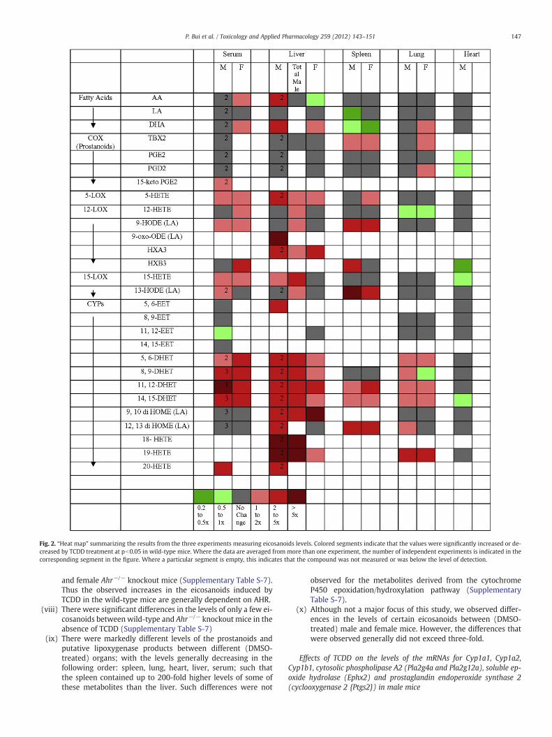
Fig. 2. “Heat map” summarizing the results from the three experiments measuring eicosanoids levels. Colored segments indicate that the values were significantly increased or de-creased by TCDD treatment at pb0.05 in wild-type mice. Where the data are averaged from more than one experiment, the number of independent experiments is indicated in thecorresponding segment in the figure. Where a particular segment is empty, this indicates that the compound was not measured or was below the level of detection.
147P. Bui et al. / Toxicology and Applied Pharmacology 259 (2012) 143–151
and female Ahr−/− knockout mice (Supplementary Table S-7).Thus the observed increases in the eicosanoids induced byTCDD in the wild-type mice are generally dependent on AHR.
(viii) There were significant differences in the levels of only a few ei-cosanoids between wild-type and Ahr−/− knockout mice in theabsence of TCDD (Supplementary Table S-7)
(ix) There were markedly different levels of the prostanoids andputative lipoxygenase products between different (DMSO-treated) organs; with the levels generally decreasing in thefollowing order: spleen, lung, heart, liver, serum; such thatthe spleen contained up to 200-fold higher levels of some ofthese metabolites than the liver. Such differences were not
observed for the metabolites derived from the cytochromeP450 epoxidation/hydroxylation pathway (SupplementaryTable S-7).
(x) Although not a major focus of this study, we observed differ-ences in the levels of certain eicosanoids between (DMSO-treated) male and female mice. However, the differences thatwere observed generally did not exceed three-fold.
Effects of TCDD on the levels of the mRNAs for Cyp1a1, Cyp1a2,Cyp1b1, cytosolic phospholipase A2 (Pla2g4a and Pla2g12a), soluble ep-oxide hydrolase (Ephx2) and prostaglandin endoperoxide synthase 2(cyclooxygenase 2 {Ptgs2}) in male mice

Fig. 3. Effect of TCDD on Cyp1a1, Cyp1a2, Cyp1b1, and Pla2g12a mRNA levels in liver, spleen, lung and heart of male wild-type mice. The levels of each mRNA are represented rel-ative to the levels of the constitutively expressed GAPDH glycolytic enzyme in each organ. The means and standard errors were derived from four mice treated with TCDD (filledbars) or vehicle (open bars). *, ** and *** represent significantly different from the DMSO control at pb0.05, pb0.01 and 0.001, respectively.
148 P. Bui et al. / Toxicology and Applied Pharmacology 259 (2012) 143–151
The levels of the Cyp1a1 and Cyp1b1 mRNAs were increased byTCDD treatment in the wild-type male mouse in the liver, lung andheart (although the mRNA levels were 10-fold or more lower in theheart). Cyp1a2 increased only in the liver. None of the mRNA levelswere increased by TCDD in the spleen. Interestingly, the levels ofCyp1a1 mRNAs in the different organs correlate with the known dis-tribution of TCDD to these organs (De Jongh et al., 1995). No induc-tion of any of these enzymes occurred in the Ahr−/− mouse (Fig. 3).It should be noted that TCDD characteristically affects the levels ofthese enzymes exclusively at the transcriptional level, and increasesin their mRNAs are reflected in increases in the corresponding pro-teins (Hankinson, 1995). The mRNA for the phosopholipase A2 formPlag2g12a has been reported to be inducible by TCDD in the liver ofboth male and female C57BL/6 mice in whole genome microarraystudies (Tijet et al., 2005: Boutros et al., 2008; Kopec et al., 2010).We found that Plag2g12a mRNA was inducible in the male liverabout 2.5-fold in our studies (Fig. 3). Pla2g4a was reported to be in-creased by TCDD treatment in the Hepa-1 mouse hepatoma cell line(Kinehara et al., 2009). This enzyme has been reported to representa major form of phosopholipase A2 with regard to the metabolismof arachidonic acid to eicosanoids (Kita et al., 2006). However, neitherthis enzyme, nor Ephx2 were induced by TCDD in our studies in mice(Supplementary Fig. S1). The levels of the prostaglandin endoperox-ide synthase 2 mRNA was not increased in any of the organs after athree day treatment with (50 ug/kg) TCDD (Supplementary Fig. S1).This is in agreement with the results of Vogel and coworkers (Vogelet al., 1998). However, those workers did find that 10 μg/kg TCDDtransiently increased cyclooxygenase 2 mRNA levels: inducing themRNA maximal 4.5-fold and 3.5-fold in the lung and spleen, respec-tively, of female C57BL/6 mice after 24 hours of treatment. (Noincrease occurred in the liver.) Nevertheless, any increase in cycloox-ygenase that might have occurred at earlier time points had minimaleffects on the levels of prostanoids in our experiments. In conclusion,TCDD induction of Cyp1a1, Cyp1a2, Cyp1b1 and Plag2g12a correlatedwith TCDD-induced increases in eicosanoid levels in some organs(lung and liver), but not others (spleen and heart).
Discussion
In this study, we show that TCDD increases the levels of a numberof eicosanoids in several organs/tissues of the mouse. DHETs wereelevated in many of the organs/tissues by TCDD. The levels of theDHETs we measured in serum fell in the 4 to 40 nM range in the ab-sence of TCDD. These values are similar to those previously reportedfor mouse serum (Kubala et al., 2010). Although these compoundsare generally considered to be inactive metabolites of EETs, they doexhibit some of the same properties as EETs, including vasodilatoryactivity (Oltman et al., 1998), and they are ligands for perioxisomeproliferator activated receptors (PPAR) α and γ (Buczynski et al.,2009; Konkel and Schunck, 2011). Importantly, they are rapidlygenerated from the corresponding EETs by epoxide hydrolase, andso their levels probably reflect the levels of the more biologicallyactive EETs. We did not report the levels of the EETs themselves inmost organs/tissues either because they were below the level ofdetection, or because we had not developed the means for theiranalysis. Nevertheless, we found that 5,6-EET was increased byTCDD in the liver and spleen. Schlezinger and coworkers previouslyreported that TCDD treatment increased the levels of three EETs inthe liver of fish (Schlezinger et al., 1998). The equivalent derivativesof linoleic acid, 9,10-diHOME and 12,13-diHOME, which are proin-flammatory (Slim et al., 2001), were increased by TCDD in someorgans.
We did not possess the means for measuring the terminal hydrox-ides in most experiments. However, where measured, these metabo-lites were consistently elevated by TCDD in all organs/tissues studied(except the heart). 20-HETE is known to be biologically active, for ex-ample exhibiting potent vasoconstrictive activity (Ishizuka et al.,2007). Little is known about the potential functions of 18-HETE and19-HETE, however, they can induce vasodilation by inhibiting the ef-fects of 20-HETE (Carroll et al., 1996). Interestingly, manymetabolitesthat are traditionally considered products of lipoxygenase metabo-lism, including 5-HETE, 12-HETE, 9-HODE, HXA3, HXB3, 15-HETEand 13-HODE were increased by TCDD treatment in serum, liver
149P. Bui et al. / Toxicology and Applied Pharmacology 259 (2012) 143–151
and spleen. These compounds all exhibit biologically activity, of vary-ing potency (Buczynski et al., 2009).
TCDD increased the level of free arachidonic acid in the liver ofwild-type mice, as has been described previously (Lin et al., 2011),and this may explain to some degree the increases in eicosanoidsthat occur after TCDD treatment in this organ. However the increasesin these metabolites are not likely to be completely due to the in-crease in arachidonic acid, because (i) increases did not occur in thelevels of the prostanoid products of arachidonic acid metabolism,ii) increases in the many arachidonic acid metabolites occurred in fe-male liver (although to a lesser extent), despite the fact that no in-crease free in arachidonic acid occurred in this organ, and (iii) anincrease did not occur in the amount of total arachidonic acid inmale liver. The total levels of several eicosanoids were increased byTCDD in male liver. Since the metabolites are likely generated fromfree arachidonic or linoleic acid rather than the esterefied forms, thefree metabolites are probably incorporated rapidly into phospho-lipids, and the phospholipids so formed therefore represent a reser-voir for these metabolites.
The TCDD-induced increases in the levels of the eicosanoids inthe spleen and other solid organs cannot be ascribed to the (relative-ly small amount of) blood co-harvested with them, as the levels inthe organs were generally of the same magnitude or greater thanin serum. Nevertheless, it is very possible that, since several of theeicosanoids were elevated by TCDD in serum, increases in oneorgan may be transmitted to another via transport of the compoundin the blood.
Since there was little effect of TCDD in eicosanoids levels in Ahr−/−
mice, the Ahrmediates most if not all the effects of TCDD on the eicos-anoids. Cyp1a1, Cyp1a2 and Cyp1b1 are known to be among the mosthighly TCDD-induced genes in the liver in vertebrates. Purified(human) CYP1A1, CYP1A2, and CYP1B1 can metabolize arachidonicacid to midchain HETEs, terminal HETEs and EETs (Choudhary et al.,2004; Schwarz et al., 2004; Fer et al., 2008; Rifkind, 2006), and sothese cytochromes P450may be responsible for much of the increasesin these metabolites in the liver after TCDD treatment. However, insome (but not all) microarray experiments, TCDD has been shown toelevate the levels of the arachidonic acid-metabolizing Cyp2c29,Cyp2c47 and Cyp2c50 enzymes (Hayes et al., 2007; Forgacs et al.,2011), and benzo(a)pyrene, a ligand for the AHR has been shownto increase the levels of the mRNAs for the arachidonic acid-metabolizing Cyp4f4 and Cyp4f5 enzymes in the rat (Aboutabl et al.,2009). Therefore other cytochromes P450 may contribute to theincrease in eicosanoids levels occurring upon TCDD treatment. Theheart did not exhibit an increase in any eicosanoids after TCDD treat-ment, despite the fact that Cyp1a1 and Cyp1b1mRNAs were induciblein this organ. However, this may be explained by the fact that the in-duced levels of these cytochromes P450 were considerably lowerthan they were in the liver and lung.
It should be noted that although analysis of arachidonic acid me-tabolism by purified cytochromes P450 is useful, the metabolic pro-file of arachidonic acid in a particular organ is difficult to predictfrom its content of cytochromes P450, for several reasons, includingthe effects of regulatory interactions between metabolic products ofsome cytochromes P450 (Rifkind, 2006). Interestingly, Schlezingerand coworkers found that TCDD had a stronger effect on the in vivometabolism of arachidonic acid than its in vitro metabolism byliver microsomes (Schlezinger et al., 1998). Lipidomic analysis aswe report here is therefore essential for advancing understandingof the roles of the eicosanoids in biological processes in the wholeorganism.
In the absence of TCDD, Ahr−/− mice exhibited different levels ofonly a few eicosanoids compared with wild-type mice.(Supplementary Table S-7). Furthermore, the number of compoundsinvolved was much less than the number increased by TCDD treat-ment in wild-type mice; more compounds showed elevated levels
in knockout mice than showed reduced levels; and the five metabo-lites that were significantly elevated in wild-type mice did not exhibitan obvious expression pattern (i.e. they were not focused in any par-ticular metabolic group or organ). Thus, it seems unlikely that theeicosanoids that were measured contribute to the known differencesin the physiological phenotypes of Ahr−/− and wild-type mice in theabsence of exogenous ligand (Fujii-Kuriyama and Kawajiri, 2010),although this needs further study.
TCDD causes toxicity in all the organs we analyzed. TCDD inducesmany adverse effects in the liver, including hepatocellular hypertro-phy and hyperplasia, fatty change, necrosis, inflammation, portalfibrosis, and liver tumor promotion and progression (Yoshizawaet al., 2007; Bock and Kohle, 2009). TCDD causes several forms ofpulmonary disease, such as keratinizing epithelioma, bronchiolarmetaplasia, and squamous metaplasia of the alveolar epithelium(Yoshizawa et al., 2007). TCDD causes cardiomyopathy, defects inseveral heart functions, and elevated arterial blood pressure(Korashy and El-Kadi, 2006; Yoshizawa et al., 2007). AHR agonistscause modest splenic lymphoid atrophy in rodents in some but notall studies (Sulentic and Kaminski, 2010; Yoshizawa et al., 2007).(We observed a decrease in spleen weight of 44% and 27% in maleand female mice, respectively, in the current study that was depen-dent on the AHR {data not shown}). It is important to considerwhether increasese in eicosanoids levels cause or contribute to anyof the TCDD-induced toxicities of these and other organs. Insightsinto this question can be provided by studies addressing the potentialroles of Cyp1a1, Cyp1a2 and Cyp1b1 in TCDD toxicity, since these cy-tochromes P450 are probably responsible for a large portion of the in-creased levels of the eicosanoids after TCDD treatment. Many toxicresponses to a high TCDD dose, including lethality and wasting,were found to be abrogated in Cyp1a1−/− or Cyp1a2−/− null micedemonstrating that these cytochromes P450 are essential for thesetoxic responses to TCDD (Smith et al., 2001; Uno et al., 2004). Certainrat strains that were resistant to about half of the multi-organ toxic-ities of TCDD that were analyzed exhibit normal induction ofCyp1a1, Cyp1a2 and Cyp1b1, indicating that although they may benecessary, these cytochromes P450 are not sufficient for the develop-ment of these toxic manifestations (Pohjanvirta et al., 2011). With re-gard to the hepatic toxicity, induction of Cyp1a1and Cyp1a2 appear toprotect against some toxic responses but to enhance others (Nukayaet al., 2009, 2010a,b). TCDD induction of splenic lymphoid atrophyappears not to depend upon induction of Cyp1a1 (Uno et al., 2004).Interestingly, studies with Cyp1a1 knockout mice demonstrated thatthis enzyme is required for vascular dysfunction and hypertension in-duced by TCDD (Kopf et al., 2010).
These studies lay the foundation for future experiments addres-sing the potential role of eicosanoids in mediating the toxic effectsof TCDD and other ligands of the AHR. Comparing the kinetic anddose–response parameters for the TCDD-mediated increases ineicosanoids levels with those for the induction of potentially relevantenzymes could provide insight into the identities of the enzymesinvolved. Such studies would be complemented by studies usingknockout mice for the relevant genes. It will be also of interest toascertain whether other ligands for the AHR, particularly nutrient-derived ligands such as indole-3-carbinol have the same effect asTCDD. Our studies on whole organs may mask much greater changesin eicosanoids levels in individual cell types in these organs, and thiswarrants examination. In addition, analysis of additional eicosanoidsmay identify metabolites that are even more elevated by TCDD thanthose studied here. It will also be of interest to investigate the effectof TCDD on the levels of eicosanoids in other mouse organs that aretargets of TCDD toxicity. Finally, it will be most important to deter-mine whether elevated eicosanoids levels contribute to the deleteri-ous effects of TCDD and other toxic agonists of the AHR.
Supplementary materials related to this article can be found on-line at doi:10.1016/j.taap.2011.12.009.
150 P. Bui et al. / Toxicology and Applied Pharmacology 259 (2012) 143–151
Funding
This work was supported by grants from National Institute ofHealth [R01CA28868 and R01ES015384 (to O.H.]). PB and PS werepartially supported by fellowships from training grant T32ES015457from the National Institute of Environmental Health Sciences. PBwas also partially supported by a fellowship from Clinical Medical Ge-netics training grant from the NIH [T32-GM0843].
Conflict of interest statement
The authors of “2,3,7,8- tetrachlorodibenzo-p-dioxin treatmentalters eicosanoids levels in several organs of the mouse in an arylhydrocarbon receptor-dependent fashion” declare that there are noconflicts of interest. None of the authors has a financial or personalrelationship with other people or organizations that could inappro-priately influence (bias) their work.
Acknowledgements
We thank Dr. Srinivasa Reddy for advice.
References
Aboutabl, M., Zordoky, B., El-Kadi, A., 2009. 3-Methylcholanthrene and benzo(a)pyrenemodulate cardiac cytochrome P450 gene expression and arachidonic acid metabo-lism in male Sprague Dawley rats. Br. J. Pharmacol. 158, 1808–1819.
Blaho, V., Buczynski, M., Brown, C., Dennis, E., 2009. Lipidomic analysis of dynamic ei-cosanoid responses during the induction and resolution of lyme arthritis. J. Biol.Chem. 283 (32), 21599–21612.
Bock, K., Kohle, C., 2009. The mammalian aryl hydrocarbon (Ah) receptor: from medi-ator of dioxin toxicity toward physiological functions in skin and liver. J. Biol.Chem. 390, 1225–1235.
Boutros, P., Yan, R., Moffat, I., Pohjanvirta, R., Okey, A., 2008. Transcriptomic responsesto 2,3,7,8-tetrachlorodibenzi-p-dioxin (TCDD) in liver: comparison of rat andmouse. BMC Genomics 9 (419), 1471–2164.
Buczynski, M., Dumlao, D., Dennis, E., 2009. An integrated omics analysis of eicosanoidbiology. J. Lipid Res. 50, 1015–1038.
Bunger, M., Glover, E., Moran, S., Walisser, J., Lahvis, G., Hsu, E., Bradfield, C., 2008. Ab-normal liver development and resistance to 2,3,7,8-tetrachlorodibenzo-p-dioxintoxicity in mice carrying a mutation in the DNA-binding domain of the aryl hydro-carbon receptor. Toxicol. Sci. 106 (1), 83–92.
Capdevilla, J., Karara, A., Waxman, D., Martin, M., Falck, J., Guenguerich, P., 1990. Cyto-chrome P-450 enzyme-specific control of the regio- and enantiofacial selectivity ofthe microsomal arachidonic acid epoxygenase. J. Biol. Chem. 265 (19), 10865–10871.
Carroll, M., Balazy, M., Margiotta, P., Huang, D., Falck, J., McGiff, J., 1996. Cytochrome P-450-dependent HETEs: profile of biological activity and stimulation by vasoactivepeptides. Am. J. Physiol. 271 (4 Pt 2), R863–R869.
Choudhary, D., Jansson, I., Stoilov, I., Sarfarazi, M., Schenkman, J., 2004. Metabolism ofretinoids and arachidonic acid by human and mouse cytochrome P450 1B1. DrugMetab. Dispos. 32 (8), 840–847.
Dalton, T., Kerzee, K., Wang, B., Miller, M., Dieter, M., Lorenz, J., Shertzer, H., Nebert, D.,Puga, A., 2001. Dioxin exposure is an environmental risk factor for ischemic heartdisease. Cardiovasc. Toxicol. 1 (4), 285–298.
De Jongh, J., DeVito, M., Diliberto, J., Van den Berg, M., Birnbaum, L., 1995. The effects of2,2′ 4,4′,5,5′-hexachlorobiphenyl cotreatment on the disposition of 2,3,7,8-tetra-chlorodibenzo-p-dioxin in mice. Toxicol. Lett. 80, 131–137.
Denison, M., Soshilov, A., He, G., DeGroot, D., Zhao, B., 2011. Exactly the same but dif-ferent: promiscuity and diversity in the molecular mechanisms of action of thearyl hydrocarbon (dioxin) receptor. Toxicol. Sci. 124 (1), 1–22.
Fer, M., Dreano, Y., Lucas, D., Corcos, L., Salaun, J., Berthou, F., Amet, Y., 2008. Metabo-lismo f eicosapentaenoic and docosahexaenoic acids by recombinant human cyto-chromes P450. Arch. Biochem. Biophys. 471, 116–125.
Forgacs, A., Kent, M., Makley, M., Mets, B., DelRaso, N., Jahns, G., Burgoon, L., Zacharewski,T.L., Reo, N., 2012. Comparative Metabolomic and Genomic Analyses of TCDD-ElicitedMetabolic Disruption in Mouse and Rat Liver. Toxicol Sci. 125, 41-55.
Fujii-Kuriyama, Y., Kawajiri, K., 2010. Molecular mechanisms of the physiological func-tions of the aryl hydrocarbon (dioxin) receptor, a multifunctional regulator thatsenses and responds to environmental stimuli. Proc. Jpn. Acad. Ser. B Phys. Biol.Sci. 86 (1), 40–53.
Hankinson, O., 1995. The aryl hydrocarbon receptor complex. Annu. Rev. Pharmacol.Toxicol. 35, 307–340.
Hayes, K., Zastrow, G., Nukaya, M., Pande, K., Glover, E., Maufort, J., Liss, A., Liu, Y.,Moran, S., Vollrath, A., Bradfield, C., 2007. Hepatic transcriptional networks in-duced by exposure to 2,3,7,8-Tetrachlorodibenzo-p-dioxin. Chem. Res. Toxicol.20, 1573–1581.
Ishizuka, T., Cheng, J., Singh, H., Vitto, M., Manthati, V., Falk, J., Laniado-Schwartzman,M., 2007. 20-Hydroxyeicosatetraenoic acid stimulates nuclear factor-κB activation
and the production of inflammatory cytokines in human endothelial cells. J. Phar-macol. Exp. Ther. 324, 103–110.
Kinehara, M., Fukada, I., Yoshida, K., Ashida, H., 2009. High-throughput evaluation of arylhydrocarbon receptor-binding sites selected via chromatin immunoprecipitation-based screening in Hepa-1c1c7 cells stimulated with 2,3,7,8-tetrachlorodibenzo-p-dioxin. J. Biosci. Bioeng. 108 (4), 277–281.
Kita, Y., Ohto, T., Uozumi, N., Shimizu, T., 2006. Biochemical properties and pathophysio-logical roles of cytosolic phospholipase A2s. Biochim. Biophys. Acta 1761, 1317–1322.
Konkel, A., Schunck, W., 2011. Role of cytochrome P450 enzymes in the bioactivation ofpolyunsaturated fatty acids. Biochim. Biophys. Acta 1814, 210–222.
Kopec, A., Burgoon, L., Ibrahim-Aibo, D., Burg, A., Lee, A., Tashiro, C., Potter, D., Sharratt, B.,Harkema, J., Rowlands, J., Budinsky, R., Zacharewski, T., 2010. Automated dose–response analysis and comparative toxicogenomic evaluation of the hepatic effectselicited by TCDD, TCDF, and PCB126 in C57BL/6 Mice. Toxicol. Sci. 118 (1), 286–297.
Kopf, P., Scott, J., Agbor, L., Boberg, J., Elased, K., Huwe, J., Walker, M., 2010. CytochromeP4501A1 is required for vasuclar dysfunction and hypertension induced by2,3,7,8-tetrachlorodibenzo-p-dioxin. Toxicol. Sci. 117 (2), 537–546.
Korashy, H., El-Kadi, A., 2006. The role of aryl hydrocarbon receptor in the pathogenesisof cardiovascular diseases. Drug Metab. Rev. 38, 411–450.
Kubala, L., Schmelzer, K., Klinke, A., Kolarova, H., Baldus, S., Hammock, B., Eiserich, J., 2010.Modulation of arachidonic and linoleic acidmetabolites inmyeloperoxidase-deficientmice during acute inflammation. Free Radic. Biol. Med. 48, 1311–1320.
Lee, C., Lawrence, B., Kerkvliet, N., Rifkind, A., 1998. 2,3,7,8-Tetrachlorodibenzo-p-dioxininduction of cytochrome P450-dependent arachidonic acid metabolism in mouseliver microsomes: evidence for species-specific differences in responses. Toxicol.Appl. Pharmacol. 153 (1), 1–11.
Li, N., Liu, J.Y., Qiu, H., Harris, T.R., Sirish, P., Hammock, B.D., Chiamvimonvat, N., 2011.Use of metabolomic profiling in the study of arachidonic acid metabolism in car-diovascular disease. Congest. Heart Fail. 17 (1), 42–46.
Lin, S., Yang, Z., Liu, H., Cai, Z., 2011. Metabolomic analysis of liver and skeletal muscletissues in C57BL/6J and DBA/2J mice exposed to 2,3,7,8-tetrachlorodibenzo-p-dioxin. Mol. Biosyst. 7, 1956–1965.
Ma, C., Marlowe, J., Puga, A., 2009. The aryl hydrocarbon receptor at the crossroads ofmultiple signaling pathways. EXS 99, 231–257.
Nebert, D., Karp, L., 2008. Endogenous functions of the aryl hydrocarbon receptor(AHR): intersection of cytochrome P450 1 (CYP1)-metabolized eicosanoids andAHR biology. J. Biol. Chem. 283 (52), 36061–36065.
Norwood, S., Liao, J., Hammock, B., Yang, G., 2010. Epoxyeicosatrienoic acids andsoluble epoxide hydrolase: potential therapeutic targets for inflammation and itsinduced carcinogenesis. Am. J. Transl. Res. 2 (4), 447–457.
Nukaya, M., Moran, S., Bradfield, C., 2009. The role of the dioxin-responsive elementcluster between the Cyp1a1 and Cyp1a2 loci in aryl hydrocarbon receptor biology.Proc. Natl. Acad. Sci. U. S. A. 106 (12), 4923–4928.
Nukaya, M., Lin, B., Glover, E., Moran, S., Kennedy, G., Bradfield, C., 2010a. The aryl hy-drocarbon receptor-interacting protein (AIP) is required for dioxin-induced hepa-totoxicity but not for the induction of the Cyp1a1 and Cyp1a2 genes. J. Biol. Chem.285 (46), 35599–35605.
Nukaya, M., Walisser, J., Moran, S., Kennedy, G., Bradfield, C., 2010b. Aryl hydrocarbon re-ceptor nuclear translocator in hepatocytes is required for aryl hydrocarbon receptor-mediated adaptive and toxic responses in liver. Toxicol. Sci. 118 (2), 554–563.
Oltman, C., Weintraub, N., VanRollins, M., Dellsperger, K., 1998. Epoxyeicosatrienoicacids and dihydroxyeicosatrienoic acids are potent vasodilators in the canine cor-onary microcirculation. Circ. Res. 83, 932–939.
Pohjanvirta, R., Korkalainen, M., Moffat, I., Boutros, P., Okey, A., 2011. Role of the AHRand its Structure in TCDD Toxicity. The AH Receptor in Biology and Toxicology.John Wiley & Sons, New Jersey. pp. 93–97.
Rifkind, A., 2006. CYP1A in TCDD Toxicity and in Physiology-with Particular Referenceto CYP Dependent Arachidonic Acid Metabolism and Other Endogenous Substrates.Drug Metab. Rev. 38, 291–335.
Schlezinger, J., Parker, C., Zeldin, D., Stegeman, J., 1998. Arachidonic acid metabolism inthe marine fish Stenotomus chrysops (Scup) and the effects of cytochrome P450 1AInducers. Arch. Biochem. Biophys. 353 (2), 265–275.
Schmidt, J.V., Su, G.H., Reddy, J.K., Simon, M.C., Bradfield, C.A., 1996. Characterization ofa murine Ahr null allele: involvement of the Ah receptor in hepatic growth and de-velopment. Proc. Natl. Acad. Sci. U. S. A. 93 (13), 6731–6736.
Schwarz, D., Kisselev, P., Ericksen, S., Szklarz, G., Chernogolov, A., Horneck, H., Schnuck,W., Roots, I., 2004. Arachidonic and eicosapentaenoic acid metabolism by humanCYP1A1: highly stereoselective formation of 17(R),18(S)-epoxiyeicosatetraenoicacid. Biochem. Pharmacol. 67, 1445–1457.
Slim, R., Hammock, B., Toborek, M., Robertson, L., Newman, J., Morisseau, C.,Watkins, B., Saraswathi, V., Hennig, B., 2001. The role of methyl-linoleicacid epoxide and diol metabolites in the amplified toxicity of linoleic acidand polychlorinated biphenyls to vascular endothelial cells. Toxicol. Appl.Pharmacol. 171 (3), 184–193.
Smith, A., Clothier, B., Carthew, P., Childs, N., Sinclair, P., Nebert, D., Dalton, T., 2001.Protection of the Cyp1a2 (−/−) Null Mouse against Uroporphyria and Hepatic In-jury Following Exposure to 2,3,7,8-Tetrachlorodibenzo-p-dioxin. Toxicol. Appl.Pharmacol. 173, 89–98.
Sulentic, C., Kaminski, N., 2010. The long Winding Road toward Understanding theMolecular Mechanisms for B-Cell Suppression by 2,3,7,8-Tetrachlorodibenzo-p-dioxin. Toxicol. Sci. 120 (S1), S171–S191.
Tijet, N., Boutros, P., Moffat, I., Okey, A., Tuomisto, J., Pohjanvirta, R., 2005. Aryl Hydro-carbon Receptor Regulates Distinct Dioxin-Dependent and Dioxin-IndependentGene Batteries. Mol. Pharmacol. 69, 140–153.
Uno, S., Dalton, T., Sinclair, P., Gorman, N., Wang, B., Smith, A., Miller, M., Shertzer, H.,Nebert, D., 2004. Cyp1a1 (−/−) male mice: protection against high-dose

151P. Bui et al. / Toxicology and Applied Pharmacology 259 (2012) 143–151
TCDD-induced lethality and wasting syndrome, and resistance to intrahepatocytelipid accumulation and uroporphyria. Toxicol. Appl. Pharmacol. 196, 410–421.
Vogel, C., Schuhmacher, U., Degen, G., Bolt, H., Pineau, T., Abel, J., 1998. Modulation ofProstaglandin H Synthase-2 mRNA Expression by 2,3,7,8-Tetrachlorodibenzo-p-dioxin in Mice. Arch. Biochem. Biophys. 352 (2), 265–271.
White, S., Birnbaum, L., 2009. An overview of the effects of dioxins and dioxin-likecompounds on vertebrates, as documented in human and ecological epidemiology.J. Environ. Sci. Health C 27 (4), 197–211.
Yang, J., Schmelzer, K., Georgi, K., Hammock, B., 2009. Quantitative Profiling Method forOxylipin Metabolome by Liquid Chromatography Electrospray Ionization TandemMass Spectrometry. Anal. Chem. 81 (19), 8085–8093.
Yoshizawa, K., Heatherly, A., Malarkey, D., Walker, N., Nyska, A., 2007. A Critical Com-parison of Murine Pathology and Epidemiological Data of TCDD, PCB126, andPeCDF. Toxicol. Pathol. 35, 865–879.
Zeldin, D., 2001. Epoxygenase Pathways of Arachidonic Acid Metabolism. J. Biol. Chem.276 (39), 36059–36062.