2782 anal. chem. 1984, 56, 2782-2791 solids analysis ... anal. chem. 1984, 56, 2782-2791 solids...
TRANSCRIPT

2782 Anal. Chem. 1984, 56, 2782-2791
Solids Analysis Using Energetic Ion Bombardment and Multiphoton Resonance Ionization with Time-of-Flight Detection
Fred M. Kimock, James P. Baxter, David L. Pappas, Paul H. Kobrin, and Nicholas Winograd* Department of Chemistry, T h e Pennsylvania S ta te University, 152 Davey Laboratory, University Park, Pennsylvania 16802
Recently multiphoton resonance ioniratlon (MPRI ) has been coupled with energetlc Ion bombardment to yleld a highly efficient and selective tool for solids analysls. Although this method promises to yield sub-part-per-billion determlnations for many elements wlthout chemlcal alteratlon of the matrlx, there are a number of experlmental factors which may ultl- mately llmlt the sensltlvlty of the technlque. Among these factors are (a) duty cycle, (b) primary Ion current, (c) sputter yield, (d) useful fraction of ejected particles, and (e) detectlon efficlency. I n this paper we discuss the origln of these factors and their Influence on the use of MPRI of sputtered neutrals as a tool for the elemental analysls of solids.
The use of energetic ion beams as probes for the analysis of solids is now well established. Detailed measurements of charged matter ejecting from ion bombarded surfaces as in secondary ion mass spectrometry (SIMS) have been employed for a wide variety of analytical applications. Among these applications are the molecular weight determination of non- volatile molecules ( I ) , determination of surface structure (2), and trace analysis of solids (3). As a trace analysis tool, SIMS has exhibited a unique ability to examine surfaces, thin films, and interfaces. The SIMS technique has also shown consid- erable dynamic range ( lo6) ( 4 ) and can achieve sensitivity for detection of certain elements down to the few parts-per-billion level in favorable cases (5). However, determination of ele- mental concentrations in parts-per-billion regime is seldom realized by SIMS. The detection limit arises from several sources. First, the ion fraction of ejected particles is often
or less. In these cases, the great maority of all ejected species are neutral and go undetected by the mass spectrom- eter. Next, in order to obtain the mass resolution necessary for SIMS analysis, quadrupole or magnetic sector mass spectrometers are typically employed which have ion trans- missions in the range of 10-1 to Thus, the useful ion fraction (number of ions detected/number of particles ejected) can easily be or less. Although not directly related to the detection limit, a final problem with SIMS analysis is that secondary ion formation is strongly influenced by electronic effects arising from the sample matrix, making it extremely difficult to quantify most measurements. Clearly, a method to efficiently detect neutrals ejected from an ion bombarded surface would overcome these problems and could be a major breakthrough for the application of ion beam methods to chemical analysis.
Several attempts have been made to directly monitor the flux of sputtered neutrals (in both ground and excited states), although none has exhibited the sensitivity to operate in the low dose regime (<1010 incident ions/cm2.s) (6, 7). Experi- ments involving postionization of the ejected neutrals using electron bombardment (8), glow discharges (9), and plasmas (10) have been moderately successful, but none has achieved ionization efficiencies much greater than 1%. A major dif- ficulty inherent in these methods is that the ionization probability is a function of particle velocity.
Recently, we have demonstrated the selective ionization of neutral atoms sputtered from solids by multiphoton resonance ionization (MPRI) (11,12). The theory behind MPRI is well documented (13). Basically, three salient features of MPRI coupled to ion bombardment make it attractive as an ana- lytical tool. (i) All elements but He and Ne can be detected. (ii) MPRI can be made selective to a single element by a judicious choice of the excitation wavelength. (iii) Laser technology has advanced to the point where the MPRI process can be saturated using pulsed lasers, i.e., with adequate photon fluxes every atom in the laser beam can be ionized. In this work, we demonstrate that MPRI of sputtered neutrals can be employed to overcome the three main difficulties inherent in SIMS analysis. First, for the majority of cases, MPRI of sputtered neutrals will sample the major fraction of ejected particles. With the appropriate experimental geometry, a high fraction of the sputtered atoms can be made to interact with the laser beam, resulting in efficient sampling. Second, since the ionization is selective, a high mass resolution detector is not needed; thus the photoionized neutrals can be detected by a low resolution time-of-flight detector with high efficiency. Finally, examination of the neutral flux should dramatically reduce matrix effects and improve the prospects for quanti- tative analysis.
When operated with a low primary ion dose, the MPRI experiment may be more sensitive than static SIMS by several orders of magnitude and will be useful for monitoring the progress of chemical reactions (12) and as a structural probe (14). Used with higher primary ion doses, MPRI of sputtered neutrals should find application in the areas of depth profiling and trace analysis of solids. For example, the detection of 0.5 ppm of Ga sputtered from a Si matrix has recently been achieved by workers at Atom Sciences, Inc., using MPRI (15). To date, multiphoton resonance ionization has been applied to over 40 elements, mostly by investigators using hot filament evaporation techniques (16, 17).
This paper evaluates the utility of MPRI coupled with energetic ion sputtering as a tool for the elemental analysis of solids. Generally, our calculations indicate that sub- part-per-billion determinations of many elements will be possible, The sensitivity of the technique is presently limited chiefly by the laser duty cycle and ion source brightness. Using model systems of elemental targets, we illustrate the influence of the sample matrix, experimental operating conditions, and atomic photoionization spectroscopy on the sensitivity of MPRI of sputtered neutrals.
EXPERIMENTAL SECTION All experiments are conducted in an ion pumped Perkin-Elmer
Ultek TNB-X ultrahigh vacuum chamber with a base pressure of torr after bakeout. A primary beam of Ar" ions is generated by a Danfysik 911A hollow cathode source. The primary beam kinetic energy can be varied from several hundred eV to -30 keV. After extraction from the source, the ion beam is mass selected by a 90° magnetic sector, and passed through two stages of differential pumping si' ultrahigh vacuum conditions can be maintained in the analysis chamber. The beam is pulsed at 30 Hz (defined by the laser) by deflecting it through an aperture with a 200-V pulse applied to a set of parallel plates located within
0003-2700/84/0356-2782$01.50/0 0 1984 American Chemical Society

ANALYTICAL CHEMISTRY, VOL. 56, NO. 14, DECEMBER 1984 2783
ELECTRON MU LT I P t I ER
J EXTRACTION
ELECTRODE
X I
Y
LASER
Flgure 1. Experimental configuration of the MPRI experiment on sputtered neutrals. Atoms are sputtered from the sample by a pulse of primary Ar+ ions, and then postionized by the laser. All ions are extracted and detected by an electron multiplier. Secondary ions can be distinguished from laser-produced ions on the basis of their time- of-flight to the detector.
the beam line. Typically, a primary ion beam current of 2 pA (at 5 keV beam energy) can be delivered to the sample at an incident angle of 45O into a spot size of less than 1 mm in diameter. Upon impact, the pulse of primary ions produces a distribution of atoms localized in space a few millimeters above the target. A few hundred nanoseconds after the ion pulse has ended, the volume of sputtered neutrals is irradiated with a laser pulse of the appropriate wavelength(s) to cause MPRI of the desired atomic species. Ions generated in the experiment are then ex- tracted and detected with a copper-beryllium electron multiplier, an arrangement which serves as a low resolution time-of-flight analyzer. The experimental geometry is illustrated in Figure 1.
It is useful to be able to distinguish between the ions produced directly at the surface (SIMS) and those produced by interaction with the photon field. Since the secondary ions and MPRI ions are born in distinctly different regions of the same extraction field, they will have different velocities a t the time they reach the detector. This is to say that the SIMS and MPRI ions can be discriminated with an electrostatic energy filter (11), or more simply on the basis of their time-of-flight to the detector. As we will show, for maximum analytical sensitivity, it is necessary that the laser be fired <500 ns after the primary ion pulse has ended. Thus, in order to ensure both maximum MPRI yield and mini- mum overlap between the laser produced ions and any “tail” of secondary ions at the detector, it is critical that the fall time of the primary ion pulse be on the order of 100 ns or less. For primary ion pulses having longer fall times, the laser firing time will have to be further delayed and sensitivity will be sacrificed.
The laser system has been previously described (12). Briefly, the Quanta-Ray DCR-2A Nd:YAG/dye laser system is capable of generating tunable laser light from 260 nm to -800 nm. The laser is pulsed (at 30 Hz, with -66-11s pulses) and typically -5 mJ/pulse of ultraviolet light and/or -15 mJ/pulse of visible light can be obtained. The laser beam which is shaped by filled-in beam
1 p m Generators
Trigger Tr igger
B O X C A Q #2 G&TE
T I W L - C F - F L I G H T S P E C T R U M n n A U’L
Secondary M P R I Ions Ions TIME
Flgure 2. Schematic of pulse timing and data collection in the ex- periment. The timing sequence is initiated by the internal flashlamp trigger of a Nd:YAG laser. All other pulses are triggered externally by time delay generators. Boxcar integrators are used to monitor the primary ion current and MPRI signal after a time-of-flight. Typical values for delay times are D1 = 200 ps, D2 = 5 ps, D3 = 3 ps, and D4 = 4 ps. Tis the primary ion flight time from the pulsing aperture to the sample, 7 is the primary ion pulse width, and t is the laser firing time. The laser pulse width is - 6 ns. The time axis is given for illustration only; no absolute scale is Intended.
optics in the YAG is -6 mm in diameter in the unfocused con- dition and exhibits a near-Gaussian profile.
The instrument is interfaced to a Digital Equipment Corp. LSI 11/23 microcomputer which is equipped with a 40 megabyte Winchester hard disk and 256 kilobytes of random access memory. Computer control of the experiment is afforded through a BiRa Systems Model 5000 Powered CAMAC crate. The crate contiiins a card rack with 25 positions for plugging in CAMAC modules. Three positions are occupied by a BRQ bus controller for com- munication between the CAMAC modules and the computer.
The laser power, primary ion beam current, and MPRI signal can be continuously monitored by applying dc voltage signals to a Kinetic Systems 3560 Quad voltage to frequency converter (V-F). The output frequency of the V-F is integrated by a BiRa Model 2101 scaler/timer. The scaler output is then relayed to the computer for disk storage and observation on a terminal screen or immediate plotting. In the case of the laser power, the V-F receives an amplified voltage from a Scientech Model 36-2002 power and energy indicator. The output from a picoammeter can be sent to the V-F to monitor primary ion current in the dc mode. When the ion beam is pulsed, the pulsed output of the picoam- meter can be measured with a boxcar integrator, whose output voltage is then sent to the V-F. For the MPRI signal, a boxcar integrator is employed as a signal averager to measure the preamplified signal of an electron multiplier operated in the analog mode. The CAMAC crate is also equipped with two LeCroy Model 2323 programmable dual gate and delay generators which are responsible for triggering the ion beam pulse, the laser, and the boxcars. In addition to the above, the CAMAC crate has been equipped with a Standard Engineering Corp. SMC-406/H step- ping motor controller which can be used for preprogrammed scanning of the dye laser monochromator. The pulsing sequence of our experiment is illustrated in Figure 2.
Exciton DCM laser dye in methanol solution was used to generate all wavelengths for the photoionization experiments on In, AI, Co, and Mo atoms. Gallium photoionization was accom- plished by using the output of Exciton Rhodamine B laser dye in methanol solution.
The In foil was cleaned before all experiments by Ar+ ion sputtering under vacuum. Sputter cleaning was terminated when

2784 ANALYTICAL CHEMISTRY, VOL. 56, NO. 14, DECEMBER 1984
the In,/In ratio as determined by MPRI reached a maximum value (12). Oxidation experiments employed Matheson reagent grade oxygen of 99.9995% purity. The AI, Co, and Mo foil samples were cleaned by acid etching in a hot solution of 50% glacial CH,COOH, 30% HNO,, 10% H2S04, and 10% H3P04, followed by Ar+ ion sputtering under vacuum. All of the foil samples had a purity of 99.9995%. Gallium atoms were sputtered from single crystal GaAs which was cleaned under vacuum by alternately cycle heating to 500 "C and Ar+ ion sputtering.
RESULTS AND DISCUSSION Three features of MPRI of sputtered neutrals reveal the
potential sensitivity of this experiment. (i) Ion beam sput- tering is a very efficient atomization source. For example, a 1 mA primary beam of Ar+ ions could conceivably liberate 10l6 atoms/s from a solid. Also, by focusing the ion beam, atom- ization can be made to arise from a microscopic region within the solid. (ii) With adequate photon fluxes, nearly every atom which is resonantly excited can be ionized. Also, the ionization probability will be independent of particle velocity for the majority of sputtered partiels. (iii) Either ions or electrons generated by MPRI can be detected with high efficiency. The lower limit to such a measurement, the detection of single atoms, has been demonstrated by using MPRI (13). Indeed, there are many limitations to the detection of one atom in 1OI6 as alluded to in the above hypothetical experiment. For MPRI of sputtered neutrals, the measured signal intensity, I , can be expressed as a product of experimental parameters
I = DPSCUE (1)
where D is the duty cycle, P is the primary ion current, S is the sputter yield of secondary particles, C is the concentration of the element to be determined in the solid, U is the fraction of ejecting analyte atoms which are in the electronic state that is being probed by MPRI, and E corresponds to some measure of detection efficiency. In eq 1, E is influenced by the ability to inject atoms into the photon field, ionize these atoms, and subsequently detect them. In the remainder of this paper, we discuss the origin of these factors and their influence on the use of MPRI of sputtered neutrals as a probe for the elemental analysis of solids.
Duty Cycle. One of the chief shortcomings of the use of MPRI is the need to employ pulsed lasers in order to approach saturation of the ionization process. As a result, in our con- figuration the primary ion beam is fired at the pulse repetition frequency (prf) of the laser (30 Hz) in order to ensure efficient sampling of the sputtered particles by the photon field. Calculations and experiments (discussed below) indicate that the maximum MPRI signal can be achieved using a primary ion beam pulse width of 110 ps, making the sampling duty cycle of the experiment 3 x at best. This problem could be significantly improved by increasing the prf on the laser. In fact, the use of continuous wave (CW) lasers for MPRI has been shown to result in total ion yields (counts/s) which are comparable to those obtained in pulsed experiments (18). However, in CW experiments the photon flux is considerably reduced, so the high degree of ionization efficiency and hence sampling efficiency is lost. Thus, further evolution of laser technology will be necessary before the compromise between duty cycle and ionization efficiency can be overcome.
Although the use of lasers with short pulses (ns) can be viewed as a disadvantage in terms of duty cycle, it is a direct consequence of these short photon pulses that the postioni- zation probability for the sputtered atoms is independent of particle velocity. For example, a 100-eV In atom will move only 0.007 cm during a 6-ns laser pulse. A lighter atom, for example, 100 eV B, will travel 0.03 cm during the laser pulse. Since the vast majority of sputtered atoms have kinetic en- ergies less than 30 eV, most particles are virtually frozen inside
the photon field (mm in diameter) for the duration of the laser pulse.
Primary Ion Beam Current. In sputtering experiments, the primary ion beam current is the major factor governing the number of particles which are atomized. For MPRI, the ionization signal is proportional to the quantity of atoms in the ionization volume. Thus, to overcome the duty cycle problem in the MPRI experiment on sputtered neutrals, the number of incident primary ions per pulse must be relatively high. For example, in the case of low dose experiments, a duty cycle of 3 X and a primary ion beam current density of lom6 A/cm2 still provides static bombardment conditions. With similar primary ion fluxes, the yield of neutrals ejected from both a clean and chemically reacted surface has been measured with good sensitivity (12).
If MPRI is to exceed the capabilities of SIMS for trace analysis, primary ion currents should be in the range of 0.01 to 1 mA or greater. Dynamic SIMS studies normally employ about 10 pA of primary beam current. Note that when a source emitting 33 mA of ion current is pulsed at 30 Hz with 10-ps pulses, the average ion beam current is 10 pA. Although ion sources which emit such high currents are not standard equipment on most surface analysis systems, they are avail- able. With duoplasmatron type sources, up to 10 mA of Ar+ ion current can be delivered to the target at ion beam energies of -25 keV, into a spot size of several millimeters.
To maximize the spatial overlap between the ejected neu- trals and the photon field, it is important to operate the MPRI experiment with an ion beam spot which is small relative to the diameter of the laser beam. Unfortunately, it is difficult to transfer high ion currents (mA) to the sample in spot sizes much smaller than 1 mm or a t low kiloelectronvolt beam energies due to space-charge factors. The space-charge effect has two important consequences for the MPRI experiment. First, in situations where multiphoton ionization must be accomplished using a focused laser, there will be a maximum useful ion beam spot size that will cause the sputtered atoms to be injected into the photon field. Due to space-charge limitations, the current which can be delivered into this spot is not arbitrarily large. Secondly, considering microprobe analysis via MPRI, primary ion beam currents are already space-charge limited and cannot be increased to offset the duty cycle.
Sputter Yield and Analyte Concentration. Knowledge of the influence of experimental parameters upon the sputter yield is important when choosing the optimum analysis con- ditions for the MPRI experiment. Several trends regarding the sputter yield for a solid under energetic ion bombardment must be considered. First, for most metals, an examination of the sputter yield as a function of primary ion energy shows a monotonic increase up to about 5 keV, where the yield either levels off or begins to decrease (19). Secondly, the sputter yield increases as ion bombardment is performed in a non- perpendicular direction and is usually maximized by bom- barding the target a t angles between 45' and 60' from the surface normal. Last, for monoenergetic noble gas ion bom- bardment, primary ion particle mass has only a minor influ- ence on the sputter yield. For example, changing the primary ion from Ar+ to Xe+ generally results in approximately a 2-fold increase in the sputter yield (19). However, for a given ion beam energy and beam diameter, the maximum obtainable ion current density varies inversely with the ion mass. Noble gas ions are useful for our experiment since (i) they allow clean operation of the ion source and (ii) their interaction with the solid is thought to have little influence on surface electronic properties. The latter is in sharp contrast to many dynamic SIMS experiments in which bombardment is performed with an 02+ beam to enhance positive secondary ion yields, or a

ANALYTICAL CHEMISTRY, VOL. 56, NO. 14, DECEMBER 1984 2785
1 I I I t r . I t 4 I I I I 1 3
2 - 10-
c - a
Cs+ beam to enhance negative secondary ion yields. For the above reasons, many of our initial investigations have em- ployed a primary beam of 5-keV Ar+ ions, incident on the sample a t 45’.
The sputter yield is also not free from matrix effects. It is often observed that as a multicomponent surface is exposed to a flux of energetic ions, initially the species with the highest effective sputtering yields will be preferentially desorbed, resulting in the near-surface region becoming enriched in species with low effective sputtering yields (20). For our purposes, we employ Coburn’s definition of “preferential” which is referenced to the bulk composition of the solid (21). Once steady-state conditions have been reached, the surface concentrations of the residing elements are proportional to their bulk concentrations and inversely proportional to their effective sputter yields. At that point, the concentrations of atoms which are sputtered from the surface are equal to the atomic concentrations in the bulk. Thus, preferential sput- tering (according to the above definition) occurs only during the time required to establish the nonstoichiometric surface layer, and there is no preferential sputtering under steady-state conditions. This means that for MPRI of sputtered neutrals, ion intensities will be directly proportional to the bulk con- centrations of the corresponding elements, and no knowledge of effective sputtering yields is required for quantitative analysis. In light of the formation of a nonstioichiometric surface region, the combination of MPRI and ion bombard- ment has an advantage in terms of its ability to make quan- titative measurements (e.g., in sputter profiling) over tech- niques like XPS and Auger electron spectroscopy, which directly examine the surface layer, rather than the sputtered particles.
For bulk analysis of major constituents, the need to reach steady-state concentrations at the sputtered surface is a minor problem. For binary and ternary systems, this condition is often achieved by sputtering for several minutes with removal rates of tens or hundreds of angstroms per minute. However, it is not yet clear how difficult it will be to establish the enriched phase for the analysis of trace constituents, or even if the existence of such a phase will be necessary at all. Also, the accuracy and resolution of depth profiling analysis is, in part, determined by the thickness of the layer to be examined relative to the amount of sample which must be removed in order to achieve steady-state sputtering conditions. The need to form an “equilibrium” nonstoichiometric phase also in- creases the difficulty of accurately profiling constituents which have a steep concentration gradient in the solid.
Useful Fraction of Ejected Atoms. For the situation of a solid having a sputter yield of 5 atoms per incident ion, a 10-pA ion beam pulsed at 30 Hz with pulses of 10 ps duration would generate a reservoir of about lo1’ atoms/s. Under ideal detection conditions an impurity a t a concentration of 1 ppb (atomic) would produce 100 counts/s. In reality, however, only atoms which are in the electronic state that is being probed by MPRI are analytically useful.
Generally, for an element M in the atomized volume, one can express the concentration of M as c(M) = c(M0) + c(M*) + CC(M,*) + n C c ( M n ) +
i n>l nCCc(M,X,) + ... (2)
where Mo represents ground state atoms, M* represents monomeric secondary ions, M,* represents atoms in long-lived excited states, and M, and M,X, are examples of molecular species which can exist in ground or excited states, or as ions. If the element to be determined is present in trace quantities, n = 1. For the systems we have studied so far, the majority of ejected particles have been monomers. For example, from a clean indium surface bombarded by 5-keV Ar’ ions, -80%
n m
v - x x x x x x x x x x x - X Co MPRI
x x x x
X - E X >
X
WAVELENGTH (4) Flgure 3. Ion intensity vs. excitation wavelength for one-color, single resonance MPRI of sputtered Co atoms. The five peaks labeled G are ground state orlglnating. Experimental conditions were as follows: 5-keV Ar+ (2 pA, 5-ps pulses, 30 Hz), -25 mJ/(cm*.puise) of laser light. The laser doubled dye output curve is also presented (X).
of the ejected In atoms are in the ground state, - 10% of all In atoms reside in the first excited state, <lo% of the sputtered In atoms form dimers, and <1% of all secondary particles are In+ (12). This observation is consistent with previous measurements on neutrals sputtered from clean metal targets which indicated that 5 to 10% of the neutral flux may be composed of dimers and small cluster species (IO). Oechsner e t al. (22) have reported that for some oxides, the major sputtered species are of the form MO, so in certain circumstances molecule formation may be significant. Since MPRI couples ionization to a resonant bound-bound tran- sition, it is both atom- and state-selective. Therefore, generally only one of the species in eq 2 can be detected at a time. The result of this selectivity is that for MPRI, the useful fraction, U , for most experiments is given by
U = c(Mo)/c(M) (3)
U = c(M,*)/c(M) (4)
or if an excited state is being probed
The excited-state populations which can be detected by MPRI are those with relatively long lifetimes, i.e., metastables. During sputtering, fluorescent decay from atoms in high-level excited states can often be observed in the region of space just above the solid. The lifetimes of these states are typically very short (nanoseconds or subnanosecond regime), and conse- quently atoms in these states will not undergo efficient multiphoton ionization. However, after cascading, most atoms should exist in the ground state or in one of several low-lying metastable states which can be probed by MPRI. In our configuration, since the laser pulse is fired several hundred nanoseconds after the primary ion pulse has ended (producing a temporal separation of the secondary ions and the laser- produced ions at the detector) nearly all atoms in high-lying excited states should have adequate time to decay before they are exposed to the photon field.
The existence of metastable populations of sputtered atoms is illustrated in Figure 3 for Co atoms ejected from Co metal. For the wavelength region shown in the figure, we have identified 5 transitions which originate from the ground state and 18 which originate from five low-lying excited states. Although the largest peaks in the spectrum arise from MPRI of ground state atoms, the signals resulting from ionization of excited state atoms are significant. For this spectrum, the laser was unfocused and none of the ion signals exhibited saturation. No detailed analysis of the electronic partitioning of the Co excited states was attempted since the ionization efficiency for each individual transition was not determined.
Indium presents itself as a convenient model system to

2786 ANALYTICAL CHEMISTRY, VOL. 56, NO. 14, DECEMBER 1984
g 1.0 "''-7 >
O k t 1 n+ 1
0 1000 2000 3000 OXYGEN EXPOSURE (L)
Figure 4. Fraction of the total monomer yield for (0) ground state (2P112) In atoms, (0) metastable (2P3,2) In atoms, and (A) In+ sec- ondary ions vs. oxygen exposure. Approximately one monolayer of In203 is formed after 800-L exposure. Error bars present the relative standard deviation of the data. 1 L = 10" torvs. Data were collected with a quadrupole mass spectrometer as described in ref 12.
study the population of metastable states. The electronic structure is simple, with only one low-lying excited state (2P312, 0.27 eV). Transitions between the 2P3/z state and the ground state (2Pl/2) are forbidden, thus the 2P3/z state must be long-lived. The second lowest excited state lies greater than halfway between the ground state and the ionization contin- uum, and atoms excited into this state (or any higher states) could be nonresonantly ionized by a single ultraviolet photon. Since we have never detected a measurable signal which could be attributed to nonresonant photoionization of In atoms, we believe that for sputtered In, the 2P,/2 and 2P3/2 states are the only long-lived states that are significantly populated.
The photoionization schemes used to study In have been previously published (12). It is worthwhile to note that we have observed saturation of the ion yields for two-color MPRI of both the 2P1/2 and 'P3/2 states with a laser beam diameter of -0.6 cm. An ionization efficiency of 100% is consistent with our measurements at saturation powers, and a realistic measure of both the 2P1/2 and 'P3/2 populations is achieved. In the following section, we present results of investigations on polycrystalline In to begin evaluating the influence of the sample matrix and primary beam energy on the useful yield of sputtered atoms.
The yield of secondary ions is known to exhibit a strong dependence on the sample matrix. For example, the presence of surface oxygen is often observed to enhance positive ion yields by several orders of magnitude over the clean surface. It has been conjectured that for most systems, the secondary neutrals would be less subject to these matrix effects, and thus measurements on the neutrals would provide a more quan- titative measurement of surface concentrations.
In order to test this theory and evaluate the influence of the sample matrix on the useful yield, we have chosen to examine the effect of oxygen chemisorption on the sputter yield of In+ ions and both ground and excited state In atoms. In Figure 4, the population of each In monomer species, plotted as a fraction of the total monomer yield, i.e., 1(2P1/2 In) + 1(2P3/2 In) + I(In+), vs. oxygen exposure is presented. Note that from the clean surface nearly all ejected atoms are found to reside in the ground electronic state. In fact, from the clean surface, the yield of 2Plj2 In atoms is >lo00 times that of In+ secondary ions (12). From the oxygen saturated surface, however, nearly 50% of all sputtered atoms are ob- served as excited neutrals or as ions. The formation of ap-
* .
Ar+ BEAM ENERGY ( k e V )
Figure 5. The effect of prlmary Ion beam energy on the relative population of metastable ('P3/2) I n atoms to ground state (2P1,2) In atoms. The plot Is normalized to the maximum value. Error bars present the relative standard deviation of the data.
proximately one monolayer of InzO3 enhances the yield of secondary In+ by a factor of >200 over the clean surface. However, the yield of ground state In atoms decreases to -0.4 of its original value as monolayer coverage is approached (12). Thus, the neutral yield seems to be a much more direct re- flection of surface composition than does the secondary ion yield. Although the yield of neutrals is not completely free from matrix effects, these effects are much less serious than those observed in SIMS. There is a noteworthy ramification of this experiment for the use of MPRI as a tool for solids analysis. No chemical alteration of the sample (e.g., dosing the surface with oxygen or cesium) is necessary to enhance the sensitivity of the MPRI experiment. The integrity of the chemical environment of the solid can be maintained during the analysis.
Previously, we have discussed the influence of primary ion beam energy on both the sputter yield and attainable primary ion current density. The primary ion beam energy can also influence the useful yield in the MFRI experiment by affecting the relative population of excited state atoms. We have ex- amined the magnitude of this effect for atoms sputtered from a clean In surface. The relative population of metastable In atoms (plotted as the 'P3/2:2P1/2 ratio) as a function of primary Ar+ ion beam.energy from 2 to 12 keV is presented in Figure 5 . Accurate measurements could not be made below 2 keV as a result of a significant loss of primary ion current due to space-charge blow-up in the beam line and magnified effects of stray electrical fields from the detector which steered the ion beam away from the sample. The plot reveals a dramatic increase in the 'P3/2 population up to 4-keV beam energy, followed by a plateau region. The absolute 2P3/z:2P,/2 ratio for In sputtered by 5-keV Ar+ has previously been measured to be -0.1 (12). The shape of the curve reveals a possible trade-off for optimizing analysis conditions in the MPRI ex- periment. At least for this case, the relative population of excited state atoms is minimized by decreasing the primary ion energy. This increases the useful yield in the experiment by maximizing the fraction of atoms which are in the ground electronic state. However, at low kiloelectronvolt energies, reducing the primary ion beam energy will necessarily decrease both the sputter yield and primary ion current density, thereby reducing the total number of particles which can enter the photon field. Because the number of atoms in the photon field is the overriding concern, most analyses via MPRI will be conducted using 5- to 20-keV ion beams.
It is interesting to compare the excited state populations we have measured to those observed by other workers. For example, Wright et al. (23) and Pellin et al. (24) examined the populations of metastable states of sputtered uranium and
ANALYTICAL CHEMISTRY, VOL. 56, NO. 14, DECEMBER 1984 2787
iron atoms, respectively, using laser induced fluorescence. In their investigations of uranium bombarded by 500 to 3000-eV Art ions (23), the authors found the populations of ejected metastable U atoms to obey a near-Boltzmann distribution which was characterized by a temperature of 920 f 100 K. Similarly, the populations of excited state Fe atoms (sputtered from Fe foil by 3-keV Art) were characterized by a temper- ature of -700 K (24). We point out that this approach is merely an attempt to describe a population distribution long after the bombardment event has occurred. As such, these temperatures should not be confused with the very high ef- fective sputtering temperatures ( - lo4 K) which have been assigned to occur a t the impact event, resulting in surface ionization (25). Using our data for clean polycrystalline In bombarded by 5 keV Ar+, we find the sputtered In atoms to be characterized by a Boltzmann temperature of - 1180 K. Similarly, using data from Figure 5, we calculate a Boltzmann temperature of -725 K for In atoms sputtered by 2-keV Ar'. These temperatures are in reasonable agreement with those calculated for sputtered U (23) and Fe (24). It is worthwhile to note that the temperatures we have calculated are on the order of the actual temperatures employed in hot filament evaporation techniques. In the hot filament experiments up to 40% of the evaporated atoms have been observed in metastable states (26). Clearly, the mechanism that is re- sponsible for populating metastable excited levels in hot fi- lament evaporation is quite different from that in sputtering. However, in terms of influencing analytical capabilities, only the relative yield (not the mode of population) of long-lived excited states is significant, and it appears that a t the very worst, sputtering is an atomization source that is a t least as cool as filament evaporation. Although the population of metastable excited state atoms will decrease the useful yield in many cases, it opens the possibility for multielement analysis with a single laser dye.
Spatial Overlap between the Sputtered Atoms and the Photon Field. Thus far, we have discussed the major factors which influence the number of neutrals which are desorbed from an ion bombarded surface. The remainder of this paper focuses on the detection of these particles. Detection efficiency is affected by the ability to (i) achieve overlap between the spatial distribution of sputtered atoms and the photon field, (ii) ionize atoms which interact with the photon field, and (iii) extract and identify particles once they have been ionized.
In order to determine the number of sputtered atoms which are detectable, it is necessary to understand the energy and angular distributions of secondary particles. It is chiefly the energy distribution which establishes the optimum timing relationship between the primary ion beam pulse and the laser pulse. As depicted in Figure 2, the experimental timing se- quence is characterized by a primary ion pulse of width 7 ( ~ s ) , and a 6 4 s laser pulse fired at a delay time t - 7. The prob- ability, p, of an ejected particle being at some point in space above the surface is a function of its distance r from the ejection point, the time t after the particle has left the surface, and the azimuthal and polar angles of ejection, 4 and 8, re- spectively. (See Figure 1.) It has been shown for single crystal surfaces that there are preferential azimuthal angles of ejection for secondary particles (2,27). However, for polycrystalline surfaces, p is not a strong function of 4. With the additional approximation that the angular and velocity distributions are independent, we have
P = f (r , t ) - f (o) (5 ) Generally, the kinetic energy distribution of secondary
neutrals can be estimated (28) as CE
N(E) = ( E -!-
ION PULSE WIDTH, T ( p ~ )
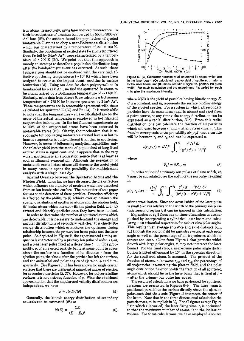
Flgure 6. (A) Calculated fraction of all sputtered I n atoms which are in the laser beam, (0) calculated relative yield of sputtered In atoms in the laser beam, and (0) measured MPRI signal vs. primary ion pulse width. For each calculation and the experiment, f is varied for each T to glve the maximum intensity.
where N(E) is the yield of particles having kinetic energy E, c is a constant, and E b represents the surface binding energy of the ejected species. For a system in which all secondary particles have the same mass (e.g., In atoms) and eject from a point source, a t any time t the energy distribution can be expressed as a radial distribution, N(r) . From this radial distribution, one can calculate the fraction of all particles which will exist between rl and r2 at any fixed time, t. This fraction corresponds to the probability p(rl,r2,t) that a particle will lie between rl and r2 and can be expressed as
where
In order to include primary ion pulses of finite width, eq 7 must be convoluted over the width of the ion pulse, resulting in
after normalization. Since the actual width of the laser pulse is small (-6 ns) relative to the width of the primary ion pulse (microsecond regime), it can be ignored in this calculation.
Expansion of eq 9 from one to three dimensions is accom- plished by incorporating a cylindrical laser beam and calcu- lating 1000 azimuthal trajectories for each of nine polar angles. This results in an average entrance and exist distance (rent, rex) through the photon field for particles ejecting at each polar angle as well as the percentage of all trajectories which in- tersect the laser. (Note from Figure 1 that particles which desorb with large polar angles, 8, may not intersect the laser beam.) For the final step, a near-cosine polar angle distri- bution (shifted off-normal by loo in the specular direction) for the sputtered atoms is assumed. The product of the fraction of atoms, p , between rent and rex, the percentage of all trajectories intersecting the photon field, and the polar angle distribution function yields the fraction of all sputtered atoms which should lie in the laser beam that is fired at t - T after the primary ion pulse has ended.
The results of calculations we have performed for sputtered In atoms are presented in Figures 6-9. The laser beam is positioned parallel to the surface directly above the ejection point such that the x axis (Figure 1) intersects the center of the beam. Note that in the three-dimensional calculation the particle mass, m, is implicit in vb. For all f i i e s except Figure 7 (in which t is varied) the laser firing time, t , is optimized so that the maximum number of atoms lie in the ionization volume. For these calculations, we have employed a source

2788 ANALYTICAL CHEMISTRY, VOL. 56, NO. 14, DECEMBER 1984
(a) x , : 0.1 cm
0.05 c m beams 4 15 z
LASER FIRING TIME, t (ps) ION PULSE WIDTH, r (ps )
figure 7. The effect of laser firing time, t , on the total intensity of sputtered In atoms localized in (a) a 1.9 cm diameter laser beam, and (b) a 0.6 cm diameter laser beam, each positioned with Its lowermost edge 0.1 cm from the sample. For both cases, T = 6 1s.
Flgwe 9. Calculated number of In atoms in the photon field vs. primary ion pulse width for three 0.05 cm diameter laser beams positioned with the lowermost edge of the beam (a) 0.1 cm, (b) 0.5 cm, and (c) 1 .O cm directly above the atom ejection point. Curve (a) is identical with Figure 8e.
as E'. For example, increasing the value Of E b to 10 eV results in a curve which more closely resembles the experiment. By the same token, using values slightly greater or less than 3 in eq 6 results in minor changes in the curve shape. From the point of view of the experiment, it is difficult to know the position of the laser to within h0.5 mm. During the analysis, the laser beam is moved toward the sample until it begins to ablate the surface, at which point it is backed away. Finally, the laser beam has a Gaussian profile, which results in dif- ferent degrees of ionization for different locations within the beam. The beam profile is not included in the model. Due to these uncertainties, one can conclude only that reasonable qualitative agreement between the experiment and theory is achieved. ION PULSE WIDTH, T (ps)
Flgure 8. Calculated number of In atoms in the photon fieid vs. primary ion pulse width for five laser beams positioned directly above the atom ejection point and intersecting the x axis between the followlng x coordinates: (a) x 1 = 0.1 cm, x z = 2.0 cm; (b) x , = 0.1 cm, x : , = 0.7 cm; (c) x , = 0.2 cm, x:, = 0.8 cm; (d) x , = 0.5 cm, x: , = 1.1 cm; (e) x 1 = 0.1 cm, x: , = 0.15 cm.
emitting 10 pA of primary ion current (which is pulsed in microsecond intervals), a sputter yield of five In atoms per incident ion, and a surface binding energy E b = 5 eV. All atoms are assumed to eject from a point source.
The calculated fraction of all sputtered atoms which lie in the ionization volume, as well as the total number of atoms in that volume are plotted in Figure 6 as a function of primary ion pulse width. The measured ion intensity for two-color MPRI of ground state In atoms vs. primary ion pulse width is also illustrated. Both the calculation and experiment were performed using a laser beam of 0.6 cm diameter positioned with its center 0.4 cm above the bombarded surface. Two regimes of the plot are of special interest. First, for a primary ion pulse width T = 200 ns, -70% of the ejected atoms should lie in the photon field; however, the total number of atoms in this region is quite low. For T = 5 ps, the fraction of all sputtered atoms which reside in the laser beam has decreased to about 25%, but the absolute number of particles in the beam has almost reached a maximum value. For primary ion pulse widths >5 ps , the total intensity of atoms localized in the photon field reaches a steady state. This is the rationale for our comment that for maximum analytical sensitivity, primary ion pulse widths need to be no greater than - 10 ps. For operation at 30 Hz, this ion pulse width fixes the maxi- mum sampling duty cycle at about 3 X lo4.
For several reasons, it is difficult to make a rigorous com- parison between the experimental and calculated results presented in Figure 6. First, for the calculation, E b must be assumed, and the energy distribution is assumed to fall off
To achieve maximum sensitivity in the MPRI experiment, it is important to know when to fire the laser pulse relative to the primary ion pulse. This question can be answered by choosing a laser beam size, a value for r, and then solving for the number of atoms in the laser beam as a function oft. The calculated result for two different diameter laser beams (0.6 cm and 1.9 cm) positioned with their lowermost edges 0.1 cm above the sample and fired both during and after a 6 - p s ion pulse is presented in Figure 7. For each curve the maximum intensity occurs for t - T of about 100 ns. Note the formation of a steady-state population of atoms in the photon field that occurs for the 0.6 cm diameter laser beam. Also, notice that the number of atoms in the ionization volume is still quite high for t - r of several hundred nanoseconds, which is when the laser is fired during the experiment. A steady-state particle flux is not observed for the 1.9 cm diameter laser beam, which encompasses greater than 60% of all sputtered atoms a t t = 6 ps.
The effect of laser beam size on the calculated number of In atoms in the photon field is illustrated in Figure 8. From this figure, it is obvious that the laser beam volume is a critical factor in determining the number of atoms which can be photoionized. In fact, for a IO-ps primary ion pulse, the 1.9 cm diameter laser beam (Figure 8a) overlaps - 70 times more atoms than does the 0.05 cm diameter laser beam (Figure 8e). Increasing the beam diameter from 0.6 cm to 1.9 cm results in a more modest gain of a factor of 3 in atom intensity. Figures 8 and 9 also illustrate the effect of displacing the laser beam along the surface normal. For laser beams placed nearest to the surface (Figures 8b and 9a), a steady-state atom flux is achieved after comparatively short primary ion pulses. Also, the number of atoms available for photoionization is increased by positioning the laser beams nearer to the ejection point. Thus, not surprisingly, the number of atoms in the photon field is maximized by using the largest diameter laser

ANALYTICAL CHEMISTRY, VOL. 56, NO. 14, DECEMBER 1984 2789
I ' I I I ' I ' Mo MPRI I .o I G
I I I I I I I I 1
2942 2943 2944 2945 2946 WAVELENGTH ( A )
Flgure 10. Ionization signal vs. excitation wavelength for two-color, single resonance MPRI of Ga under two photon fluxes: (upper spectrum) 25 rnJ/(cm2.pulse) of 2944-A 29 d/(crn2.pulse) of 5888-A light; (lower spectrum) 3.6 mJ/(cm2.pulse) of 2944-A light, 29 mJ/ (cm*-pulse) of 5888-A light. Each spectrum is normalized to the maximum intensity. Gallium atoms were sputtered from GaAs by 5keV Ar+ (2 pA, 5-ps pulses, 30 Hz).
beam oriented as close to the sample as is possible. Ionization Efficiency. Once the appropriate configuration
has been chosen to localize the maximum number of atoms inside the photon field, the next task is to accomplish efficient photoionization of these atoms. Now, a conflict is apparent. If the MPRI process is to be saturated, certain minimum photon flux and fluence requirements must be met (13). Typically, greater than 100 mJ/(cm2-pulse) is needed to saturate the ionization of atoms. The relevance of this con- dition to our experiment is as follows. By use of the laser beams in Figure 8, 1 mJ/pulse in a 0.6 cm diameter beam corresponds to 3.6 mJ/(cm2.pulse), while the same pulse en- ergy corresponds to 510 mJ/(cm2.pulse) in a beam 0.05 cm in diameter. Recall that decreasing the laser beam size from 0.6 cm to 0.05 cm leads to a loss in atom intensity in the photon field of a factor of -25. Therefore, especially for atoms which are difficult to photoionize, a compromise must be reached between maximizing the number of atoms in the photon field and maximizing the ionization efficiency.
Since the velocity of sputtered atoms can be appreciable, the possibility of signal loss due to Doppler shifting of atomic transitions must be considered. Fortunately, power broad- ening of absorption lines caused by resonance interaction of intense laser light with the atomic energy levels overcomes this problem. For example, the Doppler shift of a 100-eV Ga atom (moving parallel to the laser beam) at 2944 A is 0.17 A, but only 0.07 A for 15-eV Ga. We have often measured power broadened line widths on the order of 0.5 8, for this wavelength regime. The effect of power broadening on the 2P31z - 2D312 and 2P3/2 - 2D5/2 transitions for MPRI of metastable Ga atoms sputtered from GaAs is illustrated in Figure 10.
In order to achieve maximum ionization efficiency, it is important to employ the correct ionization scheme. Although a table of ionization shcemes for MPRI of ground state atoms has been published by Hurst et al. (13), these schemes are not necessarily the most efficient. Generally, we have observed that for schemes involving a single resonance, two-color MPRI (employing low power ultraviolet light for the resbnance step, followed normally by high-powered visible light for the ion- ization) is far more efficient than one-color MPRI. When MPRI employs a transition through a virtual level to reach an intermediate bound state, much higher photon fluxes are necessary to saturate the process. For a laser with 6-11s pulses, -2000 mJ/(cm2-pulse) is required to saturate a virtual transition in atoms (13). Initial investigations on As atoms sputtered from GaAs indicate that almost no As+ ion signal is obtained until the laser beam is tightly focused. This be- havior should be typical of most elements that require tran-
n L
I-
-1 W U
a
5
0.0 I I I I I I I I I - I I
3110 3120 3130 3140 3150 3160 WAVELENGTH ( A )
Flgure 11. Ion Intensity vs. excitation wavelength for one-color, single resonance MPRI of Mo atoms sputtered from Mo foil by 5 keV Ar' (2 pA, 5-ps pulses, 30 Hz): (upper spectrum) laser tightly focused, > 1500 mJ/(cm2.pulse); (lower spectrum) laser unfocused, 1 1 mJ/ (cm2.pulse). Each spectrum is normalized to the 3133-A peak.
sitions through virtual states for MPRI. This need to tightly focus the laser may be the limiting factor in using MPRI for trace detection of elements such as 0, C, P, As, S, and the halogens. In the case of nonresonant multiphoton ionization of atoms, still higher photon fluxes are required (29) to ap- proach saturation of the ionization; thus the effective ioni- zation volume will be smaller than for MPRI and reduced signal intensities can be expected.
Several workers using multiphoton resonance ionization have noted the appearance of untabulated spectral lines which exhibit significant intensity when the laser is focused (16,30). An example of this behavior is illustrated in Figure 11, which presents a wavelength spectrum for one-color MPRI of Mo atoms sputtered from molybdenum foil. The peaks labeled "G" correspond to ground state originating transitions. All other peaks remain unidentified. Note that when the laser is focused, several of the untabulated peaks are nearly as intense as those which have been identified. Fassett et al. (16) have reported a nearly identical spectrum for thermally va- porized Mo, with unidentified peaks at 3131.9 A, 3137.6 A, and 3140.1 8,. The appearance of untabulated peaks in MPRI experiments means that having a detector with some mass resolving capability is a necessity in order to verify the element being detected. This is especially important during an analysis for unknown elements, in which wavelength scanning could be employed to give multielement capability.
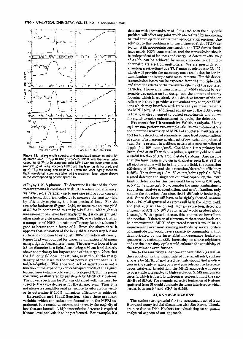
Finally, it is important to know when 100% ionization efficiency is being achieved. One should be able to make this identification by monitoring the ion intensity as a function of laser power of the ionizing wavelength. The variation of ion signal with laser power should exhibit a positive nonzero dependence on the laser power as the power is increased until 100% ionization efficiency is achieved, after which the de- pendence should be almost zero. The results of studies of this type for single resonance MPRI of sputtered In, Al, and Mo atoms are presented in Figure 12 along with the wavelength spectrum for each resonance transition.
Panels a-d of Figure 12 were obtained for In atoms. Note that for both the two-color MPRI (a-b) and one-color MPRI (c-d) the ion intensities are observed to saturate as the laser power is increased. However, the ion yield obtained using resonance excitation by 3039.4-A light is 33 times greater than that measured with 4102-A light. In addition, one-color MPRI by 3039.4-A light gives an ion yield similar to that of the two-color experiment. So far, we have not been able to explain this phenomenon; however, if it is a common observation it is obviously important for quantifying the MPRI method. Incidentally, we believe that the background in spectrum (c) is due to the photodissociation of Inz+ to In and In+, caused by the absorption of a 4102-A photon after resonant ionization
2790 ANALYTICAL CHEMISTRY, VOL. 56, NO. 14, DECEMBER 1984
0 u 25 5 75 IO 12.5 15
g o . 0 1 , , , , , , 4 1 , , , , , , , , i - 4100 4101 4102 4103 4104 0 I 2 3 4 5
00
00 0'5v UP? _.
3192 3193 3194 3195 3196 0 0.5 1.0 1.5 2.0 2.5 3.0 WAVELENGTH (a) LASER ENERGY (rn J/ pulse)
Flgure 12. Wavelength spectra and associated power spectra for sputtered (a-b) ('PI,,) In using two-color MPRI with the laser unfo- cused, (c-d) (2P,,2) In using one-color MPRI with the laser unfocused, (e-f) (*Pql2) AI using two-color MPRI with the laser tightly focused, and (9-h) (7S,) Mo uslng one-color MPRI with the laser tightly focused. Each wavelength scan was taken at the maximum laser power shown in the corresponding power spectrum.
of Inz by 4102-A photons. To determine if either of the above measurements is consistent with 100% ionization efficiency, we have used a Faraday cup to measure primary ion current, and a hemicylindrical collector to measure the sputter yield by efficiently capturing the laser-produced ions. For the two-color ionization (Figure 12a,b), we measure a sputter yield of 5.7 for In bombarded at 4 5 O by 5-keV Ar+. Although this measurement has never been made for In, it is consistent with other sputter yield measurements (19), so we believe that an assumption of 100% ionization efficiency for this scheme is good to better than a factor of 2. From the above data, it appears that saturation of the ion yield is a necessary but not sufficient condition to establish 100% ionization efficiency. Figure 12e,f was obtained for two-color ionization of A1 atoms using a tightly focused laser beam. The laser was focused from 0.6-cm diameter to a tight focus (using a 30-cm lens) directly above the primary ion impact zone of the target. Note that the Al+ ion yield does not saturate, even though the energy density of the laser a t the focal point is greater than 6000 mJ/(cmz-pulse). This apparent lack of saturation is not a function of the expanding conical-shaped profile of the tightly focused laser (which would result in a slope of 312 in the power spectrum), as illustrated by panels g-h for MPRI of Mo atoms. The power spectrum for Mo was obtained with the laser fo- cused to the same degree as for the A1 spectrum. Thus, it is not always a straightforward procedure to saturate ion yields or to determine if 100% ionization efficiency is achieved.
Extraction and Identification. Since there are many variables which can reduce ion formation in the MPRI ex- periment, it is crucial to extract and identify the majority of ions that are formed. A high transmission detector is required if trace level analysis is to be performed. For example, if a
detector with a transmission of lo4 is used, then the duty cycle problem will offset any gains which are realized by monitoring neutral atom ejection rather than secondary ion ejection. One solution to this problem is to use a time-of-flight (TOF) de- tector. With appropriate construction, the TOF device should have nearly 100% transmission, and the transmission should be independent of ion mass and energy. A detection efficiency of >40% can be achieved by using state-of-the-art micro- channel plate electron multipliers. We are presently con- structing a reflecting-type TOF mass spectrometer (31, 32) which will provide the necessary mass resolution for ion in- dentification and isotope ratio measurements. For this device, transmission losses can be expected from the multiple grids and from the effects of the transverse velocity of the sputtered particles. However, a transmission of -50% should be rea- sonable depending on the design and the amount of energy focusing which is required. An attractive feature of the ion reflector is that it provides a convenient way to reject SIMS ions which may interfere with trace analysis measurements via MPRI (32). An additional advantage of the TOF device is that it is ideally suited to pulsed experiments and allows for signal-to-noise enhancement by gating the detector.
Prospects for Ultrasensitive Solids Analysis. Using eq 1, we now perform two example calculations to demonstrate the potential sensitivity of MPRI of sputtered neutrals as a tool for the detection of elements a t trace level concentrations in solids. First, assume an element of low ionization potential (e.g., Ga) is present in a silicon matrix a t a concentration of 1 ppb (5 x 10¹³ atoms/cm3). Consider a 1-mA primary ion beam, fired at 30 Hz with 5-ps pulses, a sputter yield of 5, and a useful fraction of 50% ground-state Ga atoms. Also assume that the laser beam is 0.6 cm in diameter such that 28% of all ejected atoms will be in the photon field, the ionization efficiency is loo%, and the extraction/detection efficiency is 20%. Then from eq 1, I = 150 counts/s for 1 ppb Ga. With a gated detector and single ion counting capability, the lower limit of detection for this case could be as low as 0.01 ppb, or 5 X 10l1 atoms/cm3. Now, consider the same bombardment conditions, analyte concentration, and useful fraction, only assume the detection of an atom that is difficult to ionize (e.g., As). Since the laser will have to be tightly focused, assume that - 1 % of all sputtered As atoms will lie in the photon field, and that 25% will be ionized. For an extraction/detection efficiency of 20%, 5 X 10¹³ As atoms/cm3 would produce about 1 count/s. With a gated detector, this is about the lower limit of detection. If detection of elements a t these trace levels can be demonstrated, MPRI of sputtered neutrals would be an improvement over most existing methods by several orders of magnitude and would have a sensitivity comparable to that demonstrated by the laser ablation/resonance ionization spectroscopy technique (33). Increasing ion source brightness and/or the laser duty cycle would enhance the sensitivity of the experiment even further.
Due to the sensitivity enhancement over SIMS (as well as the reduction in the magnitude of matrix effects), surface analysis by MPRI of sputtered neutrals should find applica- tion in the study of adsorbate systems relevant to heteroge- neous catalysis. In addition, the MPRI approach will prove to be a viable alternative to high-resolution SIMS analysis for cases in which isobaric interferences seriously limit the sen- sitivity of SIMS. For example, selective ionization of P atoms sputtered from Si would eliminate the mass interference which occurs between P+ and SiH+ in SIMS.
ACKNOWLEDGMENT The authors are grateful for the encouragement of Sam
Hurst and many fruitful discussions with Jim Parks. Thanks are also due to Dick Hockett for stimulating us to pursue analytical aspects of our approach.

Anal. Chem. 1904, 56, 2791-2797 2791
Registry No. Co, 7440-48-4; In, 7440-74-6; Ga, 7440-55-3; Mo, 7439-98-7; Al, 7429-90-5.
LITERATURE CITED (1) Ens, W.; Standing, K. G.; Westmore, J. 6.; Ogllvie, K. K.; Nemer, M. J.
Anal. Chem. 1982, 54, 960-9136, (2) Glbbs, R. A.; Holland, S. P.; Foley, K. E.; Garrison, B. J.; Winograd, N.
J . Chem. Phys. 1982, 76 , 684-695. (3) Wlttmaack, K. Appl. Phys. Lett. 1978, 2 9 , 552-554. (4) Wittmaack, K.; Clegg, J. B. Appl. Phys. Lett. 1980, 378, 265-287. (5) Clegg, J. 6.; Scott, G. B.; Hallals, J.; Mlrcea-Roussel, A. J . Appl.
(6) Pellln, M. J.; Wright, R. B.; Gruen, D. M. J . Chem. Phys. 1981, 74 ,
(7) Yu, M. L.; Grischkowsky, D.; Balant, A. C. Phys. Rev. Lett. 1982, 48, 427-430.
(8) Honlg, R. E. "Advances In Mass Spectrometry"; Pergamon Press: New York, 1962; Vol. 2.
(9) Coburn, J. W.; Kay, E. Appl. Phys. Lett. 1971, 19, 350-352. (10) Oechsner, H.; Oerhard, W. Surf. Sci. 1974, 44, 480-488. (11) Winograd, N.; Baxter, J. P.; Klmock, F. M. Chem. Phys. Lett. 1982,
(12) Klmock, F. M.; Baxter, J. P.; Winograd, N. Surf. Sci. 1983, 124, L41- L46.
(13) Hurst, G. S.; Payne, M. G.; Kramer, S. D.; Young, J. P. Rev. Mod.
(14) Kobrin, P. H.; Baxter, J. P.; Winograd, N., unpublished work. (15) Parks, J. E.; Schmltt, H. W.; Hurst, G. S.; Fairbank, W. M. SPIE27th
Ann. Tech. Symp. Proc., 27th 1883. (16) Fassett, J. D.; Travis, J. C.; Moore, L. J.; Lytle, F. E. Anal. Chem.
1983, 5 5 , 765-770. (17) Donohue, D. L.; Young, J. P.; Smith, D. H. Int. J . Mass Spectrom.
Phys. 1981, 52. 1110-1112.
6448-6457.
8 8 , 581-584.
PhyS. 1979, 5 1 , 767-819.
Ion PhyS. 1982, 43, 293-307.
(18) Miller, C. M.; Nogar, N. S. Anal. Chem. 1983, 5 5 , 1606-1608. (19) Carter, G.; Colligon, J. S. "Ion Bombardment of Solids"; American
Elsevler: New York, 1968; Chapter 7. (20) Coburn, J. W. Thin Solid Films 1979, 64 , 371-382. (21) Coburn, J. W. J . Vac. Sci. Techno/. 1976, 13, 1037-1044. (22) Oechsner, H.; Schoof, H.; Stumpe, E. Surf. Sci. 1978, 76, 343-354. (23) Wright, R. B.; Pellin, M. J.; Gruen, D. M.; Young, C. E. Nucl. Instrum.
Methods 1980, 170, 295-302. (24) Pellin, M. J.; Young, C. E.; Calaway, W. F.; Gruen, D. M. Surf. Sci., in
press. (25) Wlttmaack, K. Nucl. Instrum. Methods 1980, 168, 343-356. (26) Fassett, J. D.; Moore, L. J.; Travis, J. C.; Lytle, F. E. Int. J . Mass
Spectrom. Ion Procs. 1983, 54, 201-218. (27) Onderdelinden, D. Can J . Phys. 1988, 46, 739-745. (28) Thompson, M. W. Phllos. Mag. 1968, 78, 377-414. (29) Morellac, J.; Normand, D.; Petite, G. Adv. At. Mol. Phys. 1982, 18,
(30) Young, J. P.; Donohue, D. L. Anal. Chem. 1983, 55 , 88-91. (31) Mamyrln, B. A.; Karataev, V. I . ; Shmlkk, D. V.; Zagulin, V. A. Sov.
Phys.-JETP (Engl. Transl.) 1973, 37 , 45-48. (32) Becker, C. H.; Glllen, K. T. Anal. Chem. 1984, 9 , 1671-1674. (33) Mayo, S.; Lucatorto, T. 6.; Luther, G. G. Anal. Chem. 1982, 54 ,
553-556.
97- 164.
RECEIVED for review April 30, 1984. Accepted July 23, 1984. The authors are grateful for the financial support of the National Science Foundation (Grant No. CHE 81-08382), the Office of Naval Research (Grant No. N00014-83-K-0052), the Air Force Office of Scientific Research (Grant No. AFOSR- 82-0057), and the donors of the Petroleum Research Fund, administered by the American Chemical Society.
Low Temperature Ashing Preconcentration for Elemental Localization in Biological Soft Tissues by Ion Microscopy
J. T. Brenna and G. H. Morrison* Department of Chemistry, Cornell University, Ithaca, New York 14853
Low temperature oxygen plasma ashlng (LTA) was Investi- gated as a preconcentration method for major and trace ei- ementai iocaiiratlon in biological soft tissue sections. I t was found that LTA pretreatment provides satisfactory preserva- tion of elemental morphology. Experiments with fabricated standards show that LTA enhances elemental sensitlvlties 30 to 1500-fold dependlng on the element. Copper and aluminum ion micrographs, whlch are unobtainable in intact plastic Sections, were generated from ashed sections of intestine taken from normal healthy mice. These data suggest a unique applicability of LTA in ion microscopical studies of trace e i e ment dlstrlbutlon in bloiogical samples.
The elemental microcharacterization of thin-sectioned bi- ological tissue is a subject of intense interest. Ion microscopy via secondary ion mass spectrometry (SIMS) (1) has been shown to be a useful tool for this purpose. Among its ad- vantages as an analytical technique are high sensitivity and the ability to distinguish isotopes of the same element. Major elemental constituents of tissue such as Na, K, Ca, Mg, and C1 are routinely localized by SIMS (I, 2); however, studies on transition metals and other minor elements have generally been limited to cases in which the target element concentration has been artifically raised to toxicological or pharmacological levels. These trace elements at their ambient levels are of sufficiently low concentration and ionization probability as
0003-2700/64/0356-279 1$0 1.50/0
to preclude imaging from intact resin embedded thin sections. Low temperature oxygen plasma ashing (LTA) is a well-
known and well-characterized technique used for the pre- concentration of inorganic constituents from organic material (3-6). LTA treatment consists of exposing an organic sample to a stream of oxygen excited by radio frequency to the singlet state (02, A:) and free atoms (0, 3P) which react with and remove organic material (C, H, N) at relatively low temper- atures (7, 8). In high doses, LTA is known to completely remove organic material while giving quantitative retention for most elements with no detectable contamination (4,9,10). For resin embedded biological thin sections mounted on smooth surfaces, LTA treatment in sufficient doses produces ash patterns (spodograms) of high morphological integrity (3,
Ion microscopy applied to the determination of elemental distributions in ashed sections has not previously been in- vestigated. Thus, the purpose of this study was to characterize the usefulness of LTA pretreatment for the ion microscopic localization of biologically important trace elements at their normal levels in thin tissue sections. Mouse intestine prepared by use of conventional fixation procedures and embedment in plastic served as a model system. The data from this study indicate signal enhancements of a minimum of 30-fold for Ca to 1500-fold for Co are obtained upon ashing. The absence of serious spectral interferences at masses 63,65, and 27 allows the direct imaging of Cu and A1 at their physiological con- centrations.
11-14).
0 1984 American Chemical Society