27humandissect joint of lower limb lu
TRANSCRIPT

大體老師無語良師
大體解剖學實驗HUMAN DISSECTION
THE LOWER LIMB DISSECTION IV盧家鋒助理教授
臺北醫學大學醫學系解剖學暨細胞生物學科
臺北醫學大學醫學院轉譯影像研究中心
http://www.ym.edu.tw/~cflu

REFERENCES• Dissector‘s guide
• [1] Dissection Guide for Gray's Human Anatomy, 2ed, 2006• [2] Grant’s Dissector, 15ed, 2012
• Photographic Dissector• [3] Gray's Clinical Photographic Dissector of the Human Body,
2013
• Human Atlas• [4] Gray's Atlas of Anatomy, 2ed, 2014• [5] Grant's Atlas of Anatomy 13ed, 2012• [6] Color Atlas of Anatomy: A Photographic Study of the Human
Body, 7ed, 2011• [7] Atlas of Human Anatomy, 6ed, 2014
http://www.ym.edu.tw/~cflu 2

VARIATION OF SCIATIC NERVE
Natsis K, Totlis T, Konstantinidis GA, Paraskevas G, Piagkou M, Koebke J. Anatomical variations between the sciatic nerve and the piriformis muscle: a contribution to surgical anatomy in piriformis syndrome. Surgical and Radiologic Anatomy. 2014 Apr 1;36(3):273‐80.
89.8% 6.1% 0.7%
0.7% 0.7%‐‐

VARIATION OF SCIATIC NERVE
• Number of extremities studied, 1510.• A: Usual relationships with the sciatic nerve
passing from the pelvis beneath piriformism..
• B: Piriformis m. divided into two parts with the peroneal division of the sciatic nerve passing between the two parts of piriformis.
• C: The peroneal division of the sciatic nerve passes over m. piriformis and the tibialdivision passes beneath the undivided muscle.
• D: In these cases the entire nerve passes through the divided m. piriformis.
4Beaton, L.E. and B.J. Anson. The relation of the sciatic nerve and its subdivisions to the piriformis muscle. Anat. Rec. 70:1‐5, 1938

LOWER LIMB (4/4)
• Joints of the lower limb
http://www.ym.edu.tw/~cflu 5
僅在卸下的腿進行關節解剖!
Hip joint
Knee joint
Ankle joint

JOINTS OF THE LOWER LIMB下肢關節
http://www.ym.edu.tw/~cflu 6

HIP JOINT ‐ OSTEOLOGY• Three bones form the acetabulum: ilium, ischium, and pubis.
http://www.ym.edu.tw/~cflu 7http://boneandspine.com/hip‐bone‐anatomy/
Anterior view Posterior view
Head of femur
Neck of femur
Greater trochanter
Lesser trochanter
Intertrochanteric line
Intertrochanteric Crest

HIP JOINT
http://www.ym.edu.tw/~cflu 8
Anterior surface
Iliofemoral ligament
Pubofemoral ligament
Joint Capsule
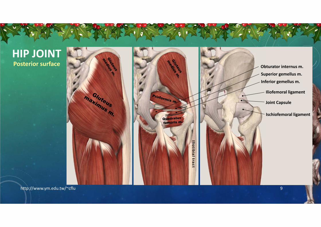
HIP JOINT
http://www.ym.edu.tw/~cflu 9
Posterior surface
Iliofemoral ligament
Ischiofemoral ligament
Joint Capsule
Iliotibial tract
Superior gemellus m.
Inferior gemellus m.
Obturator internus m.

HIP JOINT
http://www.ym.edu.tw/~cflu 10
Anterior surface
寬臼窩
寬臼唇

KNEE JOINT
http://www.ym.edu.tw/~cflu 11
• Cut the sartorius muscle at the level of apex of femoral triangle. Reflect the lower part inferiorly.
• Cut the tendons of gracillisand semitendinosus muscles at medial surface of the tibia and reflect them superiorly.
Medial view
Tibial collateral ligament
Medial patellar retinaculum
Joint capsule
Medial View

12
KNEE JOINT
http://www.ym.edu.tw/~cflu
• Cut the long and short heads of biceps femoris tendons (lower end) and reflect them superiorly.
Lateral view
Fibular collateral ligament
Lateral patellar retinaculum
Joint capsule
Lateral View
Short head
Popliteus m.

COLLATERAL LIGAMENT
• Note that the tibial collateral ligament is attached to the medial meniscus through the joint capsule.
• Note that the fibular collateral ligament is not attached to the external surface of the joint capsule.
• Observe that the popliteus tendon passes between the fibular collateral ligament and the joint capsule.
http://www.ym.edu.tw/~cflu 13
Posterior view Anterior view

KNEE JOINT
• Remove the popliteal vessels, the tibial nerve, and the common fibular nerve.
• Free the both heads of the gastrocnemius muscle and plantarismuscle from the joint capsule.
http://www.ym.edu.tw/~cflu 14
Posterior View
Oblique popliteal ligament
Arcuate popliteal ligament
popliteus m.
Joint capsule

KNEE JOINT
http://www.ym.edu.tw/~cflu 15
Posterior View
Ref. [5] p.418
Joint capsule
Open the posterior wall of the joint capsule to expose the joint cavity.

KNEE JOINT
• Observe that the quadriceps femoristendon has patellar retinacula that help to keep the patella centered.
• Reflect the patella and patellar ligament inferiorly.
• Open the joint capsule and verify that the cruciate ligaments cross each other.
http://www.ym.edu.tw/~cflu 16
Anterior View
Ref. [5] p.417

ANKLE JOINT
http://www.ym.edu.tw/~cflu 17
Medial View
Medial ligament of ankle

ANKLE JOINT
http://www.ym.edu.tw/~cflu 18
Medial ViewFlexor retinaculum
Superior extensor retinaculum
Inferior extensor retinaculum
Anterior tibiotalar ligament
Tibiocalcaneal ligamentTibionavicular ligament
Posterior tibiotalar ligament
Medial ligament of ankle

ANKLE JOINT
http://www.ym.edu.tw/~cflu 19
Lateral View
Lateral ligament of ankle

ANKLE JOINT
http://www.ym.edu.tw/~cflu 20
Lateral View
Superior Fibular retinaculum
Superior extensor retinaculum
Posterior talofibular ligament
Inferior Fibular retinaculum
Anterior talofibular ligamentCalcaneofibular ligament
Lateral ligament of ankle
