3 a novel coazervation-based process for the preparation ... · chapter 3 development of oil-loaded...
TRANSCRIPT
Chapter 3 Development of oil-loaded polyelectrolyte nanocapsules
54
3 A novel coazervation-based process for the preparation of oil-loaded polyelectrolyte nanocapsules 3.1 Introduction
Polyelectrolyte (PE) complexes are formed by complex coazervation which is the
reaction of two dispersed hydrophilic colloids of opposite electric charges.
Biopolymers, chemically modified biopolymers or synthetic polymers can function
as polyions. Widely used examples for anionic biopolymers are gelatine B, sodium
alginate, Arabic gum, pectin, xanthan gum, nucleic acids or proteins. Examples for
chemically modified biopolyanions are carboxymethylcellulose, chitosan sulphate
or dextrane sulphate. From the group of synthesized polymers poly(acrylic acid) is
suitable. There are fewer examples for polycations, the most widely used are
gelatine A as a biopolymer, chitosane as a modified biopolymer and poly(ethylene
imin) as a synthetic polymer.
Polyelectrolytes have several applications. The polyelectrolytes themselves can be
used in food industry as thickening agents for e.g. juices and chocolate. For a
potential application in food industry Ogawa et al. [146-148] utilized lecithin-
chitosan and lecithin-chitosan-pectin multilayered membranes to improve the
stability of food emulsions.
In industrial chemistry polyelectrolytes are applied as stabilizers and emulsifiers for
thixotropic paints and in textile industry as suspending agents for dyes.
Polyelectrolytes are also suitable as ion exchange material for the removal of
heavy metals from industrial wastewaters.
During the last 10 years research activity on polyelectrolytes and polyelectrolyte
complexes has been high for pharmaceutical applications as wound healing [149] or
drug-release systems. Panzner et al. [150] encapsulated liposomes with chitosan-
alginate layers to prevent liposome mixtures from aggregation. Decher and Hong
[151-153] introduced a layer-by-layer (LBL) self assembly technique for the
production of ultra-thin polyelectrolyte films on flat substrates. The technology,
using slides and beakers, is quite simple, namely a positively charged substrate on
a slide adsorbs a polyanion and a polycation consecutively from solution, using
washing steps for the removal of excess polyion in between. The driving force
behind this method is the electrostatic attraction between the incoming polymer

Chapter 3 Development of oil-loaded polyelectrolyte nanocapsules
55
and the surface. This technique has been widely used by following groups working
on polyelectrolyte complexes. Möhwald and co-workers transferred the LBL
principle from two-dimensional films to three-dimensional structures, namely
hollow polyelectrolyte capsules and crystals in the lower micrometer range
[154-157], other groups followed with hollow nanocapsules [158,159]. These
capsules have a given size because the LBL assembly of polyelectrolytes is
performed on preformed templates. Melamine formaldehyde (MF) or poly(styrene
sulfonate) (PSS) serve as templates. Due to their acid solubility, the templates can
be removed after finished LBL deposition to form hollow polyelectrolyte capsules.
Loading of and release from the polyelectrolyte multilayer capsules is dependent
on the number of adsorbed layers, the chosen PE pairs, their stoichiometry, pH
values and salt concentrations during production and at the site of action.
The capsule wall thickness can be adjusted by the number of PE layers. Mean
values for the thickness of a polyanion-polycation monolayer vary depending on the
PE pair used. Values of 2-4 nm are reported [160,161] for each double layer of
poly(styrene sulfonate)/poly(allylamine hydrochloride), chitosan/dextran sulphate
and chitosan/polyacrylic acid PE pairs. In contrary gelatine-polyanion layers can be
as thick as about 10 nm. Layer growth can be controlled by single particle light
scattering and electrophoresis.
In general polyelectrolyte capsule walls are semipermeable whereas small
molecules can easily penetrate the capsule wall but macromolecules are excluded
from the shell. Despite the coating with several PE pair layers release from
capsules can be very fast, e.g. 100% Ibuprofen release within 125 seconds from
microcapsules containing five PE pair layers was reported [156].
Since encapsulation of substances into the hollow capsules has to be performed
belated, approaches were made to switch the capsule walls between open and
closed state. This was achieved by pH-induced permeability changes of the
polyelectrolyte network. Capsules allow pH-sensitive encapsulation, namely they
offer permeability at low pHs and closed walls at high pHs, when the
polyelectrolyte pair contains a strong polyanion and a weak polycation. Möhwald
and co-workers [80] demonstrated this principle with poly(styrene sulfonate) and
poly(allylamine hydrochloride) to form multilayered microcapsules. Sauer et al.
[87] created ph-sensitive nanocontainers of 45 nm using crosslinked poly(acrylic
acid). The background of this approach is pH-sensitive swelling of poly(acrylic acid)

Chapter 3 Development of oil-loaded polyelectrolyte nanocapsules
56
due to increasing dissociation at increasing pH. Thereby the electrostatic repulsion
between the carboxylate anions within the polymer backbone is so strong that a
volume increase of factor 80 was reported which can possibly be used for belated
encapsulation of compounds.
Adjusting the pH to certain values is crucial during the adsorption process to induce
maximal ionization. Sukhorukov [162] reported that the addition of NaCl facilitates
the formation of denser PE multilayers.
Despite all features of LBL capsules, a disadvantage of this method is the time-
consuming filtration or ultracentrifugation of non-adsorbed polyelectrolyte after
each adsorption step. Furthermore liquid templates like emulsions are not suitable
for the LBL technique because these cores cannot withstand the mechanical
strength during the separation step.
In this chapter a new formulation approach for oil-loaded polyelectrolyte
nanocapsules based on complex coazervation and following high pressure
homogenization will be discussed. The objective of this study was to develop a
technique that uses oily nanodroplets as templates for PE nanocapsule production
and does not require separation steps and belated filling of hollow capsules.
First of all the optimal experimental conditions concerning choice of suitable PE
pairs, their stoichiometry, pH values and temperatures for the single production
steps were evaluated. Composite PE nanocapsules consisting of olive oil and
polyionic biopolymers were produced. � potential measurement have been used to
confirm the reloading of the nanocapsules after each adsorption step which gives
evidence for the step-wise growth of the PE shell. TEM micrographs show the
morphology of the composite PE nanocapsules in comparison to the nanoemulsion
template. PCS studies have been carried out to study the nanocapsule size.

Chapter 3 Development of oil-loaded polyelectrolyte nanocapsules
57
3.2 Materials
The used polysaccharides chitosan, carrageenan and purity gum 2000 are all food
grade, non-toxic and biodegradable materials.
Chitosan is a derivative of chitin, a natural biopolymer which is produced in the
shell of insects and crustaceans. Since chitin provides the structure and integrity to
these animals, as cellulose does to plants, it is one of the most abundant
biopolymers on earth. Chitin molecules are large chains of N-acetyl glucosamine
units. By slightly modifying the molecule through deacetylation, chitosan is
produced, which offers exposed amino groups. Chitosan has a pKS value between
6.3 and 7.0 [163] leading to a positive charge of the molecule upon exposure to
acidic media. Due to its high binding capacity and high viscosity, the main
commercial applications are the dietary supplement, where it serves as a fat
binder, and cosmetic industry.
Powdered chitosan, Chito Clear FG 95 (Food grade; degree of deacetylation: 95%)
from Primex, Iceland, was used.
Figure 3.1 Chemical structure of chitosan (degree of deacetylation: 95%).
Carrageenan is the name given to a family of linear sulphated polysaccharides
obtained from the red seaweeds, which have the ability to form all different types
of gels. The carrageenan family has three main branches named kappa (�), iota (�)
and lambda (�) which can be differentiated by their gelling properties and protein
reactivity. While � carrageenans produce strong rigid gels, � forms only flaccid ones
and � carrageenans do not gel in water. � carrageenan shows the strongest
interactions with proteins followed by �. Due to their gelling properties and protein
reactivity carrageenans are mainly used in the food and dentifrice industry, e.g. for
chocolate milk, toothpaste and bread. It is also used in low fat products, where it

Chapter 3 Development of oil-loaded polyelectrolyte nanocapsules
58
is added to replace fat and to maintain viscosity and mouth feel. In pharmaceutical
technology carrageenans are used as tabletting excipients which form hydrocolloid
matrices. Viscarin GP 209 NF from FMC Bio Polymer, which is � carrageenan, has
been used. The degree of sulfatation is 35% [164].
Figure 3.2 Chemical structure of � carrageenan (degree of sulfatation: 35%).
Purity gum 2000 is a modified starch which is derived from waxy starch. Due to the
esterification of starch with octenyl succinic acid, purity gum has good emulsion
stabilising properties even at low temperatures. Applications include food
emulsions such as flavoured emulsions, bakery emulsions, vitamin and health care
preparations. Purity gum 2000, E1450 for food use from National Starch &
Chemicals, was used. The degree of esterification is 0.2% and its aqueous solution
shows an pH of approximately 4 [165].
Figure 3.3 Chemical structure of purity gum (degree of esterification: 0.2%).
In pre-experiments the polyanion xanthan gum was also under investigation.
Xanthan gum is a microbial biopolymer which is synthesized by Xanthomonas
campestris. The primary structure of xanthan consists of a pentasaccharide
repeating unit [(1→4)-�-D-glucopyranose] to which trisaccharide side-chains are
Chapter 3 Development of oil-loaded polyelectrolyte nanocapsules
59
attached which consist of two mannose and a glucuronic acid residue. Xantural™
180 from CP Kelco was used.
For the nanoemulsion template olive oil from Sigma was used.
3.3 Methods
3.3.1. Nanocapsule preparation
Solution preparation
For the aqueous emulsifier solution 5% (w/v) purity gum was dispersed in
bidestilled water.
0.2% (w/v) of chitosan powder was dispersed in 0.1 N acetate buffer, pH 4.5
(containing 0.02 wt% sodium acide as an antimicrobial agent).
0.4% of Carrageenan were dissolved in bidestilled water by heating to 80°C to
decrease the viscosity of the solution.
Preparation of the nanoemulsion template
The aqueous purity gum solution was heated to 50 °C and applied to a rotor-stator
mixer (Ultra turrax® , IKA® T18 basic, IKA®-Works, NC). 5% of olive oil was added
stepwise under mixing. The olive oil was injected into the aqueous solution by a
pipette, which decreased the foaming compared to the addition of olive oil on top
of the aqueous phase. The aqueous emulsifier solution and the oil phase were
mixed for 90 seconds at 14.000 rpm before this coarse emulsion was introduced to
a two-stage high-pressure valve homogenizer (nG7400.270 P, Standsted Fluid Power
Ltd., UK-Stansted) to achieve droplet sizes in the nano range. Three passes were
run with 900-1000 bar for stage one and 200 bar for stage two and then a fourth
path with only one stage at 400 bar.
Preparation of PE nanocapsules I
10 ml of a 0.2% chitosan solution in acetate buffer were injected with a syringe
into 20 ml of the primary emulsion (5% olive oil and 5% purity gum) under high-
sheer mixing (14.000 rpm, Ultraturrax). Addition of the chitosan solution was
carried out very slowly. The solution was added tropwise with mixing breaks in
between. After the complete addition of 10 ml mixing was continued for 3-4
minutes.

Chapter 3 Development of oil-loaded polyelectrolyte nanocapsules
60
Preparation of composite PE nanocapsules II
30 ml of the nanocapsule I dispersion were heated to 80°C and 10 ml of the 0.4%
carrageenan solution of the same temperature were added by syringe under high
sheer mixing (18.000 rpm). The mixture was introduced to high pressure
homogenization at once (3 runs x 900-1000 bar + 200 bar, 4th run at 400 bar).
3.3.2 Experimental techniques
PCS measurements were performed with a Malvern HPPS as described in chapter
2.3.2. All samples were diluted 1:3 with water.
� potential measurements (see chapter 2.3.3) were carried out after diluting the
samples in the ratio 1:4 and mixing them with KCl solution (0.002 mol/l) in the
ratio 1:1. Measurements were performed with a ZetaSizer 3000HS (Malvern
Instruments, UK). Each sample was measured four times with automatic
measurement duration.
For TEM measurements (see chapter 2.3.4) the nanoemulsion templates were
diluted with bidestilled water (1:1). The composite PE nanocapsules II were
examined without further dilution.
Chapter 3 Development of oil-loaded polyelectrolyte nanocapsules
61
3.4. Results and discussion 3.4.1. Optimization of the production process
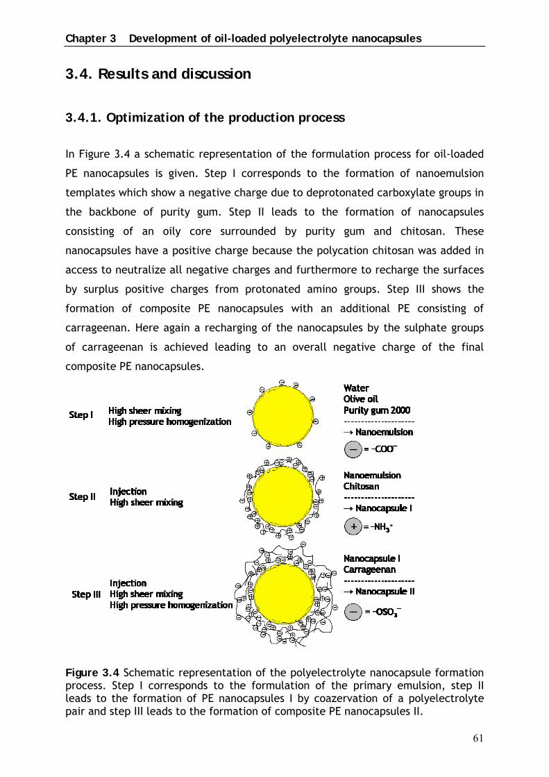
In Figure 3.4 a schematic representation of the formulation process for oil-loaded
PE nanocapsules is given. Step I corresponds to the formation of nanoemulsion
templates which show a negative charge due to deprotonated carboxylate groups in
the backbone of purity gum. Step II leads to the formation of nanocapsules
consisting of an oily core surrounded by purity gum and chitosan. These
nanocapsules have a positive charge because the polycation chitosan was added in
access to neutralize all negative charges and furthermore to recharge the surfaces
by surplus positive charges from protonated amino groups. Step III shows the
formation of composite PE nanocapsules with an additional PE consisting of
carrageenan. Here again a recharging of the nanocapsules by the sulphate groups
of carrageenan is achieved leading to an overall negative charge of the final
composite PE nanocapsules.
Figure 3.4 Schematic representation of the polyelectrolyte nanocapsule formation process. Step I corresponds to the formulation of the primary emulsion, step II leads to the formation of PE nanocapsules I by coazervation of a polyelectrolyte pair and step III leads to the formation of composite PE nanocapsules II.

Chapter 3 Development of oil-loaded polyelectrolyte nanocapsules
62
In detail the following optimizations for the three production steps have been
performed.
In step I high pressure homogenization was used to produce oily templates in the
nano range with the ability to adsorb polyelectrolytes due to a surface charge.
These nanotemplates will be referred to as nanoemulsion template or
nanoemulsion.
The anionic and cationic biopolymers xanthan gum, purity gum and chitosan were
studied for their ability to stabilize O/W nanoemulsions containing 5% (w/v) of
olive oil. This primary oil concentration was chosen to achieve a final oil load of
2.5% (w/v) for composite PE nanocapsules (Table 3.1.).
Xanthan gum was disadvantageous because of its low emulsifying property
combined with a high viscosity. Though it was well soluble in water forming
solutions of moderate viscosity in concentrations of 0.5 to 0.7% (w/v), oil was
mainly quasiemulsified by the high viscosity of the outer phase leading to large
aggregates of droplets with particle sizes in the µm range. Chitosan in
concentrations of 1% (w/v), dispersed in acetate buffer, formed nanoemulsions
with a smaller size distribution though few droplets in the µm range were
detectable by light microscopy. The weakly anionic emulfsifier purity gum was
found to produce nanoemulsions with the smallest droplet size and a narrow size
distribution.
5% of purity gum was the optimal concentration to stabilize the nanoemulsion
templates containing 5% of olive oil. Lower concentrations lead to increased
polydispersity whereas higher concentrations caused increased foaming during
processing. The ingredients bidestilled water and purity gum were premixed with a
rotor stator mixer (Ultraturrax) and the oil phase was injected with a pipette to
decrease foaming. As a following step high pressure homogenization was needed to
achieve particle sizes in the nano range. Best results were achieved at increased
temperature as high as 50 ºC. The formation process was carried out in bidestilled
water for two reasons: first to guarantee almost maximal ionization of the charged
groups of purity gum and second to achieve a low influence of additional ions in the
formulation.
The produced nanoemulsion templates had a z-average particle size of
212.01 ± 16.16 nm and showed a very narrow polydispersity index of 0.136 ± 0.017.

Chapter 3 Development of oil-loaded polyelectrolyte nanocapsules
63
In step II chitosan was used as a corresponding polycation. Having a pKs value of
6.3-7.0, chitosan is not soluble in water. Therefore the biopolymer was dissolved at
pH 4.5 in acetate buffer, 0.1N. At this pH almost maximal ionization of the amino
groups should be achieved leading to good solubility and offering a high quantity of
charged groups for layer formation. The chitosan solution was slowly injected by
syringe under high sheer mixing to prevent the formation of polymer bridges
between single droplets leading to flocculation.
To decrease the viscosity of carrageenan in step III this biopolymer was processed
at 80ºC. After injection of the aqueous biopolymer solution to nanocapsules I, the
final PE nanocapsules were introduced to high pressure homogenization to
mechanically force polyelectrolytes to become detacled from all but one of the
droplets. The particle size of the final composite PE nanocapsules was
236.10 (± 12.67 nm) with a PDI of 0.341 (± 0.026).
Table 3.1 gives an overview of the sample compositions. The rational background
for the stoichiometry of the three polyions will be discussed in the following
chapter with regard to electrical charge of the samples.
Table 3.1 Sample composition Sample
Nr. Sample
olive oil
% [v/v]
purity gum
% [w/v]
chitosan
% [w/v]
carrageenan
% [w/v]
1 Nanoemulsion
template 5 5
2 PE nanocapsules
I 2.5 2.5 0.05
3 PE nanocapsules
II 2.5 2.5 0.05 0.1

Chapter 3 Development of oil-loaded polyelectrolyte nanocapsules
64
3.4.2. Layer formation followed by zeta potential measurements
The primary emulsion droplets in aqueous environment had a zeta potential of
-12.7 mV due to the negative charge of purity gum molecules at the oil–water
interface. The relatively low zeta potential can be explained by the low
substitution degree of the starch backbone with octenyl succinic acid of 0.2%. Free
oleic acid or other ionic impurities might also contribute to the electrical charge of
the emulsion droplets. Upon addition of the polycation chitosan the electrical
charge on the droplets changed from negative to positive. The mixing ratio of the
polyelectrolyte layer pair was 50 parts of the polyanion purity gum with a
carboxylation degree of 0.2% with 1 part of the polycation chitosan with a
substitution degree of free amino groups of 95% (Table 3.1.). There was obviously
sufficient charge from the protonated amino groups of chitosan to cause surface re-
charging as evident by the reversal of the zeta potential values from -12.7 mV to
+8.6 mV. Following exposure to the polyanion carrageenan caused adsorption of
this PE, the resulting nanocapsules having a zeta potential of -28.5 mV. The mixing
ratio of chitosan to carrageenan was 1:2 (Table 3.1.), whereas the ratio of cationic
to anionic groups was 95% to 35%. As the zeta potential values demonstrate there
was enough negative charge provided by the sulphate groups of carrageenan to
neutralize the free protonated amino groups of the chitosan layer and to re-charge
the electrical charge of the nanocapsules by surplus negative sulphate groups.
Figure 3.5 � potential reversals for three PE adsorption steps.

Chapter 3 Development of oil-loaded polyelectrolyte nanocapsules
65
The charge reversals obtained (Figure 3.5.) represent credible evidence for the
step-wise growth of a composite polyelectrolyte layer on the nanoemulsion
templates.
3.4.3 Morphology of polyelectrolyte nanocapsules
Transmission electron microscopy studies confirm the existence of shell formation
on the oily template which was observed by � potential measurements.
Figure 3.6 TEM photomicrographs of a nanoemulsion template (left) and a composite PE nanocapsule treated by freeze fracture and etching. The arrow (left) marks a “shadow” which emerges from sample processing for TEM.
The TEM photomicrograph on the left shows a nanoemulsion template composed of
olive oil and purity gum (Table 3.1, sample 1). The arrow marks a “shadow”, which
emerges from the sample processing for TEM as follows. Surfaces, which result
from the freeze-fracture, are shadowed with platinum to produce good topographic
contrast. Due to the shadowing angle of 45º a microscopic object on the surface
provides a “shadow”, as it can be seen on the left photomicrograph, which remains
uncoated with metal. On the right a composite PE nanocapsule composed of a
nanoemulsion template which is coated with purity gum, chitosan and carrageenan
(sample 3) is visible.
The difference between the two photomicrographs clearly shows that coating of
the nanoemulsion template with polyelectrolytes was successful. The nanoemulsion
droplet exhibits a thin coating from the emulsifying starch purity gum which
arranges at the oil-water interface to stabilise the system. On the other hand the
final PE nanocapsule shows an oily core which is surrounded by a composite PE

Chapter 3 Development of oil-loaded polyelectrolyte nanocapsules
66
coating. A highly swollen shell is visible. The observed PE shell is much thicker than
the shell of PLA and PEG-PLA nanocapsules (Figure 2.3). The diameter of the
encapsulated oily core can be estimated with 200 nm, the shell has an extent of
30 nm, leading to a total nanocapsule diameter of 260 nm.
In Figure 3.7 an unbroken composite PE nanocapsule is visible having also a particle
size in this range.
Figure 3.7 TEM photomicrograph of an unbroken composite PE nanocapsule.
These results are in accordance with PCS results. The increased layer thickness of
the shell might be explained by loop formation of uncharged parts of the polymer
backbone.

Chapter 3 Development of oil-loaded polyelectrolyte nanocapsules
67
3.5 Conclusion
Complex coazervation combined with high pressure homogenization has been
carried out successfully to design novel composite polyelectrolyte nanocapsules
composed of olive oil and three different biopolymers. � potential measurements
confirmed the charge reversal after each adsorption step involved and thereby
proved the step-wise growth of the polyelectrolyte shell. TEM studies on these
nanocapsules revealed the formation of a composite shell on the oily core. PCS
measurements of the final PE nanocapsules showed a z-average value of 230 nm.
In comparison to the existing interfacial layer-by-layer deposition process, the
method developed here for nanocapsule preparation saves the time-consuming
separation of non-adsorbed polyelectrolyte in between the adsorption steps.
Furthermore it is the first work on polyelectrolyte nanocapsules which describes in
detail the usage of a liquid colloidal template. The elsewhere used solid templates,
which are necessary for the separation process by filtration or ultracentrifugation,
have to be dissolved after nanocapsule production leaving hollow carriers. In the
here presented approach the liquid template composed of olive oil serves as an
ideal solvent for lipophilic drugs. Injection of the polyelectrolytes and subsequent
high pressure homogenization caused disruption of aggregates due to polymer
bridges between single droplets leading to acceptable oil loading of 2.5% (v/v).