3+ functions influence the relaxation properties
TRANSCRIPT

processes
Article
Gd3+ Complexes Conjugated to Cyclodextrins: HydroxylFunctions Influence the Relaxation Properties
Anais Biscotti 1, François Estour 1 , Berthe-Sandra Sembo-Backonly 1, Sébastien Balieu 1, Michaël Bosco 1 ,Cécile Barbot 1 , Agnès Pallier 2, Éva Tóth 2 , Célia S. Bonnet 2,* and Géraldine Gouhier 1,*
�����������������
Citation: Biscotti, A.; Estour, F.;
Sembo-Backonly, B.-S.; Balieu, S.;
Bosco, M.; Barbot, C.; Pallier, A.; Tóth,
É.; Bonnet, C.S.; Gouhier, G. Gd3+
Complexes Conjugated to
Cyclodextrins: Hydroxyl Functions
Influence the Relaxation Properties.
Processes 2021, 9, 269. https://
doi.org/10.3390/pr9020269
Academic Editor: Francesco Parrino
Received: 18 December 2020
Accepted: 26 January 2021
Published: 30 January 2021
Publisher’s Note: MDPI stays neutral
with regard to jurisdictional claims in
published maps and institutional affil-
iations.
Copyright: © 2021 by the authors.
Licensee MDPI, Basel, Switzerland.
This article is an open access article
distributed under the terms and
conditions of the Creative Commons
Attribution (CC BY) license (https://
creativecommons.org/licenses/by/
4.0/).
1 Normandie Université, COBRA, UMR 6014, FR 3038, INSA Rouen, CNRS, IRIB, IRCOF, 1 rue Tesnière,76821 Mont Saint Aignan, France; [email protected] (A.B.); [email protected] (F.E.);[email protected] (B.-S.S.-B.); [email protected] (S.B.);[email protected] (M.B.); [email protected] (C.B.)
2 Centre de Biophysique Moléculaire, CNRS UPR 4301, Université d’Orléans, Rue Charles Sadron,CEDEX 2, 45071 Orléans, France; [email protected] (A.P.); [email protected] (É.T.)
* Correspondence: [email protected] (C.S.B.); [email protected] (G.G.);Tel.: +33-238-255-593 (C.S.B.); +33-235-522-909 (G.G.)
Abstract: In the search for improvement in the properties of gadolinium-based contrast agents,cyclodextrins (CDs) are interesting hydrophilic scaffolds with high molecular weight. The impact ofthe hydrophilicity of these systems on the MRI efficacy has been studied using five β-CDs substitutedwith DOTA or TTHA ligands which, respectively, allow for one (q = 1) or no water molecule (q = 0)in the inner coordination sphere of the Gd3+ ion. Original synthetic pathways were developed toimmobilize the ligands at C-6 position of various hydroxylated and permethylated β-CDs via anamide bond. To describe the influence of alcohol and ether oxide functions of the CD macrocycleon the relaxation properties of the Gd3+ complexes, 1H Nuclear Magnetic Relaxation Dispersion(NMRD) profiles, and 17O transverse relaxation rates have been measured at various temperatures.The differences observed between the hydroxylated and permethylated β-CDs bearing non-hydratedGdTTHA complexes can be rationalized by a second sphere contribution to the relaxivity in thecase of the hydroxylated derivatives, induced by hydrogen-bound water molecules around thehydroxyl groups. In contrast, for the DOTA analogs the exchange rate of the water molecule directlycoordinated to the Gd3+ is clearly influenced by the number of hydroxyl groups present on theCD, which in turn influences the relaxivity and gives rise to a very complex behavior of thesehydrophilic systems.
Keywords: cyclodextrin; gadolinium; contrast agent; magnetic resonance imaging; second hydra-tion sphere
1. Introduction
Magnetic resonance imaging (MRI) is currently used to diagnose diseases and tomonitor treatment progress in deep tissues. This noninvasive technique is based on themeasurement of nuclear spin relaxation times of water protons of the organism. MR im-ages are obtained thanks to the variation of the longitudinal or the transverse relaxationtimes (T1- or T2-weighted images, respectively) between different tissues [1–3]. In orderto improve the image contrast and reduce the examination time, contrast agents are com-monly injected to patients. These compounds are stable gadolinium chelates formed withpolyamino-polycarboxylate ligands, such as the macrocyclic GdDOTA (H4DOTA = 1,4,7,10-tetraazacyclododecane-1,4,7,10-tetraacetic acid). Via dipole–dipole interactions betweenthe water proton spins and the electron spin of Gd3+, such paramagnetic complexes inducean acceleration of the proton spin relaxation. In the GdDOTA chelate, the metal ion isnine-coordinated with one water molecule in the inner coordination sphere (IS), q = 1. Thepresence of this water molecule, directly coordinated to Gd3+, is important for an efficient
Processes 2021, 9, 269. https://doi.org/10.3390/pr9020269 https://www.mdpi.com/journal/processes

Processes 2021, 9, 269 2 of 13
relaxation effect [4–6]. Indeed, its exchange with surrounding water molecules transmitsthe paramagnetic effect of the Gd3+ to bulk water, which is then detectable on the MRimages. In the absence of inner sphere coordination water, a Gd3+ complex has a morelimited effect on the water proton relaxation times.
The efficiency of a T1 contrast agent is assessed by its longitudinal relaxivity, r1,defined as the paramagnetic relaxation rate enhancement referred to 1 mM concentrationof the metal ion. A high r1 value translates to good contrast enhancing capability in MRimaging. Relaxivity is influenced by different relaxation mechanisms [7]. Inner sphererelaxivity arises from the interaction of the Gd3+ electron spin with the inner sphere waterprotons and their exchange with bulk water. This mechanism is described by the theory ofSolomon, Bloembergen, and Morgan for paramagnetic nuclear relaxation, which relates therelaxivity to microscopic parameters of the Gd3+ complex, such as the hydration numberq, the water exchange rate kex, and the rotational correlation time τr [8,9]. The outersphere (OS) relaxation mechanism originates from interactions of the Gd3+ electron spinwith water molecules randomly diffusing around the complex. Finally, a second sphere(2S) mechanism might be also operating for systems containing water molecules stronglyhydrogen-bonded to the functional groups of the complex. This second sphere effect isusually negligible, and it is difficult to describe when it exists. The contribution of eachmechanism to the relaxivity depends on the structure of the complex, especially the size, thepresence of hydrogen bonding acceptors, the charge, and the hydrophilicity. Commercialcontrast agents are small, monohydrated Gd3+ complexes of polyamino-polycarboxylateligands for which the IS and OS relaxivity contributions are similar. With an appropriatechemical design, the inner and second sphere contributions can be substantially increased,while the outer sphere relaxivity can practically not be modified.
The inner sphere relaxivity term is linearly proportional to the hydration number.Therefore, increasing q is a straightforward way to improve relaxivity. However, the pres-ence of two or more inner sphere water molecules in the complex can seriously compromiseits stability, thereby increasing the potential risk of releasing free and toxic Gd3+ [10–12].Relaxivity can be also increased by optimizing the water exchange rate and the rotationalmotion time of the complex. The rate of water exchange is correlated with the exchangemechanism. The majority of the polyamino-polycarboxylate complexes of Gd3+ undergo adissociative exchange, i.e., the leaving of the bound water molecule precedes the enteringof the incoming water molecule. In this case, the steric hindrance around the water bind-ing site and the global charge of the complex are important parameters to determine theexchange rate, and water exchange can be accelerated by increasing the steric hindrancearound the water binding site. The most common way to reach higher relaxivity, with majorimprovements at medium frequencies (20–60 MHz), has been to increase the rotationalcorrelation time, τr, by increasing the molecular weight of the complex. Indeed, for lowmolecular weight complexes, fast rotation limits the relaxation efficiency. Thus, a largenumber of bulkier ligands coordinating the Gd3+ ion was developed to create more efficientMRI contrast agents. Gd3+ complexes were therefore incorporated into macromolecular sys-tems such as proteins [12–15], dendrimers [16], cyclodextrins [17], polyrotaxanes [18–20],etc. However, the relaxivity enhancements has been often less than those expected, due tothe flexibility of the macromolecule which implies faster local motion for Gd3+ than theoverall slow motion of the entire macromolecule.
We have focused our study on cyclodextrins (CDs) as interesting and versatile scaffoldsto design potential MRI contrast agents. CDs are natural cyclic oligosaccharides with 6,7, or 8 glucose units, issued from starch degradation by glucosyltransferase (CGTase)and so-called α-, β-, and γ-CD, respectively [21]. Cyclodextrins have primary (C-6) andsecondary hydroxyl groups (C-2, C-3) forming the smaller and larger crowns of the cone,respectively. For β-CD, this cone shape is especially reinforced by hydrogen bondingbetween two adjacent units (C-2-OH and C-3-OH). The 21 alcohol functions also favorinteractions with water molecules and improve the solubility in aqueous medium. Theinternal cavity (host), composed of glycosidic oxygen and C-H groups, is less hydrophilic

Processes 2021, 9, 269 3 of 13
and makes possible intermolecular interactions with organic molecules (guests). Host–guest inclusion complexes improve the water solubility and the stability of the guests, suchas small bioactive molecules. In the context of MRI contrast agents, cyclodextrins have beenmainly explored in three distinct approaches in the objective of modulating the rotationalmotion, thus improving relaxivity: (i) as host to form inclusion complexes with contrastagents functionalized by lipophilic groups [22–24], (ii) as high molecular weight scaffoldsby covalently immobilizing one or more Gd3+ complexes [25,26], and (iii) as a platform forLn3+ complexation [27,28].
In the first two approaches, dimers, trimers [23], polymers [29], and polyrotaxanes [18–20]of CDs were developed in order to maximize the number of Gd3+ complexes per molecule.For the development of platform for Ln3+ complexation, CDs were modified, for instance,into per-3,6-anhydro derivatives, which affected their structure leading to a hydrophiliccavity capable of metal binding. The replacement of the hydroxyl groups by carboxylatefunctions gives a ligand, which can complex hard Ln3+ ions to form mono- and bimetallicspecies [27]. The relaxivity of the monometallic Gd3+ has been investigated in details usinga rigorous approach, where a maximum of the microscopic parameters was determinedindependently. It has been demonstrated that the high relaxivity obtained was due to thehigh hydration number of the complex and a relatively long rotational correlation timeexplained by the hydrophilic character of the complex [28].
6-O-Peracetylated-β-CDs were also synthesized to coordinate Gd3+ in the axis of themacrocycle [30–32]. In particular, the effect of the second hydration sphere has been studiedusing the native and permethylated CDs. It has been proved that this functionalizationhad an impact on the relaxivity. Indeed, a 40% relaxivity enhancement was observedwith perhydroxylated β-CDs (4.6 mM−1 s−1 and 6.5 mM−1 s−1, respectively). As thesecomplexes had similar structure and identical hydration number (q = 2), the relaxivitydifference was attributed to the presence of hydrogen-bound water molecules around thehydroxyl groups inducing an important second-sphere (2S) contribution to relaxivity.
In order to improve the thermodynamic stability of the Gd3+ chelate and further studythe influence of the hydrogen-bonding network provided by the hydrophilic cyclodex-trins on the relaxivity, several functionalized cyclodextrins were synthesized and studied.Modified DOTA and TTHA (3,6,9,12-tetrakis(carboxymethyl)-3,6,9,12-tetraazatetradecane-1,14-dioic acid) ligands were conjugated at one of the C-6 position of various β-CDs byreplacing a carboxylate function by an amide (1–5, Figure 1).
The DOTA monoamide (DOTAMA) ligand is known to form thermodynamically sta-ble and kinetically inert complexes with Ln3+. The GdDOTAMA complex was introducedat one of the O-6-position on the small rim of the native (hydroxylated) β-CD (1), of the 6-O-permethylated β-CD (2), and of the 2,3,6-O-permethylated β-CD (3). With contrast agent2, the hydroxyl groups are in the vicinity of the GdDOTAMA complex, while the methoxygroups are located on the opposite side on the larger rim of the CD. In order to betterdecipher the relaxation behavior of those compounds, TTHA monoamide (TTHAMA) wasalso introduced on the native (5) and 2,3,6-O-permethylated β-CD (4) (Figure 1). TTHA isa commercially available linear ligand with six carboxylic acid and four amine functionswhich can chelate the Gd3+ ion. This full coordination by the ligand prevents inner sphere(IS) binding of any water molecule. Indeed, the Gd3+ complex of TTHAMA has no innersphere hydration water molecule (q = 0), which means that the relaxivity will be governedby outer sphere, and possibly second sphere mechanisms [33].

Processes 2021, 9, 269 4 of 13
Processes 2021, 9, x FOR PEER REVIEW 4 of 15
Figure 1. Structure of new contrast agents based on β-CDs-DOTAMA (1–3) and β-CDs-TTHAMA (4 and 5) ligands.
2. Results and Discussion 2.1. Synthesis of CDs Functionalized with DOTA Ligand
The monofunctionalization of native β-CD 6 was based on the difference of reactivity between the hydroxyl groups (Scheme 1). Indeed, the secondary alcohols are more acidic: the position 3 less accessible. We substituted the more nucleophile primary alcohol at 6 position to keep the larger cavity available to form inclusion complex. We reported herein the introduction of DOTA and TTHA ligands on native and methylated-β-CDs.
All synthesis used the same precursor mono(6-amino-6-deoxy)-β-CD 7 obtained after monotosylation of primary face of β-CD 6 and substitution reaction by sodium azide (Scheme 1) [34]. The CD 7 was permethylated after deprotonation and treatment with methyl iodide (Supplementary Materials). The synthesis of the mono(6-azido-6-deoxy)-6-O-permethylated-β-CD 9 required two additional steps, the protection of secondary alcohol functions by tert-butyldimethylsilyl groups and the deprotection using ammonium fluoride reagent (Supplementary Materials).
The azide reduction using Staudinger reaction led to mono(6-amino-6-deoxy)-β-cyclodextrins precursors 10–12 with yields between 40–52%, which was confirmed by the disappearance of the signal in IR spectroscopy of azide function at 2199 cm−1 and appearance of amine function at 2920 cm−1. A shift of 10 ppm corresponding to the methylene carbon bearing the amine function was observed by 13C DEPT confirming the reduction step (Supplementary Materials). A peptide coupling was then applied with DOTA structure 13 protected by three tert-butyl groups (Scheme 1) [35]. The activation of the free acid function in presence of DCC and HOBt led to the three precursors 14–16 in 21%, 12%, and 70% yields, respectively. In order to improve the yields, uronium salt
O
R1OOR1 O O
R1OOR1
O
6
OR2
NH
1 R1 = H, R2 = H2 R1 = Me, R2 = H3 R1 = Me, R2 = Me
N N
NN COO-
COO-
COO-
O
GdH2O
Figure 1. Structure of new contrast agents based on β-CDs-DOTAMA (1–3) and β-CDs-TTHAMA (4 and 5) ligands.
2. Results and Discussion2.1. Synthesis of CDs Functionalized with DOTA Ligand
The monofunctionalization of native β-CD 6 was based on the difference of reactivitybetween the hydroxyl groups (Scheme 1). Indeed, the secondary alcohols are more acidic:the position 3 less accessible. We substituted the more nucleophile primary alcohol at6 position to keep the larger cavity available to form inclusion complex. We reported hereinthe introduction of DOTA and TTHA ligands on native and methylated-β-CDs.
All synthesis used the same precursor mono(6-amino-6-deoxy)-β-CD 7 obtained aftermonotosylation of primary face of β-CD 6 and substitution reaction by sodium azide(Scheme 1) [34]. The CD 7 was permethylated after deprotonation and treatment withmethyl iodide (Supplementary Materials). The synthesis of the mono(6-azido-6-deoxy)-6-O-permethylated-β-CD 9 required two additional steps, the protection of secondaryalcohol functions by tert-butyldimethylsilyl groups and the deprotection using ammoniumfluoride reagent (Supplementary Materials).
The azide reduction using Staudinger reaction led to mono(6-amino-6-deoxy)-β-cyclodextrins precursors 10–12 with yields between 40–52%, which was confirmed bythe disappearance of the signal in IR spectroscopy of azide function at 2199 cm−1 andappearance of amine function at 2920 cm−1. A shift of 10 ppm corresponding to themethylene carbon bearing the amine function was observed by 13C DEPT confirming thereduction step (Supplementary Materials). A peptide coupling was then applied withDOTA structure 13 protected by three tert-butyl groups (Scheme 1) [35]. The activationof the free acid function in presence of DCC and HOBt led to the three precursors 14–16in 21%, 12%, and 70% yields, respectively. In order to improve the yields, uronium saltHATU was tested but it did not improve the reactivity [36]. Consequently, another strategyhas been developed using the activation of the primary amine function by chloroacetylchloride reagent (Scheme 2). However, only the permethylated β-CD 12 was substitutedin this case in 82% yield. In the case of mono(6-amino-6-deoxy)-perhydroxylated β-CD10 and mono(6-amino-6-deoxy)-2,3-O-permethylated-β-CD 11, the reactions led to manysecondary products due to polysubstitution reactions of the alcohol functions. In orderto control the monosubstitution reaction, other precursors 17 and 18 have to be used(Scheme 2).

Processes 2021, 9, 269 5 of 13
Processes 2021, 9, x FOR PEER REVIEW 5 of 15
HATU was tested but it did not improve the reactivity [36]. Consequently, another strategy has been developed using the activation of the primary amine function by chloroacetyl chloride reagent (Scheme 2). However, only the permethylated β-CD 12 was substituted in this case in 82% yield. In the case of mono(6-amino-6-deoxy)-perhydroxylated β-CD 10 and mono(6-amino-6-deoxy)-2,3-O-permethylated-β-CD 11, the reactions led to many secondary products due to polysubstitution reactions of the alcohol functions. In order to control the monosubstitution reaction, other precursors 17 and 18 have to be used (Scheme 2).
Scheme 1. Synthesis route of 6-O-monoamino-β-cyclodextrins precursors 10–12 and DOTAMA derivatives 14–16.
Thus, the mono(6-amino-6-deoxy)-β-CD 7 was quantitatively peracetylated using anhydride acetic in pyridine and the azido function was then reduced by catalytic hydrogenation in 50% yield (see Scheme 1 in experimental section) (Supplementary Materials). From the same precursor 7, the mono(6-amino-6-deoxy)-2,3-permethyl-β-CD protected with silyl groups at 6 positions 18 was obtained in three steps by silylation of the residual primary alcohols of the intermediate mono(6-azido-6-deoxy)-β-CD 7 (Scheme 1), followed by a permethylation of the secondary face and after Staudinger reduction of azido group in 51% over yield (see Scheme 2 in experimental section) (Supplementary Materials). The two mono(6-amino-6-deoxy)-CDs 17 and 18 were substituted with chloromethylacetyl group in 89% and 77% yields, respectively (Supplementary Materials). From precursor 18, an additional deprotection step of the TBS group was required using ammonium fluoride, providing quantitatively compound 20 (see Scheme 3 in
7, R1 = H, R2 = H, 62%8, R1 = Me, R2 = H, 65%9, R1 = Me, R2 = Me, 74%
10, R1 = H, R2 = H, 52%11, R1 = Me, R2 = H, 47%12, R1 = Me, R2 = Me, 40%
O
R1OOR1 O O
R1OOR1
O
6
OR2
N3
O
R1OOR1 O O
R1OOR1
O
6
OR2
NH2
6
O
HOOH
O O
HOOH
O
6
OH
OHa
d
a. 1) TsCl, Pyr. 2) NaN3. b. 1) TBSCl, Pyr. 2) NaH, MeI, THF. 3) NH4+F-, MeOH. c. NaH, MeI, THF. d. PPh3, NH4OH
cb
14, R1 = H, R2 = H, 21%15, R1 = Me, R2 = H, 12%16, R1 = Me, R2 = Me, 70%
N N
NNCOOtBu
COOtBuHOOC
ButOOC
13
O
R1OOR1 O O
R1OOR1
O
6
OR2
NH
N N
NN
COOtBu
COOtBu
COOtBu
O
DCC, HOBt, DMF
Scheme 1. Synthesis route of 6-O-monoamino-β-cyclodextrins precursors 10–12 and DOTAMA derivatives 14–16.
Processes 2021, 9, x FOR PEER REVIEW 6 of 15
experimental section) (Supplementary Materials). The coupling was carried out from the free secondary amine of the commercially available DO3A derivative 22 bearing three tert-butylester groups. The DOTAMA ligand was introduced with yields varying between 59% and 88% leading the compounds 23, 15, and 16, thereby improving the initial yields obtained with the direct strategy (21% and 12% yields for 14 and 15, respectively) (Schemes 1 and 2).
Scheme 2. Second strategy to the synthesis of β-CDs-DOTAMA ligands 24–26.
The 1H NMR spectra confirmed that the substitution of DOTAMA on 15, 16, and 23 compounds was effective by the presence of tert-butyl groups clearly observed at 1.45 ppm; the other signals being hidden by those of the CD scaffold (Supplementary Materials). For example, the 13C NMR spectrum of precursor 19 revealed a characteristic peak at 42.6 ppm corresponding to the carbon at alpha position of chlorine atom. This signal disappeared in the DOTAMA-substituted 23 and the tert-butyl groups signals appeared at 81.8 and 28.0 ppm, respectively. Finally, the methylene groups of the DOTAMA ligand were observed between 55.7 and 62.7 ppm (N-CH2-CH2-N and CH2-COOH) proving the substitution reaction. All the structural analysis was confirmed by mass spectrometry analyses (Supplementary Materials).
The ester functions of compounds 23, 15, and 16 were quantitatively deprotected using trifluoroacetic acid in a mixture of dichloromethane/toluene (1/1). In the case of the
17 R1 = Ac, R2 = Ac18 R1 = Me, R2 = TBS12 R1 = Me, R2 = Me
O
R1OOR1 O O
R1OOR1
O
6
OR2
NH2ClCH2COCl
Et3N, Et2O
NH N
NNCOOtBu
COOtBu
ButCOO
CsCO3, ACN
O
R1OOR1 O O
R1OOR1
O
6
OR2
NH
N N
NN
COOtBu
COOtBu
COOtBu
O
19 R1 = Ac, R2 = Ac, 89%20 R1 = Me, R2 = H, 77% (+ NH4
+F-, MeOH)21 R1 = Me, R2 = Me, 82%
O
R1OOR1 O O
R1OOR1
O
6
OR2
NH
O
Cl
23 R1 = Ac, R2 = Ac, 59%15 R1 = Me, R2 = H, 88%16 R1 = Me, R2 = Me, 70%
O
R1OOR1 O O
R1OOR1
O
6
OR2
NH
N N
NN
COOH
COOH
COOH
O
24 R1 = H, R2 = H, 85%(+ MeONa/MeOH)
25 R1 = Me, R2 = H, 100%26 R1 = Me, R2 = Me, 100%
TFACH2Cl2Toluene
DO3A 22
Scheme 2. Second strategy to the synthesis of β-CDs-DOTAMA ligands 24–26.
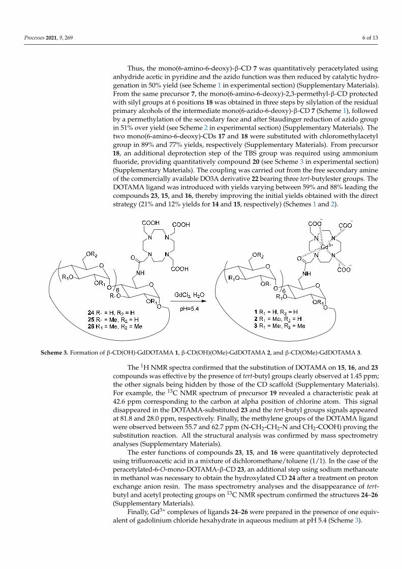
Processes 2021, 9, 269 6 of 13
Thus, the mono(6-amino-6-deoxy)-β-CD 7 was quantitatively peracetylated usinganhydride acetic in pyridine and the azido function was then reduced by catalytic hydro-genation in 50% yield (see Scheme 1 in experimental section) (Supplementary Materials).From the same precursor 7, the mono(6-amino-6-deoxy)-2,3-permethyl-β-CD protectedwith silyl groups at 6 positions 18 was obtained in three steps by silylation of the residualprimary alcohols of the intermediate mono(6-azido-6-deoxy)-β-CD 7 (Scheme 1), followedby a permethylation of the secondary face and after Staudinger reduction of azido groupin 51% over yield (see Scheme 2 in experimental section) (Supplementary Materials). Thetwo mono(6-amino-6-deoxy)-CDs 17 and 18 were substituted with chloromethylacetylgroup in 89% and 77% yields, respectively (Supplementary Materials). From precursor18, an additional deprotection step of the TBS group was required using ammoniumfluoride, providing quantitatively compound 20 (see Scheme 3 in experimental section)(Supplementary Materials). The coupling was carried out from the free secondary amineof the commercially available DO3A derivative 22 bearing three tert-butylester groups. TheDOTAMA ligand was introduced with yields varying between 59% and 88% leading thecompounds 23, 15, and 16, thereby improving the initial yields obtained with the directstrategy (21% and 12% yields for 14 and 15, respectively) (Schemes 1 and 2).
Processes 2021, 9, x FOR PEER REVIEW 7 of 15
peracetylated-6-O-mono-DOTAMA-β-CD 23, an additional step using sodium methanoate in methanol was necessary to obtain the hydroxylated CD 24 after a treatment on proton exchange anion resin. The mass spectrometry analyses and the disappearance of tert-butyl and acetyl protecting groups on 13C NMR spectrum confirmed the structures 24–26 (Supplementary Materials).
Finally, Gd3+ complexes of ligands 24–26 were prepared in the presence of one equivalent of gadolinium chloride hexahydrate in aqueous medium at pH 5.4 (Scheme 3).
Scheme 3. Formation of β-CD(OH)-GdDOTAMA 1, β-CD(OH)(OMe)-GdDOTAMA 2, and β-CD(OMe)-GdDOTAMA 3.
Scheme 3. Formation of β-CD(OH)-GdDOTAMA 1, β-CD(OH)(OMe)-GdDOTAMA 2, and β-CD(OMe)-GdDOTAMA 3.
The 1H NMR spectra confirmed that the substitution of DOTAMA on 15, 16, and 23compounds was effective by the presence of tert-butyl groups clearly observed at 1.45 ppm;the other signals being hidden by those of the CD scaffold (Supplementary Materials).For example, the 13C NMR spectrum of precursor 19 revealed a characteristic peak at42.6 ppm corresponding to the carbon at alpha position of chlorine atom. This signaldisappeared in the DOTAMA-substituted 23 and the tert-butyl groups signals appearedat 81.8 and 28.0 ppm, respectively. Finally, the methylene groups of the DOTAMA ligandwere observed between 55.7 and 62.7 ppm (N-CH2-CH2-N and CH2-COOH) proving thesubstitution reaction. All the structural analysis was confirmed by mass spectrometryanalyses (Supplementary Materials).
The ester functions of compounds 23, 15, and 16 were quantitatively deprotectedusing trifluoroacetic acid in a mixture of dichloromethane/toluene (1/1). In the case of theperacetylated-6-O-mono-DOTAMA-β-CD 23, an additional step using sodium methanoatein methanol was necessary to obtain the hydroxylated CD 24 after a treatment on protonexchange anion resin. The mass spectrometry analyses and the disappearance of tert-butyl and acetyl protecting groups on 13C NMR spectrum confirmed the structures 24–26(Supplementary Materials).
Finally, Gd3+ complexes of ligands 24–26 were prepared in the presence of one equiv-alent of gadolinium chloride hexahydrate in aqueous medium at pH 5.4 (Scheme 3).
Processes 2021, 9, 269 7 of 13
2.2. Synthesis of CDs Functionalized with TTHA Ligand
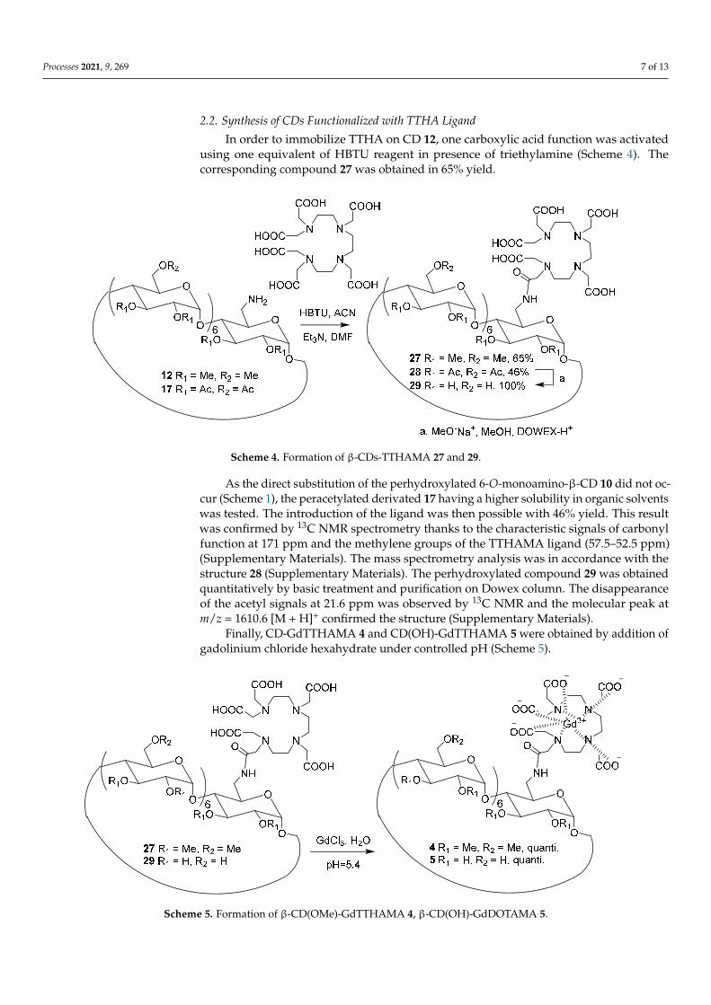
In order to immobilize TTHA on CD 12, one carboxylic acid function was activatedusing one equivalent of HBTU reagent in presence of triethylamine (Scheme 4). Thecorresponding compound 27 was obtained in 65% yield.
Processes 2021, 9, x FOR PEER REVIEW 8 of 15
2.2. Synthesis of CDs Functionalized with TTHA Ligand In order to immobilize TTHA on CD 12, one carboxylic acid function was activated
using one equivalent of HBTU reagent in presence of triethylamine (Scheme 4). The corresponding compound 27 was obtained in 65% yield.
Scheme 4. Formation of β-CDs-TTHAMA 27 and 29.
Scheme 4. Formation of β-CDs-TTHAMA 27 and 29.
As the direct substitution of the perhydroxylated 6-O-monoamino-β-CD 10 did not oc-cur (Scheme 1), the peracetylated derivated 17 having a higher solubility in organic solventswas tested. The introduction of the ligand was then possible with 46% yield. This resultwas confirmed by 13C NMR spectrometry thanks to the characteristic signals of carbonylfunction at 171 ppm and the methylene groups of the TTHAMA ligand (57.5–52.5 ppm)(Supplementary Materials). The mass spectrometry analysis was in accordance with thestructure 28 (Supplementary Materials). The perhydroxylated compound 29 was obtainedquantitatively by basic treatment and purification on Dowex column. The disappearanceof the acetyl signals at 21.6 ppm was observed by 13C NMR and the molecular peak atm/z = 1610.6 [M + H]+ confirmed the structure (Supplementary Materials).
Finally, CD-GdTTHAMA 4 and CD(OH)-GdTTHAMA 5 were obtained by addition ofgadolinium chloride hexahydrate under controlled pH (Scheme 5).
Processes 2021, 9, x FOR PEER REVIEW 9 of 15
As the direct substitution of the perhydroxylated 6-O-monoamino-β-CD 10 did not occur (Scheme 1), the peracetylated derivated 17 having a higher solubility in organic solvents was tested. The introduction of the ligand was then possible with 46% yield. This result was confirmed by 13C NMR spectrometry thanks to the characteristic signals of carbonyl function at 171 ppm and the methylene groups of the TTHAMA ligand (57.5–52.5 ppm) (Supplementary Materials). The mass spectrometry analysis was in accordance with the structure 28 (Supplementary Materials). The perhydroxylated compound 29 was obtained quantitatively by basic treatment and purification on Dowex column. The disappearance of the acetyl signals at 21.6 ppm was observed by 13C NMR and the molecular peak at m/z = 1610.6 [M + H]+ confirmed the structure (Supplementary Materials).
Finally, CD-GdTTHAMA 4 and CD(OH)-GdTTHAMA 5 were obtained by addition of gadolinium chloride hexahydrate under controlled pH (Scheme 5).
Scheme 5. Formation of β-CD(OMe)-GdTTHAMA 4, β-CD(OH)-GdDOTAMA 5.
2.3. Relaxometric Analysis of the TTHA-Derived Complexes 4 and 5 Nuclear Magnetic Relaxation Dispersion (NMRD) profiles describe the efficacy of the
complex in terms of relaxivity as a function of the magnetic field, and they are helpful to characterize the parameters governing proton relaxivity [37,38]. Typically, the analysis of NMRD curves allows for estimating some of the physicochemical parameters that determine relaxivity, in particular the rotational correlation time (τR), the water exchange rate (kex), the number of water molecules directly coordinated to Gd3+ (q), and the electronic relaxation rates. However, it is important to determine a maximum of these parameters independently for the reliability of the results.
In the case of TTHA complexes, as there is no water molecule directly coordinated to Gd3+, the relaxivity is a sum of outer sphere and, if present, second sphere contributions [33]. The NMRD profiles of 4 and 5 were measured between 10 kHz and 400 MHz and are presented in Figure 2 and in supporting information (Figure S6 in Supplementary Materials).
Scheme 5. Formation of β-CD(OMe)-GdTTHAMA 4, β-CD(OH)-GdDOTAMA 5.

Processes 2021, 9, 269 8 of 13
2.3. Relaxometric Analysis of the TTHA-Derived Complexes 4 and 5
Nuclear Magnetic Relaxation Dispersion (NMRD) profiles describe the efficacy of thecomplex in terms of relaxivity as a function of the magnetic field, and they are helpful tocharacterize the parameters governing proton relaxivity [37,38]. Typically, the analysisof NMRD curves allows for estimating some of the physicochemical parameters thatdetermine relaxivity, in particular the rotational correlation time (τR), the water exchangerate (kex), the number of water molecules directly coordinated to Gd3+ (q), and the electronicrelaxation rates. However, it is important to determine a maximum of these parametersindependently for the reliability of the results.
In the case of TTHA complexes, as there is no water molecule directly coordinated toGd3+, the relaxivity is a sum of outer sphere and, if present, second sphere contributions [33].The NMRD profiles of 4 and 5 were measured between 10 kHz and 400 MHz and are pre-sented in Figure 2 and in supporting information (Figure S6 in Supplementary Materials).
Processes 2021, 9, x FOR PEER REVIEW 10 of 15
Figure 2. 1H NMRD profiles of 4 () and 5 () at 25 °C and pH = 7.0.
The low relaxivities observed are in accordance with the absence of inner sphere water molecules in the complexes. At 20 MHz, 25 °C, the relaxivities are 2.99 and 3.75 mM−1 s−1 for 4 and 5, respectively; therefore, an increase of 25% is observed when replacing OMe by OH groups on the CDs. As the two complexes are supposed to have similar size, thus rotational dynamics, the higher relaxivity for 5 can be ascribed to the presence of second sphere water molecules contributing to the overall relaxivity, which is consistent with the presence of an H-bonding network in the case of the TTHA-substituted native CD 5. It should be noted that rough simulations of a purely outer sphere mechanism give a relaxivity of 2.3 mM−1 s−1 at 20 MHz and 25 °C, in the same order of magnitude as that measured for 4. This is also the value reported for GdTTHA in the same conditions [33].
2.4. Relaxometric Analysis of DOTA-Derived Complexes 1–3 The NMRD profiles of 1–3 were also recorded between 10 kHz and 400 MHz, at 25
°C, 37 °C, and 50 °C (see Figure 3 and Figures S3–S5 in Supplementary Materials). It should be noted that the relaxivity profile of 3 was already partially measured and analyzed by Botta et al. [25], but for the sake of direct comparison, it was re-measured in identical conditions as those of 1 and 2. The relaxivity values of 3 were similar (within 5–10%) to those previously reported.
The relaxivities determined at 20 MHz and 25 °C were 9.67, 10.50, and 9.64 mM−1 s−1 for 1, 2, and 3, respectively. These values are ~2.5 times higher than the relaxivity of clinically used contrast agents like GdDOTA (3.5 mM−1 s−1). In the NMRD profiles, we note however the absence of a relaxivity “hump” at intermediate fields, which is characteristic of slowly rotating macromolecular Gd3+ complexes. This is also in accordance with previous data from Botta et al. [25] and us [18], and indicates that the CD-based systems are not characterized by very slow motion as they are relatively flexible, and do not aggregate in aqueous solution. The relaxivity (measured at 25 °C and 20 MHz) was found independent of the concentration (between 0.5 and 5 mM), evidencing again the absence of intermolecular interactions in this concentration range.
The relaxivity of the native CD 1 and permethylated one 3 were similar, in contrast to what had been observed previously in the case of CD substituted by TTHA derivatives 4 and 5. A modest increase of relaxivity of ~9% was observed for the partially methylated CD 2. On the simple assumption that a higher hydrogen bonding network would result in a higher number of second sphere water molecules contributing to relaxivity, we would have expected a relaxivity increase in the following order: 3 < 2 < 1.
0.0
2.0
4.0
6.0
0.01 0.1 1 10 100 1000
r 1/ m
M-1
.s-1
ν (1H) / MHz
Figure 2. 1H NMRD profiles of 4 (�) and 5 (�) at 25 ◦C and pH = 7.0.
The low relaxivities observed are in accordance with the absence of inner sphere watermolecules in the complexes. At 20 MHz, 25 ◦C, the relaxivities are 2.99 and 3.75 mM−1 s−1
for 4 and 5, respectively; therefore, an increase of 25% is observed when replacing OMeby OH groups on the CDs. As the two complexes are supposed to have similar size, thusrotational dynamics, the higher relaxivity for 5 can be ascribed to the presence of secondsphere water molecules contributing to the overall relaxivity, which is consistent withthe presence of an H-bonding network in the case of the TTHA-substituted native CD5. It should be noted that rough simulations of a purely outer sphere mechanism give arelaxivity of 2.3 mM−1 s−1 at 20 MHz and 25 ◦C, in the same order of magnitude as thatmeasured for 4. This is also the value reported for GdTTHA in the same conditions [33].
2.4. Relaxometric Analysis of DOTA-Derived Complexes 1–3
The NMRD profiles of 1–3 were also recorded between 10 kHz and 400 MHz, at 25 ◦C,37 ◦C, and 50 ◦C (see Figure 3 and Figures S3–S5 in Supplementary Materials). It shouldbe noted that the relaxivity profile of 3 was already partially measured and analyzed byBotta et al. [25], but for the sake of direct comparison, it was re-measured in identicalconditions as those of 1 and 2. The relaxivity values of 3 were similar (within 5–10%) tothose previously reported.

Processes 2021, 9, 269 9 of 13
Processes 2021, 9, x FOR PEER REVIEW 11 of 15
The temperature dependence of the relaxivities provides qualitative information on the parameter that limits relaxivity for a given system. Indeed, upon temperature increase, both the water exchange and the rotational dynamics become faster. If fast rotational dynamics is a limiting factor, the relaxivity will decrease upon increasing the temperature. Conversely, if relaxivity is limited by slow water exchange, increasing the temperature will lead to the acceleration of the water exchange, thus an increase in relaxivity. If slow water exchange and fast rotation are both limiting factors, as a result of an interplay between the two, relaxivity can be relatively independent of temperature. The temperature dependence of the different systems (1–3) showed similar relaxivities at 25 °C and 37 °C, whereas r1 became lower at 50 °C (Supplementary Materials). This suggests that at the lower temperatures slow water exchange starts to become the limiting parameter, rather than fast rotational dynamics. In order to better decipher the relaxivity dependence, we performed 17O NMR measurements on the different 1–3 complexes.
Figure 3. 1H NMRD profiles of 1 () 2 (), and 3 () at 25 °C and pH = 7.0.
2.5. 17O NMR Data of Complexes 1–3 Variable temperature 17O T2 measurements give access to the water exchange rate,
kex. The reduced 17O transverse relaxation rates for 1–3 are presented in Figure 4. The behavior of 3 is classical with an increase of the reduced transverse relaxation rates (up to ~55 °C), followed by a decrease with increasing temperature, indicating that the complex is in the slow kinetic region below 55 °C. In this region, 1/T2r is directly determined by the water exchange rate constant kex, allowing for a reliable determination of kex value. The 17O data have been fitted to the Swift–Connick equations, where the number of water molecules coordinated to Gd3+ was fixed to 1, and the scalar coupling constant, A/ħ, was fixed to −3.6 × 10⁶ rad s−1. The fit yielded a value of kex298 = (1.49 ± 0.08) × 106 s−1, while ΔH≠ = (37 ± 3) kJ mol−1 was obtained for the activation enthalpy of the water exchange. The kex298 value is similar to what was previously estimated by Botta et al. from the fitting of the NMRD profile (kex298 = 1.7 × 10⁶ s−1) [25], and in the same order of magnitude as water exchange rate constants typical of monoamide DOTA complexes of Gd3+ [7]. It is nearly three times lower than the water exchange rate of GdDOTA (kex298 = 4.1 × 10⁶), and higher than that of GdDOTAM (see Table 1), which is consistent with previous observations on analogous systems. Indeed, in the case of dissociative exchange for DOTA-derivatives (which is expected here), it is generally observed that the replacement of one negatively
0.0
2.0
4.0
6.0
8.0
10.0
12.0
14.0
16.0
0.01 0.1 1 10 100 1000
r 1/m
M-1
.s-1
ν (1H) /MHz
Figure 3. 1H NMRD profiles of 1 (�) 2 (•), and 3 (�) at 25 ◦C and pH = 7.0.
The relaxivities determined at 20 MHz and 25 ◦C were 9.67, 10.50, and 9.64 mM−1 s−1
for 1, 2, and 3, respectively. These values are ~2.5 times higher than the relaxivity ofclinically used contrast agents like GdDOTA (3.5 mM−1 s−1). In the NMRD profiles,we note however the absence of a relaxivity “hump” at intermediate fields, which ischaracteristic of slowly rotating macromolecular Gd3+ complexes. This is also in accordancewith previous data from Botta et al. [25] and us [18], and indicates that the CD-basedsystems are not characterized by very slow motion as they are relatively flexible, and donot aggregate in aqueous solution. The relaxivity (measured at 25 ◦C and 20 MHz) wasfound independent of the concentration (between 0.5 and 5 mM), evidencing again theabsence of intermolecular interactions in this concentration range.
The relaxivity of the native CD 1 and permethylated one 3 were similar, in contrast towhat had been observed previously in the case of CD substituted by TTHA derivatives 4and 5. A modest increase of relaxivity of ~9% was observed for the partially methylatedCD 2. On the simple assumption that a higher hydrogen bonding network would result ina higher number of second sphere water molecules contributing to relaxivity, we wouldhave expected a relaxivity increase in the following order: 3 < 2 < 1.
The temperature dependence of the relaxivities provides qualitative information onthe parameter that limits relaxivity for a given system. Indeed, upon temperature increase,both the water exchange and the rotational dynamics become faster. If fast rotationaldynamics is a limiting factor, the relaxivity will decrease upon increasing the temperature.Conversely, if relaxivity is limited by slow water exchange, increasing the temperature willlead to the acceleration of the water exchange, thus an increase in relaxivity. If slow waterexchange and fast rotation are both limiting factors, as a result of an interplay between thetwo, relaxivity can be relatively independent of temperature. The temperature dependenceof the different systems (1–3) showed similar relaxivities at 25 ◦C and 37 ◦C, whereasr1 became lower at 50 ◦C (Supplementary Materials). This suggests that at the lowertemperatures slow water exchange starts to become the limiting parameter, rather than fastrotational dynamics. In order to better decipher the relaxivity dependence, we performed17O NMR measurements on the different 1–3 complexes.
2.5. 17O NMR Data of Complexes 1–3
Variable temperature 17O T2 measurements give access to the water exchange rate,kex. The reduced 17O transverse relaxation rates for 1–3 are presented in Figure 4. Thebehavior of 3 is classical with an increase of the reduced transverse relaxation rates (up to

Processes 2021, 9, 269 10 of 13
~55 ◦C), followed by a decrease with increasing temperature, indicating that the complexis in the slow kinetic region below 55 ◦C. In this region, 1/T2r is directly determined bythe water exchange rate constant kex, allowing for a reliable determination of kex value.The 17O data have been fitted to the Swift–Connick equations, where the number of watermolecules coordinated to Gd3+ was fixed to 1, and the scalar coupling constant, A/h̄, wasfixed to −3.6 × 10⁶ rad s−1. The fit yielded a value of kex
298 = (1.49 ± 0.08) × 106 s−1, while∆H 6= = (37 ± 3) kJ mol−1 was obtained for the activation enthalpy of the water exchange.The kex
298 value is similar to what was previously estimated by Botta et al. from the fittingof the NMRD profile (kex
298 = 1.7 × 10⁶ s−1) [25], and in the same order of magnitude aswater exchange rate constants typical of monoamide DOTA complexes of Gd3+ [7]. It isnearly three times lower than the water exchange rate of GdDOTA (kex
298 = 4.1 × 10⁶), andhigher than that of GdDOTAM (see Table 1), which is consistent with previous observationson analogous systems. Indeed, in the case of dissociative exchange for DOTA-derivatives(which is expected here), it is generally observed that the replacement of one negativelycharged carboxylate in the complex with a neutral amide decreases the water exchangerate of about one-third [7].
Processes 2021, 9, x FOR PEER REVIEW 12 of 15
charged carboxylate in the complex with a neutral amide decreases the water exchange rate of about one-third [7].
Figure 4. Temperature dependence of the reduced 17O transverse relaxation rates of 1 (, 10.04 mM) 2 (, 3.2 mM), and 3 (, 10.33 mM) at 9.4 T and pH = 7.0. The continuous curve represents the best fit to the experimental data points of 3.
Table 1. Water exchange rates (kex298) of various GdDOTA derivative complexes.
3 3 a GdDO3A-bz-NO2 Gd2-Wazaby6 GdDOTA GdDOTAM Coordinating unit DOTA-monoamide DOTA-monoamide DOTA-monoamide + COO- DOTA-monoamide DOTA DOTA-tetramide
kex298 (106 s−1) 1.49 ± 0.08 1.7 1.6 2.8 4.1 0.053 Reference This work [25] [39] [40] [38] [41]
a Obtained from fitting of NMRD data from ref. [25].
In contrast to 3, 1 and 2 have a very different behavior. Indeed, at low temperatures the 17O ln(1/T2r) values are rather constant. This might be indicative of the presence of more than one species (isomers) in solution with different water exchange properties. The SAP (square antiprismatic) and TSAP (twisted square antiprismatic) isomers of macrocyclic systems such as DOTA derivatives are known to have different water exchange rate and their ratio can be very different depending on the systems [42,43]. In order to obtain information on the potential coexistence of different species in solution, we recorded 1H NMR spectra on the corresponding Eu3+ complexes obtained using similar protocol as for the Gd complexes (Figures S7 and S8 in the Supplementary Materials). Europium is the neighboring element to Gd in the lanthanide series, so they are expected to have similar coordination environment. Eu3+ is also paramagnetic; it causes large chemical shifts but much less line-broadening than Gd3+. Unfortunately, the 1H NMR spectra of the Eu3+ analogs of 1 and 2, recorded at different temperatures, show broad resonances, combined with the presence of many protons of the cyclodextrins in the diamagnetic window which dominate the spectra. Overall, this prevents distinguishing different isomers. In the absence of information about the presence of different species in solution and their ratio, the analysis of the 17O transverse relaxation rates could not be realized for 1 and 2. Nevertheless, the temperature dependence of the transverse 17O relaxation rates at low temperature clearly showed a very different water exchange for 1–3. Although we can only speculate on the origin of this difference, it is plausible to hypothesize that it could be related to the different H-bonding network generated by the three different
11
11.5
12
12.5
13
13.5
14
14.5
15
2.7 2.8 2.9 3 3.1 3.2 3.3 3.4 3.5 3.6 3.7
Ln (1
/T2r
)
1000/T
Figure 4. Temperature dependence of the reduced 17O transverse relaxation rates of 1 (�, 10.04 mM)2 (•, 3.2 mM), and 3 (�, 10.33 mM) at 9.4 T and pH = 7.0. The continuous curve represents the best fitto the experimental data points of 3.
Table 1. Water exchange rates (kex298) of various GdDOTA derivative complexes.
3 3 a GdDO3A-bz-NO2 Gd2-Wazaby6 GdDOTA GdDOTAM
Coordinating unit DOTA-monoamide DOTA-monoamide DOTA-monoamide + COO- DOTA-monoamide DOTA DOTA-tetramidekex
298 (106 s−1) 1.49 ± 0.08 1.7 1.6 2.8 4.1 0.053Reference This work [25] [39] [40] [38] [41]
a Obtained from fitting of NMRD data from ref. [25].
In contrast to 3, 1 and 2 have a very different behavior. Indeed, at low temperaturesthe 17O ln(1/T2r) values are rather constant. This might be indicative of the presence ofmore than one species (isomers) in solution with different water exchange properties. TheSAP (square antiprismatic) and TSAP (twisted square antiprismatic) isomers of macrocyclicsystems such as DOTA derivatives are known to have different water exchange rate andtheir ratio can be very different depending on the systems [42,43]. In order to obtaininformation on the potential coexistence of different species in solution, we recorded 1HNMR spectra on the corresponding Eu3+ complexes obtained using similar protocol as for

Processes 2021, 9, 269 11 of 13
the Gd complexes (Figures S7 and S8 in the Supplementary Materials). Europium is theneighboring element to Gd in the lanthanide series, so they are expected to have similarcoordination environment. Eu3+ is also paramagnetic; it causes large chemical shifts butmuch less line-broadening than Gd3+. Unfortunately, the 1H NMR spectra of the Eu3+
analogs of 1 and 2, recorded at different temperatures, show broad resonances, combinedwith the presence of many protons of the cyclodextrins in the diamagnetic window whichdominate the spectra. Overall, this prevents distinguishing different isomers. In the absenceof information about the presence of different species in solution and their ratio, the analysisof the 17O transverse relaxation rates could not be realized for 1 and 2. Nevertheless, thetemperature dependence of the transverse 17O relaxation rates at low temperature clearlyshowed a very different water exchange for 1–3. Although we can only speculate on theorigin of this difference, it is plausible to hypothesize that it could be related to the differentH-bonding network generated by the three different cyclodextrins scaffolds, which canhave an influence on the water exchange rate of the Gd3+ complexes.
In overall, the combined 17O NMR and NMRD data suggest that these highly hy-drophilic systems have a complex behavior in which the hydrogen bonding network doesnot only contribute to a second sphere proton relaxation mechanism, but it also affects theexchange rate of the inner sphere water molecule of the Gd3+ complexes. The complexityof the systems prevents any reliable fit of the NMRD data.
3. Conclusions
We described β-CDs bearing derivatives of DOTA and TTHA ligands for Gd3+ com-plexation. The molecules have been obtained using novel synthetic routes. We studiedthe influence of the numerous hydrophilic OH groups of the CD structure, which createa strong hydrogen bonding network involving second sphere water molecules, on theproton relaxivity and on the water exchange rate of the Gd3+ complexes. In the absence ofinner sphere water molecule in the Gd3+ complex (TTHA ligand), the relaxivity increaseswith the increasing number of hydroxyl groups on the CD, confirming a strong secondsphere contribution to the relaxivity, induced by the hydrophilicity of the molecule. In thecase of DOTA derivatives, the situation is more complicated. Indeed, the variation of therelaxivity between the systems containing a different number of OH groups on the CDis not guided by the increase of hydroxyl functions. 17O NMR measurements revealeddifferent water exchange processes depending on the number of hydroxyls on the CD.For the permethylated system 3, a classical water exchange rate is found, consistent withtypical GdDOTA-monoamide complexes. In contrast, when hydroxyls are present on theCD 1 and 2, the water exchange process becomes clearly different, as evidenced by 17O T2data. These different water exchange properties will very likely impact the relaxivity. Inoverall, these highly hydrophilic systems have a hydrogen-bound network that induces asecond sphere relaxivity, but it also influences the water exchange process. Altogether thisleads to a complicated relaxation behavior.
Supplementary Materials: The materials, method, synthesis, and characterization details of products7, 9, 12, 15–21, 23–29, 1–5, and europium complexes are available online at https://www.mdpi.com/2227-9717/9/2/269/s1. 1H NMRD profiles of contrast agents 1–4 at 25 ◦C, 37 ◦C, and 1H NMRspectra of europium complexes of 24 and 25 at 9.4 T and 5 ◦C, 25 ◦C, and 37 ◦C are also reported.
Author Contributions: Conceptualization, G.G. and F.E.; Methodology, G.G., F.E., É.T., and C.S.B.;Validation, G.G., F.E., É.T., and C.S.B.; Formal Analysis, A.B., B.-S.S.-B., S.B., M.B., C.B., A.P. andC.S.B.; Investigation, A.B., B.-S.S.-B., S.B., M.B., C.B., and A.P.; Writing—Original draft preparationG.G. and C.S.B.; Writing—review and editing, F.E. and É.T.; Supervision, G.G., F.E., S.B., C.S.B.,and É.T.; Project administration, Funding acquisition, G.G. All authors have read and agreed to thepublished version of the manuscripts.
Funding: This research was funded by the Interreg IV AI-Chem Channel (PhD A.B.).
Institutional Review Board Statement: Not applicable.

Processes 2021, 9, 269 12 of 13
Informed Consent Statement: Not applicable.
Data Availability Statement: No data.
Acknowledgments: We thank Raphaël Tripier from UMR 6521, University of Bretagne Occidentale(Brest) for sending some tritertbutylDOTA 13. We are also grateful for the compagny Cyclolab,https://cyclolab.hu/, for its contribution by sending β-CD-NH2 10.
Conflicts of Interest: The authors declare no conflict of interest.
References1. Caravan, P.; Ellison, J.J.; McMurry, T.J.; Lauffer, R.B. Gadolinium(III) Chelates as MRI Contrast Agents: Structure, Dynamics, and
Applications. Chem. Rev. 1999, 99, 2293–2352. [CrossRef] [PubMed]2. Aime, S.; Botta, M.; Terreno, E. Gd(III)-based contrast agents for MRI. Adv. Inorg. Chem. 2005, 57, 173–237. [CrossRef]3. Hermann, P.; Kotek, J.; Kubícek, V.; Lukes, I. Gadolinium(III) Complexes as MRI Contrast Agents: Ligand Design and Properties
of the Complexes. Dalton Trans. 2008, 23, 3027–3047. [CrossRef] [PubMed]4. Botta, M. Second Coordination Sphere Water Molecules and Relaxivity of Gadolinium(III) Complexes: Implications for MRI
Contrast Agents. Eur. J. Inorg. Chem. 2000, 2000, 399–407. [CrossRef]5. Rudovsky, J.; Kotek, J.; Hermann, P.; Lukes, I.; Mainero, V.; Aime, S. Synthesis of a bifunctional monophosphinic acid DOTA
analogue ligand and its lanthanide(III) complexes. A gadolinium(III) complex endowed with an optimal water exchange rate forMRI applications. Org. Biomol. Chem. 2005, 3, 112–117. [CrossRef] [PubMed]
6. Bonnet, C.S.; Fries, P.H.; Crouzy, S.; Delangle, P. Outer-Sphere Investigation of MRI Relaxation Contrast Agents. Example of aCyclodecapeptide Gadolinium Complex with Second-Sphere Water. J. Phys. Chem. B 2010, 114, 8770–8781. [CrossRef]
7. Merbach, A.; Helm, L.; Toth, E. The Chemistry of Contrast Agents in Medical Magnetic Resonance Imaging, 2nd ed.; Wiley: Hoboken,NJ, USA; Chichester, UK, 2013. [CrossRef]
8. Solomon, I.; Bloembergen, N. Nuclear Magnetic Interactions in the HF Molecule. J. Chem. Phys. 1956, 25, 261–266. [CrossRef]9. Bloembergen, N.; Morgan, L.O. Proton Relaxation Times in Paramagnetic Solutions. Effects of Electron Spin Relaxation. J. Chem.
Phys. 1961, 34, 842–850. [CrossRef]10. Idée, J.-M.; Port, M.; Medina, C.; Lancelot, E.; Fayoux, E.; Ballet, S.; Corot, C. Possible Involvement of Gadolinium Chelates in the
Pathophysiology of Nephrogenic Systemic Fibrosis: A Critical Review. Toxicology 2008, 248, 77–88. [CrossRef]11. Port, M.; Idée, J.-M.; Medina, C.; Robic, C.; Sabatou, M.; Corot, C. Efficiency, thermodynamic and kinetic stability of marketed
gadolinium chelates and their possible clinical consequences: A critical review. Biometals 2008, 21, 469–490. [CrossRef]12. Fraum, T.J.; Ludwig, D.R.; Bashir, M.R.; Kathryn, J.F. Toxicity Gadolinium-Based Contrast Agents: A Comprehensive Risk
Assessment: Gadolinium Risk Assessment. J. Magn. Reson. Imaging 2017, 46, 338–353. [CrossRef] [PubMed]13. Sieving, P.F.; Watson, A.D.; Roklage, S.M. Preparation and Characterization of Paramagnetic Polychelates and Their Protein
Conjugates. Bioconjugate Chem. 1990, 1, 65–71. [CrossRef] [PubMed]14. Tóth, E.; Connac, F.; Helm, L.; Adzamli, K.; Merbach, A.E. Direct assessment of water exchange on a Gd(III) chelate bound to a
protein. J. Biol. Inorg. Chem. 1998, 3, 606–613. [CrossRef]15. Zech, S.G.; Eldredge, H.B.; Lowe, M.P.; Caravan, P. Protein Binding to Lanthanide(III) Complexes Can Reduce the Water Exchange
Rate at the Lanthanide. Inorg. Chem. 2007, 46, 3576–3584. [CrossRef]16. Rudovsky, J.; Botta, M.; Hermann, P.; Hardcastle, K.I.; Lukes, I.; Aime, S. PAMAM Dendrimeric Conjugates with a Gd-DOTA
Phosphinate Derivative and Their Adducts with Polyaminoacids: The Interplay of Global Motion, Internal Rotation, and FastWater Exchange. Bioconjugate Chem. 2006, 17, 975–987. [CrossRef] [PubMed]
17. Champagne, P.-L.; Barbot, C.; Zhang, P.; Han, X.; Gaamoussi, I.; Hubert-Roux, M.; Bertolesi, G.E.; Gouhier, G.; Ling, C.-C.Synthesis and Unprecedented Complexation Properties of β-Cyclodextrin-Based Ligand for Lanthanide Ions. Inorg. Chem. 2018,57, 8964–8977. [CrossRef]
18. Fredy, J.W.; Scelle, J.; Guenet, A.; Morel, E.; Adam de Beaumais, S.; Menand, M.; Marvaud, V.; Bonnet, C.S.; Toth, E.; Sollogoub,M.; et al. Cyclodextrin Polyrotaxanes as a Highly Modular Platform for the Development of Imaging Agents. Chem. Eur. J. 2014,20, 10915–10920. [CrossRef]
19. Fredy, J.W.; Scelle, J.; Ramniceanu, G.; Doan, B.-T.; Bonnet, C.S.; Tóth, É.; Ménand, M.; Sollogoub, M.; Vives, G.; Hasenknopf, B.Mechanostereoselective One-Pot Synthesis of Functionalized Head-to-Head Cyclodextrin [3] Rotaxanes and Their Application asMagnetic Resonance Imaging Contrast Agents. Org. Lett. 2017, 19, 1136–1139. [CrossRef]
20. Mondjinou, Y.A.; Loren, B.P.; Collins, C.J.; Hyun, S.-H.; Demoret, A.; Skulsky, J.; Chaplain, C.; Badwaik, V.; Thompson, D.H. Gd3+:DOTA-Modified 2-Hydroxypropyl-β-Cyclodextrin/4-Sulfobutyl Ether-β-Cyclodextrin-Based Polyrotaxanes as Long CirculatingHigh Relaxivity MRI Contrast Agents. Bioconjugate Chem. 2018, 29, 3550–3560. [CrossRef]
21. D’Souza, V.T.; Lipkowitz, K.B. Cyclodextrins: Introduction. Chem. Rev. 1998, 98, 1741–1742. [CrossRef]22. Aime, S.; Gianolio, E.; Terreno, E.; Menegotto, I.; Bracco, C.; Milone, L.; Cravotto, G. β-Cyclodextrin adducts of Gd(III) chelates:
Useful models for investigating the structural and dynamic determinants of the relaxivity of gadolinium-based systems. Magn.Reson. Chem. 2003, 41, 800–805. [CrossRef]

Processes 2021, 9, 269 13 of 13
23. Aime, S.; Gianolio, E.; Arena, F.; Alessandro Barge, A.; Martina, K.; Heropoulos, G.; Cravotto, G. New cyclodextrin dimers andtrimers capable of forming supramolecular adducts with shape-specific ligands. Org. Biomol. Chem. 2009, 7, 370–379. [CrossRef][PubMed]
24. Cao, Y.; Zu, G.; Kuang, Y.; He, Y.; Mao, Z.; Liu, M.; Xiong, D.; Pei, R. Biodegradable Nanoglobular Magnetic Resonance ImagingContrast Agent Constructed with Host-Guest Self-Assembly for Tumor-Targeted Imaging. ACS Appl. Mater. Interfaces 2018, 10,26906–26916. [CrossRef] [PubMed]
25. Skinner, P.J.; Beeby, A.; Dickins, R.S.; Parker, D.; Aime, S.; Botta, M. Conjugates of cyclodextrins with charged and neutralmacrocyclic europium, terbium and gadolinium complexes: Sensitized luminescence and relaxometric investigations and anexample of supramolecular relaxivity enhancement. J. Chem. Soc. Perkin Trans. 2000, 2, 1329–1338. [CrossRef]
26. Hana, Y.; Qian, Y.; Zhoub, X.; Hub, H.; Liua, X.; Zhoua, Z.; Tanga, J.; Shena, Y. Biodegradable Nanoglobular Magnetic ResonanceImaging Contrast Agent Constructed with Host-Guest Self-Assembly for Tumor-Targeted Imaging. Polym. Chem. 2016, 7,6354–6362. [CrossRef]
27. Bonnet, C.; Gadelle, A.; Pecaut, J.; Friesa, P.H.; Delangle, P. Inclusion complexes of trivalent lutetium cations with an acidicderivative of per(3,6-anhydro)-α-cyclodextrin. Chem. Commun. 2005, 5, 625–627. [CrossRef] [PubMed]
28. Bonnet, C.S.; Fries, P.H.; Gadelle, A.; Gambarelli, S.; Delangle, P. A Rigorous Framework to Interpret Water Relaxivity. The CaseStudy of a Gd(III) Complex with an α-Cyclodextrin Derivative. J. Am. Chem. Soc. 2008, 130, 10401–10413. [CrossRef]
29. Battistini, E.; Gianolio, E.; Gref, R.; Couvreur, P.; Fuzerova, S.; Othman, M.; Aime, S.; Badet, B.; Durand, P. High-RelaxivityMagnetic Resonance Imaging (MRI) Contrast Agent Based on Supramolecular Assembly between a Gadolinium Chelate, aModified Dextran, and Poly-β-Cyclodextrin. Chem. Eur. J. 2008, 14, 4551–4561. [CrossRef]
30. Idriss, H.; Estour, F.; Zgani, I.; Barbot, C.; Biscotti, A.; Petit, S.; Galaup, C.; Hubert-Roux, M.; Nicol, L.; Mulder, P.; et al. Effect ofthe Second Coordination Sphere on New Contrast Agents Based on Cyclodextrin Scaffolds for MRI Signals. RSC Adv. 2013, 3,4531–4534. [CrossRef]
31. Zgani, I.; Idriss, H.; Barbot, C.; Djedaïni-Pilard, F.; Petit, S.; Hubert-Roux, M.; Estour, F.; Gouhier, G. Positive Variation of the MRISignal via Intramolecular Inclusion Complexation of a C-2 Functionalized β-Cyclodextrin. Org. Biomol. Chem. 2017, 15, 564–569.[CrossRef]
32. Biscotti, A.; Barbot, C.; Nicol, L.; Mulder, P.; Sappei, C.; Hubert-Roux, M.; Déchamps-Olivier, I.; Estour, F.; Gouhier, G. MRI probesbased on C6-peracetate β-cyclodextrins: Synthesis, gadolinium complexation and in vivo relaxivity studies. Polyhedron 2018, 148,32–43. [CrossRef]
33. Zitha-Bovens, E.; Muller, R.N.; Laurent, S.; Vander, E.L.; Geraldes, C.F.G.C.; van Bekkum, H.; Peters, J.A. Structure and Dynamicsof Lanthanide Complexes of Triethylenetetramine-N, N, N′, N′ ′, N′ ′ ′, N′ ′ ′-hexaacetic Acid (H6ttha) and of Diamides H4ttha(NHR) Derived from H6ttha as Studied by NMR, NMRD, and EPR. Helv. Chim. Acta 2005, 88, 618–632. [CrossRef]
34. Martinelli, J.; Thangavel, K.; Tei, L.; Botta, M. Dendrimeric b-Cyclodextrin/GdIII Chelate SupramolecularHost–Guest Adducts asHigh-Relaxivity MRI Probes. Chem. Eur. J. 2014, 20, 10944–10952. [CrossRef] [PubMed]
35. Laakso, J.; Rosser, G.A.; Szíjjártó, C.; Beeby, A.; Borbas, K.E. Synthesis of Chlorin-Sensitized Near Infrared-Emitting LanthanideComplexes. Inorg. Chem. 2012, 51, 10366–10374. [CrossRef]
36. Montalbetti, C.A.G.N.; Falque, V. Amide bond formation and peptide coupling. Tetrahedron 2005, 61, 10827–10852. [CrossRef]37. Swift, T.J.; Connick, R.E. NMR Relaxation Mechanisms of O17 in Aqueous Solutions of Paramagnetic Cations and the Lifetime of
Water Molecules in the First Coordination Sphere. J. Chem. Phys. 1962, 37, 307–320. [CrossRef]38. Powell, D.H.; Ni Dhubhghaill, O.M.; Pubanz, D.; Helm, L.; Lebedev, Y.S.; Schlaepfer, W.; Merbach, A.E. Structural and Dynamic
Parameters Obtained from 17O NMR, EPR, and NMRD Studies of Monomeric and Dimeric Gd3+ Complexes of Interest inMagnetic Resonance Imaging: An Integrated and Theoretically Self-Consistent Approach. J. Am. Chem. Soc. 1996, 118, 9333–9346.[CrossRef]
39. Tóth, É.; Pubanz, D.; Vauthey, S.; Helm, L.; Merbach, A.E. The Role of Water Exchange in Attaining Maximum Relaxivities forDendrimeric MRI Contrast Agents. Chem. Eur. J. 1996, 2, 1607–1615. [CrossRef]
40. Florès, O.; Pliquett, J.; Galan, L.A.; Lescure, R.; Denat, F.; Maury, O.; Pallier, A.; Bellaye, P.-S.; Collin, B.; Même, S.; et al. Aza-BODIPY Platform: Toward an Efficient Water-Soluble Bimodal Imaging Probe for MRI and Near-Infrared Fluorescence. Inorg.Chem. 2020, 59, 1306–1314. [CrossRef]
41. Aime, S.; Barge, A.; Bruce, J.I.; Botta, M.; Howard, J.A.K.; Moloney, J.M.; Parker, D.; De Sousa, A.S.; Woods, M. NMR, Relaxometric,and Structural Studies of the Hydration and Exchange Dynamics of Cationic Lanthanide Complexes of Macrocyclic TetraamideLigands. J. Am. Chem. Soc. 1999, 121, 5762–5771. [CrossRef]
42. Aime, S.; Barge, A.; Botta, M.; De Sousa, A.S.; Parker, P. Direct NMR Spectroscopic Observation of a Lanthanide-CoordinatedWater Molecule whose Exchange Rate Is Dependent on the Conformation of the Complexes. Angew. Chem. Int. Ed. 1998, 37,2673–2675. [CrossRef]
43. Platas-Iglesias, C. The Solution Structure and Dynamics of MRI Probes Based on Lanthanide(III) DOTA as Investigated by DFTand NMR Spectroscopy. Eur. J. Inorg. Chem. 2012, 12, 2023–2033. [CrossRef]