3-neurological_emergencies-mfs.pdf -...
TRANSCRIPT
Neurological Emergencies Ivo Bekavac, MD,PhD
The Practice of Emergency and Critical Care Neurology
1. The Presenting Neurologic Emergency
2. Criteria of Triage
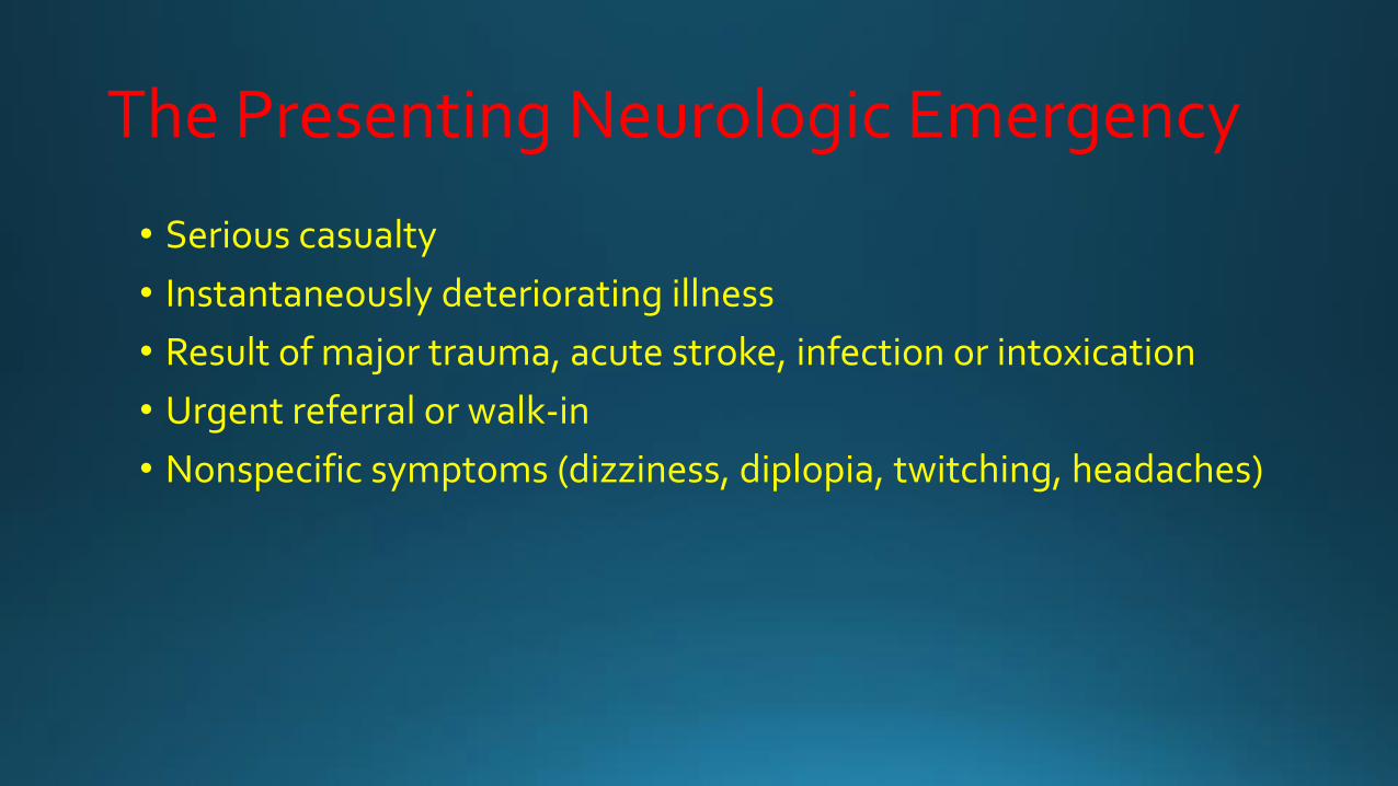
The Presenting Neurologic Emergency
• Serious casualty
• Instantaneously deteriorating illness
• Result of major trauma, acute stroke, infection or intoxication
• Urgent referral or walk-in
• Nonspecific symptoms (dizziness, diplopia, twitching, headaches)
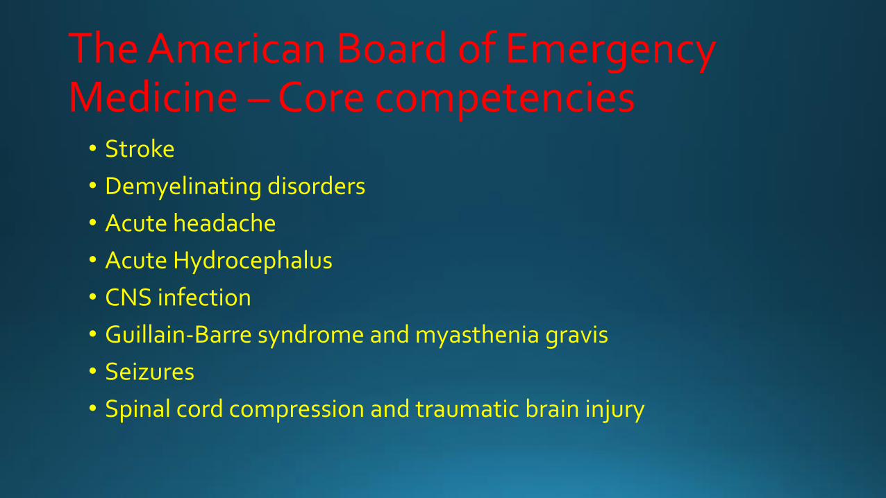
The American Board of Emergency Medicine – Core competencies • Stroke
• Demyelinating disorders
• Acute headache
• Acute Hydrocephalus
• CNS infection
• Guillain-Barre syndrome and myasthenia gravis
• Seizures
• Spinal cord compression and traumatic brain injury
The Emergency Department
• Highly dependent on location (inner-city vs rural)
• Many ED are packed
• Mixed pathology (nonurgent visits, “frequent flyers”)
• Designated critical care area
• Trauma activation (Level 1)
The Neurologic Emergency and its Assessment • Specific clinical presentation
• Abnormal neuroimaging
• Progression of symptoms
• Neurologic symptoms often fluctuate (VBI)
Signs and Symptoms of Neurologic emergency • Worsening and changing of neurologic signs
• Abnormal consciousness
• Seizure
• Inability to stand or walk
• Acute cranial nerve deficit/focal neurological deficit
• Severe unexpected headache
Neurologic Tests Available in ED
• CT
• MRI/A
• Cerebrospinal fluid examination
• Electroencephalography
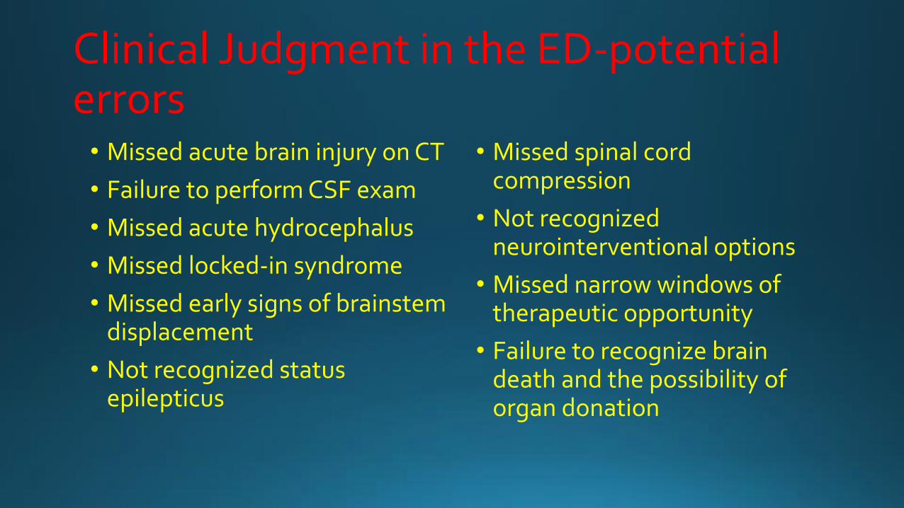
Clinical Judgment in the ED-potential errors • Missed acute brain injury on CT
• Failure to perform CSF exam
• Missed acute hydrocephalus
• Missed locked-in syndrome
• Missed early signs of brainstem displacement
• Not recognized status epilepticus
• Missed spinal cord compression
• Not recognized neurointerventional options
• Missed narrow windows of therapeutic opportunity
• Failure to recognize brain death and the possibility of organ donation
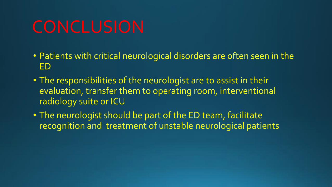
CONCLUSION
• Patients with critical neurological disorders are often seen in the ED
• The responsibilities of the neurologist are to assist in their evaluation, transfer them to operating room, interventional radiology suite or ICU
• The neurologist should be part of the ED team, facilitate recognition and treatment of unstable neurological patients
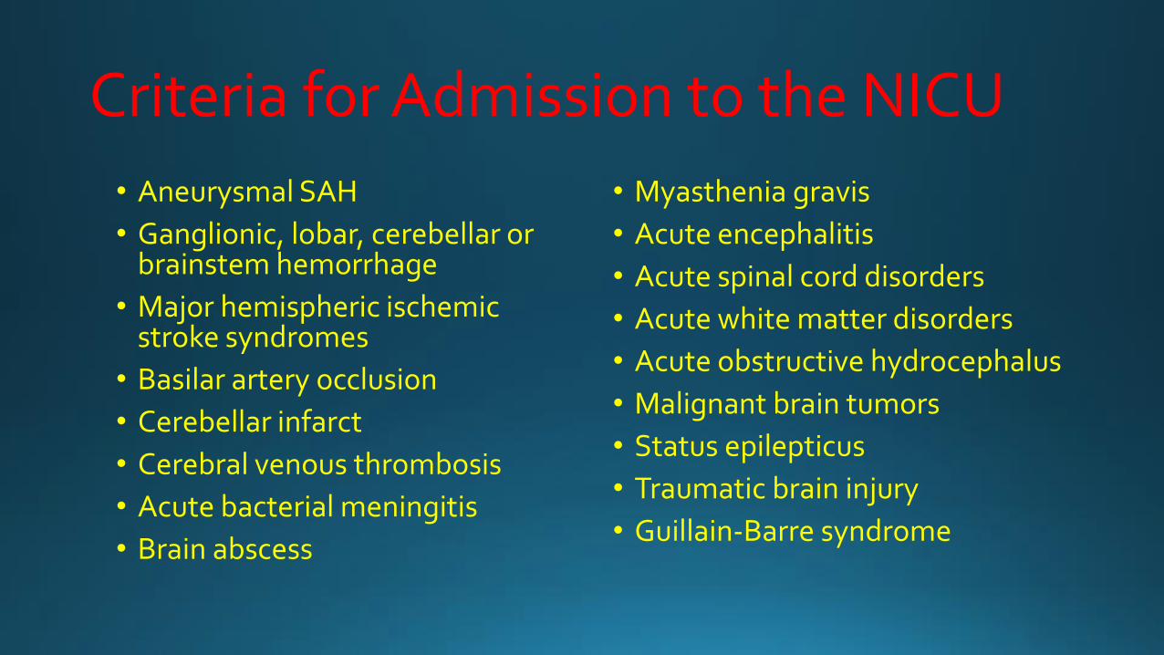
Criteria for Admission to the NICU
• Aneurysmal SAH
• Ganglionic, lobar, cerebellar or brainstem hemorrhage
• Major hemispheric ischemic stroke syndromes
• Basilar artery occlusion
• Cerebellar infarct
• Cerebral venous thrombosis
• Acute bacterial meningitis
• Brain abscess
• Myasthenia gravis
• Acute encephalitis
• Acute spinal cord disorders
• Acute white matter disorders
• Acute obstructive hydrocephalus
• Malignant brain tumors
• Status epilepticus
• Traumatic brain injury
• Guillain-Barre syndrome
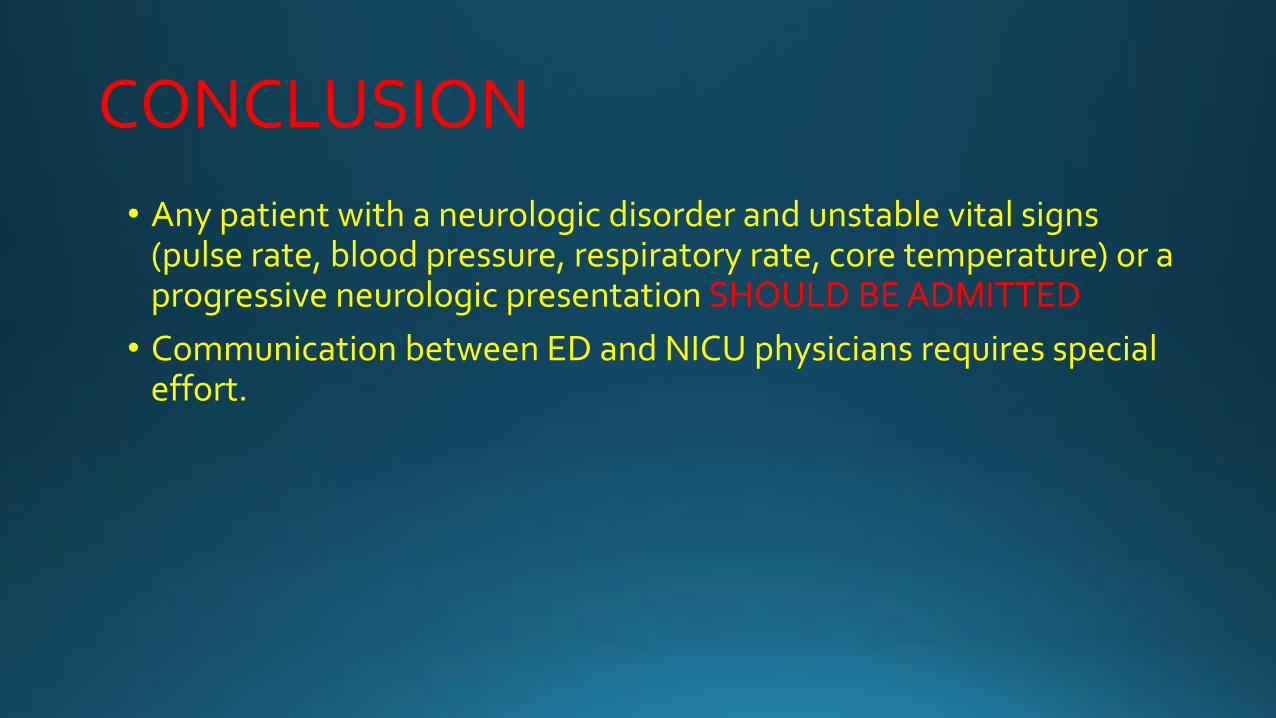
CONCLUSION
• Any patient with a neurologic disorder and unstable vital signs (pulse rate, blood pressure, respiratory rate, core temperature) or a progressive neurologic presentation SHOULD BE ADMITTED
• Communication between ED and NICU physicians requires special effort.
Evaluation of Presenting Symptoms Indicating Urgency

“Confused and Febrile”
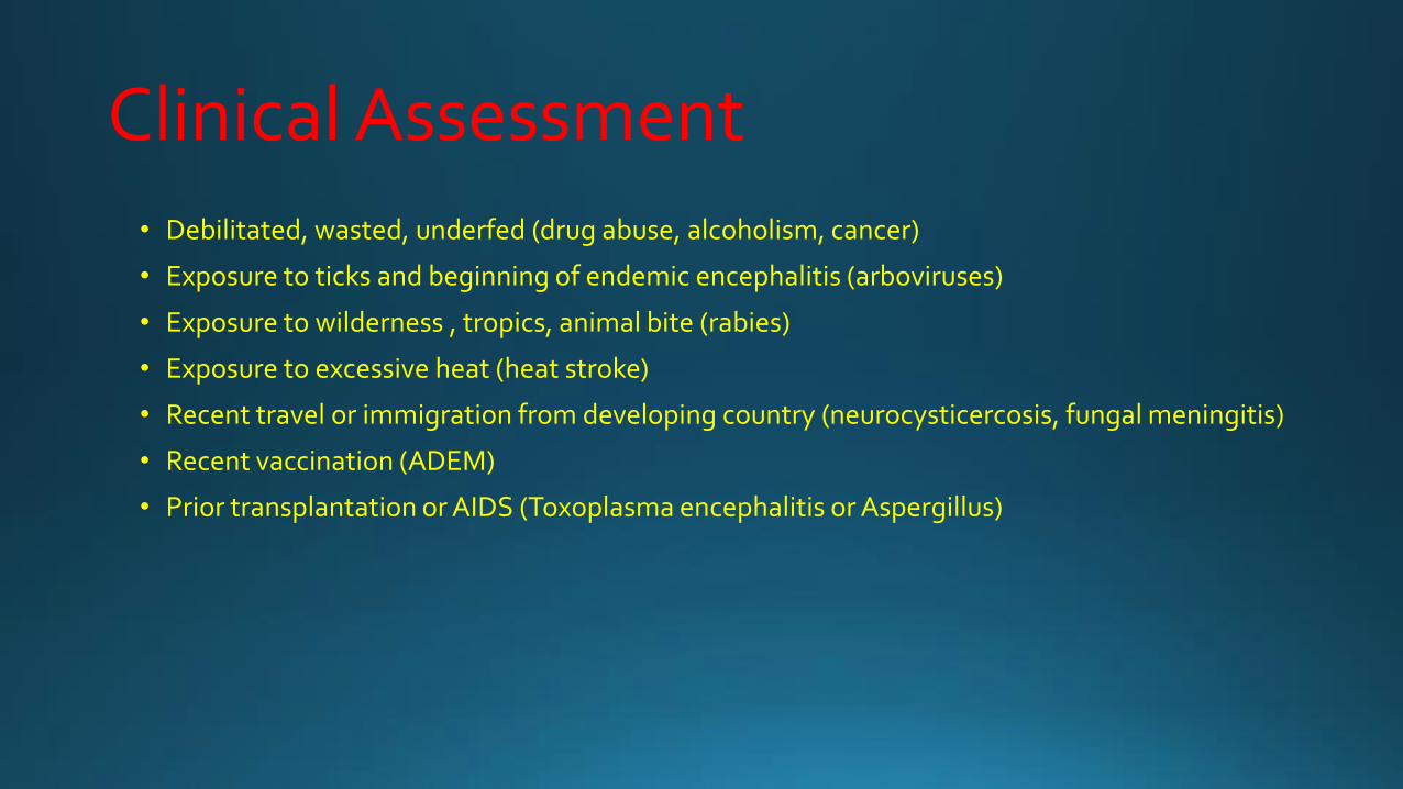
Clinical Assessment
• Debilitated, wasted, underfed (drug abuse, alcoholism, cancer)
• Exposure to ticks and beginning of endemic encephalitis (arboviruses)
• Exposure to wilderness , tropics, animal bite (rabies)
• Exposure to excessive heat (heat stroke)
• Recent travel or immigration from developing country (neurocysticercosis, fungal meningitis)
• Recent vaccination (ADEM)
• Prior transplantation or AIDS (Toxoplasma encephalitis or Aspergillus)
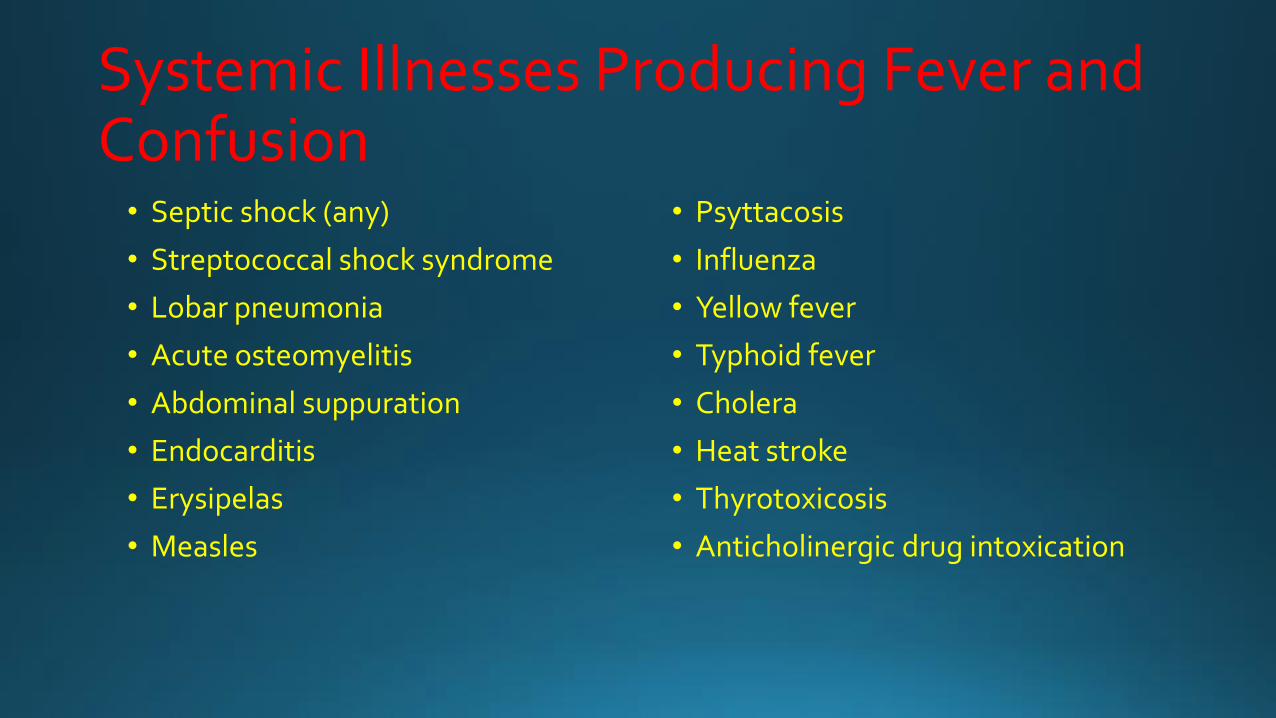
Systemic Illnesses Producing Fever and Confusion
• Septic shock (any)
• Streptococcal shock syndrome
• Lobar pneumonia
• Acute osteomyelitis
• Abdominal suppuration
• Endocarditis
• Erysipelas
• Measles
• Psyttacosis
• Influenza
• Yellow fever
• Typhoid fever
• Cholera
• Heat stroke
• Thyrotoxicosis
• Anticholinergic drug intoxication
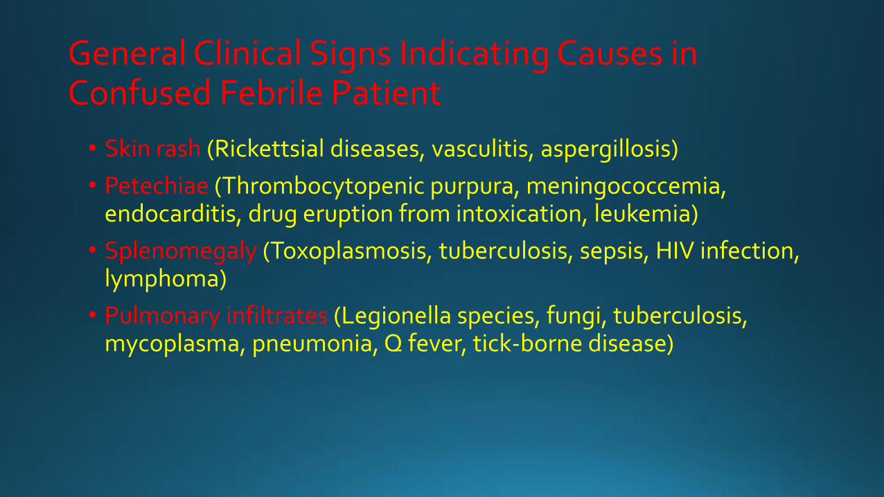
General Clinical Signs Indicating Causes in Confused Febrile Patient
• Skin rash (Rickettsial diseases, vasculitis, aspergillosis)
• Petechiae (Thrombocytopenic purpura, meningococcemia, endocarditis, drug eruption from intoxication, leukemia)
• Splenomegaly (Toxoplasmosis, tuberculosis, sepsis, HIV infection, lymphoma)
• Pulmonary infiltrates (Legionella species, fungi, tuberculosis, mycoplasma, pneumonia, Q fever, tick-borne disease)
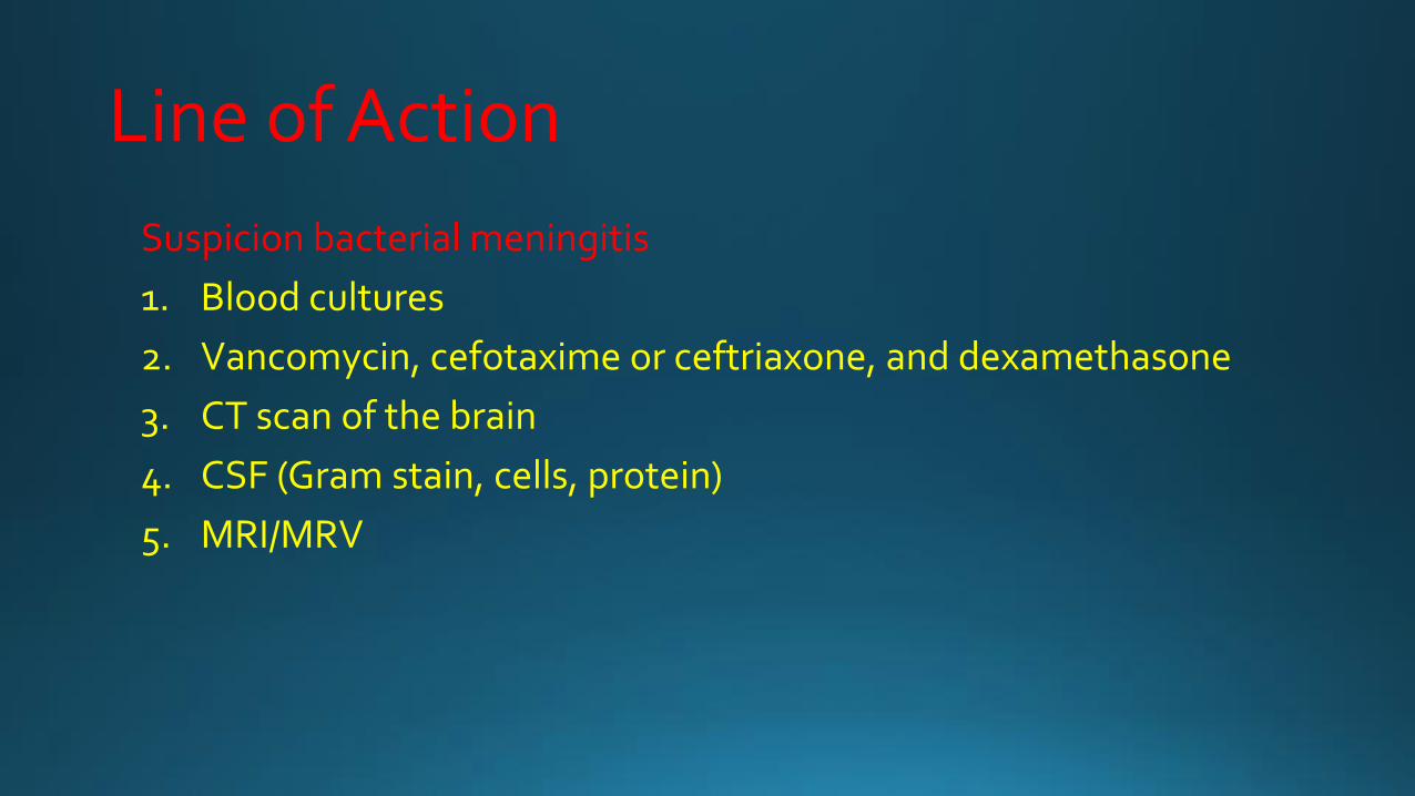
Line of Action
Suspicion bacterial meningitis
1. Blood cultures
2. Vancomycin, cefotaxime or ceftriaxone, and dexamethasone
3. CT scan of the brain
4. CSF (Gram stain, cells, protein)
5. MRI/MRV
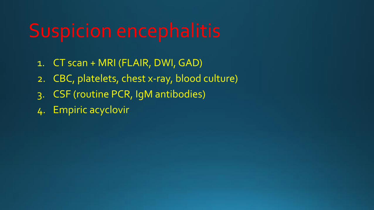
Suspicion encephalitis
1. CT scan + MRI (FLAIR, DWI, GAD)
2. CBC, platelets, chest x-ray, blood culture)
3. CSF (routine PCR, IgM antibodies)
4. Empiric acyclovir
CONCLUSION
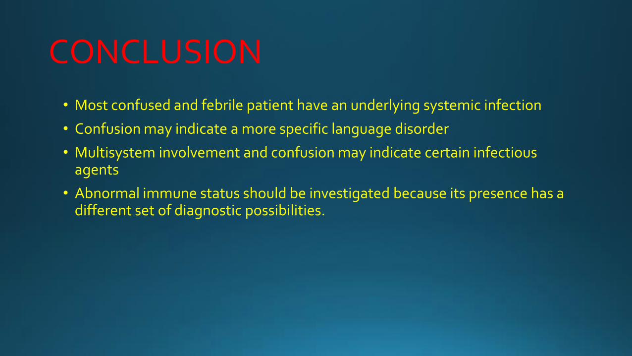
• Most confused and febrile patient have an underlying systemic infection
• Confusion may indicate a more specific language disorder
• Multisystem involvement and confusion may indicate certain infectious agents
• Abnormal immune status should be investigated because its presence has a different set of diagnostic possibilities.

“A Terrible Headache”
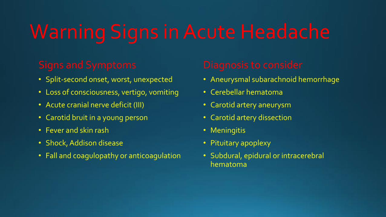
Warning Signs in Acute Headache
Signs and Symptoms • Split-second onset, worst, unexpected
• Loss of consciousness, vertigo, vomiting
• Acute cranial nerve deficit (III)
• Carotid bruit in a young person
• Fever and skin rash
• Shock, Addison disease
• Fall and coagulopathy or anticoagulation
Diagnosis to consider • Aneurysmal subarachnoid hemorrhage
• Cerebellar hematoma
• Carotid artery aneurysm
• Carotid artery dissection
• Meningitis
• Pituitary apoplexy
• Subdural, epidural or intracerebral hematoma
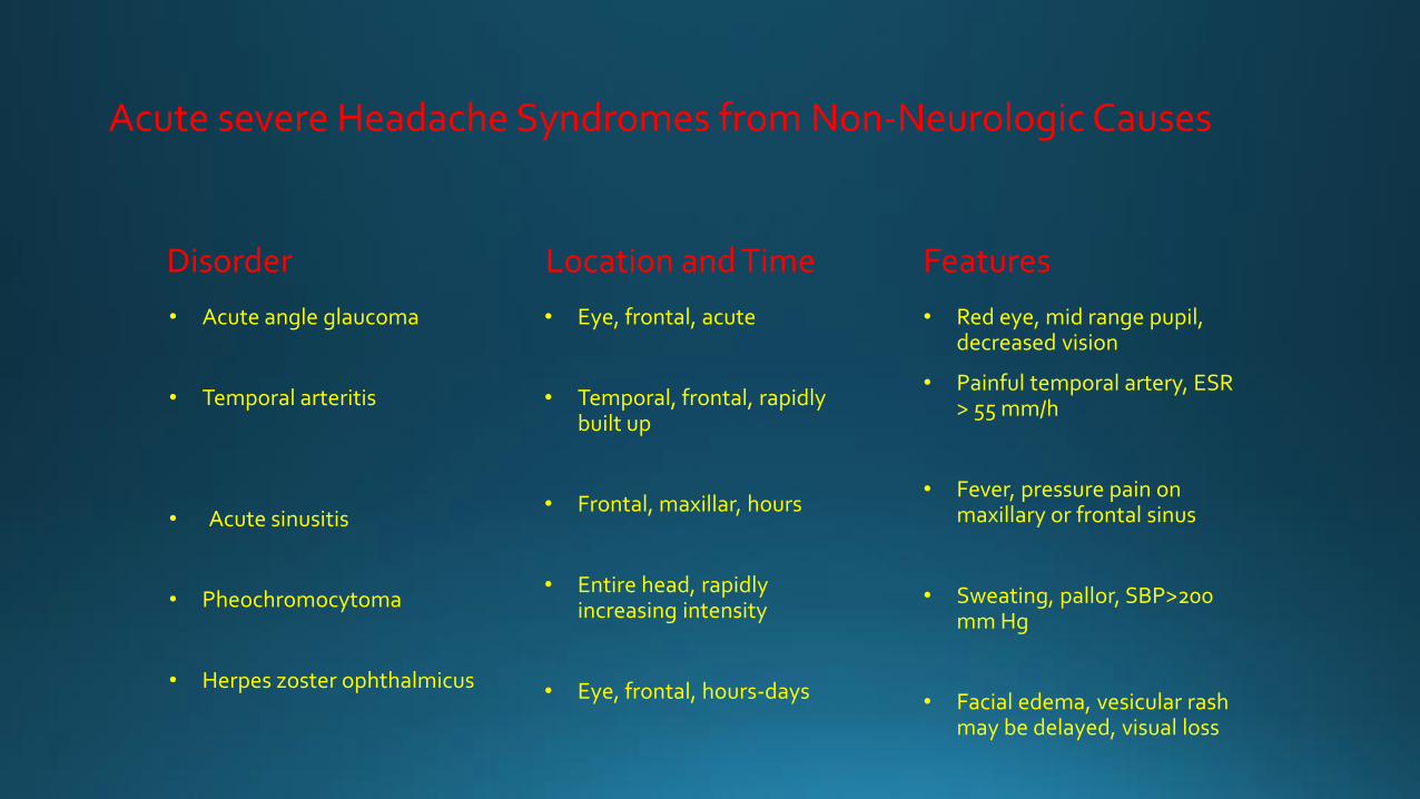
Acute severe Headache Syndromes from Non-Neurologic Causes
Disorder
• Acute angle glaucoma
• Temporal arteritis
• Acute sinusitis
• Pheochromocytoma
• Herpes zoster ophthalmicus
Location and Time
• Eye, frontal, acute
• Temporal, frontal, rapidly built up
• Frontal, maxillar, hours
• Entire head, rapidly increasing intensity
• Eye, frontal, hours-days
Features
• Red eye, mid range pupil, decreased vision
• Painful temporal artery, ESR > 55 mm/h
• Fever, pressure pain on maxillary or frontal sinus
• Sweating, pallor, SBP>200 mm Hg
• Facial edema, vesicular rash may be delayed, visual loss
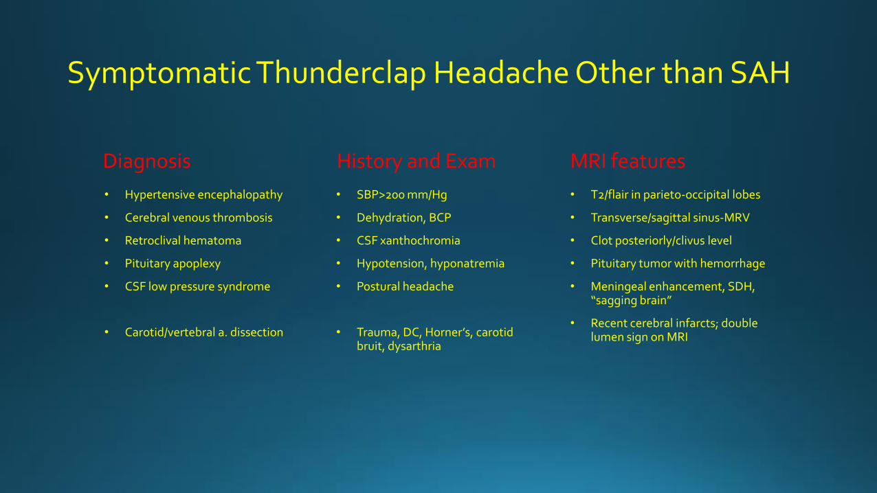
Symptomatic Thunderclap Headache Other than SAH
Diagnosis
• Hypertensive encephalopathy
• Cerebral venous thrombosis
• Retroclival hematoma
• Pituitary apoplexy
• CSF low pressure syndrome
• Carotid/vertebral a. dissection
History and Exam
• SBP>200 mm/Hg
• Dehydration, BCP
• CSF xanthochromia
• Hypotension, hyponatremia
• Postural headache
• Trauma, DC, Horner’s, carotid bruit, dysarthria
MRI features
• T2/flair in parieto-occipital lobes
• Transverse/sagittal sinus-MRV
• Clot posteriorly/clivus level
• Pituitary tumor with hemorrhage
• Meningeal enhancement, SDH, “sagging brain”
• Recent cerebral infarcts; double lumen sign on MRI
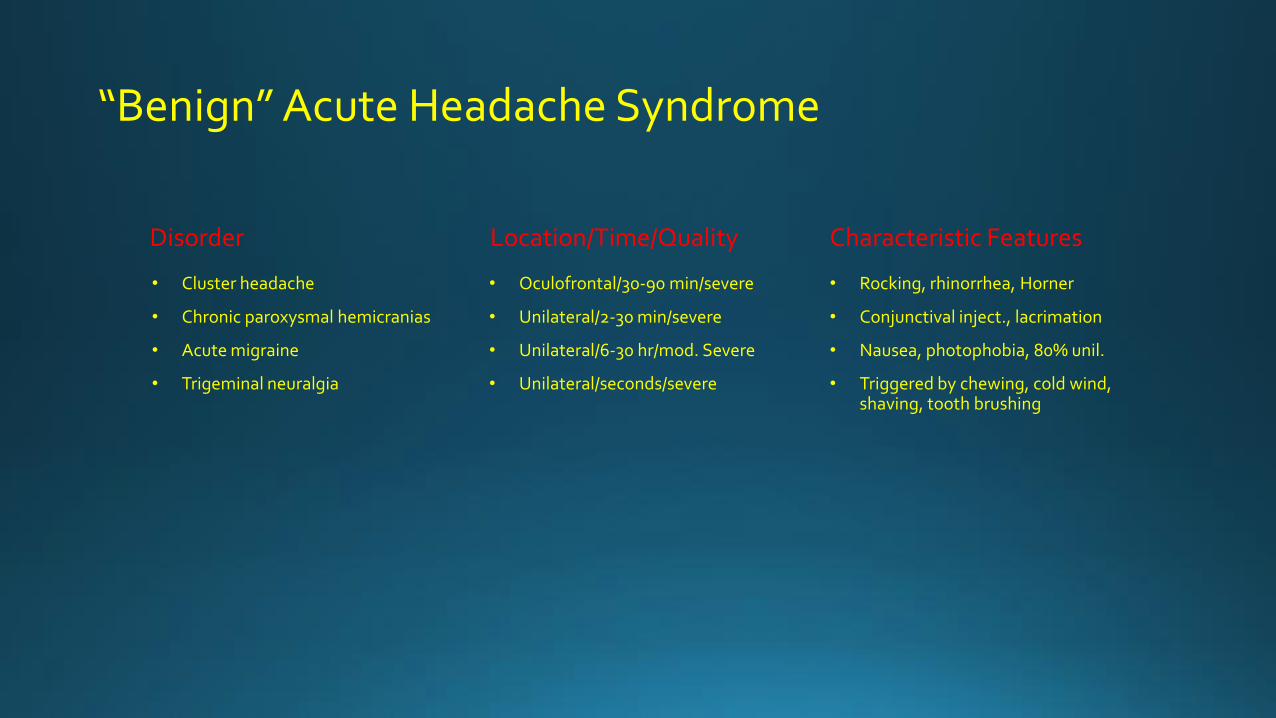
“Benign” Acute Headache Syndrome
Disorder
• Cluster headache
• Chronic paroxysmal hemicranias
• Acute migraine
• Trigeminal neuralgia
Location/Time/Quality
• Oculofrontal/30-90 min/severe
• Unilateral/2-30 min/severe
• Unilateral/6-30 hr/mod. Severe
• Unilateral/seconds/severe
Characteristic Features
• Rocking, rhinorrhea, Horner
• Conjunctival inject., lacrimation
• Nausea, photophobia, 80% unil.
• Triggered by chewing, cold wind, shaving, tooth brushing
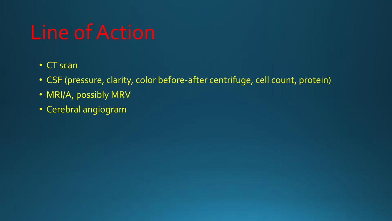
Line of Action
• CT scan
• CSF (pressure, clarity, color before-after centrifuge, cell count, protein)
• MRI/A, possibly MRV
• Cerebral angiogram
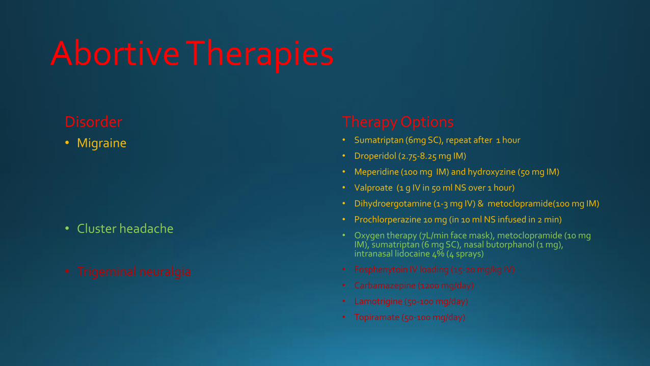
Abortive Therapies
Disorder • Migraine
• Cluster headache
• Trigeminal neuralgia
Therapy Options • Sumatriptan (6mg SC), repeat after 1 hour
• Droperidol (2.75-8.25 mg IM)
• Meperidine (100 mg IM) and hydroxyzine (50 mg IM)
• Valproate (1 g IV in 50 ml NS over 1 hour)
• Dihydroergotamine (1-3 mg IV) & metoclopramide(100 mg IM)
• Prochlorperazine 10 mg (in 10 ml NS infused in 2 min)
• Oxygen therapy (7L/min face mask), metoclopramide (10 mg IM), sumatriptan (6 mg SC), nasal butorphanol (1 mg), intranasal lidocaine 4% (4 sprays)
• Fosphenytoin IV loading (15-20 mg/kg IV)
• Carbamazepine (1200 mg/day)
• Lamotrigine (50-100 mg/day)
• Topiramate (50-100 mg/day)
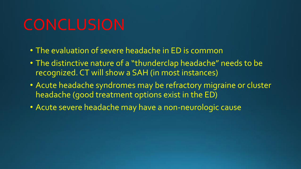
CONCLUSION
• The evaluation of severe headache in ED is common
• The distinctive nature of a “thunderclap headache” needs to be recognized. CT will show a SAH (in most instances)
• Acute headache syndromes may be refractory migraine or cluster headache (good treatment options exist in the ED)
• Acute severe headache may have a non-neurologic cause

“Blacked Out and Stumped Down”
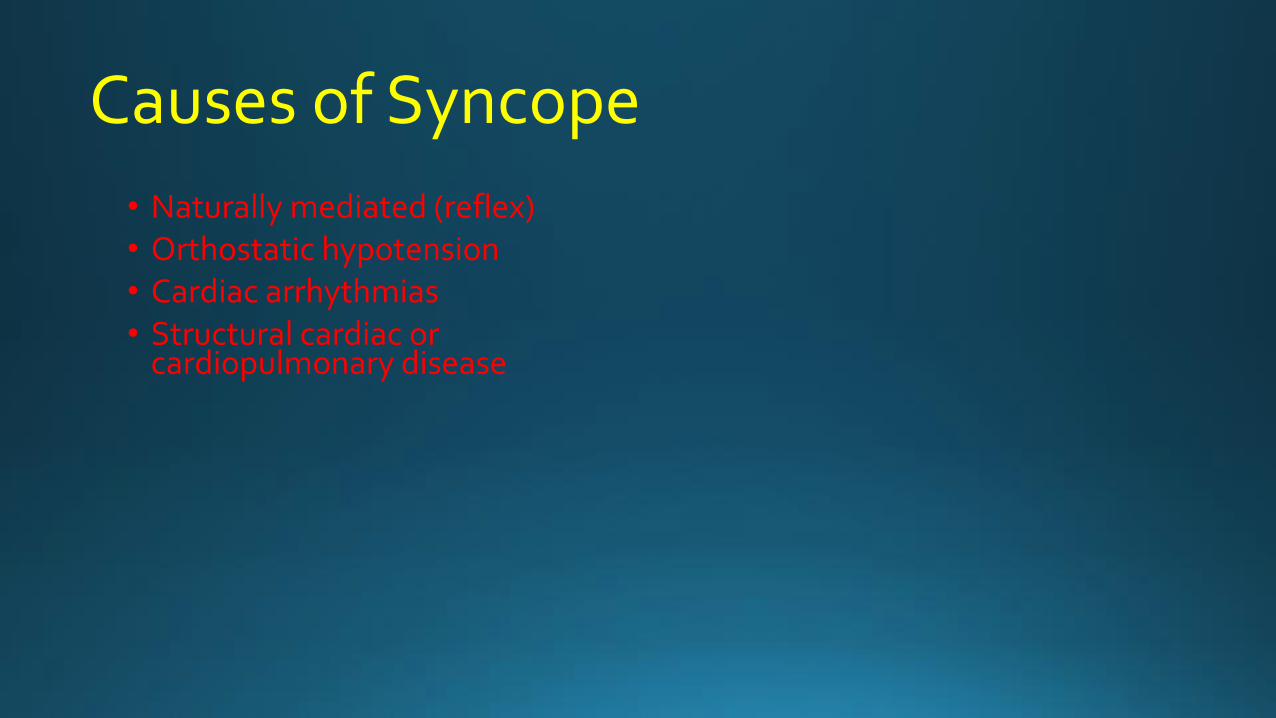
Causes of Syncope
• Naturally mediated (reflex) • Orthostatic hypotension • Cardiac arrhythmias • Structural cardiac or
cardiopulmonary disease
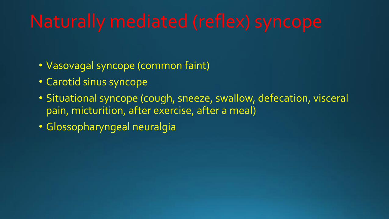
Naturally mediated (reflex) syncope • Vasovagal syncope (common faint)
• Carotid sinus syncope
• Situational syncope (cough, sneeze, swallow, defecation, visceral pain, micturition, after exercise, after a meal)
• Glossopharyngeal neuralgia
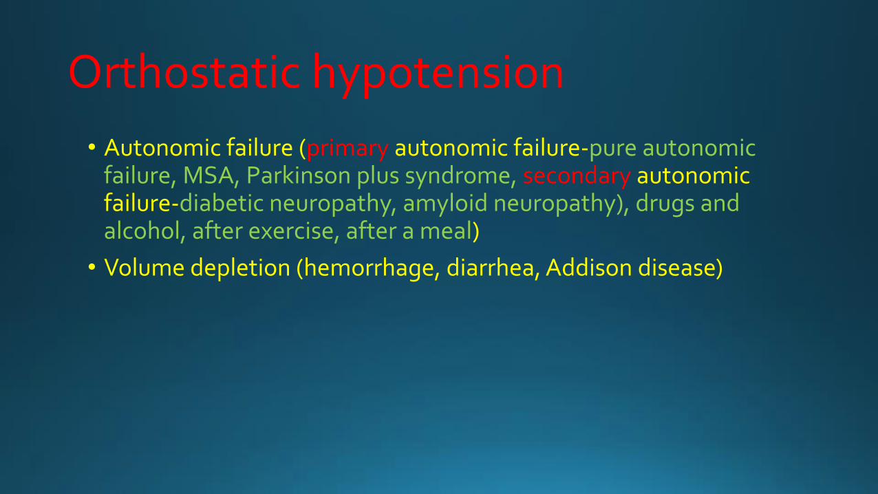
Orthostatic hypotension
• Autonomic failure (primary autonomic failure-pure autonomic failure, MSA, Parkinson plus syndrome, secondary autonomic failure-diabetic neuropathy, amyloid neuropathy), drugs and alcohol, after exercise, after a meal)
• Volume depletion (hemorrhage, diarrhea, Addison disease)
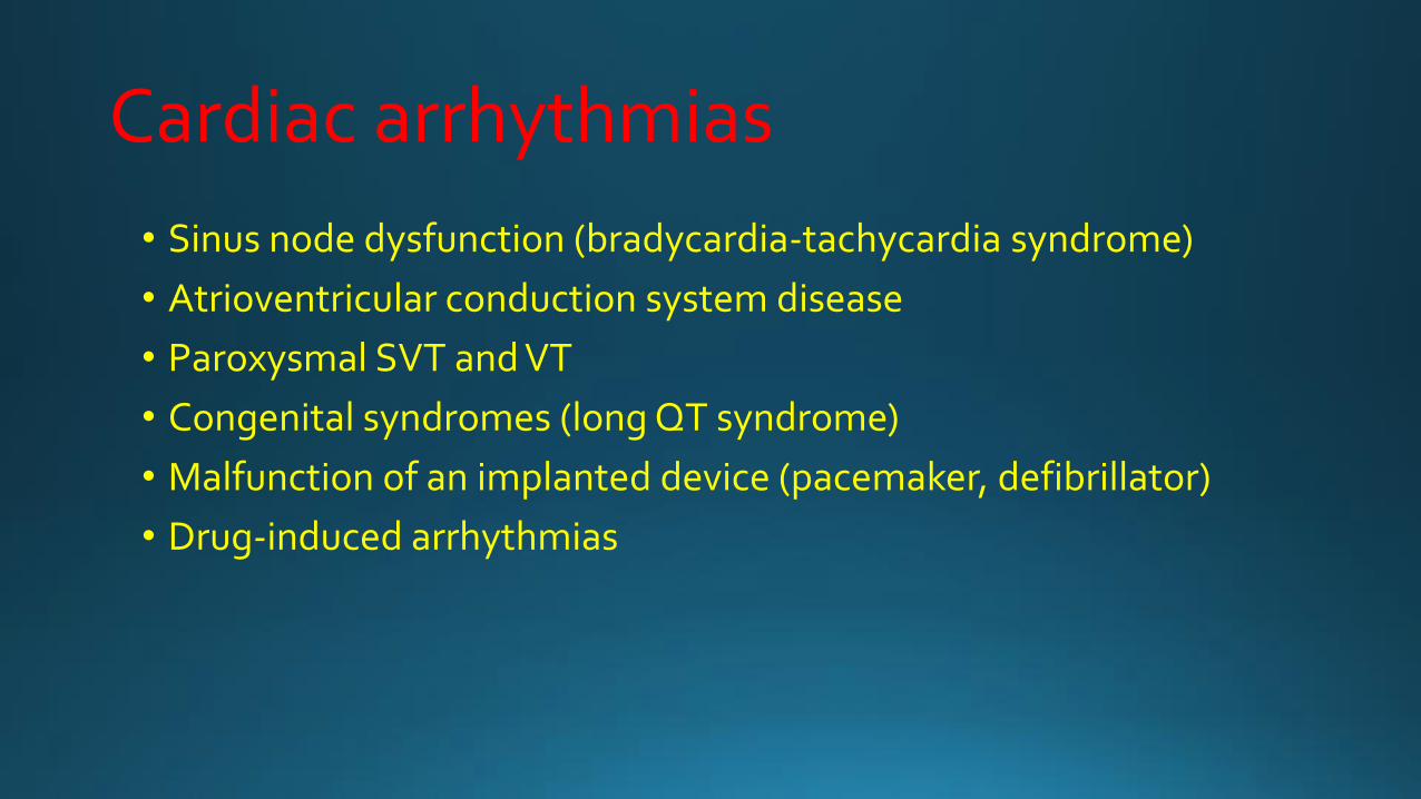
Cardiac arrhythmias
• Sinus node dysfunction (bradycardia-tachycardia syndrome)
• Atrioventricular conduction system disease
• Paroxysmal SVT and VT
• Congenital syndromes (long QT syndrome)
• Malfunction of an implanted device (pacemaker, defibrillator)
• Drug-induced arrhythmias
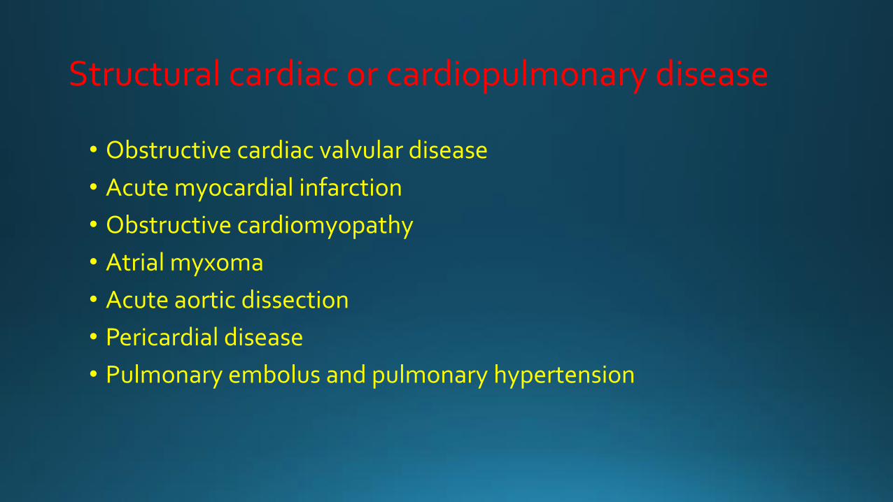
Structural cardiac or cardiopulmonary disease
• Obstructive cardiac valvular disease
• Acute myocardial infarction
• Obstructive cardiomyopathy
• Atrial myxoma
• Acute aortic dissection
• Pericardial disease
• Pulmonary embolus and pulmonary hypertension
CONCLUSION
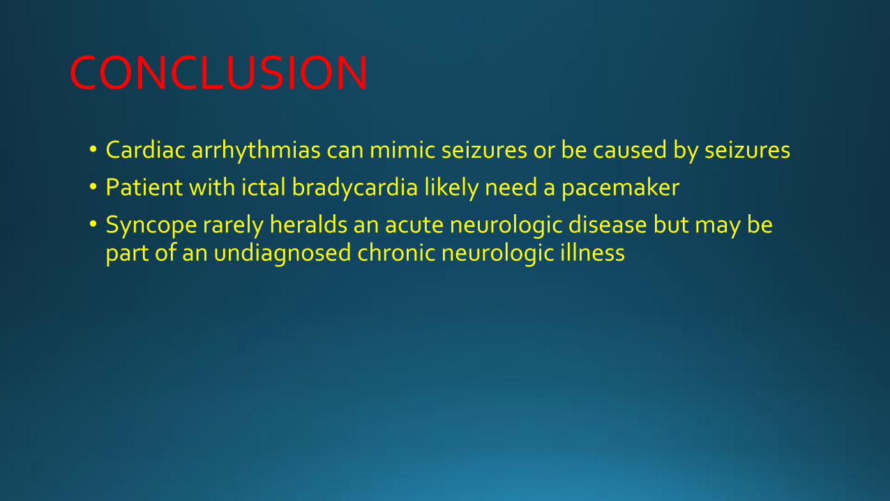
• Cardiac arrhythmias can mimic seizures or be caused by seizures
• Patient with ictal bradycardia likely need a pacemaker
• Syncope rarely heralds an acute neurologic disease but may be part of an undiagnosed chronic neurologic illness

“See Nothing, See Double, See Shapes”
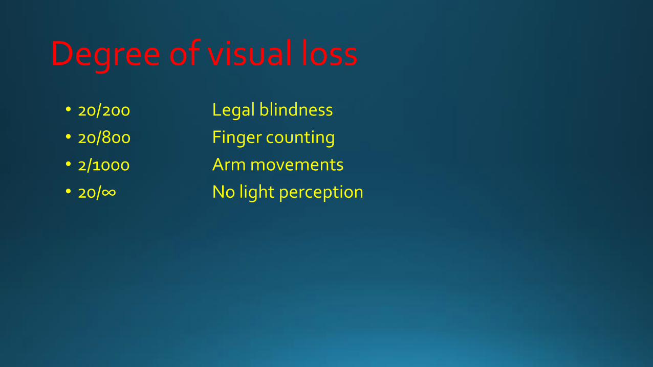
Degree of visual loss
• 20/200 Legal blindness
• 20/800 Finger counting
• 2/1000 Arm movements
• 20/∞ No light perception
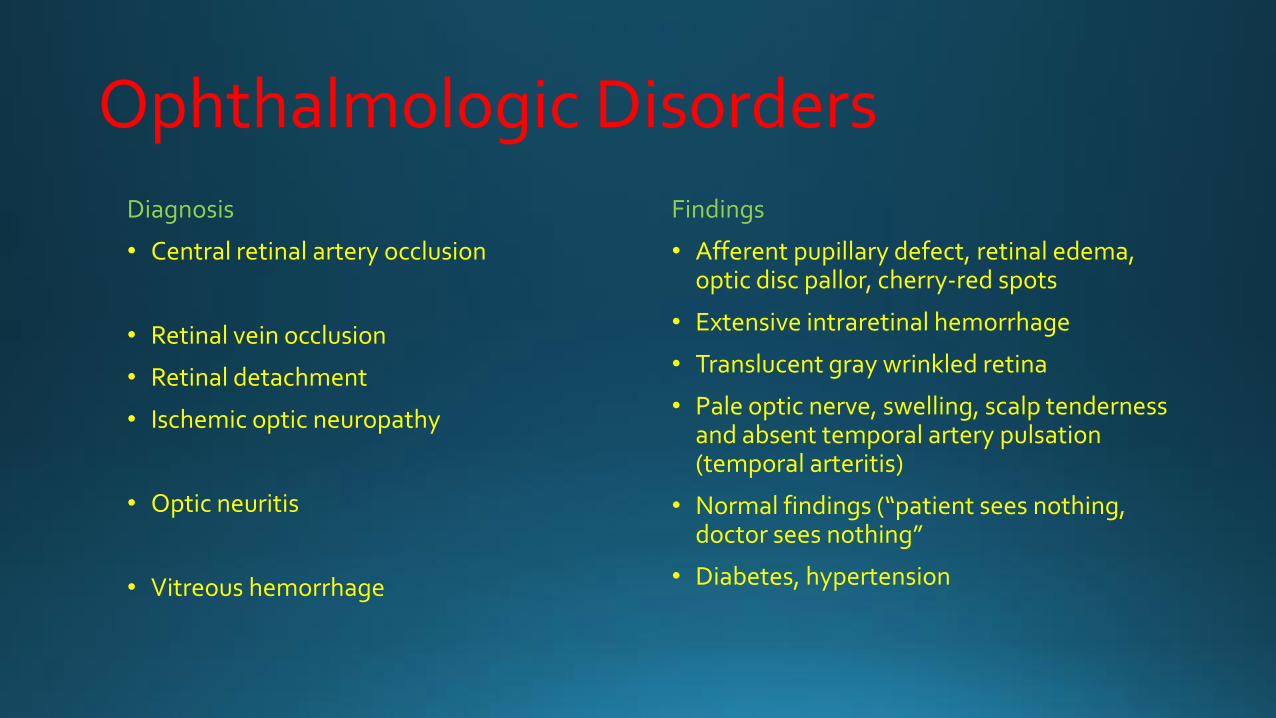
Ophthalmologic Disorders
Diagnosis
• Central retinal artery occlusion
• Retinal vein occlusion
• Retinal detachment
• Ischemic optic neuropathy
• Optic neuritis
• Vitreous hemorrhage
Findings
• Afferent pupillary defect, retinal edema, optic disc pallor, cherry-red spots
• Extensive intraretinal hemorrhage
• Translucent gray wrinkled retina
• Pale optic nerve, swelling, scalp tenderness and absent temporal artery pulsation (temporal arteritis)
• Normal findings (“patient sees nothing, doctor sees nothing”
• Diabetes, hypertension
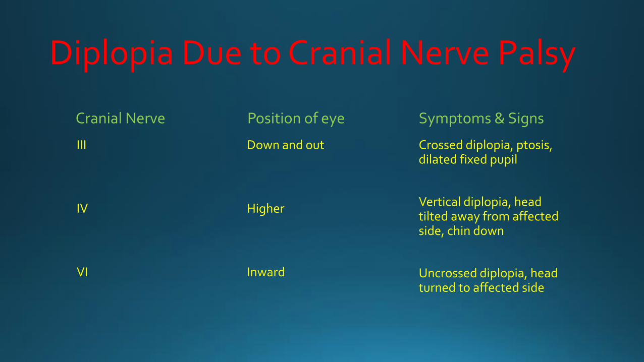
Diplopia Due to Cranial Nerve Palsy
Cranial Nerve
III
IV
VI
Position of eye
Down and out
Higher
Inward
Symptoms & Signs
Crossed diplopia, ptosis, dilated fixed pupil
Vertical diplopia, head tilted away from affected side, chin down
Uncrossed diplopia, head turned to affected side
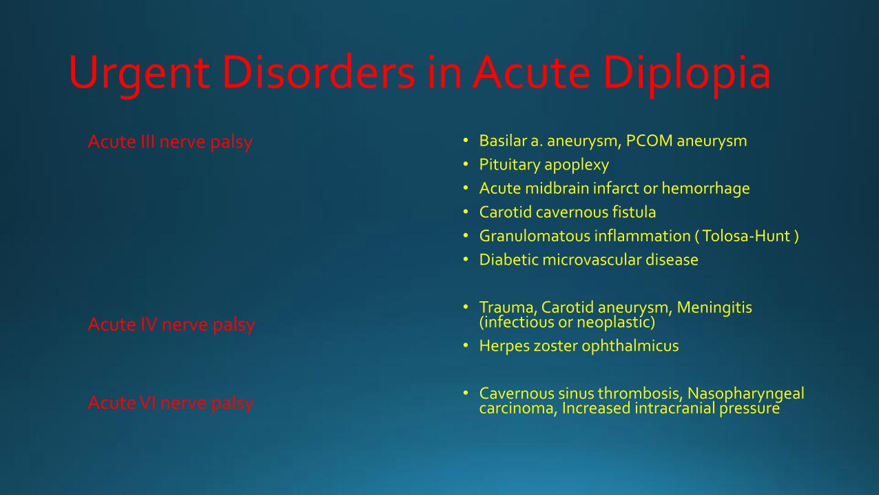
Urgent Disorders in Acute Diplopia Acute III nerve palsy
Acute IV nerve palsy
Acute VI nerve palsy
• Basilar a. aneurysm, PCOM aneurysm
• Pituitary apoplexy
• Acute midbrain infarct or hemorrhage
• Carotid cavernous fistula
• Granulomatous inflammation ( Tolosa-Hunt )
• Diabetic microvascular disease
• Trauma, Carotid aneurysm, Meningitis (infectious or neoplastic)
• Herpes zoster ophthalmicus
• Cavernous sinus thrombosis, Nasopharyngeal carcinoma, Increased intracranial pressure
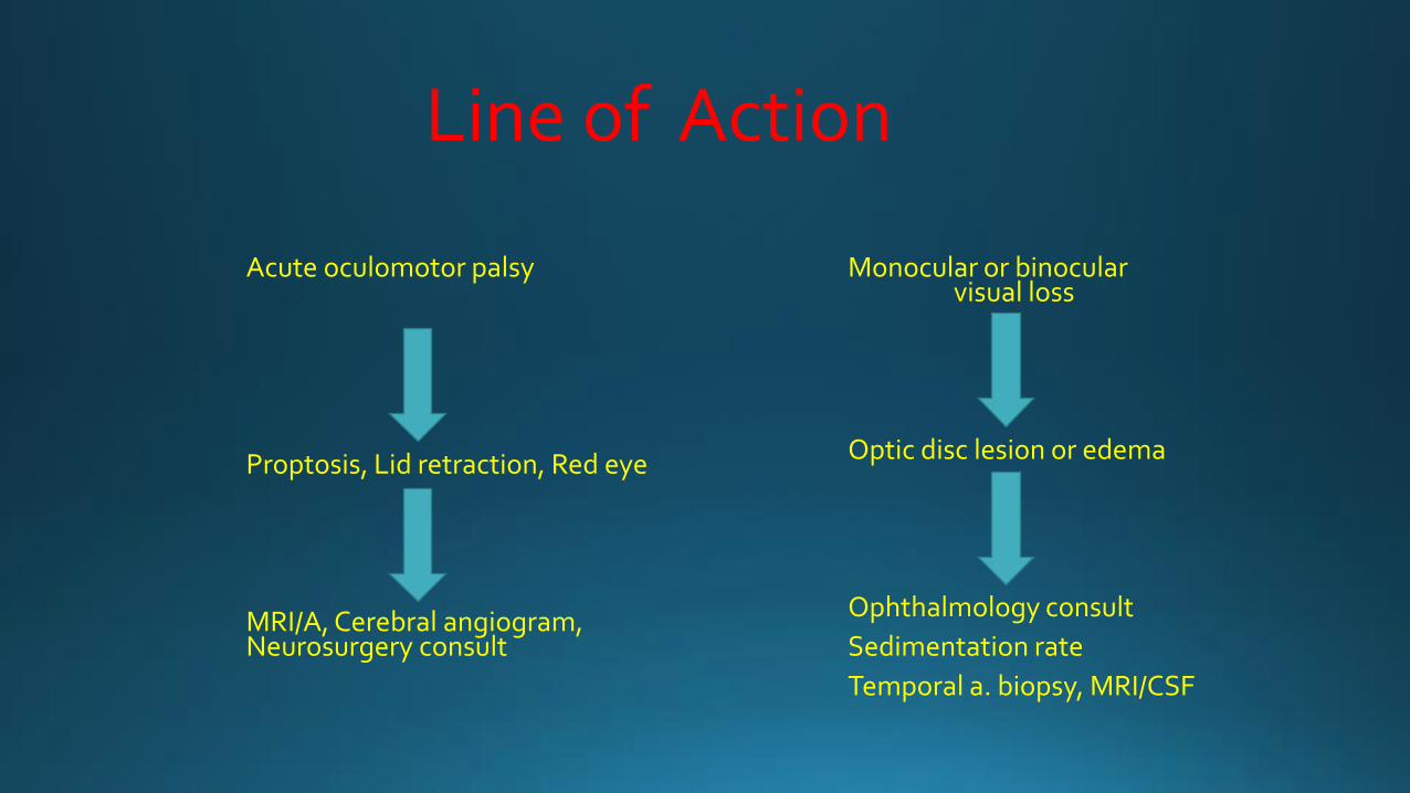
Line of Action
Acute oculomotor palsy
Proptosis, Lid retraction, Red eye
MRI/A, Cerebral angiogram, Neurosurgery consult
Monocular or binocular visual loss
Optic disc lesion or edema
Ophthalmology consult
Sedimentation rate
Temporal a. biopsy, MRI/CSF
CONCLUSION
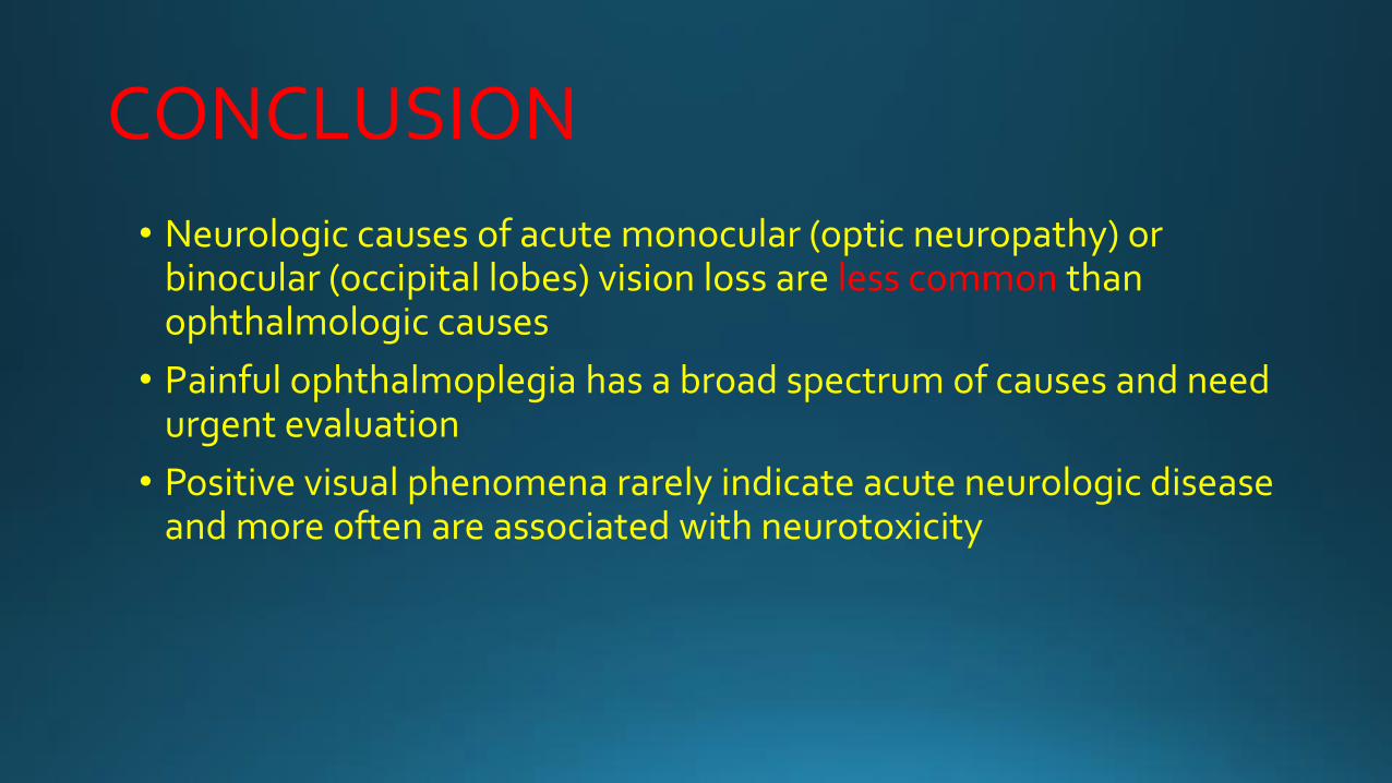
• Neurologic causes of acute monocular (optic neuropathy) or binocular (occipital lobes) vision loss are less common than ophthalmologic causes
• Painful ophthalmoplegia has a broad spectrum of causes and need urgent evaluation
• Positive visual phenomena rarely indicate acute neurologic disease and more often are associated with neurotoxicity

“Spinning”
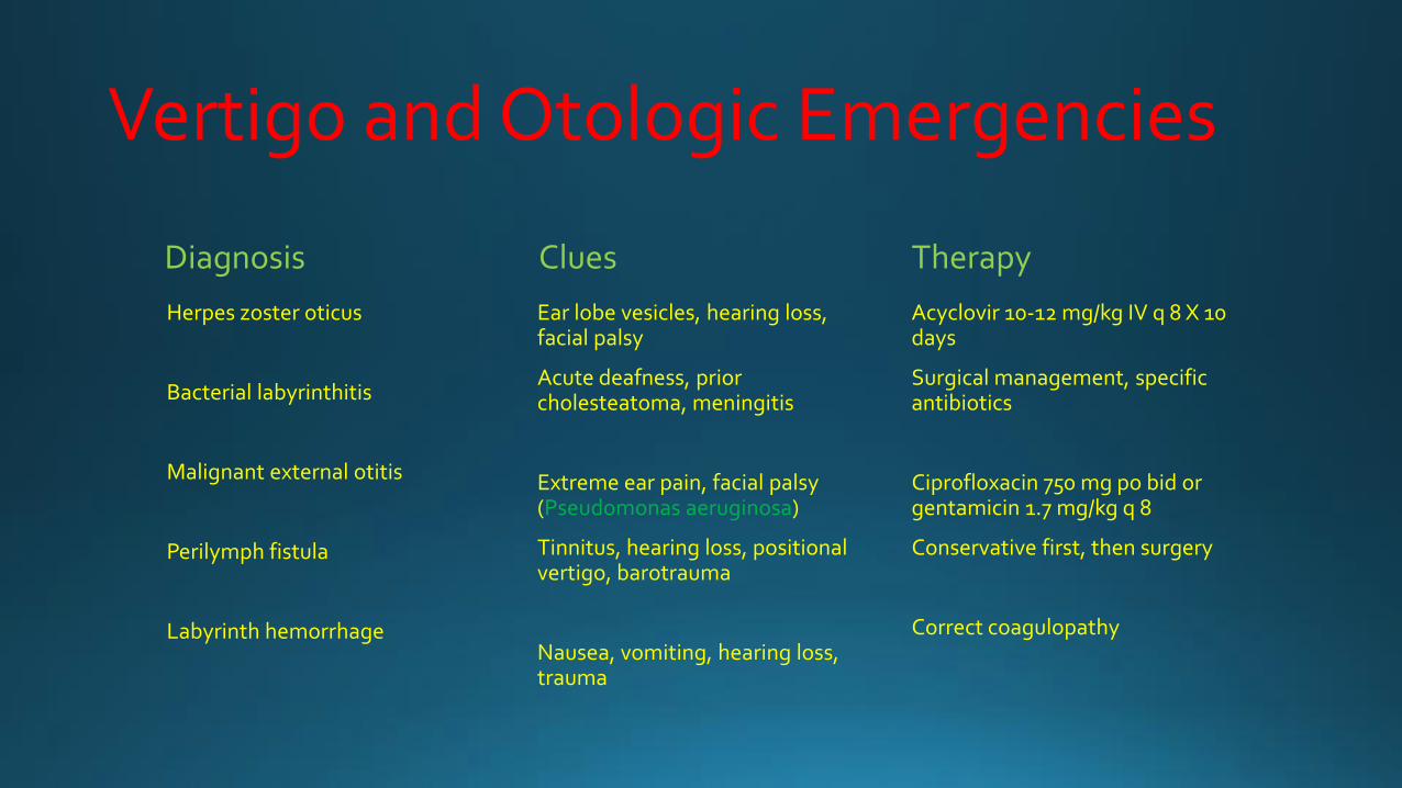
Vertigo and Otologic Emergencies
Diagnosis
Herpes zoster oticus
Bacterial labyrinthitis
Malignant external otitis
Perilymph fistula
Labyrinth hemorrhage
Clues
Ear lobe vesicles, hearing loss, facial palsy
Acute deafness, prior cholesteatoma, meningitis
Extreme ear pain, facial palsy (Pseudomonas aeruginosa)
Tinnitus, hearing loss, positional vertigo, barotrauma
Nausea, vomiting, hearing loss, trauma
Therapy
Acyclovir 10-12 mg/kg IV q 8 X 10 days
Surgical management, specific antibiotics
Ciprofloxacin 750 mg po bid or gentamicin 1.7 mg/kg q 8
Conservative first, then surgery
Correct coagulopathy
“Can’t Walk or Stand”
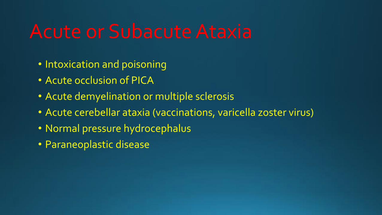
Acute or Subacute Ataxia
• Intoxication and poisoning
• Acute occlusion of PICA
• Acute demyelination or multiple sclerosis
• Acute cerebellar ataxia (vaccinations, varicella zoster virus)
• Normal pressure hydrocephalus
• Paraneoplastic disease

Acute paraplegia Disorder
Myelitis
Myelopathy
Polyradiculopathy
Neoplastic meningitis
Neuromuscular Junction Disorders
Myopathy
History Vaccination, febrile illness, optic neuritis, travel, tick bite, immunosuppression, AIDS
Cancer, AA or recent catheterization, low back pain, connective tissue disease (SLE), anticoagulation
Diarrhea, URI, CMV, HSV, EBV, DM, leukemia, sarcoidosis
Carcinoma,lymphoma
Dysphagia, diplopia, ptosis, small lung cancer
Autoimmune disorder, thyrotoxicosis, exercise intolerance
Suggest
Postvaccination myelopathy, postinfectious transverse myelitis, MS or Devic disease, schistosomiasis, cysticercosis, Lyme disease, TB, syphilis
Acute necrotic myelopathy, cord infarction, vasculitis, radiation myelopathy, paraneoplastic myelopathy, epidural hematoma, spinal AVM, dural AV fistula
GBS, acute diabetic polyradiculopathy
Infiltrative leptomeningeal spread
Myasthenia gravis, Lambert-Eaton syndrome, botulism
Polymyositis, dermatomyositis, metabolic myopathy
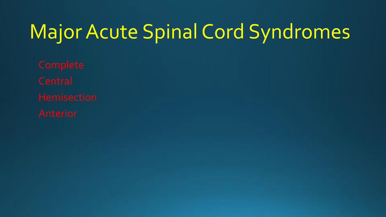
Major Acute Spinal Cord Syndromes
Complete
Central
Hemisection
Anterior

CONCLUSION
• Acute gait disorder may be caused by viral infections or recent vaccinations in children and acute cerebellar infarction or medication overdose in the elderly
• Acute paraplegia may be a consequence of spinal cord compression, and neuroimaging with MRI is urgently needed
• Acute chest pain and paraplegia requires immediate evaluation for aortic dissection
“Short of Breath”
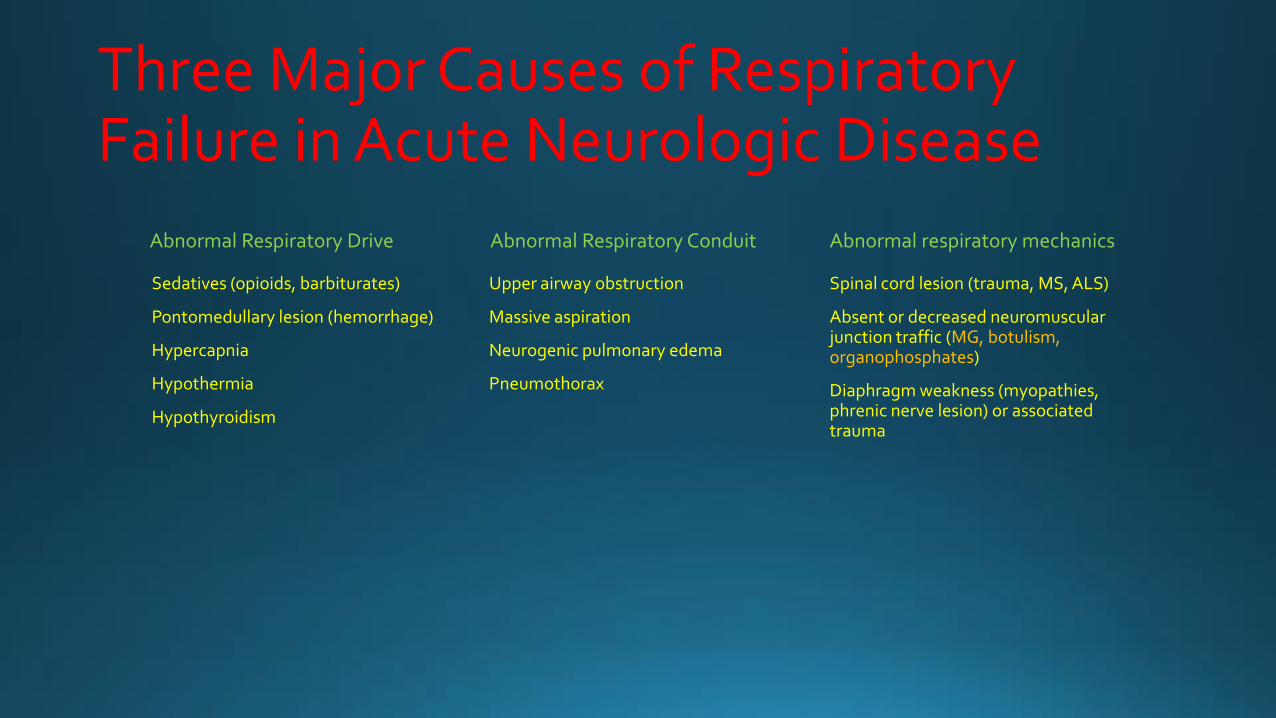
Three Major Causes of Respiratory Failure in Acute Neurologic Disease
Abnormal Respiratory Drive
Sedatives (opioids, barbiturates)
Pontomedullary lesion (hemorrhage)
Hypercapnia
Hypothermia
Hypothyroidism
Abnormal Respiratory Conduit
Upper airway obstruction
Massive aspiration
Neurogenic pulmonary edema
Pneumothorax
Abnormal respiratory mechanics
Spinal cord lesion (trauma, MS, ALS)
Absent or decreased neuromuscular junction traffic (MG, botulism, organophosphates)
Diaphragm weakness (myopathies, phrenic nerve lesion) or associated trauma
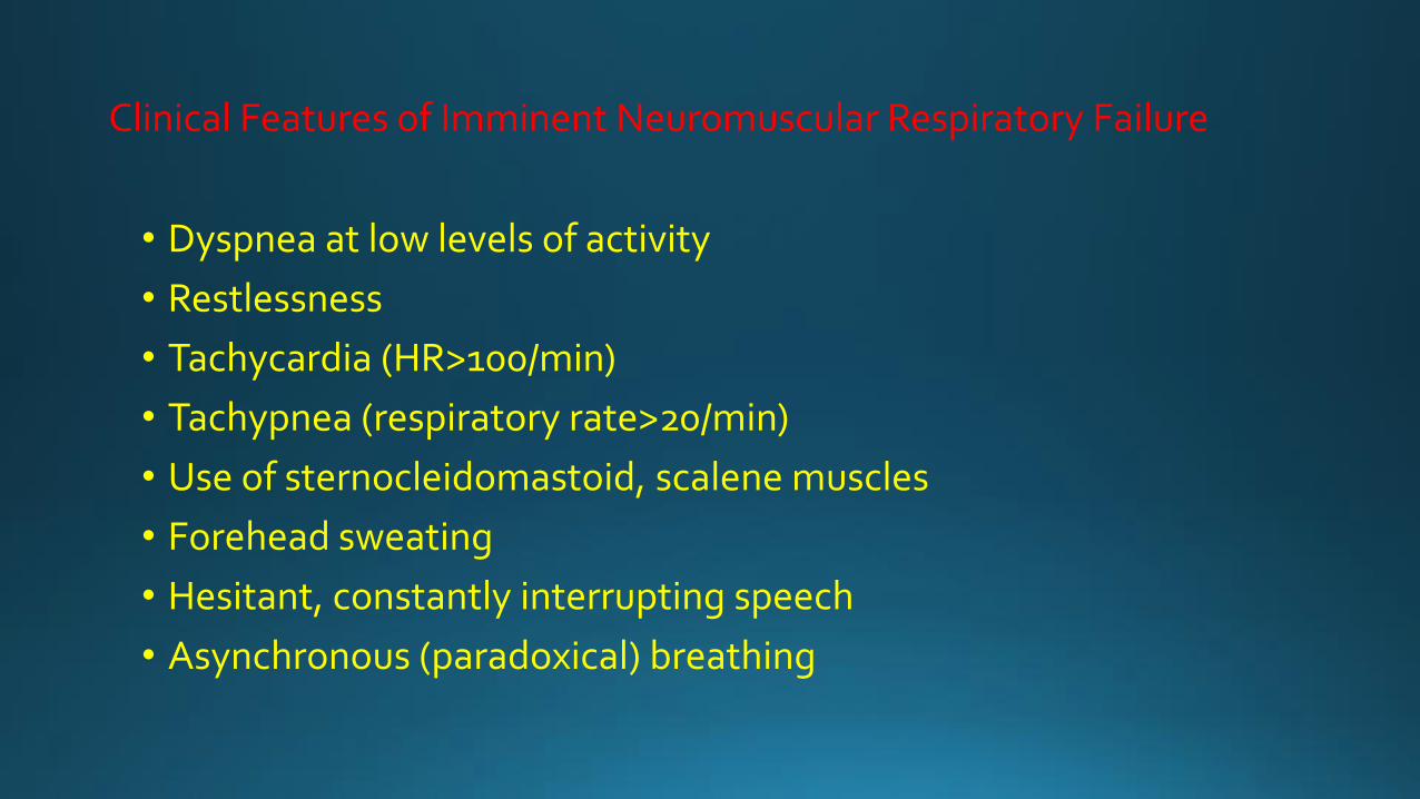
Clinical Features of Imminent Neuromuscular Respiratory Failure
• Dyspnea at low levels of activity
• Restlessness
• Tachycardia (HR>100/min)
• Tachypnea (respiratory rate>20/min)
• Use of sternocleidomastoid, scalene muscles
• Forehead sweating
• Hesitant, constantly interrupting speech
• Asynchronous (paradoxical) breathing
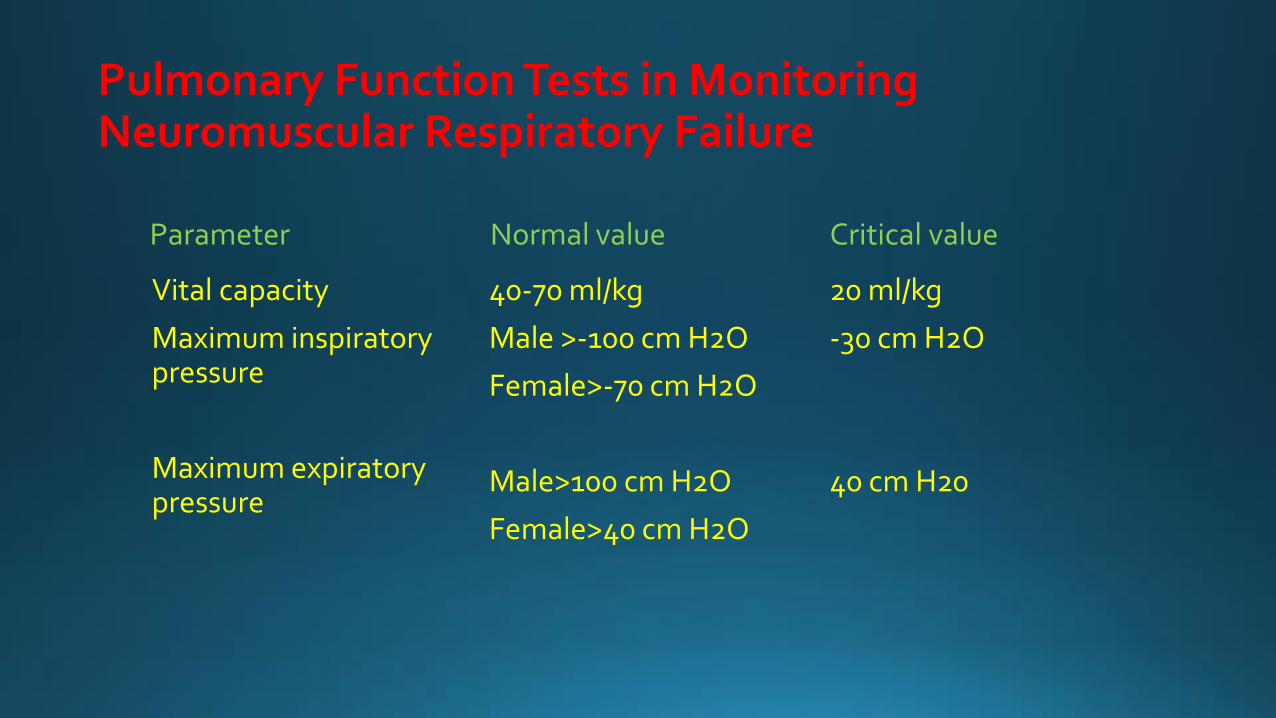
Pulmonary Function Tests in Monitoring Neuromuscular Respiratory Failure
Parameter
Vital capacity
Maximum inspiratory pressure
Maximum expiratory pressure
Normal value
40-70 ml/kg
Male >-100 cm H2O
Female>-70 cm H2O
Male>100 cm H2O
Female>40 cm H2O
Critical value
20 ml/kg
-30 cm H2O
40 cm H20