3-o-ethyl ascorbic acid a stable, vitamin c-derived agent...
TRANSCRIPT

!!!
P.O. Box 810472 • Dallas, Texas 75381• 972. 620. 9730 • Fax: 972. 421. 1860!
!Formulating !
3-O-Ethyl Ascorbic Acid: A Stable, Vitamin C-Derived Agent for Skin Whitening !Sep 1, 2013 | View online to Contact Author | By: Jill Hsu, Corum Inc. !Keywords: 3-O-ethyl ascorbic acid | whitening | tyrosinase | TRP-1 | TRP-2 | melanin biosynthesis A flawless complexion is without doubt a desirable attribute across many cultures. Especially in Asia, the obsession with whiter skin has greatly evolved, shifting from beauty to a sign of social class and representing sophistication, innocence, femininity and higher social stature. To achieve this lightening effect, product developers leverage active ingredients such as vitamin C, or L-ascorbic acid, which is not only a septicemia inhibitor, but also an effective reducing agent that can momentarily retard the melanin-biosynthesis pathway in skin. Vitamin C is also known to protect skin from ultraviolet B (UVB) damage and enhance collagen synthesis, therefore it is widely used in cosmetics as a whitening ingredient, especially in Asia. Due to its instability, however, several derivatives of it have been developed for cosmetic application; for example, ethyl ascorbic acid, magnesium L-ascorbyl-2-phosphate, L- ascorbic acid 2-glucoside and sodium ascorbyl phosphate. 3-O-Ethyl ascorbic acid, shown in Figure 1, is a derivative consisting of vitamin C with an ethyl groupa bound to the third carbon position. With its reducing ability, this structure helps to increase the stability of the compound when used in cosmetic products. The present article explores the function and efficacy of this material to determine its mechanism of action and potential for application against melanin biosynthesis.
Melanin Biosynthesis !
To understand the potential mechanisms of skin lightening, it is helpful to review the melanin biosynthesis process. In brief, after exposure to sunlight, melanin is produced by melanocytes through melanogensis, a biological process that helps to protect human DNA from harmful UV rays. The amount of melanin produced by melanocytes embedded in the basal layer of the epidermis defines the skin color, but it is interesting to note that all individuals from different countries and climates tend to have the same number of melanocytes. The melanin biosynthesis pathway is depicted in Figure 2. In this pathway, tyrosinase inhibition is the key to skin lightening and it is the first rate-limiting action that catalyzes the first two steps of melanin biosynthesis. During the first step, tyrosine is oxidized to dopa, which is then oxidized to dopaquinone via the action of tyrosinase.
Figure 1. Structure of 3-O-ethyl ascorbic acid 3-O-Ethyl ascorbic acid, shown in Figure 1, is a derivative consisting of vitamin C with an ethyl group bound to the third carbon position.
http://www.cosmeticsandtoiletries.com/formulating/function/active/premium-3-O-Ethyl-Ascorbic-Acid-A-Stable-Vitamin-C-Derived-Agent-for-Skin-Whitening-225540872.html?c=n !

!!!
P.O. Box 810472 • Dallas, Texas 75381• 972. 620. 9730 • Fax: 972. 421. 1860!
Moreover, tyrosinase accelerates the reaction of monomer aggregation, which leads to the production of eumelanin. Three enzymes are known to be involved in melanin biosynthesis in mammals—tyrosinase, tyrosinase-related protein-1 (TRP-1) and dopachrome tautomerase (DCT, also known as TRP-2) (see Figure 2).1 Tyrosinase is the focus of this review because it directly regulates the amount of melanin produced, whereas other enzymes modify the type of melanin synthesized.2
TRP-1 functions as a 5,6-dihydroxyindole-2-carboxylic acid (DHICA) oxidase in the melanogenic pathway. The catalytic activity of TRP-1 promotes the oxidation and polymerization of DHICA monomers into melanin.3 However, TRP-2 catalyzes the transformation of dopachrome to DHICA. TRP-2, similar to TRP-1, is considered a eumelanogenic enzyme that also stabilizes tyrosinase activity. Nevetheless, this enzyme is known to control the quantity and quality of melanin production.4 Thus, both TRP-1 and TRP-2 can act as enzymes modifying eumelanogenesis velocity, as regulators and stabilizers of the eumelanogenic apparatus in vivo, and perhaps as regulators of other melanocyte functions. Ultimately, a skin lightener would potentially downregulate TRP-1 and TRP-2 activity. !
Materials and Methods !
As described here, 3-O-ethyl ascorbic acid was first tested to determine its ability to inhibit tyrosinase and TRP-2, which are the important melanin-generating enzymes. It was then tested for melanin-inhibiting activity via in vitro and ex vivo studies. Clinical whitening efficacy on age spots and skin tone was measured by chromameter, and DNA protection and collagen synthesis abilities were assessed to support anti-aging claims. !
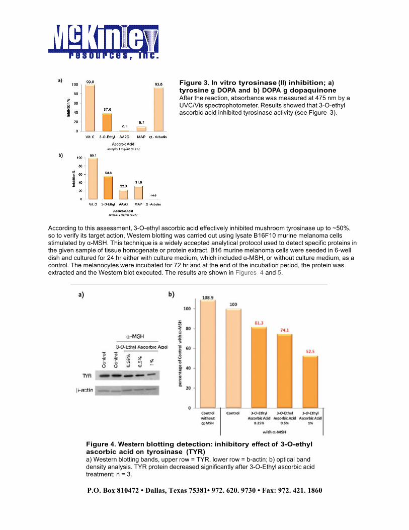
Tyrosinase inhibition: Mushroom tyrosinaseb has widely been used as the target enzyme for screening potential tyrosinase inhibitors. An in-house experiment measured the conversion of L-dopa, a substrate of tyrosinase, into dopaquinone, then monitored dopachrome’s absorption at 475 nm using a spectrometer. Being a colored substance, the dopachrome absorbs light at a wavelength of 475 nm, so a quantitative measurement of the time-related conversion and thus the activity of tyrosinase can be made. All samples were dissolved in dimethyl sulfoxide (DMSO) and mixed with tyrosinase substrate (L-DOPA or tyrosine) solution. Tyrosinase was then added to each sample, and the reaction was carried out for 30 min at 37°C. Simultaneously, a blank control tested only the tyrosinase substrate solution and DMSO without tyrosinase, under the same test conditions. After the reaction, absorbance was measured at 475 nm by a UVC/Vis spectrophotometer. Results showed that 3-O-ethyl ascorbic acid inhibited tyrosinase activity (see Figure 3).
Figure 2. Melanin biosynthesis schematic Three enzymes are known to be involved in melanin biosynthesis in mammals—tyrosinase, tyrosinase-related protein-1 (TRP-1) and dopachrome tautomerase (DCT, also known as TRP-2) (see Figure 2).1 !
!!!
P.O. Box 810472 • Dallas, Texas 75381• 972. 620. 9730 • Fax: 972. 421. 1860!
According to this assessment, 3-O-ethyl ascorbic acid effectively inhibited mushroom tyrosinase up to ~50%, so to verify its target action, Western blotting was carried out using lysate B16F10 murine melanoma cells stimulated by α-MSH. This technique is a widely accepted analytical protocol used to detect specific proteins in the given sample of tissue homogenate or protein extract. B16 murine melanoma cells were seeded in 6-well dish and cultured for 24 hr either with culture medium, which included α-MSH, or without culture medium, as a control. The melanocytes were incubated for 72 hr and at the end of the incubation period, the protein was extracted and the Western blot executed. The results are shown in Figures 4 and 5.
Figure 3. In vitro tyrosinase (II) inhibition; a) tyrosine g DOPA and b) DOPA g dopaquinone After the reaction, absorbance was measured at 475 nm by a UVC/Vis spectrophotometer. Results showed that 3-O-ethyl ascorbic acid inhibited tyrosinase activity (see Figure 3).
Figure 4. Western blotting detection: inhibitory effect of 3-O-ethyl ascorbic acid on tyrosinase (TYR) a) Western blotting bands, upper row = TYR, lower row = b-actin; b) optical band density analysis. TYR protein decreased significantly after 3-O-Ethyl ascorbic acid treatment; n = 3.

!!!
P.O. Box 810472 • Dallas, Texas 75381• 972. 620. 9730 • Fax: 972. 421. 1860!
When cells were stimulated by α-MSH, the protein expression of tyrosinase and TRP-2 increased significantly. β-Actin was used as an internal control and it showed no change in protein levels. Further, it was clearly observed that with the addition of 3-O-ethyl ascorbic acid, α-MSH-stimulated cells could inhibit the expression of tyrosinase and TRP-2 in a dose-dependent manner. In Figure 4, the x axis shows the test sample and the y axis shows tyrosinase expression; 3-O-ethyl ascorbic acid inhibited tyrosinase expression by 47.5%, in comparison with the control, which stimulated it by 100%. Moreover, as shown in Figure 5, 3-O-ethyl ascorbic acid presented an excellent dose-dependent inhibition on TRP-2 formation. On the other hand, TRP-1 expression showed no significant change in this experiment (see Figure 6).
Figure 6. Western blotting detection: inhibitory effect of 3-O-ethyl ascorbic acid on tryrosinase (TRP-1) On the other hand, TRP-1 expression showed no significant change in this experiment (see Figure 6).!
Figure 5. Western blotting detection: inhibitory effect of 3-O-ethyl ascorbic acid on tyrosinase (TRP-2) a) Western blotting bands, upper row = TRP-2, lower row = b-actin; b) optical band density analysis. TRP-2 protein decreased significantly after 3-O-ethyl ascorbic acid treatment; n = 4.!

!!!
P.O. Box 810472 • Dallas, Texas 75381• 972. 620. 9730 • Fax: 972. 421. 1860!
Melanin Reduction !
Although tyrosinase inhibitors can decrease melanogenesis by inhibiting the key enzyme, pre-existing melanin cannot be removed or eliminated via this pathway. Melanin-reduction tests are required to evaluate whether samples have the ability to reduce or destroy melanin and simulate age spot reduction. A melanin reduction/whitening study was thus conducted, by IDEA France, using theophylline, a molecule that accelerates the production of melanin. Melanocytes from the melanoms of mice were cultivated for 24 hr before treatment with test media at 37°C under 5% CO2 conditions. Their cellular morphology was checked under a microscope, and the media were discarded and replaced by 500 µL of the various concentrations of samples, or of the kojic acid control. These were incubated for 16 hr, again at 37°C and 5% CO2. !
After 16 hr, the media were again discarded, then 500 µL of theophyllin (0.5 mM) was added to the wells in order to stimulate melanin synthesis. The plates were incubated again for 48 hr, after which the culture media were recovered, the samples transferred to a 96-well microtiter plate, and melanin production assessed by a direct reading of the plate at 405 nm. Results showed 3-O-ethyl ascorbic acid exhibited effective whitening, observed both at 15 mg/mL and 20 mg/mL concentrations (see Figure 7).
Ex-vivo assay: In relation, another study was carried out, by BioInnovation Laboratories, Inc., to assess the potential of the material to induce changes in tissue pigmentation. An in vitro tissue model of the human epidermisc prepared from cultured human keratinocytes and melanocytes was employed. The tissue was stored at 4°C until used. Upon use, the tissue was removed from the agarose-shipping tray and placed into a 6-well plate containing 1.0 mL of assay medium (37±2°C). All of the agarose was removed from the outside of the tissue culture insert since any residual agarose could prevent the assay medium from reaching the tissue. A 50-µL sample of the test material was then applied to the surface of the tissue, which was incubated at 37±2°C and 5±1% CO2. !
Every 24 hr, the tissue was rinsed with PBS, fresh test material was applied, and the media was changed. The treatments were carried out for 9 days. Two sets of tissues were prepared for this study. One set was used to determine changes in melanin content while the second was used assess changes in pigmentation via histology. The results are presented in Figure 8. The black area shown is melanin from melanocytes; as can be seen, after 9 days, a 3% concentration of 3-O-ethyl ascorbic acid was found to have excellent skin brightening activity, in comparison with day 1.
Figure 7. In vitro whitening activity study: melanin assessment Results showed 3-O-ethyl ascorbic acid exhibited effective whitening, observed both at 15 mg/mL and 20 mg/mL concentrations (see Figure 7).!

!!!
P.O. Box 810472 • Dallas, Texas 75381• 972. 620. 9730 • Fax: 972. 421. 1860!
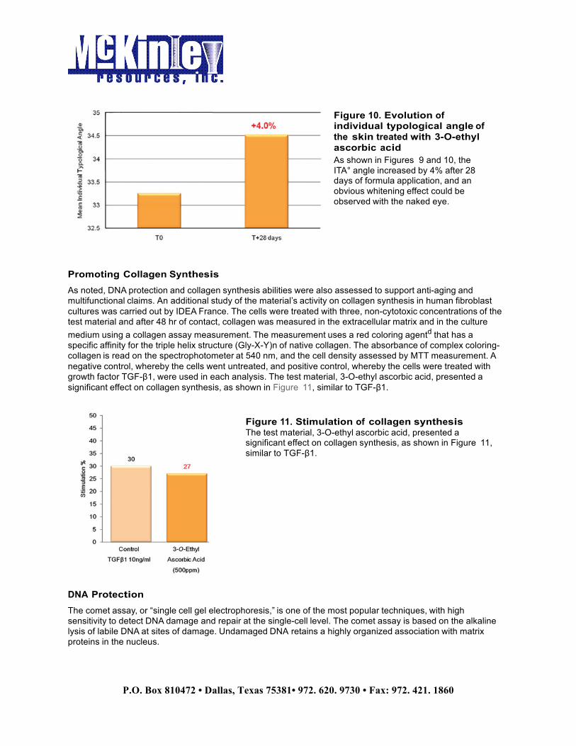
Clinical efficacy: Further, a clinical skin lightening test was carried out by Spincontrol France using a test formulation containing 2% 3-O-ethyl ascorbic acid, which was applied twice daily for 28 days on 20 healthy Asian females, ages 25-40, having skin type III. Skin colorimetric measurements was taken using a chromameter, which converts colors perceived by the human eye to a digital code composed of L*, a* and b* values. These two last parameters are characterized by chromatic axes, such as green to red, and blue to yellow. Both parameters are exploited through the calculation of the Individual Typological Angle (ITA°), which defines the skin pigmentation degree of a subject taking into account the skin clarity (L*) and the melanin parameter (b*). The higher the ITA°, the lighter the skin. As shown in Figures 9 and 10, the ITA° angle increased by 4% after 28 days of formula application, and an obvious whitening effect could be observed with the naked eye. Overall, this clinical study confirmed the ability of the material to improve skin radiance via delivery by a cosmetic product. !!!
Figure 8. The result of ex vivo melanin assay for nine days; untreated (left) vs. treated (right) One set was used to determine changes in melanin content while the second was used assess changes in pigmentation via histology. The results are presented in Figure 8.!
Figure 9. In vivo evaluation of the efficacy of one whitening lotion in healthy Asian subjects by chromatography As shown in Figures 9 and 10, the ITA° angle increased by 4% after 28 days of formula application, and an obvious whitening effect could be observed with the naked eye. !
!!!
P.O. Box 810472 • Dallas, Texas 75381• 972. 620. 9730 • Fax: 972. 421. 1860!
Promoting Collagen Synthesis !
As noted, DNA protection and collagen synthesis abilities were also assessed to support anti-aging and multifunctional claims. An additional study of the material’s activity on collagen synthesis in human fibroblast cultures was carried out by IDEA France. The cells were treated with three, non-cytotoxic concentrations of the test material and after 48 hr of contact, collagen was measured in the extracellular matrix and in the culture medium using a collagen assay measurement. The measurement uses a red coloring agentd that has a specific affinity for the triple helix structure (Gly-X-Y)n of native collagen. The absorbance of complex coloring-collagen is read on the spectrophotometer at 540 nm, and the cell density assessed by MTT measurement. A negative control, whereby the cells went untreated, and positive control, whereby the cells were treated with growth factor TGF-β1, were used in each analysis. The test material, 3-O-ethyl ascorbic acid, presented a significant effect on collagen synthesis, as shown in Figure 11, similar to TGF-β1. !
DNA Protection !
The comet assay, or “single cell gel electrophoresis,” is one of the most popular techniques, with high sensitivity to detect DNA damage and repair at the single-cell level. The comet assay is based on the alkaline lysis of labile DNA at sites of damage. Undamaged DNA retains a highly organized association with matrix proteins in the nucleus.
Figure 10. Evolution of individual typological angle of the skin treated with 3-O-ethyl ascorbic acid As shown in Figures 9 and 10, the ITA° angle increased by 4% after 28 days of formula application, and an obvious whitening effect could be observed with the naked eye. !
Figure 11. Stimulation of collagen synthesis The test material, 3-O-ethyl ascorbic acid, presented a significant effect on collagen synthesis, as shown in Figure 11, similar to TGF-β1.!

!!!
P.O. Box 810472 • Dallas, Texas 75381• 972. 620. 9730 • Fax: 972. 421. 1860!
When damaged, this organization is disrupted. The individual strands of DNA lose their compact structure and relax, expanding out of the cavity into the agarose. The intensity of the comet tail relative to the head reflects the number of DNA breaks. As a result, damaged DNA has a longer tail, as shown in Figure 12. When treated with the 3-O-ethyl ascorbic acid vitamin C derivative for 24 hr, however (see Figure 12), the DNA of human fibroblast cells (HS-68) was protected. !
Stability Testing !
In addition to being effective, 3-O-ethyl ascorbic acid showed excellent stability. The 2% aqueous 3-O-ethyl ascorbic acid samples were retained for 1 month in a 45°C oven to assess any color change and compared with other vitamin C derivatives. The absence of color change indicated the sample could tolerate the high temperature and remain stable. As shown in Figure 13, 3-O-ethyl ascorbic acid was more stable than the others, and its color remained unchanged. Further, its purity also was analyzed vie HPLC, and as Figure 14 shows, the purity of 3-O-ethyl ascorbic acid crystalline powder remained stable at 45˚C for 60 days.
Figure 12. Comet assay shows: a) the appearance of undamaged/damaged DNA by electrophoresis, and b) the result of DNA protection As a result, damaged DNA has a longer tail, as shown in Figure 12. !
Figure 13. Heat stability As shown in Figure 13, 3-O-ethyl ascorbic acid was more stable than the others, and its color remained unchanged.!

!!!
P.O. Box 810472 • Dallas, Texas 75381• 972. 620. 9730 • Fax: 972. 421. 1860!
!
Conclusions !
3-O-Ethyl ascorbic acid was shown to pinpoint specified targets in the melanin biosynthesis process, suggesting it could reduce the expression of tyrosinase and TRP-2 at the protein level. This slowed eumelanogenesis velocity and overall, reduced melanin production. Conclusively, 3-O-ethyl ascorbic acid is an effective melanin inhibitor, reducing tyrosinase and TRP-2 protein expression, stimulating collagen synthesis, protecting DNA and lightening skin, all while exhibiting excellent stability. References 1. T Kobayashi et al, Tyrosinase related protein 1 (TRP1) functions as a DHICA oxidase in melanin biosynthesis, J EMBO 13 5818–25 (1994) 2. H Ando, H Kondoh, M Ichihashi and VJ Hearing, Approaches to identify inhibitors of melanin biosynthesis via the quality control of tyrosinase, Soc Invest Derm 127, 751–761 (2007) 3. T Kobayashi, G Imokawa, DC Bennett, and VJ Hearing, Tyrosinase stabilization by Tyrp1 (the brown locus protein), J Biologic Chem 273(48) 31801–31805 (1998) 4. K Tsukamoto, IJ Jackson, K Urabe, PM Montague and VJ Hearing, A second tyrosinase-related protein, TRP-2 is a melanogenic enzyme termed dopachrome tautomerase, J EMBO 11 519–526 (1992)!
! Footnotes !!
Footnotes (CT1309 Hsu) !a Corum 9515 (INCI: 3-O-Ethyl Ascorbic Acid) is a product of Corum, Inc., www.corum.com.tw. b Mushroom Tyrosinase T3824 is a product of Sigma, www.sigmaaldrich.com. c MelanoDerm tissues are manufactured by MatTek Corp., www.mattek.com. d Sirius Direct Red 80 is a product of Sigma-Aldrich, www.sigma-aldrich.com.
!
Copyright © 2014 Allured Business Media.
Figure 14. Purity analysis Further, its purity also was analyzed vie HPLC, and as Figure 14 shows, the purity of 3-O-ethyl ascorbic acid crystalline powder remained stable at 45˚C for 60 days.!