32nd annual combined orthopaedic spring … annual combined orthopaedic spring symposium friday, ......
TRANSCRIPT

32nd AnnualCombined Orthopaedic
Spring Symposium
April 7-8, 2017Hawaii Prince Hotel - Honolulu

Welcome from HOA President
2007 HOA Officers
Aloha & Welcome to the 32nd Annual Combined OrthopaedicSpring Symposium! Months of effort have gone into planning
this signature event, at which attendees can learn about thelatest advances in orthopaedic surgery from nationallyrenowned experts in their fields. The symposium provides a
constructive forum for discussions among HOA members,residents, medical students and allied health professionals fromacross our state. It also features the research being conducted
by University of Hawaii and Tripler Army Medical Centerresidents. And, it provides HOA members with a wonderfulopportunity to network with fellow specialists during the
sessions and at the awards banquet. This is truly a greatopportunity to gain knowledge and earn CME credit,participate in discussions and catch-up with our local
orthopaedic ohana over the course of just a few days. Mahalofor joining us!
Wei Chin Chen, MDHOA President & Symposium Chair
HOA Membership InformationContact HOA Executive Director Cathy Iwai at 808-673-0234 or [email protected] if
you are interested in becoming a member of the Hawaii Orthopaedic Association.
Hawaii Orthopaedic AssociationP.O. Box 61207Honolulu, HI 96839Fax: 808-956-1315
Americans with Disability Act (ADA)Participants with special needs should contact Cathy Iwai at 808-630-1586 or [email protected] todiscuss desired accommodation(s).
PresidentWei Chin Chen, MD
Vice-PresidentJoseph Varcadipane, MD
Secretary/TreasurerPaul Ryan, MD
Immediate Past PresidentDarren Egami, MD
Board of CouncilorsJerry Van Meter, MD
Executive DirectorCathy Iwai
2017 HOAOfficers
1
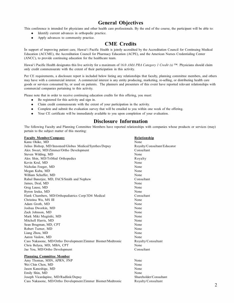
CME CreditsIn support of improving patient care, Hawai‘i Pacific Health is jointly accredited by the Accreditation Council for Continuing MedicalEducation (ACCME), the Accreditation Council for Pharmacy Education (ACPE), and the American Nurses Credentialing Center(ANCC), to provide continuing education for the healthcare team.
Hawai‘i Pacific Health designates this live activity for a maximum of 16.0 AMA PRA Category 1 Credit (s) ™. Physicians should claimonly credit commensurate with the extent of their participation in this activity.
Per CE requirements, a disclosure report is included below listing any relationships that faculty, planning committee members, and othersmay have with a commercial interest. A commercial interest is any entity producing, marketing, re-selling, or distributing health caregoods or services consumed by, or used on patients. The planners and presenters of this event have reported relevant relationships withcommercial companies pertaining to this activity.
Please note that in order to receive continuing education credits for this offering, you must: Be registered for this activity and sign in. Claim credit commensurate with the extent of your participation in the activity. Complete and submit the evaluation survey that will be emailed to you within one week of the offering. Your CE certificate will be immediately available to you upon completion of your evaluation.
Disclosure InformationThe following Faculty and Planning Committee Members have reported relationships with companies whose products or services (may)pertain to the subject matter of this meeting:
Faculty Member/Company RelationshipKanu Okike, MD NoneJulius Bishop, MD/Innomed/Globus Medical/Synthes/Depuy Royalty/Consultant/EducatorAlex Sweet, MD/Zimmer/Ortho Development ConsultantSteven Wilding, MD NoneAlex Shin, MD/TriMed Orthopedics RoyaltyKevin Krul, MD NoneNicholas Foeger, MD NoneMegan Kuba, MD NoneWilliam Schaffer, MD NoneRahul Banerjee, MD, FACS/Smith and Nephew ConsultantJames, Deal, MD NoneGreg Lause, MD NoneByron Izuka, MD NoneHank Chambers, MD/Orthopediatrics Corp/3D4 Medical ConsultantChristina Wu, MS III NoneAdam Groth, MD NoneJoshua Dworkin, MD NoneZack Johnson, MD NoneMark Miki Mugiishi, MD NoneMitchell Harris, MD NoneSean Brugman, MD, CPT NoneRobert Turner, MD NoneLiang Zhou, MD NoneAaron Vaslow, MD NoneCass Nakasone, MD/Ortho Development/Zimmer Biomet/Medtronic Royalty/ConsultantChris Belyea, MD, MBA, CPT NoneJae You, MD/Ortho Development Consultant
Planning Committee MemberAmy Thomas, MSN, APRN, FNP NoneWei Chin Chen, MD NoneJason Kaneshige, MD NoneEmily Shin, MD NoneJoseph Vicardapine, MD/Radlink/Depuy Shareholder/ConsultantCass Nakasone, MD/Ortho Development/Zimmer Biomet/Medtronic Royalty/Consultant
General ObjectivesThis conference is intended for physicians and other health care professionals. By the end of the course, the participant will be able to:
Identify current advances in orthopedic practice. Apply advances to community practice.
2

AcknowledgementsThank you for the Support of all of Our Exhibitors...
All Island Surgical
Automated Healthcare Solutions
Carbofix
Depuy Synthes
DJO Global
Ferring Pharmaceuticals
Halyard
Medical Innovative Technologies
Special Thanks to...
Laura Abby Reed & Amy Thomas @ Hawaii Pacific Health
Shriners Hospital for Children - Honolulu
Tripler Army Medical Center Orthopaedic Residency Program
University of Hawaii Orthopaedic Residency Program
...and a Big Mahalo to...
HOA Executive Director Cathy Iwai for all of your work
in overseeing another successful year!
Mid Pac Medical
Smith & Nephew Inc.
Sportstek Medical
Stryker Sports Medicine
TeDan Surgical Innovations
Vericel
Wright Medical Group
Zimmer Biomet
Best Resident Paper AwardsRichardson Awards: The Richardson Fund was established in 1982 to honor the memory of B. AllenRichardson, MD. Dr. Richardson was one of the first Board-Certified Orthopaedic Surgeons in Honolulu,where he practiced for nearly 30 years. He was an active member of the teaching staff of the University ofHawaii Orthopaedic Residency Training Program from its inception in the mid-1960s, and was a staunchsupporter for the creation of the John A. Burns School of Medicine. The proceeds of the Richardson Fund areused to award first, second and third place prizes for the best resident papers presented at the Annual Com-bined Orthopaedic Spring Symposium.
Shriners Award: The Shriners Award is presented annually and was established to honor an orthopaedicresident who has completed a rotation at the Shriners Hospital for Children in Honolulu. Residents present theircompleted papers to medical staff and allied health professionals at the Shriners Hospital's patient care confer-ence. The paper must be written to meet standards for publishing in clinical publications.
3

32nd Annual Combined Orthopaedic Spring SymposiumFriday, April 7, 2017
6:00 Registration / Continental Breakfast / Exhibits6:30 Welcome & Opening Remarks - WeiChin Chen, MD6:45 Introduction of Julius Bishop - Kanu Okike, MD
Julius Bishop, MD - Tips and Tricks for Optimizing Resident Education7:30 Alex Sweet, MD - Orthopaedic Injuries in Hawaii Shark Attack Victims
Steven Wilding, MD - Are Surgical Skills Under-Emphasized in Disaster Response Literature?7:45 Kanu Okike, MD - Single-Blind vs. Double-Blind Peer Review in the Setting of Author Prestige8:30 Break & Visit Exhibits8:45 Introduction of Alex Shin - Emily Shin, MD
Alex Shin MD - Current Concepts of Treatment of Scaphoid Nonunions9:30 Mariya Opanova, MD - Anatomic Relationship Between the Dorsal Intercarpal Ligament
and Scaphoid RidgeKevin Krul, MD - Should Arthroscopic Treatment of Occult Dorsal Wrist Ganglions be theGold Standard?Nicholas Foeger, MD - The Effects of Aging and Diabetes Mellitus on Hand SomatosensationMegan Kuba, MD - Anatomic Anterior Proximal Ulnar Angle Differences Based on Arm Positionand a Case Report of its Use
10:00 Break & Visit Exhibits10:15 AAOS Representative Talk on MACRA/MIPS - William Shaffer, MD11:00 Introduction to Rahul Banerjee - Jason Kaneshige, MD
Rahul Banerjee, MD - Periprosthetic Femur Fractures11:45 James Deal, MD - The Incidence of Lumbar Disc Herniation in Military Helicopter Pilots
vs.Matched Controls Over a 10-Year PeriodGreg Lause, MD - The Impact of Implant Density on Curve Correction for Large StiffIdiopathic Curves
12:00 Lunch12:15 HMSA Insurance Talk - Mark Mugiishi, MD1:00 Introduction to Hank Chambers - Byron Izuka, MD
Hank Chambers, MD - Treatment of ACLs and OCDs in Children and Adolescents1:45 Byron Izuka, MD & Christina Wu MS III - Use of Live Video Recordings in the Outpatient
Setting: A Study Update and Final Results2:15 Break & Visit Exhibits2:30 Introduction to Adam Groth - WeiChin Chen, MD
Adam Groth, MD - Total Ankle Arthroplasty: A Lateral Approach3:30 Joshua Dworkin, MD - Occupational Outcomes of the Modified Broström Procedure:
A Retrospective ReviewZack Johnson, MD - Comparison of MR Imaging and Stress Radiographs in the Evaluationof Chronic Lateral Ankle Instability
3:45 Break & Visit Exhibits4:00 Alex Shin, MD - Distal Radius Fractures: A Personal Journey Over 20 Years4:45 Rahul Banerjee, MD - Orthopedic Trauma Tips5:15 Adjournment & Reception to follow
4

32nd Annual Combined Orthopaedic Spring SymposiumSaturday, April 8, 2017
6:00 Registration / Continental Breakfast / Exhibits
6:30 Appreciation for Guest Speakers
6:45 Julius Bishop, MD - Tibial Plateau Fractures: Best Practices for 2017
7:15 Rahul Banerjee, MD - Acetabular Fractures in the Elderly
8:00 Break & Visit Exhibits
8:15 Shawn Brugman, MD - Rotator Cuff Repairs in Active Patients 40 Years and Younger
Robert Turner, MD - Return to Duty After Multiligamentous Knee Injuries Active Duty Members
Liang Zhou, MD - A Comparison of 'On-Track' and 'Off-Track' Assessment With Clinical Failure
in a 13-year Follow-Up of Open vs. Arthroscopic Shoulder Stabilizations
Mitchell Harris, MD - Prospective Evaluation of Acute ACL Reconstructions Using Patellar
Tendon Autografts
Aaron Vaslow, MD - Smoking: A Prognostic Indicator for Bankart Repair
9:00 Introduction to Cass Nakasone - Joe Vicardapine, MD
Cass Nakasone, MD - Joint Arthroplasty Talk
9:45 Chris Belyea, MD - Early Outcomes of Total Hip Arthroplasty in an Adolescent Population
Jae You, MD - Addressing Large Tibial Osseous Defects in Primary TKA Using Trabecular
Metal Cones
10:15 Hank Chambers, MD - PRiSM: Pediatric Research in Sports Medicine
11:00 Adam Groth, MD - Osteobiologics in Foot and Ankle Reconstruction
11:30 Alex Shin, MD - Adult Traumatic Brachial Plexus Injuries: An Overview of Concepts
and Treatment Options
12:15 Lunch
1:00 Julius Bishop, MD - The Syndesmosis: Not as Simple as You Thought
1:45 Hank Chambers, MD - Cerebral Palsy: A View From Both Sides
2:30 Adam Groth, MD - Barefoot Running: Myths and Controversies
3:15 Adjournment & Banquet to follow
5
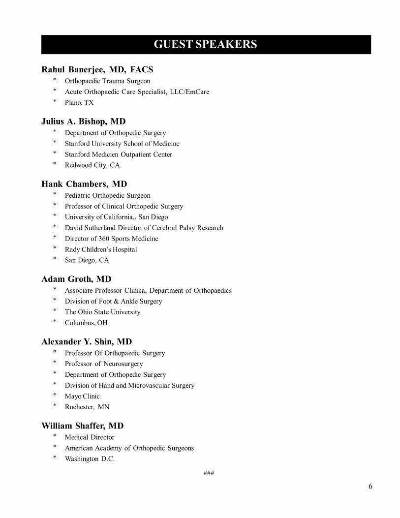
Rahul Banerjee, MD, FACS* Orthopaedic Trauma Surgeon* Acute Orthopaedic Care Specialist, LLC/EmCare* Plano, TX
Julius A. Bishop, MD* Department of Orthopedic Surgery* Stanford University School of Medicine* Stanford Medicien Outpatient Center* Redwood City, CA
Hank Chambers, MD* Pediatric Orthopedic Surgeon* Professor of Clinical Orthopedic Surgery* University of California,, San Diego* David Sutherland Director of Cerebral Palsy Research* Director of 360 Sports Medicine* Rady Children’s Hospital* San Diego, CA
Adam Groth, MD* Associate Professor Clinica, Department of Orthopaedics* Division of Foot & Ankle Surgery* The Ohio State University* Columbus, OH
Alexander Y. Shin, MD* Professor Of Orthopaedic Surgery* Professor of Neurosurgery* Department of Orthopedic Surgery* Division of Hand and Microvascular Surgery* Mayo Clinic* Rochester, MN
William Shaffer, MD* Medical Director* American Academy of Orthopedic Surgeons* Washington D.C.
###
6
GUEST SPEAKERS

Sy
mp
os
ium
Ab
str
ac
ts -
Fri
da
y,
Apr
il 7
Tips and Tricks for Optimizing Resident Education
Julius Bishop, MD
Abstract available upon request
7###

Sy
mp
os
ium
Ab
str
ac
ts -
Fri
da
y,
Apr
il 7
8
Orthopaedic Injuries in Hawaii Shark Attack Victims
Sweet, R. Opanova, M. Murray, P. Moroz, P. OBJECTIVE: To review of all shark attacks in the state of Hawaii for the past 10 years (7/1/06 to7/1/16). The aim was be to explore the severity and types of injuries associated with the nativeshark population of Hawaii. METHODS: This study involves a case series consisting of a retrospective medical chart review ofpatients with a shark related injury. Patient names were obtained from the Hawaii shark incidentslist at http://dlnr.hawaii.gov/sharks/shark-incidents/incidents-list/ and patient’s charts found by thename and age, at the treating medical center. The names of the victims that are withheld from thepublic website will be obtained directly from the Division of Aquatic Resources. Inclusion criteriawas any patient who presented with a shark related injury. There were no exclusion criteria, alllisted shark attack patients were included.
RESULTS: We have IRB approval for Queens Hospital but are still awaiting final approval fromthe Western IRB to access Maui Memorial, Kaiser, and HPH hospital records. The addition ofthese hospital records will greatly strengthen our study. DISCUSSION: Not yet finalized CONCLUSION: Not yet finalized.
###
Sy
mp
os
ium
Ab
str
ac
ts -
Fri
da
y,
Apr
il 7
9
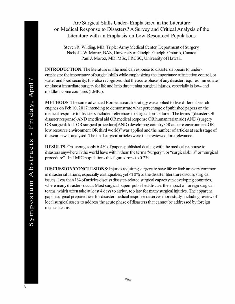
Are Surgical Skills Under- Emphasized in the Literatureon Medical Response to Disasters? A Survey and Critical Analysis of the
Literature with an Emphasis on Low-Resourced Populations
Steven R. Wilding, MD. Tripler Army Medical Center, Department of Surgery.Nicholas W. Moroz, BAS, University of Guelph, Guelph, Ontario, Canada
Paul J. Moroz, MD, MSc, FRCSC, University of Hawaii.
INTRODUCTION: The literature on the medical response to disasters appears to under-emphasize the importance of surgical skills while emphasizing the importance of infection control, orwater and food security. It is also recognized that the acute phase of any disaster requires immediateor almost immediate surgery for life and limb threatening surgical injuries, especially in low- andmiddle-income countries (LMIC).
METHODS: The same advanced Boolean search strategy was applied to five different searchengines on Feb 10, 2017 intending to demonstrate what percentage of published papers on themedical response to disasters included references to surgical procedures. The terms “(disaster ORdisaster response) AND (medical aid OR medical response OR humanitarian aid) AND (surgeryOR surgical skills OR surgical procedure) AND (developing country OR austere environment ORlow resource environment OR third world)” was applied and the number of articles at each stage ofthe search was analysed. The final surgical articles were then reviewed fore relevance.
RESULTS: On average only 6.4% of papers published dealing with the medical response todisasters anywhere in the world have within them the terms “surgery”, or “surgical skills” or “surgicalprocedure”. In LMIC populations this figure drops to 0.2%.
DISCUSSION/CONCLUSIONS: Injuries requiring surgery to save life or limb are very commonin disaster situations, especially earthquakes, yet <10% of the disaster literature discuss surgicalissues. Less than 1% of articles discuss disaster-related surgical capacity in developing countries,where many disasters occur. Most surgical papers published discuss the impact of foreign surgicalteams, which often take at least 4 days to arrive, too late for many surgical injuries. The apparentgap in surgical preparedness for disaster medical response deserves more study, including review oflocal surgical assets to address the acute phase of disasters that cannot be addressed by foreignmedical teams.
###
Sy
mp
os
ium
Ab
str
ac
ts -
Fri
da
y,
Apr
il 7
10
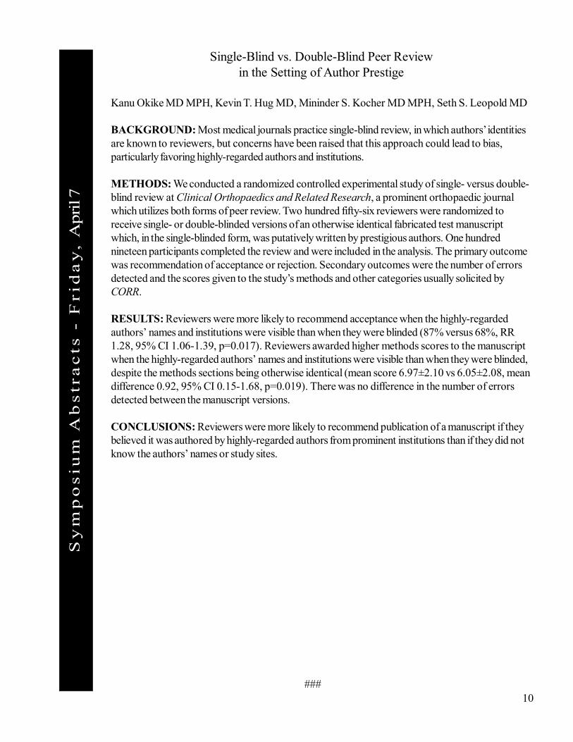
Single-Blind vs. Double-Blind Peer Reviewin the Setting of Author Prestige
Kanu Okike MD MPH, Kevin T. Hug MD, Mininder S. Kocher MD MPH, Seth S. Leopold MD
BACKGROUND: Most medical journals practice single-blind review, in which authors’ identitiesare known to reviewers, but concerns have been raised that this approach could lead to bias,particularly favoring highly-regarded authors and institutions.
METHODS: We conducted a randomized controlled experimental study of single- versus double-blind review at Clinical Orthopaedics and Related Research, a prominent orthopaedic journalwhich utilizes both forms of peer review. Two hundred fifty-six reviewers were randomized toreceive single- or double-blinded versions of an otherwise identical fabricated test manuscriptwhich, in the single-blinded form, was putatively written by prestigious authors. One hundrednineteen participants completed the review and were included in the analysis. The primary outcomewas recommendation of acceptance or rejection. Secondary outcomes were the number of errorsdetected and the scores given to the study’s methods and other categories usually solicited byCORR.
RESULTS: Reviewers were more likely to recommend acceptance when the highly-regardedauthors’ names and institutions were visible than when they were blinded (87% versus 68%, RR1.28, 95% CI 1.06-1.39, p=0.017). Reviewers awarded higher methods scores to the manuscriptwhen the highly-regarded authors’ names and institutions were visible than when they were blinded,despite the methods sections being otherwise identical (mean score 6.97±2.10 vs 6.05±2.08, meandifference 0.92, 95% CI 0.15-1.68, p=0.019). There was no difference in the number of errorsdetected between the manuscript versions.
CONCLUSIONS: Reviewers were more likely to recommend publication of a manuscript if theybelieved it was authored by highly-regarded authors from prominent institutions than if they did notknow the authors’ names or study sites.
###
Sy
mp
os
ium
Ab
str
ac
ts -
Fri
da
y,
Apr
il 7
11
Current Concepts of Treatment of Scaphoid Nonunions
Alex Shin MD
Abstract available upon request
###

Sy
mp
os
ium
Ab
str
ac
ts -
Fri
da
y,
Apr
il 7
###12
Scapholunate Instability, Anatomic Study
Opanova, M, Foeger, N, Dupaix, J and Medoff, R
OBJECTIVE: Explore the anatomic relationship of the dorsal scaphotriquetral (ST) ligament andthe scapholunate (SL)interosseous ligaments. Suggest that these dorsal structures have a key role inguiding the capitate via a direct cam effect during wrist movement.
METHODS: Cadaver wrists were dissected using a dorsal approach, and the superficialretinaculum, extensor tendons and synovial tissue over the dorsal capsule reflected. An incision wasmade along the distal margin of the dorsal intercarpal ligament, leaving its radial attachment on thescaphoid and its ulnar attachment on the triquetrum intact. The scaphoid, triquetrum, and lunatewere dissected en bloc and from a volar perspective, attachments between the dorsal scapholunateinterosseous ligament and the dorsal scaphotriquetral ligament were photographed and measured.
RESULTS: Total of 5 cadaver wrists were dissected, 2 female and 3 male. One of the malespecimens had a disrupted scapholunate ligament and was excluded from the study. The averageage of the cadavers was 73 years. Digital calipers were used to get 3 measurements for eachdimension, the table below represents the average for each specimen.
DISCUSSION: During our dissection we visualized the dorsal structures of the proximal carpalrow in particular the ST ligament, the dorsal ridge of the scaphoid and the dorsal part of the SLligament. These structures appear to provide a significant amount of support to the capitate and arelikely to contribute to the motion of the capitate during wrist flexion and extension. Dividing the STligament during the dorsal approach to the wrist may destabilize this mechanical support andcontribute to wrist instability seen in SL injuries. One major limitation to our study is the limitednumber of specimens as well as the relative older age of the cadavers. Another limitation is therelative difficulty of measuring soft tissues using calipers. We attempted to minimize this by havingonly one person measure the specimens and repeat those measurements 3 times.Further studies are necessary to determine the mechanical role of the ST ligament and the scaphoidridge in directing capitate motion.
CONCLUSION: Scapholunate instability is a complex problem that can result in prolongedmorbidity and permanent impairment of function. Addressing SL instability surgically is complex andnot always successful. One aspect that may contribute is the incomplete restoration of the dorsalstructures which could play an important role in directing capitate movement. In particular disruptingthe ST ligament attachment and failing to repair it could result in alteration of normal wristbiomechanics. Further studies are necessary to clarify the importance of the dorsal structures but wesuggest that preserving the ST ligament when possible may help with restoring normal wristbiomechanics and avoiding future instability.
Sy
mp
os
ium
Ab
str
ac
ts -
Fri
da
y,
Apr
il 7
###13
Should Arthroscopic Treatment of Occult Dorsal Wrist Ganglionsbe the Gold Standard?
Major Kevin P. Krul, M.D.; Lieutenant Colonel Rey D. Gumboc M.D.Tripler Army Medical Center Department of Orthopaedics
INTRODUCTION: Originally described in 1999 through open exploration, occult dorsal wristganglions are one of the multiple causes of dorsal wrist pain over the scapholunate interval. With theincreased availability of MRI, the diagnosis is becoming more common and presents a challenge toboth the generalist and the specialty trained hand surgeon.
METHODS: A retrospective review was conducted using an automated search of electronicpatient medical records from March 2012 through March 2015 for patients who underwent wristarthroscopy for occult dorsal wrist ganglions. Patients were excluded who had other primarydiagnoses, or who had radiographic evidence of scapholunate instability.
RESULTS: 20 operative cases met the inclusion criteria in 19 patients. All patients had dorsal andradial sided wrist pain, and MRI’s that demonstrated the occult ganglion. Of these 20 cases, 14(70%) had instability noted from the midcarpal row. The grades of instability ranged from Geissler1-3. In 12 of the 14 cases of instability, (85%) the surgical plan was changed to address thescapholunate instability.
DISCUSSION: While numerous studies have evaluated the prevalence of scapholunate instabilitywith dorsal wrist ganglions, this is the first to report on instability in occult dorsal wrist ganglions. Incontrast to non-occult ganglions, where the association with scapholunate instability has ranged from0-16%, our incidence in occult ganglions is much higher. Due to the difficulty in assessing low gradeand pre-dynamic scapholunate instability in the open surgery for an occult ganglion, there is a strongrecommendation for primary arthroscopic treatment.
Sy
mp
os
ium
Ab
str
ac
ts -
Fri
da
y,
Apr
il 7
14
Effects of Aging and Diabetes Mellitus on Hand Somatosensation
Nicholas Foeger, MD, PhD; Edward Chan, MD; Robert Atkinson, MD
Functional use and protective sensation in the hand are critical to maintaining independence for theelderly patient. Previous studies have suggested that a natural decline in both motor andsomatosensory hand function occurs with aging. Importantly, these decrements correlate not onlywith other somatosensory parameters including vibration thresholds and pain/temperature sensation,but also with a decline in motor and functional performance of the hand. Reductions in handsomatosensation, therefore, herald the reduced ability for elderly patients to live autonomously.Somatosensory deficits are also a component of diabetic polyneuropathy. A multifactorial disease,diabetes is a modern medical epidemic that disproportionately affects the elderly and patients ofPacific Island descent. The interplay between natural aging and diabetes in the decline of fingertipsensation, however, remains unclear. Additionally, a paucity of data exists for elderly patients ofPacific Island descent as a majority of studies were conducted on homogenously Caucasian patientpopulations from either Europe or North America. We conducted a cross-sectional cohort study,therefore, to characterize the interplay between the effects of physiologic aging and diabetes onhand somatosensation.
Research subjects are elderly patients (aged 60-89), with and without diabetes, recruited from thepractice of Dr. Robert Atkinson. Patients were excluded if they had a history of any hand conditionthat specifically contains a neuropathic component or a history of any previous surgery on theirhands which could conceivably result in nerve injury. Static and moving two-point discrimination, aswell as Semmes-Weinstein monofilament sensation were determined for all ten digits by certifiedhand therapists and demographic data for each patient were recorded. Patients with diabetesmellitus were queried regarding their most recent HgbA1c value. Continued collection of patientdata remains ongoing.
###

Sy
mp
os
ium
Ab
str
ac
ts -
Fri
da
y,
Apr
il 7
15
Anatomic Anterior Proximal Ulnar Angle DifferencesBased on Arm Position and A Case Report of Its Use
Megan H.M. Kuba, MD, Byron H. Izuka, MD, Ian G. Hasegawa, MD
PURPOSE: The goals of this study are three-fold: (i) describe a novel angle, the “anatomic anteriorproximal ulna angle” (aAPUA), for detecting and quantifying proximal ulnar malalignment in thesagittal plane, (ii) determine if the aAPUA is altered by differing positions of the arm duringradiographic examination, and (iii) to demonstrate its use in a case report.
METHODS: The aAPUA is measured on a true lateral radiograph of the elbow. The first lineconnects a point from the most anterior/proximal tip of the olecranon to a point at the tip of thecoronoid process. The second line connects a mid-metaphyseal point to a mid-diaphyseal point ofthe proximal ulna. Four cadaveric specimens were radiographically examined and the aAPUAmeasured and compared in 10 different positions: a true lateral view of the elbow and at 10°, 20°and 30° each of relative shoulder adduction, external rotation and internal rotation.
RESULTS: The aAPUA ranged from 22-32° on a true lateral view of the elbow. With increasingdegrees of relative shoulder rotation the angle did change though no consistent pattern was seen forany direction of rotation tested.
CONCLUSION: The aAPUA defines the sagittal alignment of the proximal ulna and is bestdetermined on a true lateral view as even small changes in position of the arm may alter the angle.Comparison films to the contralateral normal elbow should be made to determine the exact anglefor each individual patient.
SIGNIFICANCE: The aAPUA was used to detect and quantify a subtle residual proximal ulnarmalalignment of a Monteggia fracture-dislocation that previously underwent surgical correction withseemingly appropriate reduction. This information aided both the surgical planning prior to and theintraoperative assessment during the revision surgery. The aAPUA should be a part of theorthopaedic surgeon’s armamentarium when assessing the sagittal alignment of the proximal ulna.
###

Sy
mp
os
ium
Ab
str
ac
ts -
Fri
da
y,
Apr
il 7
16###
AAOS Representative Talk on MACRA/MIPS
William Shaffer, MD
Notes available upon request

Sy
mp
os
ium
Ab
str
ac
ts -
Fri
da
y,
Apr
il 7
###17
Periprosthetic Femur Fractures
Rahul Banerjee, MD
Abstract available upon request

Sy
mp
os
ium
Ab
str
ac
ts -
Fri
da
y,
Apr
il 7
###18
The Incidence of Lumbar Disc Herniation in Military Helicopter PilotsVersus Matched Controls Over a 10 Year Period
Knox Jeffrey B1, Deal J Banks Jr1, Knox Jennifer A2
1Tripler Army Medical Center 2 25th Combat Aviation BrigadeDepartment of Orthopaedic Surgery Wheeler Army Airfield, HawaiiHonolulu, Hawaii
INTRODUCTION: Lumbar disc herniation (LDH) represents a common injury among youngactive individuals and a significant cause of missed duty hours and limited duty. Little is known aboutthe relative risk of lumbar disc herniation among military rotary winged aviators and which, if any, ofthese individuals carry a higher risk of herniation.
METHODOLOGY: A query was made using the Defense Medical Epidemiology Database(DMED) which includes all patient encounters for the US Military over a ten year period. The ICD-9 code for LDH was used to identify appropriate patients. Incidence rates were calculated forpatients with the occupation of helicopter pilot and stratified by age, gender, and branch of service.These results were compared to matched controls using a Poisson regression analysis. Then, datafrom a seventeen year period was evaluated to establish long term trends.
DISCUSSION & CONCLUSION: We identified 1,218 cases of LDH among 141,383 person-years among helicopter pilots with a 1.22-fold higher incidence versus non-pilots. This increasedrisk remained significant among pilots over 30 years old and among Army helicopter pilots aftercontrolling for other variables. Male pilots had higher overall rates of LDH than female pilots but thiswas not significant after controlling for other variables. Long term data revealed a 2.6-fold increasein incidence since 1997. Our study represents the largest study of LDH in pilots available in theliterature. We identified higher rates among older pilots and those in the Army. This information willallow targeted prevention strategies and further investigation to potential aircraft-specific causes ofincreased risk in Army pilots.

Sy
mp
os
ium
Ab
str
ac
ts -
Fri
da
y,
Apr
il 7
###19
Impact of Implant Density on Curve Correction for LargeStiff Idiopathic Scoliosis Curves
Gregory E. Lausé, MD1, J. Matthew Cage, DO1,2,3, Connor Delman, BS2, David M Prior, MD2,3,Rolando Roberto, MD2,3, Yashar 1, MD2,3 ,Munish Gupta, MD4, Eric Klineberg, MD2,3
1. Tripler Army Medical Center,2. University of California, Davis Medical Center,3. Shriners Hospital for Children, Northern California,4. Washington University Orthopaedics
Despite significant improvements in posterior spinal instrumentation over the past twodecades, there is controversy regarding the optimal implant density for correction of idiopathicscoliosis curves from 40º-70º.1,2,3 For large curves > 70º there is even less literature to guide thetreating surgeon. The purpose of our investigation is to determine optimal implant density for stiffidiopathic scoliosis curves greater than 70º who have undergone an anterior release and posteriorspinal instrumentation and fusion.
A retrospective review was performed at a single center for patients with juvenile andadolescent idiopathic scoliosis who received an anterior release (either open thoracotomy or videoassisted thoracoscopic surgery (VATS)) and posterior spinal fusion between the years 2006 and2014. All patients had a major structural curves greater than 70º on standing radiographs with lessthan 50% correction on pre-operative bending radiographs. Post-operative radiographs from 1, 3,6 months, 1 year and final follow up were reviewed. The data was then divided into two groupsbased upon a previously established cutoff number for implant density (Low Implant Density (LID)< 1.54, High Implant Density (HID) >1.54).3
Twenty-seven patients met the inclusion criteria and were enrolled into the study. There wasno difference between the groups regarding age at time of surgery, pre-operative major structuralCobb angle (LID 94.2º vs HID 86.5º, p = 0.14), curve flexibility (LID 23.4% vs HID 22.3%, p =0.84) and pre-operative kyphosis (LID 37.2º vs HID 31.7º, p = 0.5). At final follow up (mean 37months, range 12 m - 74 m) the major curve correction in the coronal plane was 76% in LID and82% in HID (p = 0.04). In the LID group the thoracic kyphosis decreased from 37.2ºpreoperatively to 33º at final follow up (p = 0.6), while in the HID group the pre-operative thoracickyphosis increased from 31.7º to 39º at final follow up (p = 0.04).
Controversy still exists over optimal implant density for smaller idiopathic curves, however,we found improved coronal cobb angle and thoracic kyphosis associated with higher implant densityin large (>70º), stiff (<50% flexibility) idiopathic curves with concomitant anterior release andposterior spinal instrumentation and fusion. Higher implant density may be required in these largestiff curves for optimal surgical correction, and to maintain that correction overtime.
REFERENCES:1. Kleuver MD et al. Optimal Surgical Care for Adolescent Idiopathic Scoliosis: an International Consensus. Eur Spine J.
2014 June; 23:2603–2618.
2 . Larsen AN et al. Does Higher Anchor Density Result in Increased Curve Correction and Improved Clinical Outcomesin Adolescent Idiopathic Scoliosis. Spine 2014; 39:571–578.
3 . Quan GMY et al. Correction of Main Thoracic Adolescent Idiopathic Scoliosis Using Pedicle Screw Inst rumentation.Does Higher Implant Density Improve Correction. Spine 2010; 35:562–567.
20###
Sy
mp
os
ium
Ab
str
ac
ts -
Fri
da
y,
Apr
il 7
HMSA Insurance Talk
Mark Mugiishi, MD
Notes available upon request

21###
Sy
mp
os
ium
Ab
str
ac
ts -
Fri
da
y,
Apr
il 7
Treatment of ACLs and OCDs in Children and Adolescents
Hank Chambers, MD
Abstract available upon request

###22
Sy
mp
os
ium
Ab
str
ac
ts -
Fri
da
y,
Apr
il 7
Use of Live Video Recordings in the Outpatient Setting: A Study Update andFinal Results
Byron Izuka, MD & Christina Wu MS III
Abstract available upon request

23
Sy
mp
os
ium
Ab
str
ac
ts -
Fri
da
y,
Apr
il 7
Total Ankle Arthroplasty: A Lateral Approach
Adam Groth, MD
Abstract available upon request
###
24###
Sy
mp
os
ium
Ab
str
ac
ts -
Fri
da
y,
Apr
il 7
Occupational Outcomes of the Modified Broström Procedure:A Retrospective Review
Joshua Dworkin, MD
BACKGROUND: Ankle sprains are common injuries and typically treated conservatively. Chronicankle instability, however, may require surgery when non-operative measures fail. The purpose ofthis study was to evaluate the clinical outcomes of the modified Broström procedure in an activeduty population.METHODS: A retrospective study was performed of active duty patients who underwent modifiedBroström at our facility from January 2010 through April 2014 by a single surgeon. The electronicmedical record and Army E-profile database were reviewed for each patient to determine whetherthey were able to return to active duty, and if they had any permanent post-operative lowerextremity activity restrictions.
RESULTS: 127 patients who met the inclusion criteria underwent modified Broström during thestudy period. 26.8% (34/127) underwent military separation following their operation. 23 of thosepatients were found to be unfit for reasons related to their ankle, while another 11 patients requiredmilitary separation for reasons unrelated to their ankle. 73.2% (93/127) were able to remain onactive duty after undergoing a Broström procedure. Of those patients who were able to remain onactive duty, 40.9% (38/93) required activity modifications and 59.1% (55/93) were able to returnto full duty.
CONCLUSION: Chronic ankle instability in active duty patients can be severely limiting. Themodified Broström can provide significant improvement in symptoms and allow patients to remainon active duty. In our high demand population, approximately 73% of patients were able to remainon active duty after their injury and subsequent surgery. 60% of these patients returned to theirprevious level of duty without any physical restrictions.

25###
Sy
mp
os
ium
Ab
str
ac
ts -
Fri
da
y,
Apr
il 7
Comparison of MR Imaging and Stress Radiographs in the Evaluation ofChronic Lateral Ankle Instability
CPT Zackary Johnson, MD; LTC Paul Ryan, MD
BACKGROUND: Injuries of the ankle account for approximately 10% of patient encounters atprimary care practices and emergency departments, and the majority of these encounters involveinjury to the lateral ankle ligaments. In those patients who develop chronic instability, clinicians oftenobtain magnetic resonance imaging (MRI) as part of the evaluation prior to surgical referral. Thepurpose of this study is to analyze the diagnostic efficacy of MRI in the diagnosis of chronic lateralankle instability. Our hypothesis was that magnetic resonance imaging is not a specific diagnostictool in the evaluation of chronic lateral ankle instability.
MATERIALS & METHODS: A retrospective chart review of one hundred and eighty-sevenconsecutive patients (one hundred and ninety ankles) was performed. Inclusion criteria for the studygroup required a primary complaint of instability which required surgical repair or reconstruction, adocumented clinical evaluation consistent with instability, stress radiographs, and MRI. Stressradiographs and clinical examinations for the study group and a control group were reviewedindependently by both a musculoskeletal radiologist and a board certified orthopaedic foot andankle surgeon. Predictive values in terms of sensitivity, specificity and prevalence were performed.
RESULTS: One hundred and twelve patients (115 ankles) were identified who underwent asurgical reconstruction of their lateral ligaments with a history, physical examination, and stressradiographs consistent with lateral ankle instability. All of these patients were evaluated with MRIduring their pre-operative evaluation. A control group was selected consisting of seventy-fivepatients seen in the foot and ankle clinic with a diagnosis other than lateral ankle instability. Allpatients in the control group were required to have had a MRI. Thirty-seven of the patients in thecontrol group had stress radiographs performed in the clinic to rule out instability as part of theirevaluation and this allowed for an evaluation of the efficacy of stress radiographs in addition toMRI. Stress radiographs were commonly performed on patients with diagnoses associated withinstability such as osteochondral defects of the talus or peroneal tendon tears. Statistical analysiswas performed utilizing predictive values from sensitivity, specificity and prevalence. The sensitivity,specificity, positive and negative predictive values in regards to MRI in the evaluation of patientsfound to have clinical lateral ankle instability and those who did not were found to have statisticalsignificance. Sensitivity of MRI was found to be 82.6%, specificity was 53.3%, negative predictivevalue (NPV) of 66.66%, and positive predictive value (PPV) of 73%. Since thirty-seven patients inthe control group also had stress radiographs, a sub analysis was performed to identify the samevalues with stress radiographs. Sensitivity, specificity, NPV, PPV was 66%, 97%, NPV 48%, andPPV 98.7% respectively. The overall accuracy of MRI within this study was found to be 71% andfor stress radiographs to be 74%.
CONCLUSION: This study demonstrated that MRI has high sensitivity but low specificity in theevaluation of clinical ankle instability. While MRI has value as a screening tool for concomitant anklepathology, it should not be considered diagnostic in 48 terms of lateral ankle instability.
26###
Sy
mp
os
ium
Ab
str
ac
ts -
Fri
da
y,
Apr
il 7
Distal Radius Fractures: A Personal Journey Over 20 Years
Alex Shin, MD
Abstract available upon request

27###
Sy
mp
os
ium
Ab
str
ac
ts -
Fri
da
y,
Apr
il 7
Orthopedic Trauma Tips
Rahul Banerjee, MD
Abstract available upon request

###28
Sy
mp
osi
um
Ab
stra
cts
- S
atur
da
y,
Apr
il 8
Tibial Plateau Fractures: Best Practices for 2017
Julius Bishop, MD
Abstract available upon request

Sy
mp
osi
um
Ab
stra
cts
- S
atur
da
y,
Apr
il 8
29
Acetabular Fractures in the Elderly
Rahul Banerjee, MD
Abstract available upon request
###
Sy
mp
osi
um
Ab
stra
cts
- S
atur
da
y,
Apr
il 8
30
Arthroscopic Rotator Cuff Repair in Active Duty Patients 40 Years or Younger
Brugman SC, Bottoni CTripler Army Medical Center
INTRODUCTION: Arthroscopic treatment of rotator cuff repairs (RCR) has been shown to be asuccessful treatment modality in both and older population. Limited case series have demonstratedgood results in patients less than 40 years old. To date, there have been no studies reporting on ayoung active population with rotator cuff repair.
METHODS: Retrospective review was performed of all primary RCR at a single institution fromJan 2011 to Jan 2015. Patients were excluded if they were older than 40 years at time of surgery orhad a history of any previous shoulder surgery prior to the RCR. Preoperative demographics andmagnetic resonance imaging (MRI) were analyzed. Operative reports were reviewed forintraoperative findings, concomitant procedures and fixation used. Clinic notes were used to trackreturn to duty (RTD), revision surgery, pain scores, range of motion (ROM), mental health (MH)visits (3 or more visits up to 12 months post-op), and if the patient was unable to return to work orphysical activity at a pre-injury level. Student T-Test was utilized for statistical analysis.
RESULTS: 56 shoulders in 55 patients (1 bilateral) were identified that met inclusion and exclusioncriteria. Average age was 34.6 year (22.1-40.9). The majority of patients were male (n=51).Patient’s military grade distribution showed 7 E5 and below, 33 E6-E9 and 16 officers. MRIdemonstrated High-grade partial or full thickness tears in 27 patients, partial tear versustendinopathy in 23 patients and no specific cuff disease in 6 patients. The most commonconcomitant procedures were subacromial decompression (41) and distal clavicle excision (16).Median preoperative pain was 5 (SD: 2.4) versus 1 (SD:2.0) (p<0.001). ROM was similarbetween pre and post-operative values. Revision surgery rate was 23.6% (13/55). The return toduty rate was 72.7% (40/55). Medical board for shoulder disability occurred in 19.6% (11/55). 13patients had revision surgery: 5 patients had a revision rotator cuff repair, 5 patients had bicepstenodesis, 1 distal clavicle excision (DCE), 1 arthroscopic lysis of adhesions, and 1 MUA. OFthose that had revision surgery 4/13 were eventually medically retired (at least in part) for shoulderpathology. There were 22 patients who had consistent MH visits during convalescence. Those whofailed to RTD were more likely to have mental health visits during their recovery (12/15 vs 10/40,p<0.0001). When all patients with consistent MH visits at any time were eliminated 30/33 returnedto active duty, the remaining 3 were medically retired 1 with associated scapular dyskinesis (LTNinjury from IED), the remaining two for persistent pre-operative symptoms. Pre-operative MRI: Ofthe 27 pts with full thickness or high-grade partial thickness tears 5 were medically retired, and 4required revision surgery. The 29 patients with partial thickness, tendinopathy or no specific cuffpathology had 9 revision surgeries, and 6 were medically retired. Those with full thickness or highgrade partial tears on pre-op MRI were less likely to have revision surgery than those with partial orno tears, although this was not statistically significant (p=0.089). When analyzed by rank it wasfound that enlisted soldiers of any grade had lower return to duty (24/37) vs officers (15/16)(p=0.014).
(continued on next page)
Sy
mp
osi
um
Ab
stra
cts
- S
atur
da
y,
Apr
il 8
31###
(continued from previous page)
DISCUSSION: Overall, this group of patients represents largest cohort of young active dutypatients undergoing arthroscopic rotator cuff repair. Patients generally had good preoperative ROMwhich was maintained in the post-operative period. Pain symptoms significantly improved. Thefailure to return to work was much lower than that seen in the civilian literature. There does appearto be an association with patients’ military grade as well as mental health visits within the recoveryperiod. Presence of a full thickness tear trended towards fewer revision surgeries, although this wasnot statistically significant.
CONCLUSION: Rotator cuff surgery in high demand young active patients has a high success rateof 72.7%. A multidisciplinary approach may be needed for patients with a concominant mentalhealth diagnosis to improve patient outcomes. Patients maintained their ROM and had significantimprovement in shoulder pain.

Sy
mp
osi
um
Ab
stra
cts
- S
atur
da
y,
Apr
il 8
###32
Return to Duty after Multiligamentous Knee Reconstructions in Active DutyService Members
Robert Turner, CPT, MD; Alicia Unangst, CPT, DO; Kevin Krul, MAJ, MD; Andrew Pike, LTC,MD; Craig Bottoni, MD
INTRODUCTION: Traumatic knee dislocations and associated multiligamentous knee injuries,while uncommon, are a disabling problem. They are also commonly associated with other injuries.Reconstruction and rehabilitation can therefore be a challenge, especially in cases of associatedneurovascular injury. These injuries present a unique challenge to active duty service members asmany rely on a stable and functional knee to perform their duties. There is one outcome studypublished to date on these injuries in active duty service members. The purpose of this study is toanalyze the rate of return to full duty after multiligamentous knee reconstructions in active dutyservice members treated at a single facility.
METHODS: A total of 48 patients were identified in a retrospective medical chart review that metinclusion criteria (Active Duty service member with a primary multiligamentous knee reconstruction).The majority of these patients were male, in the Army, and underwent a two-ligamentreconstruction. The primary outcome was determination of return to duty, defined as full dutywithout restrictions, limitations, or a profile. This was determined by statements made in the medicalchart. Those who did not return to full duty were categorized as limited duty, medically separated,or having ended their time in service prior to returning to full duty.
RESULTS: Overall approximately 37.5% of the patients returned to full duty. Those undergoing atwo-ligament reconstruction had a better chance of returning to duty compared to those in thethree-ligament group (p=0.03). There was no difference in time to return to duty between the two-and three-ligament groups.
CONCLUSIONS: Multiligamentous knee injuries requiring reconstruction are disabling and, basedon these findings, there is greater than a 60% chance that after such an injury an individual will fail toreturn to full duty in their service. With this goal in mind, there may be an advantage to requiring atwo-ligament reconstruction versus a three-ligament reconstruction.

Sy
mp
osi
um
Ab
stra
cts
- S
atur
da
y,
Apr
il 8
33###
A Comparison of ‘On-Track’ and ‘Off-Track’ Assessment With Clinical Failurein a 13-Year Follow Up of Open vs. Arthroscopic Shoulder Stabilization
Liang Zhou MD
INTRODUCTION: Glenoid and humeral bone loss has recently been proposed as a majorcontributing factor to the success of arthroscopic stabilization for anterior shoulder instability.Several studies have suggested critical threshold values of glenohumeral bone loss, above whichgreater rates of failure following arthroscopic stabilization are reported. Magnetic ResonanceImaging (MRI) has been described to accurately quantify glenoid and humeral bone loss. The MRIcalculation of the glenoid bone deficit in comparison to the Hill-Sachs lesion, a concept known asthe glenoid track, has never been applied to a cohort of patients prospectively randomized to eitheropen or arthroscopic stabilization. We performed a retrospective evaluation of perioperative MRIstudies to calculate the glenoid track in patients randomized to arthroscopic and open repair with aminimum 13-year follow-up. We hypothesize that an 3 off-track3 shoulder predisposes to higherrates of recurrent instability in both treatment groups.
METHODS: Three independent observers reviewed the perioperative imaging studies of 61consecutive patients prospectively randomized to either open or arthroscopic shoulder stabilizationbetween 2001 and 2002, to designate the shoulders as either 3 on-track3 or 3 off-track3 usingestablished criteria. Using a digital PACS system, glenoid bone loss was quantified on sagittal cutsusing a perfect-circle technique, while Hill-Sachs lesions were measured on axial cuts. As previouslypublished, an 3 off-track3 lesion was defined as one in which the calculated glenoid track wasgreater than the size of the Hill-Sachs lesion. Two fellowship-trained musculoskeletal radiologistsand an orthopaedic surgery resident reviewed and calculated the data independently. These resultswere then correlated to clinical results at minimum 13-year follow-up. Clinical failure was defined aseither any recurrent dislocation post-operatively or persistent subjective instability.
RESULTS: Sixty of 61 patients were contacted for clinical follow-up (1 deceased), and 56 ofthese patients (93%) had perioperative MRIs available for review. The mean age at surgery was24.6 years (range 19-42 years), and the mean follow-up was 14 years (range 13-15 years). Theaverage glenoid bone loss was 8.5% (range 0-30.3%), the average Hill-Sachs lesion measured12.9 mm (range 0-29.8 mm), and the average biceps angle was 152 degrees (range 112-227degrees). No statistically significant differences were found between groups of clinical failures andsuccesses (p-values 0.54, 0.78, and 0.67, respectively). Inter-observer reliability for glenoid trackstatus was calculated to be 0.92. Eight of the 56 patients (14%) demonstrated a shoulder that wasdeemed 3 off-track.” Four of these patients were treated arthroscopically, 4 were treated open.There were only 2 clinical failures among the off-track group (1 arthroscopic, 1 open).
DISCUSSION & CONCLUSION: Using established methods to calculate bone loss andmeasure the glenoid track in a young, athletic population, the presence of an 3 off-track3 lesionwas not predicative of recurrent instability following either arthroscopic or open shoulderstabilization surgery at 13-year follow-up.

Sy
mp
osi
um
Ab
stra
cts
- S
atur
da
y,
Apr
il 8
###34
Prospective Evaluation of Acute ACL Reconstructions Using Patellar TendonAutograft
CPT Mitchell C. Harris MD, CPT Jay B. Cook MD, CPT Adam C. Hines MD, COL KennethLindell MD, CMD (Ret) Douglas J. Rowles MD, Steven H. Shaha PhD, COL (Ret) John M.
Tokish MD, LTC (Ret) Craig R. Bottoni MD
INTRODUCTION: There is a common belief that surgical reconstruction of an acutely tornanterior cruciate ligament (ACL) should be delayed for at least three weeks due to the high risk ofpost-operative motion loss (arthrofibrosis) and suboptimal clinical results. The null hypothesis of thisstudy was that there is no difference in post-operative range of motion or stability compared to thecontralateral knee following ACL reconstructions performed acutely using a patellar tendonautograft.
METHODS: Patients (>18 yrs) who presented within 10 days of an ACL tear, irrespective of thecondition or preoperative range of motion of the injured knee, were reconstructed using autograftpatellar tendon. Previous knee surgery on the index extremity and a multi-ligamentous injury wereexclusionary criteria. A standard surgical technique and postoperative rehabilitation were employedand were identical for all patients. Postoperative evaluations were performed by an independentphysical therapist, blinded to the operative side. Post-operative assessments included active rangeof motion measurements using a goniometer and KT-1000 testing. Subjective outcomes wereassessed using the International Knee Documentation Committee (IKDC) and Knee Injury andOsteoarthritis Outcome (KOOS) scores.
RESULTS: Twenty-five consecutive patients were enrolled who met the inclusion criteria. Theaverage age was 28.0 years (range 20-48) and 19 were males. The time from injury to surgeryaveraged 4.5 days (range 1-8). Average follow up was 9 months and range of motion was regainedat an average of 4.4 months (range 1-9). Two meniscal repairs and two microfractures wereperformed concomitantly, and one repeat surgery was performed for resection of symptomaticbone overgrowth at the tibial tunnel. There was no loss of active extension or flexion > 2°compared to the contralateral side in any patient. There was no difference greater than 1mm onKT-1000 testing. The mean improvement in IKDC and KOOS scores from pre-operativeassessment were 43 and 33 points, respectively, at a mean of 13 months follow-up.
DISCUSSION & CONCLUSION: Excellent clinical results can be achieved following ACLreconstructions performed acutely after injury using autograft patellar tendon. Although we do notadvocate that all reconstructions should be performed acutely, we found that early ACLreconstructions do not result in loss of motion or suboptimal clinical results as long as a rehabilitationprotocol emphasizing extension and early range of motion is employed.
Sy
mp
osi
um
Ab
stra
cts
- S
atur
da
y,
Apr
il 8
###
35
Tobacco Use as a Risk Factor for Failure of Bankart Repairs for ShoulderInstability in Active Duty Military Population.
Aaron S. Vaslow , Kevin Krul1, James Shaha1, Craig Bottoni11 Tripler Army Medical Center, Honolulu, HI
INTRODUCTION: No prior study has investigated smoking as an independent risk factor forfailure of anterior Bankart repair either arthroscopic or open. While smoking has been implicated inoutcomes across multiple disciplines, its application and risk in soft tissue shoulder procedures islacking.
PURPOSE: To evaluate the effect of smoking on outcomes in anterior shoulder stabilization.
METHODS: We retrospectively reviewed 72 consecutive anterior instability patients (73shoulders) who underwent isolated anterior arthroscopic labral repair at a single military institutionby 1 of 3 sports medicine fellowship-trained orthopedic surgeons. Data were collected ondemographics, the Western Ontario Shoulder Instability (WOSI) score, Single AssessmentNumeric Evaluation (SANE) score, and failure rates. Failure was defined as recurrent dislocation.
RESULTS: The mean age at surgery was 26.3 years (range, 20-42 years), and the mean follow-up was 48.3 months (range, 23-58 months). Our patient cohort used tobacco products with aprevalence of over 39.7% (29/73) at the time of surgery. Within the cohort, a total of 9 primaryBankart repairs failed, 7 of which were in patients using tobacco at the time of surgery. Surgery incurrent tobacco users failed at a rate of 17.2% (7/29) versus 9.1% (2/44) in non-tobacco users(p=.035). The WOSI and SANE scores were lower in the tobacco than in the non-tobacco grouphowever these failed to achieve statistical significance.
DISCUSSION: Bankart repairs failed at a higher rate in patients who were smokers. While thereappears to be a trend toward lower outcome scores in tobacco users of primary arthroscopicBankart repairs multiple factors may play a role. Further study is needed including the outcomes inconjunction with pre-operative symptoms and bone loss.

Sy
mp
osi
um
Ab
stra
cts
- S
atur
da
y,
Apr
il 8
###36
Joint Arthroplasty Talk
Cass Nakasone, MD
Abstract available upon request

Sy
mp
osi
um
Ab
stra
cts
- S
atur
da
y,
Apr
il 8
###37
Wrist Arthroscopy for the Treatment ofScaphoid Lunate Predynamic Instability and Return to Duty
Christopher Belyea, Kevin Krul, Emily Shin, Rey Gumboc
Undertook a retrospective cohort review of wrist arthroscopic electrothermal shrinkage surgery forof Scaphoidlunate (SL) ligament for treatment of SL dynamic instability done in active duty militarypatients at Tripler Army Medical Center. With minimum of two year follow up analyzed the rate ofreturn to full duty.
Sy
mp
osi
um
Ab
stra
cts
- S
atur
da
y,
Apr
il 8
###38
Addressing Large Tibial Osseous Defects in Primary TKAUsing Trabecular Metal Cones
Jae You MD
BACKGROUND: Tibial osseous defects can present a serious challenge in primary total kneearthroplasty. We describe a new technique of using porous tantalum metal cones along with primaryarthroplasty implants to address large tibial osseous defects in primary total knee arthroplasty andanalyze the short term results.
KEY WORDS: total knee arthroplasty, tibial defect, trabecular metal cone
METHODS: We analyzed 14 cases (12 patients) of primary total knee arthroplasty usingtrabecular metal cone augmentation for tibial bony defects. Clinical results were evaluated usingKnee Society Scores, pre and postoperative knee range of motion as we all as serial radiographs.
RESULTS: At an average of 2.5-year follow-up all 14 knees and functioning implants and stablemetaphyseal cones with radiographic evidence of osteointegration. At a minimum follow up of 1year, no patient had signs of osteolysis, instability, infection, or systemic complications. All 13patients had excellent results with an average Knee Society Score of 96.2. Knee flexion improvedan average of 12.1 degrees and extension improved to neutral in all patients.
CONCLUSION: Primary total knee arthroplasty with trabecular metal cone augmentationproduced excellent results for the minimum 1 year follow up and should be considered as aneffective method to address large tibial osseous defects in primary total knee arthroplasty.

Sy
mp
osi
um
Ab
stra
cts
- S
atur
da
y,
Apr
il 8
###
PRiSM: Pediatric Research in Sports Medicine
Hank Chambers, MD
Abstract available upon request

Sy
mp
osi
um
Ab
stra
cts
- S
atur
da
y,
Apr
il 8
###
Osteobiologics in Foot and Ankle Reconstruction
Adam Groth, MD
Abstract available upon request

Sy
mp
osi
um
Ab
stra
cts
- S
atur
da
y,
Apr
il 8
###
Adult Traumatic Brachial Plexus Injuries: An Overview of Concepts andTreatment Options
Alex Shin, MD
Abstract available upon request

Sy
mp
osi
um
Ab
stra
cts
- S
atur
da
y,
Apr
il 8
###
The Syndesmosis: Not as Simple as You Thought
Julius Bishop, MD
Abstract available upon request

Sy
mp
osi
um
Ab
stra
cts
- S
atur
da
y,
Apr
il 8
Cerebral Palsy: A View From Both Sides
Hank Chambers, MD
Abstract available upon request
Sy
mp
osi
um
Ab
stra
cts
- S
atur
da
y,
Apr
il 8
Barefoot Running: Myths and Controversies
Adam Groth, MD
Abstract available upon request

Mahalo for attending the32nd Annual Combined Orthopaedic Spring Symposium!