364 howell, shoulder architecture
TRANSCRIPT
[Auk 364 HOWELL, Shoulder Architecture [July
MORPHOGENESIS OF THE SHOULDER
ARCHITECTURE: AVES
BY A. BRAZIER HOWELL
THE topographical myology of birds is well known, but the hornology of their muscles with those of other vertebrates has not been determined in
all particulars. For avian royology one still turns to the classical works of Gadow and Fiirbringer. The latter's 'Untersuchungen zur Morphologic und Systematik der VSgel' (1888) is particularly valuable. Shufeldt's 'Myology of the Raven' (1890) is helpful in a topographic sense, but this author ignores innervatlons and thereby gets into inextricable difficulties.
The present contribution is the seventh of a series of papers. After hav- ing worked on the architecture of the shoulder in other vertebrate classes, it seems desirable to present that of birds, with interpretations in terms of tetrapods. The Domestic Fowl (Gallus gallus) forms a suitable subject for this purpose. The descriptions are as brief as is consistent, since the exact topographical conditions are of slight moment. Three of the papers of this series are particularly pertinent in the present connection: those on Amphibia (Quart. Rev. Biol., vol. 10, pp. 397-431, 1935), Reptilia (ibid., vol. 11, pp. 183-208, 1936), and therian Mammalia (ibid., 1937, in press).
SKELETON
There are a number of skeletal features that merit accentuation in this
study, although no extensive description will be offered. The sternum is noteworthy in all particulars, but chiefly in the present
connection because of its hypertrophied keel, or carina, and the variable parasternal processes.
The membranous girdle is represented by the clavicles, both of which have fused in the toldline to become the furculum. In some birds they are rudimentary or lacking, but in Gallus they are connected with the sternum by a ligament of considerable length. At the opposite end the articulation is with a process of the scapula.
The scapula is very narrow and falcate. It has assumed this shape cause of the suppression of the coracovertebral angle of tetrapods. This, in turn, is attributable largely to the lack of min. levator scapulae and trapezlus in Aves. In flying birds at least the scapula has a considerably reduced mobility as compared with Mammalia, and this is practically re- strlcted to the vertical plane. The rhomboids and dorsalis scapulae are the chief muscles of anchorage.
The coracold extends between sternum and shoulder joint, and forms an acute angle with the scapula. It is typically a 'long bone' in birds, cylindri-

Vol. 54] 1937 .t HOWELL, Shoulder Architecture 365
cal rather than broad, as in lacertilians. Unlike the condition in Crocodilia, in which the bone must be considered as an undivided coracoid-procoracoid complex, because part of the procoracoid musculature still arises therefrom, in Aves none of the procoracoid musculature arises from the coracold, and accordingly the bone is the coracoid proper.
A development of basic importance in birds is the fact that the shoulder joint is so arranged that the appendage is directed laterally. In other words, if the lacertilian arrangement be taken as a mean, birds exhibit a change in one direction and mammals in the other. So the dorsal surface of the hu-
metal head in lacertilians is the medial surface, and the ventral surface of the former is the lateral surface in birds.
The humerus of Aves is of a modified reptilian type. As compared with the humerus of Iguana, the characters of that bone in Gallus that are of interest in the present connection comprise the more spherical articular head, relative reduction in the definition of the dorsal tuberosity and in- crease in the ventral tuberosity, and the curvature of the shaft.
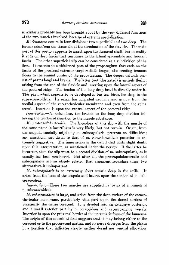
In comparing the muscle attachments upon the proximal humerus of I#•,ana with those of Gallus, it is at first apparent that in the bird the in- sertions are more localized and tendinous, and less discrete. Insertion of latissimus and origin of brachialis have moved distally. Insertions of dor- salis scapulae and pectoralis have migrated from the extensor aspect of the dorsal (lateral) tuberosity to the flexor aspect of this tuberosity, while the coracohumeralis (supracoracoid) has moved in the opposite direction. The attachment of the coracobrachialis posterior of Gallus is not quite compar- able with conditions in Iguana, as in the latter the insertion is fascial. The other muscle attachments are about the same in both forms.
NERVES
As in Reptilia, the position of the brachial plexus of Aves is much more variable than in Mammalia. According to Ftirbringer (lot. cit.) the posi- tion varies from nn. 10-15 (as in Coluraba), to nn. 22-26 (as in Cygnus). The three-trunk plan, basic for tetrapods, may be obscured, and one of these trunks may be reduced in size so that a two-trunk plan is simulated. The number of nerves may be reduced to three (Bueorvus), but usually filaments from neighboring nerves are included, at times to the total num- ber of six plexus nerves (as in Coluraba). These accessory filaments seldom or never reach the size of the three basic trunks. In flightless birds the size of the nerves in the plexus is reduced, but not their number. More distally the grouping is into a single dorsal (extensor) cord, and a single ventral (flexor) cord,--not two of the latter as in Mammalia.
Birds have no spinal accessory nerve. See m. craniocervicalis.
[Auk 366 HOWELL, Shoulder Architecture [July
Dorsal (extensor) division
Suprazonal group
Nn. thoracales posteriores are high branches arising from nn. 14 and 15, supplying min. rhomboidei and serrati. Thus these nerves have shifted relatively cranialward, when compared with lacertilians, as they have in Mammalia.
Shoulder group
N. thoracodorsalis is a branch of the extensor trunk and supplies both divisions of m. latissimus dorsi. It appears to occupy about the same relative position as in terrestrial tetrapods.
Nn. axillares.--Probably because of extreme specialization and the fact that min. dorsalis scapulae and deltoideus have such different functions in birds, the branches to these muscles come off the dorsal cord not together, but slightly separated. N. deltoideus diverges from this cord farther dist- ally than in mammals. Associated with the branch to m. dorsalis scapulae is n. subcoraco•deus, and from that nerve are given off twigs comprising nn. subscapularis anal proscapulohumeralis. The latter is probably homo- logous to that nerve of the same name in lacertilians; yet it arises from the same branch as n. subscapularis, rather than distal to n. thoracodorsalis as in lacertilians. Nevertheless, the branches of the axillary group of nerves are closely knit, and it is believed that variations of this sort are easily ac- complished as a result of high specialization.
Brachio-antebrachial group
N. radialis.--Although all roots of the plexus seem to contribute to this nerve, the contribution from n. 4 appears to be minor, and hence it is situated slightly more cranially than the common flexor nerve. It enters the brachlure between the divisions of m. triceps, which it innervates.
Ventral (flexor) division
Infrazonal group
N. sternocoracoideus diverges from the cranial part of the plexus (n. 15 (14)) in close association with n. supracoracoideus, and innervates m. sternocoracoideus.
Shoulder group
N. pectoralis is a high, stout branch of the common flexor cord. It divides and passes in the axilla to min. pectoralis and panniculus carnosus.
N. coracoideus (supracoracoideus) arises from the base of the dorsal cord upon the cranial aspect of the plexus in close association with n. sterno- coracoideus. Its course is diagnostic, for it passes craniomedial to the cora-
Vol. o41 367 1957 i •'•OWELL, Shoulder A rchitectur•
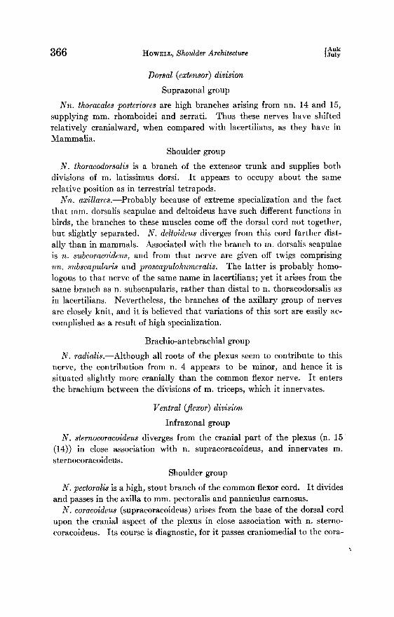
P•CT. .............. t,•,/? /./ •BICEPS ( PART} PO ST.
DELT. PROF. BREV. /
DOR$. ,SCAR .,x,
PROSC, AP. HUM. - .....
LATIS.- .........
Fro, 1.--The proximal end of the right humerus of Gallus, showing areas of muscle at- tachments, A, lateral (flexor) aspect; B, me(Hal (extensor) aspect.
........ SUPRACOR.
COR. BRACH, PST:"(/ /,,I 1',;;'•[1.•'...' ............................... PROSCAI•. HUM.
• • •"• :.•//•/ /l I! I '•!'• ...... .o o.•. = •.
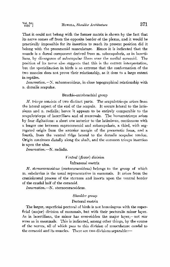
FLEX BRACH • •"•TIS FIe, 2.--Dia•am of ghe righg brachial plexus of Gallus • ventral •ew; doral (ex•eBsor)
elemengs shaded.
368 HOWEl,I,, Shoulder Archilecture [jAu•y
coid, piercing m. deltoideus profundus longus, and innervates m. supracora- coideus.
Nn. coracobrachiales are two in number, a high branch from the root of n. pectoralis to m. coracobrachialis posterior, and a lower branch diverging more distally from n. flexor brachii to m. coracobrachlalis anterior. Their separation doubtless is due to the topography of these muscles following extreme specialization.
Brachio-antebrachial group
N. flexor brachii probably arises from the three. posterior roots of the plexus. It enters the brachlure between mm. biceps and triceps, and sup- plies all parts of the former muscle, as well as m. brachialis, before passing to the forearm.
M•scL•s
The muscular system of Aves is as extremely specialized as the skeletal system, and yet it is possible, without encountering major difficulties, to interpret the muscles in terms of those of tetrapods.
M. craniocervicalis.--Birds have no nerve homologous to n. aceessorius; hence there is no accessory field of musculature. There is, however, a muscle field that simulates it, innervated by n. hypoglosso-accessorius, so called, which arises from the occipital myotomes, suppressed in mammals. The question of its homology is one that will necessitate long investigation before it is solved, and it will not be discussed here.
The part of this complex reaching the shoulder region may be said to con- sist of two parts: a tissue-thin sheet to the fascia of the anterior pectoralis and extending to the propatagium, with stronger convergence to the junc- tion of the clavicles; and a thicker, restricted part from the mid-dorsum that extends in a laterocaudal direction, between shoulder and neck, to in- sert tenuously upon the cranial part of the clavicle. Apparently in the fowl neither is innervated by cervical nerves proper, as is the case in some birds.
Hypobranchial division
The only muscle of the hypobranchial series reaching the girdle in Gallus is one from the hyoid to the anterior process of the sternum, the apparent homologue of m. sternohyoideus of 1V[ammalia.
Dorsal (extensor) division
Suprazonal matrix It is interesting that no bird, so far as I am aware, possesses a true levator
scapulae arising from the more cranial of the cervical vertebrae, although the anterior slip of the serratus frequently is so termed in the literature.

Vol. 54] 1937 J HOWELL, •houlder Archiiecfure 369
In this connection it may be noted that unlike tetrapods, Aves have no muscle of any effectiveness for protracting the shoulder, but only for ad- ducting, depressing, and retracting it, particularly for the latter action.
Mm. serrati are represented by three slips: a long, slender serratus meta- patagii, from the tissue over the last ribs to the border of the metapatagium; a small, anterior serrams from the vicinity of rib 1 to the proximal third of the seapula; and a posterior serratus, extremely variable individually, which seems typically to arise by a broad sheet from all of the ribs except the first, and to converge to insertion upon the distal seapula.
Mm. rhomboidei comprise two layers practically coextensive. Fibers of the superficial sheet extend from the dotsum eranioventrally to all but the caudal part, and of the deep sheet, eaudoventrally to all but the cranial part, of the seapula. The deep rhomboid in birds, accordingly, has sub- stantially the same function as the spinal part of the trapezius in mammals.
Innervation.--This matrix is supplied by high branches of nn. 14, 15, 16.
Shoulder group
Thoracodorsal (latissimus) matrix M. latissimus dorsi also occurs'in two parts. The more superficial, smaller,
and anterior part arises from the dorsum, with fibers extending laterally to the medial aspect of the proximal third of the humerus. The deeper and more posterior part arises from the anterior margin of the pelvis and neigh- boring fascia. It inserts upon the membranous fascia covering the side of the thorax, which in turn is connected with the deep surface of the super- ficial latissimus.
There is no m. teres major in birds, for this is an exclusively mammalian muscle.
Innervation.--N. .thoracodorsalis.
Axillary matrix M. dorsalis scapulae (scapulohumeralis posterior).--This extremely
heavy muscle arises from the entire ventral border of the scapula, deep to the latissimus, and extensively from the ventrally adjacent surface of the thorax. It inserts mainly upon the ventral process of the humerus, but also upon the strong, closely adherent axillary fascia, connected in one direction with the pectoralis insertion, and in the other, converging to a strong, slender tendon that passes between the two cords of the braehal plexus and is attached fanwise to the fascia of the ventral border of the sub- coracoideus. There is no doubt that this muscle is the homologue of m. dorsalis scapulae of lacertilians and of m. teres minor of mammals.
Innervation.--By a nerve adjoining n. deltoideus, but associated directly rather with n. subcoracoideus. The separation of these two components of
[Auk 370 HOWELL, Shoulder Architecture [.July
n. axillaris probably has been brought about by the very different functions of the two muscles involved, because of extreme specialization.
M. deltoideus occurs in four divisions: two supertidal and two deep. The former arise from the tissue about the termination of the clavicle. The main
part of this portion appears to insert upon the humeral shaft, but in reality it ends on deep fascia that continues to the lateral epieondyle and forearm fascia. The other superficial slip can be considered as a subdivision of the first. It extends to a thickened part of the propatagium that ends on the fascia of the proximal extensor earpi radialis longus, also sending tenuous fibers to the cranial border of the propatagium. The deeper deltoids con- sist of partes 1ongi and brevis. The latter (not illustrated) is entirely fleshy, arising from the end of the clavicle and inserting upon the lateral aspect of the pectoral ridge. The tendon of the long deep head is directly under it. This part, which appears to be developed in but few birds, lies deep to the supraeoraeoideus. Its origin has migrated eaudally and is now from the roedial aspect of the eoraeoelavieular membrane and even from the spina sterni. Insertion is upon the ventral aspect of the pectoral ridge.
Innervation.--N. deltoideus, the branch to the long deep division fol- lowing the tendon of insertion to the muscle substance.
M. proscapulohumeralls.--The hornology of this slip with the muscle of the same name in laeertilians is very likely, but not certain. Origin, from the seapula eaudally adjoining m. subseapularis, presents no difficulties; and insertion, just distal to that of m. eoraeobraehialis posterior, is ex- tremely suggestive. The innervation is the detail that easts slight doubt upon this interpretation, as mentioned under the nerves. If the latter be incorrect, then the slip must be a second diGsion of m. subseapularis, as it usually has been considered. But after all, the proseapulohumeralis and subseapularis are so dosely related that argument regarding these two alternatives is unimportant.
M. subscapularis is an extremely short muscle deep in the axilla. It arises from the base of the seapula and inserts upon the tendon of m. sub- eoraeoideus.
Innerrations---These two muscles are supplied by twigs of a branch of n. subeoraeoideus.
M. subcoracoideus is large, and arises from the deep surface of the eoraeo- elavieular membrane, particularly that part upon the dorsal surface of practically the entire eoraeoid. It is divided into an extensive posterior, and a small anterior part by n. eoraeoideus and accompanying vessels. Insertion is upon the proximal border of the pneumatic fossa of the humerus. The origin of this muscle at first suggests that it may belong either to the eoraeoid or to the proeoraeoid matrix, and its nerve diverges from the plexus in a position that indicates clearly neither dorsal nor ventral allocation.
Vol. 541 1937 J HOWELL, Shoulder Architecture 371
That it could not belong with the former matrix is shown by the fact that its nerve comes off from the opposite border of the plexus, and it would be practically impossible for its insertion to reach its present position did it belong with the procoracoid musculature. Hence it is indicated that the muscle is a dorsal component derived from m. subscapularis, as in lacerti- lians, by divergence of subscapular fibers over the roedial coracold. The position of its nerve also suggests that this is the correct interpretation, but the specialization in birds is so extreme that the conformation of the two muscles does not prove their relationship, as it does to a large extent in reptiles.
Innervation.--N. subcoracoideus, in close topographicai relationship with n. dorsalis scapulae.
Brachio-antebrachial group
M. triceps consists of two distinct parts. The scapulotriceps arises from the lateral aspect of the end of the scapula. It occurs lateral to the latis- simus and n. radialis; hence it appears to be entirely comparable to the scapulotriceps of lacertilians and of mammals. The humerotriceps arises by four digitations: a short one anterior to the latissimus, continuous with a longer one between supracoracoid and subscapularis, a third, with seg- regated origin from the anterior margin of the pnemnatic fossa, and a fourth, from the ventral ridge lateral to the dorsalis scapulae tendon. Origin continues distally along the shaft, and the common triceps insertion is upon the ulna.
Ianervation.--N. radialis.
Ventral (flexor) division Infrazonal matrix
M. sternocoracoideus (costocoracoideus) belongs to the group of which m. subclavius is the usual representative in mammals. It arises from the craniolateral process of the sternum and inserts upon the ventral border of the caudal half of the coracold.
Innervation.--N. sternocoracoideus.
Shoulder group Pectoral matrix
The larger, superficial pectoral of birds is not homologous with the super- ficial (major) division of mammals, but with their pectoralis minor layer. As in lacertilians, the minor has over-ridden the major layer,--not vice versa as in mammals. This is indicated, among other things, by the course of the nerves, all of which pass to this division of musculature caudal to the coracoid and its muscles. There are two divisions separable:--

Auk 372 •owm•, Shoulder Architecture [July
M. pectoralis superficialis is enormous, arising from the carina, the par- asternal processes or trabeculae, the clavicle, and a part of the coracoclavic- ular membrane. The deeper fibers from the membrane are partly separ- able. A few of the superficial fibers converge to a fascial connection with the propatagium, but the main insertion is upon the ventral aspect of the pectoral ridge of the humerus, with a fascial extension in one direction to the capsule of the joint, and in the other, a tendinous band that passes to the deep surface of m. dorsalis scapulae.
M. panniculus carnosus probably, in the Fowl, takes the place of the ab- dominal pectoral found in a number of birds. It arises from the axillary fascia lateral to the dorsalis scapulae, and from the tendinous sling of the superficial pectoral. As a very thin sheet it passes ventrally to insert upon the integument. At first glance this muscle appears to be innervated by n. 18, but this nerve was demonstrated to pierce the muscle without sup- plying it.
Innervatlon.--N. pectoralis.
Anterior eoraeoid matrix
M. supracoracoideus is the 'deep pectoral' of birds, arising from the base of the carina and sternum proper. It converges and passes deep to the distal end of the coraeoid and deep to the deltoideus profundus brevis, to insert by a stout tendon upon the medial side of the dorsal border of the humerus. There is thus formed a pulley, whereby the muscle elevates, rather than depresses, the wing.
This muscle is strictly comparable with the peetoralis major of mammals. The supra- and infraspinati of mammals are entirely unrepresented, prob- ably never having become differentiated from the supraeoraeoid matrix.
Innervation.--N. supraeoraeoideus.
Posterior coracold matrix
M. coracobrachialis posterior.--This muscle adjoins m. supracoracoideus and is covered by the pectoralis. It arises from the lateroventral border of most of the coracold and a small part of the adjoining sternum, and inserts upon the central process of the medial aspect of the humeral head. At first glance it might appear to be a division of the pectoral complex, for its nerve comes off the base of n. pectoralis. That this allocation is erroneous is attested first, by the fact that its insertion is far from that of the pectoral, with insurmountable barriers intervening. Its innervation offers no diffi- culty, for in lacertilians the coracobrachial nerve immediately adjoins n. pectoralis. Finally, its origin is fairly diagnostic.
M. coracobrachialia anterior is a feeble muscle deep to the origin of the biceps. It is very short and relatively broad. It arises from the capsule,
Vol. 54] 1037 J HOWELL, Shoulder Archilecture 373
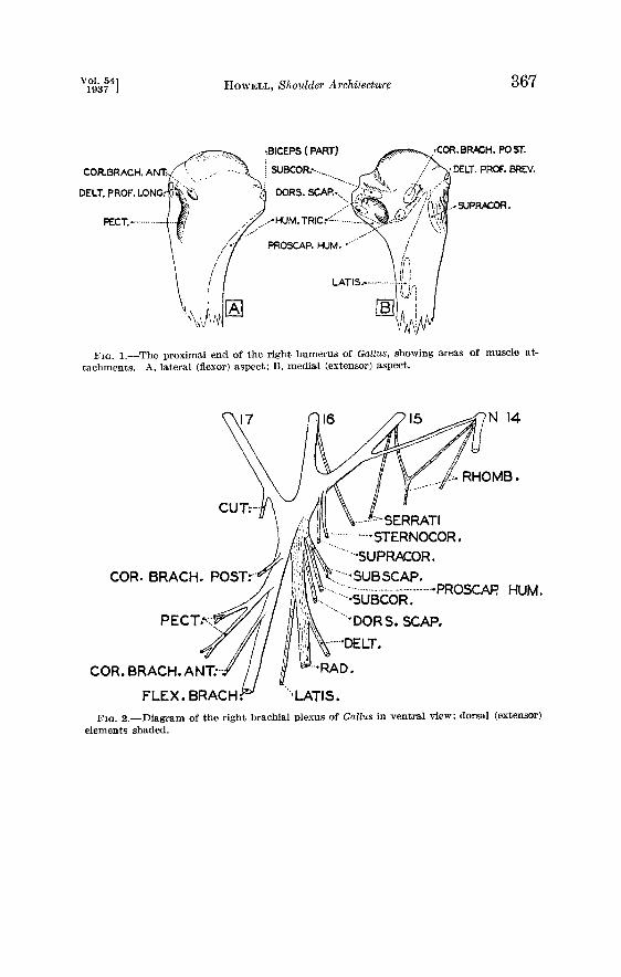
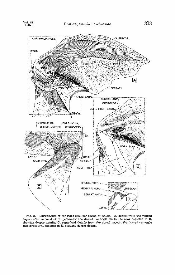
LATI$:
SCAR TRIG: BICEPS'/- i i
HUM, TRIC. •'*}'"'• *"
Fz•. 3.--Musculature of the right shoulder region of Gallus. A, details from the ventral aspect after removal of m. pectoralis; the dotted rectangle marks the area depicted in B, showing deeper details; O, superficial details from the dorsal aspect; the dotted rectangle marks the area depicted in D, showing deeper deta/ls,
fAuk 374 HOWELL, Shoulder Architecture [July
uo!s!,x!p I•S.•O(I uo!s.•!p IS.qU•A

Vol. 54] 1037 J HOWELL, Shoulder Architecture 375
rather than from the coracoid, and inserts upon the lateroventral aspect of the head of the humerus.
There may be some doubt regarding the identity of this muscle. At first glance it appears to have been derived from deeper fibers of the biceps, and its nerve, from the common flexor trunk, does not disprove this thesis. The nerve to m. eoraeobraehialis, however, frequently is situated similarly in mammals, and it seems better to consider a muscle with such a high in- sertion as belonging to the present group.
Brachio-antebrachial group
M. brachJails is very much reduced in birds. It arises as a triangular muscle from the medial aspect of the distal humerus and inserts upon the ulna.
M. biceps consists of a single primary muscle and a small accessory slip. The former arises by a broad tendon from the dorsal part of the end of the coracold also with tendinous anchorage to the dorsal margin of the humeral head (Fig. 1). It sometimes shows a tendency to divide into two parts near origin. InsertJinx is upon the ulna. The accessory head arises as a small, superficial slip from the surface of the prime head, and disappears on the propatagium. Its continuing fascia, however, may be separated from that coming from the deltoid, and the former is attached to the ulna, rather than to the forearm muscles.
Innervation.--By branches of the common flexor nerve; that to the ac- cessory head pierces the primary biceps.
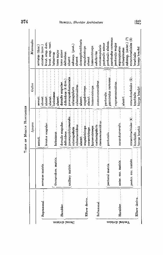
On page 374 is presented a table of homologies of the shoulder muscula- ture of Iguana, Gallus, and Mam•nalia in general.
SUMMARY
The feature of the present paper is the homology of the arian muscles of the shoulder with those of other classes of vertebrates as given in the ac- companying table. Noteworthy details comprise the absence of the spinal accessory nerve and its musculature since these are replaced by a suboc- cipital field; the lack of m. levator scapulae; and the occurrence of the thombolds in two layers. The latissimus is in two separated portions, and the subscapularis is depauperate. The dorsalis scapulae is very robust and the deltoid is noteworthy in Gallu. s. The large breast muscle in birds is the pectorahs minor element; the major is small and deep. Supra- and infra- spinati are absent. The brachialis is a feeble muscle near the elbow.
Department of Anatomy John Hopkins Medical School
Baltimore, Maryland