3d dosimetry in the clinic and research: special...
TRANSCRIPT
VALIDATING SPECIAL TECHNIQUES3D Dosimetry in the Clinic and Research
Titania Juang, PhDResident, Radiation Physics DivisionStanford Cancer Center
Learning objectives
1: 3D Dosimetry in the Clinic: Background and Motivation
2: 3D Dosimetry in the Clinic: Motion interplay effects in dynamic radiotherapy
3: 3D Dosimetry in the Clinic and Research:
Validating Special Techniques
• Understand the potential for 3D dosimetry in validating dose
accumulation in deformable systems.
• Observe the benefits of high resolution measurements for
precision therapy in SRS and in microSBRT for small animal
irradiators.
4: 3D Dosimetry in end-to-end dosimetry QA
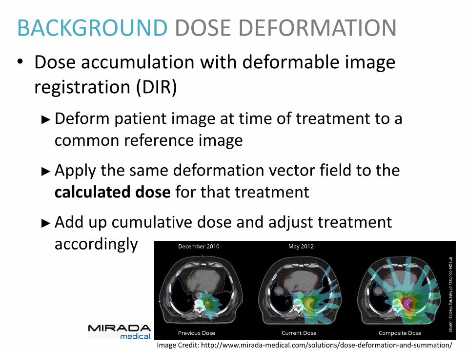
BACKGROUND DOSE DEFORMATION
• Dose accumulation with deformable image registration (DIR)
►Deform patient image at time of treatment to a common reference image
►Apply the same deformation vector field to the calculated dose for that treatment
►Add up cumulative dose and adjust treatment accordingly
Image Credit: http://www.mirada-medical.com/solutions/dose-deformation-and-summation/
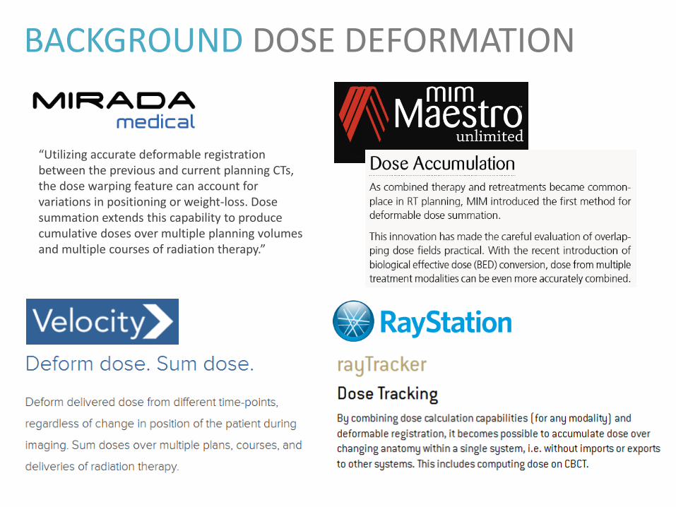
BACKGROUND DOSE DEFORMATION
“Utilizing accurate deformable registration between the previous and current planning CTs, the dose warping feature can account for variations in positioning or weight-loss. Dose summation extends this capability to produce cumulative doses over multiple planning volumes and multiple courses of radiation therapy.”
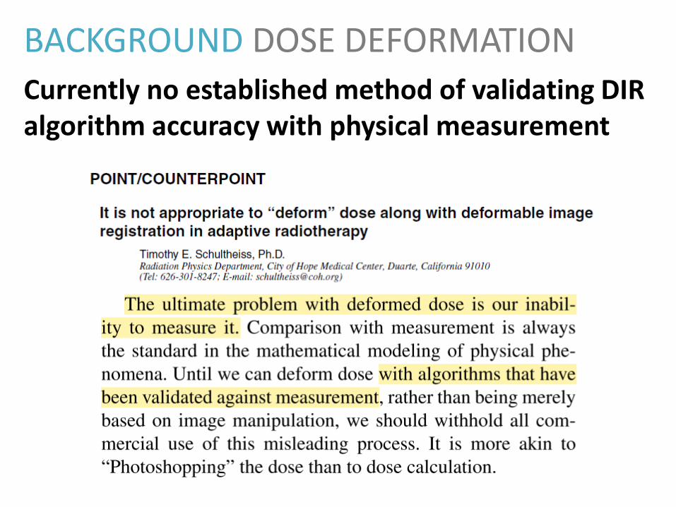
BACKGROUND DOSE DEFORMATION
Currently no established method of validating DIR algorithm accuracy with physical measurement
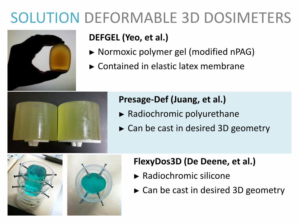
SOLUTION DEFORMABLE 3D DOSIMETERSDEFGEL (Yeo, et al.)
► Normoxic polymer gel (modified nPAG)
► Contained in elastic latex membrane
Presage-Def (Juang, et al.)
► Radiochromic polyurethane
► Can be cast in desired 3D geometry
► []
FlexyDos3D (De Deene, et al.)
► Radiochromic silicone
► Can be cast in desired 3D geometry
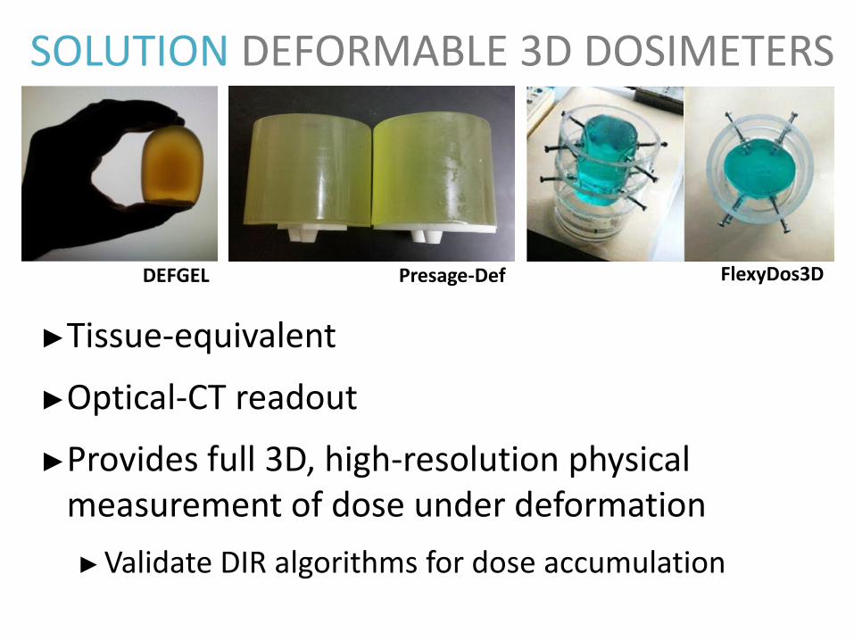
SOLUTION DEFORMABLE 3D DOSIMETERS
►Tissue-equivalent
►Optical-CT readout
►Provides full 3D, high-resolution physical measurement of dose under deformation
►Validate DIR algorithms for dose accumulation
DEFGEL Presage-Def FlexyDos3D
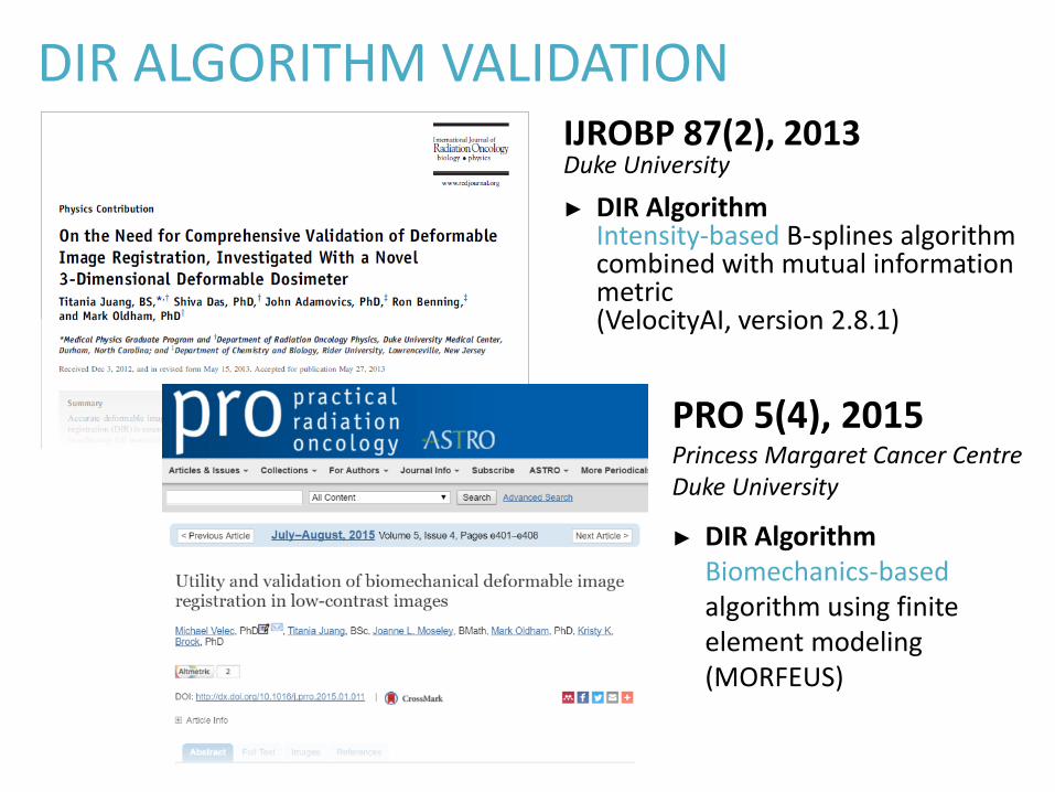
DIR ALGORITHM VALIDATIONIJROBP 87(2), 2013Duke University
► DIR AlgorithmIntensity-based B-splines algorithm combined with mutual information metric(VelocityAI, version 2.8.1)
PRO 5(4), 2015Princess Margaret Cancer CentreDuke University
► DIR AlgorithmBiomechanics-based algorithm using finite element modeling(MORFEUS)
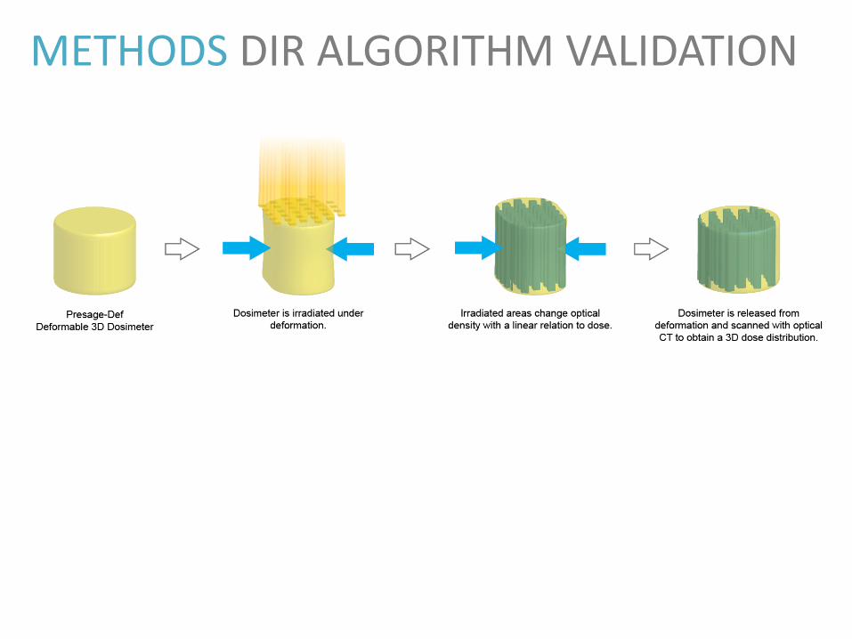
METHODS DIR ALGORITHM VALIDATION
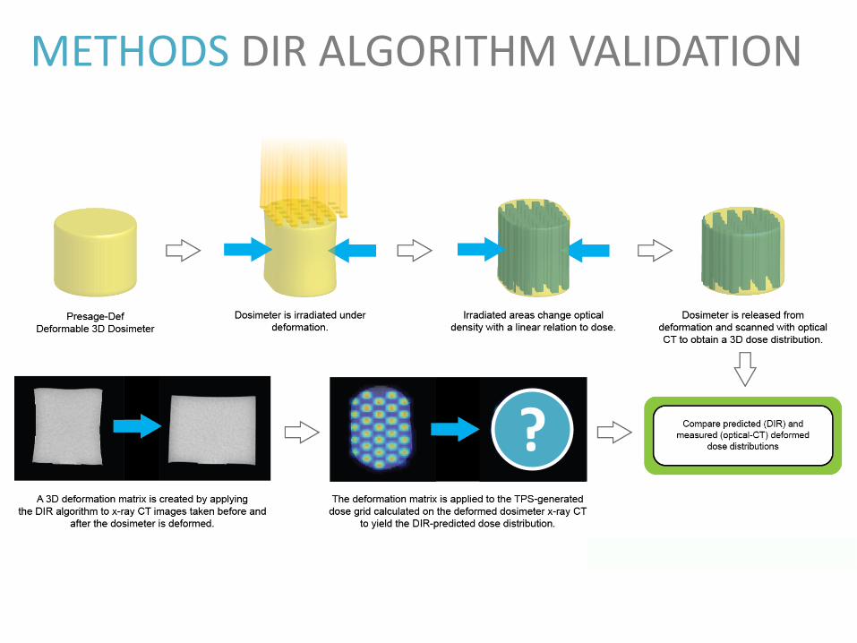
METHODS DIR ALGORITHM VALIDATION
?
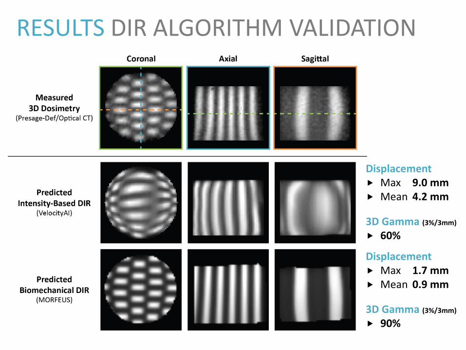
RESULTS DIR ALGORITHM VALIDATION
Displacement Max 9.0 mm Mean 4.2 mm
3D Gamma (3%/3mm)
60%
Displacement Max 1.7 mm Mean 0.9 mm
3D Gamma (3%/3mm)
90%
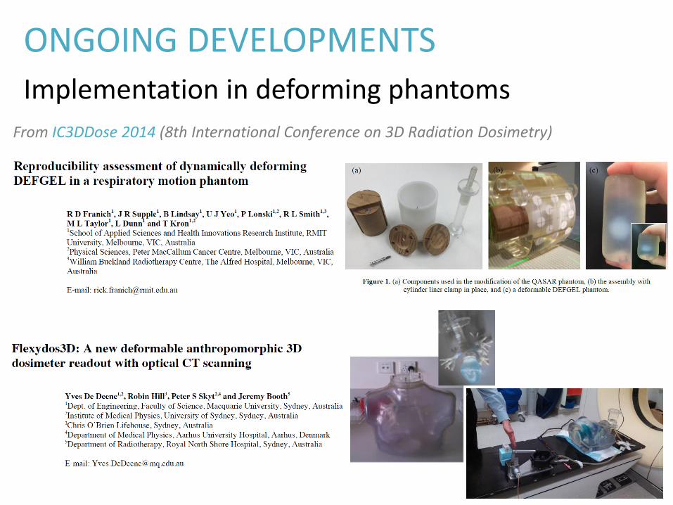
ONGOING DEVELOPMENTS
Implementation in deforming phantoms
From IC3DDose 2014 (8th International Conference on 3D Radiation Dosimetry)
TAKE-HOME MESSAGES
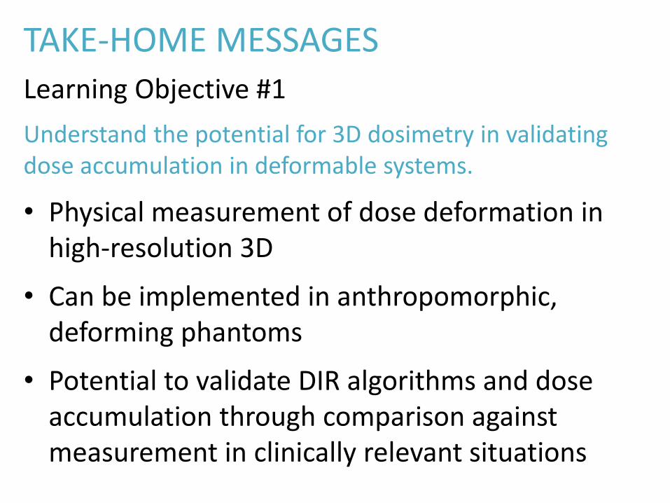
Learning Objective #1
Understand the potential for 3D dosimetry in validating dose accumulation in deformable systems.
• Physical measurement of dose deformation in high-resolution 3D
• Can be implemented in anthropomorphic, deforming phantoms
• Potential to validate DIR algorithms and dose accumulation through comparison against measurement in clinically relevant situations

LEARNING OBJECTIVES
1. Understand the potential for 3D dosimetry in validating dose accumulation in deformable systems.
2. Observe the benefits of high resolution measurements for precision therapy in SRS and in microSBRT for small animal irradiators.
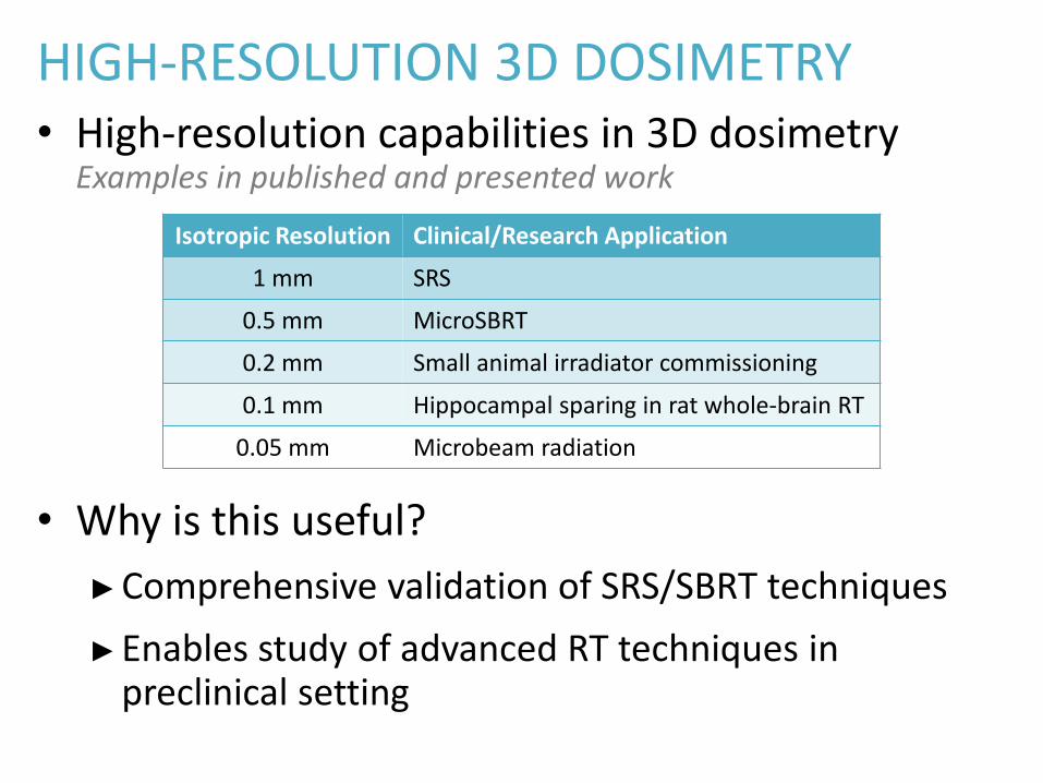
HIGH-RESOLUTION 3D DOSIMETRY• High-resolution capabilities in 3D dosimetry
Examples in published and presented work
• Why is this useful?
►Comprehensive validation of SRS/SBRT techniques
►Enables study of advanced RT techniques in preclinical setting
Isotropic Resolution Clinical/Research Application
1 mm SRS
0.5 mm MicroSBRT
0.2 mm Small animal irradiator commissioning
0.1 mm Hippocampal sparing in rat whole-brain RT
0.05 mm Microbeam radiation
EXAMPLES HIGH-RES 3D DOSIMETRY
1. Multi-target, single isocenter VMAT SRS
2. Small-field irradiator commissioning
3. MicroSBRT with rodent-morphic dosimeters
4. Hippocampal sparing in rat whole-brain RT

MULTITARGET SINGLE ISOCENTER VMAT SRS
Med. Phys. 40(12), 2013Thomas, et al. (Duke University)
► 1 mm isotropic 3D dosimetry
► 5 SRS targets (5-24 cm3)
► 5 arcs, 1 isocenter
► Verified end-to-end accuracy & reproducibility
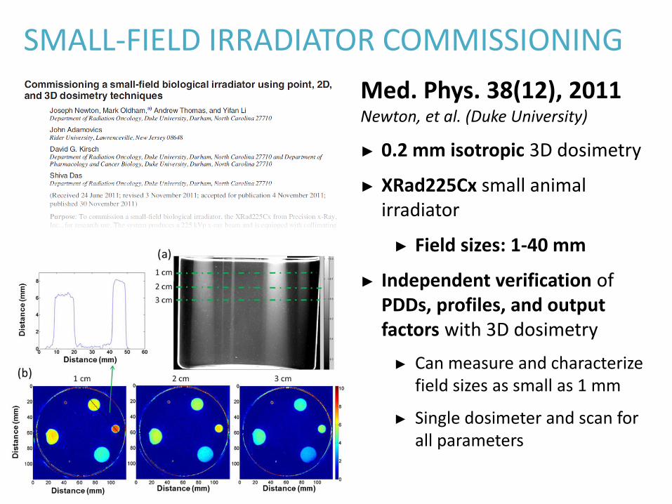
SMALL-FIELD IRRADIATOR COMMISSIONING
Med. Phys. 38(12), 2011Newton, et al. (Duke University)
► 0.2 mm isotropic 3D dosimetry
► XRad225Cx small animal irradiator
► Field sizes: 1-40 mm
► Independent verification of PDDs, profiles, and output factors with 3D dosimetry
► Can measure and characterize field sizes as small as 1 mm
► Single dosimeter and scan for all parameters
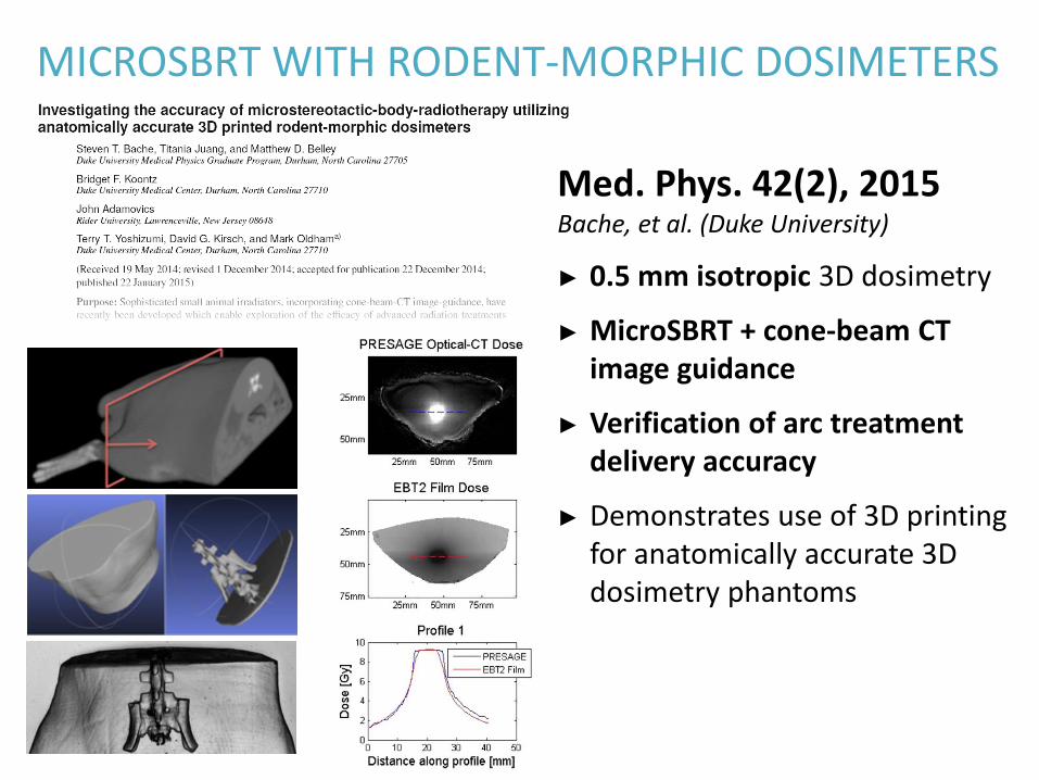
MICROSBRT WITH RODENT-MORPHIC DOSIMETERS
Med. Phys. 42(2), 2015Bache, et al. (Duke University)
► 0.5 mm isotropic 3D dosimetry
► MicroSBRT + cone-beam CT image guidance
► Verification of arc treatment delivery accuracy
► Demonstrates use of 3D printing for anatomically accurate 3D dosimetry phantoms
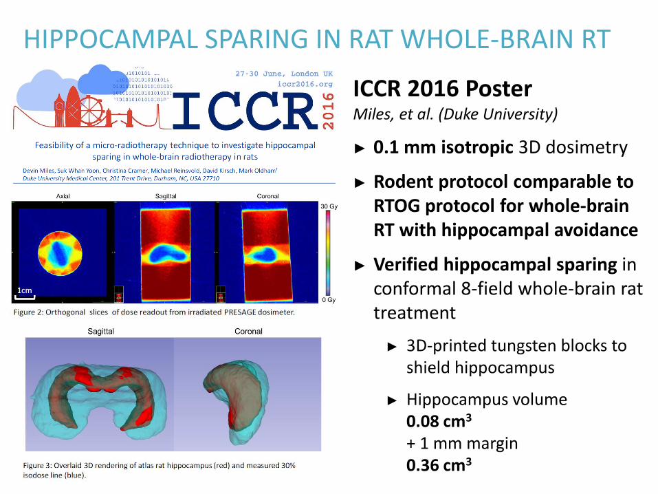
HIPPOCAMPAL SPARING IN RAT WHOLE-BRAIN RT
ICCR 2016 PosterMiles, et al. (Duke University)
► 0.1 mm isotropic 3D dosimetry
► Rodent protocol comparable to RTOG protocol for whole-brain RT with hippocampal avoidance
► Verified hippocampal sparing in conformal 8-field whole-brain rat treatment
► 3D-printed tungsten blocks to shield hippocampus
► Hippocampus volume0.08 cm3
+ 1 mm margin0.36 cm3
TAKE-HOME MESSAGESLearning Objective #2
Observe the benefits of high resolution measurements for precision therapy in SRS and in microSBRT for small animal irradiators.
• Comprehensive, full 3D dose validation for complex SRS plans
• Dosimetry option for small radiation fields down to 1 mm diameter
• Treatment delivery verification for preclinical small animal studies, including models of advanced radiation treatment techniques
ACKNOWLEDGEMENTS
DUKE UNIVERSITYMark Oldham
Shiva DasAndy Thomas
Michael NiebanckZhiheng Wang
Joe NewtonYifan Li
David G. KirschSteve Bache
Matt D. BelleyBridget F. Koontz
Terry YoshizumiDevin Miles
Suk Whan (Paul) YoonChristina Cramer
Michael Reinsvold
RIDER UNIVERSITYJohn AdamovicsRon Benning
PMCC / UNIVERSITY OF MICHIGANMichael VelecJoanne MoseleyKristy Brock
3D DOSIMETRY EDUCATIONAL COURSEL. John Schreiner
Sofie CebergGeoffrey Ibbott
FLEXYDOS3DY. De DeeneP. S. SkytR. HilJ. T. BoothC. O. B. Lifehouse
DEFGELU. J. YeoM. L. TaylorL. DunnT. KronR. L. SmithR. D. FranichJ. R. SuppleB. LindsayP. Lonski
PAPERS (1/2)• T. E. Schultheiss, W. A. Tomé, and C. G. Orton, “It is not appropriate to ‘ deform ’ dose
along with deformable image registration in adaptive radiotherapy,” Med. Phys., vol. 39, no. November, pp. 6531–6533, 2012.
• U. J. Yeo, M. L. Taylor, L. Dunn, T. Kron, R. L. Smith, and R. D. Franich, “A novel methodology for 3D deformable dosimetry,” Med. Phys., vol. 39, no. 4, pp. 2203–13, Apr. 2012.
• U. J. Yeo, M. L. Taylor, J. R. Supple, R. L. Smith, L. Dunn, T. Kron, and R. D. Franich, “Is it sensible to ‘deform’ dose? 3D experimental validation of dose-warping.,” Med. Phys., vol. 39, no. 8, pp. 5065–72, Aug. 2012.
• U. J. Yeo, J. R. Supple, M. L. Taylor, R. Smith, T. Kron, and R. D. Franich, “Performance of 12 DIR algorithms in low-contrast regions for mass and density conserving deformation.,” Med. Phys., vol. 40, no. 10, p. 101701, Oct. 2013.
• T. Juang, S. Das, J. Adamovics, R. Benning, and M. Oldham, “On the need for comprehensive validation of deformable image registration, investigated with a novel 3-dimensional deformable dosimeter.,” Int. J. Radiat. Oncol. Biol. Phys., vol. 87, no. 2, pp.
• M. Velec, T. Juang, J. L. Moseley, M. Oldham, and K. K. Brock, “Utility and validation of biomechanical deformable image registration in low-contrast images,” Pract. Radiat. Oncol., vol. 5, no. 4, pp. e401–e408, 2015.
PAPERS (2/2)• Y. De Deene, P. S. Skyt, R. Hil, and J. T. Booth, “FlexyDos3D: a deformable
anthropomorphic 3D radiation dosimeter: radiation properties,” Phys. Med. Biol., vol. 60, no. 4, pp. 1543–1563, 2015.
• R. D. Franich, J. R. Supple, B. Lindsay, U. J. Yeo, P. Lonski, R. L. Smith, M. L. Taylor, L. Dunn, and T. Kron, “Reproducibility assessment of dynamically deforming DEFGEL in a respiratory motion phantom,” J. Phys. Conf. Ser., vol. 573, no. i, p. 012024, 2015.
• Y. De Deene, R. Hill, P. S. Skyt, J. Booth, and C. O. B. Lifehouse, “Flexydos3D : A new deformable anthropomorphic 3D dosimeter readout with optical CT scanning,” IC3DDose 2014 - 8th Int. Conf. 3D Radiat. Dosim., 2014.
• A. Thomas, M. Niebanck, T. Juang, Z. Wang, and M. Oldham, “A comprehensive investigation of the accuracy and reproducibility of a multitarget single isocenter VMAT radiosurgery technique.,” Med. Phys., vol. 40, no. 12, p. 121725, Dec. 2013.
• J. Newton, M. Oldham, A. Thomas, Y. Li, J. Adamovics, D. G. Kirsch, and S. Das, “Commissioning a small-field biological irradiator using point, 2D, and 3D dosimetry techniques.,” Med. Phys., vol. 38, no. 12, pp. 6754–62, Dec. 2011.
• S. T. Bache, T. Juang, M. D. Belley, B. F. Koontz, J. Adamovics, T. T. Yoshizumi, D. G. Kirsch, and M. Oldham, “Investigating the accuracy of microstereotactic-body-radiotherapy utilizing anatomically accurate 3D printed rodent-morphic dosimeters,” Med. Phys., vol. 846, pp. 1–11, 2015.