3d model simulation and patient surgery in guided bone … › wp-content › uploads ›...
TRANSCRIPT
Citation: Grisa A, Maurino CD, Valladares A, Muchhala S, Yu PY. 3D Model Simulation and Patient Surgery in Guided Bone Regeneration. J Oral Biol. 2019; 6(1): 6
3D Model Simulation and Patient Surgery in Guided Bone Regeneration
AbstractThe anterior maxilla has traditionally been a challenge when it comes
to successfully placing dental implants. This is due to a combination of poor bone quality, ridge atrophy and bone resorption following extraction. Many techniques are available today for the experienced surgeon to rebuild lost bone, including guided bone regeneration (GBR). Despite GBR being a predictable procedure, complications can and do arise that may compromise outcomes. The most frequent of these include membrane exposure, fenestration/dehiscence, infection, graft particle leakage, collapse of the grafted site and excessive bleeding. However, careful pre-surgical planning is crucial and will reduce risk and incidence of complications. Cone Beam Computed Tomography (CBCT) provides greater detail and has become a commonly used diagnostic tool for implant treatment planning. Patient 3D printed models can be used to gain insight and become familiar with a patient’s exact anatomy prior to the surgical procedure. Using such models can aid in reducing surgical time, limiting the amount of soft tissue manipulation, familiarizing the surgeon with the patient’s specifi c anatomy, therefore reducing the risk of intra-operative complications, and decreasing the potential for error. The purpose of this article is to report on the use of a 3D printed model to familiarize with the anatomy of the patient prior to the surgery to plan and avoid possible complications.
Grisa A, Maurino CD, Valladares A, Muchhala S and Yu PY*
Arthur Ashman Department of Periodontology and Implant Dentistry, New York University College of Dentistry, USA
*Address for CorrespondenceYu PY, Arthur Ashman Department of Periodontology and Im-plant Dentistry, New York University College of Dentistry, New York 10010, USA; E-mail: [email protected]
Submission: 03 September, 2018Accepted: 25 February, 2019Published: 27 February, 2019
Copyright: © 2018 Grisa A, et al. This is an open access article distributed under the Creative Commons Attribution License, which permits unrestricted use, distribution, and reproduction in any medium, provided the original work is properly cited.
Case ReportOpen Access
Journal of
Oral Biology
IntroductionTh e anterior maxilla is demanding and challenging when it comes
to establishing clinical success while placing dental implants. Th is is due to a combination of esthetic expectations, poor bone quality, ridge atrophy and bone resorption following extraction. Various techniques are available today for experienced surgeons to reconstruct lost bone, such as Autologous onlay block graft s [1], allograft block graft s [2], distraction-osteogenesis and guided bone regeneration (GBR) [3,4]. Studies in animals and humans have shown that GBR is an eff ective technique to augment atrophic ridges.
Despite GBR being a predictable procedure, complications can arise that may compromise the fi nal outcomes of this procedure. Th e most frequent complications include membrane exposure, fenestration or dehiscence, infection, graft particle leakage, collapse of the graft ed site and excessive bleeding [5,6]. Although GBR has a high rate of success, it is surgically challenging and presents various risks and diffi culties. However careful pre-surgical planning is crucial and will reduce the risk and incidence of complications.
Cone Beam Computed Tomography (CBCT) provides greater detailed images of the bone and has become a common diagnostic tool for implant treatment planning. In spite of these advantages, it can still be challenging to convert the two-dimensional cross sectional images from CBCT into the three-dimensional geometrical structure of the atrophic ridge. For this purpose 3D printing technology has been introduced in dentistry as a useful and cost eff ective tool for educational purposes and to improve pre-surgical preparation [7,8]. More recent advances in digital technology have made 3D printing
J Oral BiolFebruary 2019 Volume 6 Issue 1 © All rights are reserved by Grisa A, et al.
Avens Publishing Group
more accessible and more economical, gaining ground in mainstream dentistry. 3D-printed models can be used to gain insight, carefully study and become familiar with the exact anatomy of the patient’s maxillary bone prior to any surgical procedures [9].
Furthermore, 3D models can be used for preoperative simulation of the surgical procedure itself, which is advantageous to the surgeon who will perform the procedure. Using such models can aid in reducing surgical time, limiting the amount of soft tissue manipulation, familiarizing the surgeon with the patient’s specifi c anatomy, reducing the risk of intra-operative complications, and decreasing the potential for errors [10-13]. Th e purpose of this case report is to report the use of a 3D model prior to a ridge augmentation procedure to get familiar with the patient’s maxillary anatomy and
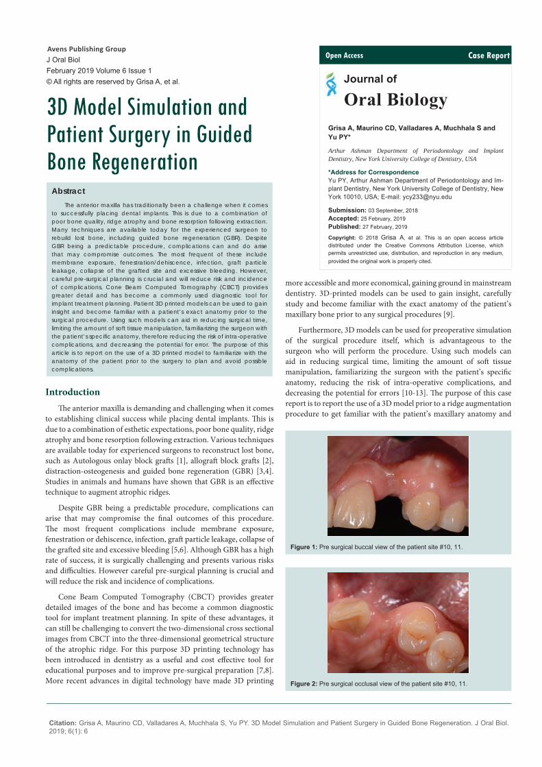
Figure 1: Pre surgical buccal view of the patient site #10, 11.
Figure 2: Pre surgical occlusal view of the patient site #10, 11.

Citation: Grisa A, Maurino CD, Valladares A, Muchhala S, Yu PY. 3D Model Simulation and Patient Surgery in Guided Bone Regeneration. J Oral Biol. 2019; 6(1): 6
J Oral Biol 6(1): 6 (2019) Page - 02
ISSN: 2377-987X
plan the treatment to avoid possible complications.
Case Report In 2016, a 32-year-old male was referred to the Ashman
Department of Periodontology and Implant Dentistry of New York University College of Dentistry. Th e patient was a non-smoker with an unremarkable medical history. His chief complaint was the missing left lateral incisor that doesn’t allow him to smile confi dently. He desired a fi xed restoration. Tooth #10 was extracted several years before with the subsequent loss of supporting bone and soft tissue (Figure 1 and 2). A CBCT scan was taken to carefully evaluate the anatomy of the alveolar ridge and revealed a defi cient volume of buccolingual crestal bone and the need for a bone regeneration procedure prior to implant placement (Figure 3). Digital Imaging and Communications in Medicine (DICOM) images from the patient’s CBCT were then converted to STL fi les (OsiriX Lite, Geneva, Switzerland) and transferred to a 3D printer (Formlabs, USA) for production of a polymer model of the maxilla. Medical adhesive tape was added to the model to mimic the oral mucosa for a more realistic simulation of the actual surgical environment (Figure 4 and 5). Th e GBR was performed on the 3D model prior to treating the patient.
Th e treatment plan was to fi rst augment the bone volume in the area of tooth #10 and aft er four months of healing, the placement of an implant and an immediate provisional restoration.
Surgical procedures
Th e bone augmentation procedure was performed as follows. Th e patient was given a prescription of amoxicillin 2 g 1 hour prior to surgery. Figures 6-22 depict the surgical simulation on the 3D printed model with the corresponding stages in the live surgery.
Aft er anesthesia was performed a full thickness fl ap was elevated with 2 vertical incisions; one papilla sparing incision distal to tooth # 9 and one intrasulcular incision distal to # 11. Aft er decortication, a 2 mm diameter trephine bur was used to harvest a bone core apical to the area being augmented. A 2 mm diameter hole with a depth of 3mm was made perpendicular to the buccal bone wall to allow the placement of the trephined bone core inside allowing it to be used as tent pole. A CopiOs Pericardium Membrane (Zimmer Biomet, USA www.zimmerbiometdental.com) was secured with three apical tacks (truFIX, ACE Surgical, www.acesurgical.com) and the space was fi lled with Bio-Oss (Geistlich, CH www.geistlich-na.com). A periosteal releasing incision was made to achieve tension-free closure using resorbable sutures. Figures 23 and 24 show the pre and post operative radiographs of the surgical site.
Following surgery, amoxicillin 500 mg TID for 10 days and chlorhexidine 0.12% mouth-rinse (PeridexTM, 3M ESPE, www.3MESPE.com) BID for 2 weeks were prescribed. Th e healing process was uneventful (Figure 25). Figure 26 depicts the fi nal restoration for #11 aft er 1 year.
Discussion3D-printed models can be used to gain insight and become
familiar with a patient’s anatomy prior to surgical procedures. Furthermore, 3D models can be used for preoperative simulation of the surgical procedure itself, which is advantageous to the surgeon who will perform the procedure. Using such models can aid in reducing surgical time, limiting the amount of soft tissue manipulation, familiarizing the surgeon with the patient’s specifi c anatomy, reducing the risk of intra-operative complications oand decreasing the potential for error [7].
GBR is surgically challenging and eff ective training and education is required to ensure successful outcomes. Although considered a predictable procedure, care must be taken so as not to disturb the healing process and to maintain the health and well-being of the patient. Simulation of the procedure on the patient’s 3D model can enable seamless execution on the day of surgery leading to a more predictable result. Th e use of 3D-printed models for such training is also preferable to training on cadavers since it is patient-specifi c and availability and cost are not limitations [14].
Flap design should be considered prior to surgery and the 3D model allows the surgeon to plan the incisions correctly to maximize visibility and access to the surgical site. Incision design and fl ap elevation, once made, are irreversible and it is crucial that primary closure without tension can be attained [15].
Th e periosteum is a dense layer of vascular connective tissue enveloping bone. In GBR a periosteal releasing incision increases the fl ap elasticity and enables the advancement of the soft tissue over the surgical site to achieve a tension-free [16]. During surgery, a proper manipulation of the periosteum, while achieving primary closure, is essential for the healing of the soft tissues due to the enhanced blood supply provided by the connective tissue above the periosteum. Th e periosteum is therefore a crucial soft tissue layer that infl uences the success of implant surgery [17].
A resorbable collagen membrane is then cut to the same shape as the template and placed over the model surgical site. Once the membrane is in the correct position, a tack is positioned apically through the membrane into the 3D model for stabilization. It is then adjusted to extend 2-3 mm beyond the augmented area. Th e graft
Clinical Procedure 3D model Simulation Comments
Incision design ++ On the model it is possible to design the fl ap as in patient's mouth
Flap elevation + Simulation
Periosteal releasing incision + Material used to simulate soft tissue allows releasing incision
Decortication ++ Decortication can be performed on the model
Membrane Tacking ++ Tacking using real tags is possible on the model
Table 1: Comparison of the clinical procedure against the 3D model.
+: Simulation procedure comparable to the clinical procedure++: Simulation procedure and tactile sensation comparable to the clinical procedure

Citation: Grisa A, Maurino CD, Valladares A, Muchhala S, Yu PY. 3D Model Simulation and Patient Surgery in Guided Bone Regeneration. J Oral Biol. 2019; 6(1): 6
J Oral Biol 6(1): 6 (2019) Page - 03
ISSN: 2377-987X
Figure 3: Patient cross sectional view of the defect site #10, 11.
Figure 4: Pre surgical facial view of the 3D model of the patient.
Figure 5: Pre surgical occlusal view of the 3D model of the patient.
Figure 6: Incision design on 3D model of the patient.
material (e.g. mineralized allograft bone and/or organic bovine bone matrix) is placed and condensed to fi ll the defect, ensure proper space maintenance, bone contact and support the membrane.
Th e fi nal accuracy of the 3D printed models is dependent on
Figure 7: 3D model fl ap elevation and decortication.
Figure 8: Patient fl ap elevation and decortication.
Figure 9: 3D model releasing incision.
Figure 10: Patient releasing incision.
data taken from the CBCT scan, the segmentation technique used to generate the STL fi le from the DICOM data and from the printing technique. CBCT accuracy discrepancies are the result of voxel size in association with segmenting, thus aff ecting the accuracy of the printing process. Th e study by Szymor et al. showed a high accuracy

Citation: Grisa A, Maurino CD, Valladares A, Muchhala S, Yu PY. 3D Model Simulation and Patient Surgery in Guided Bone Regeneration. J Oral Biol. 2019; 6(1): 6
J Oral Biol 6(1): 6 (2019) Page - 04
ISSN: 2377-987X
Figure 11: 3D model membrane stabilizing suture.
Figure 12: Patient membrane stabilizing suture.
Figure 13: 3D model membrane secured by tack and suture.
Figure 14: Patient membrane secured by tack and suture.
Figure 15: 3D model horizontal mattress suture.
Figure 16: Patient horizontal mattress suture.
Figure 17: 3D model tension-free fl ap adaptation.
Figure 18: Patient tension free fl ap adaptation.
between the segmented model and the virtual model of 0.05 ± 0.18 mm [18], and the printer used (Form 2, Form labs, USA) in this study has an accuracy of 0.025 mm. Th is indicates minimal inaccuracy in the fi nal 3D printed models.
As a result, the new 3D model was fabricated with soft tissue elements that present the opportunity to simulate the incision design, fl ap elevation, decortications, tacking, and graft material placement Table 1. While further improvements are to be made, the revised model presents a signifi cant advantage in implant education

Citation: Grisa A, Maurino CD, Valladares A, Muchhala S, Yu PY. 3D Model Simulation and Patient Surgery in Guided Bone Regeneration. J Oral Biol. 2019; 6(1): 6
J Oral Biol 6(1): 6 (2019) Page - 05
ISSN: 2377-987X
Figure 19: 3D model key sutures from the occlusal view.
Figure 20: Patient key sutures from the occlusal view.
Figure 21: 3D model key sutures from the facial view.
Figure 22: Patient key sutures from the facial view.
and the patients’ surgical complications with a positive impact on patient healthcare and management, more data from the Ashman Department of Periodontology and Implant Dentistry will be collected and presented in a future publication.
Figure 23: Pre surgical peri-apical of the site #10, 11.
Figure 24: Post surgical peri-apical Apical of site #10, 11.
Figure 25: Patient follow-up after 3 months of uneventful healing.
Figure 26: Patient with fi nal restoration after implant placement.
ConclusionGuided Bone Regeneration is a predictable but complex
procedure. Having an exact three-dimensional model of a patient’s

Citation: Grisa A, Maurino CD, Valladares A, Muchhala S, Yu PY. 3D Model Simulation and Patient Surgery in Guided Bone Regeneration. J Oral Biol. 2019; 6(1): 6
J Oral Biol 6(1): 6 (2019) Page - 06
ISSN: 2377-987X
bone defect available for a clinician to study and use for simulation of the guided augmentation procedure is a great advantage compared to relying solely on a CBCT scan, which is viewed in 2D on a screen.
With the use of patient-specifi c surgical assistive tools, the risk of surgical errors and outliers can be reduced and operations will be less dependent on the experience of the physician and can lead to a better intervention outcome and a reduction of operation time.
Data Availability Clinical data in this study was obtained from the anonymous
Implant Database (ID) IRB approved (IRB approval number: H12209-01 A) at the Ashman Department of Periodontology and Implant Dentistry at the New York University College of Dentistry. Th is data was extracted as de-identifi ed information from the routine treatment of patients. Th e ID was certifi ed by the Health Insurance Portability and Accountability Act (HIPAA) and approved by the University Committee on the Activities Involving Human Subjects (UCAIHS).
References1. Levin L, Nitzan D, Schwartz-Arad D (2007) Success of dental implants placed
in intraoral block bone grafts. J Periodontol 78: 8-21.
2. Nissan J, Marilena V, Gross O, Chaushu G (2012) Histomorphometric analysis following augmentation of the anterior atrophic maxilla with cancellous bone block allograft. Int J Oral Maxillofac Implants 27: 84-89.
3. Chiapasco M, Consolo U, Bianchi A, Ronchi P (2004) Alveolar distraction osteogenesis for the correction of vertically defi cient edentulous ridges: a multicenter prospective study on humans. Int J Oral Maxillofac Implants 19: 399-407.
4. Dahlin C, Linde A, Gottlow J, Nyman S (1988) Healing of bone defects by guided tissue regeneration. Plast Reconstr Surg 81: 672-676.
5. Froum SJ, Cho SC, Pariente L (2015) A surgical protocol for guided bone
regeneration procedures using absorbable membranes to minimize and treat complications. Dental Learning.
6. Fontana F, Maschera E, Rocchietta I, Simion M (2011) Clinical classifi cation of complications in guided bone regeneration procedures by means of a nonresorbable membrane. Int J Perio Restor Dent 31:265-273.
7. Georgantza A, Loomer P, Sang-Choon C, Froum S, Suzuki T, et al. (2016) The Use of 3D Printing in Dental Implant Education. Dent Learning.
8. Dawood A, Marti BM, Sauret-Jackson V, Darwood A (2015) 3D printing in dentistry. Br Dent J 219: 521-529.
9. Schweiger J, Beuer F, Stimmelmayr M, Edelhoff D, Magne P, et al. (2009) Histo-anatomic 3D printing of dental structures. Br Dent J 9: 555-560.
10. Cohen A, Laviv A, Berman P, Nashef R, Abu-Tair J (2009) Mandibular reconstruction using stereolithographic 3-dimensional printing modeling technology. Oral Surg Oral Med Oral Pathol Oral Radiol Endod 108: 661-666.
11. Sze-Wing M, Nizak R, Sai-Chuen Fu, Ki-Wai KH, Qin L, et al. (2016) From the printer: Potential of three-dimensional printing for orthopaedic applications. J Orthop Translation 6: 42-49.
12. Kröger E, Dekiff M, Dirksen D (2016) 3D printed simulation models based on real patient situations for hands-on practice. Eur J Dent Educ 21: e119-e125.
13. Bammani SS, Birajdar PR, Metan SS (2013) Application of CAD and SLA method in dental prosthesis. AMAE Int J Manufacturing Mater Sci 14-18.
14. Banks J (2013) Adding value in additive manufacturing: researchers in the United Kingdom and Europe look to 3D printing for customization. IEEE Pulse 4: 22-26.
15. Al-Juboori MJ, bin Abdulrahaman S, Dawood HF (2012) Principles of fl ap design in dental implantology. Dent Implantol Update 6: 41-44.
16. Romanos GE (2010) Periosteal releasing incision for successful coverage of augmented sites. A technical note. J Oral Implantol 36: 25-30.
17. Cho EH, Park JC, Cha JK, Kim YT, Jung UW, et al. (2011) Dimensional change of the healed periosteum on surgically created defects. J Periodontal Implant Sci 41: 176-184.
18. Szymor P, Kozakiewicz M, Olszewski R (2016) Accuracy of open-source softwaresegmentation and paper-based printed three-dimensional models. J Craniomaxillofal Surg 44: 202-209.