456585 ijb international journal of bilingualism brain...
TRANSCRIPT
International Journal of Bilingualism2014, Vol. 18(1) 6 –34
© The Author(s) 2012Reprints and permissions:
sagepub.co.uk/journalsPermissions.navDOI: 10.1177/1367006912456585
Ijb.sagepub.com
Brain structural correlates of individual differences at low- to high-levels of the language processing hierarchy: A review of new approaches to imaging research
Narly GolestaniUniversity of Geneva, Switzerland; University College London, UK
AbstractIn the domain of language and audition, studies have shown large individual differences, within the normal range (i.e. in healthy, non-expert individuals), in performance on tasks involving speech sound processing, vocabulary knowledge, and reading, these in both monolingual and bilingual participants and in native and non-native language contexts. These individual differences have often been related to individual differences in brain structure. Evidence for structural differences is especially striking since brain structure can be assumed to be more stable, or less malleable, than brain function. Brain function, on the other hand, can be expected to change, or be plastic, after only very short periods of training/learning. The present paper provides a review of studies that have investigated the brain structural correlates of normative individual differences in aspects of language-related performance, these spanning a hierarchy in terms of the underlying complexity of processing and brain networks involved. Specifically, the review is structured so as to describe work examining the following domains, which involve progressively increasing levels of complexity in terms of the posited perceptual/cognitive sub-functions involved: 1) lower-level acoustic processing; 2) phonetic processing, including non-native speech sound learning, learning to use pitch information linguistically, non-native speech sound articulation, and phonetic expertise; 3) working memory for verbal and for pitch information; 4) semantics, in the context of lexical knowledge and of semantic memory; 5) reading; 6) syntax, both natural and artificial; 7) bilingualism; and finally 8) executive control of language in the contexts of fluency and of speech-in-noise processing. Results are discussed and synthesized in the context of lower to higher-level brain regions thought to be functionally involved in these respective domains, which are very often, if not always, the very ones that structurally partly predict domain-specific performance.
Corresponding author:Narly Golestani, Department of Fundamental Neuroscience, University Medical School, 1, Rue Michel-Servet, CH - 1211 Geneva, Switzerland. Email: [email protected]
456585 IJB18110.1177/1367006912456585International Journal of BilingualismGolestani2012
Article
Golestani 7
KeywordsBrain structure, anatomical magnetic resonance imaging, diffusion tensor imaging, individual differences, language, phonetics, semantics, syntax, (audition), (executive function), (brain plasticity)
Introduction
The development of high resolution anatomical magnetic resonance imaging (aMRI) and of related techniques such as diffusion tensor imaging (DTI) has revolutionized research on brain morphology and its relation to cognition and perception. The vast majority of research in the domain of human cognitive neuroscience has examined brain function using methods such as functional magnetic resonance imaging (fMRI), electroencephalography (EEG), and magne-toencephalography (MEG), thus providing information about the location and dynamics of neu-ral function during active or passive tasks, or during resting state. In the last 10–15 years, however, the number of studies examining brain structure on a gross, macroscopic level using aMRI as well as DTI in relation to behaviour has grown dramatically. Evidence for structural differences, or change over time (i.e. plasticity) is especially striking since brain structure can be assumed to be more ‘stable’ (i.e. less malleable) than brain function. Brain function, on the other hand, can be expected to change, or be ‘plastic’, after only short periods of training/learning. For example, certain forms of brain functional plasticity (e.g. driven by reduced inhibition of regions adjacent to those receiving input from a region that is damaged) can be observed almost imme-diately after a change in experience (e.g. induction of an artificial scotoma for studying visual cortex plasticity) (Parks & Corballis, 2012). It has recently been shown that brain structural plasticity of white matter as observed macroscopically using DTI in human adults can be surpris-ingly rapid; it can be observed after only two hours of training (Sagi et al., 2012). The time window required for observable macroscopic functional change is, however, still much faster than that required for observable macroscopic structural change using methods such as aMRI, fMRI or DTI.
Behavioural and physiological relevance of differences in brain structure and connectivity
Importantly, as will be illustrated in this review, findings of systematic relationships between brain structure and domain-specific aspects of cognitive performance are often located in the very same brain areas that are known to functionally underlie the cognitive processes in question. More gen-erally, research on brain structure and/or structural connectivity is complementary to activation studies (i.e. studies of brain function) in that structural differences at the gross, macroscopic level necessarily arise from underlying cellular and microscopic differences (e.g. synaptic density, degree of myelinization), differences which are likely to modulate aspects of local brain function such as neuronal processing and transmission efficiency. For example, differences in gray matter can in part arise from the growth of neuronal dendrites and spines (Buchs & Muller, 1996; De Roo, Klauser, Garcia, Poglia, & Muller, 2008; Holtmaat, Wilbrecht, Knott, Welker, & Svoboda, 2006; Luscher, Nicoll, Malenka, & Muller, 2000) as well as by neurogenesis in certain brain regions (Gould, 2007; Kempermann, Wiskott, & Gage, 2004), both of which have been shown to occur following learning. Zatorre, Fields, and Johansen-Berg (2012) provide a recent review of studies having examined gray and white matter plasticity macroscopically, and speculate about possible underlying cellular and molecular level changes. In relation to brain white matter, it has been
8 International Journal of Bilingualism 18(1)
shown that white matter connectivity measures obtained using macroscopic methods such as DTI predict differences in cognitive processing speed, as assessed by behavioural testing (Turken et al., 2008). Another elegant example of the functional relevance of DTI comes from DTI and EEG test-ing in 1–4 month old human infants (Dubois et al., 2008a). Here, a correlation was found between individual differences in white matter connectivity in visual regions and an electrophysiological measure (a visual event-related potential, measured using EEG) which is thought to reflect the speed of neural transmission. This finding demonstrates that even in young infants, macroscopic magnetic resonance measures reflecting white matter architecture are sensitive enough to allow correlations to be made with electrophysiological measures that are of functional relevance (i.e. greater myelination ought to result in faster neural transmission).
Functional relevance of white matter measures. The two most common measures reported in DTI studies are the following. A) Fractional anisotropy (FA) is an exploratory (i.e. looking throughout the brain, as opposed to in a priori regions of interest) measure that reflects the directionality of movement of water molecules, with higher FA values in white compared to gray matter, and with relatively higher FA values within white matter regions reflecting better white matter microstruc-tural integrity. B) Tractography is typically a region of interest approach in which one or several ‘seed’ regions (starting points) are selected and used for virtual tracking of white matter fibres in order to, for example, show greater connectivity between these and other regions (Behrens et al., 2003). FA and tractography differences in DTI can be driven by factors including underlying dif-ferences in the number of white matter fibres and by the degree of myelination of the fibres. The latter can be expected to lead to faster transmission of neural information between connected brain regions (Sabatini & Regehr, 1999). A recent review by Zatorre and colleagues (2012) provides an overview of the cellular and molecular level changes that could underlie macroscopically mea-sured brain grey and white matter structural plasticity (Zatorre et al, 2012).
Measures used and their meaning. The methods used for in vivo studies of brain structure and struc-tural connectivity in humans include aMRI, DTI, and diffusion spectrum imaging (DSI). A number of other MRI methods also exist that allow us to obtain different measures of brain structure, including for example magnetisation transfer (MT) (Schmierer, Scaravilli, Altmann, Barker, & Miller, 2004; Stanisz, Kecojevic, Bronskill, & Henkelman, 1999) and quantitative multiparameter mapping (Weiskopf & Helms, 2008). Also, many different statistical measures can be derived from analyses of these various MRI data, but a comprehensive review of these is beyond the scope of this paper, and they have been reviewed/described elsewhere (Good et al., 2001; Mechelli, Price, Friston, & Ashburner, 2005). In this paper, studies having used the following brain structural mea-sures will be reported. For aMRI studies, papers having used voxel-based morphometry (VBM) (Ashburner & Friston, 2000), deformation-based morphometry (DBM) (Chung et al., 2001), corti-cal thickness (Fischl & Dale, 2000), regional extraction of volumes and surface areas using the Freesurfer software (Fischl et al., 2004), and manual labelling (Penhune, Zatorre, MacDonald, & Evans, 1996) will be reported. VBM can be implemented using the DARTEL algorithm (Ash-burner, 2007). Similarly, for DTI studies, papers having used fractional anisotropy and tractogra-phy will be reported.
Previous work on brain structure–function relationships
A large body of work has been dedicated to the study of brain structure in language disorders such as aphasia, dyslexia, specific language impairment (SLI), and stuttering (Best & Demb, 1999;
Golestani 9
Chang, Erickson, Ambrose, Hasegawa-Johnson, & Ludlow, 2008; Eckert & Leonard, 2000; Geva, Correia, & Warburton, 2011; Leonard et al., 2001; Watkins et al., 2002), but this literature is beyond the scope of this review. Richardson and Price provide a comprehensive review of studies on brain structure and language, including dyslexia (Richardson & Price, 2009). Here, only studies of healthy variability in brain structure and behaviour will be reviewed.
Early studies having examined normative (i.e. spanning the normal range) aspects of brain structure that are relevant to human language, which is known to be functionally left-hemisphere lateralized in most individuals, have involved studying brain structural asymmetries in auditory brain regions including Heschl’s gyrus (HG), which is known to include primary auditory cortex, and the planum temporale (PT), which includes secondary auditory cortex, as well as in other language-related brain areas. Many of these studies have shown structural asymmetries favouring the left hemisphere in HG and the PT (Jancke, Schlaug, Huang, & Steinmetz, 1994; Penhune et al., 1996; Shapleske, Rossell, Woodruff, & David, 1999; Steinmetz, 1996). Other studies have com-pared brain structure in left- versus right-handed individuals (Hagmann et al., 2006; Sequeira et al., 2006; Steinmetz, 1996; Steinmetz, Herzog, Schlaug, Huang, & Jancke, 1995). This comparison is relevant to language in that it is known that language function is more likely to be left-lateralized in the brain in right- compared to left-handed individuals (Knecht et al., 2000). Other studies have gone further and compared brain structure in individuals for whom brain language lateralization is known based on invasive methods such as the sodium amytal test (also known as the Wada test), or in whom it has been predicted based on indirect methods such as functional brain imaging or dichotic listening ear-advantages (Dorsaint-Pierre et al., 2006; Foundas, Leonard, Gilmore, Fennell, & Heilman, 1994; Propper et al., 2010; Sequeira et al., 2006). Generally, studies show greater left > right structural asymmetry of auditory regions in right-handed individuals, and in people in whom language is known to be left-lateralized in the brain.
The first studies having examined the relationship between ‘higher-level’ domain-specific cog-nitive performance and brain structure in healthy individuals have included studies on musical expertise and learning, non-native speech sound learning, as well as absolute pitch (Bengtsson et al., 2005; Bermudez, Lerch, Evans, & Zatorre, 2009; Bermudez & Zatorre, 2005; Foster & Zatorre, 2010b; Gaser & Schlaug, 2003a, 2003b; Golestani, Paus, & Zatorre, 2002; Hyde et al., 2009; Luders, Gaser, Jancke, & Schlaug, 2004; Schlaug, Jancke, Huang, & Steinmetz, 1995; Schneider et al., 2002; Zatorre, Perry, Beckett, Westbury, & Evans, 1998). Subsequently, the num-ber of studies having examined brain structural correlates of individual differences in aspects of perception and/or cognition has increased dramatically. Indeed, recent papers illustrate the grow-ing interest in examining systematic relationships between inter-individual variability in aspects of perception (Kanai, Bahrami, & Rees, 2010), cognition (Schwarzkopf, Song, & Rees, 2011), and even personality (Pujol et al., 2002), introspection (Fleming, Weil, Nagy, Dolan, & Rees, 2010) and political orientation (Kanai, Feilden, Firth, & Rees, 2011) and variability in brain structure and structural connectivity. For reviews and opinions, see papers by Johansen-Berg (2010) and Kanai and Rees (2011).
Present review
This paper will provide an overview of studies that have investigated the brain structural correlates, using aMRI and DTI, of normative individual differences in aspects of language-related perfor-mance, these spanning a hierarchy in terms of the underlying complexity of processing and brain networks involved. The review is structured so as to describe work examining the following sub-lexical–lexical–supralexical levels of processing, which involve progressively increasing levels of
10 International Journal of Bilingualism 18(1)
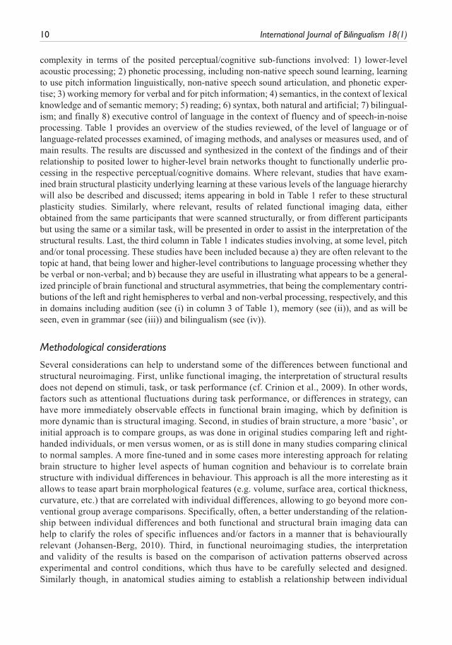
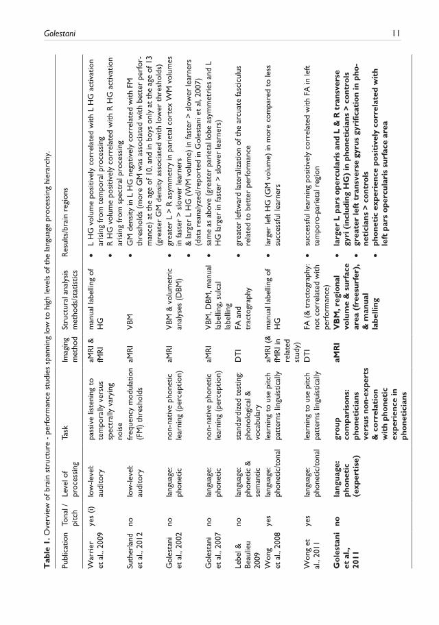
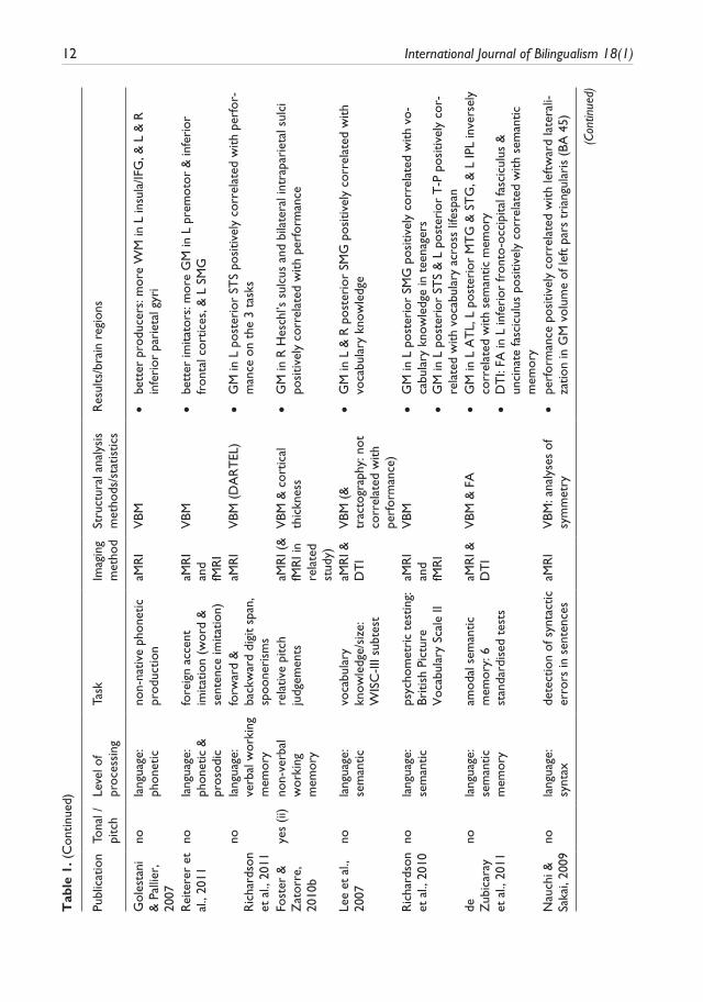
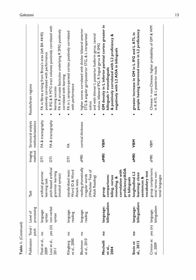
complexity in terms of the posited perceptual/cognitive sub-functions involved: 1) lower-level acoustic processing; 2) phonetic processing, including non-native speech sound learning, learning to use pitch information linguistically, non-native speech sound articulation, and phonetic exper-tise; 3) working memory for verbal and for pitch information; 4) semantics, in the context of lexical knowledge and of semantic memory; 5) reading; 6) syntax, both natural and artificial; 7) bilingual-ism; and finally 8) executive control of language in the context of fluency and of speech-in-noise processing. Table 1 provides an overview of the studies reviewed, of the level of language or of language-related processes examined, of imaging methods, and analyses or measures used, and of main results. The results are discussed and synthesized in the context of the findings and of their relationship to posited lower to higher-level brain networks thought to functionally underlie pro-cessing in the respective perceptual/cognitive domains. Where relevant, studies that have exam-ined brain structural plasticity underlying learning at these various levels of the language hierarchy will also be described and discussed; items appearing in bold in Table 1 refer to these structural plasticity studies. Similarly, where relevant, results of related functional imaging data, either obtained from the same participants that were scanned structurally, or from different participants but using the same or a similar task, will be presented in order to assist in the interpretation of the structural results. Last, the third column in Table 1 indicates studies involving, at some level, pitch and/or tonal processing. These studies have been included because a) they are often relevant to the topic at hand, that being lower and higher-level contributions to language processing whether they be verbal or non-verbal; and b) because they are useful in illustrating what appears to be a general-ized principle of brain functional and structural asymmetries, that being the complementary contri-butions of the left and right hemispheres to verbal and non-verbal processing, respectively, and this in domains including audition (see (i) in column 3 of Table 1), memory (see (ii)), and as will be seen, even in grammar (see (iii)) and bilingualism (see (iv)).
Methodological considerations
Several considerations can help to understand some of the differences between functional and structural neuroimaging. First, unlike functional imaging, the interpretation of structural results does not depend on stimuli, task, or task performance (cf. Crinion et al., 2009). In other words, factors such as attentional fluctuations during task performance, or differences in strategy, can have more immediately observable effects in functional brain imaging, which by definition is more dynamic than is structural imaging. Second, in studies of brain structure, a more ‘basic’, or initial approach is to compare groups, as was done in original studies comparing left and right-handed individuals, or men versus women, or as is still done in many studies comparing clinical to normal samples. A more fine-tuned and in some cases more interesting approach for relating brain structure to higher level aspects of human cognition and behaviour is to correlate brain structure with individual differences in behaviour. This approach is all the more interesting as it allows to tease apart brain morphological features (e.g. volume, surface area, cortical thickness, curvature, etc.) that are correlated with individual differences, allowing to go beyond more con-ventional group average comparisons. Specifically, often, a better understanding of the relation-ship between individual differences and both functional and structural brain imaging data can help to clarify the roles of specific influences and/or factors in a manner that is behaviourally relevant (Johansen-Berg, 2010). Third, in functional neuroimaging studies, the interpretation and validity of the results is based on the comparison of activation patterns observed across experimental and control conditions, which thus have to be carefully selected and designed. Similarly though, in anatomical studies aiming to establish a relationship between individual
Golestani 11T
able
1. O
verv
iew
of b
rain
str
uctu
re -
per
form
ance
stu
dies
spa
nnin
g lo
w t
o hi
gh le
vels
of t
he la
ngua
ge p
roce
ssin
g hi
erar
chy.
Publ
icat
ion
Tona
l /
pitc
hLe
vel o
f pr
oces
sing
Task
Imag
ing
met
hod
Stru
ctur
al a
naly
sis
met
hods
/sta
tistic
sR
esul
ts/b
rain
reg
ions
War
rier
et
al.,
200
9ye
s (i)
low
-leve
l: au
dito
rypa
ssiv
e lis
teni
ng t
o te
mpo
rally
ver
sus
spec
tral
ly v
aryi
ng
nois
e
aMR
I &
fMR
Im
anua
l lab
ellin
g of
H
G •
L H
G v
olum
e po
sitiv
ely
corr
elat
ed w
ith L
HG
act
ivat
ion
aris
ing
from
tem
pora
l pro
cess
ing
•R
HG
vol
ume
posi
tivel
y co
rrel
ated
with
R H
G a
ctiv
atio
n ar
isin
g fr
om s
pect
ral p
roce
ssin
gSu
ther
land
et
al.,
201
2no
low
-leve
l: au
dito
ryfr
eque
ncy
mod
ulat
ion
(FM
) th
resh
olds
aMR
IV
BM •
GM
den
sity
in L
HG
neg
ativ
ely
corr
elat
ed w
ith F
M
thre
shol
ds (
mor
e G
M w
as a
ssoc
iate
d w
ith b
ette
r pe
rfor
-m
ance
) at
the
age
of 1
0, a
nd in
boy
s on
ly a
t th
e ag
e of
13
(gre
ater
GM
den
sity
ass
ocia
ted
with
low
er t
hres
hold
s)G
oles
tani
et
al.,
200
2no
lang
uage
: ph
onet
icno
n-na
tive
phon
etic
le
arni
ng (
perc
eptio
n)aM
RI
VBM
& v
olum
etri
c an
alys
es (
DBM
) •
grea
ter
L >
R a
sym
met
ry in
par
ieta
l cor
tex
WM
vol
umes
in
fast
er >
slo
wer
lear
ners
•
& la
rger
L H
G (
WM
vol
ume)
in fa
ster
> s
low
er le
arne
rs
(dat
a re
anal
yzed
/rep
orte
d in
Gol
esta
ni e
t al
, 200
7)G
oles
tani
et
al.,
200
7no
lang
uage
: ph
onet
icno
n-na
tive
phon
etic
le
arni
ng (
perc
eptio
n)aM
RI
VBM
, DBM
, man
ual
labe
lling
, sul
cal
labe
lling
•sa
me
as a
bove
(gr
eate
r pa
riet
al lo
be a
sym
met
ries
and
L
HG
larg
er in
fast
er >
slo
wer
lear
ners
)
Lebe
l &
Beau
lieu
2009
nola
ngua
ge:
phon
etic
&
sem
antic
stan
dard
ized
tes
ting:
ph
onol
ogic
al &
vo
cabu
lary
DT
IFA
and
tr
acto
grap
hy •
grea
ter
left
war
d la
tera
lizat
ion
of t
he a
rcua
te fa
scic
ulus
re
late
d to
bet
ter
perf
orm
ance
Won
g
et a
l., 2
008
yes
lang
uage
: ph
onet
ic/t
onal
lear
ning
to
use
pitc
h pa
tter
ns li
ngui
stic
ally
aMR
I (&
fM
RI i
n re
late
d st
udy)
man
ual l
abel
ling
of
HG
•la
rger
left
HG
(G
M v
olum
e) in
mor
e co
mpa
red
to le
ss
succ
essf
ul le
arne
rs
Won
g et
al
., 20
11ye
sla
ngua
ge:
phon
etic
/ton
alle
arni
ng t
o us
e pi
tch
patt
erns
ling
uist
ical
lyD
TI
FA (
& t
ract
ogra
phy:
no
t co
rrel
ated
with
pe
rfor
man
ce)
•su
cces
sful
lear
ning
pos
itive
ly c
orre
late
d w
ith F
A in
left
te
mpo
ro-p
arie
tal r
egio
n
Go
lest
ani
et a
l.,
2011
nola
ngua
ge:
pho
neti
c (e
xper
tise
)
gro
up
com
pari
sons
: ph
one
tici
ans
vers
us n
on-
expe
rts
& c
orr
elat
ion
wit
h ph
one
tic
expe
rien
ce in
ph
one
tici
ans
aMR
IV
BM
, reg
iona
l vo
lum
e &
sur
face
ar
ea (
free
surf
er),
&
man
ual
labe
lling
•la
rger
L p
ars
ope
rcul
aris
and
L &
R t
rans
vers
e gy
ri (
incl
udin
g H
G)
in p
hone
tici
ans
> co
ntro
ls •
grea
ter
left
tra
nsve
rse
gyru
s gy
rific
atio
n in
pho
-ne
tici
ans
> co
ntro
ls •
pho
neti
c ex
peri
ence
po
siti
vely
co
rrel
ated
wit
h le
ft p
ars
ope
rcul
aris
sur
face
are
a
12 International Journal of Bilingualism 18(1)
Publ
icat
ion
Tona
l /
pitc
hLe
vel o
f pr
oces
sing
Task
Imag
ing
met
hod
Stru
ctur
al a
naly
sis
met
hods
/sta
tistic
sR
esul
ts/b
rain
reg
ions
Gol
esta
ni
& P
allie
r,
2007
nola
ngua
ge:
phon
etic
non-
nativ
e ph
onet
ic
prod
uctio
naM
RI
VBM
•be
tter
pro
duce
rs: m
ore
WM
in L
insu
la/IF
G, &
L &
R
infe
rior
par
ieta
l gyr
i
Rei
tere
r et
al
., 20
11no
lang
uage
: ph
onet
ic &
pr
osod
ic
fore
ign
acce
nt
imita
tion
(wor
d &
se
nten
ce im
itatio
n)
aMR
I an
d fM
RI
VBM
•be
tter
imita
tors
: mor
e G
M in
L p
rem
otor
& in
feri
or
fron
tal c
ortic
es, &
L S
MG
Ric
hard
son
et a
l., 2
011
nola
ngua
ge:
verb
al w
orki
ng
mem
ory
forw
ard
&
back
war
d di
git
span
, sp
oone
rism
s
aMR
IV
BM (
DA
RT
EL)
•G
M in
L p
oste
rior
ST
S po
sitiv
ely
corr
elat
ed w
ith p
erfo
r-m
ance
on
the
3 ta
sks
Fost
er &
Z
ator
re,
2010
b
yes
(ii)
non-
verb
al
wor
king
m
emor
y
rela
tive
pitc
h ju
dgem
ents
aMR
I (&
fM
RI i
n re
late
d st
udy)
VBM
& c
ortic
al
thic
knes
s •
GM
in R
Hes
chl’s
sul
cus
and
bila
tera
l int
rapa
riet
al s
ulci
po
sitiv
ely
corr
elat
ed w
ith p
erfo
rman
ce
Lee
et a
l.,
2007
nola
ngua
ge:
sem
antic
voca
bula
ry
know
ledg
e/si
ze:
WIS
C-II
I sub
test
aMR
I &
DT
IV
BM (
&
trac
togr
aphy
: not
co
rrel
ated
with
pe
rfor
man
ce)
•G
M in
L &
R p
oste
rior
SM
G p
ositi
vely
cor
rela
ted
with
vo
cabu
lary
kno
wle
dge
Ric
hard
son
et a
l., 2
010
nola
ngua
ge:
sem
antic
psyc
hom
etri
c te
stin
g:
Briti
sh P
ictu
re
Voc
abul
ary
Scal
e II
aMR
I an
d fM
RI
VBM
•G
M in
L p
oste
rior
SM
G p
ositi
vely
cor
rela
ted
with
vo-
cabu
lary
kno
wle
dge
in t
eena
gers
•G
M in
L p
oste
rior
ST
S &
L p
oste
rior
T-P
pos
itive
ly c
or-
rela
ted
with
voc
abul
ary
acro
ss li
fesp
ande
Z
ubic
aray
et
al.,
201
1
nola
ngua
ge:
sem
antic
m
emor
y
amod
al s
eman
tic
mem
ory:
6
stan
dard
ised
tes
ts
aMR
I &
DT
IV
BM &
FA
•G
M in
L A
TL,
L p
oste
rior
MT
G &
ST
G, &
L IP
L in
vers
ely
corr
elat
ed w
ith s
eman
tic m
emor
y •
DT
I: FA
in L
infe
rior
fron
to-o
ccip
ital f
asci
culu
s &
un
cina
te fa
scic
ulus
pos
itive
ly c
orre
late
d w
ith s
eman
tic
mem
ory
Nau
chi &
Sa
kai,
2009
nola
ngua
ge:
synt
axde
tect
ion
of s
ynta
ctic
er
rors
in s
ente
nces
aMR
IV
BM: a
naly
ses
of
sym
met
ry •
perf
orm
ance
pos
itive
ly c
orre
late
d w
ith le
ftw
ard
late
rali-
zatio
n in
GM
vol
ume
of le
ft p
ars
tria
ngul
aris
(BA
45)
(Con
tinue
d)
Tab
le 1
. (C
ontin
ued)
Golestani 13
Publ
icat
ion
Tona
l /
pitc
hLe
vel o
f pr
oces
sing
Task
Imag
ing
met
hod
Stru
ctur
al a
naly
sis
met
hods
/sta
tistic
sR
esul
ts/b
rain
reg
ions
Floe
l et
al.,
2009
nola
ngua
ge:
synt
axar
tific
ial g
ram
mar
le
arni
ng t
ask
DT
IFA
& t
ract
ogra
phy
•FA
in fi
bres
ari
sing
from
Bro
ca’s
are
a (le
ft B
A 4
4/45
) po
sitiv
ely
corr
elat
ed w
ith p
erfo
rman
ceLo
ui e
t al
., 20
11ye
s (ii
i)no
n-ve
rbal
sy
ntax
pitc
h-ba
sed
artif
icia
l gr
amm
ar le
arni
ng
(mus
ical
syn
tax)
DT
IFA
& t
ract
ogra
phy
•R
IFG
& R
MT
G t
ract
vol
umes
pos
itive
ly c
orre
late
d w
ith
lear
ning
suc
cess
•FA
in R
arc
uate
fasc
icul
us u
nder
lyin
g R
SM
G p
ositi
vely
co
rrel
ated
with
lear
ning
Klin
gber
g et
al.,
200
0no
lang
uage
: re
adin
gst
anda
rdiz
ed t
ests
: W
ord
ID &
Wor
d A
ttac
k te
sts
DT
IFA
•FA
in L
tem
poro
-par
ieta
l cor
tex
posi
tivel
y co
rrel
ated
w
ith p
erfo
rman
ce
Blac
kmon
et
al.,
201
0no
lang
uage
: re
adin
gre
adin
g ph
onet
ical
ly
irre
gula
r w
ords
(W
echs
ler
Tes
t of
A
dult
Rea
ding
)
aMR
Ico
rtic
al t
hick
ness
•hi
gher
sco
res
corr
elat
ed w
ith t
hick
er b
ilate
ral a
nter
ior
STG
& a
ngul
ar g
yri/p
oste
rior
ST
G, &
L in
trap
arie
tal
sulc
us •
and
with
thi
nner
L p
oste
rior
fusi
form
gyr
us, c
entr
al
sulc
us, b
ilate
ral I
FG, R
ling
ual g
yrus
& R
SM
GM
eche
lli
et a
l.,
2004
nola
ngua
ge:
bilin
gual
ism
gro
up
com
pari
sons
: bi
ling.
ver
sus
mo
nolin
g. &
co
rrel
atio
n w
ith
pro
ficie
ncy/
AO
A
in b
iling
uals
aMR
IV
BM
•G
M d
ensi
ty in
L in
feri
or
pari
etal
co
rtex
gre
ater
in
bilin
gual
s >
mo
nolin
gual
s •
& p
osi
tive
ly c
orr
elat
ed w
ith
L2
pro
ficie
ncy
&
nega
tive
ly w
ith
L2
AO
A in
bili
ngua
ls
Ste
in e
t al
., 20
12no
lang
uage
: bi
lingu
alis
mlo
ngit
udin
al L
2 le
arni
ng: c
loze
se
nten
ces
&
voca
bula
ry t
est
aMR
IV
BM
•gr
eate
r in
crea
se in
GM
in L
IF
G a
nd L
AT
L in
pe
opl
e ha
ving
impr
ove
d m
ore
in L
2 pr
ofic
ienc
y
Cri
nion
et
al.,
2009
yes
(iv)
lang
uage
: bi
lingu
alis
mgr
oup
com
pari
sons
: to
nal v
ersu
s no
n-to
nal l
angu
ages
aMR
IV
BM •
Chi
nese
> n
on-C
hine
se: h
ighe
r pr
obab
ility
of G
M &
WM
in
R A
TL
& L
pos
teri
or in
sula
Tab
le 1
. (C
ontin
ued)
14 International Journal of Bilingualism 18(1)
Publ
icat
ion
Tona
l /
pitc
hLe
vel o
f pr
oces
sing
Task
Imag
ing
met
hod
Stru
ctur
al a
naly
sis
met
hods
/sta
tistic
sR
esul
ts/b
rain
reg
ions
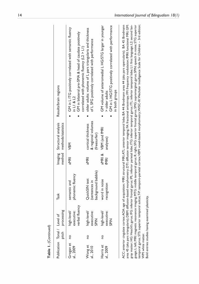
Gro
gan
et
al.,
2009
nohi
gh-le
vel/
exec
utiv
e:
verb
al fl
uenc
y
sem
antic
and
ph
onem
ic fl
uenc
yaM
RI
VBM
•G
M in
L IT
G p
ositi
vely
cor
rela
ted
with
sem
antic
flue
ncy
in L
1 &
L2
•G
M in
bila
tera
l pre
-SM
A &
hea
d of
cau
date
pos
itive
ly
corr
elat
ed w
ith p
hone
mic
flue
ncy
(L2
> L
1)W
ong
et
al.,
2010
nohi
gh-le
vel/
exec
utiv
e:
SPIN
Qui
ckSI
N t
est
(sen
tenc
es in
ba
ckgr
ound
bab
ble)
aMR
Ico
rtic
al t
hick
ness
&
reg
iona
l vol
umes
(fr
eesu
rfer
)
•ol
der
adul
ts: v
olum
e of
L p
ars
tria
ngul
aris
and
thi
ckne
ss
of L
SFG
pos
itive
ly c
orre
late
d w
ith p
erfo
rman
ce
Har
ris
et
al.,
2009
nohi
gh-le
vel/
exec
utiv
e:
SPIN
wor
d in
noi
se
reco
gniti
onaM
RI &
fM
RI
VBM
(an
d fM
RI
anal
yses
) •
GM
vol
ume
of a
nter
omed
ial L
HG
/ST
G la
rger
in y
oung
er
> o
lder
adu
lts •
GM
in L
HG
/ST
G p
ositi
vely
cor
rela
ted
with
per
form
ance
in
bot
h gr
oups
AC
C: a
nter
ior
cing
ulat
e co
rtex
; AO
A: a
ge o
f acq
uisi
tion;
a MR
I: st
ruct
ural
MR
I; AT
L: a
nter
ior
tem
pora
l lob
e; B
A 4
4: B
rodm
ann
area
44
(aka
par
s op
ercu
lari
s);
BA
45:
Bro
dman
n ar
ea 4
5 (a
ka p
ars
tria
ngul
aris
); D
BM: d
iffus
ion-
base
d m
orph
omet
ry; D
TI:
diffu
sion
ten
sor
imag
ing;
FA: f
ract
iona
l ani
sotr
opy;
FM: f
requ
ency
mod
ulat
ion;
fMR
I: fu
nctio
nal M
RI;
GM
: gr
ay m
atte
r; H
G: H
esch
l’s g
yrus
; IFG
: inf
erio
r fr
onta
l gyr
us; I
PL: i
nfer
ior
pari
etal
lobe
; IT
G: i
nfer
ior
tem
pora
l gyr
us; I
TL:
infe
rior
tem
pora
l lob
e; L
1: fi
rst
lang
uage
; L2:
sec
ond
lan-
guag
e; L
: lef
t; M
RI:
mag
netic
res
onan
ce im
agin
g; M
TG
: mid
dle
tem
pora
l gyr
us; R
: rig
ht; S
FG: s
uper
ior
fron
tal g
yrus
; SM
G: s
upra
mar
gina
l gyr
us; S
PIN
: spe
ech
in n
oise
; ST
G: s
uper
ior
tem
pora
l gyr
us; S
TS:
supe
rior
tem
pora
l sul
cus;
T-P:
tem
poro
-par
ieta
l cor
tex;
VBM
: vox
el-b
ased
mor
phom
etry
; WIS
C-II
I: Wec
hsle
r In
telli
genc
e Sc
ale
for
Chi
ldre
n –
3rd
editi
on;
WM
: whi
te m
atte
r.Bo
ld e
ntri
es: s
tudi
es h
avin
g ex
amin
ed p
last
icity
.
Tab
le 1
. (C
ontin
ued)
Golestani 15
differences in aspects of behavioural performance and brain structure, aspects of performance are measured and subsequently correlated with brain structural measures. Thus, even in struc-tural studies, careful selection of behavioural measures is required in order to yield valid results that can be interpreted meaningfully and in a more general context. Fourth, and related to the above, only behavioural measures that are relatively stable over time within individuals and that do not fluctuate widely as a function of the specific testing session (due to attentional or other variables, or simply due to lack of test–retest stability) can be used to correlate with brain struc-ture. If a particular behavioural measure were to vary more widely over time when retested in the same individual compared to the variability observed across individuals (due to individual dif-ferences) then statistically robust correlations with brain anatomy would be very difficult to establish. Last, in brain structural studies that aim to compare different participant groups (e.g. monolinguals versus bilinguals), the interpretation and validity of findings depends on the care-ful selection and matching of the experimental and control groups (for example with respect to age, sex, educational level, experience, etc.) so as to ensure that group differences are not driven by confounds of non-interest.
Brain structure – performance relationships spanning low to high levels of the language processing hierarchy
Table 1 provides an overview of the studies reviewed in this section.
Lower-level acoustic processing, relative pitch and auditory short-term memory
Several studies have examined individual differences in local brain structure and its relationship to lower-level, acoustic processing of sounds. One of these, by Warrier and colleagues (2009), can be seen as an extension of the original brain structure–function studies described in the introduction that examined normative left–right structural asymmetries in auditory cortex morphology and their relationship to handedness and hemispheric lateralization for language. In this study, fMRI was used to measure brain function while participants listened to noise stimuli varying orthogonally in temporal and spectral dimensions (Warrier et al., 2009). The functional activation patterns were then related to volumetric measurements of left and right Heschl’s gyrus (HG). As expected, Warrier and colleagues found that the anatomy of HG was very variable across individuals (Abdul-Kareem & Sluming, 2008; Penhune et al., 1996). In terms of brain function, they found leftward lateralization in the neural response to varying rates of stimulus change (i.e. stimuli containing rapidly changing information), and rightward lateralization in response to increasing spectral infor-mation. Importantly, they found that larger left HG volumes were associated with larger extents of ‘rate-related’ cortex on the left, and that right HG volumes were associated with larger extents of spectral-related cortex on the right. These findings are in line with a body of literature suggesting functional asymmetries in the way in which the auditory cortices process rapidly changing ‘tempo-ral’ information, which is proposed to be left-lateralized, versus steady-state spectral (pitch, or frequency) information, which is thought to be right-lateralised (Zatorre, Belin, & Penhune, 2002; Zatorre, Evans, Meyer, & Gjedde, 1992; Zatorre & Gandour, 2008). They are also in line with microscopic studies showing that the cellular organization of left HG makes it more suited to pro-cessing rapidly changing auditory information than the right HG (Anderson, Southern, & Powers, 1999; Galuske, Schlote, Bratzke, & Singer, 2000; Seldon, 1981).
A very recent study by Sutherland and colleagues (2012) has also examined brain structural correlates of low-level auditory processing using a task requiring acuity in the temporal domain.
16 International Journal of Bilingualism 18(1)
Here, adolescents heard two tones and had to decide which of the two contained 2-Hz frequency modulation (FM). Based on their performance, modulation depth was adjusted and FM thresholds were calculated. The participants were scanned longitudinally, several times each, at the ages of 10, 11.5, and 13 years. It was found that the 2-Hz FM thresholds were significantly correlated with gray-matter density in left HG (higher GM density was associated with better performance) at the age of 10 years, but that this correlation weakened with age. Also, at Time 3 (i.e. age 13) there was a relationship between gray-matter density in ‘left HG and FM thresholds in boys but not in girls. Interestingly, FM thresholds correlated with spelling abilities at the age of 10, again only in boys, suggesting the importance of being able to detect low-level changes in frequency for language and literacy. These findings suggest that at least in a subportion of individuals, the lower-level process-ing of sounds and of how they change over time are relevant to higher-level aspects of language processing such as literacy. This study highlights the relevance of the approach taken in this review, which is to examine the different lower to higher-level sub-processes required for speech and lan-guage and their relationship to brain structure, since it is shown here that low-level abilities predict higher-level ones, as they predict individual differences in brain structure. The results also extend the above-described ones by Warrier and colleagues in that it is here demonstrated that the relation-ship between lower-level temporal processing and left auditory cortex morphology can be linked to higher-level aspects of language processing.
Phonetic perception and production
Non-native phonetic perception. Phonetic perception can be seen as the lowest-level, most elemental sub-unit of speech since successful phonetic perception is required for subsequent speech/phonetic production, word perception, verbal working memory, semantic processing, and other ‘upstream’ cognitive speech and language related processes. Similarly, the ability to imitate foreign accents on the word and sentence levels is dependent not only on successful perception of foreign speech sounds but also on the ability to accurately imitate speech on the phonetic level. In this section studies examining brain structural correlates of phonetic perception, production, and accent imita-tion will be described.
A number of studies, including several from our group, have examined structural correlates of phonetic perception. First, behaviourally, it has been shown that there are large individual differ-ences in how well people can hear non-native phonetic contrasts and also in how quickly they can learn to hear them (Golestani & Zatorre, 2009; Hattori & Iverson, 2009). In three related brain imag-ing studies, performed in three different groups of participants, a synthetic Hindi dental- retroflex contrast was used to examine brain structural (Golestani, Molko, Dehaene, LeBihan, & Pallier, 2007; Golestani et al., 2002) and functional (Golestani & Zatorre, 2004) correlates of indi-vidual differences in non-native phonetic perception. Functionally, it was shown that once the non-native contrast was learned, regions including bilateral auditory cortices and the left inferior frontal gyrus were recruited. These regions are the same as were recruited during the perception of a native phonetic contrast. Further, people who were more successful at learning to hear the non-native con-trast showed less activation, or more efficient neural processing, in the left inferior frontal gyrus (Golestani & Zatorre, 2004). Functional brain imaging and transcranial magnetic stimulation studies have shown that the posterior portion of the left inferior frontal gyrus, the left pars opercularis, or Brodmann’s area (BA) 44, and the left supramarginal gyrus within the left inferior parietal cortex, are involved in and even necessary for phonological processing and for subvocal rehearsal in verbal working memory (Hartwigsen et al., 2010; Paulesu, Frith, & Frackowiak, 1993). Further, the left pars opercularis has been shown to play a role in the extraction and manipulation of phonetic
Golestani 17
segments in verbal working memory (Burton, Small & Blumstein, 2000; Zatorre, Meyer, Gjedde, & Evans, 1996). The finding of less activation of the left inferior frontal gyrus in ‘faster’ learners is consistent with the idea that phonetic processing is less effortful in faster than for ‘slower’ phonetic learners.
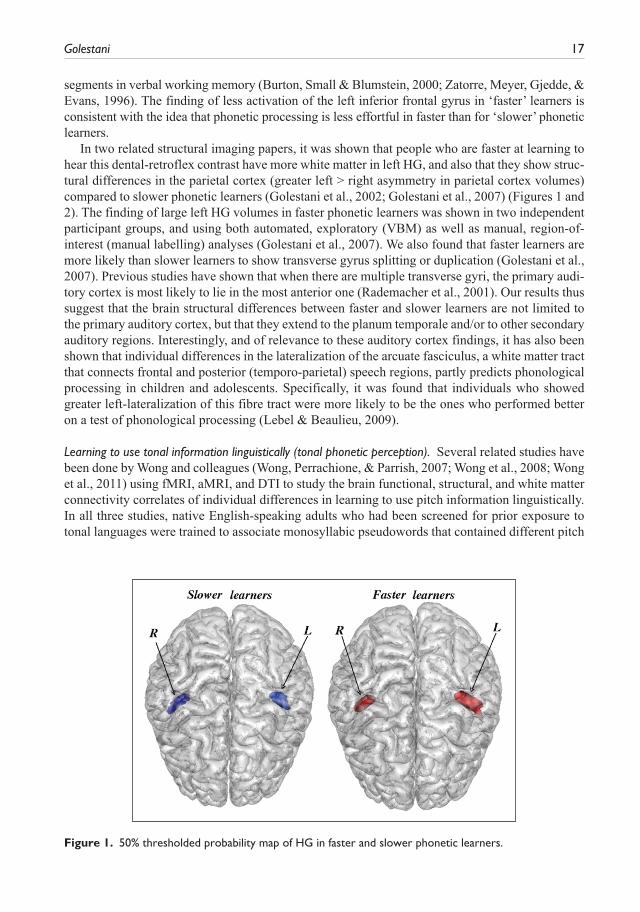
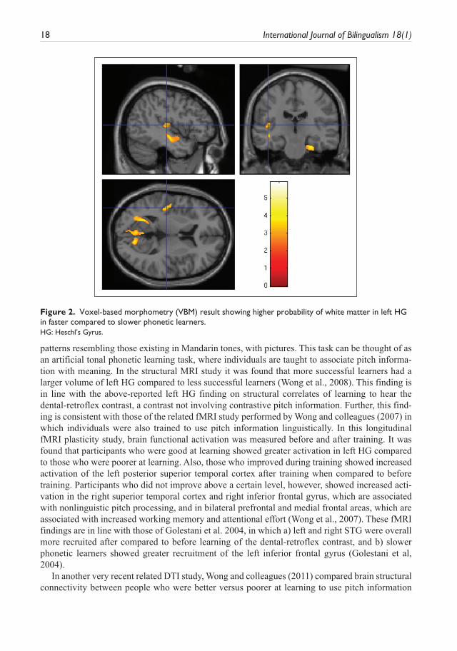
In two related structural imaging papers, it was shown that people who are faster at learning to hear this dental-retroflex contrast have more white matter in left HG, and also that they show struc-tural differences in the parietal cortex (greater left > right asymmetry in parietal cortex volumes) compared to slower phonetic learners (Golestani et al., 2002; Golestani et al., 2007) (Figures 1 and 2). The finding of large left HG volumes in faster phonetic learners was shown in two independent participant groups, and using both automated, exploratory (VBM) as well as manual, region-of-interest (manual labelling) analyses (Golestani et al., 2007). We also found that faster learners are more likely than slower learners to show transverse gyrus splitting or duplication (Golestani et al., 2007). Previous studies have shown that when there are multiple transverse gyri, the primary audi-tory cortex is most likely to lie in the most anterior one (Rademacher et al., 2001). Our results thus suggest that the brain structural differences between faster and slower learners are not limited to the primary auditory cortex, but that they extend to the planum temporale and/or to other secondary auditory regions. Interestingly, and of relevance to these auditory cortex findings, it has also been shown that individual differences in the lateralization of the arcuate fasciculus, a white matter tract that connects frontal and posterior (temporo-parietal) speech regions, partly predicts phonological processing in children and adolescents. Specifically, it was found that individuals who showed greater left-lateralization of this fibre tract were more likely to be the ones who performed better on a test of phonological processing (Lebel & Beaulieu, 2009).
Learning to use tonal information linguistically (tonal phonetic perception). Several related studies have been done by Wong and colleagues (Wong, Perrachione, & Parrish, 2007; Wong et al., 2008; Wong et al., 2011) using fMRI, aMRI, and DTI to study the brain functional, structural, and white matter connectivity correlates of individual differences in learning to use pitch information linguistically. In all three studies, native English-speaking adults who had been screened for prior exposure to tonal languages were trained to associate monosyllabic pseudowords that contained different pitch
Figure 1. 50% thresholded probability map of HG in faster and slower phonetic learners.
18 International Journal of Bilingualism 18(1)
patterns resembling those existing in Mandarin tones, with pictures. This task can be thought of as an artificial tonal phonetic learning task, where individuals are taught to associate pitch informa-tion with meaning. In the structural MRI study it was found that more successful learners had a larger volume of left HG compared to less successful learners (Wong et al., 2008). This finding is in line with the above-reported left HG finding on structural correlates of learning to hear the dental-retroflex contrast, a contrast not involving contrastive pitch information. Further, this find-ing is consistent with those of the related fMRI study performed by Wong and colleagues (2007) in which individuals were also trained to use pitch information linguistically. In this longitudinal fMRI plasticity study, brain functional activation was measured before and after training. It was found that participants who were good at learning showed greater activation in left HG compared to those who were poorer at learning. Also, those who improved during training showed increased activation of the left posterior superior temporal cortex after training when compared to before training. Participants who did not improve above a certain level, however, showed increased acti-vation in the right superior temporal cortex and right inferior frontal gyrus, which are associated with nonlinguistic pitch processing, and in bilateral prefrontal and medial frontal areas, which are associated with increased working memory and attentional effort (Wong et al., 2007). These fMRI findings are in line with those of Golestani et al. 2004, in which a) left and right STG were overall more recruited after compared to before learning of the dental-retroflex contrast, and b) slower phonetic learners showed greater recruitment of the left inferior frontal gyrus (Golestani et al, 2004).
In another very recent related DTI study, Wong and colleagues (2011) compared brain structural connectivity between people who were better versus poorer at learning to use pitch information
Figure 2. Voxel-based morphometry (VBM) result showing higher probability of white matter in left HG in faster compared to slower phonetic learners.HG: Heschl’s Gyrus.
Golestani 19
linguistically. Analyses of FA maps revealed that white matter connectivity in the left temporo-parietal region correlated positively with learning success (Wong et al, 2011). Once again, the location of this result is consistent with the above-reported parietal cortex structural differences between people who are faster versus slower at learning to hear the Hindi dental-retroflex contrast.
Phonetic expertise. In a more recent study, we were interested in understanding whether the struc-tural differences observed in HG between faster and slower phonetic learners could be due to individual differences in aspects of linguistic experience. We also wanted to test whether extensive phonetic training can influence other aspects of brain structure. Brain structure was compared in phoneticians to that of healthy, matched controls. Phoneticians are language experts who are highly trained to listen to speech and to transcribe it into an international phonetic alphabet. They thus have extensive experience with phonetic segmentation and analysis. It was found that phoneticians have a larger left pars opercularis and larger bilateral transverse gyri, which include HG, compared to non-experts. Further, it was found that the size of the left pars opercularis, the Broca’s area sub-region described above, is known to be functionally involved in phonetic parsing and segmentation (Burton et al., 2000; Zatorre et al, 1996), is larger in phoneticians with more compared to those with fewer years of transcription training experience. These cross-sectional results suggest that intensive phonetic training shapes this Broca’s area subregion (Golestani, Price, & Scott, 2011).
Non-native phonetic and speech imitation/articulation. The accurate pronunciation of foreign speech sounds can only occur in a spontaneous and relatively automatic fashion if the sounds are per-ceived accurately. The ability to produce or to imitate foreign speech sounds therefore depends on initial successful phonetic perception, and can therefore be conceptualized as being next in the language processing hierarchy put forth in this paper. Several studies have examined brain struc-tural correlates of speech and of speech sound articulation (Golestani & Pallier, 2007; Reiterer et al., 2011). In one study, VBM was used to examine brain structural correlates of the ability to accurately pronounce the Farsi voiced uvular plosive. It was found that people who are more skilled at articulating this sound have more white matter in the left anterior insula/inferior prefron-tal cortex and the inferior parietal cortices bilaterally (Golestani & Pallier, 2007). More white mat-ter could be due to greater myelination, which would allow more efficient neural transmission between relevant brain regions.
In a more recent study by Reiterer and colleagues (2011), 140 late bilingual adults were tested on how well they could imitate foreign accents. They were asked to imitate sentences and words in either their second language (English), or in Indian languages (Hindi and Tamil) that they had never been exposed to. Structural and functional MRI data was acquired in a subset of individu-als. Functional imaging revealed individual differences in the recruitment of left-hemisphere speech areas during sentence and word imitation. People with poorer abilities showed higher hemodynamic activation in a distinct fronto-parietal network, and conversely, people with higher abilities showed less activation in these regions but also more gray matter in this same network, including the left inferior parietal cortex (supramarginal gyrus) and in the left inferior frontal/premotor area (Reiterer et al., 2011).
As it can be seen from the above studies, and as would be expected based on functional neuro-anatomy, structural correlates of speech sound processing are found in a) HG but also in the adja-cent secondary auditory cortex and in the parietal cortex for phonetic perception/learning, b) in the inferior parietal cortices bilaterally and in the left anterior insula for phonetic production/articula-tion, and in c) the left supramarginal gyrus and left inferior frontal/premotor area for speech/accent
20 International Journal of Bilingualism 18(1)
imitation at the word and sentence levels. Taken together, the studies on phonetic perception and production show a partial dissociation between the structural correlates of phonetic perception and production. Further, the respective locations of the structural correlates are in the very regions that have been shown, in functional neuroimaging and in lesion studies, to functionally underlie these cognitive processes (Binder et al., 1994; Dronkers, 1996; Hickok, Buchsbaum, & Humphries, 2003; Zatorre et al., 1992). Findings of individual differences in foreign speech sound learning and their relationship with local aspects of brain structure can be further explored by linking these find-ings to electrophysiological research on functional brain mechanisms that may underlie these indi-vidual differences (Diaz, Baus, Escera, Costa, & Sebastian-Galles, 2008), and by looking for convergence across different brain imaging modalities. They can also be further explored by gain-ing a better understanding of the functional mechanisms underlying speech sound processing and learning during adulthood, both in perception (Raizada, Tsao, Liu, & Kuhl, 2010; Zhang et al., 2009) and in production (Moser et al., 2009; Shuster & Lemieux, 2005).
Verbal and non-verbal working memory
Verbal working memory. Verbal working memory involves actively retaining verbal information in short-term memory for a relatively short period of time, as required by the task or by the context (e.g. holding a phone number in memory before dialling it). It can thus in principle be performed using only phonetic information, but typically involves words and thus meaning as for example in the digit span subtest of the Wechsler Adult Intelligence Scale. It can be thought of as being one step ‘upstream’ in the language processing hierarchy, since after hearing speech sounds or words, in order to ensure speech comprehension, the information has to be main-tained in working memory while it is processed and subsequently related to meaning and to its surrounding context (e.g. sentence).
In a very recent study, performance on a verbal short-term memory task was related to brain structure. Verbal short-term memory was tested using the digit span subtests of the Wechsler Adult Intelligence Scale, 3rd edition (Wechsler, 1998). It was found that better performance was associ-ated with a higher probability of gray matter in the left posterior superior temporal sulcus (STS) in normal and also in dyslexic adults (Richardson et al., 2011). The location of this effect was near that of a lesion site that has recently been associated with reduced auditory short-term-memory capacity in patients with stroke damage (Leff et al., 2009).
Non-verbal working memory. Non-verbal working memory is typically assessed using visual-spatial information, but can in principle also be tested using non-verbal auditory information. Two related studies by Foster and Zatorre (2010a & 2010b) have examined brain functional and structural cor-relates of relative pitch judgments. Relative pitch judgements involve transforming high-level auditory information by comparing and recognizing melodies by their interval structure. Function-ally, it was shown that brain activity within the right intraparietal sulcus predicted performance in both musicians and non-musicians on this task (Foster & Zatorre, 2010a). In the related structural imaging study, it was found that gray matter density and cortical thickness in the right HG and in the intraparietal sulci positively predicted relative pitch performance (Foster & Zatorre, 2010b). These results are in the vicinity of the region homologous to that described above on structural correlates of verbal working memory, with verbal working memory being related to brain structure in the left posterior superior temporal sulcus (STS) and non-verbal working memory to that in the right intraparietal sulcus, just posterior and superior to the right STS. The lateralization of these complementary findings parallels the left versus right auditory cortex structural lateralization
Golestani 21
related to verbal versus tonal/pitch information, respectively, described in the section entitled ‘Lower-level acoustic processing, relative pitch and auditory short-term memory’, above.
Semantics: Lexical knowledge and semantic memory
Lexical knowledge. As described at the beginning of the above section, semantic processing occurs after verbal information is retained in working memory while it is related to meaning stored in long-term semantic memory. There are large individual differences in the vocabulary of individu-als, in other words in how much verbal semantic information they have stored in long-term mem-ory, depending on many factors including education, socio-cultural environment and multilingualism Several studies have examined brain structural correlates of vocabulary knowledge, and have pre-viously been reviewed elsewhere (Richardson & Price, 2009). Results will also be described here as they can then be linked to those on structural correlates of language processing at lower and higher levels in the language processing hierarchy.
Lee and colleagues (2007) used aMRI and DTI to show that vocabulary knowledge in monolin-gual teenagers is positively correlated with gray matter ‘density’, or probability, in bilateral poste-rior supramarginal gyri. The effect was specific to the number of words learned, regardless of verbal fluency or other cognitive abilities. The study also showed that this brain region is structur-ally connected to other parts of the inferior parietal cortex, ones that process either the sounds of words (the anterior supramarginal gyrus) or their meaning (the angular gyrus), suggesting that the posterior supramarginal gyrus may be a binding site for phonological and semantic information (Lee et al., 2007). In a related study, Richardson, Thomas, Filippi, Harth, and Price (2010) tested a larger number of individuals on vocabulary knowledge using different methods than those that were used in the above study, and in an age range spanning a large part of the human life span (7 to 75 years). Despite the methodological differences, they replicated the finding of a positive asso-ciation between gray matter probability in the posterior supramarginal gyrus and vocabulary knowledge, but only in the teenagers that were included in their study. They showed that gray matter density in two other, left posterior temporal regions (the posterior STS and the posterior temporo-parietal junction) was positively correlated with vocabulary across the lifespan. They sug-gest that the finding in the teenagers reflects changes arising from a mode of learning that is imple-mented during formal education, whereas that the findings across the lifespan reflect vocabulary learning that arises from linking semantic and syntactic information to prior knowledge in the context of day to day exposure to language (Richardson et al., 2010).
Semantic memory. Semantic memory refers to long-term memory for information related to ideas, meanings, and concepts that are not related to personal experiences. Semantic memory can be accessed with words, but also with images and information occurring in other modalities. In this sense it can be viewed as partly encompassing the lexicon, but also information that is based much more broadly in human cognition.
In a recent study by de Zubicaray, Rose, and McMahon (2011), aMRI and DTI were used to examine the relationship between semantic memory and brain structure in healthy older adults ranging from 55–85 years of age. They found that amodal semantic memory, as assessed by six standardized neuropsychological tests, was inversely correlated with gray matter volumes in a predominantly left lateralized network including the anterior temporal lobe and the posterior tem-poral and posterior inferior parietal lobes. It was further positively correlated with FA of the left inferior fronto-occipital fasciculus and uncinate fasciculus fibre pathways, ones which connect the posterior occipitotemporal to the anterior temporal lobe, and the anterior temporal lobe to the
22 International Journal of Bilingualism 18(1)
orbitofrontal cortex, respectively. These regions are all known to be functionally involved in aspects of language and semantic memory (de Zubicaray et al., 2011). The inverse relationship with gray matter volumes was attributed to a potential mechanism of synaptic pruning during development.
Syntax
Syntax refers to the rules that govern the sentence structure of language. As such, syntactic infor-mation in addition to semantics is used to derive meaning from speech at the sentence level, and can be conceptualized as being next in the language processing hierarchy here described.
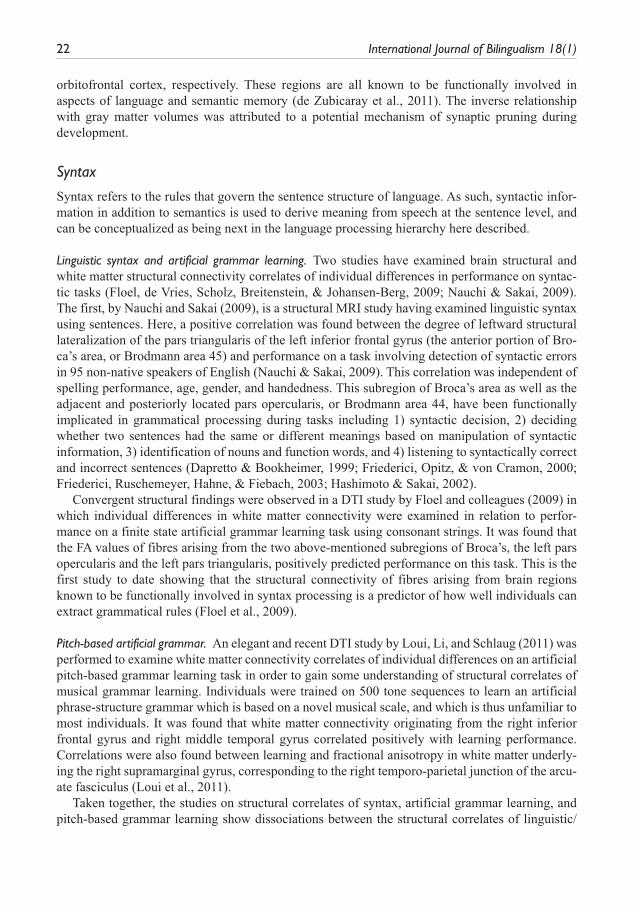
Linguistic syntax and artificial grammar learning. Two studies have examined brain structural and white matter structural connectivity correlates of individual differences in performance on syntac-tic tasks (Floel, de Vries, Scholz, Breitenstein, & Johansen-Berg, 2009; Nauchi & Sakai, 2009). The first, by Nauchi and Sakai (2009), is a structural MRI study having examined linguistic syntax using sentences. Here, a positive correlation was found between the degree of leftward structural lateralization of the pars triangularis of the left inferior frontal gyrus (the anterior portion of Bro-ca’s area, or Brodmann area 45) and performance on a task involving detection of syntactic errors in 95 non-native speakers of English (Nauchi & Sakai, 2009). This correlation was independent of spelling performance, age, gender, and handedness. This subregion of Broca’s area as well as the adjacent and posteriorly located pars opercularis, or Brodmann area 44, have been functionally implicated in grammatical processing during tasks including 1) syntactic decision, 2) deciding whether two sentences had the same or different meanings based on manipulation of syntactic information, 3) identification of nouns and function words, and 4) listening to syntactically correct and incorrect sentences (Dapretto & Bookheimer, 1999; Friederici, Opitz, & von Cramon, 2000; Friederici, Ruschemeyer, Hahne, & Fiebach, 2003; Hashimoto & Sakai, 2002).
Convergent structural findings were observed in a DTI study by Floel and colleagues (2009) in which individual differences in white matter connectivity were examined in relation to perfor-mance on a finite state artificial grammar learning task using consonant strings. It was found that the FA values of fibres arising from the two above-mentioned subregions of Broca’s, the left pars opercularis and the left pars triangularis, positively predicted performance on this task. This is the first study to date showing that the structural connectivity of fibres arising from brain regions known to be functionally involved in syntax processing is a predictor of how well individuals can extract grammatical rules (Floel et al., 2009).
Pitch-based artificial grammar. An elegant and recent DTI study by Loui, Li, and Schlaug (2011) was performed to examine white matter connectivity correlates of individual differences on an artificial pitch-based grammar learning task in order to gain some understanding of structural correlates of musical grammar learning. Individuals were trained on 500 tone sequences to learn an artificial phrase-structure grammar which is based on a novel musical scale, and which is thus unfamiliar to most individuals. It was found that white matter connectivity originating from the right inferior frontal gyrus and right middle temporal gyrus correlated positively with learning performance. Correlations were also found between learning and fractional anisotropy in white matter underly-ing the right supramarginal gyrus, corresponding to the right temporo-parietal junction of the arcu-ate fasciculus (Loui et al., 2011).
Taken together, the studies on structural correlates of syntax, artificial grammar learning, and pitch-based grammar learning show dissociations between the structural correlates of linguistic/
Golestani 23
symbolic versus musical syntax learning in regions including the left and right inferior frontal gyri, respectively. Once again, these asymmetries appear to be in line with the more general principle of cerebral asymmetries, with verbal processing being left-lateralized and with non-verbal processing being right-lateralized.
Reading
Reading involves linking semantic and syntactic information contained in written words and sen-tences to information stored in semantic memory to derive meaning. As such, successful and expe-rienced reading requires lexical and syntactic knowledge as well as access to semantic memory.
As mentioned in the introduction, numerous studies have been done on brain structure in indi-viduals with dyslexia, but are beyond the scope of this review. Several studies examining structural correlates of reading at the word level in healthy, non-dyslexic individuals will, however, be reviewed here. The first is a DTI study by Klingberg and colleagues (2000) done both in individu-als with dyslexia and healthy individuals. Reading was evaluated using two standardized tests: a) the Word ID test (Woodcock, 1987), in which participants are required to pronounce increasingly difficult words, and b) the Word Attack test (Woodcock, 1987) in which subjects have to read, or decode, pseudowords. It was found that fractional anisotropy in the left temporo-parietal cortex was positively correlated with reading scores within the reading-impaired adults and, interestingly, also within the control group. This result suggests that individual differences in aspects of white matter connectivity between this important language hub and other brain regions involved in vis-ual, auditory, and language processing predict individual differences in reading skill, and this even within the normal range (Klingberg et al., 2000).
The second is a structural MRI study by Blackmon and colleagues (2010), who tested the performance of 60 healthy individuals on a visual word reading test composed of phonetically irregular words using the Wechsler Test of Adult Reading (Wechsler, 2001). They found that higher scores were associated with thicker cortex (which is in turn usually associated with more gray matter) in bilateral anterior superior temporal gyri, bilateral angular gyri/posterior supe-rior temporal gyri, and the left intraparietal sulcus. Higher scores were also associated with a thinner cortex in the left posterior fusiform gyrus, the central sulcus, bilateral inferior frontal gyri, and the right lingual and supramarginal gyri. These findings suggest that the ability to correctly pronounce phonetically irregular words is associated with greater cortical thickness in brain areas that are commonly activated in functional neuroimaging studies of word reading, including areas associated with grapheme-to-phonemic conversion, and with thinner cortical thickness in regions including ones such as the left fusiform gyrus, associated with visual word form recognition (Blackmon et al., 2010). The results of Blackmon and colleagues converge with those of Klingberg and colleagues described above in that both show an association between brain structure (either white matter microstructure or cortical thickness) in regions including the left temporo-parietal cortex, and success at decoding phonetically irregular words or pseudo-words.
Bilingualism
Monolinguals versus bilinguals. Bilingualism involves all of the levels of language that have been described in the above sections, but in two languages rather than in one. Thus, in addition to knowl-edge and use of two sets of phonologies, lexicons, grammars, etc., bilingualism involves language control mechanisms beyond those required in one language in monolinguals. These additional
24 International Journal of Bilingualism 18(1)
language control requirements in bilinguals compared to monolinguals include selection of which language to speak (depending on factors such as context), and therefore also which to inhibit. Lan-guage control can be thought of as being the highest level aspect of language processing in the hierarchy proposed in this review since it engages, in addition to speech networks per se, a high-level, left-lateralized fronto-parieto-subcortical brain network which is also engaged during non- linguistic executive functioning. For a review of executive control in the bilingual brain see Her-vais-Adelman, Moser-Mercer, and Golestani (2011). Note also that in the next and last section of this review, studies will be reviewed that have explicitly examined brain structural correlates of tasks requiring executive control over language, as well as of top-down influences on language processing.
Here several studies having examined brain structural correlates of speaking one versus several languages, and of bilinguals who do or who don’t speak a tonal language, will be reviewed. Mechelli and colleagues (2004) compared brain structure in monolinguals versus bilinguals using VBM. They found that in bilinguals, there is a higher probability of more gray matter in the left inferior parietal cortex compared to monolinguals. They also found that this structural difference is more pronounced in bilinguals who are more proficient in or who learned their second language earlier, suggesting that greater experience and/or expertise with a second language results in structural reorganization in this brain region (Mechelli et al., 2004). Note that the region identified in this study is in close vicinity to the region found to structurally predict vocabulary knowledge in the studies reviewed in the ‘Lexical knowledge’ section above (Lee et al., 2007; Richardson et al., 2010). The differences observed here in bilinguals could be driven by higher overall knowledge of vocabulary in two languages in bilinguals compared to in one language in monolinguals. The review paper by Richardson and Price (2009) provides a detailed comparison of this bilingualism study and the ones on lexical knowledge reviewed above.
A very recent study by Stein and colleagues (2012) examined brain structural plasticity arising from second language learning longitudinally in native English speakers having moved to Switzerland to learn German. Individuals were scanned using aMRI upon their arrival and again five months later, when their proficiency in German had increased. VBM analyses revealed that structural change over time in the left inferior frontal gyrus and in the left anterior temporal lobe was positively correlated with individual differences in the increase in second language profi-ciency over time (Stein et al., 2012). Taken together with the above study, the lack of findings in the left inferior parietal cortex here could be due to the fact that a long enough time window of learning was not examined.
Tonal languages. A study by Crinion and colleagues (2009) has examined brain structure in bilinguals who spoke a tonal language, Chinese, compared to ones who did not speak any tonal language. Speakers of the tonal language included a) native Chinese speakers (21 individuals) and b) European participants who had learned Chinese as a non-native language late in life (seven individuals). They found that in all speakers of Chinese compared to people who did not speak a tonal language, there was a higher probability of gray and white matter in regions including the right anterior STG and the underlying right middle longitudinal fasciculus, and the left posterior insula (medial to HG) and the underlying white matter. These brain regions have previously been functionally implicated in linking the pitch of words to their meaning (Crinion et al., 2009). The fact that these findings occurred both in native and non-native speakers of tonal languages suggests that the differences are not related to ethnicity effects. Also, the fact that the finding also occurred in the seven individuals who learned Chinese late
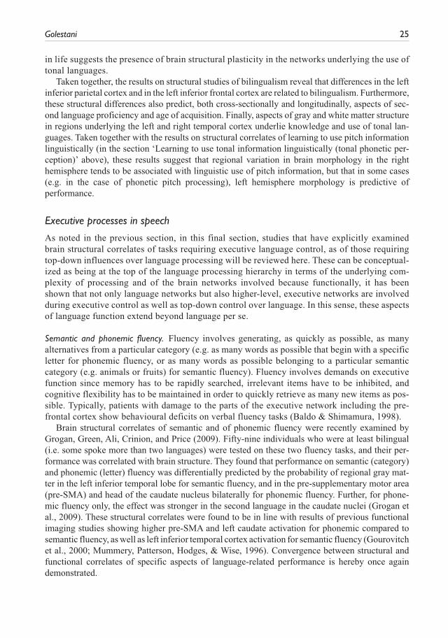
Golestani 25
in life suggests the presence of brain structural plasticity in the networks underlying the use of tonal languages.
Taken together, the results on structural studies of bilingualism reveal that differences in the left inferior parietal cortex and in the left inferior frontal cortex are related to bilingualism. Furthermore, these structural differences also predict, both cross-sectionally and longitudinally, aspects of sec-ond language proficiency and age of acquisition. Finally, aspects of gray and white matter structure in regions underlying the left and right temporal cortex underlie knowledge and use of tonal lan-guages. Taken together with the results on structural correlates of learning to use pitch information linguistically (in the section ‘Learning to use tonal information linguistically (tonal phonetic per-ception)’ above), these results suggest that regional variation in brain morphology in the right hemisphere tends to be associated with linguistic use of pitch information, but that in some cases (e.g. in the case of phonetic pitch processing), left hemisphere morphology is predictive of performance.
Executive processes in speech
As noted in the previous section, in this final section, studies that have explicitly examined brain structural correlates of tasks requiring executive language control, as of those requiring top-down influences over language processing will be reviewed here. These can be conceptual-ized as being at the top of the language processing hierarchy in terms of the underlying com-plexity of processing and of the brain networks involved because functionally, it has been shown that not only language networks but also higher-level, executive networks are involved during executive control as well as top-down control over language. In this sense, these aspects of language function extend beyond language per se.
Semantic and phonemic fluency. Fluency involves generating, as quickly as possible, as many alternatives from a particular category (e.g. as many words as possible that begin with a specific letter for phonemic fluency, or as many words as possible belonging to a particular semantic category (e.g. animals or fruits) for semantic fluency). Fluency involves demands on executive function since memory has to be rapidly searched, irrelevant items have to be inhibited, and cognitive flexibility has to be maintained in order to quickly retrieve as many new items as pos-sible. Typically, patients with damage to the parts of the executive network including the pre-frontal cortex show behavioural deficits on verbal fluency tasks (Baldo & Shimamura, 1998).
Brain structural correlates of semantic and of phonemic fluency were recently examined by Grogan, Green, Ali, Crinion, and Price (2009). Fifty-nine individuals who were at least bilingual (i.e. some spoke more than two languages) were tested on these two fluency tasks, and their per-formance was correlated with brain structure. They found that performance on semantic (category) and phonemic (letter) fluency was differentially predicted by the probability of regional gray mat-ter in the left inferior temporal lobe for semantic fluency, and in the pre-supplementary motor area (pre-SMA) and head of the caudate nucleus bilaterally for phonemic fluency. Further, for phone-mic fluency only, the effect was stronger in the second language in the caudate nuclei (Grogan et al., 2009). These structural correlates were found to be in line with results of previous functional imaging studies showing higher pre-SMA and left caudate activation for phonemic compared to semantic fluency, as well as left inferior temporal cortex activation for semantic fluency (Gourovitch et al., 2000; Mummery, Patterson, Hodges, & Wise, 1996). Convergence between structural and functional correlates of specific aspects of language-related performance is hereby once again demonstrated.
26 International Journal of Bilingualism 18(1)
Speech in noise. The comprehension of speech in noise also extends beyond language per se as it involves being able to use top-down linguistic and semantic context and knowledge in order to assist perception of degraded sensory input. (Golestani et al, 2009). As such it involves effortful listening to speech under challenging conditions, and involvement of executive processes such as anticipation and use of context can be expected.
Two studies have examined brain structural correlates of the ability to comprehend degraded speech (Harris, Dubno, Keren, Ahlstrom, & Eckert, 2009; Wong, Ettlinger, Sheppard, Gunasekera, & Dhar, 2010). In a structural MRI study by Wong and colleagues (2010), it was found that in older but not in younger adults, the volume of the left pars triangularis and the cortical thickness of the left superior frontal gyrus positively predicted performance on a speech in noise test involving hearing sentences in background babble. These findings suggest that a larger volume of these regions, which are functionally involved in higher-level aspects of linguistic (semantic) and execu-tive (inhibition, attention, cross-modal compensation, anticipation) processing, might compensate for declining peripheral hearing (Wong et al., 2010).
A second study by Harris and colleagues (2009) also tested for brain structural but also func-tional differences related to comprehension of speech in noise, this time using single words for which intelligibility was varied parametrically. It was found that the gray matter volume of an anteromedial left HG/superior temporal gyrus (STG) region was larger in younger compared to older adults, and the volume of this region also positively predicted word recognition in both groups. Further, greater functional activation of the anterior cingulate cortex (ACC) in older com-pared to younger adults suggested greater error monitoring demands in older compared to younger participants. Last, causal path modelling was used to show that individual differences in left HG/STG morphology affected word recognition performance, which was reflected by error monitoring activity in the dorsal ACC. Results suggest that changes in left HG/STG morphology during aging contribute to poorer speech in noise recognition performance in older compared to younger adults (Harris et al., 2009).
Taken together, the above studies having examined tasks involving executive control and top-down control of language show structural correlates of performance in brain regions including ones known to be functionally implicated in executive control. These include the superior frontal gyrus, the anterior cingulate cortex, and the caudate nuclei.
Discussion and conclusions
This review describes studies that have investigated the brain structural correlates of normal individual differences in aspects of language-related performance, spanning from lower-level acoustic processing, to the phonetic, lexical, and grammatical levels of language, and finally to studies on reading, bilingualism, and executive aspects of linguistic processing. Studies having examined brain structural plasticity related to language learning and expertise in these domains have also been described. Overall, it can be seen that brain structural correlates of behavioural measures reflecting lower to higher-level perceptual and cognitive aspects of language process-ing involve regions known to functionally subserve these very lower to higher-level processes. Specifically: a) studies on structural correlates of auditory processing show relationships between performance and brain structure in the auditory cortex; b) studies on phonetic processing show structural correlates in the auditory cortex, in the parietal cortices and in the inferior frontal gyrus; c) studies on verbal and non-verbal working memory show correlations in the left poste-rior STS and in the right Heschl’s suclus/bilateral intraparietal sulcus, respectively; d) studies on vocabulary knowledge show correlates in the left and right posterior supramarginal gyri and in
Golestani 27
the posterior STS and temporo-parietal cortex, areas that also show differences between bilin-guals and monolinguals; e) studies on syntactic processing show correlates in the left pars oper-cularis and pars triangularis subregions of Broca’s area; f) studies on reading show correlations with brain structure in the left angular gyrus and in the left temporo-parietal cortex; and finally, g) studies on executive aspects of speech processing show correlations with brain morphology in regions including the caudate nucleus and the superior frontal gyrus. Also, when comparing results of studies on verbal processing versus ones on non-verbal equivalents of such processing, findings illustrate structural asymmetries, with dissociations between structural correlates of verbal processing in the left hemisphere and of non-verbal processing in the right hemisphere. These dissociations are observed in domains including audition, working memory, grammatical processing and bilingualism. Last, generally speaking, and as expected from functional imaging and clinical data, studies on structural correlates of speech perception versus production reveal associations with brain structure in more posterior versus in more anterior brain regions, respectively.
Avenues for future research on these and related themes include more generally exploring the relationship between individual differences in aspects of language and communication, brain structure and function, and more broad cognitive capacities such as fluid intelligence (Wartenburger et al., 2010). Other avenues for future research include examining the relative contributions of experience-dependent plasticity, and of factors that may be more ‘innate’ in explaining not only individual differences, but also in explaining the potential for learning and plasticity, or in domain-specific aptitudes. Indeed, in the above described study on brain struc-tural plasticity in phonetics experts, we also found evidence for a brain structural feature which likely existed before phoneticians began their expertise training. Specifically, we found that phoneticians are more likely than non-experts to have multiple or split transverse gyri in the left auditory cortex (Golestani et al., 2011). It is thought that the transverse gyri are established in utero (Chi, Dooling, & Gilles, 1977); our results thus suggest that this gross morphological feature predates phonetic training (which only starts in adulthood), and that its presence may predispose individuals to become phoneticians or to work in other domains requiring detailed auditory processing (e.g. sound technicians, acousticians, musicians, other language experts). In line with this interpretation, it has been shown that cortical gyrification at birth in preterm infants predicts later functional outcome (Dubois et al., 2008b). In other words, we found evi-dence for a potential brain structural ‘intermediate phenotype’ (Meyer-Lindenberg & Weinberger, 2006) not for disorder or disease, but for a domain-specific aptitude which can, with adequate opportunity and training, lead to expertise. Consistent with our findings, genetic studies show that the morphology of Broca’s area may be more malleable due to experience than the morphology of HG (Peper, Brouwer, Boomsma, Kahn, & Poll, 2007). More generally, studies show different degrees of heritability in different parts of the cortex (Chen et al., 2012; Peper et al., 2007; Wright, Sham, Murray, Weinberger, & Bullmore, 2002) and also different heritability estimates in different domains of performance (Vinkhuyzen et al, 2009). It is likely that both genetics and the environment play a role in shaping brain structure and cognitive aptitudes, with different relative contributions in different brain areas. Further, genetics and the environment likely interact in that the environment itself is partly selected based on genetically influenced preferences.
Last but not least, it is important to highlight the importance of seeking convergent evidence regarding brain structure and function and their relationship to behaviour using complimentary brain imaging methodologies, ideally within the same participants. Such an approach is critical to elucidating the mechanisms underlying plasticity, learning, and domain-specific aptitudes.
28 International Journal of Bilingualism 18(1)
Funding
This work was supported by a Swiss National Science Foundation grant awarded to N.G.
References
Abdul-Kareem, I. A., & Sluming, V. (2008). Heschl gyrus and its included primary auditory cortex: Structural MRI studies in healthy and diseased subjects. Journal of Magnetic Resonance Imaging 28, 287-299.
Anderson, B., Southern, B. D., & Powers, R. E. (1999). Anatomic asymmetries of the posterior superior tem-poral lobes: A postmortem study. Cognitive and Behavioral Neurology 12, 247-254.
Ashburner, J. (2007). A fast diffeomorphic image registration algorithm. Neuroimage, 38, 95-113.Ashburner, J., & Friston, K. J. (2000). Voxel-based morphometry--the methods. Neuroimage 11: 805-821.Baldo, J. V., & Shimamura, A. P. (1998). Letter and category fluency in patients with frontal lobe lesions.
Neuropsychology 12, 259-267.Behrens, T. E. J., Johansen-Berg, H., Woolrich, M. W., Smith, S. M , Wheeler-Kingshott, C. A. M., Boulby, P. A.,
…Matthews, P. M. (2003). Non-invasive mapping of connections between human thalamus and cortex using diffusion imaging. Nature Neuroscience 6, 750-757.
Bengtsson, S. L., Nagy, Z., Skare, S., Forsman, L., Forssberg, H., & Ullen, F. (2005). Extensive piano prac-ticing has regionally specific effects on white matter development. Nature Neuroscience 8, 1148-1150.
Bermudez, P., Lerch, J. P., Evans, A. C., & Zatorre, R. J. (2009). Neuroanatomical correlates of musicianship as revealed by cortical thickness and voxel-based morphometry. Cerebral Cortex 19, 1583-1596.
Bermudez, P., & Zatorre, R. J. (2005). Differences in gray matter between musicians and nonmusicians. In G. Avanzini, S.Koelsch, L. Lopez, & M.Majno (Eds.), Neurosciences and Music II: From Perception to Performance (pp. 395-399).
Best, M., & Demb, J. B. (1999). Normal planum temporale asymmetry in dyslexics with a magnocellular pathway deficit. Neuroreport 10, 607-612.
Binder, J. R., Rao, S. M., Hammeke, T. A., Frost, J. A., Bandettini, P. A., & Hyde, J. S. (1994). Effects of stimulus rate on signal response during functional magnetic resonance imaging of auditory cortex. Brain Brain Research Cognitive Brain Research, 31-38.
Blackmon, K., Barr, W. B., Kuzniecky, R., DuBois, J., Carlson, C., Quinn, B. T., …Thesen, T. (2010). Phonetically irregular word pronunciation and cortical thickness in the adult brain. Neuroimage 51, 1453-1458.
Buchs, P. A., & Muller, D. (1996). Induction of long-term potentiation is associated with major ultrastructural changes of activated synapses. Proceedings of the National Academy of Sciences of the United States of America 93, 8040-8045.
Burton, M. W., Small, S. L., & Blumstein, S. E. (2000). The role of segmentation in phonological processing: An fMRI investigation. Journal of Cognitive Neuroscience, 679-690.
Chang, S. E., Erickson, K. I., Ambrose, N. G., Hasegawa-Johnson, M. A., & Ludlow, C. L. (2008). Brain anatomy differences in childhood stuttering. Neuroimage 39, 1333-1344.
Chen, C-H., Gutierrez, E. D., Thompson, W., Panizzon, M. S., Jernigan, T. L., Eyler, L. T., …Dale, A. M. (2012). Hierarchical genetic organization of human cortical surface area. Science 335, 1634-1636.
Chi, J. G., Dooling, E. C., & Gilles, F. H. (1977). Left-right asymmetries of the temporal speech areas of the human fetus. Archives of Neurology 34, 346-348.
Chung, M. K., Worsley, K. J., Paus, T., Cherif, C., Collins, D. L., Giedd, J. N., …Evans, A. C. (2001). A uni-fied statistical approach to deformation-based morphometry. Neuroimage 14, 595-606.
Crinion, J. T., Green, D. W., Chung, R., Ali, N., Grogan, A., Price, G. R., …Price, C. J. (2009). Neuroanatomical markers of speaking Chinese. Human Brain Mapping 30, 4108-4115.
Dapretto, M., & Bookheimer, S. Y. (1999). Form and content: Dissociating syntax and semantics in sentence comprehension. Neuron 24, 427-432.
De Roo, M., Klauser, P., Garcia, P. M., Poglia, L., & Muller, D. (2008). Spine dynamics and synapse remod-eling during LTP and memory processes. In W.S. Sossin, J. C. Lacaille, V. F. Castellucci, & S. Belleville (Eds.), Essence of Memory (pp 199-207). Amsterdam, Holland: Elsevier Science Bv.
Golestani 29
de Zubicaray, G. I., Rose, S. E., & McMahon, K. L. (2011). The structure and connectivity of semantic memory in the healthy older adult brain. Neuroimage 54, 1488-1494.
Dediu, D., & Ladd, D. R. (2007). Linguistic tone is related to the population frequency of the adaptive hap-logroups of two brain size genes, ASPM and Microcephalin. Proceedings of the National Academy of Sciences of the United States of America 104, 10944-10949.
Diaz, B., Baus, C., Escera, C., Costa, A., & Sebastian-Galles, N. (2008). Brain potentials to native phoneme discrimination reveal the origin of individual differences in learning the sounds of a second language. Proceedings of the National Academy of Sciences of the United States of America 105, 16083-16088.
Dorsaint-Pierre, R., Penhune, V. B., Watkins, K. E., Neelin, P., Lerch, J. P., Bouffard, M., & Zatorre, R. J. (2006). Asymmetries of the planum temporale and Heschl’s gyrus: Relationship to language lateraliza-tion. Brain 129, 1164-1176.
Dronkers, N. F. (1996). A new brain region for coordinating speech articulation. Nature 384, 159-161.Dubois, J., Benders, M., Borradori-Tolsa, C., Cachia, A., Lazeyras, F., Leuchter, R. H. V., …Hüppi, P. S.
(2008b). Primary cortical folding in the human newborn: An early marker of later functional develop-ment. Brain 131, 2028-2041.
Dubois, J., Dehaene-Lambertz, G., Soares, C., Cointepas, Y., Le Bihan, D., & Hertz-Pannier, L. (2008a). Microstructural correlates of infant functional development: Example of the visual pathways. Journal of Neuroscience 28, 1943-1948.
Eckert, M. A., & Leonard, C. M. (2000). Structural imaging in dyslexia: The planum temporale. Mental Retardation and Developmental Disabilities Research Reviews 6, 198-206.
Fischl, B., & Dale, A. M. (2000). Measuring the thickness of the human cerebral cortex from magnetic reso-nance images. Proceedings of the National Academy of Sciences of the United States of America 97, 11050-11055.
Fischl, B., van der Kouwe, A., Destrieux, C., Halgren, E., Segonne, F., Salat, D. H., …Dale, A. M. (2004). Automatically parcellating the human cerebral cortex. Cerebral Cortex 14, 11-22.
Fleming, S. M., Weil, R. S., Nagy, Z., Dolan, R. J., & Rees, G. (2010). Relating introspective accuracy to individual differences in brain structure. Science 329, 1541-1543.
Floel, A., de Vries, M. H., Scholz, J., Breitenstein, C., & Johansen-Berg, H. (2009). White matter integrity in the vicinity of Broca’s area predicts grammar learning success. Neuroimage 47, 1974-1981.
Foster, N. E. V., & Zatorre, R. J. (2010a). A role for the intraparietal sulcus in transforming musical pitch information. Cerebral Cortex 20, 1350-1359.
Foster, N. E. V., & Zatorre, R. J. (2010b). Cortical structure predicts success in performing musical transfor-mation judgments. Neuroimage 53, 26-36.
Foundas, A. L., Leonard, C. M., Gilmore, R., Fennell, E., & Heilman, K. M. (1994). Planum temporale asym-metry and language dominance. Neuropsychologia 32, 1225-1231.
Friederici, A. D., Opitz, B., & von Cramon, D. Y. (2000). Segregating semantic and syntactic aspects of processing in the human brain: An fMRI investigation of different word types. Cerebral Cortex 10, 698-705.
Friederici, A. D., Ruschemeyer, S. A., Hahne, A., & Fiebach, C. J. (2003). The role of left inferior frontal and superior temporal cortex in sentence comprehension: Localizing syntactic and semantic processes. Cerebral Cortex 13, 170-177.
Galuske, R. A., Schlote, W., Bratzke, H., & Singer, W. (2000). Interhemispheric asymmetries of the modular structure in human temporal cortex. Science 289, 1946-1949.
Gaser, C., & Schlaug, G. (2003a). Gray matter differences between musicians and nonmusicians. Annals of the New York Academy of Sciences 999, 514-517.
Gaser, C., & Schlaug, G. (2003b). Brain structures differ between musicians and non-musicians. Journal of Neuroscience 23, 9240-9245.
Geva, S., Correia, M., & Warburton, E. A. (2011). Diffusion tensor imaging in the study of language and aphasia. Aphasiology 25, 543-558.
Golestani, N., Molko, N., Dehaene, S., LeBihan, D., & Pallier, C. (2007). Brain structure predicts the learning of foreign speech sounds. Cerebral Cortex 17, 575-582.
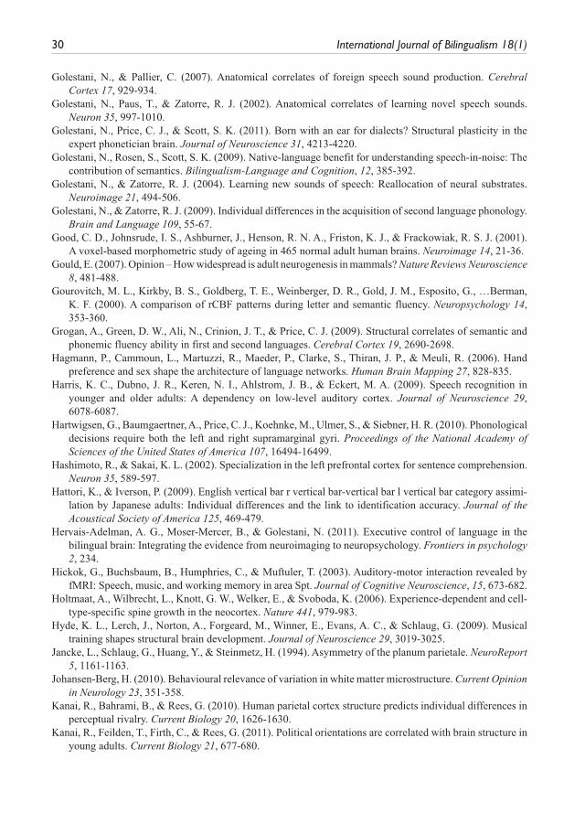
30 International Journal of Bilingualism 18(1)
Golestani, N., & Pallier, C. (2007). Anatomical correlates of foreign speech sound production. Cerebral Cortex 17, 929-934.
Golestani, N., Paus, T., & Zatorre, R. J. (2002). Anatomical correlates of learning novel speech sounds. Neuron 35, 997-1010.
Golestani, N., Price, C. J., & Scott, S. K. (2011). Born with an ear for dialects? Structural plasticity in the expert phonetician brain. Journal of Neuroscience 31, 4213-4220.
Golestani, N., Rosen, S., Scott, S. K. (2009). Native-language benefit for understanding speech-in-noise: The contribution of semantics. Bilingualism-Language and Cognition, 12, 385-392.
Golestani, N., & Zatorre, R. J. (2004). Learning new sounds of speech: Reallocation of neural substrates. Neuroimage 21, 494-506.
Golestani, N., & Zatorre, R. J. (2009). Individual differences in the acquisition of second language phonology. Brain and Language 109, 55-67.
Good, C. D., Johnsrude, I. S., Ashburner, J., Henson, R. N. A., Friston, K. J., & Frackowiak, R. S. J. (2001). A voxel-based morphometric study of ageing in 465 normal adult human brains. Neuroimage 14, 21-36.
Gould, E. (2007). Opinion – How widespread is adult neurogenesis in mammals? Nature Reviews Neuroscience 8, 481-488.
Gourovitch, M. L., Kirkby, B. S., Goldberg, T. E., Weinberger, D. R., Gold, J. M., Esposito, G., …Berman, K. F. (2000). A comparison of rCBF patterns during letter and semantic fluency. Neuropsychology 14, 353-360.
Grogan, A., Green, D. W., Ali, N., Crinion, J. T., & Price, C. J. (2009). Structural correlates of semantic and phonemic fluency ability in first and second languages. Cerebral Cortex 19, 2690-2698.
Hagmann, P., Cammoun, L., Martuzzi, R., Maeder, P., Clarke, S., Thiran, J. P., & Meuli, R. (2006). Hand preference and sex shape the architecture of language networks. Human Brain Mapping 27, 828-835.
Harris, K. C., Dubno, J. R., Keren, N. I., Ahlstrom, J. B., & Eckert, M. A. (2009). Speech recognition in younger and older adults: A dependency on low-level auditory cortex. Journal of Neuroscience 29, 6078-6087.
Hartwigsen, G., Baumgaertner, A., Price, C. J., Koehnke, M., Ulmer, S., & Siebner, H. R. (2010). Phonological decisions require both the left and right supramarginal gyri. Proceedings of the National Academy of Sciences of the United States of America 107, 16494-16499.
Hashimoto, R., & Sakai, K. L. (2002). Specialization in the left prefrontal cortex for sentence comprehension. Neuron 35, 589-597.
Hattori, K., & Iverson, P. (2009). English vertical bar r vertical bar-vertical bar l vertical bar category assimi-lation by Japanese adults: Individual differences and the link to identification accuracy. Journal of the Acoustical Society of America 125, 469-479.
Hervais-Adelman, A. G., Moser-Mercer, B., & Golestani, N. (2011). Executive control of language in the bilingual brain: Integrating the evidence from neuroimaging to neuropsychology. Frontiers in psychology 2, 234.
Hickok, G., Buchsbaum, B., Humphries, C., & Muftuler, T. (2003). Auditory-motor interaction revealed by fMRI: Speech, music, and working memory in area Spt. Journal of Cognitive Neuroscience, 15, 673-682.
Holtmaat, A., Wilbrecht, L., Knott, G. W., Welker, E., & Svoboda, K. (2006). Experience-dependent and cell-type-specific spine growth in the neocortex. Nature 441, 979-983.
Hyde, K. L., Lerch, J., Norton, A., Forgeard, M., Winner, E., Evans, A. C., & Schlaug, G. (2009). Musical training shapes structural brain development. Journal of Neuroscience 29, 3019-3025.
Jancke, L., Schlaug, G., Huang, Y., & Steinmetz, H. (1994). Asymmetry of the planum parietale. NeuroReport 5, 1161-1163.
Johansen-Berg, H. (2010). Behavioural relevance of variation in white matter microstructure. Current Opinion in Neurology 23, 351-358.
Kanai, R., Bahrami, B., & Rees, G. (2010). Human parietal cortex structure predicts individual differences in perceptual rivalry. Current Biology 20, 1626-1630.
Kanai, R., Feilden, T., Firth, C., & Rees, G. (2011). Political orientations are correlated with brain structure in young adults. Current Biology 21, 677-680.
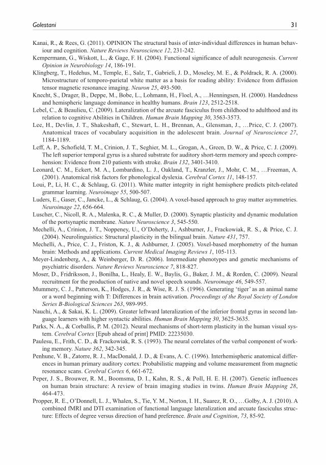
Golestani 31
Kanai, R., & Rees, G. (2011). OPINION The structural basis of inter-individual differences in human behav-iour and cognition. Nature Reviews Neuroscience 12, 231-242.
Kempermann, G., Wiskott, L., & Gage, F. H. (2004). Functional significance of adult neurogenesis. Current Opinion in Neurobiology 14, 186-191.
Klingberg, T., Hedehus, M., Temple, E., Salz, T., Gabrieli, J. D., Moseley, M. E., & Poldrack, R. A. (2000). Microstructure of temporo-parietal white matter as a basis for reading ability: Evidence from diffusion tensor magnetic resonance imaging. Neuron 25, 493-500.
Knecht, S., Drager, B., Deppe, M., Bobe, L., Lohmann, H., Floel, A., …Henningsen, H. (2000). Handedness and hemispheric language dominance in healthy humans. Brain 123, 2512-2518.
Lebel, C., & Beaulieu, C. (2009). Lateralization of the arcuate fasciculus from childhood to adulthood and its relation to cognitive Abilities in Children. Human Brain Mapping 30, 3563-3573.
Lee, H., Devlin, J. T., Shakeshaft, C., Stewart, L. H., Brennan, A., Glensman, J., …Price, C. J. (2007). Anatomical traces of vocabulary acquisition in the adolescent brain. Journal of Neuroscience 27, 1184-1189.
Leff, A. P., Schofield, T. M., Crinion, J. T., Seghier, M. L., Grogan, A., Green, D. W., & Price, C. J. (2009). The left superior temporal gyrus is a shared substrate for auditory short-term memory and speech compre-hension: Evidence from 210 patients with stroke. Brain 132, 3401-3410.
Leonard, C. M., Eckert, M. A., Lombardino, L. J., Oakland, T., Kranzler, J., Mohr, C. M., …Freeman, A. (2001). Anatomical risk factors for phonological dyslexia. Cerebral Cortex 11, 148-157.
Loui, P., Li, H. C., & Schlaug, G. (2011). White matter integrity in right hemisphere predicts pitch-related grammar learning. Neuroimage 55, 500-507.
Luders, E., Gaser, C., Jancke, L., & Schlaug, G. (2004). A voxel-based approach to gray matter asymmetries. Neuroimage 22, 656-664.
Luscher, C., Nicoll, R. A., Malenka, R. C., & Muller, D. (2000). Synaptic plasticity and dynamic modulation of the portsynaptic membrane. Nature Neuroscience 3, 545-550.
Mechelli, A., Crinion, J. T., Noppeney, U., O’Doherty, J., Ashburner, J., Frackowiak, R. S., & Price, C. J. (2004). Neurolinguistics: Structural plasticity in the bilingual brain. Nature 431, 757.
Mechelli, A., Price, C. J., Friston, K. J., & Ashburner, J. (2005). Voxel-based morphometry of the human brain: Methods and applications. Current Medical Imaging Reviews 1, 105-113.
Meyer-Lindenberg, A., & Weinberger, D. R. (2006). Intermediate phenotypes and genetic mechanisms of psychiatric disorders. Nature Reviews Neuroscience 7, 818-827.
Moser, D., Fridriksson, J., Bonilha, L., Healy, E. W., Baylis, G., Baker, J. M., & Rorden, C. (2009). Neural recruitment for the production of native and novel speech sounds. Neuroimage 46, 549-557.
Mummery, C. J., Patterson, K., Hodges, J. R., & Wise, R. J. S. (1996). Generating ‘tiger’ as an animal name or a word beginning with T: Differences in brain activation. Proceedings of the Royal Society of London Series B-Biological Sciences 263, 989-995.
Nauchi, A., & Sakai, K. L. (2009). Greater leftward lateralization of the inferior frontal gyrus in second lan-guage learners with higher syntactic abilities. Human Brain Mapping 30, 3625-3635.
Parks, N. A., & Corballis, P. M. (2012). Neural mechanisms of short-term plasticity in the human visual sys-tem. Cerebral Cortex [Epub ahead of print] PMID: 22235030.
Paulesu, E., Frith, C. D., & Frackowiak, R. S. (1993). The neural correlates of the verbal component of work-ing memory. Nature 362, 342-345.
Penhune, V. B., Zatorre, R. J., MacDonald, J. D., & Evans, A. C. (1996). Interhemispheric anatomical differ-ences in human primary auditory cortex: Probabilistic mapping and volume measurement from magnetic resonance scans. Cerebral Cortex 6, 661-672.
Peper, J. S., Brouwer, R. M., Boomsma, D. I., Kahn, R. S., & Poll, H. E. H. (2007). Genetic influences on human brain structure: A review of brain imaging studies in twins. Human Brain Mapping 28, 464-473.
Propper, R. E., O’Donnell, L. J., Whalen, S., Tie, Y. M., Norton, I. H., Suarez, R. O., …Golby, A. J. (2010). A combined fMRI and DTI examination of functional language lateralization and arcuate fasciculus struc-ture: Effects of degree versus direction of hand preference. Brain and Cognition, 73, 85-92.
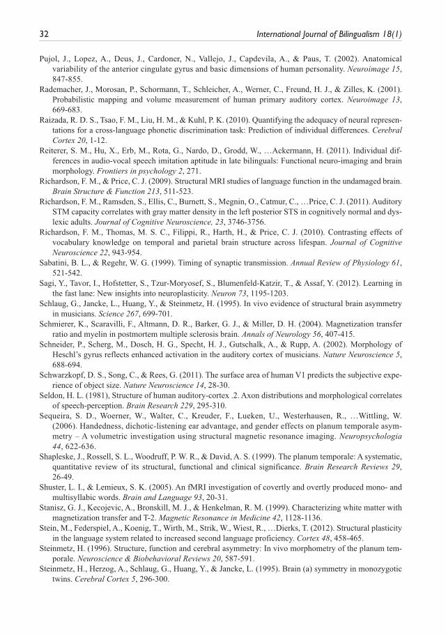
32 International Journal of Bilingualism 18(1)
Pujol, J., Lopez, A., Deus, J., Cardoner, N., Vallejo, J., Capdevila, A., & Paus, T. (2002). Anatomical variability of the anterior cingulate gyrus and basic dimensions of human personality. Neuroimage 15, 847-855.
Rademacher, J., Morosan, P., Schormann, T., Schleicher, A., Werner, C., Freund, H. J., & Zilles, K. (2001). Probabilistic mapping and volume measurement of human primary auditory cortex. Neuroimage 13, 669-683.
Raizada, R. D. S., Tsao, F. M., Liu, H. M., & Kuhl, P. K. (2010). Quantifying the adequacy of neural represen-tations for a cross-language phonetic discrimination task: Prediction of individual differences. Cerebral Cortex 20, 1-12.
Reiterer, S. M., Hu, X., Erb, M., Rota, G., Nardo, D., Grodd, W., …Ackermann, H. (2011). Individual dif-ferences in audio-vocal speech imitation aptitude in late bilinguals: Functional neuro-imaging and brain morphology. Frontiers in psychology 2, 271.
Richardson, F. M., & Price, C. J. (2009). Structural MRI studies of language function in the undamaged brain. Brain Structure & Function 213, 511-523.
Richardson, F. M., Ramsden, S., Ellis, C., Burnett, S., Megnin, O., Catmur, C., …Price, C. J. (2011). Auditory STM capacity correlates with gray matter density in the left posterior STS in cognitively normal and dys-lexic adults. Journal of Cognitive Neuroscience, 23, 3746-3756.
Richardson, F. M., Thomas, M. S. C., Filippi, R., Harth, H., & Price, C. J. (2010). Contrasting effects of vocabulary knowledge on temporal and parietal brain structure across lifespan. Journal of Cognitive Neuroscience 22, 943-954.
Sabatini, B. L., & Regehr, W. G. (1999). Timing of synaptic transmission. Annual Review of Physiology 61, 521-542.
Sagi, Y., Tavor, I., Hofstetter, S., Tzur-Moryosef, S., Blumenfeld-Katzir, T., & Assaf, Y. (2012). Learning in the fast lane: New insights into neuroplasticity. Neuron 73, 1195-1203.
Schlaug, G., Jancke, L., Huang, Y., & Steinmetz, H. (1995). In vivo evidence of structural brain asymmetry in musicians. Science 267, 699-701.
Schmierer, K., Scaravilli, F., Altmann, D. R., Barker, G. J., & Miller, D. H. (2004). Magnetization transfer ratio and myelin in postmortem multiple sclerosis brain. Annals of Neurology 56, 407-415.
Schneider, P., Scherg, M., Dosch, H. G., Specht, H. J., Gutschalk, A., & Rupp, A. (2002). Morphology of Heschl’s gyrus reflects enhanced activation in the auditory cortex of musicians. Nature Neuroscience 5, 688-694.
Schwarzkopf, D. S., Song, C., & Rees, G. (2011). The surface area of human V1 predicts the subjective expe-rience of object size. Nature Neuroscience 14, 28-30.
Seldon, H. L. (1981), Structure of human auditory-cortex .2. Axon distributions and morphological correlates of speech-perception. Brain Research 229, 295-310.
Sequeira, S. D., Woerner, W., Walter, C., Kreuder, F., Lueken, U., Westerhausen, R., …Wittling, W. (2006). Handedness, dichotic-listening ear advantage, and gender effects on planum temporale asym-metry – A volumetric investigation using structural magnetic resonance imaging. Neuropsychologia 44, 622-636.
Shapleske, J., Rossell, S. L., Woodruff, P. W. R., & David, A. S. (1999). The planum temporale: A systematic, quantitative review of its structural, functional and clinical significance. Brain Research Reviews 29, 26-49.
Shuster, L. I., & Lemieux, S. K. (2005). An fMRI investigation of covertly and overtly produced mono- and multisyllabic words. Brain and Language 93, 20-31.
Stanisz, G. J., Kecojevic, A., Bronskill, M. J., & Henkelman, R. M. (1999). Characterizing white matter with magnetization transfer and T-2. Magnetic Resonance in Medicine 42, 1128-1136.
Stein, M., Federspiel, A., Koenig, T., Wirth, M., Strik, W., Wiest, R., …Dierks, T. (2012). Structural plasticity in the language system related to increased second language proficiency. Cortex 48, 458-465.
Steinmetz, H. (1996). Structure, function and cerebral asymmetry: In vivo morphometry of the planum tem-porale. Neuroscience & Biobehavioral Reviews 20, 587-591.
Steinmetz, H., Herzog, A., Schlaug, G., Huang, Y., & Jancke, L. (1995). Brain (a) symmetry in monozygotic twins. Cerebral Cortex 5, 296-300.
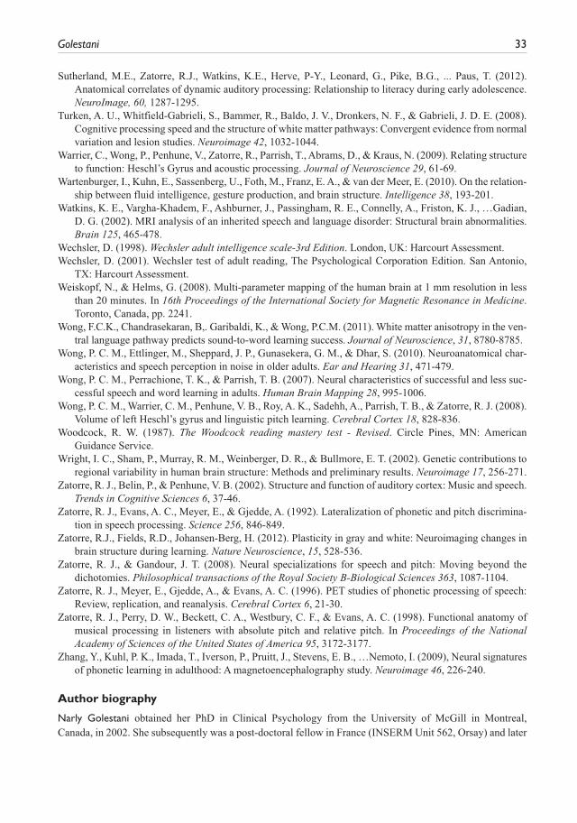
Golestani 33
Sutherland, M.E., Zatorre, R.J., Watkins, K.E., Herve, P-Y., Leonard, G., Pike, B.G., ... Paus, T. (2012). Anatomical correlates of dynamic auditory processing: Relationship to literacy during early adolescence. NeuroImage, 60, 1287-1295.
Turken, A. U., Whitfield-Gabrieli, S., Bammer, R., Baldo, J. V., Dronkers, N. F., & Gabrieli, J. D. E. (2008). Cognitive processing speed and the structure of white matter pathways: Convergent evidence from normal variation and lesion studies. Neuroimage 42, 1032-1044.
Warrier, C., Wong, P., Penhune, V., Zatorre, R., Parrish, T., Abrams, D., & Kraus, N. (2009). Relating structure to function: Heschl’s Gyrus and acoustic processing. Journal of Neuroscience 29, 61-69.
Wartenburger, I., Kuhn, E., Sassenberg, U., Foth, M., Franz, E. A., & van der Meer, E. (2010). On the relation-ship between fluid intelligence, gesture production, and brain structure. Intelligence 38, 193-201.
Watkins, K. E., Vargha-Khadem, F., Ashburner, J., Passingham, R. E., Connelly, A., Friston, K. J., …Gadian, D. G. (2002). MRI analysis of an inherited speech and language disorder: Structural brain abnormalities. Brain 125, 465-478.
Wechsler, D. (1998). Wechsler adult intelligence scale-3rd Edition. London, UK: Harcourt Assessment.Wechsler, D. (2001). Wechsler test of adult reading, The Psychological Corporation Edition. San Antonio,
TX: Harcourt Assessment.Weiskopf, N., & Helms, G. (2008). Multi-parameter mapping of the human brain at 1 mm resolution in less
than 20 minutes. In 16th Proceedings of the International Society for Magnetic Resonance in Medicine. Toronto, Canada, pp. 2241.
Wong, F.C.K., Chandrasekaran, B,. Garibaldi, K., & Wong, P.C.M. (2011). White matter anisotropy in the ven-tral language pathway predicts sound-to-word learning success. Journal of Neuroscience, 31, 8780-8785.
Wong, P. C. M., Ettlinger, M., Sheppard, J. P., Gunasekera, G. M., & Dhar, S. (2010). Neuroanatomical char-acteristics and speech perception in noise in older adults. Ear and Hearing 31, 471-479.
Wong, P. C. M., Perrachione, T. K., & Parrish, T. B. (2007). Neural characteristics of successful and less suc-cessful speech and word learning in adults. Human Brain Mapping 28, 995-1006.
Wong, P. C. M., Warrier, C. M., Penhune, V. B., Roy, A. K., Sadehh, A., Parrish, T. B., & Zatorre, R. J. (2008). Volume of left Heschl’s gyrus and linguistic pitch learning. Cerebral Cortex 18, 828-836.
Woodcock, R. W. (1987). The Woodcock reading mastery test - Revised. Circle Pines, MN: American Guidance Service.
Wright, I. C., Sham, P., Murray, R. M., Weinberger, D. R., & Bullmore, E. T. (2002). Genetic contributions to regional variability in human brain structure: Methods and preliminary results. Neuroimage 17, 256-271.
Zatorre, R. J., Belin, P., & Penhune, V. B. (2002). Structure and function of auditory cortex: Music and speech. Trends in Cognitive Sciences 6, 37-46.
Zatorre, R. J., Evans, A. C., Meyer, E., & Gjedde, A. (1992). Lateralization of phonetic and pitch discrimina-tion in speech processing. Science 256, 846-849.
Zatorre, R.J., Fields, R.D., Johansen-Berg, H. (2012). Plasticity in gray and white: Neuroimaging changes in brain structure during learning. Nature Neuroscience, 15, 528-536.
Zatorre, R. J., & Gandour, J. T. (2008). Neural specializations for speech and pitch: Moving beyond the dichotomies. Philosophical transactions of the Royal Society B-Biological Sciences 363, 1087-1104.
Zatorre, R. J., Meyer, E., Gjedde, A., & Evans, A. C. (1996). PET studies of phonetic processing of speech: Review, replication, and reanalysis. Cerebral Cortex 6, 21-30.
Zatorre, R. J., Perry, D. W., Beckett, C. A., Westbury, C. F., & Evans, A. C. (1998). Functional anatomy of musical processing in listeners with absolute pitch and relative pitch. In Proceedings of the National Academy of Sciences of the United States of America 95, 3172-3177.
Zhang, Y., Kuhl, P. K., Imada, T., Iverson, P., Pruitt, J., Stevens, E. B., …Nemoto, I. (2009), Neural signatures of phonetic learning in adulthood: A magnetoencephalography study. Neuroimage 46, 226-240.
Author biography
Narly Golestani obtained her PhD in Clinical Psychology from the University of McGill in Montreal, Canada, in 2002. She subsequently was a post-doctoral fellow in France (INSERM Unit 562, Orsay) and later
34 International Journal of Bilingualism 18(1)
in the United Kingdom (Institute for Cognitive Neuroscience at University College London). In 2009 she received a Prize of Excellence for research at the University of Geneva, and has been Assistant Professor, funded by the Swiss National Science Foundation, since 2012. Her research revolves around brain imaging methods such as structural and functional magnetic resonance imaging (MRI) as well as diffusion weighted MRI imaging for understanding the neural bases of bilingualism, language learning, and expertise.