483 trail agonist resistance is associated with death receptor mutations

1
AGA Abstracts 481 Noninvasive Detection of Colorectal Neoplasia (CRN) in Inflammatory Bowel Disease (IBD) by Stool DNA Testing: A Pilot Study John B. Kisiel, Tracy C. Yab, Fareeda Taher Nazer Hussain, William R. Taylor, Jonathan J. Harrington, Mary E. Devens, Julie A. Simonson, Megan Garrity-Park, William J. Sandborn, Edward V. Loftus, Bruce G. Wolff, David T. Rubin, Steven H. Itzkowitz, Hongzhi Zou, David A. Ahlquist Background: Conventional colonoscopic surveillance is not sensitive for detection of CRN (cancer+dysplasia) associated with IBD (IBD-CRN). Complementary detection methods are needed. Assay of exfoliated molecular markers in stool, which can detect sporadic CRN, is unexplored in IBD-CRN. Aim: To test the feasibility of stool DNA testing for detection of IBD-CRN. Methods: To select markers for stool testing, pre-study gene sequencing and methylation assays were performed on candidate genes using tissue-extracted DNA from 25 IBD-CRNs and 25 samples of non-dysplastic IBD mucosa. No mutational sites on P53, APC, KRAS, BRAF or PIK3CA genes were sufficiently informative, but aberrant methylation on EYA-4 (mEYA) and vimentin (mVIM) were highly discriminant (areas under receiver operator characteristics curve >0.85). Accordingly, we performed a prospective blinded study of stool mEYA and mVIM. Ten cases (patients with known IBD-CRN) and 10 controls (un-matched IBD patients without CRN) were recruited. Stool was collected with preservative buffer prior to or >1 week after colonoscopy. From stool-extracted DNA, target genes were enriched by sequence capture, bisulfite treated, and quantitatively assayed by methylation-specific PCR. Results: Cases included 3 with Crohn's disease (CD) and 7 with ulcerative colitis (UC); 50% were male, median age was 59 years (range 46-73), and median IBD disease duration was 30 (2-40) years. Controls included one with CD and 9 with UC; 50% were male, median age was 49 years (29-70), and median IBD duration was 12 (4-24) months. Case neoplasms included 5 cancers [median size 7 (2-8) cm; 4/5 (80%) were proximal to splenic flexure, median stage was IIA (range IIA-IV), and 2/5 (40%) were missed by colonoscopy and discovered on resected colons)] and 5 dysplastic lesions [4 focal adenomas (1 high grade dysplasia) with median size 3 (1-6) cm and one flat lesion detected by random biopsy]. At 90% specificity cutoffs, stool assay sensitivities of mEYA and mVIM were 90% and 60%, respectively. Stool assay of mEYA alone detected 5/5 cancers (100%) and 4/5 precancers (80%) at 90% specificity. In this small series, assay of combined markers did not improve discrimination over mEYA alone. Conclusions: These pilot data demonstrate proof-of- concept for a stool DNA assay to detect IBD-CRN. Stool assay of mEYA was especially discriminant for IBD-CRN. Further studies are indicated to optimize assay techniques, corroborate and extend these observations, and test this noninvasive approach as a complement to endoscopic surveillance strategies. 482 Tumor Suppressor XAF1 Reverses TRAIL-Resistance in Hepatocellular Carcinoma Cells Through Upregulation of Death Receptor 4 and 5 Liming Zhu, Qiang Dai, Dongmei Shi, Weiyan Yao, Pinghu Sun, Minmin Qiao, Jihong Tan, Xiaoguang Qi, Shihu Jiang, Shuiping Tu Background and Aims: Tumor necrosis factor-related apoptosis-inducing ligand (TRAIL) selectively triggers rapid apoptosis tumor cells but not in normal cells, and is a potential anticancer agent. However, several types of cancer cells including hepatocellular carcinoma (HCC) are resistant to TRAIL. XIAP-associated factor 1(XAF1) is a novel identified XIAP antagonist and a potential tumor suppressor.The aim of this study is to investigate the effect and mechanism of XAF1 on reversing resistance to TRAIL in HCC. Materials and methods: Four HCC cells were pre-infected with Ad5/F35-XAF1 adenovirusor control virus for 4 hours and then incubated with rhTRAIL peptide at different dose for 48 hours. Cell proliferation was measured by MTT method. Cell apoptosis was detected by FACS. The microarray was performed to identify downstream gene of XAF1. Gene expressions were determined by RT-PCR, Western Blot and immunohistochemistry (IHC). Tumor growth was evaluated in a nude mice xenograft model.Results: MTT results confirmed that Hep3B cells were a TRAIL- resistant cell lines. FACS analysis results showed that Hep3B cell were significantly resistant to TRAIL-induced cell apoptosis. Combination of adeno-XAF1 and rhTRAIL treatment signi- ficantly decreased the cell proliferation and increased cell apoptosis in all 4 HCC cells compared to the treatment of adeno-XAF1 or rhTRAIL alone. Combination of adeno-XAF1 and TRAIL increased the activation of caspase-3,-8,-9, and the cleavage of PARP-r and release of cytochrome c, downregulated expression of FLIP, XIAP and Bcl-2 and upregulated expression of Bax compared with XAF1 or rhTRAIL treated alone in all four HCC cells. Furthermore, the microarray data showed that death receptor 4 (DR4) and death receptor 5(DR5), two TRAIL receptors, were significantly upregulated in adeno-XAF1 -infected SMMC7721 cells. RT-PCR and Western blot confirmed that adeno-XAF1 significantly increased the expression of DR4 and DR5 in SMMC7721 and Hep3B cells. Moreover, DR5 siRNA treatment abrogated the effect of aden-XAF1 on sensitization of Hep3B to TRAIL- induced apoptosis, leading to decrease the expression of caspase-8, 9 and PARP. Intra- tumoral injection of Ad5/F35-XAF1 combined with TRAIL significantly inhibited tumor growth in both TRAL-sensitive and TRAIL-resistant HCC tumor xenografts model. Conclu- sion: Ad5/F35-XAF1 can sensitize TRAIL-resistant HCC to TRAIL-induce apoptosis and tumor inhibition through upreguation of DR5 and DR4 expression. Our results suggest that the combination of XAF1 with TRAIL may be a potential strategy for TRAIL-resistance HCC therapy. The project was supported by NSFC grant No. 30500221(Tu SP). 483 TRAIL Agonist Resistance is Associated With Death Receptor Mutations Joy Liu, Haizhen Zhu, Hongyan Liu, David R. Nelson, Chen Liu Background: Tumor-necrosis factor-related apoptosis-inducing ligand (TRAIL) is an agonist for death receptor 4 (DR4) and death receptor 5 (DR5).TRAIL agonist-based cancer therapy is promising because of the specificity of TRAIL to cancerous cells. A major problem related to this therapy is the cancer cell resistance to the TRAIL agonists. The underlying molecular mechanisms are unknown. Aim: The objective of our study is to determine the molecular S-68 AGA Abstracts mechanisms by which liver cancer cells are resistant to TRAIL agonists. Materials and Methods: A monoclonal antibody specifically triggers DR5 was used to treat a human hepatoma cell line, LH86. The cancer cells that are resistant to the treatment were selected. Total genomic DNA was extracted from the resistant cells and followed by DNA sequencing of DR5 gene. The sequences were compared with original cells that were sensitive to the TRAIL agonist. Results: Majority of LH86 cells were undergoing apoptosis upon TRAIL agonist. The surviving cells eventually grow in the presence of TRAIL agonist (monoclonal antibody) after more than three weeks of continuous culture. These cells are referred to as DR5 resistant cell line. Genomic sequencing of the DR5 gene was performed in the resistant cell line and the original LH86 cell line. Sequence alignment with the DR5 from the original LH86 cells show that there are missense mutations in the death domain region of the DR5 gene in the resistant cell line. Conclusion: We have demonstrated that the liver cancer cells that are resistant to the TRAIL agonist have mutations in the DR5 gene. We speculate that these mutations constitute evasive action of liver cancer cells, which may impede the effectiveness of a TRAIL-based cancer therapy. 484 PEMT (+/-) Fed a Methionine and Choline Deficient (MCD) Diet Demonstrate Increased Severity of Non-Alcoholic Steatohepatitis and Activation of the Unfolded Protein Response (UPR) Elizabeth A. Newell, Richard M. Green The Methionine and Choline deficient (MCD) diet is an established murine model of experi- mental steatohepatitis (NASH). Recently phosphatidyethanolamine N-methyltransferase (PEMT) gene polymorphisms were associated with human NASH. PEMT is a hepatic enzyme that is rate-limiting for the synthesis of phosphatidylcholine from phosphatidylethanolamine. In addition, PEMT(-/-) mice fed a choline-deficient diet rapidly die of liver failure. However, the role of PEMT in the pathogenesis of steatohepatitis remains unexplored. Aim: The purposes of the study is to investigate whether PEMT(+/-) mice fed a MCD diet develop enhanced steatohepatitis; and define the role of endoplasmic reticulum stress and activation of the unfolded protein response (UPR). Methods: Female PEMT(+/-) and wildtype (+/+) mice (8-12 wks) were treated with a MCD or control diet for 2 weeks. Body weight, liver weight, serum ALT and hepatic triglyceride levels were measured. Liver histology was assessed for steatosis and inflammation. Gene expression was determined by quantitative RT-PCR. Results: PEMT(+/-) mice fed a MCD diet had 3-fold higher serum ALT levels than PEMT(+/ +) mice, being 222 ± 32 vs. 71 ± 7 IU, respectively (p<0.001). PEMT(+/-) mice fed a MCD diet also had a significantly greater decrease in body weight (24 ± 4% vs 11 ± 2%, p<0.001), more hepatomegaly (5.9 ± 1.4% vs 4.6 ± 0.6% liver/body weight, p<0.001) and a trend toward higher hepatic triglyceride levels. Histological analyses of inflammation were consistent with the ALT levels. Hepatic UPR and TNFα gene expression revealed significant increases in activation in the PEMT(+/-) mice (see table). Hepatic ATF6, XBP1s and TNFα were greater in the PEMT(+/-) mice fed a MCD diet compared to PEMT(+/+) mice (p<0.05), and there were strong trends toward increased activation of ATF4 and CHOP. All control diet fed mice had lower levels of UPR markers than MCD fed mice. Conclusions: PEMT(+/-) mice fed a MCD diet have markedly higher serum ALT levels and hepatic inflammation than wildtype mice. PEMT(+/-) mice also have enhanced activation of the hepatic UPR and hepatic TNFα expression. Since polymorphisms of PEMT, and hepatic UPR activation, have been associated with human NASH, these data suggest that reduced PEMT activity may be mechanistically important in UPR activation during steatohepatitis. We speculate that this provides a causative mechanism for the role of PEMT in human NASH. 485 Il10 Deficiency Markedly Ameliorates and Provides Prolonged Protection Against Cholesterol Gallstone Formation in a Susceptible Mouse Model Kirk J. Maurer, Jacqueline J. Fremont-Rahl, Shou-An Liu, Martin C. Carey, James G. Fox Introduction: We recently demonstrated that acquired immunity is instrumental in choles- terol gallstone formation (Gastroenterology. 2007: 1304-15). The current study expands upon these findings and analyzes the role of the cytokine IL10 in cholesterol gallstone pathogenesis. Methods: We compared male wild-type C57BL/6J (B6) (n=13) with congenic IL10 -/- mice (n=11). Beginning at 8-10 weeks of age, all mice were fed a lithogenic diet (1% cholesterol, 0.5% cholic acid, 15% triglyceride) for 10 weeks. At the conclusion of the study, mice were euthanized with CO2, and gallbladder bile phenotype was analyzed by microscopy. A follow-up study was then conducted on IL10 -/- mice in which 4 male and 4 female mice were fed the lithogenic diet for 6 months and gallbladder bile was analyzed microscopically. In addition, after 4 weeks of dietary feeding, gallbladders were harvested from male B6 (n= 10) and IL10 -/- (n=10) mice, and bile was chemically analyzed to determine concentrations of major biliary lipids. Results: After ten weeks of feeding, normalized gallbladder weight (1.4 +/-0.2mg/g) and mucin gel formation score (1.2mg/g +/-0.18) were significantly (P<0.05) reduced in IL10 -/- compared to B6 mice (2.4+/-0.3mg/g; 2.5 +/-0.9). IL10 -/- mice displayed a significantly reduced prevalence of cholesterol monohydrate crystals (55%) compared to B6 mice (92%, P<0.05). Furthermore, cholesterol gallstone prevalence was significantly reduced in IL10 -/- mice (36%) compared to B6 mice (85%, P<0.05). After six months of

Upload: joy-liu

Post on 03-Dec-2016

213 views

Category:

Documents


1 download

TRANSCRIPT

AG

AA

bst

ract

s481

Noninvasive Detection of Colorectal Neoplasia (CRN) in Inflammatory BowelDisease (IBD) by Stool DNA Testing: A Pilot StudyJohn B. Kisiel, Tracy C. Yab, Fareeda Taher Nazer Hussain, William R. Taylor, Jonathan J.Harrington, Mary E. Devens, Julie A. Simonson, Megan Garrity-Park, William J.Sandborn, Edward V. Loftus, Bruce G. Wolff, David T. Rubin, Steven H. Itzkowitz,Hongzhi Zou, David A. Ahlquist

Background: Conventional colonoscopic surveillance is not sensitive for detection of CRN(cancer+dysplasia) associated with IBD (IBD-CRN). Complementary detection methods areneeded. Assay of exfoliated molecular markers in stool, which can detect sporadic CRN, isunexplored in IBD-CRN. Aim: To test the feasibility of stool DNA testing for detection ofIBD-CRN. Methods: To select markers for stool testing, pre-study gene sequencing andmethylation assays were performed on candidate genes using tissue-extracted DNA from 25IBD-CRNs and 25 samples of non-dysplastic IBD mucosa. No mutational sites on P53, APC,KRAS, BRAF or PIK3CA genes were sufficiently informative, but aberrant methylation onEYA-4 (mEYA) and vimentin (mVIM) were highly discriminant (areas under receiver operatorcharacteristics curve >0.85). Accordingly, we performed a prospective blinded study of stoolmEYA and mVIM. Ten cases (patients with known IBD-CRN) and 10 controls (un-matchedIBD patients without CRN) were recruited. Stool was collected with preservative buffer priorto or >1 week after colonoscopy. From stool-extracted DNA, target genes were enriched bysequence capture, bisulfite treated, and quantitatively assayed by methylation-specific PCR.Results: Cases included 3 with Crohn's disease (CD) and 7 with ulcerative colitis (UC);50% were male, median age was 59 years (range 46-73), and median IBD disease durationwas 30 (2-40) years. Controls included one with CD and 9 with UC; 50% were male,median age was 49 years (29-70), and median IBD duration was 12 (4-24) months. Caseneoplasms included 5 cancers [median size 7 (2-8) cm; 4/5 (80%) were proximal to splenicflexure, median stage was IIA (range IIA-IV), and 2/5 (40%) were missed by colonoscopyand discovered on resected colons)] and 5 dysplastic lesions [4 focal adenomas (1 highgrade dysplasia) with median size 3 (1-6) cm and one flat lesion detected by random biopsy].At 90% specificity cutoffs, stool assay sensitivities of mEYA and mVIM were 90% and 60%,respectively. Stool assay of mEYA alone detected 5/5 cancers (100%) and 4/5 precancers(80%) at 90% specificity. In this small series, assay of combined markers did not improvediscrimination over mEYA alone. Conclusions: These pilot data demonstrate proof-of-concept for a stool DNA assay to detect IBD-CRN. Stool assay of mEYA was especiallydiscriminant for IBD-CRN. Further studies are indicated to optimize assay techniques,corroborate and extend these observations, and test this noninvasive approach as acomplement to endoscopic surveillance strategies.

482

Tumor Suppressor XAF1 Reverses TRAIL-Resistance in HepatocellularCarcinoma Cells Through Upregulation of Death Receptor 4 and 5Liming Zhu, Qiang Dai, Dongmei Shi, Weiyan Yao, Pinghu Sun, Minmin Qiao, JihongTan, Xiaoguang Qi, Shihu Jiang, Shuiping Tu

Background and Aims: Tumor necrosis factor-related apoptosis-inducing ligand (TRAIL)selectively triggers rapid apoptosis tumor cells but not in normal cells, and is a potentialanticancer agent. However, several types of cancer cells including hepatocellular carcinoma(HCC) are resistant to TRAIL. XIAP-associated factor 1(XAF1) is a novel identified XIAPantagonist and a potential tumor suppressor.The aim of this study is to investigate the effectand mechanism of XAF1 on reversing resistance to TRAIL in HCC. Materials and methods:Four HCC cells were pre-infected with Ad5/F35-XAF1 adenovirusor control virus for 4 hoursand then incubated with rhTRAIL peptide at different dose for 48 hours. Cell proliferation wasmeasured by MTT method. Cell apoptosis was detected by FACS. The microarray wasperformed to identify downstream gene of XAF1. Gene expressions were determined byRT-PCR, Western Blot and immunohistochemistry (IHC). Tumor growth was evaluated ina nude mice xenograft model.Results: MTT results confirmed that Hep3B cells were a TRAIL-resistant cell lines. FACS analysis results showed that Hep3B cell were significantly resistantto TRAIL-induced cell apoptosis. Combination of adeno-XAF1 and rhTRAIL treatment signi-ficantly decreased the cell proliferation and increased cell apoptosis in all 4 HCC cellscompared to the treatment of adeno-XAF1 or rhTRAIL alone. Combination of adeno-XAF1and TRAIL increased the activation of caspase-3,-8,-9, and the cleavage of PARP-r andrelease of cytochrome c, downregulated expression of FLIP, XIAP and Bcl-2 and upregulatedexpression of Bax compared with XAF1 or rhTRAIL treated alone in all four HCC cells.Furthermore, the microarray data showed that death receptor 4 (DR4) and death receptor5(DR5), two TRAIL receptors, were significantly upregulated in adeno-XAF1 -infectedSMMC7721 cells. RT-PCR and Western blot confirmed that adeno-XAF1 significantlyincreased the expression of DR4 and DR5 in SMMC7721 and Hep3B cells. Moreover, DR5siRNA treatment abrogated the effect of aden-XAF1 on sensitization of Hep3B to TRAIL-induced apoptosis, leading to decrease the expression of caspase-8, 9 and PARP. Intra-tumoral injection of Ad5/F35-XAF1 combined with TRAIL significantly inhibited tumorgrowth in both TRAL-sensitive and TRAIL-resistant HCC tumor xenografts model. Conclu-sion: Ad5/F35-XAF1 can sensitize TRAIL-resistant HCC to TRAIL-induce apoptosis andtumor inhibition through upreguation of DR5 and DR4 expression. Our results suggest thatthe combination of XAF1 with TRAIL may be a potential strategy for TRAIL-resistance HCCtherapy. The project was supported by NSFC grant No. 30500221(Tu SP).

483

TRAIL Agonist Resistance is Associated With Death Receptor MutationsJoy Liu, Haizhen Zhu, Hongyan Liu, David R. Nelson, Chen Liu

Background: Tumor-necrosis factor-related apoptosis-inducing ligand (TRAIL) is an agonistfor death receptor 4 (DR4) and death receptor 5 (DR5).TRAIL agonist-based cancer therapyis promising because of the specificity of TRAIL to cancerous cells. A major problem relatedto this therapy is the cancer cell resistance to the TRAIL agonists. The underlying molecularmechanisms are unknown. Aim: The objective of our study is to determine the molecular

S-68AGA Abstracts

mechanisms by which liver cancer cells are resistant to TRAIL agonists. Materials andMethods: A monoclonal antibody specifically triggers DR5 was used to treat a humanhepatoma cell line, LH86. The cancer cells that are resistant to the treatment were selected.Total genomic DNA was extracted from the resistant cells and followed by DNA sequencingof DR5 gene. The sequences were compared with original cells that were sensitive to theTRAIL agonist. Results: Majority of LH86 cells were undergoing apoptosis upon TRAILagonist. The surviving cells eventually grow in the presence of TRAIL agonist (monoclonalantibody) after more than three weeks of continuous culture. These cells are referred to asDR5 resistant cell line. Genomic sequencing of the DR5 gene was performed in the resistantcell line and the original LH86 cell line. Sequence alignment with the DR5 from the originalLH86 cells show that there are missense mutations in the death domain region of the DR5gene in the resistant cell line. Conclusion: We have demonstrated that the liver cancer cellsthat are resistant to the TRAIL agonist have mutations in the DR5 gene. We speculatethat these mutations constitute evasive action of liver cancer cells, which may impede theeffectiveness of a TRAIL-based cancer therapy.

484

PEMT (+/-) Fed a Methionine and Choline Deficient (MCD) Diet DemonstrateIncreased Severity of Non-Alcoholic Steatohepatitis and Activation of theUnfolded Protein Response (UPR)Elizabeth A. Newell, Richard M. Green

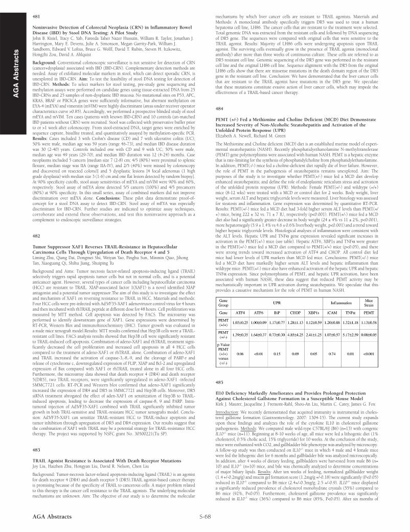
The Methionine and Choline deficient (MCD) diet is an established murine model of experi-mental steatohepatitis (NASH). Recently phosphatidyethanolamine N-methyltransferase(PEMT) gene polymorphisms were associated with human NASH. PEMT is a hepatic enzymethat is rate-limiting for the synthesis of phosphatidylcholine from phosphatidylethanolamine.In addition, PEMT(-/-) mice fed a choline-deficient diet rapidly die of liver failure. However,the role of PEMT in the pathogenesis of steatohepatitis remains unexplored. Aim: Thepurposes of the study is to investigate whether PEMT(+/-) mice fed a MCD diet developenhanced steatohepatitis; and define the role of endoplasmic reticulum stress and activationof the unfolded protein response (UPR). Methods: Female PEMT(+/-) and wildtype (+/+)mice (8-12 wks) were treated with a MCD or control diet for 2 weeks. Body weight, liverweight, serumALT and hepatic triglyceride levels were measured. Liver histologywas assessedfor steatosis and inflammation. Gene expression was determined by quantitative RT-PCR.Results: PEMT(+/-) mice fed a MCD diet had 3-fold higher serum ALT levels than PEMT(+/+) mice, being 222 ± 32 vs. 71 ± 7 IU, respectively (p<0.001). PEMT(+/-) mice fed a MCDdiet also had a significantly greater decrease in body weight (24 ± 4% vs 11 ± 2%, p<0.001),more hepatomegaly (5.9 ± 1.4% vs 4.6 ± 0.6% liver/body weight, p<0.001) and a trend towardhigher hepatic triglyceride levels. Histological analyses of inflammation were consistent withthe ALT levels. Hepatic UPR and TNFα gene expression revealed significant increases inactivation in the PEMT(+/-) mice (see table). Hepatic ATF6, XBP1s and TNFα were greaterin the PEMT(+/-) mice fed a MCD diet compared to PEMT(+/+) mice (p<0.05), and therewere strong trends toward increased activation of ATF4 and CHOP. All control diet fedmice had lower levels of UPR markers than MCD fed mice. Conclusions: PEMT(+/-) micefed a MCD diet have markedly higher serum ALT levels and hepatic inflammation thanwildtype mice. PEMT(+/-) mice also have enhanced activation of the hepatic UPR and hepaticTNFα expression. Since polymorphisms of PEMT, and hepatic UPR activation, have beenassociated with human NASH, these data suggest that reduced PEMT activity may bemechanistically important in UPR activation during steatohepatitis. We speculate that thisprovides a causative mechanism for the role of PEMT in human NASH.

485

Il10 Deficiency Markedly Ameliorates and Provides Prolonged ProtectionAgainst Cholesterol Gallstone Formation in a Susceptible Mouse ModelKirk J. Maurer, Jacqueline J. Fremont-Rahl, Shou-An Liu, Martin C. Carey, James G. Fox

Introduction: We recently demonstrated that acquired immunity is instrumental in choles-terol gallstone formation (Gastroenterology. 2007: 1304-15). The current study expandsupon these findings and analyzes the role of the cytokine IL10 in cholesterol gallstonepathogenesis. Methods: We compared male wild-type C57BL/6J (B6) (n=13) with congenicIL10-/- mice (n=11). Beginning at 8-10 weeks of age, all mice were fed a lithogenic diet (1%cholesterol, 0.5% cholic acid, 15% triglyceride) for 10 weeks. At the conclusion of the study,mice were euthanized with CO2, and gallbladder bile phenotype was analyzed bymicroscopy.A follow-up study was then conducted on IL10-/- mice in which 4 male and 4 female micewere fed the lithogenic diet for 6 months and gallbladder bile was analyzed microscopically.In addition, after 4 weeks of dietary feeding, gallbladders were harvested from male B6 (n=10) and IL10-/- (n=10) mice, and bile was chemically analyzed to determine concentrationsof major biliary lipids. Results: After ten weeks of feeding, normalized gallbladder weight(1.4 +/-0.2mg/g) and mucin gel formation score (1.2mg/g +/-0.18) were significantly (P<0.05)reduced in IL10-/- compared to B6 mice (2.4+/-0.3mg/g; 2.5 +/-0.9). IL10-/- mice displayeda significantly reduced prevalence of cholesterol monohydrate crystals (55%) compared toB6 mice (92%, P<0.05). Furthermore, cholesterol gallstone prevalence was significantlyreduced in IL10-/- mice (36%) compared to B6 mice (85%, P<0.05). After six months of