5 2011 ap slide seminar: tumors and tumor-like...
TRANSCRIPT

5 2011 AP Slide Seminar: Tumors and Tumor-like Lesions of the Urinary Bladder
David Grignon MD, FASCP
2011 Annual Meeting – Las Vegas, NV
AMERICAN SOCIETY FOR CLINICAL PATHOLOGY 33 W. Monroe, Ste. 1600
Chicago, IL 60603

5 2011 AP Slide Seminar: Tumors and Tumor-like Lesions of the Urinary Bladder Urinary bladder biopsies and transurethral resections are among the most common specimens generated by urologists. The nature of the material including small sample size and frequent artifacts also makes them among the most challenging. The natural history of bladder cancer, the 6th most common cancer overall in the USA, results in many patients having repeated endoscopic procedures and the frequent use of intravesical therapy complicates the interpretation of post treatment specimens. This slide seminar will focus on the key diagnostic features of urothelial carcinoma including classification, grading and pathologic staging. The wide ranging morphologic spectrum of urothelial carcinoma will also be explored and particularly problematic and clinically significant variants highlighted. Benign proliferative epithelial lesions that can be confused with urothelial tumors will also be covered. Other types of carcinomas occasionally arise in the urinary bladder and these can cause significant diagnostic challenges; the most important and problematic of these will be discussed. Finally, spindle cell proliferations are infrequent but can be particularly difficult to deal with and these will be reviewed.
• To understand the critical morphologic aspects of urothelial carcinoma and provide a complete and accurate pathology report sufficient for optimal patient care decisions to be made.
• To recognize the wide morphologic spectrum of urothelial carcinoma, identify the unusual but clinically significant variants and provide guidance to clinical colleagues on what these mean to patient management.
• To identify benign proliferative processes that can closely mimic bladder cancers. FACULTY: David Grignon MD, FASCP Entire Pathology Team Surgical Pathology Surgical Pathology (GI, GU, Etc.) 3.0 CME/CMLE Credits Accreditation Statement: The American Society for Clinical Pathology (ASCP) is accredited by the Accreditation Council for Continuing Medical Education to provide continuing medical education (CME) for physicians. This activity has been planned and implemented in accordance with the Essential Areas and Policies of the Accreditation Council for Continuing Medical Education (ACCME). Credit Designation: The ASCP designates this enduring material for a maximum of 3 AMA PRA Category 1 Credits™. Physicians should only claim credit commensurate with the extent of their participation in the activity. ASCP continuing education activities are accepted by California, Florida, and many other states for relicensure of clinical laboratory personnel. ASCP designates these activities for the indicated number of Continuing Medical Laboratory Education (CMLE) credit hours. ASCP CMLE credit hours are acceptable to meet the continuing education requirements for the ASCP Board of Registry Certification Maintenance Program. All ASCP CMLE programs are conducted at intermediate to advanced levels of learning. Continuing medical education (CME) activities offered by ASCP are acceptable for the American Board of Pathology’s Maintenance of Certification Program.

1
ASCP AP SLIDE SEMINAR 2011 TUMOR AND TUMOR-LIKE LESIONS OF THE URINARY BLADDER
David Grignon, MD
Centennial Professor & Vice Chair for Clinical Programs Indiana University & Indiana University Health
Indianapolis, IN [email protected]
INTRODUCTION
The vast majority of malignant tumors that arise throughout the urinary tract are urothelial carcinomas. Information from the pathology report, in particular biopsy and TURBT specimens is critical in therapeutic decision making.
CLASSIFICATION OF UROTHELIAL TUMORS
The last two decades has seen a tremendous upheaval in this very important area that only recently seems to have settled down to a level of general concurrence. For over 2 decades the 1973 World Health Organization (WHO) classification and grading of urothelial neoplasms dominated although several variations and different schemes were published. In the early 1990’s several factors emerged that resulted in the need to re-evaluate this approach. First, the controversy of calling grade 1 papillary tumors “carcinoma” arose with several groups led by Dr. William Murphy calling all tumors in the low-grade end papilloma. Second, the use of intravesical therapy as a standard practice in the treatment of high-risk non-invasive papillary tumors demanded that high-risk tumors be clearly identified. Third, the WHO (1973) system was criticized for the imprecision of the published criteria (Table 1), leading many pathologists to essentially use this 3 grade system to create 5 grade groups (1, 1-2, 2, 2-3, and 3); one result being that only a small percentage of cases were placed in the grade 3 group. For example, in a review of 3 clinical studies only 25 of 280 (8.9%) newly diagnosed non-invasive papillary tumors (pTa) were called grade 3. The effect of the latter was confusion as to how to treat grade 2 pTa tumors, a category that included many high-risk patients that could benefit from intravesical therapy as well as many patients with low-risk disease for whom intravesical therapy may not be appropriate or necessary. Multiple studies have demonstrated that the grade 2 group included high risk patients with progression to invasive carcinoma reported in up to 20% of patients and cancer specific death in 13 to 20%.
Table 1. 1973 WHO grading criteria Grade 1 Tumors with the least degree of cellular anaplasia compatible with a diagnosis
of malignancy Grade 2 Histologic features between grades 1 and 3 Grade 3 Tumors with the most severe degrees of cellular anaplasia
A consensus conference to address this issue was held under the auspices of the
International Society of Urologic Pathology (ISUP) in March of 1998. The results of this consensus were adopted by the WHO and the results published in 1998 as the World Health Organization/International Society of Urologic Pathology consensus classification. Most important was the adoption of the grading system that had been described by Malmström et al. The value of the latter was viewed as two-fold; first, the morphologic criteria for applying the scheme were well defined and second, it appeared to place the majority of patients with high-risk disease into the high-grade category.

2
Subsequently it was agreed to essentially reproduce the 1998 WHO/ISUP classification as the 2004 WHO recommended classification scheme (Table 2). Similarly the authors of the 4th Series Armed Forces Institute of Pathology fascicle covering bladder neoplasia have followed this system. The success of this in addressing the key issues outlined in the first paragraph has been stressed, in particular the reclassification of high-risk grade 2 tumors (WHO 1973) in the high-grade papillary carcinoma category. For example, in the study by Yin et al,, 13 of 46 WHO (1973) grade 2 tumors (28%) were placed in the WHO (2004) high-grade category resulting in 23% of all cases being considered high-grade in WHO 2004 compared to only 4% being called grade 3 in the 1973 WHO system.
In a series of 215 patients with low grade (papilloma, papillary urothelial neoplasm of low
malignant potential and low grade papillary carcinoma) pTa tumors from the Memorial Sloan Kettering Cancer Center treated by transurethral resection only, progression to high grade pTa or invasive (pT1) carcinoma occurred in only 3% and 5% of patients respectively with a median follow up of 8 years. A recent prospective study has lent further support to these findings. This experience has led to the suggestion that patients with low grade pTa tumors can be followed less frequently. In December, 2007 the American Urological Association reinforced the current approach in its guidelines for the treatment of non muscle invasive bladder cancers by separating non invasive papillary tumors into 2 groups – low grade and high grade with different treatment recommendations for each group. The College of American Pathologists also utilizes WHO 2004 in its recommendations for the reporting of urothelial tumors. Most recently an International Review of Bladder Cancer has advocated this system.
Table 2. 2004 WHO/1998 ISUP CLASSIFICATION
• NORMAL • HYPERPLASIAS • FLAT LESIONS WITH ATYPIA
♦ Reactive (inflammatory) atypia ♦ Atypia of unknown significance ♦ Dysplasia (low grade intra-urothelial neoplasia) ♦ Carcinoma in situ (high grade intra-urothelial neoplasia)
• PAPILLARY NEOPLASMS
♦ Papilloma ♦ Inverted papilloma ♦ Papillary urothelial neoplasm of low malignant potential ♦ Papillary carcinoma, low grade ♦ Papillary carcinoma, high grade
• INVASIVE NEOPLASMS
References Amin MB, Delahunt B, Bochner B, et al. Protocol for the examination of specimens from patients with carcinoma of the urinary bladder. Protocol applies primarily to invasive carcinomas and/or associated epithelial lesions including carcinoma in situ. (Protocol revision date, October 2009; CAP Web page) Amin MB, Epstein JI, Grignon DJ, et al. Pathology Consensus Guidelines by The Pathology of Bladder Cancer Work Group. International Consultation of Urologic Diseases – Bladder 2011 (In press)

3
Malmström PU, Busch C, Norlen BJ. Recurrence, progression and survival in bladder cancer: a retrospective analysis of 232 patients with greater than or equal to 5-year follow-up. Scand J Urol Nephrol 1987;21:185-195. Epstein JI, Amin MB, Reuter VR, Mostofi FK and the Bladder Consensus Conference Committee. The World Health Organization/International Society of Urologic Pathology consensus classification of urothelial (transitional) neoplasms of the urinary bladder. Am J Surg Pathol 1998;22:1435-1448. Hall MC, Chang SS, Dalbagni G, et al. Guideline for the management of non muscle invasive bladder cancer (stages Ta, T1, and Tis): 2007 update. J Urol 2007;178:2314-2330. Herr HW, Donat SM, Reuter VE. Management of low grade papillary bladder tumors. J Urol 2007;178:1201-1205. Herr HW. Low risk bladder tumors – less is more. J Urol 2008;179:13-14. Kiemeney LALM, Witjes JA, Verbeek ALM, et al. The clinical epidemiology of superficial bladder cancer. Br J Cancer 1993;67:806-812. Malmström PU, Busch C, Norlen BJ. Recurrence, progression and survival in bladder cancer: a retrospective analysis of 232 patients with greater than or equal to 5-year follow-up. Scand J Urol Nephrol 1987;21:185-195. Mostofi FK, Sobin LH. Histologic typing of urinary bladder tumors. Geneva. World Health Organization, 1973. Murphy WM, Beckwith JB, Farrow GM. Atlas of Tumor Pathology: Tumors of the Kidney, Bladder, and related Urinary Structures. Armed Forces Institute of Pathology, 3rd series fascicle, American Registry of Pathology, Washington, DC, 1994. Murphy WM, Grignon DJ, Perlman E. Atlas of Tumor Pathology: Tumors of the Kidney, Bladder, and related Urinary Structures. Armed Forces Institute of Pathology, 4th series fascicle, American Registry of Pathology, Washington, DC, 2004. Witjes JA, Moonen PMJ, van der Heijden AG. Review pathology in a diagnostic bladder cancer trial: effect on patient risk category. Urology 2006;67:751-755. Yin H, Leong AS-Y. Histologic grading of noninvasive papillary urothelial tumors: validation of the 1998 WHO/ISUP system by immunophenotyping and follow up. Am J Clin Pathol 2004;121:679-687.
CASE 1. UROTHELIAL CARCINOMA IN SITU Case history. This 69 year old woman was undergoing routine cystoscopy following a right nephroureterectomy for a renal pelvis urothelial carcinoma one year earlier. At cystoscopy the urologist noted a reddened area on the right lateral wall. The lesion was resected. Diagnosis: The sections show partial involvement of the urothelium by urothelial carcinoma in situ. The neoplastic cells have enlarged nuclei and moderate cytoplasm. It is useful to compare the lesion with the adjacent benign epithelium This degree of atypia may have been called moderate to severe dysplasia in the past but in the current system does qualify for a diagnosis of CIS. FLAT LESIONS

4
Hyperplasia: The current classification recognizes hyperplasia as when there is a “markedly thickened mucosa without atypia.” Counting cell layers is not recommended. There is no significant cytologic or architectural atypia. Hyperplasia can have a pseudo-papillary architecture. Hyperplasia is usually part of a reactive process in response to inflammation or other causes of irritation. The relationship between hyperplasia and neoplasia is unknown. Reactive Atypia: In the presence of acute and/or chronic inflammation, the urothelium shows a wide range of reactive changes. There is usually a history of instrumentation, infection or treatment with intravesical agents. In reactive atypia the epithelium may or may not be thickened. Although a thickened epithelium is typically associated with a reactive process, carcinoma in situ can also produce a thicker than normal epithelium. Nuclei are uniformly enlarged, vesicular, and may have prominent usually centrally located nucleoli. Mitoses can be frequent and are located in the lower epithelial layers. Inflammation is almost always present. Atypia of Unknown Significance: One of the major grey zones in any consideration of intraepithelial lesions is between reactive atypia and true neoplastic (dysplastic) alterations. Reproducibility studies have clearly demonstrated the lack of consistency in this particular area. This category was created to include those instances where a lesion cannot be confidently placed in the reactive versus dysplastic categories. Histologically there is usually an inflammatory background. The degree of cytologic atypia is judged to be outside of the accepted range for reactive processes although this possibility cannot be excluded. In one study, none of 35 patients diagnosed with atypia of unknown significance developed urothelial neoplasia with a median 3.5 years of follow up. Dysplasia (Low Grade Intra-urothelial Neoplasia): This category also suffers from a significant problem in diagnostic reproducibility. The natural history of lesions with dysplastic features of a lesser degree than the moderate to severe categories is unknown. There is however some evidence, largely genetic that it shares some abnormalities with carcinoma in situ and therefore likely represents a precursor lesion. It is most often diagnosed in the context of known urothelial neoplasia. Histologically there is some architectural distortion. The nuclei are irregularly enlarged with some hyperchromasia and pleomorphism present. Overall the features are those of a neoplastic atypia but fall short of the criteria for carcinoma in situ outlined below. Studies that have applied the 1998 WHO/ISUP criteria indicated a 15% – 19% risk of developing cancer with a mean follow up of 4.9 to 8.2 years. Carcinoma In Situ (CIS, High Grade Intra-urothelial Neoplasia): The current classification recognized the need to expand the category of CIS to include lesions that had been graded in the moderate to severe dysplasia categories in previous systems. Carcinoma in situ is accepted as a precursor of invasive carcinoma.
A variety of descriptive terms such as large cell, small cell, denuding, pagetoid and others have been applied to CIS. These are helpful in highlighting the morphologic variability of the lesion but have no clinical significance. Histologically carcinoma in situ is characterized by architectural disorder with haphazard orientation of nuclei and nuclear crowding and clustering. There is nuclear pleomorphism with variable nuclear enlargement, hyperchromasia, and single to multiple nucleoli. The atypical cells need not involve the full thickness of the epithelium and at the minimum single malignant cells growing in a pagetoid fashion are sufficient for the diagnosis. Individual cells tend to show marked cytologic atypia but an increased nuclear to cytoplasmic ratio is not a prerequisite (not present in the large cell type of carcinoma in situ). In some cases only a few isolated cells are present clinging to the basement membrane (denuding CIS). In other cases the surface may be completed denuded with the carcinoma in situ present in von Brunn’s nests only. This pattern should not be over diagnosed as invasive carcinoma.
Carcinoma in situ is most often seen in association with high grade papillary or invasive
urothelial carcinoma. De novo CIS accounts for only 1 to 3% of newly diagnosed cases of bladder

5
cancer. These patients are at significant risk for the development of invasive carcinoma. In contemporary series progression to invasive carcinoma occurs in up to 25% of patients and the cancer specific survival is in the 75 to 85% range. Value of Immunohistochemistry in Flat Lesions: A variety of immunohistochemical markers have been studied as adjuncts to the diagnosis of flat lesions. Most (>80%) cases of CIS show diffuse and intense nuclear expression of p53 and diffuse reactivity for cytokeratin 20 throughout the full thickness of the urothelium. In normal or reactive urothelium p53 generally is expressed by only a few cells and with weak intensity while cytokeratin 20 expression is limited to the umbrella cell layer. In my experience the results for both are often equivocal in difficult cases and do not contribute to a final diagnosis. Proliferation is significantly higher than in reactive atypia but there is sufficient overlap to make this a difficult feature to use in individual cases. Other markers that have been applied to this question include CD44 and p16 but there is less experience with both. Selected References Cheng L, Cheville JC, Neumann RM, Bostwick DG. Natural history of urothelial dysplasia of the bladder. Am J Surg Pathol 1999;23:443-447. Cheng L, Cheville JC, Neumann RM, et al. Flat intraepithelial lesions of the urinary bladder. Cancer 2000;88:625-631. Gofrit ON, Pode D, Pizov G, et al. The natural history of bladder carcinoma in situ after an initial response to bacillus Calmette-Guerin immunotherapy. Urol Oncol 2009;27:258-262.. McKenney JK, Amin MB. The role of immunohistochemistry in the diagnosis of urinary bladder neoplasms. Semin Diagn Pathol 2005;22:69-87. Murata S, Iseki M, Kinjo M, et al. Molecular and immunohistologic analyses cannot reliably solve diagnostic variation of flat intraepithelial lesions of the urinary bladder. Am J Clin Pathol 2010;134:862-872. Murphy WM, Busch C, Algaba F. Intraepithelial lesions of urinary bladder: morphologic considerations. Scand J Urol Nephrol Suppl 2000;205:67-81. Sylvester RJ, van der Meijden A, Witjes JA, et al. High-grade Ta urothelial carcinoma and carcinoma in situ of the bladder. Urology 2005;66[Suppl 1]:90-107. Yin H, He Q, Li T, Leong AS. Cytokeratin 20 and Ki-67 to distinguish carcinoma in situ from flat non-neoplastic urothelium. Appl Immunohistochem Mol Morphol 2006;14:260-265. Yin M, Bastacky S, Parwani AV, et al. p16(ink4) immunoreactivity is a reliable marker for urothelial carcinoma in situ. Hum Pathol 2008;39:527-535.
CASE 2. HIGH GRADE PAPILLARY UROTHELIAL CARCINOMA WITH FOCAL LAMINA PROPRIA INVASION
Clinical history. This 59 year old man presented with a history of painless gross hematuria. At cystoscopy the urologist noted “several large papillary tumors.” These were resected. Diagnosis. The section shows a papillary lesion. The degree of atypia is quite variable with much of the lesion showing low grade carcinoma but there are areas with more marked cytologic and architectural atypia that are high grade. Current recommendations are to assign grade based on the highest grade component. There is a focus of lamina propria invasion present in one chip.

6
PAPILLARY LESIONS
In the majority of cases the diagnosis of a lesion as being “papillary” in the urinary bladder is straight forward and the features to be determined are related to proper classification (grading) and evaluation of stage. Occasionally there can be problems in differential diagnosis with lesions that can mimic a papillary process (polypoid or papillary cystitis, fragmented or tangentially cut flat lesions) or papillary lesions that are not strictly speaking urothelial (nephrogenic adenoma, condyloma acuminatum).
For cases with an undulating surface or tenting of the urothelium where no fibrovascular
core is evident, the term papillary hyperplasia has been suggested. In some instances small capillaries are present in the stroma at the base of the pseudo-papillae but these do not extend upwards into the stroma. These are therefore more “pseudo-papillary” than truly papillary and that is the terminology I use in my practice. Urothelial Papilloma: There has been a long standing controversy regarding the nature of papillary lesions with minimal cytologic atypia. An early definition by Mostofi restricted the term papilloma to non invasive papillary lesions covered by urothelium that was indistinguishable from normal urothelium. This definition was adopted in the WHO (1973) classification. The current classification retains the restrictive traditional WHO criteria. Histologically, papilloma is characterized by fine papillary fronds without fusion or complexity. Individual fronds are covered by an essentially normal urothelium without architectural or cytologic atypia. The absolute number of cell layers is not a criterion for diagnosis but the urothelium should not be thickened.
Lesions meeting these restricted criteria occur at a younger age than other urothelial bladder tumors and often present with only one or a few papillary processes. Using the restrictive criteria recommended, these lesions account for approximately 1% of papillary tumors. They have a low recurrence rate with a low risk for the subsequent development of higher grade tumors. Inverted Urothelial Papilloma: Inverted urothelial papilloma is a distinct clinical pathologic entity that occurs over a wide age range. These develop throughout the urinary tract. In rare cases they can be multifocal. Inverted papilloma is associated with a low risk of recurrence (< 5%), distinctly different from papillary urothelial neoplasms. Genetic data supports the concept that these are not related to papillary urothelial tumors. Cases of synchronous inverted papilloma and papillary carcinoma are well described though in my experience very rare; distinction from papillary carcinoma with an inverted growth pattern can be problematic.
By cystoscopy these lesions typically have an exophytic polypoid growth pattern and can be pedunculated. Histologically inverted papilloma consists of anastomosing trabeculae of urothelium covered by a normal or attenuated urothelium. Multiple sites of origin from the surface urothelium are usually present. The basal layer is often prominent with the basilar nuclei lined up perpendicular to the basement membrane. The cells in the central part of the trabeculae can be spindled with a streaming growth pattern. In general there is no significant nuclear pleomorphism although occasionally mild atypia can be present. Mitotic figures are rare or absent. Squamous or glandular differentiation may be present. In transurethral resection material the fragmentation of the lesion may result in apparent papillary structures making diagnosis difficult. Papillary Urothelial Neoplasm of Low Malignant Potential: The 1998 consensus statement acknowledged that the lower grade papillary neoplasms were not intrinsically malignant but were associated with a significant risk for the development of new papillary tumors (recurrence). These lesions at the lower end of the spectrum were accepted as clinically significant with close clinical follow up necessary but further intravesical therapy not indicated. Morphologically this category largely though not completely corresponds to grade 1 papillary carcinoma in the 1973 WHO system. The tumor consists of delicate papillae with little or no fusion. The covering urothelium is usually thickened but shows minimal if any architectural irregularity. Nuclei are normal in size to

7
slightly enlarged and lack significant nuclear hyperchromasia or pleomorphism. The chromatin is fine and nucleoli are inconspicuous. Mitoses are infrequent and basally located when present (Tables 1,2).
These tumors have a significantly lower rate of recurrence than either low- or high-grade papillary carcinomas and have a very low rate of stage progression. In a review of published studies, Lopez-Beltran et al found the mean tumor recurrence rate to be 36% and the stage progression rate to be 3.7%. Papillary Urothelial Carcinoma, Low-Grade: This category contains the intermediate group of lesions. In the 1973 WHO system this would include roughly the lower one-half to two-thirds of grade 2 papillary carcinoma. The papillae are largely delicate and separate but some fusion may be seen. At low magnification there is a generally ordered appearance to the cells within the epithelium. The nuclei tend to be uniformly enlarged and retain the elongated to oval shape of normal urothelial cells. The chromatin remains fine with small and generally inconspicuous nucleoli. Mitoses may be present but are few and generally remain basally located (Tables 3,4).
These tumors have a significantly higher recurrence rate than for papillary urothelial neoplasm of low malignant potential and similar to high-grade papillary carcinomas. They also have a higher rate of stage progression than papillary urothelial neoplasm of low malignant potential but significantly lower than for high-grade papillary carcinoma. Low grade tumors can be invasive though this is distinctly uncommon. A review of the literature revealed a mean recurrence rate of 50% and mean stage progression rate of 10%. Patients with these tumors require close clinical follow up though recently it has been suggested that this can be less frequent than for patients with high grade tumors. A single dose of intravesical therapy (usually mitomycin C) is optional but maintenance intravesical therapy (with BCG) is not recommended. Papillary Urothelial Carcinoma, High-Grade: As discussed above, many WHO grade 2 (1973) tumors (roughly the upper one-half to one-third) have a significant risk of invasion and biologically have more in common with the grade 3 tumors. These tumors not only have a significant risk of recurrence but have a substantial risk of invasion and progression to muscle invasive disease. For this reason the consensus was that these were better included in a high-grade category with the traditional WHO grade 3 neoplasms.
The papillae are frequently fused forming apparent solid masses. The overall impression is of disordered growth. The epithelium is of variable thickness and may resemble “denuding carcinoma in situ” in some instances. Individual cells are haphazardly arranged within the epithelium and have a generally discohesive nature. Nuclei are hyperchromatic and pleomorphic. The chromatin is dense, irregularly distributed and often clumped. Nucleoli may be single or multiple and are often prominent. Mitoses are generally frequent and may be seen at any level of the epithelium (Tables 1,2). Several studies have looked at a variety of biologic markers in papillary tumors and their relationship to the 3 groups; for the most part these have demonstrated significant differences of the respective marker in the different categories.
The overall progression rate (to invasive carcinoma) ranges from 15% to 40%. For high
grade pTa tumors, intravesical bacillus Calmette-Guérin therapy with an induction course and maintenance is recommended. Heterogeneity of grade is recognized in papillary lesions and the consensus is that tumors should be graded on their worst part although this needs further study. Table 1. PAPILLARY UROTHELIAL NEOPLASMS: ARCHITECTURAL FEATURES PUNLMP* LOW GRADE HIGH GRADE PAPILLAE Delicate
Rarely fused Delicate Occasionally fused
Delicate Fused and branching
ORGANIZATION Polarity normal
Predominantly ordered with minimal crowding and loss of polarity
Predominantly disordered with crowding, overlapping

8
Any thickness Most increased Cohesive cells
Any thickness Most increased Cohesive cells
cells and loss of polarity Any thickness – may be single cells (denuding) Often discohesive
Table 2. PAPILLARY UROTHELIAL NEOPLASMS: NUCLEAR FEATURES PUNLMP* LOW GRADE HIGH GRADE SIZE Mildly enlarged
Uniform Enlarged Some variation
Enlarged Marked variability
SHAPE Elongated Uniform
Elongated – oval – round Slight variation
Pleomorphic
CHROMATIN Fine Fine with some variation Frequently coarse with marked variation
NUCLEOLI Absent to inconspicuous
Usually inconspicuous Prominent Single to multiple
MITOSES Rare Basal if present
Infrequent Most basal if present
Frequent Any level
* papillary urothelial neoplasm of low malignant potential Selected References Eble JN, Young RH. Benign and low grade papillary lesions of the urinary bladder: a review of the papilloma-papillary carcinoma controversy and a report of 5 typical papillomas. Semin Diag Pathol 1987;6:351-371. Eble JN, Epstein JI, Sauter G, Sesterhenn I. World Health Organization Histologic and Genetic Typing of Tumors of the Kidney, Urinary Bladder, Prostate Gland and Testis. IARC Press, Lyon, 2004. Epstein JI, Amin MB, Reuter VR, Mostofi FK and the Bladder Consensus Conference Committee. The World Health Organization/International Society of Urologic Pathology consensus classification of urothelial (transitional) neoplasms of the urinary bladder. Am J Surg Pathol 1998;22:1435-1448. Cheng L, Neumann RM, Nehra A, et al. Cancer heterogeneity and its biological implications in the grading of urothelial carcinoma. Cancer 2000;88:1663-1670. Hall MC, Chang SS, Dalbagni G, et al. Guideline for the management of non muscle invasive bladder cancer (stages Ta, T1, and Tis): 2007 update. J Urol 2007;178:2314-2330. Herr HW, Donat SM, Reuter VE. Management of low grade papillary bladder tumors. J Urol 2007;178:1201-1205. Herr HW. Low risk bladder tumors – less is more. J Urol 2008;179:13-14. Jones TD, Zhang S, Lopez-Beltran A, et al. Urothelial carcinoma with an inverted growth pattern can be distinguished from inverted papilloma by fluorescence in situ hybridization immunohistochemistry and morphologic analysis. Am J Surg Pathol 2007;31:1861-1867. Lopez-Beltran A, Montironi R. Non-invasive urothelial neoplasms: according to the most recent WHO classification. Eur Urol 2004;46:170-176.

9
Malmström PU, Busch C, Norlen BJ. Recurrence, progression and survival in bladder cancer: a retrospective analysis of 232 patients with greater than or equal to 5-year follow-up. Scand J Urol Nephrol 1987;21:185-195. McKenney JK, Amin MB, Young RH. Urothelial (transitional cell) papilloma of the urinary bladder: a clinicopathologic study of 26 cases. Mod Pathol 2003;16:623-629. Mostofi FK, Sobin LH. Histologic typing of urinary bladder tumors. Geneva. World Health Organization, 1973. Murphy WM, Grignon DJ, Perlman E. Atlas of Tumor Pathology: Tumors of the Kidney, Bladder, and related Urinary Structures. Armed Forces Institute of Pathology, 4th series fascicle, American Registry of Pathology, Washington, DC, 2004. Pan CC, Chang YH, Chen KK, et al. Prognostic significance of the 2004 WHO/ISUP classification for prediction of recurrence, progression, and cancer specific mortality of non-muscle-invasive urothelial tumors of the urinary bladder: a clinicopathologic study of 1,515 cases. Am J Clin Pathol 2010;133:788-795. Sung M-T, MacLennan GT, Lopez-Beltran A, et al. Natural history of urothelial inverted papilloma. Cancer 2006;107:2622-2627. Yin H, Leong AS-Y. Histologic grading of noninvasive papillary urothelial tumors: validation of the 1998 WHO/ISUP system by immunophenotyping and follow up. Am J Clin Pathol 2004;121:679-687.
CASE 3. HIGH GRADE PAPILLARY UROTHELIAL CARCINOMA WITH INVASION OF MUSCULARIS PROPRIA
Case history. This 69 year old man presented with gross hematuria. Cystoscopy revealed a bladder tumor and a TURBT was performed. Pathology showed a high grade urothelial carcinoma with lamina propria invasion (T1). He was referred to our institution where a restaging TURBT was performed (the slide comes from this procedure). The patient subsequently underwent a cystoprostatectomy that revealed a large tumor directly invading the perivesical fat, the seminal vesicles and the prostate gland (pT4). Multiple peritoneal metastases were present. Diagnosis. Sections show a high grade papillary urothelial carcinoma. There are two chips with invasive carcinoma. The case highlights the difficulty in evaluating muscle invasion due to artifact and separating desmoplasia from muscle invasion. There are definitely some muscle fibers present and to me there are just too many to be explained by muscularis mucosae alone. These fibers can be highlighted with a trichrome stain or desmin by immunohistochemistry. We are not comfortable relying on the smoothelin stain for this. Diagnosis of invasion: Having made a diagnosis of either a papillary neoplasm or carcinoma in situ, the next decision that must be reached is whether there is or is not invasion of the underlying tissue. In most cases this is straightforward however it is not uncommon to face cases where this can be quite problematic. Several reports have documented significant differences in the diagnosis of the presence or absence of invasion and in the presence or absence of muscularis propria invasion. In two large series, 35% and 53% of cases reported to be pT1 were considered to be noninvasive on review. Any case of high-grade papillary carcinoma or CIS should be studied with the mindset that invasion is common and must be excluded. For papillary lesions invasion can occur at the tumor base or within the fibrovascular cores so both areas must be evaluated. The literature contains many series that include Grade 1 tumors reported to be invasive (pT1); the occurrence of such cases has been challenged. In my experience all tumors

10
reported as grade 1, pT1 tumors have not demonstrated convincing invasion and I have either down staged them to pTa or upgraded them.
A variety of histologic features are clues to invasion into the underlying lamina propria/submucosa (see table 1). With even early invasion the individual cells often have different morphological features than the in situ tumor including more abundant eosinophilic cytoplasm or higher nuclear grade. There is often an associated stromal response including desmoplasia, edema (myxoid), fibrosis and inflammation. Retraction artifact around small clusters of cells suggests that they are invasive. In some cases however there is no apparent stromal reaction. The type of stromal reaction has not been found to have prognostic significance in pT1 tumors. If there is a marked inflammatory infiltrate care must be taken to look for obscured tumor cells or nests of cells and occasionally a cytokeratin stain is needed to clarify the nature of suspicious cells.
Table 1. Criteria for Diagnosis of Invasion into Lamina Propria by Urothelial Carcinoma
Histologic grade: Invasion seen much more frequently, although not exclusively, in high
grade (2004 WHO/ISUP classification) lesions. Invading epithelium: Irregularly shaped nests Single cell infiltration Irregular or absent basement membrane Tentacular finger-like projections Invasive component with higher nuclear grade and/or more cytoplasm
(or morphologically different) than overlying non-invasive component (“paradoxical differentiation”)
Stromal response: Desmoplasia or sclerosis Retraction artifact Inflammation Myxoid stroma Pseudosarcomatous stroma Absent stromal response
Depth of Invasion: Having determined that invasion is present, the next most critical feature is assessing the specimen for muscularis propria invasion. This is often viewed as the most critical parameter in treatment algorithms with pT1 tumors managed primarily by TURBT and intravesical therapy (most often BCG) and pT2 tumors by cystectomy although the latter is increasingly performed for T1 disease. Muscularis propria must be distinguished from muscularis mucosae (MM). The former is characterized by thick, compact bundles of smooth muscle cells forming a “solid” tissue. In contrast, MM is characterized by wispy collections of smooth muscle cells that only rarely form a “solid” layer. The MM layer is typically associated with a layer of large vascular channels. The trigone region is problematic in that the muscularis propria has a pattern of interweaving smooth muscle bundles with a thin submucosa. In this region the muscularis mucosae is for practical purposes impossible to define. Over diagnosis of MP invasion has been documented to occur in a significant number of cases.
Thermal artifact related to cautery can also result in the submucosa having a dense eosinophilic appearance mimicking the MP – on occasion I have resorted to doing a trichrome or desmin stain to try to elucidate the nature of cauterized tissue. The potential for using a recently described smooth muscle marker “smoothelin” has been reported but experience is limited and it is uncertain whether this will hold up in further studies. In our experience smoothelin works quite well in the normal bladder but seems less reliable in problem cases with cautery artifact and stromal desmoplasia (see Paner et al and Miyomoto et al).

11
If no muscle invasion is present, the concept of substaging pT1 tumors has been advocated. Two approaches have been taken. In 1982 Farrow introduced the idea of microinvasion based on measuring the depth of invasion with invasion less than 5 mm considered microinvasive. More recently, Cheng et al applied this approach in a study of TURBT specimens and found 4 mm to be the best cut off for predicting the likelihood of extravesical extension in the subsequent cystectomy specimen. In a similar study van der Aa et al found 5 mm to be a significant cut point. At present there is no accepted definition of microinvasion though this may eventually be defined with increased experience and study.
The possibility of using the muscularis mucosae as a layer to substage pT1 tumors was
first proposed by Younes et al who demonstrated that tumors invading to the level of the MM or deeper behaved more like pT2 tumors than those with invasion superficial to this layer. Many studies have subsequently repeated this with most having similar findings. Angulo et al reported that in many cases it is not possible to accurately apply this method of substaging although in a recent report by Orsola et al they were able to substage 87% of cases. In current urologic practice, many urologists are interested in identifying patients with pT1 disease at high risk for progression and offering them more aggressive treatment (radical surgery). In this regard it is appropriate to give the clinician at least some descriptive information regarding the depth or volume of invasive disease. In my practice if I see MM invasion I report it, if there is minimal invasion I use terms such as superficial invasion with a clarifier such as < 1 mm or superficial to the MM, etc.
Finally the diagnosis of pT3 disease in biopsy or TURBT material is complicated by the
fact that fat can be present throughout the bladder wall, including the submucosa and so is largely restricted to cystectomy specimens.
Table 2. AJCC-TNM Staging Urinary Bladder (2010) Primary Tumor (T)
TX Primary tumor cannot be assessed T0 No evidence of primary tumor Ta Non-invasive papillary tumor. Tis Carcinoma in situ “flat tumor” T1 Tumor invades subepithelial connective tissue
T2a Tumor invades superficial muscularis propria (inner one-half) T2b Tumor invades deep muscularis propria (outer one-half)
T3a Tumor invades perivesical tissue – microscopically
T3b Tumor invades perivesical tissue – macroscopically T4a Tumor invades prostate, uterus, vagina T4b Tumor invades pelvic wall, abdominal wall
Regional Lymph Nodes (N) NX Lymph nodes cannot be assessed N0 No lymph node metastases N1 Single regional lymph node metastasis in the true pelvis
(hypogastric, obturator, external iliac, or presacral LNs) N2 Multiple regional lymph node metastasis in the true pelvis N3 Lymph node metastasis to the common iliac lymph node
Distant Metastases (M) MX Distant metastasis cannot be assessed M0 No distant metastasis M1 Distant metastasis

12
Selected References Amin MB, Gomez JA, Young RH. Urothelial transitional cell carcinoma with endophytic growth patterns: a discussion of patterns of invasion and problems associated with assessment of invasion in 18 cases. Am J Surg Pathol 1997;21:1057-1068. Angulo JC, Lopez JI, Grignon DJ, et al. The muscularis mucosae distinguishes two populations with different prognosis in stage T1 bladder cancer. Urology 1995;45:47-53. Cheng L, Weaver AL, Bostwick DG. Predicting extravesical extension of bladder carcinoma: a novel method based on micrometer measurement of the depth of invasion in transurethral resection specimens. Urology 2000;55:668-672. Farrow GM, Utz DC. Observations on microinvasive transitional cell carcinoma of the urinary bladder. Clin Oncol 1982;1:609. Mikulowski P, Hellsten S. T1 G1 urinary bladder carcinoma: fact or fiction? Scand J Urol Nephrol 2005;39:135-137. Miyamoto H, Sharma RB, Illei PB, Epstein JI. Pitfalls in the use of smoothelin to identify muscularis propria invasion by urothelial carcinoma. Am J Surg Pathol 2010;34:418-422. Orsola A, Trias A, Raventos CX, et al. Initial high-grade T1 urothelial cell carcinoma: feasibility and prognostic significance of lamina propria invasion microstaging (T1a/b/c) in BCG-treated and BCG-non-treated patients. Eur Urol 2005;48:231-238. Paner GP, Ro JY, Wojcik EM, et al. Further characterization of the muscle layers and lamina propria of the urinary bladder by systematic histologic mapping: implications for pathologic staging of invasive urothelial carcinoma. Am J Surg Pathol 2007;31:1420-1429. Paner GP, Shen SS, Lapetino S, et al. Diagnostic utility of antibody to smoothelin in the distinction of muscularis propria from muscularis mucosae of the urinary bladder: a potential ancillary tool in the pathologic staging of invasive urothelial carcinoma. Am J Surg Pathol 2009;33:91-98. Tosoni I, Wagner U, Sauter G, et al. Clinical significance of inter observer differences in the staging and grading of superficial bladder cancer. BJU International 2000;85:48-53. van der Aa MNM, van Leenders GJLH, Steyerberg EW, et al. A new system for substaging pT1 papillary bladder cancer: a prognostic evaluation. Hum Pathol 2005;36:981-986. van der Meijden A, Sylvester R, Collette L, et al. The role and impact of pathology review on stage and grade assessment of stages Ta and T1 bladder tumors: a combined analysis of 5 European organizations for research and treatment of cancer trials. J Urol 2000;164:1533-1537. Witjes JA, Moonen PMJ, van der Heijden AG. Review pathology in a diagnostic bladder cancer trial: effect on patient risk category. Urology 2006;67:751-755. Younes M, Sussman J, True LD. The usefulness of the level of the muscularis mucosae in the staging of invasive transitional cell carcinoma of the urinary bladder. Cancer 1990;66:543-548.
CASE 4. NEPHROGENIC ADENOMA (METAPLASIA)

13
Case history. This 58 year old man presented with painless gross hematuria. Past history included a renal transplant 10 years earlier and diabetes mellitus. At cystoscopy the urologist note “multiple papillary areas” involving the dome, right lateral wall and left lateral wall. The lesions were resected. Diagnosis. The chips show several foci of a lesion characterized predominantly by the proliferation of small tubules a few of which are dilated. There is a minor papillary component. The tubules are lined by a single cell layer with cells having uniform round nuclei. In some areas a hobnail morphology is evident. A few tubules are surrounded by a thick basement membrane. The papillae and surface is covered by a single layer of cuboidal to low columnar cells. The features are typical of nephrogenic adenoma.
Nephrogenic adenoma was first described by Davis who considered the lesion to be a hamartoma; although it had generally become accepted that the process is metaplastic in nature, recent data indicates that at least in some cases these develop from the “seeding” on the bladder by renal tubular epithelial cells (see elegant study of Mazal et al). The entity is more common in males than females and most often arises in a setting of prior surgery or trauma to the urinary bladder or occasionally in association with chronic irritants such as bladder calculi. More recently, cases have been reported in association with malakoplakia, interstitial cystitis and renal transplantation. Nephrogenic adenoma is not restricted to adults with approximately 10% of cases occurring in patients under the age of 18 years. In children, they occur more commonly in girls.
The lesions are usually single and most often found at the bladder base. They may have an exophytic polypoid appearance and frequently have small papillary projections at the surface often suggesting a diagnosis of urothelial carcinoma. Although nephrogenic adenoma most often occurs within the urinary bladder, it can be seen with lesser frequency involving the ureter and urethra. Involvement of the latter, particularly the prostatic urethra in males, may cause considerable diagnostic difficulty as has been recently reported.
Histologically, nephrogenic adenoma characteristically shows two architectural patterns. At the surface, there are small papillary projections covered by a single layer of cuboidal cells often having somewhat cleared cytoplasm. This should immediately distinguish these from papillary urothelial neoplasms which are covered by a multilayered epithelium. In high grade papillary tumors, there may be extensive denudation of the surface epithelium resulting in only a single layered epithelium, however, in these cases the cells are highly atypical and completely out of keeping with the appearance seen in nephrogenic adenoma. Within the lamina propria, nephrogenic adenoma is characterized by the presence of multiple small tubules with the tubules characteristically surrounded by a thick eosinophilic basement membrane. The glandular spaces can contain eosinophilic or basophilic secretion that may stain weakly for mucin. The tubular structures can be quite small and, in some instances appear as single cells with cytoplasmic vacuoles mimicking signet ring cell carcinoma. A mucin stain can be helpful in difficult cases where this pattern predominates with the cells of nephrogenic adenoma showing negative staining for cytoplasmic mucin while signet ring carcinomas of the urinary bladder are characterized by positivity for mucin. In other areas, the tubules may become cystically dilated and are lined by a flattened epithelium. The epithelium itself can show considerable variability ranging from cuboidal cells with scant pale cytoplasm to enlarged elongated cells having a "hobnail" appearance. In some instances, the nuclei can be enlarged and hyperchromatic, some with a somewhat smudged chromatin pattern. Mitotic activity is variable but for the most part mitoses are extremely uncommon. Differential diagnosis The major differential diagnosis is with clear cell adenocarcinoma which typically is composed of tubules and cystic spaces but usually has an associated solid component. Clear cell adenocarcinoma is an extremely rare neoplasm within the urinary bladder in contrast to the relatively common occurrence of nephrogenic adenoma. If the possibility of clear cell
14
adenocarcinoma is seriously entertained, then the presence of significant atypia and clear cytoplasm containing abundant glycogen is characteristic. Clinically, the major significance of nephrogenic adenoma is its distinction of urothelial carcinoma. Nephrogenic adenoma is treated by transurethral resection and although it may recur has no known risk for malignant change. Selected References Alsanjari N, Lynch MJ, Fisher C, et al. Vesical clear cell adenocarcinoma vs nephrogenic adenoma: a diagnostic problem. Histopathology 1995;27:43-49. Amin W, Parwani AV. Nephrogenic adenoma (review). Arch Pathol Lab Med 2011;35:1186-1194. Berger BW, Bhagavan S, William R, et al. Nephrogenic adenoma: Clinical features and therapeutic considerations. J Urol 1981;126:824-826. Ford TF, Watson GM, Cameron KM. Adenomatous metaplasia (nephrogenic adenoma) of urothelium : An analysis of 70 cases. Br J Urol 1985;57:427-433. Fromont G, Barcat L, Gaudin J, Irani J. Revisiting the immunophenotype of nephrogenic adenoma. Am J Surg Pathol 2009;33:1654-1658. Hansel DE, Nadasdy T, Epstein JI. Fibromyxoid nephrogenic adenoma: a newly recognized variant mimicking mucinous adenocarcinoma. Am J Surg Pathol 2007;31:1231-1237. Hartmann A, Junker K, Dietmaier W, et al. Molecular evidence for progression of nephrogenic metaplasia of the urinary bladder to clear cell adenocarcinoma. Hum Pathol 2007;37:117-120. Mazal PR, Schaufler R, Altenhuber-Müller R, et al. Derivation of nephrogenic adenomas from renal tubular cells in kidney-transplant recipients. N Engl J Med 2002;347:653-659. Oliva E, Young RH. Nephrogenic adenoma of the urinary tract: a review of the microscopic appearances of 80 cases with emphasis on unusual features. Mod Pathol 1992;8:722-730. Young RH. Tumor-like lesions of the urinary bladder. Mod Pathol 2009;22(suppl 2):S37-52.
CASE 5. UROTHELIAL CARCINOMA, PLASMACYTOID VARIANT (100%) WITH INVASION OF MUSCULARIS PROPRIA
Case history. This 76 year old woman presented with gross hematuria and pelvic pain. At cystoscopy the mucosa was oedematous without a well defined lesion. Biopsies were performed. Diagnosis. The biopsy shows edema of the superficial lamina propria. In the mid to deep lamina propria there is diffuse permeation of the stroma by discohesive cells with centrally or peripherally located uniform round nuclei. There is little nuclear pleomorphism. The cells are reminiscent of monocytes and plasma cells. Occasional cells have a signet ring morphology. The overlying urothelium is unremarkable. The features are typical of the plasmacytoid variant of urothelial carcinoma. An anterior pelvic exenteration was performed and the tumor diffusely permeated the bladder wall, perivesical adipose tissue, uterus and cervix. Multiple lymph node metastases were present. The morphologic diversity of tumors arising from the urothelium is well recognized. In this seminar we will focus only two those tumors that are considered variants of urothelial carcinoma. The table below provides a modified listing of these variants as described within the WHO classification scheme as well as in more recent publications.

15
Table 1. Variants of Urothelial Carcinoma (modified from WHO 2004)
Mixed differentiation With squamous differentiation With glandular differentiation With small cell differentiation
Nested Microcystic Micropapillary Lymphoepithelioma-like Plasmacytoid/Lymphoma-like Inverted papilloma-like carcinoma Giant cell carcinoma Urothelial carcinoma with trophoblastic differentiation Clear cell (glycogen-rich) urothelial carcinoma Lipid-rich (lipoid) urothelial carcinoma Urothelial carcinoma with myxoid stroma Urothelial carcinoma with chordoid features Urothelial carcinoma with villoglandular architecture Sarcomatoid carcinoma (carcinosarcoma) Undifferentiated carcinoma
Plasmacytoid variant: Rare carcinomas of the urinary bladder can mimic malignant lymphoma. Zukerberg et al described two cases of bladder carcinoma that diffusely permeated the bladder wall and were composed of cells with a monotonous appearance mimicking lymphoma. Similar tumors have been referred to as plasmacytoid and have been included in some series as signet-ring carcinomas. In Saphir’s original description of signet ring cell carcinoma of the bladder reported two cases made up of “monocyte-like” cells. The reminiscence to lobular carcinoma of the breast has also been noted. The incidence is not well documented but these probably represent less than 1% of bladder cancers.
These tumors may not produce a discrete mass but rather produce thickening of the bladder wall with a linitis plastica-like appearance. Edema of the mucosa can be present and may be the only abnormality on cystoscopy. The tumor is composed of discohesive medium-sized cells with pale eosinophilic cytoplasm and eccentric nuclei producing the plasmacytoid appearance. Many cells have an area of perinuclear clearing that may be mucin positive. In almost all cases a minority of cells have true signet-ring morphology. Typical urothelial carcinoma is often present. The diagnosis of carcinoma can be confirmed by positive immunoreactivity for cytokeratin, EMA and CEA with negative reactivity for lymphoid markers. Because of the diffusely infiltrative nature, only isolated tumor cells may be present within the muscular wall or periureteral tissue resulting in false negative frozen section diagnoses of the ureteral margins at the time of cystectomy
These are aggressive tumors and are often widely invasive at the time of diagnosis. In the limited cases reported to date prognosis has been poor. Differential diagnosis The differential diagnostic considerations include plasmacytosis, lymphoma and multiple myeloma. Identification of an epithelial component confirms the diagnosis, but IHC may be necessary to confirm the histologic impression. There is variable expression of usual urothelial markers such as cytokeratins 7 and 20, high molecular weight cytokeratin (34ßE12), p63, thrombomodulin and uroplakin III. These tumors consistently show loss of E-cadherin expression. There is uniform strong expression of CD138, a plasma cell marker. Plasmacytoid urothelial carcinoma can also be confused with metastatic breast carcinoma or signet ring cell adenocarcinoma of gastrointestinal tract origin.

16
Selected References Amin MB. Histologic variants of bladder cancer: diagnostic, therapeutic and prognostic implications. Mod Pathol 2009;22(suppl 2):S96-S118. Baldwin L, Lee AHS, Al-Talib RK, Theaker JM. Transitional cell carcinoma of the bladder mimicking lobular carcinoma of the breast: a discohesive variant of urothelial carcinoma. Histopathology 2005;46:50-56. Eble JN, Epstein JI, Sauter G, Sesterhenn I. World Health Organization Histologic and Genetic Typing of Tumors of the Kidney, Urinary Bladder, Prostate Gland and Testis. IARC Press, Lyon, 2004. Grignon DJ, Ro JY, Ayala AG, et al. Primary signet-ring cell carcinoma of the urinary bladder. Am J Clin Pathol 1991;95:13-20. Keck B, Stoehr R, Wach S, et al. The plasmacytoid carcinoma of the bladder – rare variant of aggressive urothelial carcinoma. Int J Cancer 2011;129:346-354. Lim MG, Adsay NV, Grignon DJ, Osunkoya AO. E-cadherin expression in plasmacytoid, signet-ring cell and micropapillary variants of urothelial carcinoma. Mod Pathol 2011;24:241-247. Mai KT, Park PC, Yazdi HM, et al. Plasmacytoid urothelial carcinoma of the urinary bladder: report of seven new cases. Eur Urol 2006;50:1111-1114. Murphy WM, Grignon DJ, Perlman E. Atlas of Tumor Pathology: Tumors of the Kidney, Bladder, and related Urinary Structures. Armed Forces Institute of Pathology, 4th series fascicle, American Registry of Pathology, Washington, DC, 2004. Nigwekar P, Tamboli P, Amin MB, et al. Plasmacytoid urothelial carcinoma: detailed analysis of morphology with clinicopathologic correlation in 17 cases. Am J Surg Pathol 2009;33:417-424. Patriarca C, Di Pasquale M, Giunta P, Bergamaschi F. CD138-positive plasmacytoid urothelial carcinoma of the bladder. Int J Surg Pathol 16:215-217, 2008. Ro JY, Shen SS, Lee HI, et al. Plasmacytoid transitional cell carcinoma of urinary bladder: a clinicopathologic study of 9 cases. Am J Surg Pathol 2008;32:752-7. Saphir O. Signet ring cell carcinoma of the urinary bladder. Am J Pathol 1955;31:223-231. Zukerberg LR, Harris NL, Young RH: Carcinomas of the urinary bladder simulating malignant lymphoma. Am J Surg Pathol 1991;15:569-576. CASE 6. UROTHELIAL CARCINOMA, MICROPAPILLARY VARIANT (> 90%) WITH INVASION
OF MUSCULARIS PROPRIA Case history. This 67 year old woman presented with dysuria, micro-hematuria and pelvic pain. Past history included SLE and a renal transplant 4 years earlier. At cystoscopy the urologist noted a “flat, solid tumor involving the entire left half of the bladder wall.” A transurethral resection was performed but the urologist noted that “the entire full thickness of the tumor could not be resected.”

17
Diagnosis. Several chips are involved by an invasive tumor characterized by small cohesive clusters of cells almost all of which are surrounded by a clear space. These mostly appear to represent retraction artefact but some do seem to have an endothelial cell lining. In some of the nests the nuclei are present around the outside of the nests. These features are diagnostic of the micropapillary variant of urothelial carcinoma.
The occurrence of this distinctive morphological variant of carcinoma in the urinary bladder was first presented in detail by Amin et al in a description of 18 patients. There are no distinctive epidemiological features. Although one series suggests that the greater the proportion of the micropapillary component the more aggressive the tumor, most recommend diagnosing this variant irrespective of the proportion present. It is estimated to comprise 0.6 – 2.0% of urothelial carcinomas though series have reported up to 8.2%.
There are no specific gross features. The tumor is composed of cytologically malignant
cells arranged in small pseudopapillary (there are no central fibrovascular cores) clusters with the cell nuclei oriented towards the outside of the nests. These clusters are usually floating free in an empty space suggesting angiolymphatic invasion. Although most of these spaces appear related to tissue retraction artifact, extensive angiolymphatic invasion is generally present. Morphologic criteria for this variant are not clearly established. Tumors may have a pure micropapillary appearance but the majority have an associated typical urothelial carcinoma component. The tumor is often associated with overlying carcinoma in situ. A surface micropapillary pattern has been described in some cases with slender delicate filiform projections that lack a central fibrovascular core. The variant histology should be reported no matter what percentage of the tumor is makes up – it is a good idea to specify the percentage in the report.
Immunohistochemistry demonstrates variable expression of urothelial associated
markers such as cytokeratins 7 and 20, high molecular weight cytokeratin (34ßE12), p63, thrombomodulin and uroplakin. TTF-1, WT-1 and PAX8 are negative. These tumors are considered to be highly aggressive and most present at advanced local stage with muscularis propria invasion. In the absence of demonstrable muscularis propria invasion, some groups have still advocated immediate cystectomy because of the rapid progression and frequent under staging (see Kamal et al). The proportion of the micropapillary component may be significant though even cases with only 10% micropapillary histology have been aggressive in some series, Differential diagnosis
Morphologically the tumor bears a striking resemblance to serous papillary carcinoma of the ovary, a differential diagnosis that may require clinical correlation for exclusion in female patients. One feature of note is that psammoma bodies are extremely rare in the bladder tumor. Immunohistochemistry may be helpful with papillary serous carcinoma expressing WT1, PAX8 and ER (the latter can be expressed in some urothelial carcinomas however). Selected References Amin MB, Ro JY, El-Sharkawy T, et al. Micropapillary variant of transitional cell carcinoma of the urinary bladder: histologic pattern resembling ovarian papillary serous carcinoma. Am J Surg Pathol 1994;18:1224-1232. Comperat E, Roupret M, Yaxley J, et al. Micropapillary urothelial carcinoma of the urinary bladder: a clinicopathologic analysis of 72 cases. Pathology 2010;42:650-654. Kamat AM, Dinney CPN, Gee JR, et al. Micropapillary bladder cancer: a review of the University of Texas M. D. Anderson Cancer Center experience with 100 consecutive cases. Cancer 2007;110:62-67.

18
Kamat AM, Gee JR, Dinney CPN, et al. The case for early cystectomy in the treatment of nonmuscle invasive micropapillary bladder carcinoma. J Urol 2007;175:881-885. Lotan TL, Ye H, Melamed J, et al. Immunohistochemical panel to identify the primary site of invasive micropapillary carcinoma. Am J Surg Pathol 2009;33:1037-1041. Samaratunga H, Khoo K. Micropapillary variant of urothelial carcinoma of the urinary bladder; a clinicopathologic and immunohistochemical study. Histopathology 2004;45:55-64. Sangoi AR, Beck AH, Amin MB, et al. Interobserver reproducibility in the diagnosis of invasive micropapillary carcinoma of the urinary tract among urologic pathologists. Am J Surg Pathol 2010;34:1367-1376. CASE 7. SMALL CELL CARCINOMA (50%) MIXED WITH UROTHELIAL CARCINOMA (30%) AND SQUAMOUS CELL CARCINOMA (20%); MUSCULARIS PROPRIA INVASION PRESENT Clinical history. This 48 year old man presented with gross hematuria and abdominal pain. At cystoscopy the urologist noted a “large necrotic tumor involving the right anterior wall. A transurethral resection was performed. Diagnosis. The tumor shows striking morphologic heterogeneity with three major patterns. The predominant pattern shows typical features of small cell carcinoma. This is mixed with a second component having large cells with abundant cytoplasm and nuclear pleomorphism. Finally there are foci of squamous differentiation. This represents a urothelial carcinoma with mixed differentiation with a predominant small cell carcinoma component. Small cell differentiation histologically identical to that occurring in the lung is being reported with increasing frequency, with over 500 cases now described. The tumor has been estimated to represent 0.5 to 1.0% of bladder malignancies, but this is probably an underestimate. It develops much more frequently in men than women (ratio, 4:1) and is essentially a tumor of older patients (range, 20 to 85 years; mean, 66 years). The patients often present with locally advanced or metastatic cancer. Paraneoplastic syndromes rarely occur. Approximately 50% of cases have pure small cell histology without an urothelial component. In mixed tumors a common origin has been demonstrated. The small cell areas meet the light microscopic criteria used for small cell carcinoma of the lung. It can show either an oat cell or intermediate cell pattern, and both may be present in the same tumor. The oat cell type consists of a relatively uniform population of cells with scant cytoplasm, hyperchromatic nuclei with dispersed chromatin, and absent or inconspicuous nucleoli. The intermediate cell type has more abundant cytoplasm, larger nuclei with less hyperchromasia, and similar chromatin pattern and nucleolar features. Large cell neuroendocrine carcinoma has also been described in the bladder. In the majority of cases, evidence of neuroendocrine differentiation can be found by IHC with NSE and CD56 immunoreactivity being most frequent; synaptophysin and chromogranin are positive in about half of cases studied. The tumors are usually CK20 and p63 negative. TTF-1 expression is often present
The aggressive behaviour of this tumor has been repeatedly demonstrated and overall survival is poor. Reports suggest that patients may respond to aggressive combination therapy if given at the time of diagnosis. In a series from the M.D. Anderson Cancer Center, 26 patients treated with cystectomy or cystoprostatectomy alone had a 36% 5-year disease free survival compared to 78% for surgery combined with neoadjuvant and adjuvant multi-agent chemotherapy (see Siefker-Radtke et al). These reports underscore the importance of recognizing this distinct form of bladder cancer.

19
Differential diagnosis: Considerations include poorly differentiated urothelial carcinoma, malignant lymphoma and secondary involvement from another site. The distinction from urothelial carcinoma is based on light microscopic criteria that can be assisted with immunohistochemical demonstration of neuroendocrine differentiation by IHC. Lymphoma is readily sorted out by IHC as long as it is thought of. Metastasis from another site requires clinical correlation though a primary elsewhere presenting with bladder symptoms has rarely been reported. Selected References Abrahams NA, Moran C, Reyes AO, et al. Small cell carcinoma of the bladder: a contemporary clinicopathological study of 21 cases. Histopathology 2005;46:57-63. Akamatsu S, Kanamaru S, Ishihara M, et al. Primary large cell neuroendocrine carcinoma of the urinary bladder. Int J Urol 2008;15:1080-1083. Buza N, Cohen PJ, Hui P, Parkash V. Inverse p16 and p63 expression in small cell carcinoma and high-grade urothelial cell carcinoma of the urinary bladder. Ind J Surg Pathol 2010;18:94-102. Cheng L, Pan C-X, Yang XJ, et al. Small cell carcinoma of the urinary bladder: a clinicopathologic analysis of 64 patients. Cancer 2004;101:957-962. Cheng L, Jones TD, McCarthy RP, et al. Molecular genetic evidence for a common clonal origin of urinary bladder small cell carcinoma and co-existing urothelial carcinoma. Am J Pathol 2005;166:1533-1539. Choong NWW, Quevedo JF, Kaur JS. Small cell carcinoma of the urinary bladder: the Mayo Clinic experience. Cancer 2005;103:1172-1178. Grignon DJ, Ro JY, Ayala AG, et al. Small cell carcinoma of the urinary bladder: A clinicopathologic analysis of 22 cases. Cancer 1992;69:527-536. Serrano FA, Sanchez-Mora N, Arranz JA, et al. Large cell and small cell neuroendocrine bladder carcinoma: immunohistochemical and outcome study in a single institution. Am J Clin Pathol 2007;128:733-739. Siefker-Radtke AO, Dinney CP, Abrahams NA, et al. Evidence supporting preoperative chemotherapy for small cell carcinoma of the bladder: a retrospective review of the MD Anderson experience. J Urol 2004;172:481-484. Sved P, Gomez P, Manoharan M, et al Small cell carcinoma of the bladder. BJU International 2004;94:12-17. Vakar-Lopez F, True LD. How wide is the spectrum of neuroendocrine carcinoma of the urinary bladder? Am J Clin Pathol 2007;128:723-725.
CASE 8. FLORID CYSTITIS GLANDULARIS WITH INTESTINAL METAPLASIA AND HIGH GRADE DYSPLASIA
Clinical history. This 35 year old man presented with a history of repeated urinary tract infections and difficulty urinating. There was no other significant history. At cystoscopy the urologist noted a 4 cm polypoid mass with a smooth surface in the trigone area. A TURBT was performed.

20
Diagnosis. This remarkable case shows florid cystitis cystica and glandularis with extensive intestinal metaplasia. There are areas of extravasated mucin within the stroma but no cytologically malignant cells are present within the mucin. In other areas the epithelium shows “adenomatous” changes with pseudostratification, significant nuclear enlargement and pleomorphism, and frequent mitoses. For most GI pathologists the changes would not qualify for high grade dysplasia, however because of the frequent association of this change with invasive adenocarcinoma, I do use that term as one way of alerting the clinician to the significance of this lesion.
Primary adenocarcinoma accounts for less than 1% of malignant bladder tumors. Adenocarcinoma of the bladder is divided into 2 major categories: those arising in the urachus, and those developing in the bladder proper.
Non-urachal adenocarcinoma (NUA) accounts for 61% to 80% of primary bladder adenocarcinomas. These occur over a wide age range, with a mean of 59 years and are more common in males than females (3:1).1,4-6,10,11 Hematuria is the most common presentation, followed by irritative symptoms and rarely mucusuria. The tumors are often advanced, with metastases in up to 40% of patients at the time of presentation.
Most cases of primary adenocarcinoma arise from intestinal metaplasia of the urothelium. Support for this mechanism comes from cases arising in patients with longstanding diffuse intestinalization of the bladder mucosa associated with a nonfunctioning bladder, chronic irritation, obstruction and cystocele. Intestinal metaplasia is identifiable in 14% to 67% of patients with NUA. Origin from metaplasia is also considered to be the mechanism in patients with exstrophy. Most cancers arising in association with exstrophy are adenocarcinoma. The risk of adenocarcinoma in patients with exstrophy is in the range of 4.1% to 7.1%. There is also an increased risk of adenocarcinoma in patients with pelvic lipomatosis, and this is attributed to its association with cystitis glandularis. Adenocarcinoma also arises in patients with S. hematobium infection. Rare cases of adenocarcinoma and adenosarcoma have arisen in association with endometriosis involving the bladder.
There has been some variability in defining adenocarcinoma in the literature. Most have excluded any case containing a recognizable urothelial carcinoma component, preferring to classify these as urothelial carcinoma with glandular differentiation. The current WHO classification follows this approach. Grignon et al recognized six histologic variants of adenocarcinoma of the urinary bladder; (i) adenocarcinoma of no specific type when the tumor did not resemble an other recognized pattern; (ii) enteric, when the cancer was composed of pseudostratified columnar cells forming glands, often with central necrosis, typical of colonic adenocarcinoma; (iii) mucinous (colloid), when the tumor cells were single or in nests floating in extracellular mucin; (iv) signet ring, when the tumor consisted of signet ring cells diffusely infiltrating the bladder wall; (v) clear cell, when the tumor was composed of papillary and tubular structures with cytologic features identical to mesonephric adenocarcinoma; and (vi) mixed, when two or more of the described patterns were found. For NUA, the enteric type is most common.
A uniform grading system has not been applied to adenocarcinoma of the bladder. The histologic pattern did not correlate with outcome in the M.D. Anderson series although the poor prognosis of the signet ring variant was noted (see Grignon et al).
Surgery is the preferred therapy for NUA with radical cystectomy or cystoprostatectomy with pelvic lymph node dissection. NUA is staged using the standard AJCC-TNM staging system. Stage is considered to be the most significant prognostic indicator in bladder adenocarcinoma. For urachal adenocarcinoma (UA), surgery requires inclusion of the entire urachal tract including the umbilicus as tumors can recur along the tract if it is not removed.

21
Prognosis for this tumor is poor. The overall 5 year and 10 year survivals for the 48 cases of nonurachal adenocarcinoma reported by Grignon et al were 31% and 28%, respectively. These data indicate that most patients dying of this tumor do so in the first 5 years, with uncommon late recurrences and deaths. Differential diagnosis. The differential diagnosis of adenocarcinoma is extensive. First, benign mimics of adenocarcinoma need to be excluded. Cystitis cystica and cystitis glandularis may be florid, producing pseudo-papillary or polypoid lesions that can mimic a tumor. The benign cytology of the lining cells and lack of invasion are important features. In unusual cases, extracellular mucin is present. Patients with longstanding intestinal metaplasia are at risk for the development of adenocarcinoma, and such cases should be carefully evaluated for early evidence of neoplastic transformation. Nephrogenic adenoma must be distinguished from adenocarcinoma, particularly the clear cell variant (see below). Endometriosis, endocervicosis and Müllerianosis can mimic adenocarcinoma. Secondary gastrointestinal adenocarcinoma. Secondary involvement of the bladder must always be excluded and requires clinicopathologic correlation. It is unwise to commit to a diagnosis of primary adenocarcinoma of the bladder without this possibility having been excluded clinically. Mucin histochemistry does not distinguish UA or NUA from colorectal adenocarcinoma. Immunohistochemistry has been reported to be of some value with colonic adenocarcinoma being CK20 positive and CK7 negative in the majority of cases and with primary tumors being variably reactive to both CK7 and CK20 however this is not reliable. Thrombomodulin has been reported to be positive in primary but not secondary tumors and ß-catenin staining is thought to be more in keeping with a GI origin. I do not however use these markers to make a specific diagnosis of primary or secondary tumor. Urothelial carcinoma with glandular differentiation is much more common that primary adenocarcinoma, being present in up to 18% of high grade urothelial carcinomas. Glandular differentiation is defined as the presence of true glandular spaces within the tumor. Pseudoglandular spaces caused by necrosis or artefact should not be considered evidence of glandular differentiation. Mucin-containing cells are common in high-grade urothelial carcinoma. For example, Donhuijsen et al found mucin-positive cells in 14% of grade 1, 49% of grade 2, and 63% of grade 3 urothelial carcinomas. The diagnosis of adenocarcinoma is reserved for pure tumors and a tumor with mixed glandular and urothelial differentiation is classified as urothelial carcinoma with glandular differentiation regardless of the extent of the glandular differentiation. Immunohistochemistry is not of great value in these cases as the glandular component assumes a gastrointestinal profile while the urothelial profile is lost. None the less, finding p63 or high molecular weight cytokeratin positivity in a pure glandular tumor in the bladder would suggest the diagnosis of urothelial carcinoma with glandular differentiation. Villous adenoma in the bladder is morphologically identical to those occurring in the gastrointestinal tract. There are tall villous projections covered by an intestinal type epithelium with variable numbers of goblet cells. The nuclei are oval to fusiform, enlarged, and frequently pseudostratified with variable degrees of atypia. Nucleoli can be prominent. Mitoses are present but are not frequent. In some cases the degree of atypia is sufficient to warrant the designation of adenocarcinoma in situ . Selected References Cheng L, Montironi R, Bostwick DG. Villous adenoma of the urinary tract: a report of 23 cases including eight with associated adenocarcinoma. Am J Surg Pathol 1999;23:764-771. Donhuijsen K, Schmidt U, Richter HJ, et al: Mucoid cytoplasmic inclusions in urothelial carcinomas. Hum Pathol 1992;23:860-864.

22
Eble JN Sauer SG, Epstein JI, Sesterhenn I, editors. World Health Organization Classification of Tumors. Pathology and Genetics of Tumors of the Urinary System and Male Genital Organs. Lyon: IARC; 2004. El-Mekresh MM, el-Baz MA, Abol-Enein H, et al. Primary adenocarcinoma of the urinary bladder: a report of 185 cases. Br J Urol 1998;82:206-212. Grignon DJ, Ro JY, Ayala AG, et al: Primary adenocarcinoma of the urinary bladder: A clinicopathologic analysis of 72 cases. Cancer 1991;67:2165-2172. Heyns CF, De Kock MLS, Kirsten PH, van Velden DJJ. Pelvic lipomatosis associated with cystitis glandularis and adenocarcinoma of the bladder. J Urol 1991;145:364-366. McKenney JK, Amin MB. The role of immunohistochemistry in the diagnosis of urinary bladder neoplasms. Semin Diagn Pathol 2005;22:69-87. Morton MJ, Zhang S, Lopez-Beltran A, et al. Telomere shortening and chromosomal abnormalities in intestinal metaplasia of the urinary bladder. Clin Cancer Res 2007;13:6232-6. Murphy WM, Grignon DJ, Perlman E. Tumors of the kidney, bladder, and related urinary structures. Washington: Armed Forces Institute of Pathology 4th series fascicle, 2004. Paner GP, McKenney JK, Barkan GA, et al. Immunohistochemical analysis in a morphologic spectrum of urachal epithelial neoplasms: diagnostic implications and pitfalls. Am J Surg Pathol 2011;35:787-798. Paulhac P, Maisonnette F, Bourg S, et al. Adenocarcinoma in the exstrophic bladder. Urology 1999;54:744. Seibel JL, Prasad S, Weiss RE, et al. Villous adenoma of the urinary tract: a lesion frequently associated with malignancy. Hum Pathol 2002;33:236-241. Suh N, Yang XJ, Tretiakova MS, et al. Value of CDX-2, villin, and alpha-methylacyl coenzyme A racemase immunostains in the distinction between primary adenocarcinoma of the bladder and secondary colorectal adenocarcinoma. Mod Pathol 2005;18:1217-1222. Torenbeek R, Lagendijk JH, van Diest PJ, et al. Value of a panel of antibodies to identify the primary origin of adenocarcinoma presenting as bladder carcinoma. Histopathology 1998;32:20-27. Young RH, Bostwick DG. Florid cystitis glandularis of intestinal type with mucin extravasation: a mimic of adenocarcinoma. Am J Surg Pathol 1996;20:1462-1468. Young RH. Tumor-like lesions of the urinary bladder. Mod Pathol 2009;22(suppl 2):S37-52. Wang HL, Lu DW, Yerian LM, et al. Immunohistochemical distinction between primary adenocarcinoma of the bladder and secondary colorectal adenocarcinoma. Am J Surg Pathol 2001;25:1380-1387.
CASE 9. MÜLLERIANOSIS INVOLVING THE URINARY BLADDER
Clinical history. This 51 year old woman presented with pelvic pain. During the work up a CT scan of the pelvis revealed a poorly defined 3 cm mass in the bladder wall in the area of the dome. The radiologist suggested invasive carcinoma as being most likely. At cystoscopy no
23
mucosal lesion was identified and a biopsy of the area was read as “negative.” A partial cystectomy was performed. Diagnosis. Sections show tubules, cysts and complex branching tubular structures within disorganized smooth muscle. The epithelial lining ranges from attenuated to hobnail to columnar cells some of which are mucin secreting and other that are ciliated. There is no significant cytologic atypia. No endometrial-type stroma or collections of hemosiderin-laden macrophages are present. Because of the mixed type of epithelium, a diagnosis of Müllerianosis is made. Endometriosis The urinary bladder is the organ of the urinary tract most frequently harboring foci of ectopic endometrial tissue. Most cases occur in women during the reproductive years and many follow pelvic surgery. By cystoscopy the lesions appear as an elevated, congested mucosa extending over dilated dark blue or red brown cystic spaces. The involved areas may become ulcerated especially during menstruation resulting in hematuria. The histologic appearance is similar to implants of endometriosis in other organ systems with a variable mixture of endometrial glands, stroma and hemosiderin laden macrophages. These should not cause diagnostic difficulty if the diagnosis is thought of at the time of review. If the implants are located deep in the muscularis propria and are associated with extensive fibrosis a mass lesion may result. Rare examples of malignancy arising from foci of endometriosis in the urinary bladder have been described including examples of adenocarcinoma and adenosarcoma. Additionally rare cases of endometriosis involving the bladder in males have been reported; these presumably are related to remnants of the Müllerian ducts. Endocervicosis This lesion occurs in women during their reproductive years. Cystoscopically they present with mass lesions usually located on the posterior wall or dome and can measure up to 5 cm in size. Histologically, there are small to cystically dilated endocervical type glands. The glands can involve the submucosa, muscularis and perivesical fat. The lining epithelium shows minimal cytologic atypia, however in some cases there is abundant mucin production with extravasated mucin in the stroma. The latter feature may raise the possibility of a mucinous adenocarcinoma. Endosalpingiosis Clinically these have similar features to those describe for endocervicosis. In these cases the lining epithelium has features of the ciliated serous type fallopian tube epithelium. Müllerianosis This term has been applied to cases where glandular structures of endosalpingiosis, endocervicosis and endometriosis are found in combination. It has been recommended that this term be applied whenever any 2 of the 3 processes are found. Differential diagnosis. A number of benign glandular lesions such as cystitis cystica, cystitis glandularis and nephrogenic adenoma can be considered. The latter are all mucosa based conditions that do not involve the muscular wall. Well differentiated adenocarcinoma can also be considered particularly in limited samples where the apparent infiltrative growth may be concerning. Cytologically the cells do not show features of malignancy however. Particularly in patients without a history of endometriosis the clinical impression may be of a malignant neoplasm. Selected References

24
Al-Izzi MS, Horton LWL, Kelleheri J, Fawcett D. Malignant transformation in endometriosis of the urinary bladder. Histopathology 1989;14:191-198. Antonelli A, Simeone C, Zani D, et al. Clinical aspects and surgical treatment of urinary tract endometriosis: our experience with 31 cases. Eur Urol 2006;49:1093-1097. Clement PB, Young RH. Endocervicosis of the urinary bladder: A report of six cases of a benign mullerian lesion that may mimic adenocarcinoma. Am J Surg Pathol 1992;16:533-542. Garavan F, Grainger R, Jeffers M. Endometrioid carcinoma of the urinary bladder complicating vesical Mullerianosis: a case report and review of the literature. Virchows Arch 2004;444:587-9. Gustilo-Ashby AM, Paraiso MF. Treatment of urinary tract endometriosis. J Minim Invasive Gynecol 2006;13:559-565. Koren J, Mensikova J, Mukensnabl P, Zamecnik M. Mullerianosis of the urinary bladder: report of a case with suggested metaplastic origin. Virchows Arch 2006;449:268-71. Mai KT, Burns BF, Gerridzen RG. Clear cell adenocarcinoma of the urinary bladder associated with mullerian duct remnants. J Urol Pathol 1996;4:175-182. Nazeer T, Ro JY, Tornos C, et al. Endocervical type glands in the urinary bladder: A clinicopathologic study of six cases. Hum Pathol 1996;27:816-820. Oliva Vella JE, Nair N, Ferryman SR, et al. Müllerianosis of the urinary bladder. Int J Surg Pathol 2011;19:548-551. Vara AR, Ruzics EP, Moussabeck O, Martin DC. Endometrioid adenosarcoma of the bladder arising from endometriosis. J Urol 1990;143:813-815. Young RH, Clement PB. Mullerianosis of the urinary bladder. Mod Pathol 1996;9:731-737. Young RH. Tumor-like lesions of the urinary bladder. Mod Pathol 2009;22(suppl 2):S37-52.
CASE 10. INFLAMMATORY MYOFIBROBLASTIC TUMOR
Case history. This 20 year old pregnant (27weeks) woman presented with an episode of gross hematuria. An MRI was read as showing “a left lateral wall mass with full thickness invasion and concern for an extra-cystic component.” Following a biopsy, the patient was referred to our institution and a partial cystectomy was performed. Diagnosis. The section shows a spindle cell proliferation with a prominent myxoid stroma. The cells have oval to fusiform nuclei and abundant eosinophilic cytoplasm with long cytoplasmic processes. The nuclei are oval to fusiform and many have prominent nucleoli. There is moderate nuclear atypia. Mitotic figures are easily found but are not frequent and abnormal forms are not present. The lesion is superficially infiltrating into the muscularis propria. The features are consistent with an inflammatory myofibroblastic tumor.
Benign spindle cell proliferations can be among the most challenging lesions encountered in the urinary bladder. There has been a tendency in the recent literature to lump reactive processes with inflammatory myofibroblastic tumors. This is not an approach that I follow as despite the similar morphologic appearance, it seems to me that these represent distinct clinicopathologic entities that at least for now warrant separation.

25
Inflammatory (fibromyxoid) myofibroblastic (pseudo)tumor. Many terms have been used to describe these lesions including pseudosarcomatous fibromyxoid tumor, inflammatory pseudotumor and pseudosarcomatous myofibroblastic proliferation. It is unclear whether this represents a single entity or whether it represents two or even more distinctive lesions. Some authors have distinguished inflammatory myofibroblastic tumors of children from the pseudosarcomatous myofibroblastic proliferations in adults (see recent review by Gleason and Hornick for a general discussion on this topic). The demonstration of ALK-1 over expression and translocations involving the ALK-1 gene in many of these cases suggests that at least some represent neoplastic proliferations. There are no consistent morphologic features to distinguish between cases with and without ALK-1 abnormalities. These lesions can occur at any age with a group occurring in childhood and a second group in middle aged adults. Hematuria is the most frequent presentation. Grossly they are typically exophytic and often polypoid. Most are in the 4 to 5 cm range but small and very large lesions can occur. Infiltration of the muscularis propria is common. Microscopically the characteristic features are the myxoid background with bland spindle to stellate shaped cells with a sprinkling of inflammatory cells (lymphocytes, plasma cells and eosinophils). Ganglion-like cells are also seen in some cases. There is minimal cytologic atypia and mitotic figures are usually infrequent and without atypical forms. The vasculature is prominent often with slit like vessels and extravasated red blood cells. In some cases there are more cellular areas and occasionally the mitoses are more frequent. Hypocellular fibrous areas can also be seen. The cells show myofibroblastic differentiation. By immunohistochemistry there can be strong expression of cytokeratin. ALK-1 over expression is seen in about 50% of cases. The clinical behaviour of these lesions is indolent and even with incomplete resection recurrences are uncommon. Metastases are not described. Postoperative spindle cell nodule. Post operative spindle cell nodule is a lesion that develops at the site previous trauma (usually a surgical procedure). Some authors include these in the same category as inflammatory myofibroblastic pseudotumor while others consider them to be a distinct entity. The lesions are usually small (1 cm or less) and often polypoid. Histologically they are highly cellular with a granulation tissue-like vascular pattern. The spindle cells are arranged in intersecting fascicles and can closely mimic a smooth muscle tumor. There is minimal cytologic atypia but mitotic figures are typically frequent without atypical forms. The background may be myxoid and inflammatory cells are often present including a prominent eosinophil component. The cells show myofibroblastic features. The major differential diagnosis is with myxoid leiomyosarcoma and myxoid sarcomatoid carcinoma. Features distinguishing these are summarized in the table below.
Feature PSCN IMPT Leiomyosarc Sarc CarcinomaSize Small (< 1 cm) 4-5 cm Most large Most large History of prior instrumentation
Yes No No Yes if prior TUR or Bx for UC
Cellularity High Low High Low with myxoid
High
Pleomorphism Minimal Minimal Variable Marked Mitotic rate High Low Variable High Abnormal MF No No Yes/No Yes Vessels Granulation
tissue-like Slit-like Not specific Not specific
Growth pattern Fascicles Haphazard - myxoid
Fascicles or haphazard-myxoid
Fascicles or haphazard
Keratin (pan) >50% pos >50% pos <10% pos >90% pos CK 34ßE12 Neg Neg Neg 25% pos
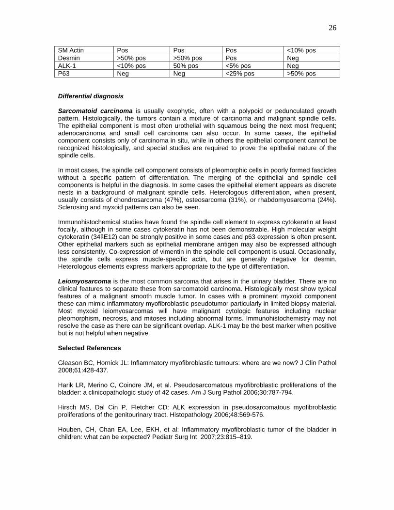
26
SM Actin Pos Pos Pos <10% pos Desmin >50% pos >50% pos Pos Neg ALK-1 <10% pos 50% pos <5% pos Neg P63 Neg Neg <25% pos >50% pos Differential diagnosis Sarcomatoid carcinoma is usually exophytic, often with a polypoid or pedunculated growth pattern. Histologically, the tumors contain a mixture of carcinoma and malignant spindle cells. The epithelial component is most often urothelial with squamous being the next most frequent; adenocarcinoma and small cell carcinoma can also occur. In some cases, the epithelial component consists only of carcinoma in situ, while in others the epithelial component cannot be recognized histologically, and special studies are required to prove the epithelial nature of the spindle cells. In most cases, the spindle cell component consists of pleomorphic cells in poorly formed fascicles without a specific pattern of differentiation. The merging of the epithelial and spindle cell components is helpful in the diagnosis. In some cases the epithelial element appears as discrete nests in a background of malignant spindle cells. Heterologous differentiation, when present, usually consists of chondrosarcoma (47%), osteosarcoma (31%), or rhabdomyosarcoma (24%). Sclerosing and myxoid patterns can also be seen. Immunohistochemical studies have found the spindle cell element to express cytokeratin at least focally, although in some cases cytokeratin has not been demonstrable. High molecular weight cytokeratin (34ßE12) can be strongly positive in some cases and p63 expression is often present. Other epithelial markers such as epithelial membrane antigen may also be expressed although less consistently. Co-expression of vimentin in the spindle cell component is usual. Occasionally, the spindle cells express muscle-specific actin, but are generally negative for desmin. Heterologous elements express markers appropriate to the type of differentiation. Leiomyosarcoma is the most common sarcoma that arises in the urinary bladder. There are no clinical features to separate these from sarcomatoid carcinoma. Histologically most show typical features of a malignant smooth muscle tumor. In cases with a prominent myxoid component these can mimic inflammatory myofibroblastic pseudotumor particularly in limited biopsy material. Most myxoid leiomyosarcomas will have malignant cytologic features including nuclear pleomorphism, necrosis, and mitoses including abnormal forms. Immunohistochemistry may not resolve the case as there can be significant overlap. ALK-1 may be the best marker when positive but is not helpful when negative. Selected References Gleason BC, Hornick JL: Inflammatory myofibroblastic tumours: where are we now? J Clin Pathol 2008;61:428-437. Harik LR, Merino C, Coindre JM, et al. Pseudosarcomatous myofibroblastic proliferations of the bladder: a clinicopathologic study of 42 cases. Am J Surg Pathol 2006;30:787-794. Hirsch MS, Dal Cin P, Fletcher CD: ALK expression in pseudosarcomatous myofibroblastic proliferations of the genitourinary tract. Histopathology 2006;48:569-576. Houben, CH, Chan EA, Lee, EKH, et al: Inflammatory myofibroblastic tumor of the bladder in children: what can be expected? Pediatr Surg Int 2007;23:815–819.

27
Jones EC, Young RH. Myxoid and sclerosing sarcomatoid transitional cell carcinoma of the urinary bladder: a clinicopathologic and immunohistochemical study of 25 cases. Mod Pathol 1997;10:908-916. Lopez-Beltran A, Pacelli A, Rothenberg HJ, et al. Carcinosarcoma and sarcomatoid carcinoma of the bladder: clinicopathological study of 41 cases. J Urol 1998;159:1497-1503. Mills SE, Bova GS, Wick MR, Young RH. Leiomyosarcoma of the urinary bladder: a clinicopathologic and immunohistochemical study of 15 cases. Am J Surg Pathol 1989;13:480-489. Spiess PE, Tuziak T, Tibbs RF, et al. Pseudosarcomatous and sarcomatous proliferations of the bladder. Hum Pathol 2007;38:753-761. Sukov, WR, Cheville JC, Carlson AW, et al : Utility of ALK-1 protein expression and ALK rearrangements in distinguishing inflammatory myofibroblastic tumor from malignant spindle cell lesions of the urinary bladder. Modern Pathology 2007;20:592–603. Westfall DE, Folpe AL, Paner GP, et al. Utility of a comprehensive immunohistochemical panel in the differential diagnosis of spindle cell lesions of the urinary bladder. Am J Surg Pathol 2009;33:99-105.

ASCP AP SLIDE SEMINAR 2011 CASE HISTORIES
Case 1. This 69 year old woman was undergoing routine cystoscopy following a right nephroureterectomy for a renal pelvis urothelial carcinoma one year earlier. At cystoscopy the urologist noted a reddened area on the right lateral wall. The lesion was resected (transurethral resection of bladder tumor – TURBT). Case 2. This 59 year old man presented with a history of painless gross hematuria. At cystoscopy the urologist noted “several large papillary tumors.” A TURBT was performed. Case 3. This 69 year old man presented with gross hematuria. Cystoscopy revealed a bladder tumor and a TURBT was performed. Pathology showed a high grade urothelial carcinoma with lamina propria invasion (T1). He was referred to our institution where a restaging TURBT was performed (the slide comes from this procedure). Case 4. This 58 year old man presented with painless gross hematuria. Past history included a renal transplant 10 years earlier and diabetes mellitus. At cystoscopy the urologist note “multiple papillary areas” involving the dome, right lateral wall and left lateral wall. The lesions were resected. Case 5. This 76 year old woman presented with gross hematuria and pelvic pain. At cystoscopy the mucosa was edematous without a well defined lesion. Biopsies were performed. Case 6. This 67 year old woman presented with dysuria, micro-hematuria and pelvic pain. Past history included SLE and a renal transplant 4 years earlier. At cystoscopy the urologist noted a “flat, solid tumor involving the entire left half of the bladder wall.” A transurethral resection was performed but the urologist noted that “the entire full thickness of the tumor could not be resected.” Case 7. This 48 year old man presented with gross hematuria and abdominal pain. At cystoscopy the urologist noted a “large necrotic tumor involving the right anterior wall. A transurethral resection was performed. Case 8. This 35 year old man presented with a history of repeated urinary tract infections and difficulty urinating. There was no other significant history. At cystoscopy the urologist noted a 4 cm polypoid mass with a smooth surface in the trigone area. A TURBT was performed. Case 9. This 51 year old woman presented with pelvic pain. There was no significant past history. During the clinical work up a CT scan of the pelvis revealed a poorly defined 3 cm mass in the bladder wall in the area of the dome. At cystoscopy no mucosal lesion was identified and a biopsy from the area was read as “negative for malignancy.” A partial cystectomy was subsequently performed. Case 10. This 20 year old pregnant (27weeks) woman presented with an episode of gross hematuria. An MRI was read as showing “a left lateral wall mass with full thickness invasion and concern for an extra-cystic component.” Following a biopsy, the patient was referred to our institution and a partial cystectomy was performed.