5 sms research roundtable symposium - … full...sms research roundtable symposium abstracts reston...
TRANSCRIPT


5TH SMS RESEARCH ROUNDTABLE SYMPOSIUM ABSTRACTS
Reston Hyatt Hotel Regency Ballroom A
Thursday, September 17, 2009
An invited scientific symposium Co-sponsored by:
SMS Research Team Office of the Clinical Director Professional
National Human Genome Research Institute Advisory Board National Institutes of Health, HHS
With Funding from:
ORDR Conference Grant 2009
Partial funding also provided by: Friends of PRISMS – donations
Frank Greenberg Memorial Education Fund, &
Acknowledgements: Special thanks the Office of Rare Diseases Research, NIH and Office of Clinical Director, National Human Genome Research Institute, NIH for financial and administrative support and to Alan Kleinfeld & Pat Brown for assistance with on-site logistics.

5TH SMS RESEARCH ROUNDTABLE SYMPOSIUM
SEPTEMBER 17, 2009 HYATT RESTON
RESTON, VIRGNIIA
TABLE OF CONTENTS Agenda Page 5th SMS Research Roundtable Agenda 3
Submitted Abstracts 7 SESSION I: LESSONS FROM THE BEDSIDE – CLINICAL RESEARCH UPDATE 7
Moderated by Ann C.M. Smith, M.A., D.Sc.(hon) Expanding the SMS Phenotype Development & Behavior Family Support & Respite Discussion & Summary 25
SESSION II: LESSONS FROM THE BENCH 26 Moderated by Sarah Elsea, Ph.D. Molecular Genetic Aspects - RAI1 & other genes Mouse models – What can we learn? Discussion & Summary 35
2009 Research Roundtable Participant List 36
PRISMS Board of Directors 39
PRISMS Professional Advisory Board 40
SMS Medical Management Guidelines 41 (Last revised 2008 for GeneReviews)
Recent Publications (abstracts) on Smith-Magenis Syndrome 44 2009 Publications 44
2008 Publications 48
2007 Publications 49
SMSRR 9.17.2009 - 2 -

5TH SMS RESEARCH ROUNDTABLE SYMPOSIUM Reston Hyatt Hotel ~ Regency Ballroom A
Thursday, September 17, 2009 AGENDA
7:15-8:15am Continental Breakfast & Gathering
8:15am Welcome and Introduction Ann C.M. Smith, M.A., D.Sc.(Hon) Chair, Professional Advisory Board Head SMS Research Team, OCD/NHGRI/NIH/HHS
8:30am SESSION I: LESSONS FROM THE BEDSIDE - CLINICAL RESEARCH UPDATE Expanding the SMS Phenotype; Development & Behavior; Family Support & Respite Moderated by Ann C.M. Smith, M.A., D.Sc. (Hon)
8:30am Recent Progress on 3D Face Analysis in Smith-Magenis Syndrome (SMS) Peter Hammond, Mauren Abreu de Sousa (UCL, London), Judith Allanson (CHEO, Ottawa), Ann Smith (NHGRI/NIH, Bethesda)
8:45am UPDATE FROM FRANCE Electromyography and pain in SMS Hélène De Leersnyder, Pediatrician Paris, France
9:00am 4 years study of activity of the treatment with beta-blockers and a new melatonin antagonist, Agomelatine Hélène De Leersnyder, Pediatrician Hopital Robert Debré, Department of Genetics, Paris, France
9:15am A Psychiatrist’s Experience with SMS in France Didier Rosch, MD, Psychiatrist Paris, France
9:30am What Goes in Must Come Out: Constipation in Smith-Magenis Syndrome Woolery, M, Duncan, F and Smith ACM CC/NIH and OCD/NHGRI/NIH, Bethesda, MD
9:45am DEVELOPMENT & BEHAVIOR Neurodevelopment of children under 3 years of age with Smith-Magenis syndrome. 1Wolters PL, 2Gropman AL, 1Martin SC, 3Smith MR, 3Hildenbrand HL, 4Brewer CC, 5Smith ACM 1NCI/NIH; 2Children’s National Medical Center; 3CC-RMD/NIH; 4NIDCD/NIH; 5OCD/NHGRI, NIH, Bethesda, MD
SMSRR 9.17.2009 - 3 -

10:00 BREAK
10:15am A Comparative Study of Sensory Processing in Children with Smith-Magenis Syndrome and Children with Autism1Hildenbrand H, 1Silberman AE, 2Golden-Williams C, 2Thurm A,3Smith ACM 1Dept. Rehabilitative Medicine, Clinical Center; 2 PDNB/NIMH; and 3
3OCD/NHGRI, NIH, Bethesda, MD
10:30am Sensory Motor and Functional Skills of Dizygotic Twins: One With Smith– Magenis Syndrome and a Twin Control Michaele R. Smith1, Hanna Hildenbrand1, Ann C. M. Smith2,3
1) Rehabilitation Medicine Department, National Institutes of Health (NIH); 2) Office of Clinical Director, NHGRI/NIH, Bethesda, MD; 3) Georgetown University, Washington, DC.
10:45am Measurement of Developmental Asynchrony in Smith-Magenis Syndrome Brenda Finucane, MS, CGC1, Barbara Haas-Givler, MEd, BCBA1, Heather M Jones2, Elliott W Simon, PhD1
1Genetic Services at Elwyn, Elwyn, PA; 2Kutztown University, Kutztown, PA
11:00am Review of disrupted sleep patterns in Smith-Magenis syndrome and normal melatonin secretion in a patient with an atypical interstitial 17p11.2 deletion. Boudreau EA1, Johnson KP2, Jackman AR2, Blancato J4, Huizing M5, Bendavid C8, Jones M6, Chandrasekharappa SC6, Lewy AJ2, Smith ACM7, Magenis RE3. 1Dept Neurology, 2Dept Psychiatry, 3Dept of Molecular & Medical Genetics, Oregon Health & Science University, Portland, OR; 4Dept Oncology, Georgetown University Med.Center, Washington, DC; 5Medical Genetics Branch, 6Genome Technology Branch, 7Office of the Clinical Director, NHGRI, NIH, Bethesda, Maryland; 8CNRS UMR 6061, Institut de Génétique et Développement de Rennes, Faculté de Médecine, Université de Rennes1, Rennes, France.
11:15am Longitudinal update- Aging & SMS – The OHSU experience R. Ellen Magenis, MD Dept of Molecular & Medical Genetics, Oregon Health & Science University, Portland, OR
11:30am MORNING DISCUSSION & SUMMARY
11:45-1:00pm LUNCH WITH PRISMS BOARD OF BIRECTORS & RESEARCH NETWORKING Randy Beall, President, PRISMS BOARD members
1:00pm FAMILY SUPPORT AND RESPITE Caring for the Caregiver: Increased risk for anxiety and depression for primary caregivers of a child diagnosed with Smith-Magenis syndrome. Rebecca H. Foster, M.S.,1 Stephanie Kozachek,2 Surbhi Kanotra,1 Marilyn Stern1,3, and Sarah Elsea2,3. 1Dept. of Psychology, 2Dept. of Human and Molecular Genetics, and 3Dept. of Pediatrics Virginia Commonwealth University, Richmond, VA.
SMSRR 9.17.2009 - 4 -
1:15pm They said it couldn’t be done -The power of one and the dedication of many, builds bridges of hope globally Jodie Davis Funding & Sponsorship Coordinator, SMS Administrator, USA Liaison Officer, Volunteer Assistant Camp Coordinator for past 3 Australian SMS Camps, Marketing Committee, Volunteer. Gae Miller M.N; F.R.C.N.A. R.N; C.M; B.A. (Ed); Grad Dip Health Soc Sci. (Med Sci); Dip ED (Nursing), Volunteer Camp Coordinator for past 2 Australian SMS Camps and Volunteer for all 3, Volunteer. Michelle Price O.A.M, R.N., B.N., P.H.F, M.R.C.N.A, Volunteer for past 3 Australian SMS Camps, Breakaway Board Member, Committee Chairperson for ‘Proposed Breakaway in USA’, Volunteer.
1:30pm SESSION I: LESSONS FROM THE BEDSIDE - DISCUSSION & SUMMARY
1:45pm Session II: LESSONS FROM THE BENCH Molecular genetic aspects of RAI1 and other genes; Mouse models Moderated by Sarah Elsea, Ph.D.
1:45pm Array comparative genomic hybridization of 52 subjects with a Smith-Magenis-like phenotype: identification of dosage-sensitive loci associated with schizophrenia, autism, and developmental delay. Stephen R. Williams1, Santhosh Girirajan1, David Tegay2, Norma Nowak3, Eli Hatchwell4, and Sarah H. Elsea1,5
1 Dept of Human and Molecular Genetics, Virginia Commonwealth University, Richmond, VA, USA 2 Dept of Medicine, New York College Osteopathic Medicine, Old Westbury, NY, USA 3 Dept of Biochemistry, University at Buffalo and Department of Cancer Genetics, Roswell Park Cancer Institute, USA 4 Dept of Pathology, SUNY, Stony Brook, NY, USA 5 Dept of Pediatrics, Virginia Commonwealth University, Richmond, VA, USA.
2:00pm Analyses of gene and protein variations of Retinoic Acid Induced 1 (RAI1) in Smith-Magenis Syndrome. T. Vilboux1, C. Ciccone1, ACM. Smith2, Marjan Huizing1
1Med. Genetics Branch, 2Office Clinical Director, NHGRI, NIH, Bethesda, MD
2:15pm MOUSE MODELS Tom1l2 hypomorphic mice exhibit increased incidence of infections and tumors and abnormal immunological response. Santhosh Girirajan1*, Paula M. Hauck2*#, Stephen Williams1, Christopher N. Vlangos2+, Barbara B. Szomju2, Sara Solaymani-Kohal2, Philip D. Mosier3, Kimber L. White, Jr.4, Kathleen McCoy5, and Sarah H. Elsea.1,2
1Depts. of Human Genetics, 2Pediatrics, 3 Medicinal Chemistry & Center for the Study of Biological Complexity, 4Pharmacology & Toxicology, & 5Microbiology and Immunology, Medical College of Virginia Campus, Virginia Commonwealth University, Richmond, VA.
2:30pm AFTERNOON BREAK
2:45pm Using a mouse CopyNumberVariation (CNV) -engineering system to study neurobehavioral phenotypes in two genomic disorders SMS and PTLS Wenli Gu1, Mel Heney1, Weimin Bi1, Jiong Yan1, Corinne Spencer1, Richard Paylor1, Jim Lupski1,2,3
1Department of Molecular and Human Genetics, 2Department of Pediatrics, 3Texas Children’s Hospital, Houston, TX.
SMSRR 9.17.2009 - 5 -
3:00pm Using mouse models for Smith-Magenis and Potocki-Lupski syndromes to study the impact of CNV on weight and metabolism M.Heney1, W. Gu1, J. Yan1, W. Bi1, P.K. Saha2, L. Chan2, J.R. Lupski1,3,4
1Department of Molecular and Human Genetics, 2Department of Molecular and Cellular Biology, 3Department of Pediatrics, 4Texas Children’s Hospital, Houston, TX.
3:15pm How much is too much? Phenotypic consequences of Rai1 overexpression in mice. Santhosh Girirajan1, Nisha Patel2, 3, Rebecca E. Slager4, Mary E. Tokarz5, Maja Bucan6, Jenny L. Wiley5, and Sarah H. Elsea.1, 2 *
Department of Human Genetics, Medical College of Virginia Campus, Virginia Commonwealth University, Richmond, VA. Department of Pediatrics, Medical College of Virginia Campus, Virginia Commonwealth University, Richmond, VA. Faculty of Health and Life Sciences, University of the West of England, Bristol, U.K. Department of Internal Medicine, University of Nebraska Medical Center, Omaha, NE. Department of Pharmacology and Toxicology, Medical College of Virginia Campus, Virginia Commonwealth University, Richmond, VA. Department of Genetics, University of Pennsylvania School of Medicine, Philadelphia, PA.
3:30pm SESSION II: LESSONS FROM THE BENCH - DISCUSSION & SUMMARY
3:50pm Concluding Remarks and Announcements Ann C.M. Smith, M.A., D.Sc. (hon)
4:00pm Adjournment (& Group Picture)
4:30-5:15pm PRISMS Professional Advisory Board (brief meeting)
5:00-7:00pm PRISMS Welcome Reception, Building Bridges of Hope
SMSRR 9.17.2009 - 6 -
ABSTRACTS 5th SMS Research Roundtable -Thursday, September 17, 2009
Hyatt Regency Reston, VA
SESSION I: LESSONS FROM THE BEDSIDE – CLINICAL RESEARCH UPDATE Moderated by Ann C.M. Smith, M.A., D.Sc. (Hon)
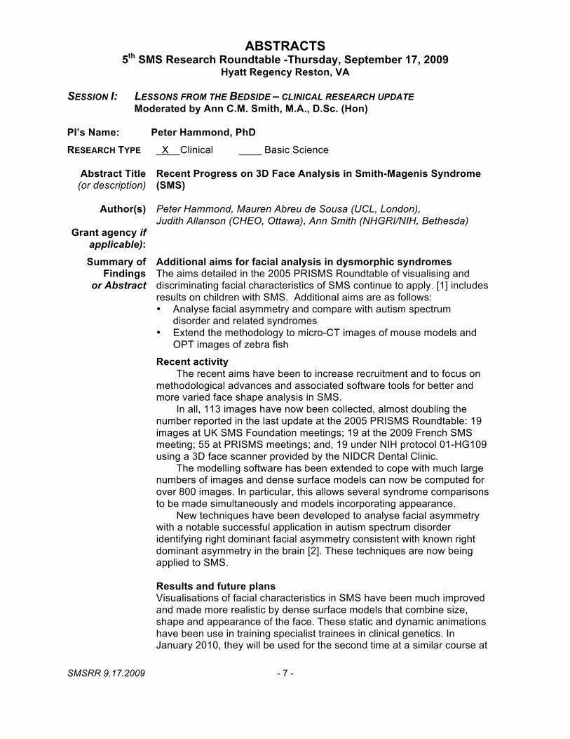
PI’s Name: Peter Hammond, PhD RESEARCH TYPE _X__Clinical ____ Basic Science
Abstract Title Recent Progress on 3D Face Analysis in Smith-Magenis Syndrome (or description) (SMS)
Author(s) Peter Hammond, Mauren Abreu de Sousa (UCL, London), Judith Allanson (CHEO, Ottawa), Ann Smith (NHGRI/NIH, Bethesda)
Grant agency if applicable): Summary of Additional aims for facial analysis in dysmorphic syndromes
Findings The aims detailed in the 2005 PRISMS Roundtable of visualising and or Abstract discriminating facial characteristics of SMS continue to apply. [1] includes
results on children with SMS. Additional aims are as follows: • Analyse facial asymmetry and compare with autism spectrum
disorder and related syndromes • Extend the methodology to micro-CT images of mouse models and
OPT images of zebra fish
Recent activity The recent aims have been to increase recruitment and to focus on
methodological advances and associated software tools for better and more varied face shape analysis in SMS.
In all, 113 images have now been collected, almost doubling the number reported in the last update at the 2005 PRISMS Roundtable: 19 images at UK SMS Foundation meetings; 19 at the 2009 French SMS meeting; 55 at PRISMS meetings; and, 19 under NIH protocol 01-HG109 using a 3D face scanner provided by the NIDCR Dental Clinic.
The modelling software has been extended to cope with much large numbers of images and dense surface models can now be computed for over 800 images. In particular, this allows several syndrome comparisons to be made simultaneously and models incorporating appearance.
New techniques have been developed to analyse facial asymmetry with a notable successful application in autism spectrum disorder identifying right dominant facial asymmetry consistent with known right dominant asymmetry in the brain [2]. These techniques are now being applied to SMS.
Results and future plans Visualisations of facial characteristics in SMS have been much improved and made more realistic by dense surface models that combine size, shape and appearance of the face. These static and dynamic animations have been use in training specialist trainees in clinical genetics. In January 2010, they will be used for the second time at a similar course at
SMSRR 9.17.2009 - 7 -

Page 2: Hammond, Peter
the Cambridge Sanger Centre. Stand-alone viewing software has been produced so that the models and animations can be distributed for general training use.
Discrimination testing using various pattern recognition algorithms score very highly on unseen or blind testing. This provides an accurate, objective means for screening children without a diagnosis for whom SMS is suspected. Positive identification of the facial features encourages appropriate genetic testing. New techniques have been developed to calculate optimal dense surface models for face classification when comparing SMS and controls or SMS and other genetic conditions. The improved discrimination accuracy is a direct result of the large increase in recruitment. Separate models for male and female subjects are also now possible.
Although face shape differences in individuals with an RAI1 mutation are detectable, the number of those fully genotyped remains too low to draw quantifiable differences. This needs to be addressed in future if genotype-phenotype analysis is to be extended to facial morphology.
The dense surface modelling techniques have been applied successfully to mouse models [3] and plans are underway to extend this to Optical Projection Tomography images of zebra fish.
Publications (List related papers and/or abstracts)
1. Hammond P, Hutton TJ, Allanson J, Buxton B, Campbell L, Clayton-Smith J, Donnai D, Karmiloff-Smith A, Metcalfe K, Murphy KC, Patton M, Pober B, Prescott K, Shaw A, Scambler, Smith ACM, Temple K, Hennekam R, Tassabehji M. (2005) Discriminating Power of Localised 3D Facial Morphology. Am. J. Hum. Genet. 77:999-1010.
2. Hammond P, Forster-Gibson C, Chudley AE, Allanson JE, Hutton TJ, Holden JJA, Lewis MES (2008) Face-brain asymmetry in autism spectrum disorders, Molecular Psychiatry, 13: 614-623.
3. Tobin JL, DiFranco M, Eichers E, May-Simera H, Garcia M, Yan J, Justice M, Briscoe J, Mayor R, Lupski JR, Hammond P, Beales PL. (2008) Inhibition of neural crest migration underlies craniofacial dysmorphology and Hirschsprung’s disease in Bardet–Biedl syndrome, PNAS, 6;105(18):6714-9; on line April 28th 2008.
SMSRR 9.17.2009 - 8 -
5th SMS Research Roundtable -Thursday, September 17, 2009 Hyatt Regency Reston, VA
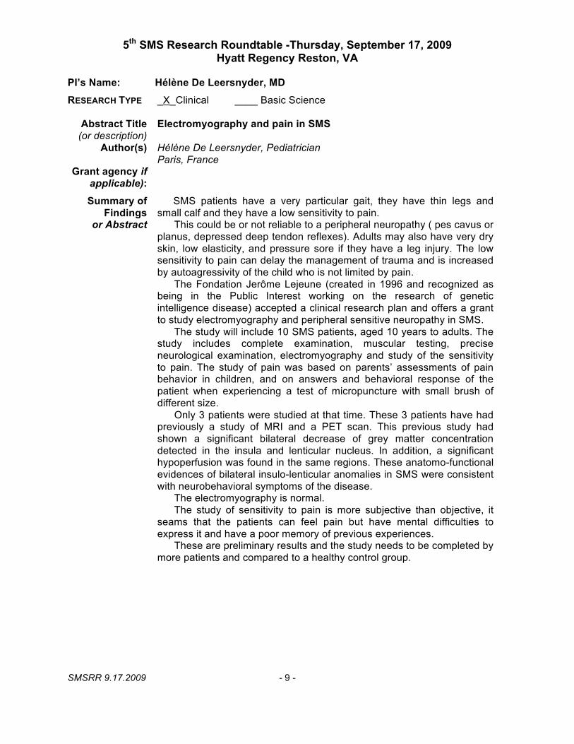
PI’s Name: Hélène De Leersnyder, MD RESEARCH TYPE _X_Clinical ____ Basic Science
Abstract Title Electromyography and pain in SMS (or description)
Author(s) Hélène De Leersnyder, Pediatrician Paris, France
Grant agency if applicable): Summary of SMS patients have a very particular gait, they have thin legs and
Findings small calf and they have a low sensitivity to pain. or Abstract This could be or not reliable to a peripheral neuropathy ( pes cavus or
planus, depressed deep tendon reflexes). Adults may also have very dry skin, low elasticity, and pressure sore if they have a leg injury. The low sensitivity to pain can delay the management of trauma and is increased by autoagressivity of the child who is not limited by pain.
The Fondation Jerôme Lejeune (created in 1996 and recognized as being in the Public Interest working on the research of genetic intelligence disease) accepted a clinical research plan and offers a grant to study electromyography and peripheral sensitive neuropathy in SMS.
The study will include 10 SMS patients, aged 10 years to adults. The study includes complete examination, muscular testing, precise neurological examination, electromyography and study of the sensitivity to pain. The study of pain was based on parents’ assessments of pain behavior in children, and on answers and behavioral response of the patient when experiencing a test of micropuncture with small brush of different size.
Only 3 patients were studied at that time. These 3 patients have had previously a study of MRI and a PET scan. This previous study had shown a significant bilateral decrease of grey matter concentration detected in the insula and lenticular nucleus. In addition, a significant hypoperfusion was found in the same regions. These anatomo-functional evidences of bilateral insulo-lenticular anomalies in SMS were consistent with neurobehavioral symptoms of the disease.
The electromyography is normal. The study of sensitivity to pain is more subjective than objective, it
seams that the patients can feel pain but have mental difficulties to express it and have a poor memory of previous experiences.
These are preliminary results and the study needs to be completed by more patients and compared to a healthy control group.
SMSRR 9.17.2009 - 9 -
5th SMS Research Roundtable -Thursday, September 17, 2009 Hyatt Regency Reston, VA
PI’s Name: Hélène De Leersnyder, Pediatrician
RESEARCH TYPE _X__Clinical ____ Basic Science
Abstract Title (or description)
Author(s)
Grant agency if applicable): Summary of
Findings or Abstract
4 years study of activity of the treatment with beta-blockers and a new melatonin antagonist, Agomelatine
Hélène De Leersnyder, Pediatrician Hopital Robert Debré, Department of Genetics, Paris, France
Smith-Magenis syndrome is a genetic disease emblematic of these sleeps disturbances in children with neurodevelopmental and neuropsychological difficulties. Indeed, in this microdeletional syndrome, sleep disturbances are extremely severe and have been ascribed to an abnormal secretion of melatonin, the main hormone of pineal gland influencing sleep. Previous studies related the benefit of the association of beta-blockers and melatonine to impove the sleep of the children.
The objective of this study was to assess the activity of a treatment with beta-blockers and Agomelatine. Agomelatine is a melatonin agonist of melatonin receptors. In animals, Agomelatine is able to resynchronize circadian rhythms in models of circadian rhythms desynchronization, free running, delayed sleep phase syndrome model through its direct action on melatonin receptors MT1, MT2 in suprachiasmatic nucleus, the biologic clock.
9 SMS patients aged 6 years to 18 years were included in the study. Beta-blocker (Acebutolol) was given at breakfast and Agomelatine at dinner. Drug dosages varied according to the weight of the subject.
At the end of the mandatory period of 7 months (1 month baseline and 6 months treatment), in view of the therapeutic improvement and of a very good feedback of the patients’ parents, the study was extended by 42 months. So the complete trial lasted 49 months and patients were treated during 48 months with regular examination and different measurements. Overall compliance was satisfactory.
The criteria for evaluation were actigraphy, sleep diaries, Achenbach questionnaires, and children’s sleep questionnaires. Safety measurements included: recording adverse events, vital signs, biological and hematological laboratory tests.
The results from actigraphy were consistent with those obtained with the sleep diary and the children’s sleep questionnaire, which shows that the nocturnal waking up was less frequent and shorter than at baseline (-60 mn) and that the sleeping time was longer than at baseline (+ 63 minutes).
The mean duration of the naps decreased over the study period. Clinical improvement was notable and the parents confirmed the benefice of the treatment. The children slept deeply and were quiet whereas in the past it used to be dramatic. The sleep was no more fragmented by prolonged nocturnal awakenings and waking up in the morning was delayed.
SMSRR 9.17.2009 - 10 -
Page 2: Hélène De Leersnyder
During the day, the number of tired patients decreased over the M0-M48 period. Patients’ behavior improved according to all lines of Achenbach questionnaire. The mean activities and social scales scores increased. The mean internalizing and externalizing scores decreased.
Only 4 patients reported emergent adverse events not considered as related to the study treatment and recovered quickly. No serious or significant adverse event occurred. No clinical relevant changes over time were detected for any biochemical and hematological parameters as well as for blood pressure, heart rate, weight, height and BMI. The child had regular growth relatively to age. According to the beta-blockers treatment this prolonged follow up is important.
Data from this exploratory, open trial indicate that Agomelatine (1 or 5 mg), when co-administrated with Acebutolol (10mg/kg) was an effective and well-tolerated treatment of sleep disturbances in SMS. At the families’ request 8 children have been receiving the treatment for 4 years with no habituation and no side effect.
PUBLICATIONS (List related papers and/or abstracts) 1. De Leersnyder, H. (2006). Inverted rhythm of melatonin secretion in Smith-Magenis
syndrome: from symptoms to treatment. Trends Endocrinol Metab 17, 291-298. 2. Smith, A. C., Dykens, E., and Greenberg, F. (1998). Sleep disturbance in Smith-Magenis
syndrome (del 17 p11.2). Am J Med Genet 81, 186-191. 3. Potocki, L., Glaze, D., Tan, D. X., Park, S. S., Kashork, C. D., Shaffer, L. G., Reiter, R. J.,
and Lupski, J. R. (2000). Circadian rhythm abnormalities of melatonin in Smith-Magenis syndrome. J Med Genet 37,
4. Olie, J. P., and Kasper, S. (2007). Efficacy of agomelatine, a MT1/MT2 receptor agonist with 5-HT2C antagonistic properties, in major depressive disorder. Int J Neuropsychopharmacol 10, 661-673.
5. Jan, J. E., and Freeman, R. D. (2004). Melatonin therapy for circadian rhythm sleep disorders in children with multiple disabilities: what have we learned in the last decade? Dev Med Child Neurol 46, 776-782.
SMSRR 9.17.2009 - 11 -
5th SMS Research Roundtable -Thursday, September 17, 2009 Hyatt Regency Reston, VA
RESEARCH TYPE _X__Clinical ____ Basic Science
Abstract Title A Psychiatrist’s Experience with SMS in France (or description) No Abstract submitted.
Author(s) Didier Rosch, MD, Psychiatrist Paris, France
SMSRR 9.17.2009 - 12 -

5th SMS Research Roundtable -Thursday, September 17, 2009 Hyatt Regency Reston, VA
PI’s Name: Ann C.M. Smith, MA,DSc(hon) - Presented by Myra Woolery,RN, MN,CPON RESEARCH TYPE
Abstract Title (or description)
Author(s)
Grant agency if applicable): Summary of
Findings or Abstract
__X_Clinical ____ Basic Science
Constipation in SMS: What Goes in Must Come Out
Woolery, M, Duncan, F and Smith ACM CC/NIH and OCD/NHGRI/NIH, Bethesda, MD
Division of Intramural Research, CC-Nursing & NHGRI, (Protocol 01-HG-0109). Background: Constipation is common in children with special needs and an unrecognized problem in children with Smith Magenis Syndrome (SMS). Smith et al. (1998) reported a 53% incidence of constipation in SMS. Children with SMS are at risk for constipation secondary to hyptonia, severe oral-sensori-motor dysfunction, and excessive drooling which impact bowel status. Symptoms of constipation are often ignored by clinicians however they can have a negative impact on the child’s appetite, activity level, mood, and comfort. Accurate assessment is essential in evaluating the effectiveness of interventions targeted at prevention and/or management of constipation. This descriptive study was conducted to identify the incidence, symptoms and contributing factors associated with constipation in SMS and strategies used to manage constipation. Methods: The Pediatric Constipation Survey (P-CAS) was administered to parents of children with SMS attending Camp Breakaway in Australia in November 2008. The survey was developed by Woolery in conjunction with a doctoral level course. It includes demographic and bowel history questions, the Bristol Stool Form Scale (BSFS), and an adapted version of the Constipation Assessment Scale (Woolery et al., 2006). Descriptive statistics were computed. Results: Seventeen parents completed the P-CAS and 16 were evaluable. There were 17 (10M/7F) children with a confirmed diagnosis of SMS between the ages of 3-20 years (mean age 10.6 years). Sixty-eight percent (11/16) had a previous history of constipation. Stool frequency was >once per day in approximately 30% (5/16). Stool consistency was consistent with constipation in 50% (8/16) and borderline between constipation and normal in 44% (7/16). Strategies and aids used to manage constipation included water intake, bowel frequency, consistency, medications and diet. Children who consumed more fluid, fruits, and fiber tended to have softer and more frequent stools. Conclusion: The incidence of constipation in SMS is similar to findings reported by Smith et al. 1998. The P-CAS provided a mechanism for assessing constipation. While the data suggests strategies such as fluid and fiber may be effective, further research is needed.
SMSRR 9.17.2009 - 13 -

Page 2: Myra Woolery
PUBLICATIONS (List related papers and/or abstracts)
Smith ACM, E Dykens and F Greenberg (1998b): Sleep disturbance in Smith-Magenis syndrome (del 17p11.2). Amer J Med Genetics (Neurospychiatric Genetics) 81:186-191.
Woolery, M., Carroll, E., Fenn, E., Wieland, H., Jarosinski, P., Corey, B., & Wallen, G.R. (2006). Constipation assessment scale use in pediatric oncology. Journal of Pediatric Oncology Nurses, 23(2), 64-74.
SMSRR 9.17.2009 - 14 -

5th SMS Research Roundtable -Thursday, September 17, 2009 Hyatt Regency Reston, VA
PI’s Name: Ann C.M. Smith, MA, DSc (hon) RESEARCH TYPE _X__Clinical ____ Basic Science
Abstract Title Neurodevelopment of children under 3 years of age with Smith-(or description) Magenis syndrome.
Author(s) 1Wolters PL, 2Gropman AL, 1Martin SC, 3Smith MR, 3Hildenbrand HL, 4Brewer CC, 5Smith ACM 1) NCI/NIH; 2) Children’s National Medical Center; 3) CC-RMD/NIH; 4) NIDCD/NIH; 5) OCD/NHGRI, NIH, Bethesda, MD
Grant agency if Division of Intramural Research, NHGRI/NIH (Protocol 01-HG-0109), applicable): Bench-to-Bedside Award (NIH-CC)
Summary of Systematic data regarding early neurodevelopmental functioning in Findings Smith-Magenis syndrome are limited. Eleven children with Smith-
or Abstract Magenis syndrome less than 3 years of age (mean, 19 months; range 5-34 months) received prospective multidisciplinary assessments using standardized measures. The total sample scored in the moderately to severely delayed range in cognitive functioning, expressive language, and motor skills and exhibited generalized hypotonia, oral-motor abnormalities, and middle ear dysfunction. Socialization skills were average, and significantly higher than daily living, communication, and motor abilities, which were below average. Mean behavior ratings were in the non-autistic range. According to exploratory analyses, the toddler subgroup scored significantly lower than the infant subgroup in cognition, expressive language, and adaptive behavior, suggesting that the toddlers were more delayed than the infants relative to their respective peers. Infants aged approximately 1 year or younger exhibited cognitive, language, and motor skills that ranged from average to delayed, but with age-appropriate social skills and minimal maladaptive behaviors. At ages 2 to 3 years, the toddlers consistently exhibited cognitive, expressive language, adaptive behavior, and motor delays and mildly to moderately autistic behaviors. Combining age groups in studies may mask developmental and behavioral differences. Increased knowledge of these early neurodevelopmental characteristics should facilitate diagnosis and appropriate intervention.
PUBLICATIONS (List related papers and/or abstracts)
Martin SC, Wolters PL, Smith AC. Adaptive and maladaptive behavior in children with Smith-Magenis Syndrome. J Autism Dev Disord. 2006 May;36(4):541-52.
Gropman AL, Duncan WC, Smith AC. Neurologic and developmental features of the Smith-Magenis syndrome (del 17p11.2). Pediatr Neurol. 2006 May;34(5):337-50. Review.
Gropman A, ACM Smith, J Allanson, and F Greenberg: Smith Magenis Syndrome: Aspects of the Infant Phenotype. Amer J Hum Genet (Suppl) 63(4): A19, 1998.
SMSRR 9.17.2009 - 15 -
5th SMS Research Roundtable -Thursday, September 17, 2009 Hyatt Regency Reston, VA
PI’s Name: ACM Smith, MA, DSc(Hon) - Presented by Hanna Hildenbrand, OT RESEARCH TYPE __XX_Clinical ____ Basic Science
Abstract Title A Comparative Study of Sensory Processing in Children with Smith-(or description) Magenis Syndrome and Children with Autism
Author(s) 1Hildenbrand H, 1Silberman AE, 2Golden-Williams C, 2Thurm A, 3Smith ACM 1Dept. Rehabilitative Medicine, Clinical Center; 2 PDNB/NIMH; and 3
3OCD/NHGRI, NIH, Bethesda, MD
Grant agency if CC-RMD, Division of Intramural Research NHGRI (Protocol 01-HG-0109) applicable): & NIMH (Protocol 06-M-0065), NIH.
Summary of Individuals with SMS exhibit a neurobehavioral phenotype with atypical Findings sensory processing, overlapping with other well recognized disorders.
or Abstract This study seeks to describe sensory processing and examine differences in patterns of sensory processing in children with SMS and in children with autism using the Short Sensory Profile (SSP). Compared to the normative sample, both groups presented with overall sensory processing difficulties with mean scores in the at risk or definite difference range for 6 out of 7 sections of the SSP; the SMS group presented with a relative strength in Taste/Smell Sensitivity, while the autism group had a relative strength in Movement Sensitivity. Between groups, statistically significant differences were found in Low Energy/Weak (p = .01), Movement Sensitivity (p = .05), Underresponsive/Seeks Sensation (p = .05), and the Total Score (p = .05), indicating more severe difficulties in the SMS group. The single SMS participant with a confirmed dual diagnosis of autism and SMS was identified as an outlier, scoring lower in the Underresponsive/Seeks Sensation and higher in Low-Energy/Weak sections relative to all other SMS participants, but within the range exhibited by autism group. Recognition of the specific patterns of sensory processing unique to SMS may promote early and accurate diagnosis and direct intervention. Further research examining this domain should address the limitations of this study. Use of the long Sensory Profile that has a bidirectional scoring system may address the potential ceiling effect observed in the autism group and may expand and further delineate patterns of sensory processing between groups. Including a separate group of children with dual diagnosis of autism and SMS may provide additional understanding of the complex neurobehavioral phenotype.
SMSRR 9.17.2009 - 16 -

Page 2: Hildenbrand, H. PUBLICATIONS (List related papers and/or abstracts)
Smith MR, Hildenbrand H, Smith ACM. Sensory Motor and Functional Skills of Dizygotic Twins: One With Smith–Magenis Syndrome and a Twin Control. Physical & Occupational Therapy in Pediatrics, Vol. 29(3), 2009
Hildenbrand H, Furst G, Smith ACM. Patterns of Sensory Processing in Children with Smith-Magenis Syndrome, Del (17) (p11.2p11.2), Poster at Occupational Therapy Association conference, Alaska, May 2004.
SMSRR 9.17.2009 - 17 -
5th SMS Research Roundtable -Thursday, September 17, 2009 Hyatt Regency Reston, VA
PI’s Name: ACM Smith, MA, DSc (hon) – Michaele Smith, BS, MEd presenting RESEARCH TYPE __X_Clinical ____ Basic Science
Abstract Title Sensory Motor and Functional Skills of Dizygotic Twins: One With (or description) Smith–Magenis Syndrome and a Twin Control
Author(s) Michaele R. Smith1, Hanna Hildenbrand1, Ann C. M. Smith2,3
1) Rehabilitation Medicine Department, National Institutes of Health (NIH); 2) Office of Clinical Director, NHGRI/NIH, Bethesda, MD; 3) Georgetown University, Washington, DC.
Grant agency if Division of Intramural Research, NHGRI (Protocol 01-HG-0109) & applicable): RMD/CC, National Institutes of Health; Bench to Bedside Award, CC-NIH
Summary of Smith–Magenis syndrome (SMS), the result of an interstitial deletion Findings within chromosome 17p11.2, is a disorder which may include minor
or Abstract dysmorphic features, brachydactyly, short stature, hypotonia, speech delays, cognitive deficits, signs of peripheral neuropathy, 10 scoliosis, and neurobehavioral problems including sleep disturbances and maladaptive repetitive and self-injurious behaviors. Physical and occupational therapists provide services for the children who have the syndrome, whose genetic disorder is frequently not identified or diagnosed before 1 year of age. Comprehensive physical and occupational therapy evaluation was completed in nonidentical twins with one having SMS, using the Sensory Profile; Brief Assessment of Motor Function (BAMF); Peabody Developmental Motor Scales, Second Edition (PDMS-2); and Pediatric Evaluation Disability Inventory (PEDI). This provides a framework for conducting assessments to enhance early detection and interdisciplinary management with this specialized population.
PUBLICATIONS (List related papers and/or abstracts)
1. Smith MR, Hildenbrand H, & Smith ACM. Sensory Motor and Functional Skills of Dizygotic Twins: One With Smith–Magenis Syndrome and a Twin Control. Physical & Occupational Therapy in Pediatrics, Vol. 29(3), 2009
2. Young, K, Smith, M, Hildenbrand H, Danoff J, Smith ACM. Assessment of Motor and Functional Performance in children with Smith-Magenis syndrome. NIH Summer Internship Program (Poster), July 2007, NIH, Bethesda, MD.
SMSRR 9.17.2009 - 18 -

5th SMS Research Roundtable -Thursday, September 17, 2009 Hyatt Regency Reston, VA
PI’s Name: Brenda Finucane, MS RESEARCH TYPE __X_Clinical ____ Basic Science
Abstract Title Measurement of Developmental Asynchrony in Smith-Magenis (or description) Syndrome
Author(s) Brenda Finucane, MS, CGC1, Barbara Haas-Givler, MEd, BCBA1
Heather M Jones2. Elliott W Simon, PhD1
1Genetic Services at Elwyn, Elwyn, PA; 2Kutztown Univ., Kutztown, PA Grant agency if
applicable): Martha WS Rogers Trust, Philadelphia, PA Summary of
Findings Developmental asynchrony is a term used to describe unevenness in the or Abstract intellectual and socio-emotional development of highly gifted children.
Such asynchrony causes gifted children to be particularly vulnerable to emotional difficulties, and requires educational and counseling strategies which specifically address the gap between cognitive and socio-emotional development. Although the phenomenon of developmental asynchrony has not been well-researched in people with intellectual disabilities, we have observed a parallel phenomenon in people with Smith-Magenis syndrome. We hypothesize that developmental asynchrony is a significant contributor to maladaptive behaviors in SMS.
This research aims to compare development across a number of cognitive, social, and emotional domains in age and IQ-matched cohorts of adolescents and young adults with SMS and Down syndrome. Measures administered include the Kaufman Adolescent and Adult Intelligence Test; The BERS-2 (Behavioral and Emotional Rating scale); the Reiss Profile; and the Carey Temperament Scales. We hypothesize that we will be able to distinguish children with SMS from those with Down syndrome based on comparison of syndrome-specific scores across developmental domains (i.e., coordinated development across domains in Down syndrome versus asynchronous development in SMS.) The ability to reliably detect developmental asynchrony could potentially offer important insights into behavior and intervention in SMS.
PUBLICATIONS (List related papers and/or abstracts)
Finucane, B. and Haas-Givler, B. (2009): Smith-Magenis syndrome: Genetic basis and clinical implications. J Ment Health Res Intell Disab 2,134-148.
Finucane B : Embracing the inner toddler in people with Smith-Magenis syndrome. Spectrum, Summer 2008, PRISMS (Parents and Researchers Interested in Smith-Magenis Syndrome).
SMSRR 9.17.2009 - 19 -

5th SMS Research Roundtable -Thursday, September 17, 2009 Hyatt Regency Reston, VA
PI’s Name: RE Magenis, MD & ACM Smith, MA, DSc(hon) RESEARCH TYPE _X__Clinical ____ Basic Science
Abstract Title Review of disrupted sleep patterns in Smith-Magenis syndrome and (or description) normal melatonin secretion in a patient with an atypical interstitial
17p11.2 deletion.
Author(s) Boudreau EA, Johnson KP, Jackman AR, Blancato J, Huizing M, Bendavid C, Jones M, Chandrasekharappa SC, Lewy AJ, Smith AC, Magenis RE. Oregon Health & Science University, Portland, OR; 4Dept Oncology, Georgetown University Med.Center, Washington, DC; 5Medical Genetics Branch, 6Genome Technology Branch, 7Office of the Clinical Director, NHGRI, NIH, Bethesda, Maryland; 8CNRS UMR 6061, Institut de Génétique et Développement de Rennes, Faculté de Médecine, Université de Rennes1, Rennes, France.
Grant agency if OHSU; Division of Intramural Research, NHGRI/NIH; applicable):
Summary of Smith-Magenis syndrome (SMS) is a disorder characterized by multiple Findings congenital anomalies and behavior problems, including abnormal sleep
or Abstract patterns. It is most commonly due to a 3.5 Mb interstitial deletion of chromosome 17 band p11.2. Secretion of melatonin, a hormone produced by the pineal gland, is the body's signal for nighttime darkness. Published reports of 24-hr melatonin secretion patterns in two independent SMS cohorts (US and France) document an inverted endogenous melatonin pattern in virtually all cases (96%), suggesting that this finding is pathognomic for the syndrome. We report on a woman with SMS due to an atypical large proximal deletion ( approximately 6Mb; cen<->TNFRSFproteinB) of chromosome band (17)(p11.2p11.2) who presents with typical sleep disturbances but a normal pattern of melatonin secretion. We further describe a melatonin light suppression test in this patient. This is the second reported patient with a normal endogenous melatonin rhythm in SMS associated with an atypical large deletion. These two patients are significant because they suggest that the sleep disturbances in SMS cannot be solely attributed to the abnormal diurnal melatonin secretion versus the normal nocturnal pattern.
PUBLICATIONS (List related papers and/or abstracts)
Boudreau EA, Johnson KP, Jackman AR, Blancato J, Huizing M, Bendavid C, Jones M, Chandrasekharappa SC, Lewy AJ, Smith AC, Magenis RE. Review of disrupted sleep patterns in Smith-Magenis syndrome and normal melatonin secretion in a patient with an atypical interstitial 17p11.2 deletion. Am J Med Genet A. 2009 Jul;149A(7):1382-91.
SMSRR 9.17.2009 - 20 -

5th SMS Research Roundtable -Thursday, September 17, 2009 Hyatt Regency Reston, VA
PI’s Name: R. Ellen Magenis, MD RESEARCH TYPE ___Clinical ____ Basic Science
Abstract Title Longitudinal update - Aging & SMS – The OHSU experience (or description)
Author(s) R. Ellen Magenis, MD Oregon Health & Sciences University, Portland, OR
Grant agency if applicable):
Summary of Follow-up data on three SMS deletion cases now in their 40’s: Findings
or Abstract Case 1: Female (del 17p11.2) diagnosed in her 20’s when she was a resident of Fairview Training School. Now in her 40’s, has moderate/severe MR and currently living in group home setting,
Case 2: Male (del 7p11.2) diagnosed in early childhood. Raised in a home setting, he is now in his 40’s.
Case 2: Male (P.D.) (del 17p11.2) now in his 40’s. His mother, Shirley Dechaine in her book All About Me, chronicles his early history. Initially evaluated for Down syndrome at one year of age and found to have no evidence of trisomy 21 (“normal “46,XY karyotype), he was diagnosed with SMS in 1988 at 18 years of age. Genetic evaluation by Dr. Judith Allanson (former PRISMS PAB member) led to a clinically suspected diagnosis of SMS that was confirmed by repeat cytogenetic study in 1988. P.D. and his family moved from Arizona to Oregon where he continues to be followed by Dr. Magenis.
PUBLICATIONS (List related papers and/or abstracts)
Dechaine S, Smith ACM and RE Magenis. All About Me! One Family’s Experience with Smith-Magenis Syndrome. Mountain Creek Publications,Tualatin, OR, 2005.
SMSRR 9.17.2009 - 21 -
5th SMS Research Roundtable -Thursday, September 17, 2009 Hyatt Regency Reston, VA
PI’s Name Sarah Elsea, Ph.D. - Presented by Rebecca Foster, MS
RESEARCH TYPE _X__Clinical ____ Basic Science
Abstract Title (or description)
Author(s)
Grant agency if applicable):
Summary of Findings
or Abstract
Caring for the Caregiver: Increased risk for anxiety and depression for primary caregivers of a child diagnosed with Smith-Magenis syndrome.
Rebecca H. Foster, M.S.,1 Stephanie Kozachek,2 Surbhi Kanotra,1 Marilyn Stern1,3, and Sarah Elsea2,3. 1Dept. of Pschychology, 2Dept. of Human and Molecular Genetics, and 3Dept. of Pediatrics Virginia Commonwealth University, Richmond, VA 23298.
SMS is a genetic disorder characterized by physical, developmental, and behavioral features including craniofacial anomalies, feeding problems, low muscle tone, motor/speech delay, decreased pain sensitivity, sleep disturbances, hyperactivity, inattentiveness, mood instability, and self-injury. Caregivers must readily adapt to the specific and ever-changing needs of the child. Not only can this be challenging daily, it is a role that the caretaker often assumes for a lifetime. Due to these demands, caregivers may encounter difficulties maintaining their own level of well-being (WB). Despite concerns among healthcare professionals and families, this is the first study to investigate WB and related constructs among SMS caregivers. Ninety-seven mothers and 15 fathers of children diagnosed with SMS completed an online survey containing several self-report questionnaires via a link posted on the PRISMS website. The questionnaires explored whether caregiver demographics significantly relate to caregiver WB among mothers or fathers caring for a child diagnosed with SMS, as well as how social support (SS), caregiver efficacy (CE) and satisfaction (CS), and symptoms of depression and anxiety predict the quality of caregiver WB. The moderating effects of counseling history were also assessed. Hierarchical regression analyses indicated that CS, CE, and SS together predicted the change in caregiver WB beyond the influence of counseling history and highest level of education completed by the mother. Furthermore, a main effect of CS on caregiver WB was observed. Additional regressions suggested that while counseling history moderates the effects of anxiety symptoms on caregiver WB, there is no significant interaction between depression symptoms and counseling history in predicting caregiver WB. With the exception of level of education obtained and counseling history, demographic variables did not influence caregiver WB in this population. Analyses indicate that while numerous factors may play roles in predicting caregiver WB among mothers and fathers caring for a child with SMS, formal counseling may play an especially important role in predicting outcomes and interacting to alleviate the anxieties experienced by these caregivers.
SMSRR 9.17.2009 - 22 -

Page 2, PI ____Elsea (Foster)____
PUBLICATIONS (List related papers and/or abstracts)
Abstracts: S. Kozachek, R. Foster, S. Konotra, M. Stern, and S. Elsea (2008) "Depression and Anxiety in Caregivers of Smith-Magenis Syndrome," National Society of Genetic Counselor's Annual Education Conference, Los Angeles, CA., Poster #37. Best Poster Award
R.H. Foster, S. Kozachek, S. Kanotra, S.H. Elsea, and M. Stern (2008) “Caring for child with Smith-Magenis syndrome: Preliminary analysis of caregiver well-being.” 4th Annual Women’s Health Research Day, a Celebration and Promotion of Research in Women’s Health, Virginia Commonwealth University. Abstract #29. Best Poster Prize.
R.H. Foster, S. Kozachek, M. Stern, and S.H. Elsea (2009) “Caring for the Caregivers: Depression and anxiety in parents caring for children with Smith-Magenis syndrome.” Genetic Alliance Annual Conference, Invited workshop.
SMSRR 9.17.2009 - 23 -

5th SMS Research Roundtable -Thursday, September 17, 2009 Hyatt Regency Reston, VA
PI’s Name: Jodie Davis RESEARCH TYPE _X__Clinical ____ Basic Science
Abstract Title They said it couldn’t be done -The power of one and the dedication of (or description) many, builds bridges of hope globally
Author(s) Jodie Davis Funding & Sponsorship Coordinator, SMS Administrator, USA Liaison Officer, Volunteer Assistant Camp Coordinator for past 3 Australian SMS Camps, Marketing Committee, Volunteer.
Gae Miller M.N; F.R.C.N.A. R.N; C.M; B.A. (Ed); Grad Dip Health Soc Sci. (Med Sci); Dip ED (Nursing), Volunteer Camp Coordinator for past 2 Australian SMS Camps and Volunteer for all 3, Volunteer.
Michelle Price O.A.M, R.N., B.N., P.H.F, M.R.C.N.A, Volunteer for past 3 Australian SMS Camps, Breakaway Board Member, Committee Chairperson for ‘Proposed Breakaway in USA’, Volunteer.
Grant agency if Camp Breakaway (San Remo, NSW Australia) applicable):
Summary of The plight and voice of one Australian Father of five, desperately seeking Findings support and respite for his three year old daughter with Smith -Magenis
or Abstract Syndrome (SMS), was heard by one Breakaway Board member and intently listened to, and supported by many other parents of children who had SMS. Accompanied by his wife, they became active and invaluable Committee Members, assisting in organising Breakaway’s Inaugural Australian SMS Camp.
This unique Model of respite encompasses the whole family incorporating four programs that run simultaneously. Australian SMS camps are delivered and organised by a team of amazing, educated, professional, dedicated volunteers from all walks of life, caring each time for over twenty families from all over Australia. Breakaway’s Model of respite has and continues to enthuse, excite and motivate national health professionals and specifically Professor Ann Smith from The National Institute of Health United States of America (USA). Professor Smith has been a significant part of Breakaways three SMS respite camps (2003, 2006, 2008) holding research clinics along with her ‘Team USA’, and using the results to add to the world’s knowledge about SMS.
This demonstrates how a unique Model of respite has and continues to have a ripple affect on many families of children with SMS. Just as the symbol of SMS beads on your bracelet are surrounded by a PRISM of colour representing your worldwide organisation, Breakaway a national organisation, offers a unique Model of respite globally.
This presentation unfolds and discusses how this unique Model of respite has built one bridge giving hope to thousands.
SMSRR 9.17.2009 - 24 -
5th SMS Research Roundtable -Thursday, September 17, 2009 Hyatt Regency Reston, VA
Session I: Lessons from the Bedside ~ Discussion & Summary
SMSRR 9.17.2009 - 25 -

5th SMS Research Roundtable -Thursday, September 17, 2009 Hyatt Regency Reston, VA
Session II: LESSONS FROM THE BENCH Moderated by Sarah Elsea, Ph.D.
PI’s Name Sarah Elsea, PhD - Presented by Stephen Williams
RESEARCH TYPE __X_Clinical (Molecular) ____ Basic Science
Abstract Title Array comparative genomic hybridization of 52 subjects with a (or description) Smith-Magenis-like phenotype: identification of dosage-sensitive
loci associated with schizophrenia, autism, and developmental delay.
Author(s) Stephen R. Williams1, Santhosh Girirajan1, David Tegay2, Norma Nowak3, Eli Hatchwell4, and Sarah H. Elsea1,5 1 Dept of Human and Molecular Genetics, Virginia Commonwealth University, Richmond, VA, USA 2 Dept of Medicine, New York College Osteopathic Medicine, Old Westbury, NY, USA 3 Dept of Biochemistry, University at Buffalo and Department of Cancer Genetics, Roswell Park Cancer Institute, USA 4
Dept of Pathology, SUNY, Stony Brook, NY, USA 5 Dept of Pediatrics, Virginia Commonwealth University, Richmond, VA, USA.
Grant agency if Jerome Lejeune Foundation applicable): Summary of SMS is caused by del(17)p11.2, including the retinoic acid induced 1
Findings gene (RAI1), or mutation of RAI1. Haploinsufficiency of RAI1 results in or Abstract developmental delay, mental retardation, sleep disturbance, self-abusive
behaviors, and most features commonly seen in SMS. Fifty-two subjects were referred for molecular analysis of RAI1 due to features consistent with SMS. For this cohort, deletion and mutation analyses of RAI1 were negative; thus, the clinical diagnosis of SMS could not be confirmed and suggested that at least one other locus was responsible for the phenotype(s) observed. Here, we present whole-genome array comparative genomic hybridization and detailed phenotypic data for these 52 subjects. Specifically, this SMS-like cohort exhibits developmental delays, sleep disturbance, self-abusive behaviors, motor dysfunction, and hyperactivity of the same type and prevalence as that seen in SMS. From this study, we have discovered at least 5 new loci that likely contribute to the SMS-like phenotype, including copy number changes that were found in more than one subject. Genes in these regions function in development, neurological integrity, and morphology, all of which are affected in SMS. In addition, as a result of the phenotypic overlap between SMS and the SMS-like cases, these data may provide some insight into the function of RAI1, including the pathways in which it may be involved and the genes it may regulate. Additionally, this information may help us to understand the molecular pathogenesis behind SMS. These data will improve diagnosis, understanding, and potentially treatment of these complex behavior and mental retardation syndromes.
SMSRR 9.17.2009 - 26 -
Page 2, PI ____Elsea________
PUBLICATIONS (List related papers and/or abstracts)
Stephen R. Williams, Santhosh Girirajan, David Tegay, Norma Nowak, Eli Hatchwell, and Sarah H. Elsea (2009) “Array comparative genomic hybridization of 52 subjects with a Smith-Magenis-like phenotype: identification of dosage-sensitive loci associated with schizophrenia, autism, and developmental delay,” in press, J. Med. Genet.
Abstracts: S. Williams, N.J. Nowak, E. Hatchwell, and S.H. Elsea (2007) "Identification of new loci responsible for an SMS-like phenotype using whole genome array CGH." Am. Soc. Hum. Genet. Abstract #509, p. 146
S.R. Williams, S. Girirajan, and S.H. Elsea (2008) “New Developments in the Smith-Magenis syndrome pathway: Array comparative genomic hybridization of 30 patients with an SMS-like phenotype.” Abstract #13, John C. Forbes Research Colloquium, p. 15.
S.R. Williams, S. Gririajan, D. Tegay, N. Nowak, E. Hatchwell, and S.H. Elsea (2008) “Seeing the trees to understand the forest: array comparative genomic hybridization of 49 subjects with a Smith-Magenis syndrome phentype,” Amer. Soc. of Human Genet. Abstract #559, p. 154.
S.R. Williams, S. Girirajan, D. Tegay, N. Nowak, E. Hatchwell, and S.H. Elsea (2008) “Seeing The Trees To Understand The Forest: Array Comparative Genomic Hybridization Of 49 Subjects With A Smith-Magenis-Like Phenotype,” VCU Watts Graduate Research Symposium, abstract #C-30.
S.H. Elsea, S.R. Williams, S. Girirajan, Helga V. Toriello, R. Ellen Magenis, D. Tegay, E. Hatchwell, and N.J. Nowak (2009) “Delineation of the critical region for brachydactyly-mental retardation syndrome by array comparative hybridization: new candidate genes for sleep disturbance and Albright’s hereditary osteodystrophy-like phenotypes.” Amer. Coll. Med. Genet., Abstract #215.
S.R. Williams, S. Girirajan, D. Tegay, N. Nowak, E. Hatchwell, and S.H. Elsea (2009) “Phenotypic and genomic evaluation of 52 subjects with a Smith-Magenis-like phenotype: identification of new syndromic regions associated with altered gene dosage.” Amer. Coll. Med. Genet., Abstract #28.
S.R. Williams, S. Girirajan, D. Tegay, N. Nowak, E. Hatchwell, S. Elsea (2009) “Phenotypic and genomic evaluation of 52 subjects with a Smith-Magenis-like phenotype: identification of new syndromic regions associated with altered gene dosage.” Eur. Soc. Hum. Genet. Annual Meeting, Vienna. Abstract #C02.1, p. 14. **Presentation received nomination for student award.
SMSRR 9.17.2009 - 27 -

5th SMS Research Roundtable -Thursday, September 17, 2009 Hyatt Regency Reston, VA
PI’s Name: Ann Smith, MA, DSc (Hon) – Thierry Vilboux, PhD presenting RESEARCH TYPE ___Clinical ___X_ Basic Science
Abstract Title Analyses of gene and protein variations of Retinoic Acid Induced 1 (or description) (RAI1) in Smith-Magenis Syndrome.
Author(s) T. Vilboux1, C. Ciccone1, ACM. Smith2, Marjan Huizing 1
1MGB, NHGRI, NIH, Bethesda, MD; 2OCD, NHGRI, NIH, Bethesda, MD
Grant agency if Division of Intramural Research, NHGRI, NIH applicable): Summary of Smith-Magenis syndrome (SMS) is ascribed to a 2-9Mb interstitial
Findings deletion on chromosome 17p11.2. However, a small number of SMS or Abstract patients lack any deletion, but carry dominant mutations in RAI1 (Retinoic
Acid Induced 1), which resides in the common 17p11.2 deletion area. Individuals with RAI1 mutations have many of the major features of SMS. RAI1 function is unknown. It is highly conserved through mammalian evolution and appears to be a transcriptional regulator, likely involved in neuronal development. Identifying new mutations and understanding how they affect RAI1 can help to define the precise cellular function of this protein. Determining how a single mutation in RAI1 can result in the varied clinical features of this disorder may also assist in understanding the pathways involved in craniofacial development, sleep and behavior.
Genomic studies on our NIH SMS cohort referred with a clinically suspected diagnosis of SMS resulted in 33 cases without a detectable 17p11.2 deletion (FISH, qPCR for all cases, MLPA for selected cases). We performed RAI1 analyses on these 33 “undeleted” cases; first we confirmed the presence of two RAI1copies by qPCR, followed by sequencing the RAI1 coding exons and intronic boundaries for genetic variations. We identified multiple RAI1 variants, including 7 known SNPs, but also 4 unreported variants, 3 of which were amino-acid changing. In total, we identified 6 patients with likely disease-causing RAI1 variations. Mutations in 2 patients were previously reported. Since most RAI1 mutations are missense variants, it is important to study the pathological consequences of each variation. This could be pursued by analyzing patients’ RNA for RAI1 transcription or splicing or analyzing patients’ RAI1 protein expression in appropriate cell types.
Our study of 33 undeleted SMS-like cases indicate that RAI1 mutations only account for 6 (18%) of the “undeleted” SMS phenotypes. It is feasible that, apart from RAI1, haploinsufficiency/mutation of another gene(s) contributes to the SMS-like phenotype. A few candidate genes have been postulated, including COPS3, MYO15A, PEMT and FLI1, and are worth pursuing in future mutation analysis.
PUBLICATIONS (List related papers and/or abstracts) T. Vilboux, A.C.M Smith, A. Garcia, C. Ciccone, J. Blancato, W. Introne, W.A. Gahl, M. Huizing: Analyses of gene and protein variations of Retinoic Acid Induced 1 (RAI1) in Smith-Magenis Syndrome. Am Soc Hum Genet., Abstract #423, p116 (Nov 11-15, 2008),
SMSRR 9.17.2009 - 28 -

5th SMS Research Roundtable -Thursday, September 17, 2009 Hyatt Regency Reston, VA
PI’s Name Sarah Elsea, Ph.D. RESEARCH TYPE ___Clinical __X_ Basic Science
Abstract Title Tom1l2 hypomorphic mice exhibit increased incidence of infections (or description) and tumors and abnormal immunological response.
Author(s)
Grant agency if applicable):
Summary of Findings
or Abstract
Santhosh Girirajan1*, Paula M. Hauck2*#, Stephen Williams1, Christopher N. Vlangos2+, Barbara B. Szomju2, Sara Solaymani-Kohal2, Philip D. Mosier3, Kimber L. White, Jr.4, Kathleen McCoy5, and Sarah H. Elsea.1,2
1Depts. of Human Genetics, 2Pediatrics, 3 Medicinal Chemistry and Center for the Study of Biological Complexity, 4Pharmacology and Toxicology, and 5Microbiology and Immunology, Medical College of Virginia Campus, Virginia Commonwealth University, Richmond, VA.
A.D. Williams Foundation Jeffress trust
Studies have shown that the TOM1 family of proteins, including TOM1 and TOML1, are actively involved in endosomal trafficking and function in the immune response. However, much less is known about the function of TOM1L2. In order to understand the biological importance of TOM1L2 and the potential significance of its cellular role, we created and evaluated Tom1l2 gene-trapped mice with reduced Tom1l2 expression. Mice hypomorphic for Tom1l2 exhibited numerous infections and tumors compared to wild type littermates. Associated with this increased risk for infection and tumor formation, apparently healthy Tom1l2 hypomorphs also had splenomegaly, elevated B- and T-cell counts, and an impaired humoral response, although at a reduced penetrance. Further, cellular localization studies showed that a Tom1l2-GFP fusion protein co-localizes with Golgi compartments, supporting the role of Tom1l2 in cellular trafficking, while molecular modeling and bioinformatic analysis of Tom1l2 illustrated a structural basis for a functional role in trafficking. These results indicate a role for Tom1l2 in the immune response and possibly in tumor suppression.
SMSRR 9.17.2009 - 29 -

PAGE 2, PI ____ELSEA________ PUBLICATIONS (List related papers and/or abstracts)
Santhosh Girirajan, Paula M. Hauck, Stephen Williams, Christopher N. Vlangos, Barbara B. Szomju, Sara Solaymani-Kohal, Phillip D. Mosier, Kimber L. White, Kathleen McCoy, and Sarah H. Elsea (2008) “Tom1l2 hypomorphic mice exhibit increased incidence of infections and tumors and abnormal immunologic response.” Mamm. Genome, Apr;19(4):246-62. Epub 2008 Mar 15. PMID: 18343975
Abstracts: S.H. Elsea, S. Girirajan, P.M. Hauck, S. Williams, C.N. Vlangos, B. Szomju, S. Solaymani-Kohal, P. Mosier, K.L. White, and K. McCoy (2008) “Role of Tom1l2 in Smith-Magenis syndrome: Tom1l2 mice exhibit increased incidence of infections and abnormal immunological response.” Amer. Coll. Med. Genet., Abstract #32, p. 109.
SMSRR 9.17.2009 - 30 -
5th SMS Research Roundtable -Thursday, September 17, 2009 Hyatt Regency Reston, VA
PI’s Name: Wenli Gu, Ph.D. RESEARCH TYPE ___Clinical __XX__ Basic Science
Abstract Title Using a mouse CopyNumberVariation (CNV) -engineering system to (or description) study neurobehavioral phenotypes in two genomic disorders SMS and
PTLS
Author(s) Wenli Gu1, Mel Heney1, Weimin Bi1, Jiong Yan1, Corinne Spencer1, Richard Paylor1, Jim Lupski1,2,3
1) Department of Molecular and Human Genetics, 2) Department of Pediatrics, 3) Texas Children’s Hospital.
Grant agency if applicable):
Summary of In the post-genomic era, more and more CNVs have been identified that are Findings responsible for Mendelian and complex traits and the susceptibility to these
or Abstract traits. How gene CNVs mediate these traits is, however, barely understood. Smith-Magenis syndrome (SMS) and Potocki-Lupski syndrome (PTLS) are two prototypical genomic disorders caused by reciprocal deletions (SMS) and duplications (PTLS), or sometimes point mutation in the major dosage-sensitive gene RAI1 (SMS); both disorders manifest a broad spectrum of phenotypes. We have established a unique CNV-engineering system that includes mouse models mimicking a diversity of genetic conditions found in patients including deletion of different sizes, duplication and point mutations in RAI1. By combining these alleles, we can obtain CNV of the SMS/PTLS region or of the single gene Rai1 ranging from 0 to 4 copies. The different sizes of deletions also allow us to observe the effect of flanking regions on the CNV manifestation, perhaps through alterations of chromatin structure; and to investigate the “cis-genetics” rather than “trans-genetics” Mandelian focus of the last century. In this study, we address the neurologic and behavioral phenotypes of SMS and PTLS including mental retardation, circadian rhythm distortion, self-injury and autism. We explored the learning and memory with Conditioned Fear and Morris Water Maze assays, their pain sensitivity with hotplate and tail-flicking assays, social novelty by tube tests and the circadian rhythms by recording their activity in the dark-dark conditions after being established in light-dark cycles. We found that a substantial portion of the human phenotypes can be recapitulated in our mouse models. In the effort to utilize these models to study the disease pathways of SMS and PTLS, we measured the long-term potentiation (LTP) of some of the strains to search for the physiological basis of the cognitive deficiency caused by CNV. Expression profiling experiments are being performed to search for the molecular basis for the same phenotypes. A wealth of CNVs have been identified in recent years and shown to be involved in human neurobehavioral phenotypes including autism and schizophrenia. Our mouse models and these experiments are a unique pilot study to begin to systematically investigate the physiological and pathological pathways downstream of these CNVs and may provide insights into how CNVs can perturb neuronal networks and elicit cognitive phenotypes.
SMSRR 9.17.2009 - 31 -
5th SMS Research Roundtable -Thursday, September 17, 2009 Hyatt Regency Reston, VA
PI’s Name: Lupski Lab, BCM – Melanie Heney presenting RESEARCH TYPE ___Clinical __X__ Basic Science
Abstract Title (or description)
Author(s)
Grant agency if applicable):
Summary of Findings
or Abstract
Using mouse models for Smith-Magenis and Potocki-Lupski syndromes to study the impact of CNV on weight and metabolism
M.Heney1, W. Gu1, J. Yan1, W. Bi1, P.K. Saha2, L. Chan2, J.R. Lupski1,3,4
1) Department of Molecular and Human Genetics, 2) Department of Molecular and Cellular Biology, 3) Department of Pediatrics, 4) Texas Children’s Hospital.
Potocki-Lupski syndrome (PTLS; MIM #610883) is associated with microduplication in chromosome 17p11.2, and it is characterized by congenital and neurobehavioral abnormalities, developmental delay, low muscle tone, poor feeding, and failure to thrive. The reciprocal 17p11.2 microdeletion is associated with Smith-Magenis Syndrome (SMS; MIM #182290), a well-characterized multiple congenital anomaly disorder with features of metabolic syndrome, including obesity and hypercholesterolemia. We have generated mouse models for PTLS, Dp(11)17/+, and SMS, Df(11)17/+, that harbor either a duplication or deletion of a ~2 Mb region syntenic to the PTLS/SMS region. These mouse models recapitulate some of the physical and neurobehavioral phenotypes seen in patients, including metabolic phenotypes. This unique mouse model system allows the study of copy number variation (CNV) in relation to specific physical, neurobehavioral, and metabolic phenotypes, because Df(11)17/+, Df(11)17/Dp(11)17, Dp(11)17/+, and Dp(11)17/Dp(11)17, mice can be analyzed to evaluate the effect of one, two, three, and four copies, respectively, of the dosage-sensitive SMS critical region. The metabolic phenotypes of these mice were studied to determine if CNV of the SMS critical region can be linked to metabolic deregulation. Dp(11)17/+ mice are significantly underweight at the time of weaning, and throughout their lifespan. In contrast, Df(11)17/+ mice are obese. Histology of adipocytes & liver, plasma chemistries, food intake, glucose tolerance, and body composition were also studied both early and later in the lifespan, and indicate a general failure to thrive in these mice. Copy number normalization in Df(11)17/Dp(11)17 mice is able to partially correct this phenotype; these results indicate that dosage imbalance in the SMS region leads to errors in metabolism, suggesting presence of gene(s) functioning in the regulation of metabolic pathways in this region. Our mouse model thus provides an opportunity to study not only the molecular mechanisms for the multiple features in SMS and PTLS, but also common traits such as obesity and metabolic disorder.
SMSRR 9.17.2009 - 32 -
5th SMS Research Roundtable -Thursday, September 17, 2009 Hyatt Regency Reston, VA
PI’s Name Sarah Elsea, Ph.D. RESEARCH TYPE ___Clinical __X__ Basic Science
Abstract Title How much is too much? Phenotypic consequences of Rai1 (or description) overexpression in mice.
Author(s) Santhosh Girirajan1, Nisha Patel2, 3, Rebecca E. Slager4, Mary E. Tokarz5, Maja Bucan6, Jenny L. Wiley5, and Sarah H. Elsea.1, 2 *
Department of Human Genetics, Medical College of Virginia Campus, Virginia Commonwealth University, Richmond, VA. Department of Pediatrics, Medical College of Virginia Campus, Virginia Commonwealth University, Richmond, VA. Faculty of Health and Life Sciences, University of the West of England, Bristol, U.K. Department of Internal Medicine, University of Nebraska Medical Center, Omaha, NE. Department of Pharmacology and Toxicology, Medical College of Virginia Campus, Virginia Commonwealth University, Richmond, VA. Department of Genetics, University of Pennsylvania School of Medicine, Philadelphia, PA.
Grant agency if applicable):
Summary of The retinoic acid induced 1 (RAI1) gene when deleted or mutated results Findings in SMS, while duplication of 17p11.2, including RAI1, results in the
or Abstract dup(17)(p11.2) syndrome characterized by mental retardation, growth and developmental delays, and hyperactivity. Mouse models for these human syndromes may help define critical roles for RAI1 in mammalian development and homeostasis that otherwise cannot be deduced from patient studies. We have created and evaluated mice with a graded series of 4 (hemizygous) and 6 copies (homozygous) of Rai1 and overexpressing Rai1 >1.5-fold and >2-fold, respectively. Data show that Rai1 transgenic mice have growth retardation, increased locomotor activity, and abnormal anxiety-related behavior compared to wild type littermates. Rai1 transgenic mice also have an altered gait with short strides and long sways, impaired ability on a cage-top hang test, decreased forelimb grip strength, and a dominant social behavior. Further, analyses of homozygous transgenic mice revealed a dosage-dependent exacerbation of the phenotype, including extreme growth retardation, severe neurological deficits, and increased hyperactivity. Our results show that Rai1 dosage has major consequences on molecular processes involved in growth, development, and neurological and behavioral functions, thus providing evidence for several dosage-thresholds for phenotypic manifestations causing dup(17)(p11.2) syndrome or SMS in humans.
SMSRR 9.17.2009 - 33 -

Page 2, PI ____Elsea_________
PUBLICATIONS (List related papers and/or abstracts)
Santhosh Girirajan, Nisha Patel, Rebecca E. Slager, Mary E. Tokarz, Maja Bucan, Jenny L. Wiley, and Sarah H. Elsea (2008) “How much is too much? Phenotypic consequences of Rai1 overexpression in mice.” Eur. J. Hum. Genet. Aug;16(8):941-54 2008 Feb 20 [Epub ahead of print]. PMID: 18285828
Abstracts: S. Girirajan, N. Patel, R.E. Slager, M.E. Tokarz, M. Bucan, J.L. Wiley, and S.H. Elsea (2007) "Growth retardation, hyperactivity, abnormal anxiety-related responses, and impaired neuromuscular and sensorineural coordination in a mouse model overexpressing Rai1." Am. Soc. Hum. Genet. Abstract #161, p. 87.
Other Elsea Lab SMS peer-reviewed publications since 2007:
Santhosh Girirajan, Roberto Mendoza-Londono, Norma Nowak, David K. Bunyan, Eli Hatchwell, and Sarah H. Elsea (2007) "Smith-Magenis syndrome and moyamoya disease in a patient with del(17)(p11.2p13.1)." Am .J. Med. Genet., Part A May 1;143(9): 999-1008.
Emily A. Edelman, Santhosh Girirajan, Brenda Finucane, Pragna Patel, James R. Lupski, Ann C. M. Smith, and Sarah H. Elsea (2007) "Gender, genotype, and phenotype differences in Smith-Magenis syndrome: A meta-analysis of 105 cases." Clin. Genet., 71: 540-550.
Hoa T. Truong, Sara Solaymani-Kohal, Kevin R. Baker, Santhosh Girirajan, Stephen R. Williams, Christopher, N. Vlangos, David J. Bunyan, Ann C.M. Smith, Christopher L. Blanchard, and Sarah H. Elsea (2008) "Diagnosing Smith-Magenis syndrome and duplication 17p11.2 syndrome by RAI1 gene copy number variation using quantitative real-time PCR." Genet. Test. Mar 12(1):67-73. PMID: 18373405
Sarah H. Elsea and Santhosh Girirajan (2008) "Smith-Magenis Syndrome." Practical Genetics Review. Eur. J. Hum. Genet. Apr 16(4): 412-421. Epub 2008 Jan 30.
Santhosh Girirajan and Sarah H. Elsea (2009) “Distorted Mendelian transmission as a function of genetic background in Rai1-haploinsufficient mice.” Eur. J. Med. Genet., 2008 Dec 24. [Epub ahead of print] PMID: 19116176
Sarah H. Elsea and Brenda Finucane (2009) “Smith-Magenis Syndrome.” Review. In Encyclopedia of Life Sciences (ELS), Chichester: John Wiley and Sons, Ltd.
Sarah H. Elsea and Santhosh Girirajan (2009) "Smith-Magenis syndrome," in Encyclopedia of Molecular Mechanisms of Disease, 2nd Ed. F. Lang, Ed., in press.
SMSRR 9.17.2009 - 34 -

5th SMS Research Roundtable -Thursday, September 17, 2009 Hyatt Regency Reston, VA
Session II: Lessons from the Bench ~ Discussion & Summary
SMSRR 9.17.2009 - 35 -

SMS Research Roundtable ~ September 17, 2009 Participants List
Jan Blancato, Ph.D. Dept., Oncology Georgetown Univ Med Ctr Washington, DC 20007 Tel 202-444-1536 Email: [email protected]
Kerry Boyd, M.D. Bethesda Services 3280 Schmon Parkway Thorold, Ontario, Canada L2V4Y6 Tel: 905-684-6918 Email: [email protected]
Christine Brennan, MA, CCC-SLP Brennan & Burns, LLC, Northwestern University 1234 Elmwood Ave. Unit 2D Evanson, IL 60202 Tel: 847-679-8635 Email: [email protected]
Carmen Brewer, Ph.D. SMS Research Team NIDCD/NIH/HHS 10 Center Drive, 10/5C306 Bethesda, MD 20892 Tel: 301-496-5368 Email: [email protected]
Carla Ciccone, M.S. SMS Research Team NIH/NHGRI/MGB 10 Center Drive, MSC 1851 Bldg 10 RM 10C107 Bethesda MD 20892-1851 Tel: 301-435-2823 Email: [email protected]
Settara Chandrasekharappa, Ph.D. Head, Gene Identification Unit/ GTB/NHGRI, Bldg 50, Room 5232, 10 Center Drive Bethesda, MD 20892 Tel: 301-402-2344 Email: [email protected]
Jodie Davis SMS Camp Coordinator, Camp Breakaway 80 HIghview Ave. San Remo, NSW 2262 Australia Tel : 0-61-2 4390-7624 Email : [email protected]
Hélène De Leersnyder, M.D. 2 rue de Saint Petersbourg 75008 PARIS- FRANCE Tel: +33 142 934 282 Email: [email protected]
Wallace Duncan, Ph.D. SMS Research Team MAP/NIMH/NIH Bldg 10, Rm 3N234 10 Center Drive, MSC 1289 Bethesda, MD 20892-1289 Tel: 301-496-9783 Email: [email protected]
Sarah H. Elsea, Ph.D. (PRISMS PAB) Depts. of Pediatrics and Human Genetics Virginia Commonwealth University 1101 E. Marshall, P.O. Box 980441 Richmond, VA 23298-0441 Tel: 804-628-0987 Email: [email protected]
Brenda Finucane, M.S. (PRISMS PAB) Executive Director, Genetics Services Elwyn Inc., 111 Elwyn R Elwyn, PA 19063 Tel: 610-891-2313 Email: [email protected]
Rebecca Foster, MS (Doctoral student-VCU) Dept. Behavioral Medicine Miami Children’s Hospital 3100 SW 62nd Ave Miami, FL 33155 Tel 305-668-5589 Mobile: 715.220.2353 Email: [email protected]
SMSRR 9.17.2009 - 36 -
Wenli Gu, Ph.D. Postdoctoral Associate – Lupski Laboratory, Anderson Bldg 604B One Baylor Plaza, Houston, TX 77030 Tel 713-798-6871 (ASHG web) Email: [email protected]
Barbara Haas-Givler, M.Ed.,BCBA (PRISMS PAB); Education Specialist/Behavior Specialist 111 Elwyn Road Elwyn, PA 19063 Tel: 610-891-2313 Email: [email protected]
Peter Hammond, Ph.D. UCL Institute of Child Health 30 Guilford St London WC1N 1EH UK Tel: +44 (0) 207 905 2399 Email: [email protected]
Melanie Heney Doctoral Student - Lupski Lab Baylor College of Medicine One Baylor Plaza, Rm 604B Houston, TX 77030 Tel: 713-798-6873 Email: [email protected]
Hanna Hildenbrand, OT SMS Research Team RMD-CC-NIH (Occupational Therapy) 10 Center Dr., MSC 1604 Bldg. 10, Rm 1-469NE Bethesda, MD 20892-1604 Tel: 301-451-7506 Email: [email protected]
Darlene Ho, MS GeneDx, Inc. 207 Perry Parkway Gaithersburg, MD 20877 Tel: 301- 519-2100 Ext. 6212 Email [email protected]
Marjan Huizing, Ph.D. SMS Research Team MGB/NHGRI/NIH Bldg 10, Rm 10C103 10 Center Drive, MSC 1851 Bethesda, MD 20892-1851 Tel: 301-402-2797 Email: [email protected]
Wendy Introne, M.D. Staff Clinician/SMS Research Team OCD/NHGRI/NIH/HHS BLDG 10, CRC 3-2551 10 Center Drive, MSC 1205 Bethesda, MD 20892-1205 Tel. 301-451-8879 Email: [email protected]
Heather Jones Undergraduate Kutztown University Genetic Services, Elwyn 111 Elwyn Road Elwyn, PA 19063 Tel: 610-891-2313 Email: [email protected]
Gonzalo Laje, M.D. SMS Research Team NIMH/NIH/HHS Bldg 35, Rm 1A205 35 convent Dr. MSC 3719 Bethesda, MD 20892-3719 Tel : 301-496-7730 Email: [email protected]
R. Ellen Magenis, M.D. (PRISMS PAB) Prof. Molec. & Med. Genetics, Pediatrics Oregon Health & Sciences Unversity 3209 NE 32nd Place (home) Portland, OR 97212 Tel: 503-494-2794 Email: [email protected]
Melissa Merideth, M.D. NHGRI/NIH 10 Center Drive, MSC 1851 Building 10, Rm 10C103 Bethesda, MD 20892-1851 Tel: 301-402-1816 Email: [email protected]
Gae Miller M.N; F.R.C.N.A. R.N; C.M; B.A. (Ed) Volunteer Camp Coord. Camp Breakaway 80 Highview Ave SAN REMO NSW 2262 AUSTRALIA Tel: 0-61-2-4390 7624 EMAIL: breakaway.org.au
Linda Moroz, B.A., ABA Behav. Therapist - Bethesda Services 3280 Schmon Parkway Thorold, Ontario, Canada L2V 4Y6 Tel: 905 684 6918 Email: [email protected]
SMSRR 9.17.2009 - 37 -

Rebecca Morse, M.A. Doctoral Student - George Mason University Special Volunteer (NHGRI) - SMS Research Team Bldg 10, Room 10C103 10 Center Drive, MSC 1851 Bethesda, MD 20892 Tel: 301-451-3085 Email: [email protected]
Lori Potocki, M.D. (PRISMS PAB) Dept. Pediatrics, Baylor College of Medicine 6621 Fannin Mail Code: CC1560 Houston,TX 77030 Tel: 832-822-4290 Email: [email protected]
Michelle Price O.A.M, R.N., B.N., P.H.F, M.R.C.N.A Camp Breakaway -BOD/Volunteer 80 Highview Ave. San Remo, NSW 2262 AUSTRALIA Tel 0-61-2 4390-7624 Email: breakaway.org.au
Johannes Rojahn, PhD Prof. Psychology, George Mason University 10340 Democracy Lane, Suite 202 Fairfax, VA 22030 Tel: 703-993-4241 Email: [email protected]
Didier Rosch, MD Psychiatrist 67, rue Buffon 75005 Paris, France Tel: + 331 47 07 65 60 Email: [email protected]
Ann C.M. Smith, M.A., D.Sc.(Hon) (PRISMS PAB) Head, SMS Research Team Chair, PRISMS PAB OCD/NHGRI/NIH 10/10C103 10 Center Drive, MSC 1851 Bethesda, MD 20892 301-435-5475 Email: [email protected]
Michaele Smith, B.S., MEd SMS Research Team (Physical Therapy) RMD-CC/NIH/HHS, MSC 1604 10 Center Dr., Bldg. 10CRC Rm. 1-1469 Bethesda, MD 20892-1604 Tel: 301-496-2844 Email: [email protected]
Beth Solomon, M.S., CCC-SLP (PRISMS PAB) SMS Research Team Speech Pathol Sect, RMD-CC/NIH 10 Center Drive , CRC Bethesda, MD 20892 Tel: 301-496-8831 Email: [email protected]
Thierry Vilboux, Ph.D. Sect. Hum Bioch Genetics, NHGRI/NIH Bldg 10 RM 10C107 10 Center Drive, MSC 1851 Bethesda MD 20892-1851 Tel: 301-594-6411 Email: [email protected]
Virginia Commonwealth University Richmond, VA Danielle Bartholomew -VCU undergraduate Anam Bashir -VCU undergraduate) Sun Kim – VCU Master’s student Melanie Moshier VCU Master’s GCR student Sureni Mullegama - VCU Master’s student Kristie Schmidt - VCU Master’s student
Stephen Williams Doctoral student-VCU Sanger Hall, Room 12-018 Virginia Commonwealth Univ. 1101 E. Marshall St, PO Box 980441 Richmond, VA 23298 Tel: 804-628-1631 Email: [email protected]
Myra Woolery, RN, MN, CPON Peds Nurse Specialist/CC Nursing, NIH Building 10, ,Room 2B01 9000 Rockville Pike Bethesda, MD 20892 Tel: 301-451-0389 Email: [email protected]
Chris Zalewski, M.A. SMS Research Team NIDCD/NIH/HHS 10 Center Drive, 10/5C306 Bethesda, MD 20892 Tel 301-496-5145 Email: [email protected]
PRISMS Board of Directors
SMSRR 9.17.2009 - 38 -

Mission Statement: “PRISMS is dedicated to providing information and support to families of persons with Smith-Magenis Syndrome (SMS) and fostering partnerships with professionals to increase awareness and understanding of SMS.” (July 9, 2000)
BOARD OF DIRECTORS (2009)
Randy Beall (President)
414 Birch Ln Richardson, TX 75081-5526 H – 972-690-1016 W – 214-209-3213 Cell – 214-707-4971 E-Mail (home): [email protected] E-Mail (work): [email protected]
Jeri Gawlowski (Treasurer)
57538 Hidden Timbers Dr. South Lyon, MI 48178 H - 248- 446-1094 W - 734-522-0800 C - 248-766-2434 E-Mail: [email protected]
Julia Heatherington (Board Member – Newsletter Editor)
228 Black Skimmer Dr. W. Beaufort, SC 29909-1808 H – 843-521-0156 C – 843-812-1146 W- 843-986-9670 E-Mail: [email protected]
Percy Huston (Board Member- Fundraising)
913 Pheasant Cove Dr. Cape Girardeau, MO 63701-3454 H - 573-339-0746 C - 573-225-8308 E-Mail: [email protected]
John Mayer (Board Member – Secretary)
68 Cabot St. Portsmouth, NH 03801 H- 603-422-9551 C- 603-969-3913 E-Mail: [email protected]
Margaret Miller (Vice-President)
1237 Cooper Station Rd Herndon, VA 20170-3018 H – 703-904-7226 E-Mail: [email protected]
Ann C.M. Smith, M.A., D.Sc. (hon) (Board Member/Professional Advisory Chair)
National Human Genome Research Institute, NIH 10 Center Drive, MSC 1851 Bldg 10, Room 10C103 Bethesda, MD 20892-1851 W – 301-435-5475 H – 703-709-0568 E-Mail (work): [email protected]
PRISMS Home Office 21800 Town Center Plaza, Suite #266A-633, Sterling, VA 20164 Tel: 972.231.0035, Fax: 413.826.6539 Website: http://www.prisms.org Email: [email protected]
SMSRR 9.17.2009 - 39 -
PROFESSIONAL ADVISORY BOARD
Kerry Boyd, M.D. Term: 2008-2010 Bethesda Services 3280 Schmon Parkway Thorold, Ontario, Canada L2V4Y6 Tel: 905-684-6918 Email: [email protected]
Sarah H. Elsea, Ph.D. Term: 2004-2007; 2007-2009 Depts. of Pediatrics and Human Genetics 1101 E. Marshall, P.O. Box 980441 Virginia Commonwealth University Richmond, VA 23298-0441 Tel: 804-628-0987 Email: [email protected]
Brenda Finucane, M.S. Term 2004-2007; 2007-2010 Genetic Counseling 111 Elwyn Rd Elwyn Inc. Elwyn, PA 19063 Tel: 610-891-2313 Email: [email protected]
Barbara Haas-Givler, M.Ed., Term 2005-2008; 2008-2011 Education Specialist/Behavior Specialist 111 Elwyn Road Elwyn, PA Tel: 610-891-2313 Email: [email protected]
Andrea Gropman, MD Term: 2005-2008; Renew 2008-2011 Dept. Neurology Children's National Medical Center 111 Michigan Avenue, NW Washington, DC 20010 Washington, DC 20010 Tel: 202-884-2120 Email: [email protected]
James Lupski, M.D., Ph.D. Term: 2003-2006; 2006-2009 Baylor College Medicine One Baylor Plaza, RM. 609E Houston, TX 77030 Tel: 713-798-6530 Email: [email protected]
R. Ellen Magenis, M.D. Term: 2003-2006; 2006-2009 Prof. Molec. & Med. Genetics, Pediatrics Oregon Health & Sciences Unversity 3209 NE 32nd Place (home) Portland, OR 97212 Tel: 503-494-2794 Email: [email protected]
Lorraine Potocki, M.D. Term: 2004-2007; 2007-2010 Dept. Pediatrics, Baylor College of Medicine 6621 Fannin Mail Code: CC1560 Houston,TX 77030 Tel: 832-822-4290 Email: [email protected]
Ann C.M. Smith, M.A., D.Sc.(Hon), Chair Term: 2003-2006; 2006-2009 Head, SMS Research Team/NIH OCD/NHGRI/NIH 10/10C103 10 Center Drive, MSC 1851 Bethesda, MD 20892 301-435-5475 Email: [email protected]
Beth Soloman, M.S., SLP Term: 2005-2008; Renew 2008-2011 PRISMS PAB/SMS Research Team Speech Pathol Sect, CC/NIH 10 Center Drive , Bldg. 10, Rm. 6N226C Bethesda, MD 20892 Tel: 301-496-8831 Email: [email protected]
SMSRR 9.17.2009 - 40 -
SMS MEDICAL MANAGEMENT GUIDELINES1
These guidelines were developed and approved by PRISMS Professional Advisory Board. They are published as part of the in-depth review of SMS that appears in GeneReviews.
Management involves evaluation for manifestations of SMS and treatment to mitigate symptoms.
EVALUATIONS: At the time of diagnosis of SMS, a series of baseline evaluations are recommended to guide medical management, including the following:
• Complete review of systems at the time of diagnosis • Physical and neurological examination • Renal ultrasound examination to evaluate for possible renal/urologic anomalies (~20%
of individuals with SMS) • Echocardiogram to evaluate for possible cardiac anomalies (<45% of individuals with
SMS); follow-up depending upon the severity of any cardiac anomaly identified • Spine radiographs to evaluate for possible vertebral anomalies and scoliosis (~60%) • Routine blood chemistries, qualitative immunoglobulins, fasting lipid profile, and thyroid
function studies • Ophthalmologic evaluation with careful attention to evidence of strabismus,
microcornea, iris anomalies and refractive error. Treatment with corrective lenses as indicated.
• Comprehensive speech/language pathology evaluation, with special emphasis during early childhood
• Assessment of caloric intake, signs and symptoms of gastroesophageal reflux (GER), swallowing abilities and oral motor skills with referral as warranted for full diagnostic evaluation
• Otolaryngologic evaluation to assess ear, nose, and throat problems, with specific attention to ear physiology and palatal abnormalities (clefting; velopharyngeal insufficiency). Recurrent otitis media may require treatment with tympanostomy tubes.
• Audiologic evaluation at regular intervals to monitor for conductive and/or sensorineural hearing loss. Amplification may be required.
• Multidisciplinary developmental evaluation, including assessment of motor, speech, language, personal-social, general cognitive, and vocational skills
• Early evaluation by physical and/or occupational therapists and implementation of services as needed
• Sleep history with particular attention to sleep/wake schedules and respiratory function. Sleep diaries may prove helpful in documenting sleep/wake schedules. Evidence of sleep-disordered breathing warrants polysomnogram and overnight sleep study to evaluate for obstructive sleep apnea.
• Assessment of family support and psychosocial and emotional needs to assist in designing family interventions
• Parental chromosome analysis to permit accurate recurrence risks and provision of genetic counseling
SMSRR 9.17.2009 - 41 -

RECOMMENDED ANNUALLY • Multidisciplinary team evaluation is optimal, including physical, occupational and speech
therapy evaluations, and pediatric assessment to assist in development of Individualized Educational Program (IEP). Periodic neurodevelopmental assessments and/or developmental/behavioral pediatric consultation can be an important adjunct to the team evaluation.
• Thyroid function • Fasting lipid profile • Routine urinalysis • Monitoring for scoliosis • Ophthalmologic evaluation • Otolaryngologic follow-up for assessment and management of otitis media and other
sinus abnormalities • Audiologic evaluation at regular internals to monitor for conductive or sensorineural
hearing loss
RECOMMENDED AS CLINICALLY INDICATED • EEG in individuals who have clinical seizures to guide the choice for antiepileptic agent.
For those without overt seizures, EEG may be helpful to evaluate for possible sub-clinical events in which treatment may improve attention and/or behavior. A change in behavior or attention warrants re-evaluation.
• Urologic workup if history of frequent urinary tract infections • Neuroimaging (MRI or CT scan) in accordance with findings, such as seizures, and/or
motor asymmetry • Individuals with SMS documented to have larger deletions:
o Specific screening for adrenal function; and o Detailed assessment and attention to peripheral neurologic function in
individuals with SMS with large deletions involving the PMP22 gene, which is associated with hereditary neuropathy with liability to pressure palsy (HNPP)
• Monitoring for hypercholesterolemia and medical treatment if indicated
TREATMENT RECOMMENDATIONS INCLUDE THE FOLLOWING: • Ongoing pediatric care with regular immunizations • From early infancy, referrals for early childhood intervention programs, followed by
ongoing special education programs and vocational training in later years • Therapies including speech/language, physical and occupational, and especially sensory
intregration o During early childhood, speech/language pathology services should initially
focus on identifying and treating swallowing and feeding problems as well as optimizing oral sensory motor development.
o Therapeutic goals of increasing sensory input, fostering movement of the articulators, increasing oral motor endurance and decreasing hypersensitivity are needed to develop skills related to swallowing and speech production.
o The use of sign language and total communication programs as adjuncts to traditional speech/language therapy is felt to improve communication skills and also have a positive impact on behavior. The ability to develop expressive language appears dependent upon the early use of sign language and intervention by speech/language pathologists.
• Published data about the optimal intervention and behavioral strategies in SMS are limited to anecdotal and experiential findings.
o Use of psychotropic medication may increase attention and/or decrease hyperactivity. No single regimen shows consistent efficacy.
SMSRR 9.17.2009 - 42 -
o Behavioral therapies are integral in behavioral management. Special education techniques that emphasize individualized instruction, structure, and routine can help minimize behavioral outbursts in the school setting.
o Therapeutic management of the sleep disorder in SMS remains a challenge for physicians and parents. There are no published well-controlled treatment trials. Early anecdotal reports of therapeutic benefit from melatonin remain encouraging. Dosages of 2.5 mg to 5.0 mg (6 mg maximum) taken at bedtime have been tried, providing general improvement of sleep without reports of major adverse reactions. However, melatonin dispensed over-the-counter is not regulated by the FDA; thus, dosages may not be exact. No formal melatonin treatment trials have been conducted. A monitored trial of four to six weeks on low-dose (1-3 mg) melatonin may be worth considerating in affected individuals with major sleep disturbance. A single uncontrolled study of nine patients withSMS treated with oral ß-1-adrenergic antagonists (Acebutolol 10 mg/kg) reported suppression of daytime melatonin peaks and subjectively improved behavior [ DeLeersnyder et al 2001 ]. This treatment, however, did not restore nocturnal plasma concentration of melatonin. A second uncontrolled trial by the same group [ DeLeersynder et al 2003 ] combined the daytime dose of Acebutolol with an evening oral dose of melatonin (6 mg at 8PM) and found that nocturnal plasma concentration of melatonin was restored and nighttime sleep improved with disappearance of nocturnal awakenings. Parents also reported subjective improvements in daytime behaviors with increased concentration.The contraindications to using ß-1-adrenergic antagonists must also be considered, including asthma, pulmonary problems, cardiovascular disease, and diabetes mellitus. Prior to beginning any trial, the child's medical status must be considered. It is also beneficial to have an understanding of the child's baseline sleep pattern.
o Enclosed bed system • Respite care and family psychosocial support help assure the optimal environment for
the affected individual
1 Developed and adopted by PRISMS Professional Advisory Board and revised 15 March, 2004. Smith-Magenis Syndrome. In: GeneReviews at GeneTests: Medical Genetics Information Resource (database online). Available at http://www.genetests.org. Updated August 2006; August 2008.
Authors: PRISMS Professional Advisory Board Ann C.M. Smith, M.A., D.Sc.(hon), CGC Kerry Boyd, M.D. Sarah H Elsea, Ph.D., FACMG Brenda M Finucane, M.S., CGC Barbara Haas-Givler, M.Ed., BCBA James R Lupski, M.D., Ph.D., FAAP, FACMG, FAAAS Ellen Magenis, M.D., FAAP, FACMG Lorraine Potocki, M.D., FACMG Beth Solomon, M.S., SLP
SMSRR 9.17.2009 - 43 -

RECENT PUBLICATIONS ON SMITH-MAGENIS SYNDROME (2007-2009)
2009 PUBLICATIONS
Boudreau EA, Johnson KP, Jackman AR, Blancato J, Huizing M, Bendavid C, Jones M, Chandrasekharappa SC, Lewy AJ, Smith AC, Magenis RE. Review of disrupted sleep patterns in Smith-Magenis syndrome and normal melatonin secretion in a patient with an atypical interstitial 17p11.2 deletion. Am J Med Genet A. 2009 Jul;149A(7):1382-91. ABSTRACT: Smith-Magenis syndrome (SMS) is a disorder characterized by multiple congenital anomalies and behavior problems, including abnormal sleep patterns. It is most commonly due to a 3.5 Mb interstitial deletion of chromosome 17 band p11.2. Secretion of melatonin, a hormone produced by the pineal gland, is the body's signal for nighttime darkness. Published reports of 24-hr melatonin secretion patterns in two independent SMS cohorts (US and France) document an inverted endogenous melatonin pattern in virtually all cases (96%), suggesting that this finding is pathognomic for the syndrome. We report on a woman with SMS due to an atypical large proximal deletion ( approximately 6Mb; cen<->TNFRSFproteinB) of chromosome band (17)(p11.2p11.2) who presents with typical sleep disturbances but a normal pattern of melatonin secretion. We further describe a melatonin light suppression test in this patient. This is the second reported patient with a normal endogenous melatonin rhythm in SMS associated with an atypical large deletion. These two patients are significant because they suggest that the sleep disturbances in SMS cannot be solely attributed to the abnormal diurnal melatonin secretion versus the normal nocturnal pattern. PMID: 19530184 [PubMed - in process]
Chik CL , Rollag MD, Duncan WC, Smith ACM. Daytime salivary melatonin levels in Smith-Magenis Syndrome. Amer J Med Genet (accepted for publication, 2009). ABSTRACT: An inverted circadian rhythm of melatonin (MT) likely contributes to the sleep disturbance in patients with Smith-Magenis syndrome (SMS). Plasma MT levels have documented this altered rhythm, but daytime levels of salivary MT has not been determined. Daytime measures of salivary MT might have utility in home/outpatient settings for assessing MT levels in undiagnosed patients with clinical features of SMS. Objective: To determine the utility of daytime salivary MT as a diagnostic test in SMS Design: 30 individuals with confirmed SMS [28 with del 17p11.2 and 2 with the retinoic acid induced 1 (RAI1) gene mutation] and five controls were studied. Single or serial daytime salivary MT levels were measured. Results: The mean midday salivary MT level was 79.0 pg/ml in SMS patients, compared with 16.3 pg/ml in controls, with 9 patients having values similar to controls. The median MT level in SMS patients was 49.0 pg/ml (first and third quartile values = 15.5 and 106.8 pg/ml). Twenty-six (90%) of 29 patients had at least one value > 15.5 pg/ml, including 70 (78%) of 90 samples from patients with del 17p11.2 and 1 (20%) of 5 samples from the two patients with the RAI1 mutation. Neither the pattern of medication use, nor age had an effect on daytime salivary MT levels. Conclusions: Although most SMS patients had elevated daytime salivary MT levels, the utility of a midday salivary MT level may be insufficient to distinguish patients with SMS from other conditions.
Cho EH, Park BY, Cho JH, Kang YS. Comparing two diagnostic laboratory tests for several microdeletions causing mental retardation syndromes: multiplex ligation-dependent amplification vs fluorescent in situ hybridization. Korean J Lab Med. 2009 Feb;29(1):71-6. ABTRACT: BACKGROUND: Microdeletion syndromes not detectable by conventional cytogenetic analysis have been reported to occur in approximately 5% of patients with unexplained mental retardation (MR). Therefore, it is essential to ensure that patients with MR are screened for these microdeletion syndromes. Mental retardation syndrome multiplex ligation-dependent probe amplification (MRS-MLPA) is a new technique for measuring sequence dosages that allows for the detection of copy number changes of several microdeletion syndromes (1p36 deletion syndrome, Williams syndrome, Smith-Magenis syndrome, Miller-Dieker syndrome, DiGeorge syndrome, Prader-Willi/Angelman syndrome, Alagille syndrome, Saethre-Chotzen syndrome, and Sotos syndrome) to be processed simultaneously, thus significantly reducing the amount of laboratory work. METHODS: We assessed the performance of MLPA (MRC-Holland, The Netherlands) for the detection of microdeletion syndromes by comparing the results with those generated using FISH assays. MLPA analysis was carried out on 12 patients with microdeletion confirmed by FISH (three DiGeorge syndrome, four Williams syndrome, four Prader-Willi syndrome, and one Miller-Dieker syndrome). RESULTS: The results of MLPA analysis showed a complete concordance with FISH in 12 patients with microdeletion syndromes. CONCLUSIONS: On the basis of these results, we conclude that MLPA is an accurate, reliable, and cost-effective alternative to FISH in the screening for microdeletion syndromes. PMID: 19262082 [PubMed - indexed for MEDLINE]
SMSRR 9.17.2009 - 44 -

Girirajan S, Truong HT, Blanchard CL, Elsea SH. A functional network module for Smith-Magenis syndrome. Clin Genet. 2009 Apr;75(4):364-74. Epub 2009 Feb 19. ABSTRACT: Disorders with overlapping diagnostic features are grouped into a network module. Based on phenotypic similarities or differential diagnoses, it is possible to identify functional pathways leading to individual features. We generated a Smith-Magenis syndrome (SMS)-specific network module utilizing patient clinical data, text mining from the Online Mendelian Inheritance in Man database, and in vitro functional analysis. We tested our module by functional studies based on a hypothesis that RAI1 acts through phenotype-specific pathways involving several downstream genes, which are altered due to RAI1 haploinsufficiency. A preliminary genome-wide gene expression study was performed using microarrays on RAI1 haploinsufficient cells created by RNAi-based approximately 50% knockdown of RAI1 in HEK293T cells. The top dysregulated genes were involved in growth signaling and insulin sensitivity, neuronal differentiation, lipid biosynthesis and fat mobilization, circadian activity, behavior, renal, cardiovascular and skeletal development, gene expression, and cell-cycle regulation and recombination, reflecting the spectrum of clinical features observed in SMS. Validation using real-time quantitative reverse transcriptase polymerase chain reaction confirmed the gene expression profile of 75% of the selected genes analyzed in both HEK293T RAI1 knockdown cells and SMS lymphoblastoid cell lines. Overall, these data support a method for identifying genes and pathways responsible for individual clinical features in a complex disorder such as SMS. PMID: 19236431 [PubMed - indexed for MEDLINE]
Girirajan S, Elsea SH. Distorted Mendelian transmission as a function of genetic background in Rai1-haploinsufficient mice. Eur J Med Genet. 2009 Jul-Aug;52(4):224-8. Epub 2008 Dec 24. ABSTRACT: The retinoic acid induced 1 gene (RAI1) is the primary causative gene for Smith-Magenis syndrome (SMS). Chromosomal deletion encompassing RAI1 or mutation in RAI1 is responsible for the majority of SMS features. Mouse models with targeted disruption of Rai1 have recapitulated overt SMS phenotypes, including craniofacial abnormalities, obesity, and neurobehavioral anomalies. Penetrance and expressivity of most phenotypes in mice were incomplete due to the mixed genetic background in which they were created. While increased penetrance of craniofacial phenotypes was observed in relatively homogeneous backgrounds, the effect of Rai1 haploinsufficiency on breeding outcome and fitness has not been studied. We analyzed mating results of Rai1+/- mice in a pure C57BL/6J background (>or=N10 generations). A significant distortion (P<0.05) of Mendelian transmission ratio with skewing against Rai1+/- mice was observed. Consequently, a decreased number of Rai1+/- pups and no Rai1-/- pups were obtained from all the breeding pairs. The decreased yield of Rai1+/- pups precluded penetrance studies of other phenotypes in these mice. However, when Rai1+/- alleles were transferred to a slightly variable (approximately 1% 129/approximately 99% C57BL/6J) genetic background expected numbers of Rai1+/- pups were obtained. Our results indicate that selection against Rai1-haploinsufficient alleles is governed primarily by modifier genes. Our data show that genetic background or modifier genes also significantly contribute to the severity of the phenotypes in SMS mouse models, mirroring the phenotypic variation observed in humans with Smith-Magenis syndrome and support the need for investigation of modifier loci for both single gene and complex genetic syndromes. PMID: 19116176 [PubMed - in process]
Hino-Fukuyo N, Haginoya K, Uematsu M, Nakayama T, Kikuchi A, Kure S, Kamada F, Abe Y, Arai N, Togashi N, Onuma A, Tsuchiya S. Smith-Magenis syndrome with West syndrome in a 5-year-old girl: a long-term follow-up study. J Child Neurol. 2009 Jul;24(7):868-73. Epub 2009 Mar 4. ABSTRACT: Smith-Magenis syndrome is characterized by multiple congenital anomalies and mental retardation caused by the heterozygous deletion of chromosomal region 17p11.2. We present a long-term follow-up study of a girl with Smith-Magenis syndrome and West syndrome. West syndrome became apparent at 7 months of age. Since then, mental retardation, particularly in terms of language development, became increasingly more obvious. The patient's spasms and hypsarrhythmia disappeared after a course of adrenocorticotropic hormone therapy, but focal seizures reappeared at the age of 3 years and 3 months. Her craniofacial dysmorphia and mental retardation became increasingly evident compared to her condition at the onset of West syndrome. Chromosome analysis detected the characteristic 17p deletion, which was then confirmed via fluorescent in situ hybridization analysis. This is the second report of a patient with Smith-Magenis syndrome and West syndrome; taken together, these results suggest that Smith-Magenis syndrome may be a further cause of West syndrome. PMID: 19264735 [PubMed - in process]
SMSRR 9.17.2009 - 45 -
Kumar D. Disorders of the genome architecture: a review. Genomic Med. 2008 Dec;2(3-4):69-76. Epub 2009 Mar 11. ABSTRACT: Genetic diseases are recognized to be one of the major categories of human disease. Traditionally genetic diseases are subdivided into chromosomal (numerical or structural aberrations), monogenic or Mendelian diseases, multifactorial/polygenic complex diseases and mitochondrial genetic disorders. A large proportion of these conditions occur sporadically. With the advent of newer molecular techniques, a number of new disorders and dysmorphic syndromes are delineated in detail. Some of these conditions do not conform to the conventional inheritance patterns and mechanisms are often complex and unique. Examples include submicroscopic microdeletions or microduplications, trinucleotide repeat disorders, epigenetic disorders due to genomic imprinting, defective transcription or translation due to abnormal RNA patterning and pathogenic association with single nucleotide polymorphisms and copy number variations. Among these several apparently monogenic disorders result from non-allelic homologous recombination associated with the presence of low copy number repeats on either side of the critical locus or gene cluster. The term 'disorders of genome architecture' is alternatively used to highlight these disorders, for example Charcot-Marie-Tooth type IA, Smith-Magenis syndrome, Neurofibromatosis type 1 and many more with an assigned OMIM number. Many of these so called genomic disorders occur sporadically resulting from largely non-recurrent de novo genomic rearrangements. Locus-specific mutation rates for genomic rearrangements appear to be two to four times greater than nucleotide-specific rates for base substitutions. Recent studies on several disease-associated recombination hotspots in male-germ cells indicate an excess of genomic rearrangements resulting in microduplications that are clinically underdiagnosed compared to microdeletion syndromes. Widespread application of high-resolution genome analyses may offer to detect more sporadic phenotypes resulting from genomic rearrangements involving de novo copy number variation. PMID: 19277903 [PubMed - in process]
Moss J, Oliver C, Arron K, Burbidge C, Berg K. The prevalence and phenomenology of repetitive behavior in genetic syndromes. J Autism Dev Disord. 2009 Apr;39(4):572-88. Epub 2008 Nov 27. ABSTRACT: We investigated the prevalence and phenomenology of repetitive behavior in genetic syndromes to detail profiles of behavior. The Repetitive Behaviour Questionnaire (RBQ) provides fine-grained identification of repetitive behaviors. The RBQ was employed to examine repetitive behavior in Angelman (N = 104), Cornelia de Lange (N = 101), Cri-du-Chat (N = 58), Fragile X (N = 191), Prader-Willi (N = 189), Lowe (N = 56) and Smith-Magenis (N = 42) syndromes and individuals with intellectual disability of heterogeneous aetiology (N = 56). Repetitive behavior was variable across syndromes. Fragile X syndrome scored highly on all subscales. Angelman syndrome demonstrated a significantly lowered probability for most behaviors. Prader-Willi, Cri-du-Chat and Smith-Magenis syndrome evidenced unique profiles of repetitive behavior. There is extreme heterogeneity of repetitive behavior across genetic syndromes, highlighting syndrome specific profiles. PMID: 19037716 [PubMed - indexed for MEDLINE]
Wolters, P.L., Gropman, A.L., Martin, S.C., Smith, M.R., Hildenbrand, H.L., Brewer, C.C., & Smith, A.C.M. Neurodevelopment of children under three years of age with Smith-Magenis syndrome. Pediatric Neurology, 2009, 41, 250-258. (IN PRESS)
ABSTRACT: Systematic data regarding early neurodevelopmental functioning in Smith-Magenis syndrome are limited. Eleven children with Smith-Magenis syndrome less than 3 years of age (mean, 19 months; range 5-34 months) received prospective multidisciplinary assessments using standardized measures. The total sample scored in the moderately to severely delayed range in cognitive functioning, expressive language, and motor skills and exhibited generalized hypotonia, oral-motor abnormalities, and middle ear dysfunction. Socialization skills were average, and significantly higher than daily living, communication, and motor abilities, which were below average. Mean behavior ratings were in the non-autistic range. According to exploratory analyses, the toddler subgroup scored significantly lower than the infant subgroup in cognition, expressive language, and adaptive behavior, suggesting that the toddlers were more delayed than the infants relative to their respective peers. Infants aged approximately 1 year or younger exhibited cognitive, language, and motor skills that ranged from average to delayed, but with age-appropriate social skills and minimal maladaptive behaviors. At ages 2 to 3 years, the toddlers consistently exhibited cognitive, expressive language, adaptive behavior, and motor delays and mildly to moderately autistic behaviors. Combining age groups in studies may mask developmental and behavioral differences. Increased knowledge of these early neurodevelopmental characteristics should facilitate diagnosis and appropriate intervention.
SMSRR 9.17.2009 - 46 -
2008 PUBLICATIONS
Carvalho CM, Lupski JR. Copy number variation at the breakpoint region of isochromosome 17q. Genome Res. 2008 Nov;18(11):1724-32. Epub 2008 Aug 19. ABSTRACT: Isochromosome 17q, or i(17q), is one of the most frequent nonrandom changes occurring in human neoplasia. Most of the i(17q) breakpoints cluster within a approximately 240-kb interval located in the Smith-Magenis syndrome common deletion region in 17p11.2. The breakpoint cluster region is characterized by a complex architecture with large ( approximately 38-49 kb), inverted and directly oriented, low-copy repeats (LCRs), known as REPA and REPB that apparently lead to genomic instability and facilitate somatic genetic rearrangements. Through the analysis of bacterial artificial chromosome (BAC) clones, pulsed-field gel electrophoresis (PFGE), and public array comparative genomic hybridization (array CGH) data, we show that the REPA/B structure is also susceptible to frequent meiotic rearrangements. It is a highly dynamic genomic region undergoing deletions, inversions, and duplications likely produced by non-allelic homologous recombination (NAHR) mediated by the highly identical SNORD3@, also known as U3, gene cluster present therein. We detected at least seven different REPA/B structures in samples from 29 individuals of which six represented potentially novel structures. Two polymorphic copy-number variation (CNV) variants, detected in 20% of samples, could be structurally described along with the likely underlying molecular mechanism for formation. Our data show the high susceptibility to rearrangements at the i(17q) breakpoint cluster region in the general population and exemplifies how large genomic regions laden with LCRs still represent a technical challenge for both determining specific structure and assaying population variation. The variant REPA/B structures identified may have different susceptibilities for inducing i(17q), thus potentially representing important risk alleles for tumor progression. PMID: 18714090 [PubMed - indexed for MEDLINE]
Nakamine A, Ouchanov L, Jiménez P, Manghi ER, Esquivel M, Monge S, Fallas M, Burton BK, Szomju B, Elsea SH, Marshall CR, Scherer SW, McInnes LA. Duplication of 17(p11.2p11.2) in a male child with autism and severe language delay. Am J Med Genet A. 2008 Mar 1;146A(5):636-43. ABSTRACT: Duplications of 17(p11.2p11.2) have been associated with various behavioral manifestations including attention deficits, obsessive-compulsive symptoms, autistic traits, and language delay. We are conducting a genetic study of autism and are screening all cases for submicroscopic chromosomal abnormalities, in addition to standard karyotyping, and fragile X testing. Using array-based comparative genomic hybridization analysis of data from the Affymetrix GeneChip(R) Human Mapping Array set, we detected a duplication of approximately 3.3 Mb on chromosome 17p11.2 in a male child with autism and severe expressive language delay. The duplication was confirmed by measuring the copy number of genomic DNA using quantitative polymerase chain reaction. Gene expression analyses revealed increased expression of three candidate genes for the Smith-Magenis neurobehavioral phenotype, RAI1, DRG2, and RASD1, in transformed lymphocytes from Case 81A, suggesting gene dosage effects. Our results add to a growing body of evidence suggesting that duplications of 17(p11.2p11.2) result in language delay as well as autism and related phenotypes. As Smith-Magenis syndrome is also associated with language delay, a gene involved in acquisition of language may lie within this interval. Whether a parent of origin effect, gender of the case, the presence of allelic variation, or changes in expression of genes outside the breakpoints influence the resultant phenotype remains to be determined. (c) 2007 Wiley-Liss, Inc. PMID: 17334992 [PubMed - indexed for MEDLINE]
Taylor L, Oliver C. The behavioural phenotype of Smith-Magenis syndrome: evidence for a gene-environment interaction. J Intellect Disabil Res. 2008 Oct;52(10):830-41. Epub 2008 May 5. ABSTRACT: BACKGROUND: Behaviour problems and a preference for adult contact are reported to be prominent in the phenotype of Smith-Magenis syndrome. In this study we examined the relationship between social interactions and self-injurious and aggressive/disruptive behaviour in Smith-Magenis syndrome to explore potential operant reinforcement of problem behaviours and thus a gene-environment interaction. METHOD: Observational data on five children with Smith-Magenis syndrome (age range 3 to 13 years) were collected for between 9 and 12 h. The associations between purported phenotypic behaviours and two environmental events (adult attention and demands) were examined using descriptive analysis. RESULTS: All participants engaged in self-injurious behaviour and aggressive/disruptive outbursts. Sequential analyses of aggressive/disruptive outbursts and self-injury revealed that these behaviours were evoked by low levels of adult attention and led to increased levels of attention following the behaviours in three and two participants respectively out of the four for whom this analysis was possible. CONCLUSIONS: Problem behaviour in Smith-Magenis syndrome was evoked by decreased social contact in three out of four children. These data, considered alongside the preference for adult contact and the significantly increased prevalence of these behaviours in Smith-Magenis syndrome, illustrate a potential gene-environment interaction for problem behaviour in this syndrome. PMID: 18466291 [PubMed - indexed for MEDLINE]
SMSRR 9.17.2009 - 47 -
2007 PUBLICATIONS
Andrieux J, Villenet C, Quief S, Lignon S, Geffroy S, Roumier C, de Leersnyder H, de Blois MC, Manouvrier S, Delobel B, Benzacken B, Bitoun P, Attie-Bitach T, Thomas S, Lyonnet S, Vekemans M, Kerckaert JP. Genotype-phenotype correlation of 30 Smith-Magenis Syndrome patients using CGH-array: cleft palate in SMS is associated with larger deletions. J Med Genet. 2007 Apr 27; [Epub ahead of print] ABSTRACT: INTRODUCTION: Smith-Magenis syndrome (SMS) is rare (prevalence 1/25.000) and associates psychomotor delay, a particular behavioral pattern and congenital anomalies. SMS is often due to less than 4 Mb chromosomal deletion at 17p11.2 locus, leading to haploinsufficiency of numerous genes. Mutations of one of them, RAI1, seems to be responsible of main criteria foud in heterozygous 17p11.2 deletion. Materials and METHODS: We studied DNA from 30 SMS patients using a 300 bp-amplimers-CGH-array encompassing 75 loci on the 22 Mb from chromosome 17 short arm. RESULTS: Three patients showed larger deletions (10%). Genotype-phenotype correlation revealed that two of them had cleft palate beside none of other SMS patients (p<0,007, Fisher's exact test). The smallest cleft palate SMS extra-deleted region of 1,4 Mb contains less than 16 genes and is located at 17p11.2-17p12. Among them, gene expression array data showed that Ubiquitin B precursor (UBB) is significantly expressed in first branchial arch at 4th and 5th of human development. CONCLUSION: Therefore, all these data may support UBB as a good candidate gene for isolated cleft palate.
Bersani G, Russo D, Limpido L, Marconi D. Mood disorder in a patient with Smith-Magenis syndrome: a case report. Neuro Endocrinol Lett. 2007 Feb;28(1):7-10. ABSTRACT: Smith-Magenis syndrome (SMS) is a microdeletion syndrome characterized by physical and neurobehavioural features. This report describes the case of a 27 year old female affected by SMS associated with a diagnosis, according to DSMIV criteria, of Mood Disorder N.O.S. and Intermittent Explosive Disorder. To our knowledge, the association of SMS with mood shifts has never been reported. Considering the genetic alterations that characterizes the SMS, further investigations on the region of the chromosome 17p11.2 could help produce more information on the role of melatonin in the genesis of mood disorder. PMID: 17273973 [PubMed - indexed for MEDLINE]
Bi W, Yan J, Shi X, Yuva-Paylor LA, Antalffy BA, Goldman A, Yoo JW, Noebels JL, Armstrong DL, Paylor R, Lupski JR. Rai1 deficiency in mice causes learning impairment and motor dysfunction, whereas Rai1 heterozygous mice display minimal behavioral phenotypes. Hum Mol Genet. 2007 Aug 1;16(15):1802-13. Epub 2007 May 21. ABSTRACT: Smith-Magenis syndrome (SMS) is associated with an approximately 3.7 Mb common deletion in 17p11.2 and characterized by its craniofacial and neurobehavioral abnormalities. The reciprocal duplication leads to dup(17)(p11.2p11.2) associated with the Potocki-Lupski syndrome (PLS), a neurological disorder whose features include autism. Retinoic acid induced 1 (RAI1) appears to be responsible for the majority of clinical features in both SMS and PLS. Mouse models of these syndromes harboring an approximately 2 Mb chromosome engineered deletion and duplication, respectively, displayed abnormal locomotor activity and/or learning deficits. To determine the contribution of RAI1 in the neurobehavioral traits in SMS, we performed a battery of behavioral tests on Rai1 mutant mice and the Df(11)17-1/+ mice that have a small deletion of approximately 590 kb. The mice with the small deletion were hypoactive like the large deletion mice and they also showed learning deficits. The Rai1+/-mice exhibited normal locomotor activity. However, they had an abnormal electroencephalogram with overt seizure observed in a subset of mice. The few surviving Rai1-/- mice displayed more severe neurobehavioral abnormalities including hind limb clasping, overt seizures, motor impairment and context- and tone-dependant learning deficits. X-gal staining of the Rai1+/- mice suggests that Rai1 is predominantly expressed in neurons of the hippocampus and the cerebellum. Our results suggest that Rai1 is a critical gene in the central nervous system functioning in a dosage sensitive manner and that the neurobehavioral phenotype is modified by regulator(s) in the approximately 590 kb genomic interval, wherein the major modifier affecting the craniofacial penetrance resides. PMID: 17517686 [PubMed -indexed for MEDLINE]
SMSRR 9.17.2009 - 48 -

Chaudhry AP, Schwartz C, Singh AK. Stroke after cardiac surgery in a patient with Smith-Magenis syndrome. Tex Heart Inst J. 2007;34(2):247-9. ABSTRACT: Patients with Smith-Magenis syndrome have a high incidence of congenital heart disease that requires open-heart surgery. These patients may have gene deletions that affect cholesterol homeostasis, although no previous association has been made with premature atherosclerosis. Herein, we report a case of such a patient, who experienced a stroke after cardiac surgery because of what we believe to be premature intracerebral atherosclerosis. PMID: 17622381 [PubMed - indexed for MEDLINE]
Edelman EA, Girirajan S, Finucane B, Patel PI, Lupski JR, Smith AC, Elsea SH. Gender, genotype, and phenotype differences in Smith-Magenis syndrome: a meta-analysis of 105 cases. Clin Genet. 2007 Jun;71(6):540-50. ABSTRACT: Smith-Magenis syndrome (SMS) is a multisystem disorder characterized by developmental delay and mental retardation, a distinctive behavioral phenotype, and sleep disturbance. We undertook a comprehensive meta-analysis to identify genotype-phenotype relationships to further understand the clinical variability and genetic factors involved in SMS. Clinical and molecular information on 105 patients with SMS was obtained through research protocols and a review of the literature and analyzed using Fisher's exact test with two-tailed p values. Several differences in these groups of patients were identified based on genotype and gender. Patients with RAI1 mutation were more likely to exhibit overeating, obesity, polyembolokoilamania, self-hugging, muscle cramping, and dry skin and less likely to have short stature, hearing loss, frequent ear infections, and heart defects when compared with patients with deletion, while a subset of small deletion cases with deletions spanning from TNFRSF13B to MFAP4 was less likely to exhibit brachycephaly, dental anomalies, iris abnormalities, head-banging, and hyperactivity. Significant differences between genders were also identified, with females more likely to have myopia, eating/appetite problems, cold hands and feet, and frustration with communication when compared with males. These results confirm previous findings and identify new genotype-phenotype associations including differences in the frequency of short stature, hearing loss, ear infections, obesity, overeating, heart defects, self-injury, self-hugging, dry skin, seizures, and hyperactivity among others based on genotype. Additional studies are required to further explore the relationships between genotype and phenotype and any potential discrepancies in health care and parental attitudes toward males and females with SMS. PMID: 17539903 [PubMed - indexed for MEDLINE]
Feinstein C, Singh S. Social phenotypes in neurogenetic syndromes. Child Adolesc Psychiatr Clin N Am. 2007 Jul;16(3):631-47. Review. ABSTRACT: Many of the known genetically based neurodevelopmental disorders are associated with a distinctive behavioral phenotype. As these behavioral phenotypes have been elucidated by clinical research, distinctive profiles of social traits have emerged as prominent syndromic features. This article reviews social phenotypic findings for fragile X syndrome, Down syndrome, Prader-Willi syndrome, Smith-Magenis syndrome, Turner syndrome, Williams syndrome, and velocardiofacial syndrome. An analysis of these social profiles raises several questions regarding the relationship between identified social impairments and autism and the relationship between social impairments in neurodevelopmental disorders and those found in normative child populations. The unique profile of certain of the known behavioral phenotypes also serves to distinguish several dimensions of sociability that are not readily observed in typical populations. PMID: 17562583 [PubMed - indexed for MEDLINE]
SMSRR 9.17.2009 - 49 -
Girirajan S, Mendoza-Londono R, Vlangos CN, Dupuis L, Nowak NJ, Bunyan DJ, Hatchwell E, Elsea SH. Smith-Magenis syndrome and Moyamoya disease in a patient with del(17)(p11.2p13.1). Am J Med Genet A. 2007 May 1;143A(9):999-1008. ABSTRACT: Chromosomal rearrangements causing microdeletions and microduplications are a major cause of congenital malformation and mental retardation. Because they are not visible by routine chromosome analysis, high resolution whole-genome technologies are required for the detection and diagnosis of small chromosomal abnormalities. Recently, array-comparative genomic hybridization (aCGH) and multiplex ligation-dependent probe amplification (MLPA) have been useful tools for the identification and mapping of deletions and duplications at higher resolution and throughput. Smith-Magenis syndrome (SMS) is a multiple congenital anomalies/mental retardation syndrome caused by deletion or mutation of the retinoic acid induced 1 (RAI1) gene and is often associated with a chromosome 17p11.2 deletion. We report here on the clinical and molecular analysis of a 10-year-old girl with SMS and moyamoya disease (occlusion of the circle of Willis). We have employed a combination of aCGH, FISH, and MLPA to characterize an approximately 6.3 Mb deletion spanning chromosome region 17p11.2-p13.1 in this patient, with the proximal breakpoint within the RAI1 gene. Further, investigation of the genomic architecture at the breakpoint intervals of this large deletion documented the presence of palindromic repeat elements that could potentially form recombination substrates leading to unequal crossover. PMID: 17357070 [PubMed - indexed for MEDLINE]
Girirajan S, Williams SR, Garbern JY, Nowak N, Hatchwell E, Elsea SH. 17p11.2p12 triplication and del(17)q11.2q12 in a severely affected child with dup(17)p11.2p12 syndrome. Clin Genet. 2007 Jul;72(1):47-58. ABSTRACT Multiple congenital anomalies/mental retardation syndromes due to genomic rearrangements involving chromosome 17p11.2 include deletion resulting in Smith-Magenis syndrome and a reciprocal duplication of the same region resulting in the 17p11.2 duplication syndrome. We present the clinical and molecular analysis of an 8-year-old male with a dup(17p11.2p12) who was evaluated for unusual severity of the phenotype. Fluorescent in situ hybridization (FISH) analysis not only confirmed the 17p duplication but also identified an approximately 25% mosaicism for tetrasomy 17p11.2p12. Whole-genome array comparative genomic hybridization (aCGH) was performed to identify other genomic rearrangements possibly contributing to the severe phenotype and the unusual features in the patient. The 17p duplication was determined by FISH and aCGH to encompass approximately 7.5 Mb, from COX10 to KCNJ12. An approximately 830 Kb deletion of 17q11.2q12, including exon 1 of an amiloride-sensitive cation channel neuronal gene, ACCN1, was also identified by aCGH; breakpoints of the deletion were confirmed by FISH. Sequencing the non-deleted allele of ACCN1 did not show any mutations. Western analysis of human tissue-specific proteins revealed that ACCN1 is expressed not only in the brain as previously reported but also in all tissues examined, including heart, liver, kidneys, and spleen. The large-sized 17p11.2p12 duplication, partial triplication of the same region, and the 17q11.2q12 deletion create a complex chromosome 17 rearrangement that has not been previously identified. This is the first case of triplication reported for this chromosome. Our study emphasizes the utility of whole-genome analysis for known cases with deletion/duplication syndromes with unusual or severe phenotypes. PMID: 17594399 [PubMed - indexed for MEDLINE]
Gropman AL, Elsea S, Duncan WC Jr, Smith AC. New developments in Smith-Magenis syndrome (del 17p11.2). Curr Opin Neurol. 2007 Apr;20(2):125-34. ABSTRACT: PURPOSE OF REVIEW: Recent clinical, neuroimaging, sleep, and molecular cytogenetic studies have provided new insights into the mechanisms leading to the Smith-Magenis phenotype and are summarized in this review. RECENT FINDINGS: Cross sectional studies of patients with Smith-Magenis syndrome have found evidence for central and peripheral nervous system abnormalities, neurobehavioral disturbances, and an inverted pattern of melatonin secretion leading to circadian rhythm disturbance. A common chromosome 17p11.2 deletion interval spanning approximately 3.5 Mb is identified in about 70% of individuals with chromosome deletion. Recently heterozygous point mutations in the RAI1 gene within the Smith-Magenis syndrome critical region have been reported in Smith-Magenis syndrome patients without detectable deletion by fluorescent in-situ hybridization. Patients with intragenic mutations in RAI1 as well as those with deletions share most but not all aspects of the phenotype. SUMMARY: Findings from molecular cytogenetic analysis suggest that other genes or genetic background may play a role in altering the functional availability of RAI1 for downstream effects. Further research into additional genes in the Smith-Magenis syndrome critical region will help define the role they play in modifying features or severity of the Smith-Magenis syndrome phenotype. More research is needed to translate advances in clinical research into new treatment options to address the sleep and neurobehavioral problems in this disorder. PMID: 17277733 [PubMed - indexed for MEDLINE]
SMSRR 9.17.2009 - 50 -
Kirchhoff M, Bisgaard AM, Bryndorf T, Gerdes T. MLPA analysis for a panel of syndromes with mental retardation reveals imbalances in 5.8% of patients with mental retardation and dysmorphic features, including duplications of the Sotos syndrome and Williams-Beuren syndrome regions. Eur J Med Genet. 2007 Jan-Feb;50(1):33-42. Epub 2006 Oct 10. ABSTRACT: MLPA analysis for a panel of syndromes with mental retardation (MRS-MLPA) was used for investigation of 258 mentally retarded and dysmorphic patients with normal conventional karyotypes (P064 probe set, MRC-Holland, for detection of (micro)deletions associated with 1p36-deletion, Sotos, Williams-Beuren, Prader-Willi, Angelman, Miller-Dieker, Smith-Magenis, and 22q11-deletion syndromes). Patients were initially referred for HR-CGH analysis and MRS-MLPA was performed retrospectively. MRS-MLPA analysis revealed imbalances in 15/258 patients (5.8%). Ten deletions were identified, including deletions of 1p36, 5q35 (Sotos syndrome), 7q11 (Williams-Beuren syndrome), 17p11 (Smith-Magenis syndrome), 15q11 (Angelman syndrome) and 22q11. Duplications were detected in 5q35, 7q11, 17p13, 17p11 and 22q11. We reviewed another 170 patients referred specifically for MRS-MLPA analysis. Eighty of these patients were referred with a clinical suspicion of a specific syndrome, which was confirmed in 17 patients (21.3%). The remaining 90 patients were referred because of mental retardation and dysmorphism but without suspicion of a specific syndrome. Seven imbalances, including four duplications, were detected in these 90 patients (7.8%). Clinical data regarding three patients investigated by MRS-MLPA are presented. The imbalances carried by these patients include a small interstitial 1p36 deletion, a small duplication of 5q35 (encompassing the NSD1 gene, which is deleted/mutated in Sotos syndrome) and a duplication of 7q11 (reciprocal of the Williams-Beuren syndrome deletion), respectively. MRS-MLPA allows testing for a number of micro-deletions/-duplications in a single experiment, thereby filling a gap between array techniques and single locus techniques. MRS-MLPA combined with Subtelomeric MLPA represents an attractive first test in a clinical algorithm for mental retardation. PMID: 17090394 [PubMed - indexed for MEDLINE]
Kosaki R, Okuyama T, Tanaka T, Migita O, Kosaki K. Monozygotic twins of Smith-Magenis syndrome. Am J Med Genet A. 2007 Apr 1;143(7):768-9. ABSTRACT not available.
Myers SM, Challman TD, Bock GH. End-stage renal failure in Smith-Magenis syndrome. Am J Med Genet A. 2007 Aug 15;143A(16):1922-4. ABSTRACT not available. PMID: 17603799 [PubMed - indexed for MEDLINE]
Niederhofer H. Efficacy of risperidone treatment in Smith-Magenis syndrome (del 17 pll. 2). Psychiatr Danub. 2007 Sep;19(3):189-92. ABSTRACT: Smith-Magenis syndrome (SMS) is a clinically recognizable multiple congenital anomaly and mental retardation syndrome caused by an interstitial deletion of chromosome 17 pll.2. Although the physical and molecular genetic features of SMS are increasingly well understood, work is more limited on SMS's behavioral phenotype, which includes self-injury, tantrums, aggression, attention deficit, and sleep disturbance. This case-report describes the lowering of the aggression level of a 13 year old individual with SMS. PMID: 17914318 [PubMed - indexed for MEDLINE]
Parks R, Cintas HL, Chaffin MC, Gerber L. Brief assessment of motor function: content validity and reliability of the fine motor scale. Pediatr Phys Ther. 2007 Winter;19(4):315-25. ABSTRACT: PURPOSE: The Brief Assessment of Motor Function Fine Motor Scale (FMS) allows rapid assessment, independent of age. This study was done to establish content validity of the FMS and to demonstrate FMS reliability. METHODS: A standard questionnaire ("Disagree" to "Agree," 1-4) was emailed to 28 expert panel members. Ten children with diagnoses including Proteus, Sheldon-Freeman, Smith-Lemli-Opitz, and Smith-Magenis syndromes were videotaped for reliability trials. RESULTS: Expert panel members agreed that all 28 items should be included (means, 3.43-3.89); were functionally relevant (means, 2.93-3.82), were clearly worded (means, 2.71-3.61), and were easily discriminated (means, 3.32-4.0). Kappa values for interrater and intrarater reliability were 0.978 and 0.993, respectively. CONCLUSIONS: Feedback from an expert Panel supported content validity of the Brief Assessment of Motor Function FMS. Kappa values for interrater and intrarater reliability suggest this is a reliable instrument for rapid, objective fine motor assessment. PMID: 18004200 [PubMed - indexed for MEDLINE]
SMSRR 9.17.2009 - 51 -

Potocki L, Bi W, Treadwell-Deering D, Carvalho CM, Eifert A, Friedman EM, Glaze D, Krull K, Lee JA, Lewis RA, Mendoza-Londono R, Robbins-Furman P, Shaw C, Shi X, Weissenberger G, Withers M, Yatsenko SA, Zackai EH, Stankiewicz P, Lupski JR. Characterization of Potocki-Lupski syndrome (dup(17)(p11.2p11.2)) and delineation of a dosage-sensitive critical interval that can convey an autism phenotype. Am J Hum Genet. 2007 Apr;80(4):633-49. Epub 2007 Feb 26. ABSTRACT: The duplication 17p11.2 syndrome, associated with dup(17)(p11.2p11.2), is a recently recognized syndrome of multiple congenital anomalies and mental retardation and is the first predicted reciprocal microduplication syndrome described--the homologous recombination reciprocal of the Smith-Magenis syndrome (SMS) microdeletion (del(17)(p11.2p11.2)). We previously described seven subjects with dup(17)(p11.2p11.2) and noted their relatively mild phenotype compared with that of individuals with SMS. Here, we molecularly analyzed 28 additional patients, using multiple independent assays, and also report the phenotypic characteristics obtained from extensive multidisciplinary clinical study of a subset of these patients. Whereas the majority of subjects (22 of 35) harbor the homologous recombination reciprocal product of the common SMS microdeletion (~3.7 Mb), 13 subjects (~37%) have nonrecurrent duplications ranging in size from 1.3 to 15.2 Mb. Molecular studies suggest potential mechanistic differences between nonrecurrent duplications and nonrecurrent genomic deletions. Clinical features observed in patients with the common dup(17)(p11.2p11.2) are distinct from those seen with SMS and include infantile hypotonia, failure to thrive, mental retardation, autistic features, sleep apnea, and structural cardiovascular anomalies. We narrow the critical region to a 1.3-Mb genomic interval that contains the dosage-sensitive RAI1 gene. Our results refine the critical region for Potocki-Lupski syndrome, provide information to assist in clinical diagnosis and management, and lend further support for the concept that genomic architecture incites genomic instability.
Shelley BP, Robertson MM, Turk J. An individual with Gilles de la Tourette syndrome and Smith-Magenis microdeletion syndrome: is chromosome 17p11.2 a candidate region for Tourette syndrome putative susceptibility genes? J Intellect Disabil Res. 2007 Aug;51(Pt 8):620-4. ABSTRACT: This is the first published case description in the current literature of the association of definite Gilles de la Tourette syndrome (GTS) and the Smith-Magenis syndrome (SMS), both confirmed by DSM-IV-TR criteria and molecular cytogenetic analysis, respectively. The co-occurrence of GTS, SMS and their common behavioural/neuropsychiatric abnormalities should warrant further genetic investigation of chromosome 17p11.2 deletion site as it may be a promising region for containing a gene(s) of aetiological importance in the development of the GTS phenotype. Alternatively, the co-occurrence may be due to the common endophenotypic mechanisms shared by these disorders, rather than being specific for GTS that could be explored using strategies of quantitative trait loci -endophenotype-based approach. Research into this genomic region may also benefit psychiatric genetic research in enhancing understanding of the biological and molecular underpinnings of common behavioural problems that are seen in both GTS and SMS. This would lead to advancement in neurobehavioural/neuropsychiatric genetics which will help in further explaining the broader perspective of gene-brain-behaviour interrelationships. PMID: 17598875 [PubMed - indexed for MEDLINE]
Tomona N, Smith AC, Guadagnini JP, Hart TC. Craniofacial and dental phenotype of Smith-Magenis syndrome. Am J Med Genet A. 2006 Dec 1;140(23):2556-61. ABSTRACT: The aim of this study was to assess and characterize dental and craniofacial findings in individuals with a confirmed diagnosis of Smith-Magenis syndrome (SMS). Extraoral and intraoral examination including dental and craniofacial radiographs and three-dimensional facial photoimaging were performed for 15 cases between ages 4 and 19 years old. Tooth agenesis (13/15 cases) affecting primarily the mandibular second premolars and taurodontism (13/15 cases) were common findings. Dilaceration of the tooth roots was present in one-third of the cases. At least one dental anomaly was present in each case. These findings occur with greater frequency than in the general population (P < 0.001). An age-related increase in decayed and restored teeth was found. Poorer oral hygiene, increased dental plaque, and increased gingival inflammation progressed from childhood to teenage years. Radiographic findings suggest the prognathic appearance is not caused by excessive mandibular growth. Other findings including protrusion of the mandibular anterior teeth, increased bony chin size, and macroglossia were noted, which may contribute to the prognathic appearance. The high prevalence of dental anomalies (>90%) further expands the phenotype and indicates that dental evaluation may aid in the diagnosis of SMS. PMID: 17001665 [PubMed - indexed for MEDLINE]
SMSRR 9.17.2009 - 52 -

Vissers LE, Stankiewicz P, Yatsenko SA, Crawford E, Creswick H, Proud VK, de Vries BB, Pfundt R, Marcelis CL, Zackowski J, Bi W, van Kessel AG, Lupski JR, Veltman JA. Complex chromosome 17p rearrangements associated with low-copy repeats in two patients with congenital anomalies. Hum Genet. 2007 Apr 25; [Epub ahead of print] ABSTRACT: Recent molecular cytogenetic data have shown that the constitution of complex chromosome rearrangements (CCRs) may be more complicated than previously thought. The complicated nature of these rearrangements challenges the accurate delineation of the chromosomal breakpoints and mechanisms involved. Here, we report a molecular cytogenetic analysis of two patients with congenital anomalies and unbalanced de novo CCRs involving chromosome 17p using high-resolution array-based comparative genomic hybridization (array CGH) and fluorescent in situ hybridization (FISH). In the first patient, a 4-month-old boy with developmental delay, hypotonia, growth retardation, coronal synostosis, mild hypertelorism, and bilateral club feet, we found a duplication of the Charcot-Marie-Tooth disease type 1A and Smith-Magenis syndrome (SMS) chromosome regions, inverted insertion of the Miller-Dieker lissencephaly syndrome region into the SMS region, and two microdeletions including a terminal deletion of 17p. The latter, together with a duplication of 21q22.3-qter detected by array CGH, are likely the unbalanced product of a translocation t(17;21)(p13.3;q22.3). In the second patient, an 8-year-old girl with mental retardation, short stature, microcephaly and mild dysmorphic features, we identified four submicroscopic interspersed 17p duplications. All 17 breakpoints were examined in detail by FISH analysis. We found that four of the breakpoints mapped within known low-copy repeats (LCRs), including LCR17pA, middle SMS-REP/LCR17pB block, and LCR17pC. Our findings suggest that the LCR burden in proximal 17p may have stimulated the formation of these CCRs and, thus, that genome architectural features such as LCRs may have been instrumental in the generation of these CCRs.
Yan J, Bi W, Lupski JR. Penetrance of craniofacial anomalies in mouse models of Smith-Magenis syndrome is modified by genomic sequence surrounding Rai1: not all null alleles are alike. Am J Hum Genet. 2007 Mar;80(3):518-25. Epub 2007 Jan 18. ABSTRACT: Craniofacial abnormality is one of the major clinical manifestations of Smith-Magenis syndrome (SMS). Previous analyses in a mixed genetic background of several SMS mouse models--including Df(11)17/+ and Df(11)17-1/+, which have 2-Mb and 590-kb deletions, respectively, and Rai1(-/+)--revealed that the penetrance of the craniofacial phenotype appears to be influenced by deletion size and genetic background. We generated an additional strain with a 1-Mb deletion intermediate in size between the two described above. Remarkably, the penetrance of its craniofacial anomalies in the mixed background was between those of Df(11)17 and Df(11)17-1. We further analyzed the deletion mutations and the Rai1(-/+) allele in a pure C57BL/6 background, to control for nonlinked modifier loci. The penetrance of the craniofacial anomalies was markedly increased for all the strains in comparison with the mixed background. Mice with Df(11)17 and Df(11)17-1 deletions had a similar penetrance, suggesting that penetrance may be less influenced by deletion size, whereas that of Rai1(-/+) mice was significantly lower than that of the deletion strains. We hypothesize that potential trans-regulatory sequence(s) or gene(s) that reside within the 590-kb genomic interval surrounding Rai1 are the major modifying genetic element(s) affecting the craniofacial penetrance. Moreover, we confirmed the influence of genetic background and different deletion sizes on the phenotype. The complicated control of the penetrance for one phenotype in SMS mouse models provides tools to elucidate molecular mechanisms for penetrance and clearly shows that a null allele caused by chromosomal deletion can have different phenotypic consequences than one caused by gene inactivation.
SMSRR 9.17.2009 - 53 -

With Special thanks to:Individuals with SMS and their families for their “partnership” in
research SMS Research Team, NIH Sarah Elsea, PhD, VCU, co-moderator PRISMS Professional Advisory Board members William Gahl, MD, PhD, Clinical Director, NHGRI Steve Groft, PhD, Office of Rare Diseases Research Annie Augustus, Printing Services Branch, OD-NIH Karen Anderson & Trish Hylla, NHGRI Administrative Support Maggie Miller, PRISMS Program Chair PRISMS Board of Directors
SMSRR 9.17.2009 - 54 -