5 the dynamic cell membrane. 5 the dynamic cell membrane 5.1 what is the structure of a biological...
TRANSCRIPT

5The Dynamic Cell Membrane

5 The Dynamic Cell Membrane
• 5.1 What Is the Structure of a Biological Membrane?
• 5.2 How Is the Plasma Membrane Involved in Cell Adhesion and Recognition?
• 5.3 What Are the Passive Processes of Membrane Transport?
• 5.4 How Do Substances Cross Membranes Against a Concentration Gradient?
• 5.5 How Do Large Molecules Enter and Leave a Cell?
• 5.6 What Are Some Other Functions of Membranes?
5.1 What Is the Structure of a Biological Membrane?
The general structure of membranes is know as the fluid mosaic model.
The phospholipid bilayer is like a “lake” in which a variety of proteins “float.”
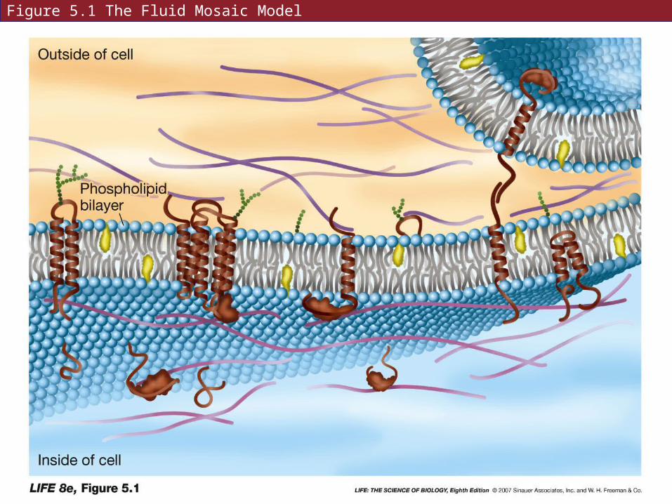
Figure 5.1 The Fluid Mosaic Model

Figure 3.20 Phospholipids (A)
Repeat Fig 3.20A here
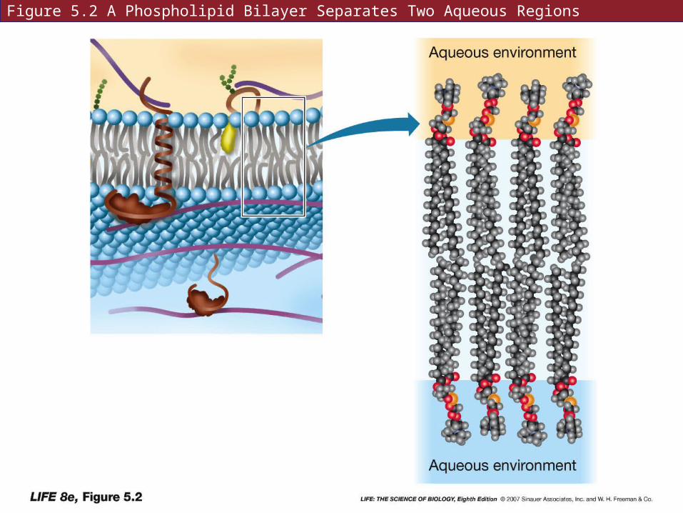
Figure 5.2 A Phospholipid Bilayer Separates Two Aqueous Regions
5.1 What Is the Structure of a Biological Membrane?
Artificial bilayers can be made in the laboratory.
Lipids maintain a bilayer organization spontaneously—helps membranes fuse during phagocytosis, vesicle formation, etc.
5.1 What Is the Structure of a Biological Membrane?
Membranes may vary in lipid composition
Phospholipids vary—fatty acid chain length, degree of saturation, phosphate groups
Membranes may be up to 25 percent cholesterol
5.1 What Is the Structure of a Biological Membrane?
Phospholipid bilayer is flexible, and the interior is fluid, allowing lateral movement of molecules.
Fluidity depends on temperature and lipid composition.
5.1 What Is the Structure of a Biological Membrane?
Membranes contain proteins, the number of proteins varies with cell function
Some membrane proteins extend across the lipid bilayer—with hydrophobic and hydrophilic regions or domains.

Figure 5.3 Membrane Proteins Revealed by the Freeze-Fracture Technique
5.1 What Is the Structure of a Biological Membrane?
The proteins and lipids in the membrane are independent and only interact noncovalently.
5.1 What Is the Structure of a Biological Membrane?
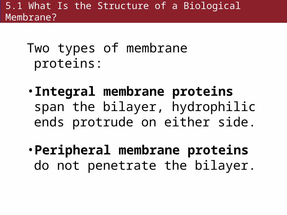
Two types of membrane proteins:
• Integral membrane proteins span the bilayer, hydrophilic ends protrude on either side.
• Peripheral membrane proteins do not penetrate the bilayer.
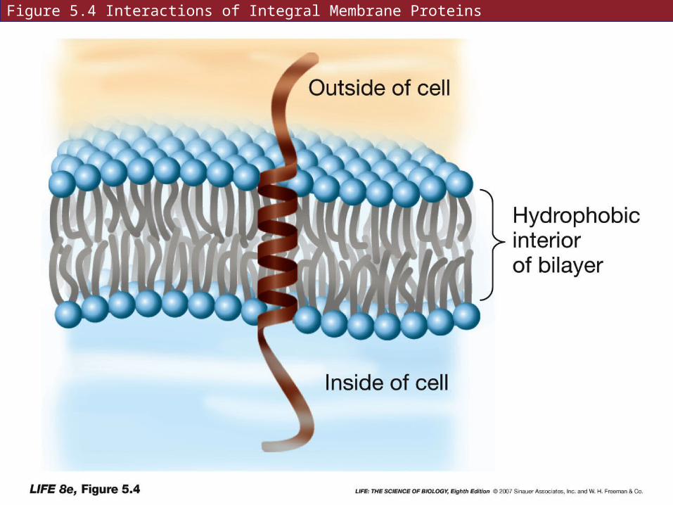
Figure 5.4 Interactions of Integral Membrane Proteins
5.1 What Is the Structure of a Biological Membrane?
Transmembrane proteins may have different domains on either side of the membrane.
The two sides of the membrane can have very different properties.
5.1 What Is the Structure of a Biological Membrane?
Some membrane proteins can move freely within the bilayer, while some are anchored to a specific region.
Some can be anchored by cytoskeleton elements, or lipid rafts—lipids in semisolid state.
5.1 What Is the Structure of a Biological Membrane?
Membranes are dynamic and are constantly forming, transforming, fusing, and breaking down.
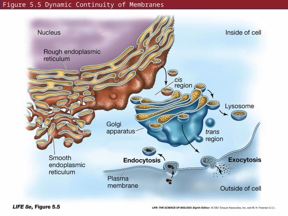
Figure 5.5 Dynamic Continuity of Membranes

5.1 What Is the Structure of a Biological Membrane?
Membranes have carbohydrates on the outer surface that serve as recognition sites for other cells and molecules.
Glycolipids
Glycoproteins

Figure 5.1 The Fluid Mosaic Model
5.2 How Is the Plasma Membrane Involved In Cell Adhesion and Recognition?
Cells arrange themselves in groups by cell recognition and cell adhesion.
These processes can be studied in sponge cells—the cells are easily separated and will come back together again.

Figure 5.6 Cell Recognition and Adhesion (A)

5.2 How Is the Plasma Membrane Involved In Cell Adhesion and Recognition?
Binding of cells is usually homotypic: the same molecule sticks out from both cells and forms a bond.
Some binding is heterotypic: the cells have different proteins.
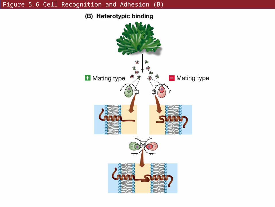
Figure 5.6 Cell Recognition and Adhesion (B)
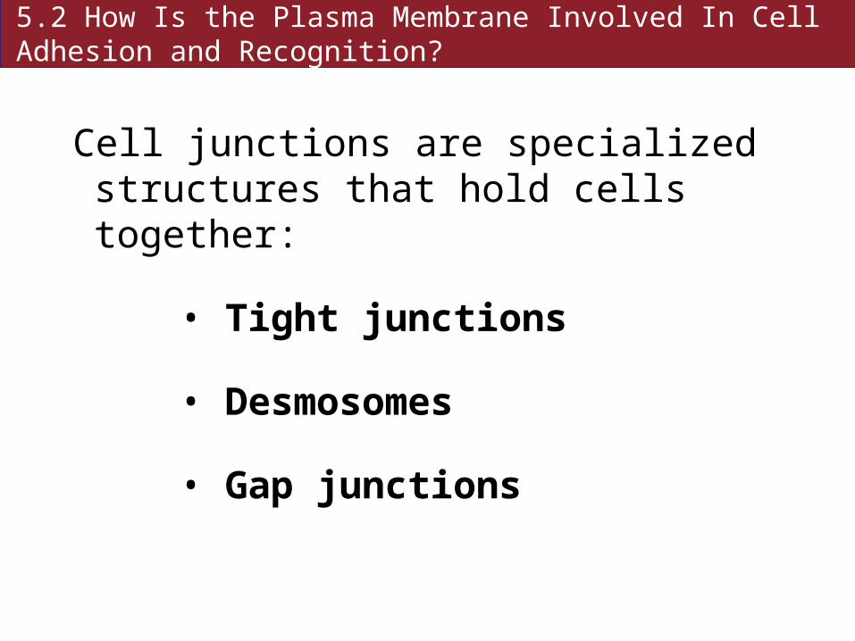
5.2 How Is the Plasma Membrane Involved In Cell Adhesion and Recognition?
Cell junctions are specialized structures that hold cells together:
• Tight junctions
• Desmosomes
• Gap junctions
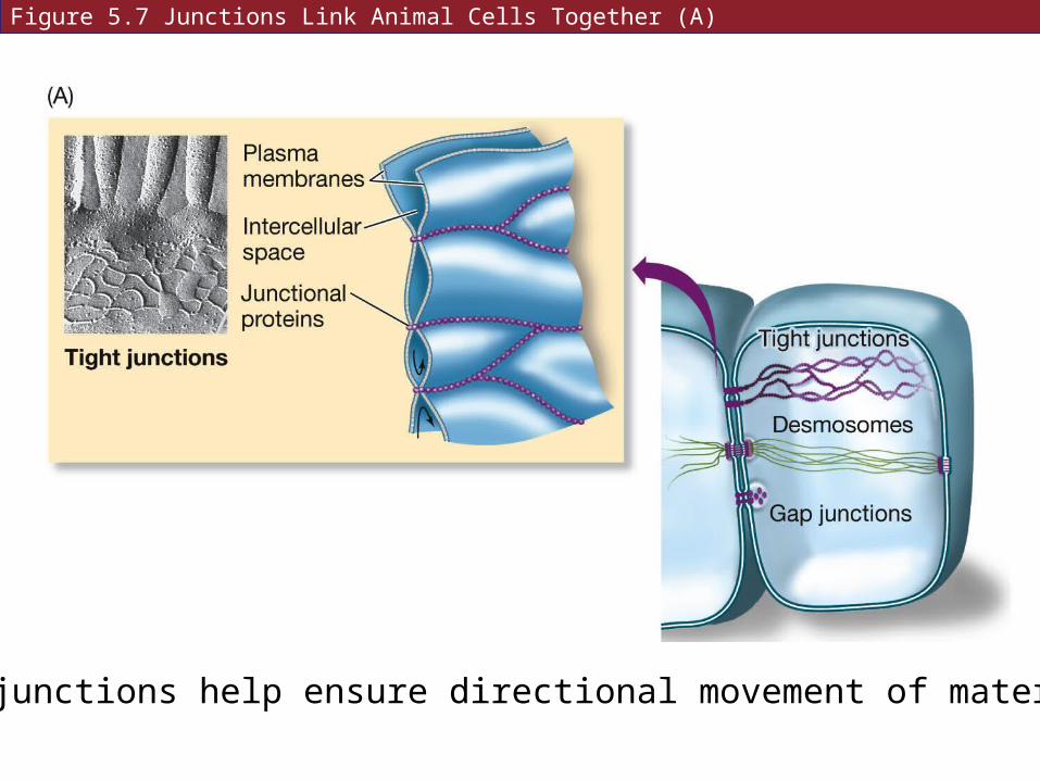
Figure 5.7 Junctions Link Animal Cells Together (A)
Tight junctions help ensure directional movement of materials.
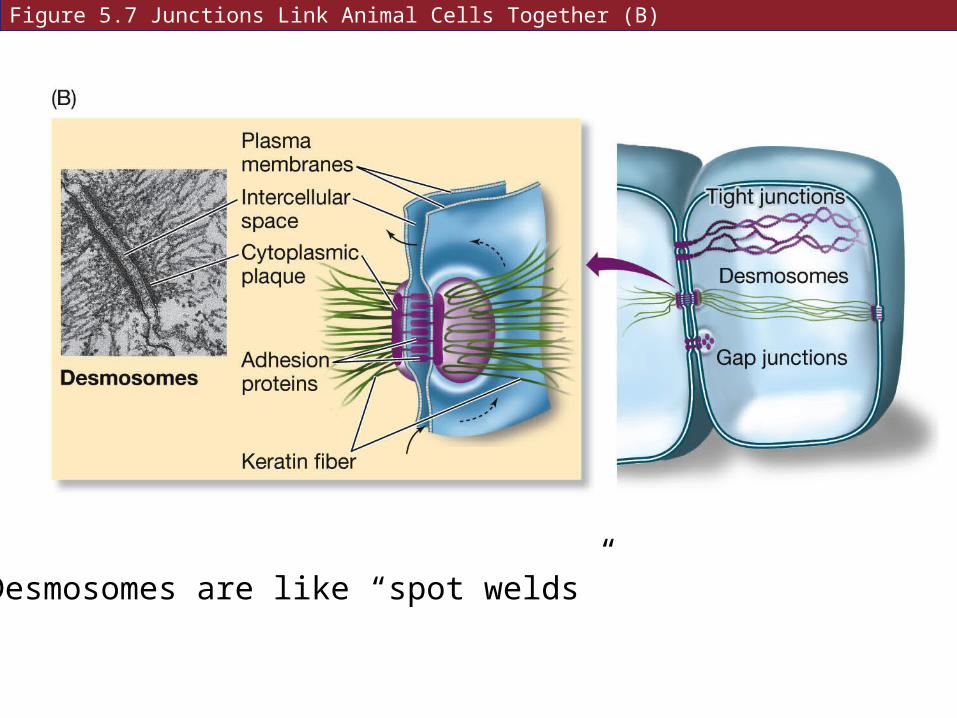
Figure 5.7 Junctions Link Animal Cells Together (B)
Desmosomes are like “spot welds”
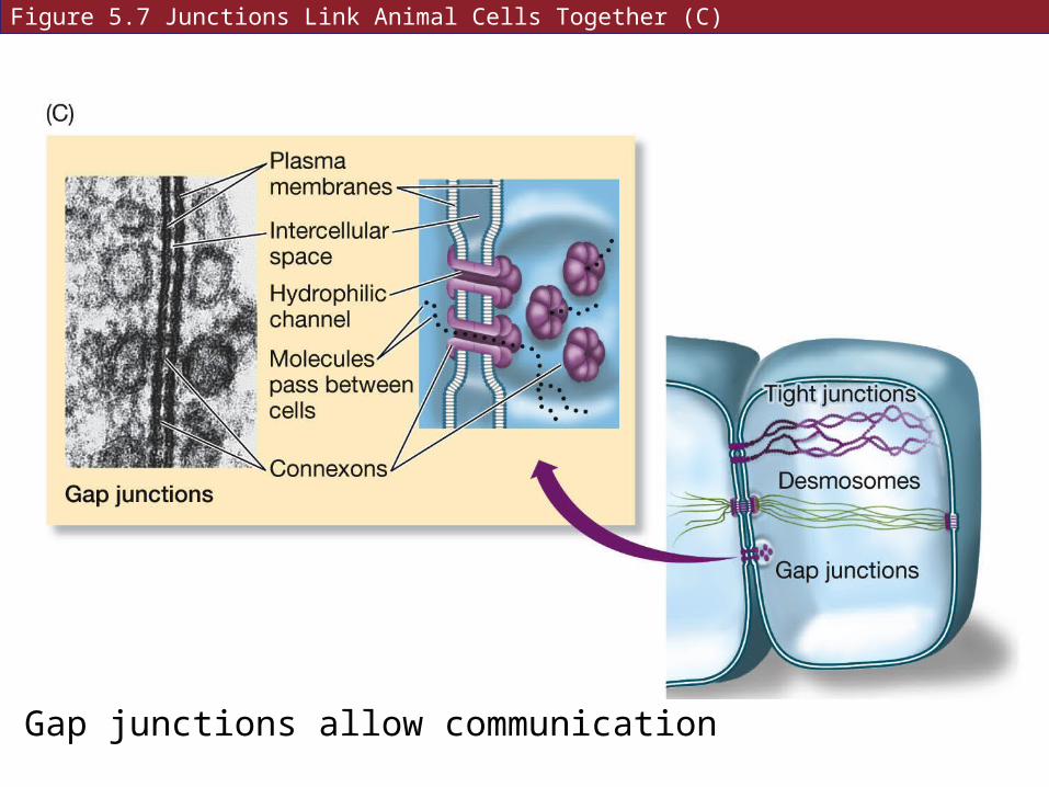
Figure 5.7 Junctions Link Animal Cells Together (C)
Gap junctions allow communication
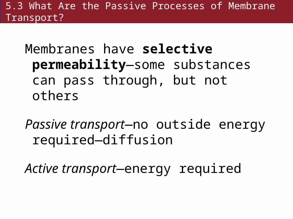
5.3 What Are the Passive Processes of Membrane Transport?
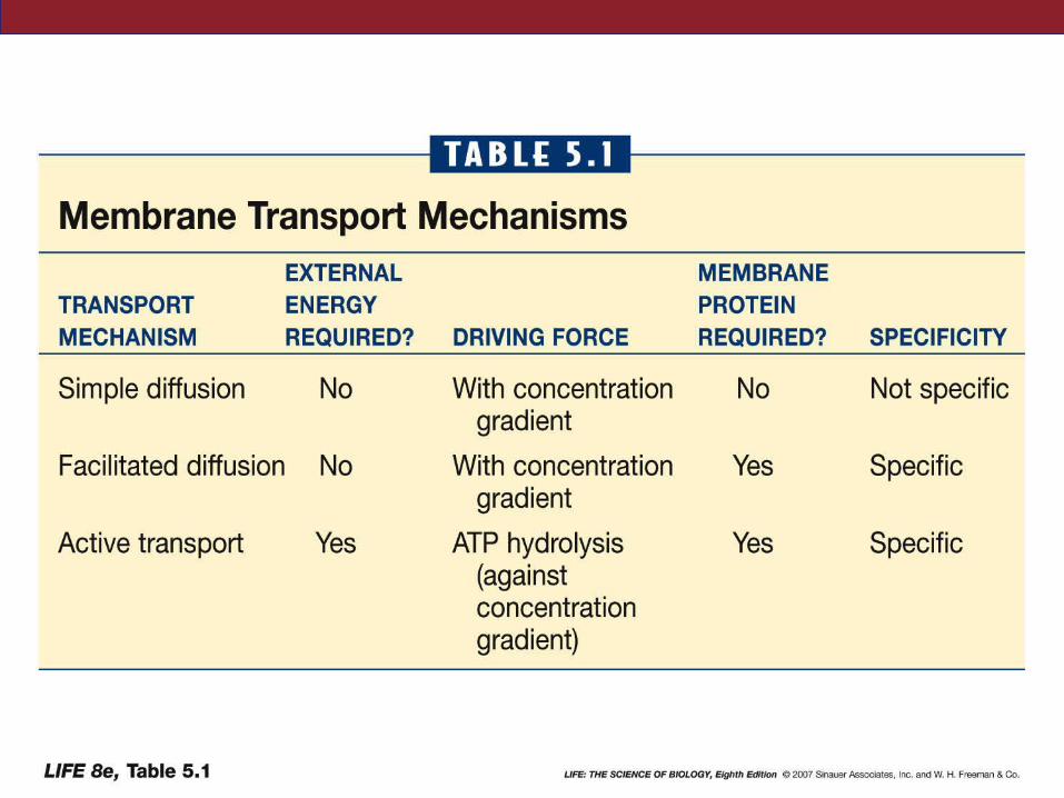
Membranes have selective permeability—some substances can pass through, but not others
Passive transport—no outside energy required—diffusion
Active transport—energy required

5.3 What Are the Passive Processes of Membrane Transport?
Diffusion: the process of random movement toward equilibrium
Equilibrium—particles continue to move, but there is no net change in distribution
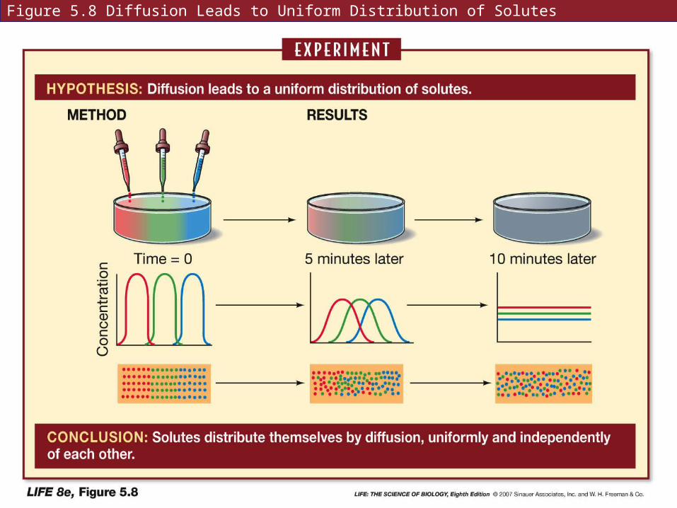
Figure 5.8 Diffusion Leads to Uniform Distribution of Solutes
5.3 What Are the Passive Processes of Membrane Transport?
Net movement is directional until equilibrium is reached.
Diffusion is net movement from regions of greater concentration to regions of lesser concentration.
5.3 What Are the Passive Processes of Membrane Transport?
Diffusion rate depends on:
• Diameter of the molecules or ions
• Temperature of the solution
• Electric charges
• Concentration gradient
5.3 What Are the Passive Processes of Membrane Transport?
Diffusion works very well over short distances.
Membrane properties affect the diffusion of solutes.
The membrane is permeable to solutes that move easily across it; impermeable to those that can’t.
5.3 What Are the Passive Processes of Membrane Transport?
Simple diffusion: small molecules pass through the lipid bilayer.
Lipid soluble molecules can diffuse across the membrane, as can water.
Electrically charged and polar molecules can not pass through easily.
5.3 What Are the Passive Processes of Membrane Transport?
Osmosis: the diffusion of water
Osmosis depends on the number of solute particles present, not the type of particles.
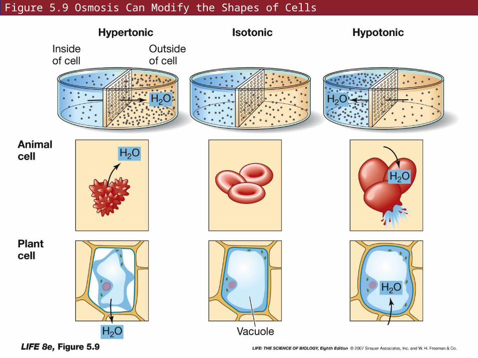
Figure 5.9 Osmosis Can Modify the Shapes of Cells
5.3 What Are the Passive Processes of Membrane Transport?
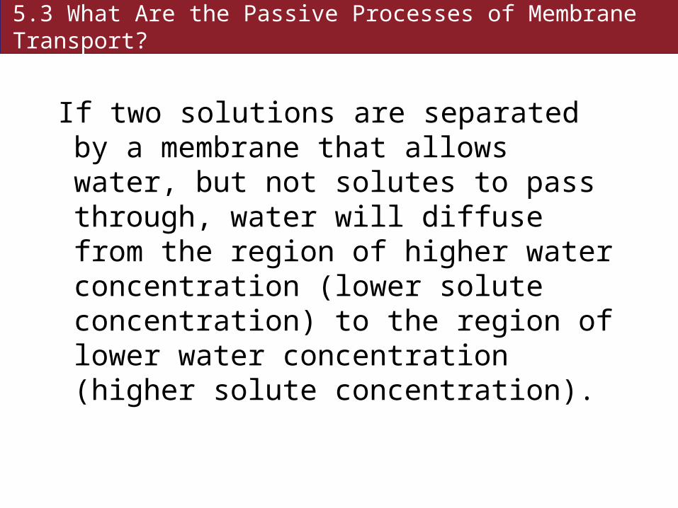
If two solutions are separated by a membrane that allows water, but not solutes to pass through, water will diffuse from the region of higher water concentration (lower solute concentration) to the region of lower water concentration (higher solute concentration).
5.3 What Are the Passive Processes of Membrane Transport?
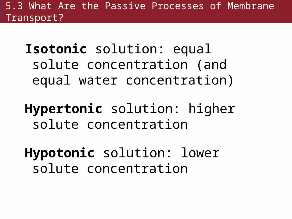
Isotonic solution: equal solute concentration (and equal water concentration)
Hypertonic solution: higher solute concentration
Hypotonic solution: lower solute concentration
5.3 What Are the Passive Processes of Membrane Transport?
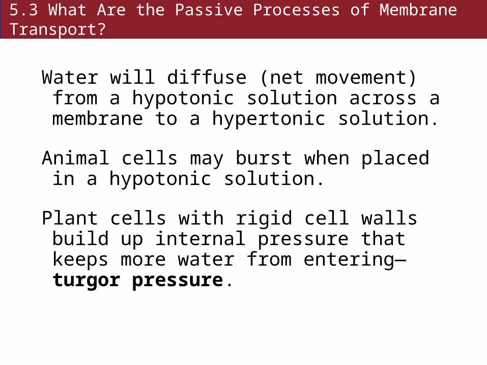
Water will diffuse (net movement) from a hypotonic solution across a membrane to a hypertonic solution.
Animal cells may burst when placed in a hypotonic solution.
Plant cells with rigid cell walls build up internal pressure that keeps more water from entering—turgor pressure.

5.3 What Are the Passive Processes of Membrane Transport?
Facilitated diffusion (passive):
• Polar molecules can cross the membrane through channel proteins and carrier proteins.
• Channel proteins have a central pore lined with polar amino acids.
5.3 What Are the Passive Processes of Membrane Transport?
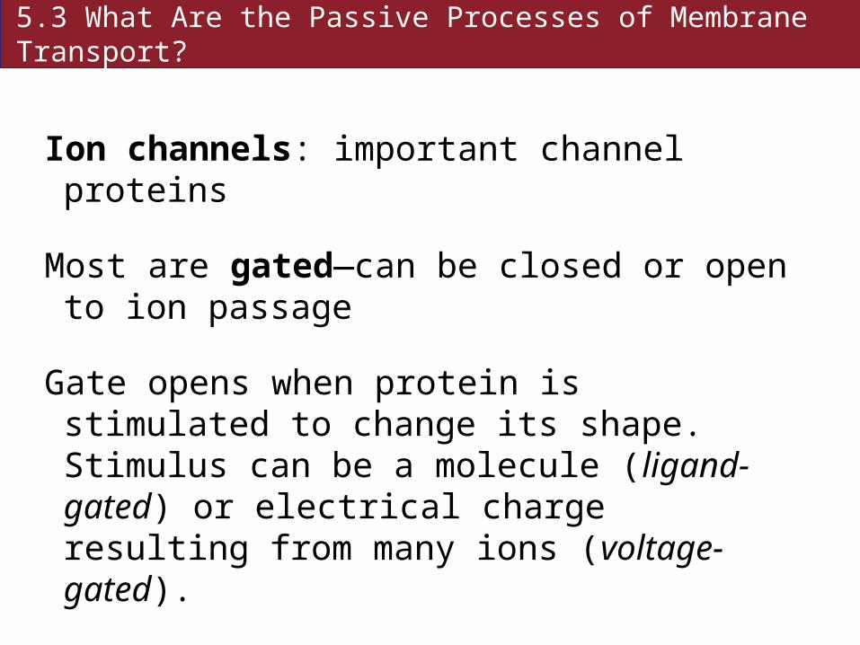
Ion channels: important channel proteins
Most are gated—can be closed or open to ion passage
Gate opens when protein is stimulated to change its shape. Stimulus can be a molecule (ligand-gated) or electrical charge resulting from many ions (voltage-gated).
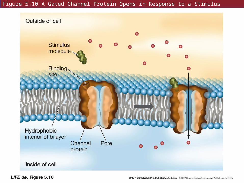
Figure 5.10 A Gated Channel Protein Opens in Response to a Stimulus

5.3 What Are the Passive Processes of Membrane Transport?
Gradients can be a concentration gradient of ions, or an electrochemical gradient resulting from a charge imbalance across the membrane.

5.3 What Are the Passive Processes of Membrane Transport?
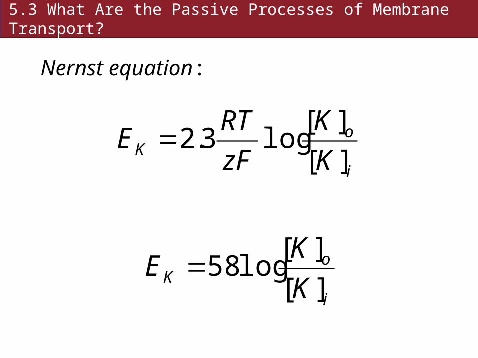
Membrane potential is a charge imbalance across a membrane.
Measured membrane potential of animal cells: –70 mV—lots of potential energy!
5.3 What Are the Passive Processes of Membrane Transport?
Nernst equation:
i
oK K
K
zF
RTE
][
][log3.2
i
oK K
KE
][
][log58

Figure 5.11 The Potassium Channel (A, B)
5.3 What Are the Passive Processes of Membrane Transport?
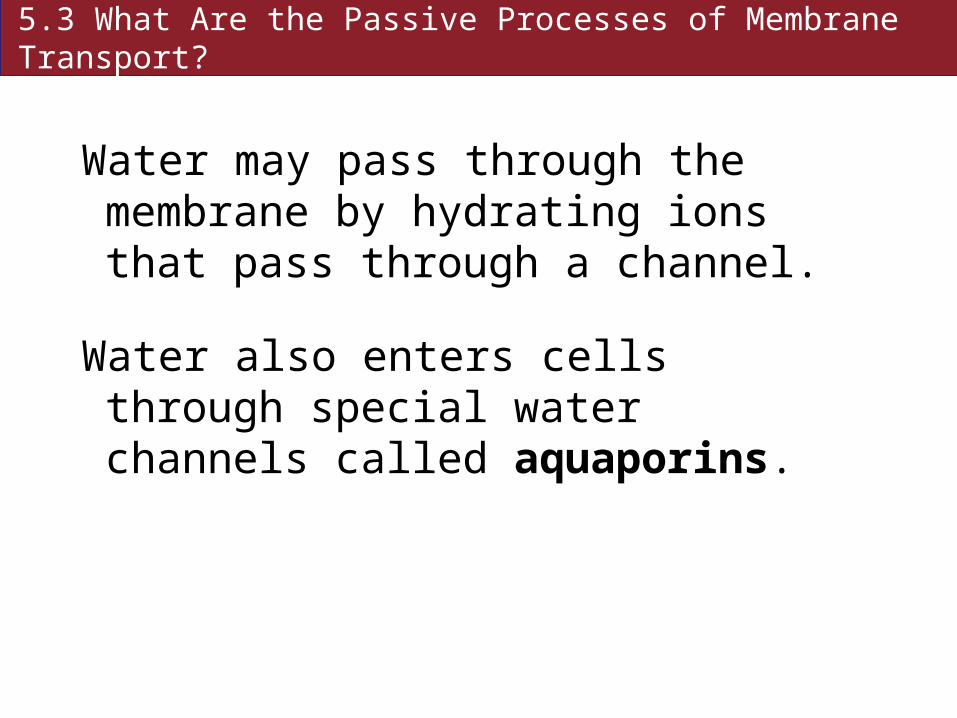
Water may pass through the membrane by hydrating ions that pass through a channel.
Water also enters cells through special water channels called aquaporins.
5.3 What Are the Passive Processes of Membrane Transport?
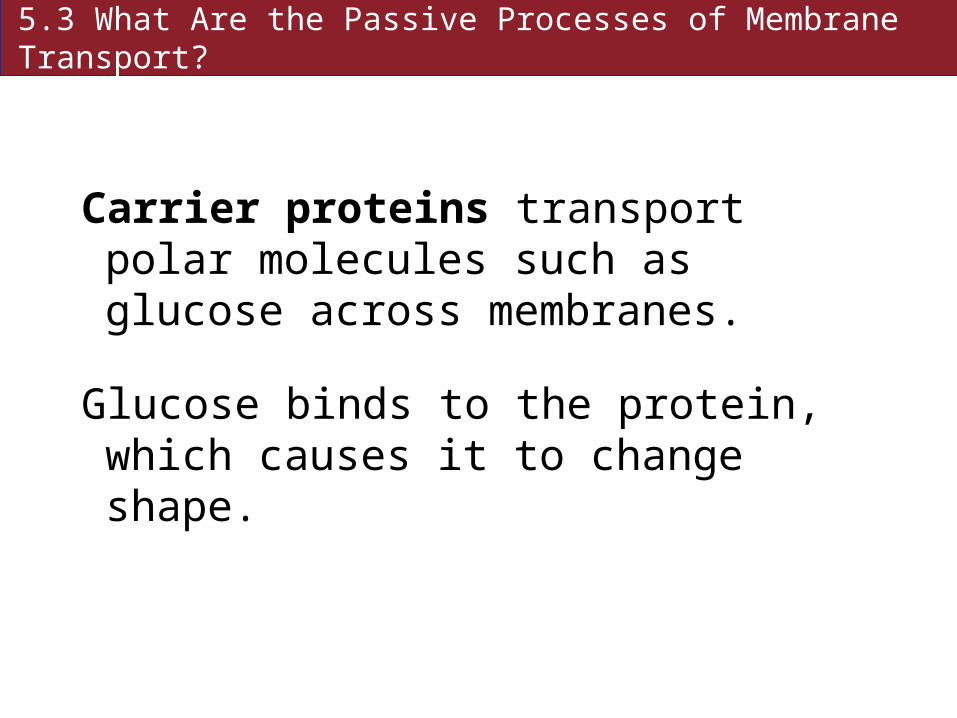
Carrier proteins transport polar molecules such as glucose across membranes.
Glucose binds to the protein, which causes it to change shape.
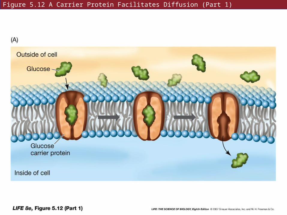
Figure 5.12 A Carrier Protein Facilitates Diffusion (Part 1)
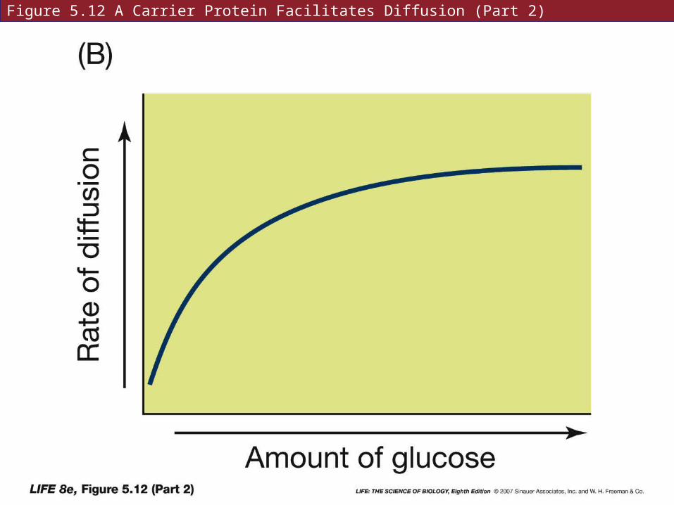
Figure 5.12 A Carrier Protein Facilitates Diffusion (Part 2)

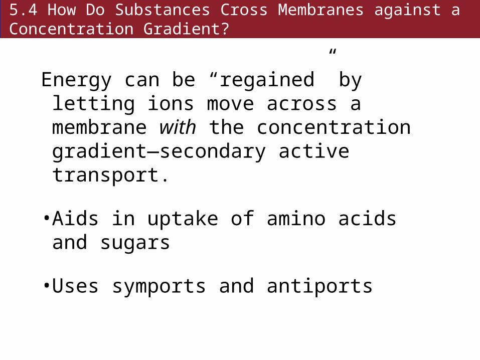
5.4 How Do Substances Cross Membranes against a Concentration Gradient?
Active transport: moves substances against a concentration gradient—requires energy.
5.4 How Do Substances Cross Membranes against a Concentration Gradient?
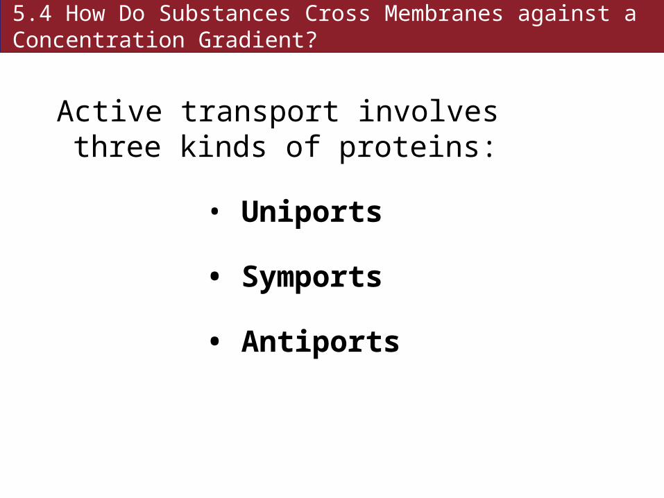
Active transport involves three kinds of proteins:
• Uniports
• Symports
• Antiports

Figure 5.13 Three Types of Proteins for Active Transport

5.4 How Do Substances Cross Membranes against a Concentration Gradient?
Primary active transport requires direct participation of ATP.
Secondary active transport: energy comes from an ion concentration gradient that is established by primary active transport.

5.4 How Do Substances Cross Membranes against a Concentration Gradient?
The sodium–potassium pump (Na+–K+) is primary active transport.
Found in all animal cells
The “pump” is an integral membrane glycoprotein. It is an antiport.

Figure 5.14 Primary Active Transport: The Sodium–Potassium Pump
5.4 How Do Substances Cross Membranes against a Concentration Gradient?
Energy can be “regained” by letting ions move across a membrane with the concentration gradient—secondary active transport.
• Aids in uptake of amino acids and sugars
• Uses symports and antiports
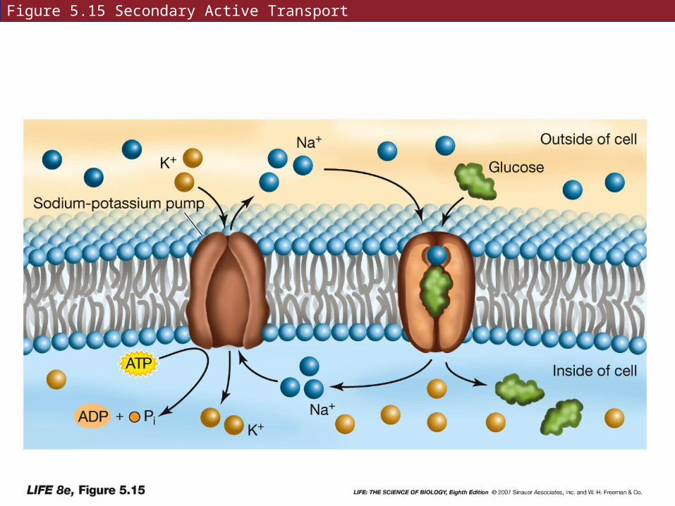
Figure 5.15 Secondary Active Transport

5.5 How Do Large Molecules Enter and Leave a Cell?
Macromolecules (proteins, polysaccharides, nucleic acids) are too large to cross the membrane.
They can be taken in or excreted by means of vesicles.
5.5 How Do Large Molecules Enter and Leave a Cell?
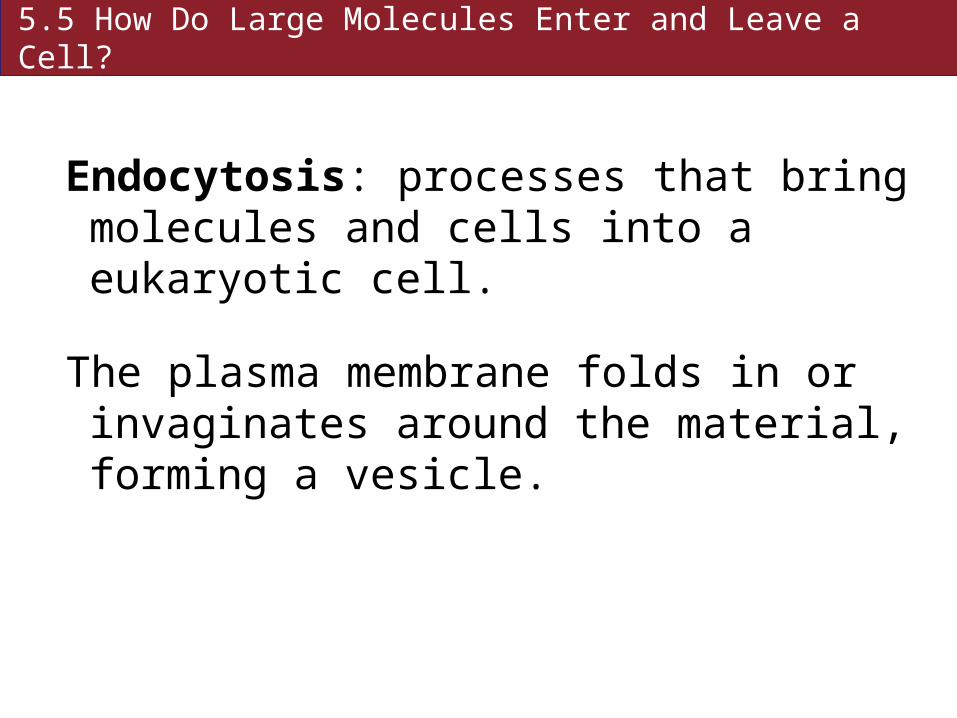
Endocytosis: processes that bring molecules and cells into a eukaryotic cell.
The plasma membrane folds in or invaginates around the material, forming a vesicle.

Figure 5.16 Endocytosis and Exocytosis (A)
5.5 How Do Large Molecules Enter and Leave a Cell?
Phagocytosis: molecules or entire cells are engulfed. Some protists feed in this way. Some white blood cells engulf foreign substances.
A food vacuole or a phagosome forms, which fuses with a lysosome.
5.5 How Do Large Molecules Enter and Leave a Cell?
Pinocytosis: a vesicle forms to bring small dissolved substances or fluids into a cell. Vesicles are much smaller than in phagocytosis.
Pinocytosis is constant in endothelial (capillary) cells.
5.5 How Do Large Molecules Enter and Leave a Cell?
Receptor mediated endocytosis: highly specific
Depends on receptor proteins—integral membrane proteins—to bind to specific substances
Sites are called coated pits—coated with other proteins such as clathrin

Figure 5.17 Formation of a Coated Vesicle (Part 1)
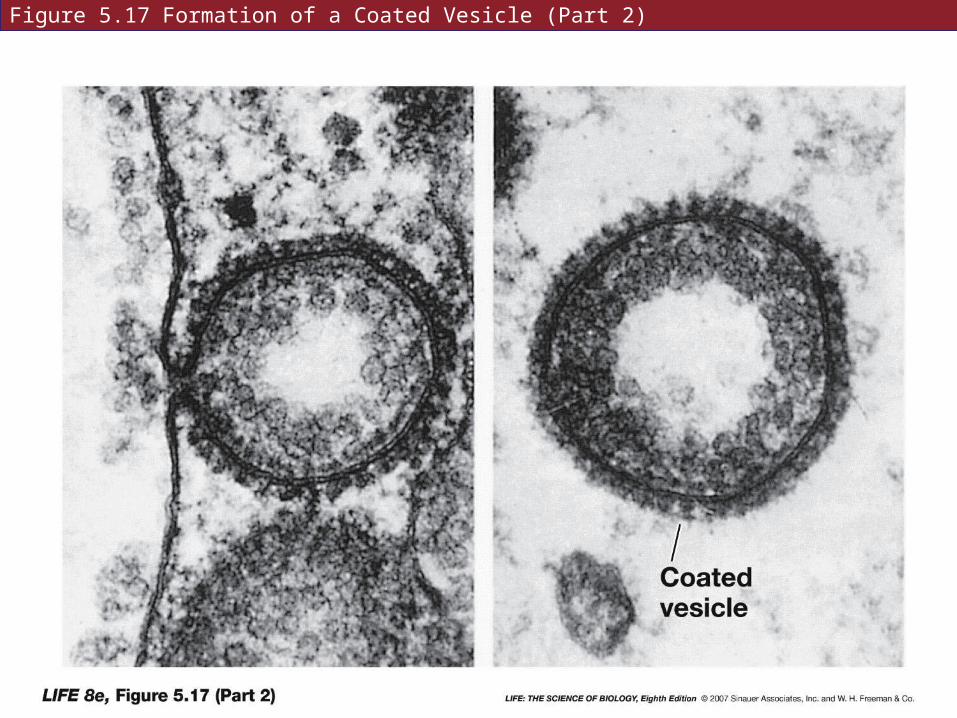
Figure 5.17 Formation of a Coated Vesicle (Part 2)
5.5 How Do Large Molecules Enter and Leave a Cell?
Mammalian cells take in cholesterol by receptor-mediated endocytosis.
Lipids are packaged by the liver into lipoproteins—secrete to bloodstream.
Liver must take up low-density lipoproteins (LDLs) for recycling. The LDLs bind to specific receptor proteins.

5.5 How Do Large Molecules Enter and Leave a Cell?
Exocytosis: material in vesicles is expelled from a cell
Indigestible materials are expelled.
Other materials leave cells such as digestive enzymes and neurotransmitters.
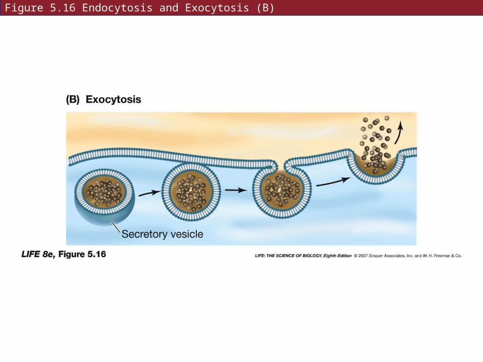
Figure 5.16 Endocytosis and Exocytosis (B)
5.6 What Are Some Other Functions of Membranes?
Keeping different materials separated:
• Endoplasmic reticulum segregates newly-formed proteins.

5.6 What Are Some Other Functions of Membranes?
Electrically excitable membranes—
• The plasma membrane of neurons conducts nerve impulses.

5.6 What Are Some Other Functions of Membranes?
Membranes help transform energy:
• Inner mitochondrial membranes—energy from fuel molecules is transformed to ATP
• Thylakoid membranes of chloroplasts transform light energy to chemical bonds.
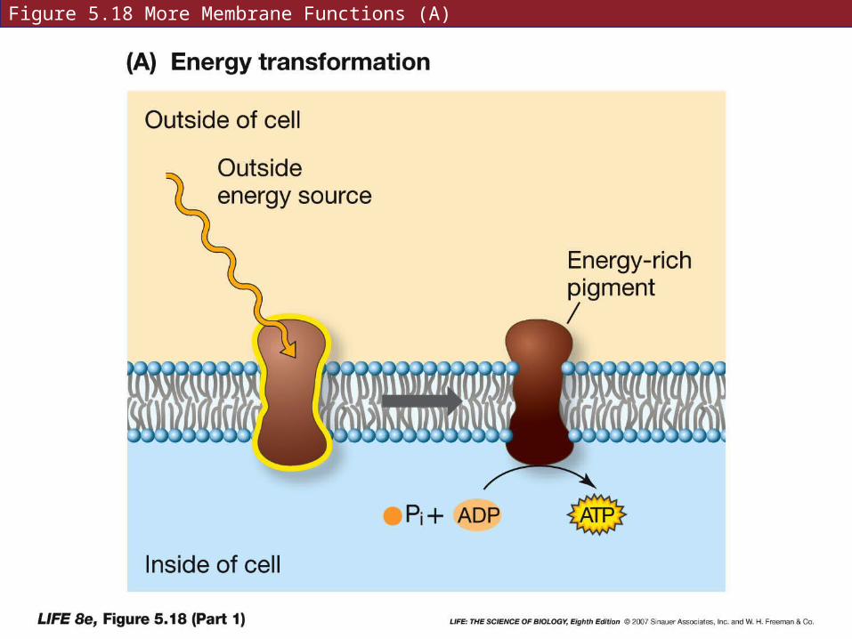
Figure 5.18 More Membrane Functions (A)
5.6 What Are Some Other Functions of Membranes?
Membrane proteins can organize chemical reactions.
Many cellular processes involve a series of enzyme-catalyzed reactions—all the molecules must come together for these to occur. Forms an “assembly line” of enzymes.
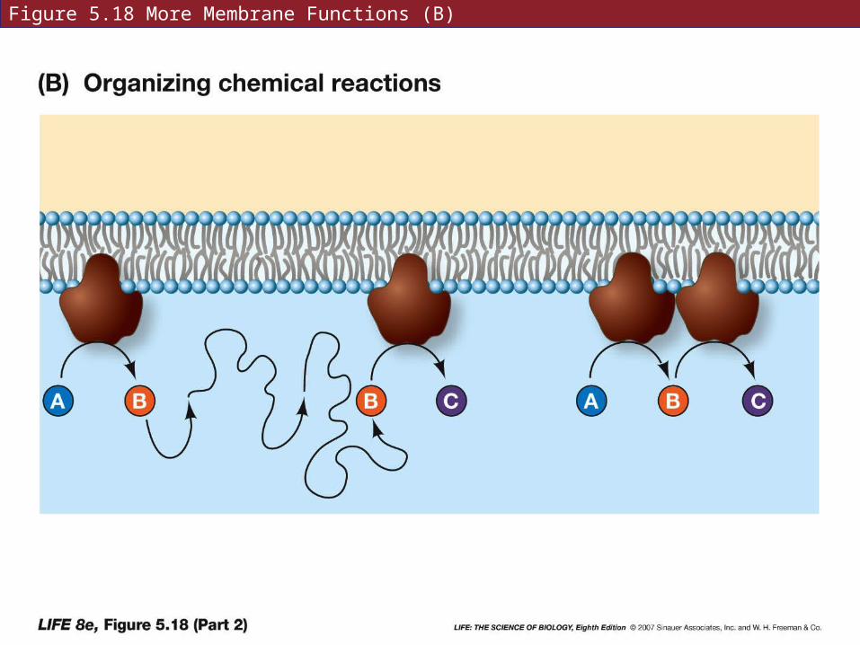
Figure 5.18 More Membrane Functions (B)

5.6 What Are Some Other Functions of Membranes?
Membrane proteins process information.
Binding of a specific ligand can initiate, stop, or change cell functions.

Figure 5.18 More Membrane Functions (C)

5.6 What Are Some Other Functions of Membranes?
The cholera toxin:
One subunit binds to a cell surface receptor—the toxin molecule changes shape and allows the other subunit to enter the cell.
The subunit acts as an enzyme to modify a peripheral protein—this opens chloride channels in the membrane.
Cl− and Na+ accumulate in the intestines, followed by osmotic loss of water.