50 years of the ieee engineering in medicine and biology ... · on the ieee engineering in medicine...
TRANSCRIPT

May/Junel 2002 IEEE ENGINEERING IN MEDICINE AND BIOLOGY 17
50 Years of the IEEE Engineering in Medicine and Biology Societyand the Emergence of a New Discipline
One of the fastest growing fields of tech-nology—a field of astounding recent
achievements and even more ambitioushopes—is biomedical engineering. Labo-ratory instrumentation, medical imaging,cardiac pacemakers, artificial limbs, andcomputer analysis of the human genomeare some of its familiar products. Definedas the use of the principles and techniquesof engineering to solve problems in biol-ogy and medicine, biomedical engineer-ing today holds a prominent place as ameans of improving medical diagnosisand treatment, as a business, and as an ac-ademic discipline. Yet 50 years ago itbarely existed.
It was in 1952 that a group of electron-ics engineers, members of the Institute ofRadio Engineers (IRE), established an or-
ganization within the IRE to consider“problems in biology and medicine whichmight be aided in solution by use of elec-tronic engineering principles and de-vices.” This Professional Group onMedical Electronics, as it was called,grew steadily and expanded its area of in-terest. In 1963 the IRE and the AmericanInstitute of Electrical Engineers (AIEE)merged to form the Institute of Electricaland Electronics Engineers (IEEE), and theIRE Professional Group on Medical Elec-tronics merged with the AIEE Committeeon Electrical Techniques in Medicine andBiology. Over the years since, the IEEEEngineering in Medicine and Biology So-ciety (EMBS), while growing into thelargest international member-based soci-ety of biomedical engineers, has made,
through its meetings, publications, andother activities, invaluable contributionsin the advancement of the field.
The 50th anniversary of the Society isan appropriate time to look back at the or-igins and growth of both the field of bio-medical engineering and the EMBS. TheSociety, under the leadership of PastPresidents Banu Onaral and AndrewSzeto and current President HenriettaGaliana, has funded and directed a his-tory project, the principal products ofwhich are a narrative history and a set oforal-history interviews.
The story to be told is a large and excit-ing one, and not all of it can be told here.The present account gives most attentionto the aspects of biomedical engineeringto which IEEE members (and, earlier,
Frederik Nebeker, IEEE History Center, Piscataway, NJ
0739-5175/02/$17.00©2002IEEE

AIEE members and IRE members) contributed, which is to saythat this account emphasizes the electrical, electronic, and com-puting aspects of biomedical engineering. Mechanical engineer-ing and chemical engineering have, of course, made enormouscontributions to biomedical engineering. These areas, thoughonly occasionally mentioned in the narrative, are well repre-sented in the oral-history interviews, excerpts of which appear inthis issue in a separate article.
It must also be said that the number of professional societiesthat have arisen to foster biomedical engineering is very large.Some of them have broad scope, such as the European Society forEngineering and Medicine, and some of them narrow scope, suchas the North American Society of Pacing and Electrophysiology.The geographic scope of professional societies may be regional,national, continental, or global. This account, of course, focuseson the IEEE Engineering in Medicine and Biology Society and itspredecessor organizations in the AIEE and the IRE, and it men-tions other societies only occasionally. And even within thebounds thus established, many choices had to be made. For exam-ple, an extremely large topic whose history is only sketchily pre-sented here is biomedical engineering education.
The research and writing of this narrative account, as well asthe conducting and editing of the oral histories, were carried outmainly by Frederik Nebeker and other staff of the IEEE HistoryCenter at Rutgers University. David Geselowitz, chair of theEMBS history committee, gave the greatest assistance, and otherEMBS members contributed, especially Herman Schwan, An-drew Szeto, and Max Valentinuzzi. Others who deserve manythanks are the EMBS staff, headed by Executive Director LauraJ. Wolf, and those who contributed oral-history interviews. Also,the section that looks at the future contains contributions by JohnW. Clark, L.A. Geddes, as well as Elise Fear and Faustina Hwangwith contributions from EMBS student members.
The author of this history is well aware of the fact that, despitethe assistance of all these people, many shortcomings remain. Inan attempt at exculpation he appeals to Murphy’s Law, anotherof the contributions of biomedical engineering to modern soci-ety. (In 1949 the U.S. Air Force performed tests of rapid deceler-ation on pilots. Volunteers were strapped to a rocket-propelledsled, and their physiological conditions were monitored throughelectrodes contained in a harness designed by Captain Edward A.Murphy. After discovering one day that the electrodes had beenwired incorrectly, Murphy commented “If there are two or moreways of doing something, and one of them can lead to catastro-phe, then someone will do it.”)
The Roots of Biomedical EngineeringHistory of the Technologies
In the 1780s Luigi Galvani, studying what was called “animalelectricity,” initiated a line of research known as elec-trophysiology. By 1900 it had established the electrical nature ofthe nerve impulse and its velocity and revealed much about elec-trolytic conduction in animal tissues. Concepts of electrical engi-neering, such as resistivity, capacitance, and polarization, wereapplicable. Indeed, the mathematical model that WilliamThomson (later Lord Kelvin) proposed in 1855 for the Atlantictelegraph cable was modified, shortly after the turn of the cen-tury, to describe the mechanism of conduction along a nervefiber.
Modern hemodynamics may be considered to have startedwith the conviction of William Harvey expressed in 1616 that the
heart propels blood around a closed system. In 1840 a Frenchphysician named J.L.M. Poiseuille showed that the major pres-sure drop in the cardiovascular system occurred in the capillar-ies. He then studied the pressure drop in small glass tubes anddeveloped the relation between pressure drop, flow, and tube di-ameter. A theory for wave propagation in elastic tubes such asblood vessels was developed by Thomas Young as early as 1808.
The physicist Hermann von Helmholtz may well be consid-ered one of the first biomedical engineers. He invented the oph-thalmoscope and the ophthalmometer. He determined the
velocity of nerve pulse transmission, and he developed the basicphysics for understanding fields in a volume conductor producedby bioelectric sources. And he studied the mechanism of hearingand invented the Helmholtz resonator.
It was especially through instrumentation—for measuringand imaging—that engineering influenced biomedicine. In 1888Augustus Desiré Waller showed that, with a capillaryelectrometer, one could record the changing heart voltages fromthe body surface. X-ray imaging, invented by Wilhelm Röntgenin 1895, had an enormous impact on medicine; already in 1896Siemens and General Electric began selling X-ray equipment.Indeed, it has been argued that it was X-ray technology that “trig-gered the transformation of the hospital from a passive receptaclefor the sick poor to an active curative institution for all membersof society.” Other important medical instruments were the elec-trocardiograph and the electroencephalograph, the former com-ing into clinical use in the 1920s, the latter in the 1930s. Devicessuch as thermocouples, galvanometers, and phototubes foundmany applications in biomedical research in the 1930s. An ex-ample of biomedical research indebted to engineering both forconcepts and instrumentation is the work of Edward D. Adrianand Charles Sherrington, recognized by the 1932 Nobel Prize forPhysiology or Medicine, for elucidating the electrical nature ofneural activity. (The 1944 Nobel Prize for Physiology or Medi-cine recognized work in the same area by Joseph Erlanger andHerbert Gasser.)
Sometimes the biomedical application stimulated the techno-logical advance, as with the string galvanometer invented byWillem Einthoven to improve electrocardiography (ECG). An-other example is the differential amplifier, a basic component ofelectronics, which was invented by B.H.C. Matthews in 1934 toamplify action potentials of nerves.
Not only research but also medical practice was changed.Many research instruments, such as X-ray machines, electrocar-diographs, and electroencephalographs, came to be regularlyused in diagnosis. Throughout the 19th century there were at-tempts to use electrical technology in treatment, but only a few,such as cardiorespiratory resuscitation by electrical stimulation,were of much effectiveness. In the early decades of the 20th cen-tury, a few techniques of obvious utility were introduced: X-ray
18 IEEE ENGINEERING IN MEDICINE AND BIOLOGY May/June 2002
It was especially throughinstrumentation—for measuringand imaging—that engineering
influenced biomedicine.

therapy, electrosurgery, and diathermy (the generation of heat inliving tissues by electromagnetic radiation).
There was an older therapeutic tradition that should be men-tioned. From the late 18th through early 20th century, promoterssold a wide variety of electrical devices, most of which passed acurrent through or near the body, to cure physical and psychologi-cal ailments. The use of magnets was popular also. Devices to ad-minister an electric shock, electric baths, and electric belts becamepopular, and the practitioners of electrical medicine, as it wascalled, attracted millions of customers. In the 1880s a cellar roomof the U.S. Capitol was equipped with medical electrical equip-
ment for the use of Congressmen, and Electrical Review reportedin 1887, “A great many members take electricity, and some go tothe basement of the Capitol for it every day during the season.”
Though such applications of electricity continue to the pres-ent, they have been vastly overshadowed by science-based treat-ments. The great expansion of medical research in the 20thcentury, coupled with new technological capabilities, led tocountless advances. It was during World War I that the manufac-ture of electron tubes (mainly for radio) began on a large scale,and beginning in the 1920s electron tubes permitted short-wavediathermy, new types of electrosurgery, and medical applica-tions of telemetry. (Short-wave diathermy began in Europe inabout 1925 and was used to treat a variety of ailments; micro-wave diathermy began after World War II when the klystron andthe magnetron became available to investigators.) New researchtechniques included the ultracentrifuge (developed by TheSvedberg) and electrophoresis (developed especially by ArneTiselius), both recognized in Nobel Prizes.
Electron-tube amplification was put to work, for example, in acommercial ECG machine introduced by Siemens & Halske in1921; this machine used an oscilloscope also. It was the highly sen-sitive string-galvanometer of Einthoven, rather than electron-tubeamplification, that made possible the first recording of brain wavesfrom scalp electrodes. This electroencephalography or EEG, as itwas called, was developed by the German psychiatrist Hans Bergerin the mid 1920s, though he did not publish his results until 1929.Clinical EEGs evolved in the 1930s and expanded quickly in theearly 1940s. It was also in the early 1940s that the field ofelectrocorticography—multichannel recordings from the exposedbrain cortex—emerged; it was used to locate epileptic foci.
One of the most important tools of 20th-century biomedicalresearch, the electron microscope, was invented at the beginningof the 1930s by two German groups independently, one at theUniversity of Berlin headed by Hans Hermann Knoll and Ernst
Ruska and the other at Siemens-Schuckert headed byReinhold Rüdenberg. This was, of course, the
transmission electron microscope, in whichthe electrons used to form the image
passed through the sample. Knoll pro-posed a scanning electron micro-
scope, in which the electrons werereflected off the sample, and anearly form was built by Manfredvon Ardenne in 1938. In theUnited States VladimirZworykin and others at RCAbuilt a scanning electron micro-scope in 1942. In 1939 the JapanSociety for the Promotion ofScience began a program to con-
struct electron microscopes, andby the end of World War II nearly
20 electron microscopes had beenplaced in use. Japanese companies
began manufacturing electron micro-scopes in the late 1940s, and in 1955 Ja-
pan had half as many electron microscopesin use as the United States and five times as
many as Germany, France, or England.Radiology was no doubt the most highly devel-
oped application of engineering to medicine. Technical advances
May/June 2002 IEEE ENGINEERING IN MEDICINE AND BIOLOGY 19
Believe It...Or Not!
Devices to deliverelectric shock fortherapeutic pur-poses were popularin the late 18th cen-tury through theearly 20th century.Top: For women, anad for an electric corset(circa 1885) proposedcures for ailments rang-ing from weak backs tokidney disorders. Right:For men, an ad for an elec-
tric belt from 1902 offers a10-day free trial.
CO
UR
TE
SY
OF
TH
EB
AK
KE
NLI
BR
AR
YA
ND
MU
SE
UM
,MIN
NE
AP
OLI
S,M
N
COURTESY OF FRANCES RICHMOND, ALFRED E.MANN INSTITUTE OF BIOMEDICAL ENGINEERING

included improved X-ray tubes, notably the high-vacuumhot-cathode tube developed by William Coolidge at GeneralElectric in 1913, and means of visualizing soft tissues. It hadbeen discovered around the turn of the century that ingestion ofradio-opaque bismuth compounds made parts of the digestivetract visible in X-ray images, and in the late 1920s Portuguese in-vestigators, including Egas Moniz and dos Santos, developedangiography, the X-ray visualization of blood vessels after injec-tion of a radio-opaque substance. The image-intensifier tube, in-vented by Irving Langmuir of General Electric, greatly improvedfluoroscopy. Though the computational demands made the tech-nique impractical, the mathematics of tomography (constructinga three-dimensional (3-D) image from two-dimensional (2-D)cross sections) were invented independently by a number of peo-ple in different countries, including André Marie EdmondBocage in France, Bernard Zeidses des Plantes in Holland,Alessandro Vallebona in Italy, and Ernst Pohl andGustave Grossmann in Germany. As we willsee in later sections, it was the electronicdigital computer that made computedtomography a practical technique.
Sonar, invented near the end ofWorld War I for detecting subma-rines, became an important mili-tary technology, and in the 1940sattempts were made to adapt thetechnique to medical imaging. In1941 Donald Sproule developed apulse-echo ultrasonic instrument;one transducer generated the pulsesand a second one registered the ech-oes in the intervals between the gener-ated pulses. In 1944 Floyd Firestonepatented what he called a “Reflectoscope”;it used the same transducer for generatingpulses and detecting echoes. In the 1950s and 1960s,as we will see in the next two sections, the technique reachedclinical usefulness.
It was also in the 1940s that nuclear magnetic resonance wasfirst demonstrated, independently by Felix Block at Stanford andEdward Purcell at Harvard, building on the work of I.I. Rabi. Theelectronic digital computer, another product of the 1940s, was de-veloped in several countries independently, though probably themost influential early machine was the ENIAC, built for the U.S.Army at the University of Pennsylvania and completed in 1946.Biomedical applications of these advances came in the 1950s.
World War II was a great stimulus to the study of control sys-tems, particularly with radar systems and guidance systems. Thisand other work, such as some of the investigations in aviation medi-cine, prompted some people after the war to take a systems ap-proach to biological and medical studies. The construction ofmathematical models of physiological systems, which becameknown as systems physiology, became an important activity. Re-search areas where mathematical models were constructed includehemodynamics, respiration, temperature regulation, nerve-impulsepropagation, muscular control systems, and eye movements.
Prehistory of the ProfessionIn the 1920s and 1930s more and more investigators used the
concepts and techniques of physics and engineering in biologicaland medical research, and a few institutions were established to
promote this approach. In the United States in the 1920s, theJohnson Foundation for Medical Physics at the University ofPennsylvania and the Biophysics Department of the ClevelandClinic were both established. The Rockefeller Foundation, es-tablished in 1913, supported research in this area. In Germany,Siemens, a major supplier of X-ray and diathermy equipment,maintained a biophysical laboratory at Erlangen. The emergenceof biomedical engineering, it should be pointed out, was inter-twined with the emergence of biophysics and medical physics;only gradually did these fields assume distinct identities.
It was in the 1920s that a particularly influential institutionwas established in Frankfurt-am-Main: the Institute for the Phys-ical Foundations of Medicine (Institut für physikalischeGrundlagen der Medizin). The founding director was FriedrichDessauer, who did important work on the biological damagecaused by X rays. In 1934 Boris Rajewsky became director of theinstitute; he did important work on the biological effects of ioniz-ing and nonionizing radiation. In 1938 Rajewsky gained thesponsorship of the Kaiser Wilhelm Society, and the Institute forthe Physical Foundations of Medicine became attached to thelarger, newly formed Kaiser Wilhelm Institute for Biophysics(Kaiser Wilhelm Institut für Biophysik). This institute became
20 IEEE ENGINEERING IN MEDICINE AND BIOLOGY May/June 2002
The Electron Microscope
©R
CA
,CO
UR
TE
SY
DA
VID
SA
RN
OF
FLI
BR
AR
Y
The electron microscope was oneof the most important tools of 20th
century biomedical research. Top:Dr. James Hillier of RCA is shown
with the first commercial electron mi-croscope in the Western hemisphere
(1940). Hillier won an award for makingelectron microscopy a practical technology
for research. Left: A more modern and com-pact version of the device from 1986.COURTESY OF DR. HANI AMASHA

affiliated with the University of Frankfurt and established aPh.D. program in biophysics.
In 1925 the first International Conference of Radiology met inLondon, and a Commission on X-ray Units was set up to defineunits of radiation. At the second International Conference on Ra-diology, which met in Stockholm in 1928, the curie and theroentgen were established as units of radiation.
In the United States the Massachusetts Institute of Technol-ogy implemented a research and teaching program in “biologicalengineering” in 1937. It consisted of five sections: bio-electricalengineering (concerned with X rays and cathode rays), elec-tro-physiology, biophysics, microbiology, and nutritional bio-chemistry. However, the program quickly evolved into plainbiology, despite the championship of Vannevar Bush and KarlCompton of the engineering approach. In the late 1940s the Uni-versity of California at Los Angeles initiated a program in “bio-technology.” It was concerned mainly with what elsewhere wascalled “human factors research.” (By 1962 some 500 engineersand physiologists in the United States were identified with hu-man factors research.)
In the 1940s those concerned with applying electrical tech-nologies to biology and medicine might have belonged to theAmerican Institute of Electrical Engineers (AIEE) or the Insti-tute of Radio Engineers (IRE) or both. The domain of the IRE,founded in 1912, had expanded from radio engineering to almostall areas of electronics, and the domain of the AIEE, founded in1884, included traditional electrical engineering and manynewer areas of development. (In 1963 the AIEE and the IREmerged to form the Institution of Electrical and Electronics Engi-neers (IEEE).)
In 1948 the American Institute of Electrical Engineers(AIEE) formed a Committee on Electrical Techniques in Medi-cine and Biology and held the first conference on the subject.Called the U.S. Conference on Medical Electronics, it took placein New York City in 1948. At about the same time the IRE
formed a committee concerned with medicine and biology. TheAIEE, the IRE, and the Instrument Society of America joined informing the Joint Executive Committee on Electrical Tech-niques in Medicine and Biology. It was this committee that orga-nized the annual conference. In the next section the activities inthe 1950s of the AIEE and the IRE, including the annual confer-ence, are described.
The 1950s: First Steps Toward aDiscipline of Biomedical Engineering
History of the TechnologiesThe 1950s was a decade of economic growth in the United
States and of economic recovery in Europe and Japan. The in-creasing prosperity allowed much greater expenditures on healthcare and biomedical research. Appropriations for the NationalInstitutes of Health (NIH), for example, increased from $52 mil-lion to $430 million. Engineering in medicine gained promi-nence for the cardiac pacemaker, the heart-lung machine, and theiron lung. The latter device, developed in 1927 by Philip Drinker,became well known for treating victims of poliomyletis in theearly 1950s.
A public-health concern of the 1950s was atomic radiation,especially from fallout from the testing of atomic weapons.(There were radiation hazards in some industries where radioac-tive substances were handled and in hospitals, where radiumtubes were used.) The Geiger counter (invented by Hans Geigerand Walther Müller in 1928) was seen as the “watchdog of theatomic age.” The devices became common, and “Volks-Geigercounters” were marketed for use by individuals. Another con-cern was the health hazard of exposure to microwaves in and nearradar installations, in hospitals (in 1955 there were some 20,000microwave diathermy machines in use by physicians in theUnited States), and from the newly developed microwave oven.In 1953 the U.S. Navy and Air Force held a series of meetingsthat considered whether radar transmitters posed serious
May/June 2002 IEEE ENGINEERING IN MEDICINE AND BIOLOGY 21
The Iron Lung
PHOTOS OF COURTESY OF MATTHIAS WITT, DRÄGER MEDICAL AG & CO.
The iron lung, developed in 1927 by Philip Drinker, became wellknown in the early 1950s for patients whose breathing capabilitieswere compromised by poliomyelitis. Left: A Drager E52 iron lungwith electric drive from 1952. Above: An iron lung installed in a1953 “emergency car.”

22 IEEE ENGINEERING IN MEDICINE AND BIOLOGY May/June 2002
dangers. In 1954 General Electric held ameeting on the hazards of microwave ra-diation, and in 1955 the Mayo Clinic helda symposium on the physiologic effects ofmicrowaves. A precursor of the environ-mental movement of the 1960s and 1970swas the establishment, by LeslieSilverman of the Harvard School of Pub-lic Health and many others, of the field ofindustrial hygiene. Another importantcontribution of chemical engineers in the1950s was in the processing of blood,such as techniques for fractionating bloodplasma.
Engineering contributed to cardiologyin numerous ways. Attracting the most at-tention was the cardiac pacemaker. In1952 Paul M. Zoll, working with engi-neers of the Electrodyne Company, devel-oped an external pacemaker, whichstimulated the heart through large elec-trodes placed on the chest wall. Somewhatmore satisfactory was the direct pacing ofthe heart that C. Walton Lillehei and col-leagues achieved in 1957: electrodesplaced in the heart muscle were connectedto an external pulse generator. A fullyimplantable pacemaker was developed in1958 and 1959 by Wilson Greatbatch andWilliam M. Chardack. (Independentlyand slightly later, Adrian Kantrowitz andGeneral Electric engineers also developedan implantable pacemaker.)
Having an even greater impact was thedefibrillator. Here too, Paul Zoll was a pi-oneer; he performed the first humantransthoracic defibrillations in 1955.(C.S. Beck had successfully performedopen-chest human defibrillation in 1947.)
A third great advance for cardiology inthis decade was the heart-lung machine,which provided a mechanical substitute,during cardiac surgery, for heart and lungsand thus made open-heart surgery a possi-bility. In 1953 John H. Gibbon used such adevice, developed with the assistance ofIBM engineers, and later C. WaltonLillehei and Richard DeWall developedan improved heart-lung machine.
The well-established technology ofelectrocardiography benefited from im-proved electronics, notably for amplifica-tion. A “Symposium on Electrodes andAmplifiers in Biological Research,” heldat the University of Pennsylvania in June1956, was influential in this and otherbranches of biomedicine. Trials of send-ing electrocardiograms through telephonelines began in 1952, though the techniquehad been attempted as far back as 1905 byWillem Einthoven. The electronic digital
Pacemakers Keep the Beat
PH
OT
OS
CO
UR
TE
SY
OF
MA
RK
BR
OW
N,M
ED
TR
ON
IC
CO
UR
TE
SY
OF
RO
BE
RT
SC
HO
EN
FE
LD,R
OC
KE
FE
LLE
RU
NIV
ER
SIT
Y
CO
UR
TE
SY
OF
ALV
INW
EIN
BE
RG
,ST
.JU
DE
ME
DIC
AL
Electrical stimulation of the heartwas accomplished in the late 1950sthrough electrodes placed in theheart muscle that were connected toan external pulse generator. Inset:The Medtronic 5800 prototype(1958) won an IEEE “EngineeringMilestones” Award. Top: Dr. C.
Walton Lillehei with a child who received one of the firstMedtronic external pacemakers.
A battery operated, transistorizedversion of a radio frequency cou-pled cardiac pacemaker (circa1960). The patient has the receivercoil implanted in his chest. The ex-ternal unit is connected to thetransmitter coil, which is taped tothe chest just above the implantedreceiver coil and inductively cou-pled to it.
The first implantable pacemakerwas developed through collabora-tion by cardiac surgeon Dr. AkeSenning and Dr. Rune Elmqvist inSweden. Arne Larsson in 1958 wasthe first person to receive the de-vice. This pacemaker used twotransistors and was the size of ahockey puck.

May/June 2002 IEEE ENGINEERING IN MEDICINE AND BIOLOGY 23
Now Hear This
Hearing aids help the hearingimpaired by amplifying soundand filtering unwanted noise.Below: An ad from 1914 showsa Mears “Ear Phone” that of-fered eight different soundstrengths and tone adjust-ments. Right: This ad from the1960s for a Zenith slip-onhearing aid is an example ofaids that combined micro-phone, transistor, and batteryinto one unit that could be con-cealed more easily.
The Sonotone 1010 was animportant transitional hear-ing aid because it mixedtransistors and tubes. In1953 it won the First AnnualAudio Engineering awardfor technical excellence inhearing aids.
Different from hearing aids, cochlear implants stimulate the auditorynerve. This House-3M cochlear implant was the first device to prove thatelectrical stimulation of the human ear could provide beneficial speech in-formation to the deaf. Despite the fact that modern multichannel implantsprovide much more speech information, there are still many people usingthe House implant who derive substantial benefit.
CO
UR
TE
SY
OF
AD
NA
NS
HE
NN
IB,I
NS
OU
ND
ME
DIC
AL
INC
.
CO
UR
TE
SY
OF
AD
NA
NS
HE
NN
IB,I
NS
OU
ND
ME
DIC
AL
INC
.;R
EP
RIN
TE
DW
ITH
PE
RM
ISS
ION
OF
ZE
NIT
HC
OR
PO
RA
TIO
N
CO
UR
TE
SY
OF
TH
EB
AK
KE
NLI
BR
AR
YA
ND
MU
SE
UM
,MIN
NE
AP
OLI
S,M
N
CO
UR
TE
SY
OF
JAY
RU
BIN
ST
EIN
,UN
IVE
RS
ITY
OF
IOW
A

computer, just then becoming a practical device, was applied tocardiology in the late 1950s by Hubert Pipberger and his col-leagues for automatic analysis of the ECG. He and others orga-nized an international conference in 1959 titled “Conference onModern Concepts of Electrocardiography and Methods of ECGData Processing,” which was extremely influential. The relatedfield of EEG, too, saw important advances. For example, in 1959MIT professor Walter Rosenblith and his colleagues published“Processing Neuroelectric Data,” which described work on aver-aging of evoked responses, the use of correlation techniques andpower spectra for the EEG, statistical models for neuroelectricphenomena, and the use of digital computers for the processingof data.
Transistors first became commercially available in the 1950s,and applications in biomedicine followed fast. Indeed, it washearing-aid companies that first marketed products containingtransistors, the first appearing in late 1952. The implantablepacemaker used transistors, as did the endoradiosonde. The lat-ter, also called the radio pill and gutnick (it appeared in 1957, theyear of Sputnik), was developed by Bertil Jacobsen, R. StuartMackay, Vladimir Zworykin, and others, and it was used inmany medical and biological studies (including in tortoises forthe 1964 Galapagos International Scientific Project). Other earlyapplications of transistors were improved physiological amplifi-ers and portable equipment for studying physiological functionsin ambulant subjects.
X-ray imaging advanced as fluoroscopic image intensifierscame onto the market. In these devices, based ultimately on a pat-ent issued to Irving Langmuir in 1934, X rays strike a screen, trig-gering the release of electronics, which are accelerated and strikephosphors at the end of the tube. By the 1960s these image inten-sifiers permitted a radiation dosage of a tenth or a hundredth ofwhat was used before to acquire the same diagnostic informa-tion. The specialized technique of mammography began to bedeveloped.
Related to X-ray imaging is the formation of images from ra-dioisotopes introduced into the patient. In 1951 Benedict Cassenbuilt the “scintiscanner,” which produced a crude picture bymoving a scintillation detector over the area to be scanned and re-cording the intensity levels with a dot-producing mechanism. Animprovement was the “photoscan,” invented by David Kuhl in1954; the output from a photomultiplier tube controlled a beamof light that exposed photographic film. More sophisticated wasthe gamma or Anger camera, which Hal Anger began developingin the late 1950s.
Two other advances in images in the 1950s should be men-tioned: the development of the scanning electron microscope(which could, unlike the earlier transmission electron micro-scope, produce images of surfaces, including those of opaque ob-jects) and a color-translating ultraviolet microscope, whichdisplayed the UV spectrum in visible light.
The therapeutic use of high-energy radiation and particlesalso made great advances in the 1950s. Megavoltage radiother-apy, first from tele-cobalt units and then from linear accelerators,came into use. Investigation of the therapeutic use of electronbeams from betatrons began in about 1948, first perhaps in Ger-many, soon in several countries. In the late 1950s high-voltageelectrons from the Stanford linear accelerator were used in clini-cal studies. Experimental trials of the therapeutic use of ionbeams (protons, deuterons, and alpha particles) from the Berke-ley synchrocyclotron were first reported in 1952. (Ion beams
produce highly localized radiation damage; the scattering of par-ticles is very small, as compared with electrons, and negligibleamounts of radiation fall outside the beam.)
One of the most significant events of the decade was the de-velopment of automatic chemical analyzers. In 1956 Leonard T.Skeggs Jr. invented an automatic analyzer, which could carry outten different tests on a single substance and could analyze 5,000substances daily. Based on Skeggs’s continuous-flow tech-niques, Technicon introduced its Auto Analyzer in 1957, and by1960 most large hospitals in the United States were using it. Thisdevice allowed a hundredfold increase in the number of labora-tory tests over a ten-year period, and in the mid 1980s, some50,000 were in use. Automatic scanning systems were also in-vented in the 1950s. Notable are the Cytoanalyzer of the Air-borne Instruments Laboratory (Mineola, New York) forautomatically reading slides to determine the presence or ab-sence of abnormal cells and a machine for automatic counting ofbacterial cultures.
It was in the 1950s that computers became important tools forresearch and for administration. At the beginning of the decadethere was much interest in analog computers. F.S. Grodin, for ex-ample, used an analog computer to simulate a respiratory system.By the end of the decade, however, digital computers prevailedin almost all applications. Their use in data handling and dataanalysis has been mentioned above. A major difficulty in theiruse, however, was the development of satisfactory techniquesfor analog-to-digital conversion (discussed in the next section).There were hopes that computers would greatly improve medicaldiagnosis. A conference devoted to computers in biology andmedicine took place in Minneapolis in 1958, and the followingyear there was a major conference on computers in medical care,held at the Rockefeller Institute in New York. At the latter con-ference it was reported, “Two features of the computer particu-larly captivated physicians: its capacity to store a prodigiousamount of data in a little space; and, with dispatch, to search forand to establish complicated associations that existed within thedata.” The same year there appeared an influential paper by R.S.Ledley and L.B. Lusted that outlined methods by which a com-puter might assist a physician in making a diagnosis.
A great many other activities of the 1950s might be reported.There were advances in instrumentation, such as the “bristleflowmeter” invented in 1952 by Gerhard Brecher (using a newlyavailable mechanoelectrical transducer) to measure blood flow;within a few years measurements made with this device had an-swered three long-standing questions. There was important workin ergonomics or human engineering in the many studies of thepilot-aircraft system, as the development of high-performancemilitary aircraft called attention to the interaction between manand machine. Control systems for medical devices, such as the
24 IEEE ENGINEERING IN MEDICINE AND BIOLOGY May/June 2002
The emergence of biomedicalengineering was intertwined
with the emergence ofbiophysics and medical physics;only gradually did these fields
assume distinct identities.

iron lung mentioned earlier, was an important area of study. Anumber of people began applying engineering concepts in biol-ogy; in a 1959 paper, for example, Lawrence Stark analyzed theaction of the pupil as a servomechanism.
One of the most important programs taking an engineeringapproach to understanding living organisms was that begun byHerman Schwan in the late 1940s in Philadelphia, first at the U.S.Navy’s Aeromedical Equipment Laboratory and then at the Uni-versity of Pennsylvania. Schwan focused on the biological mate-rials themselves, seeking to determine the full range of theirphysical properties, including how energy in various forms inter-acts with molecules, membranes, cells, and tissues. The resultingunderstanding would, he believed, serve as a basis for solvingproblems encountered in research, diagnosis, and treatment.Schwan developed appropriate instruments to measure conduc-tivity and permittivity of biological materials, greatly extendingthe frequency range of measurements of the dielectric constant oftissues and cell suspensions. He considered also the ultrasonicproperties of tissues. Schwan worked to give biophysical expla-nations of the observed properties. Such understanding couldthen be applied to practical problems, such as the health hazardsof electric fields.
History of the ProfessionEvery year the IRE held its major convention in New York City.
(Though the majority of its members lived in North America, theIRE was, from its founding in 1912, an international organization.)At the 1951 IRE convention there was a symposium on dc amplifi-ers, and the symposium elicited considerable discussion concerningthe recording of bioelectric potentials. This gave L.H. Montgomery,a professor at Vanderbilt University, the idea that there might besufficient interest to form an IRE Professional Group on MedicalElectronics. (In recognition of the rapid ramification of electronicsfollowing World War II, there emerged within the IRE, beginningin 1948, so-called Professional Groups devoted to particular areasof electronics.) Montgomery wrote to a number of colleagues andthen arranged with Vladimir Zworykin at the RCA Laboratories inPrinceton, New Jersey, to hold a meeting there to discuss the idea.(Zworykin, famous for his work in developing television, was a pio-neer in electron microscopy and by the 1950s had become primarilyconcerned with medical applications of electronics.) The meetingwas successful in that a petition to establish a Professional Groupwas started—Zworykin was given the honor of being the first tosign it—and then circulated by mail. The petition was presented tothe IRE on about 1 February 1952, and the IRE gave its tentative ap-proval on 7 April 1952 pending minor revisions to the constitutionfor the Group.
Article III, Section 1 of the constitution of the ProfessionalGroup on Medical Electronics (PGME) includes the followingstatements: “The Group will provide a forum for the presentationof research and development problems in biology and medicinewhich might be aided in solution by use of electronic engineeringprinciples and devices, and conversely, the presentation of newdevelopments in electronic engineering which might find wide,or special, application to biological and medical research. TheGroup will provide means for the personal exchange of informa-tion in this area of medical and biological research and for estab-lishing rapport between the workers in these fields.”
An early action of the newly formed IRE Professional Groupwas to collaborate with the AIEE on the Annual Conference onElectronic Instrumentation and Nucleonics in Medicine,
which, as mentioned in the previous section, the AIEE had be-gun in about 1948. For the conference held in November 1952the Professional Group gave assistance in procuring papers. Forthe 1953 annual conference the Group sponsored the meeting,along with the AIEE and the Instrument Society of America(ISA). In 1954 the AIEE, IRE, and ISA formed the Joint Execu-tive Committee on Medicine and Biology, which from thatpoint on organized the annual conferences. In 1954 the namewas changed to the Conference on Electrical Techniques inMedicine and Biology. Attendance and participation graduallyincreased throughout the decade, reaching 500 attendees and 60to 70 papers presented.
The PGME was led by an Administrative Committee, headedby a Chairman, a Vice-Chairman, and a Secretary-Treasurer. Theother members varied in number from three to 12, and there werealso, from 1956 on, one or more Advisory Members. Table 1 is alist of the chairmen during the 1950s. Membership in PGME rosefrom about 500 at the end of 1952 to more than 2000 in 1959.
The first regular publication of the Professional Group, whichbegan in 1952, was a newsletter. In November of the followingyear the IRE Transactions on Medical Electronics began; Julia F.Herrick was the first editor, and she was succeeded by Lee B.Lusted in September 1959. Local branches of the ProfessionalGroup, called Chapters, began to form. The first two, both ap-proved on 7 April 1953, were organized by, respectively, WilsonGreatbatch of the Buffalo-Niagara IRE Section and A.J. Morris ofthe San Francisco IRESection. By the end of1955 there were Chap-ters in the ConnecticutValley Section, theLos Angeles Section,the Philadelphia Sec-tion, and the Washing-ton, DC, Section. Ninemore Chapters wereestablished by the endof the decade.
There were otherorganizations con-cerned with biomedical engineering. Besides the AIEE, therewere other engineering societies, the Instrument Society ofAmerica, and medical organizations. There was increasing inter-est in biophysics, and in the United States a biophysical societywas established in the mid 1950s. In addition, there were somespecialized organizations, such as the American Society for Arti-ficial Internal Organs (hemodialysis was one of its important top-ics). Yet PGME held a prominent place. In 1956 Otto Schmittwrote that “PGME has been the primary outlet for all of biophys-ics in the fields of engineering and applied sciences and so carriesa much greater responsibility than is implied by medical elec-tronics alone. …because professional medical societies have bysome chance been less aggressive in promoting biophysics whileengineering groups have been exceptionally active, most ofthose potential biophysicists are finding themselves associatedwith PGME and, to a lesser extent, with comparable affiliates ofother engineering societies.” And at the end of the decade PGMEwas the largest organization concerned with biomedical engi-neering.
Similar organizations were formed in other countries. In June1957 Zworykin convened an international conference on medi-
May/June 2002 IEEE ENGINEERING IN MEDICINE AND BIOLOGY 25
Table 1. The Chairmen during the1950s of the IRE ProfessionalGroup on Medical Electronics.
1952 L.H. Montgomery
1953 L.H. Montgomery
1954 Julia F. Herrick
1955 Vladimir Zworykin
1956 Vladimir Zworykin
1957 Lee B. Lusted
1958 Urner Liddel
1959 Walter E. Tolles

cal electronics in Paris. This led to the establishment of the Inter-national Federation for Medical Electronics the following year,with the IRE PGME as one of the member societies.
It was also in the 1950s that the first medical engineering pro-grams were set up. R. Stuart Mackay set up a medical engineer-ing program between the Berkeley and San Francisco campusesof the University of California, and in 1956 the first student com-pleted his doctorate. Herman Schwan began training graduatestudents in the early 1950s at the University of Pennsylvania.Master’s programs were set up at Iowa State University in 1957and at Drexel University in Philadelphia in 1959. Also, in Britainbiomedical engineering emerged as an identifiable discipline inthe 1950s.
The 1950s, then, not only saw remarkable advances in bio-medical technologies but also the first steps toward establishingbiomedical engineering as a discipline, with professional societ-ies, regular meetings and publications, and formalized trainingprograms.
The 1960s: Biomedical Applications of the ComputerHistory of the Technologies
The 1960s were tumultuous years. In the United States, thecivil rights movement gained momentum, protest against U.S.involvement in the war in Vietnam escalated, and the women’sliberation movement became prominent. In both Europe and theUnited States young people questioned the established order,sometimes campaigning for change (as in the 1968 studentstrikes in Paris) and sometimes “dropping out” (as the Hippiesadvocated). The decade was, nevertheless, one of strong eco-nomic growth.
It was in the 1960s that the U.S. government institutedMedicare (health care for the elderly) and Medicaid (health carefor the indigent). These programs channeled more money to healthcare, and, because they reimbursed providers for all “necessaryand proper” expenses (that is, permitted “cost-plus” reimburse-ment), they encouraged the use of new medical technologies. Thegovernment advanced the field of biomedical engineering moredirectly through a major NIH program to promote the introductionof engineering into biomedical research. This effort, initiated byFrederick Stone and J.H.U. Brown of the NIH Division of GeneralMedical Sciences, did much to establish training programs and aresearch base for biomedical engineering.
In the 1960s there was considerable concern about exposureto radio-frequency (RF) radiation. Following development ofstandards by a project sponsored by the IEEE and the U.S. Navy,the American National Standards Institute in 1966 issued its firststandard for a safe exposure limit to RF radiation (10 mW/cm2).This standard evoked controversy; researchers in the Soviet Un-ion had recommended a safe exposure limit three orders of mag-
nitude lower. Also in 1966 it was discovered that some GE televi-sion sets emitted X rays. Congress passed the Radiation Controlfor Health and Safety Act of 1968, requiring the Secretary ofHealth, Education, and Welfare to develop standards for elec-tronic products.
In the 1960s technology became even more prominent incardiology. Indeed, a good deal of the work done by biomedicalengineers in the 1960s concerned the cardiovascular system.For example, in the four 1963 issues of the IEEE Transactionson Bio-Medical Electronics, 15 papers out of a total of 28 pa-pers dealt with cardiovascular research. Among many advancesin ECG was the application of techniques of digital signal pro-cessing, such as digital filtering and averaging. (It was in the1960s that the field of digital signal processing first becameprominent.) An example is the work done at the BiomedicalComputing Laboratory of the Washington University School ofMedicine (St. Louis) that used signal processing techniques toobtain the fetal electrocardiogram from electrodes on themother’s abdomen and shoulders.
Pacemakers became more common. In 1965 WilsonGreatbatch completed the design and building of an inhibited de-mand pacemaker (which worked only when needed); this proto-type led to the Medtronic Model 5841, which was the firstcommercially available demand pacemaker. Also in 1965 L.Lemberg and colleagues reported on a trans-chest pacemakerthat provided pacing only if needed (that is, if there was no beat-ing otherwise).
In 1965 the NIH’s National Heart Institute (NHI) hired sixfirms to do a feasibility study of the artificial heart. There was agreat deal of optimism about the prospect, and one Nobel Laure-ate remarked that the artificial heart should be no more difficultthan an earth satellite. As a result of the studies, in 1966 the NHIconcluded that there was a need for an artificial heart and thatthere were no insurmountable difficulties. Congress approvedspending at the rate of $10 to $12 million a year (a rate that con-tinued to 1984). NHI expected that an artificial heart would bedevised in four or five years. In 1969 Denton Cooley did implantan artificial heart in a patient whose heart had failed; the devicefunctioned for 64 hours until a suitable donor heart was located.
The transthoracic defibrillator, invented in the 1950s, tookessentially its modern form in 1962 when Bernard Lown, a Har-vard cardiologist, along with Barouh Berkovits, an electrical en-gineer, developed the direct-current defibrillator. The firstmechanical heart valves were implanted in 1960, and their usegrew rapidly. (What may have been the first implantation of anartificial component in the human circulatory system was the ballcheck-valve that C.A. Hufnagel placed in the descending aorta ofa patient in 1951.) Cardiac bypass surgery became common inthe late 1960s, and this intensified the development of effectiveheart-lung machines.
The U.S. Air Force had long been interested in the physiologi-cal aspects of high-altitude flight. The creation of NASA (Na-tional Aeronautics and Space Administration) in 1958 andinterest in human travel into space greatly increased the interestin biomedical telemetry (measuring physiological variables andtransmitting that information by radio). Thus in the 1950s and1960s there was a great deal of work in this area. The first confer-ences in biomedical telemetry took place in the 1960s, and sev-eral books on the subject were published, notably R. StuartMackay’s Bio-Medical Telemetry (1968). An indication of thesize of the field is that a book on biomedical telemetry published
26 IEEE ENGINEERING IN MEDICINE AND BIOLOGY May/June 2002
The 1950s saw the first stepstoward establishing biomedicalengineering as a discipline, withprofessional societies, regular
meetings and publications, andformalized training programs.

in 1970 had several thousand references. In 1974 the Interna-tional Society on Biotelemetry was founded, and it began pub-lishing a journal titled Biotelemetry and Patient Monitoring.Telemetry had many applications in research and health care,and it contributed to the efforts, which began in the 1960s, to pro-vide medical diagnosis and treatment to remote locations(telemedicine).
A great deal of the work in biomedical engineering in the1960s concerned computers. This was the first decade in whichscientists and engineers routinely had access to a computer, and itwas a decade of great enthusiasm for applying computers tomany fields. At the beginning of the decade there seemed to becountless ways computers could assist in biomedical researchand health care, and some of them had already begun to be real-ized, as we saw in the preceding section.
In 1960 the director of the National Institutes of Health, JamesShannon, set up an Advisory Committee on Computers in Re-search (ACCR). Its purpose was to stimulate the use of comput-ing in biomedical science, and in the first two years thecommittee, chaired by Lee B. Lusted, spent more than $50 mil-lion on computer-related biomedical research. The first specialmeeting of the ACCR was a ten-day “Workshop on BiomedicalComputing,” held in 1961 at Ohio State and directed by RalphW. Stacy. Such activities and the funding provided by ACCRgave great impetus to the new field.
At the beginning of the decade there was still some interest inanalog computers (especially for biological simulations) and inspecial-purpose computers. For example, in 1961, working withJerome Cox, Maynard Engebretson completed a special-purposecomputer (HAVOC) for recording evoked average responsesfrom infants to ascertain the amount of hearing deficit. There wasalso interest in so-called hybrid computers, which, it wasthought, would combine the advantages of analog and digitalcomputers.
In the 1960s a number of neurophysiologists starting usingcomputers such as the Computer of Average Transients in theirlabs to help analyze their data. The NIH funded the Lincoln Lab-oratories to develop a small laboratory computer for biomedicalapplications. Charles Molnar was a key member of the team thatproduced the so-called LINC computer, which some people con-sider to be the first personal computer (since earlier computerswere intended for a large number of users).
People interested in using a computer in research often had todevelop themselves the equipment to convert analog signals todigital signals, as for example, a technique for high-speed sam-pling of multichannel information reported by Carl Barus in1956. This continued throughout the 1960s. For example, in1967 Louis Siegel presented a method of digitizing graphic re-cords. Even at the end of the decade biomedical engineers de-signed their own systems for converting data on magnetic tape todigital form.
The computer enormously facilitated the use of statistics as anaid in medical diagnosis. A pioneer in this area, Wilfred Card, ar-gued that statistics and the computer could effect a transition: themedical knowledge applied in a particular case would no longerbe drawn exclusively from the private world of a single clinicianbut would draw explicitly on the public world of science.
One of the most important efforts to use the computer tomechanize scientific reasoning and to formalize scientificknowledge in a specific field was the DENDRAL project at Stan-ford, initiated in 1965. The program took ten years to develop. It
May/June 2002 IEEE ENGINEERING IN MEDICINE AND BIOLOGY 27
The Lab Goes High-Tech
The first decade where bioengineers routinely had accessto computers was the 1960s, and there seemed to becountless ways computers could assist in research andhealth care. Above: Dr. Irving Engelson is shown in 1960standing near the Electroencephalographic StatisticalAnalyzing Computer (ESAC), which he designed andused for electroencephalograph studies.
Front (above) and rear (below) views of a 12-channelelectroencephalograph developed in Southeastern/Cen-tral Europe in 1960 at the Institute of Electrical Engi-neering in Zagreb, Croatia.
PH
OT
OC
OU
RT
ES
YO
FD
R.I
RV
ING
EN
GE
LSO
N,I
EE
E.
PH
OT
OS
CO
UR
TE
SY
OF
DR
.AN
TE
SA
NT
IC,U
NIV
ER
SIT
YO
FZ
AG
RE
B
rivaled the skill of expert organic chemists in predicting thestructures of molecules in certain classes of compounds. (A re-cent version of the interactive structure generator, GENOA, hasbeen licensed by Stanford University for commercial use.)DENDRAL led to the development of other rule-based reason-ing programs, the most important of which was MYCIN, de-scribed in the next section.
In 1967 Frederick Brooks at the University of North Carolinastarted Project GROPE to develop a haptic interface for molecu-lar forces. GROPE II was a six-dimensional system (three forcesand three torques), but on computers available in 1976 it couldproduce forces in real time for very simple models only. The pro-ject was revived in 1986 when VAX computers became avail-able, and GROPE III was completed in 1988. The principal valueof the system seems to be in giving chemists a better understand-ing of molecular interactions.
Reflecting the interest in using computers for biomedical re-search was the establishment in the 1960s of two journals: Com-puters in Medicine and Biology and Computers and BiomedicalResearch. Databases began to assist researchers. For example, anAtlas of Protein Sequence (later the Protein Identification Re-source), a knowledge base for protein sequences, appeared in1962; it was the work mainly of Margaret Dayhoff and RobertLedley. An electronic gateway to the medical literature,MEDLINE, first became available in 1966.
One application of computers was the so-called neural net-work, an interconnection of processors in a manner suggestive ofthe interconnection of neurons in animal nervous systems. Amilestone of this work was the 1969 book Perceptrons byMarvin Minsky and Seymour Papert. Though it attracted atten-tion to neural networks, it was not optimistic about the future ofthe field and relatively little work was done on the subject untilthe early 1980s.
Computers began to be used for hospital administration, too.Important work was done by the department of Medical MethodsResearch set up in 1961 by Kaiser-Permanente in California.Morris Collen and others began to develop “a comprehensivehealth care information system to provide an integrated, continu-ing patient medical record.” Initially they concentrated on a com-puter system for multiphasic screening. There were manyvisitors to the Oakland multiphasic center in the late 1960s, andinterest in such techniques spread. In the 1960s other hospitalsserved as sites for the development of computerized handling ofpatient information. Most of the early systems, however, werefailures. From the successes came the first commercially avail-able systems in the early 1970s, such as the Technicon system,described in the next section.
Instruments for research and health care underwent great im-provement. Partly as a result, the number of laboratory tests per-formed in hospitals increased rapidly. The Yale-New HavenHospital, a typical case, performed 48,000 laboratory proceduresin 1954, 98,000 in 1959, and 200,000 in 1964, with only a slightincrease in the number of patients. Automatic equipment, such asTechnicon’s Sequential Multiple Analyzer (SMA), saved timeand reduced costs. The SMA 12/30 instrument, put on the marketin 1963 for $30,000, performed 12 tests per sample at a rate of 30samples an hour. Several years later came the SMA 12/60, whichcould handle 60 samples an hour. These products were highlysuccessful. Another example is the automatic retinoscope (foreye examinations). Aran Safir received a patent for an automatic
retinoscope in 1964, and in the 1980s there were some 20 suchdevices on the market.
Instrumentation played a large role in creating the intensivecare unit (ICU). In 1960 in the United States the concept was al-most unknown. Monitoring equipment, which became muchmore available in the 1960s, was vital. The mechanical ventila-tor, which became available in a compact, bedside form in the1950s, was also important. (The use of the ventilator led to theconcept of brain death, first described in 1959.) More and morehospitals set up intensive-care units in the 1960s and 1970s. By1979 there were 55,000 ICU beds in the United States, and theywere continuing to increase.
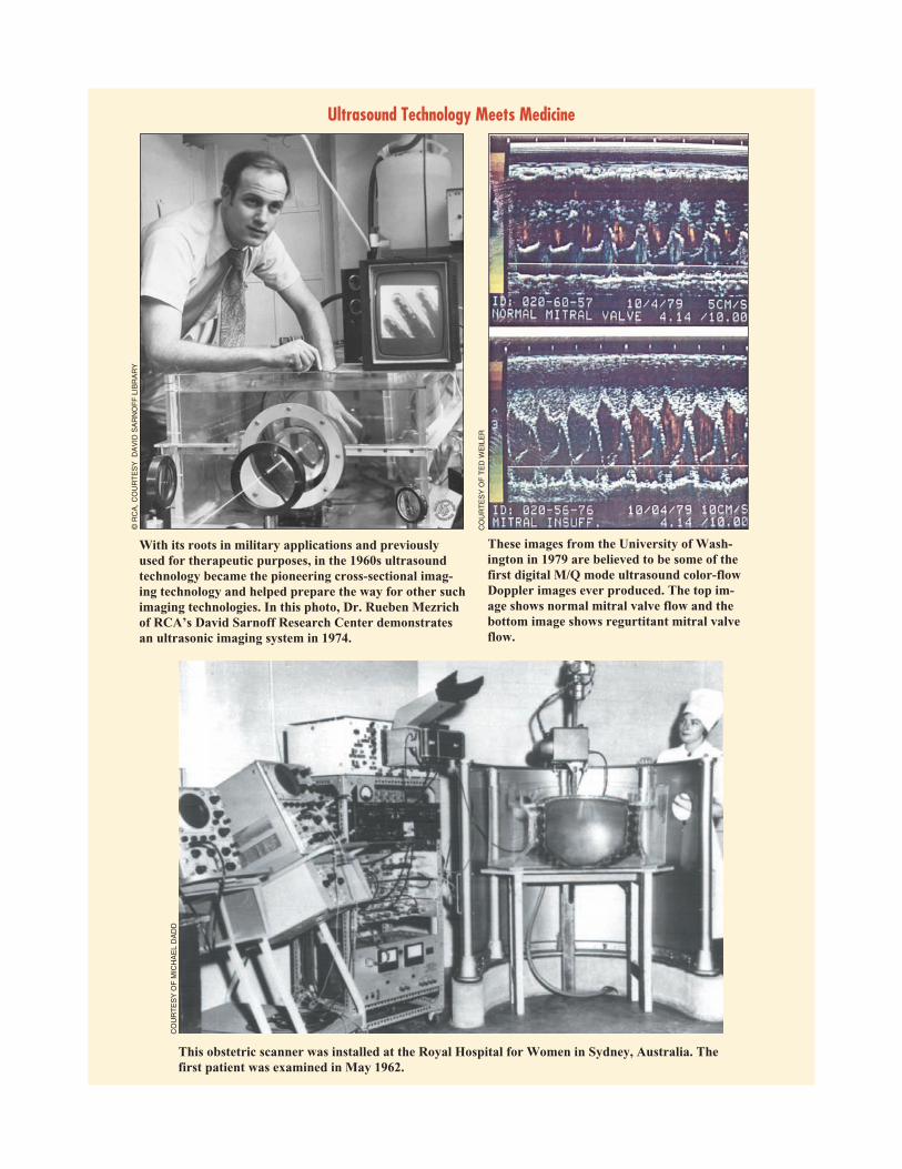
A new imaging technology attracted much attention: ultra-sound imaging. Ultrasound had been used for therapeutic pur-poses (mainly in physical therapy but also to treat cancer) in the1920s and 1930s, and in the 1940s pulsed reflected ultrasoundwas used in industry to detect flaws in materials and construc-tion. In the late 1940s and 1950s a number of groups in variouscountries—Japan, Austria, France, and the United States amongthem—pioneered in creating medical images using ultrasound.Among the most influential were George Ludwig at MIT, JohnJulian Wild and John M. Reid in Minnesota, and DouglassHowry at the University of Colorado. As the pioneeringcross-sectional imaging technology, ultrasound imaging helpedprepare the way for other such technologies discussed in the nextchapters.
In the early 1960s a number of products reached the commer-cial market, such as the Kelvin-Hughes Diasonograph and theSmith Kline echocardiograph. The range of application of ultra-sound increased, and the technology advanced. Some examplesare the work on Doppler ultrasound by a group headed by RobertRushmer and Dean Franklin at the University of Washington, thearray transducer (a ten-element concave transducer) introducedin 1965 by the German opthalmologist Werner Buschmann, andthe Vidoson, designed by Richard Soldner of Siemens, which ap-peared in 1967 (the transducer rotated at the focus of a parabolicmirror in a water-filled enclosure).
In the mid 1960s infrared thermography (which registereddifferences in the heat emitted by tissues) was developed formedical diagnosis. J. Gershon-Cohen of the Albert EinsteinMedical Center introduced IR imaging to the United States in1965. It did not become widely adopted and was used mainly as ascreening technique for breast cancer. Interest in the techniquepeaked in the mid 1970s, when there were 2,000 to 3,000thermography clinics in the United States. Following a majorcomparative study by Stephen Feig, first reported in 1975, themedical community began to lose interest in the technique, andits use faded gradually.
28 IEEE ENGINEERING IN MEDICINE AND BIOLOGY May/June 2002
Biomedical engineering wasseen as both part of the
problem—for raising the costs ofhealth care—and part of the
solution—by improvinghealth care.
May/June 2002 IEEE ENGINEERING IN MEDICINE AND BIOLOGY 29
Ultrasound Technology Meets Medicine
This obstetric scanner was installed at the Royal Hospital for Women in Sydney, Australia. Thefirst patient was examined in May 1962.
With its roots in military applications and previouslyused for therapeutic purposes, in the 1960s ultrasoundtechnology became the pioneering cross-sectional imag-ing technology and helped prepare the way for other suchimaging technologies. In this photo, Dr. Rueben Mezrichof RCA’s David Sarnoff Research Center demonstratesan ultrasonic imaging system in 1974.
These images from the University of Wash-ington in 1979 are believed to be some of thefirst digital M/Q mode ultrasound color-flowDoppler images ever produced. The top im-age shows normal mitral valve flow and thebottom image shows regurtitant mitral valveflow.
©R
CA
,CO
UR
TE
SY
DA
VID
SA
RN
OF
FLI
BR
AR
Y
CO
UR
TE
SY
OF
TE
DW
EIL
ER
CO
UR
TE
SY
OF
MIC
HA
EL
DA
DD

An important area of work, as mentioned in the previouschapter, was hearing aids. These devices, however, cannot helpthose with complete hearing loss. In the late 1950s, A. Djournoand C. Eyries began experimenting with implanted systems tostimulate the auditory nerve (today known as cochlear implants).A number of other researchers took up the work in the 1960s, andin the late 1960s and early 1970s several multiple-electrode co-chlear implant systems were developed. Success was difficult toachieve, but by the end of 1985 more than 150 patients world-wide had received multichannel systems.
History of the ProfessionOn 1 January 1963 the American Institute of Electrical Engi-
neers and the Institute of Radio Engineers merged to form the In-stitute of Electrical and Electronics Engineers. The AIEE, whichdated back to 1884, originally consisted mainly of engineers con-cerned with electric power and its applications and with telegra-phy and telephony. The IRE, which dated back to 1912,originally consisted almost entirely of engineers working on ra-dio. In the 1940s and 1950s, however, the two organizationsoverlapped more and more: electronic techniques were beingadopted in virtually all branches of electrical engineering (so thatAIEE members concerned themselves with electronics), and ra-dio engineering had ramified into numerous branches of elec-tronics (so that IRE members worked in many areas other thanradio). A contributing force for the merger was the members ofthe AIEE and IRE technical committees for biomedical engi-neering, as almost all of them favored it and had been collaborat-ing with their counterparts in the other society for years.
At the merger it was decided to carry over to the IEEE the IREsystem of Professional Groups. The IRE Professional Group onMedical Electronics became the IEEE Professional TechnicalGroup on Bio-Medical Engineering, the name change reflectingthe fact that many members, particularly former AIEE members,were concerned with nonelectronic topics. Table 2 is a list of thechairmen during the 1950s. The members who served as editor ofTransactions during the 1960s were Lee B. Lusted, Edward F.MacNichol, Robert L. Schoenfeld, and David Geselowitz.
The Society began an awards program. First to be institutedwas the William J. Morlock Memorial Award (which later be-came the EMBS Career Achievement Award). The awardees inthe 1960s are listed in Table 3. In 1968 the Society announced anew prize in memory of Samuel A. Talbot to “recognize achieve-ments by young scientists and engineers under the age of 30 for
creative contribu-tions to the field ofbiomedical engi-neering.”
The diversity ofwork in biomedicalengineering and thediversity of back-ground of thepeople contributingto this field made itdifficult for a singleorganization to rep-resent everyone. Inthe 1960s therewere effor ts bysome leaders of the
PGME to achieve greater autonomy within the IEEE in order toaccommodate a more diverse membership. These efforts metconsiderable resistance, and partly as a result the Biomedical En-gineering Society was formed in 1968.
Because there were quite a few professional groups, severalumbrella organizations were established to facilitate coopera-tion. In the late 1960s the Alliance for Engineering in Medicineand Biology was formed, with Pat Horner as its executive direc-tor. This alliance assumed the task of organizing the annual con-ference that had earlier been organized by the Joint ExecutiveCommittee on Medicine and Biology, mentioned in the preced-ing section, and the name changed from Annual Conference onElectrical Techniques in Medicine and Biology to the AnnualConference of Engineering in Medicine and Biology. Within theUnited States, the National Academy of Engineering (NAE) wasestablished in 1964. The NAE created a number of sections, in-cluding one on bio-engineering. (Theb i o e n g i n e e r selected to the NAEare listed on theNAE Web site.)
There was inter-est also on the inter-national plane infacilitating coopera-tion among profes-sional groups. TheInternational Feder-ation for Medical Electronics was formed, with the IRE PGMEas one of its members. The first full-fledged technical conferencewas held in London in July 1960. In the 1960s the name waschanged first to International Federation for Medical Electronicsand Biological Engineering, and shortly thereafter to the Interna-tional Federation for Medical and Biological Engineering. Andin 1963 the International Federation launched the journal Medi-cal Electronics and Biological Engineering.
It was in the late 1960s that the term “biomedical engineer-ing” became established. The term “medical electronics” was toonarrow. “Engineering in medicine and biology” was too long. Inthe United State the Engineers Joint Council Committee on Engi-neering Interaction with Biology and Medicine officially recom-mended the term “bioengineering” in 1966, but this did notbecome standard. J.H.U. Brown, head of the NIH program de-scribed above, promoted the term “biomedical engineering.”
Besides professional societies, publications, conferences,and educational programs, another characteristic of theprofessionalization of a discipline is the formal establishment ofstandards. The first such activity for the PGME was that of theSubcommittee on Instrumentation, set up by the PGME Commit-tee on Electrocardiography. In 1967 the Subcommittee pub-lished a set of standards for ECG, and industry paid attention tothese standards. An international milestone occurred when theInternational Electrotechnical Commission (IEC) establishedthe IEC Technical Committee 62: Electromedical Equipment.TC62, which held its first meeting in 1968, concerned itself withall aspects of standardization of biomedical equipment.
At the beginning of the decade only a handful of universitieshad formal programs in biomedical engineering. At the Univer-sity of Pennsylvania in 1960, Herman Schwan obtained univer-sity approval for an independent Ph.D. program in biomedical
30 IEEE ENGINEERING IN MEDICINE AND BIOLOGY May/June 2002
Table 2. The Chairmen during the1960s of the IRE Professional
Group on Medical Electronics andthe IEEE Professional Technical
Group on Bio-Medical Engineering.
1960 Herman P. Schwan
1961 George N. Webb
1962 Otto H. Schmitt
1963 Robert L. Schoenfeld
1964 Murray Eden
1965 Murray Eden
1966 J.E. Jacobs
1967 Herman P. Schwan
1968 Herman P. Schwan
1969 D.G. Fleming
Table 3. The winners of the WilliamJ. Morlock Memorial Award
(which later became the EMBSCareer Achievement Award)
during the 1960s.
1961 Britton Chance
1963 Otto Schmitt
1965 Edward F. MacNichol, Jr.
1967 Herman P. Schwan
1968 Wilson Greatbatch

engineering. In the United States the number of universities hav-ing a recognized program increased from 40 in 1965 to 180 in1971. The range of biomedical engineering increased. For exam-ple, in 1963 MIT began offering a course titled “Chemical engi-neering in medicine,” which may have been the first such coursein the United States. Universities also began to offer undergradu-ate programs in biomedical engineering.
The 1960s stand out in the history of biomedical engineeringas the decade in which computers became widely applied in re-search and health care and as the decade in which biomedical en-gineering was established as an academic discipline.
The 1970s: New Means of Medical ImagingHistory of the Technologies
In the early 1970s it was believed there was a crisis in healthcare in the United States, with costs out of control and perfor-mance unsatisfactory in many areas. Biomedical engineeringwas seen as both part of the problem—for raising the costs ofhealth care—and part of the solution—by improving health care.
The high cost of medical technology was brought forcefullyto U.S. public attention at the beginning of the decade, when themedia made public the way patient selection for dialysis was be-ing carried out in Seattle. In 1972 Congress agreed to meet thecost of dialysis for every patient considered likely to benefit.Costs soon far exceeded the original estimates; the 1983 cost tothe government for providing dialysis was $2.2 billion. This cre-ated alarm, as it was thought that other technologies that wereboth effective and costly would soon become available. Beforelong this indeed happened with CT scans, coronary bypass sur-gery, and heart and liver transplants.
It was in part a response to the rising cost of medical care thathealth maintenance organizations (HMOs) emerged in the 1970sas a principal way of providing medical care in the United States,as cost containment was one objective of the HMO. At the sametime, HMOs made great use of new technologies. One commen-tator wrote, “An HMO is a working version of the utilization ofmodern technology in health care, including computers, commu-nication systems, automatic analysis, and systems theory, all ap-plied to the business of modern health-care delivery.”
Another response to the rising costs was a change made in1978 to reimbursement procedures for Medicare and Medicaidthat reduced incentives for the use of new medical technologies.That same year Secretary of Health Joseph Califano Jr. said thathis department was concerned about the allegedly extravagantuse of medical technology, and the sales of computerized axialtomography (CAT) scanners fell dramatically.
The government also took a more active role in thehealth-technologies market. In 1976 Congress passed the Medi-cal Device Amendments, which required FDA approval of a de-vice before it could be marketed (through a process calledpremarket approval). And in 1978 Congress established the Na-tional Center for Health Care Technology to evaluate existingand emerging technologies. The first technology to be studiedwas CAT scanning.
The 1970s stand out for the development of new techniques ofmedical imaging. Attracting the most attention was computer-ized tomography (CT), which uses X-ray machines and comput-ers to generate cross-sectional images. Important work done inthe 1970s included two papers by Allan Cormack on a scanningmethod that projected gamma rays through an object on a rotat-ing platform and a prototype transmission CT scanner built by
David Kuhl and Roy Edwards in Philadelphia. In London,Godfrey Hounsfeld developed a CT scanner. He first produced aCT image of a patient on 1 October 1971, and his scanner waspresented at the 1972 British Institute of Radiology Congress.Within a few years there were hundreds of CT scanners through-out the world. A whole-body CT scanner was developed by Rob-ert Ledley in 1973, and it was put into routine clinical use inFebruary 1974. In 1979 Allan Cormack and Godfrey Hounsfeldreceived the Nobel Prize in Medicine for their work on comput-erized axial tomography, the former for his mathematical mod-els, the latter for his engineering of an effective machine.
Nuclear magnetic resonance (NMR) imaging, which takesadvantage of the fact that different chemical elements responddifferently in magnetic fields, began in the mid 1970s. A contrib-uting factor was advances in magnet technology, since high-in-tensity fields are necessary for high-resolution images. In 1972Raymond Damadian applied for a patent on an NMR “Apparatusand method for detecting cancer in tissue,” and in 1973 Paul C.Lauterbur published a paper in Nature that presented a methodfor forming a 2-D NMR map. A live human NMR image (of afinger) was reported in 1976 by Mansfield and Maudsley, and in1977 Damadian succeeded in making an NMR image of the hu-man chest. In April 1980 Damadian’s company displayed a pro-totype of a commercial machine and began taking orders. In 1992some 4000 NMR scanners were in use.
Positron emission tomography (PET), which forms imagesfrom the positrons emitted by radioactive chemicals injectedinto, or swallowed by, a patient, made its commercial debut in1977. A group head by Michael Ter-Pogossian at WashingtonUniversity in St. Louis built a positron imaging device in 1972,and Ter-Pogossian and Michael Phelps published a paper ontheir system of PET in 1975. This new imaging technology wasnot, however, adopted as rapidly as CT imaging: at the end of thedecade only about 40 institutions had set up PET centers.
Ultrasound imaging, which, as related above, became widelypracticed in the 1960s, underwent improvement. The 1970s sawthe advent of a related technology, the scanning acoustic micro-scope. Conceived by C.F. Quate in 1973, the device, which usesultrasound, became available commercially in 1975.
As in the preceding decades, there was much work by bio-medical engineers on the heart, circulation, and blood. Indeed,studies in these areas accounted for almost half of the articles inthe first five volumes of the journal Annals of Biomedical Engi-neering; which commenced in 1972. The principal explanationfor this focus is, of course, the well-established tradition of suchwork, but in the 1970s there was also much concern for the highdeath rate for heart-related problems, hence generous funding inthese areas.
In 1970 was the first successful use of a totally implantedstandby defibrillator (by J.C. Schuder and co-workers). A re-chargeable cardiac pacemaker was marketed from 1973 to 1978,and it was successfully used in hundreds of patients, and at aboutthe same time a nuclear-powered pacemaker was developed. Theintroduction of the lithium battery into pacemaker use by WilsonGreatbatch, however, led to the discontinuation of the manufac-ture of both rechargeable and nuclear pacemakers. Medtronicworked to develop an implantable blood pump to take over partof the heart’s workload, giving time for a donor heart to be lo-cated, but ended the program in the late 1970s because of declin-ing federal support and the limited market. In the late 1970s at theUniversity of Utah, Robert Jarvik and Wilhelm Kolff developed
May/June 2002 IEEE ENGINEERING IN MEDICINE AND BIOLOGY 31

several artificial hearts and testedtwo of them (Jarvik 5 and Jarvik7) in calves. The FDA gave ap-proval for use of the Jarvik 7 in apatient, and in 1982 a teamheaded by William C. DeVriesreplaced the diseased heart ofBarney Clark with the Jarvik 7.(Clark died after 112 days.) AtPennsylvania State University ateam led by William Pierce de-veloped the Pierce-Donachypump, a pneumatic ventricularassist device. It was first used ina patient in 1976 as a bridge totransplant. A commercial ver-sion manufactured by Thoratechas been implanted in over3,000 patients worldwide. Alsoin the 1970s was the develop-ment of an alternative to the me-chanical respirator :extracorporeal membrane oxy-genation (ECMO). The ratio-nale for ECMO is that byoxygenating the patient’s bloodoutside the body, the patient’slungs can rest and heal them-selves. The 1970s also saw ad-vances in electrocardiographictelemetry from ambulances andin the remote diagnosis of anECG through a telephone net-work.
Improvements in instrumen-tation, particularly automation,continued to affect health care. Inthe 1970s the rate of laboratory test-ing in U.S. hospitals increased just as much as in the 1960s. In1971 some 2 billion tests were performed, five years later some4.5 billion. Part of the explanation for the increase was whatcame to be called “defensive medicine,” the practice of physi-cians to order many diagnostic procedures lest they be sued formalpractice for not having used all available technology. Alarger part of the explanation was the increased efficiency andlower cost of testing that automation provided.
Already in the early 1970s roughly 80% of the 15,000 clini-cal laboratories in the United States used automatic equipment,and in the 1970s the NIH made the automation of clinical labo-ratories an area of particular emphasis in its support of researchand development. One example of a successful product wasDuPont’s ACA (Automatic Clinical Analyzer), introduced in1970. The ACA was designed for specialty and emergencytests, as well as small batches of routine tests. It thus comple-mented the large batch analyzers already available in many hos-pitals, providing automation in small hospitals, clinics, andgroup practices. Also in 1970 Beckman Instruments introducedan automatic glucose analyzer, which soon became standardworldwide for measurement of glucose in biological fluids suchas blood, and the next year Beckman introduced an automaticurea nitrogen analyzer.
An instrumentation trend of the 1960s and 1970s was the re-placement of analog techniques by digital techniques. A sub-heading of a 1974 Spectrum article on instrumentation was “Thedigital takeover is now a reality.” Another trend of the 1970s wasthe incorporation of microprocessors in instruments. (It was in1971 that Intel introduced the first microprocessor, the 4004.)For example, in 1978 Beckman introduced the ASTRA (Auto-mated Stat/Routine Analyzer) System, which combined the effi-ciency of Beckman’s dedicated analyzers with the flexibilitymade possible by the microprocessor. In the mid 1970s theTechnicon SMA 12/60, mentioned in the preceding section, wassucceeded by a computer-controlled instrument known as theSMAC (Sequential Multiple Analyzer Plus Computer). TheSMAC did 20 tests on each sample at the rate of 150 samples perhour, and in the late 1980s this machine was the workhorse ofmost large clinical laboratories. Automatic devices that classi-fied and counted blood cells began to be used in the 1970s. Forexample, in 1976 Perkin-Elmer’s introduced the “diff 3” BloodSmear Analysis System that used image processing and patternrecognition techniques to count and classify both white and redblood cells.
Microprocessors were used in many other ways. At the 1978Annual Conference on Engineering in Medicine and Biology
32 IEEE ENGINEERING IN MEDICINE AND BIOLOGY May/June 2002
X Rays +Computers =ComputerizedTomography
COURTESY OF DR. ROBERT SENZIG, GE MEDICAL SYSTEMS CT IMAGING
Computerized tomography(CT), which uses X-ray ma-chines and computers to gener-ate cross-sectional images, was
the imaging technique attracting the most attention in the 1970s. GodfreyHounsfeld first produced a CT image of patient in 1971. His scanner was presentedat the 1972 British Institute of Radiology Congress and within a few years therewere hundreds of CT scanners throughout the world. Hounsfeld won a Nobel Prizein Medicine for his engineering of the CT machine. Above: EMI’s original lathe bedscanner used by Hounsfeld in early CT experiments in 1968. Below: A GE MedicalSystems CT scanner from 1976.
COURTESY OF DR. ROBERT SENZIG, GE MEDICAL SYSTEMS CT IMAGING

May/June 2002 IEEE ENGINEERING IN MEDICINE AND BIOLOGY 33
Chemicals + Magnets =NMR Imaging
CO
UR
TE
SY
OF
DR
.BR
ITT
ON
CH
AN
CE
With the help of improved magnet technology, nuclear magnetic resonance (NMR)imaging, which takes advantage of the fact that different chemical elements responddifferently in magnetic fields, began in the mid 1970s. The high-intensity fields arenecessary for high-resolution images. Above: Dr. Britton Chance has his leg insideone of the early NMR magnets in 1980 at the University of Pennsylvania.
PH
OT
OS
CO
UR
TE
SY
OF
RO
LAN
DS
AA
M
This double-decker bus with an installed NMR scan-ner (circa 1990) provides a portable scanning servicefor preventative noninvasive medical check ups, andit is still in use today by a research team at the RoyalBrompton Clinical Magnetic Resonance Unit, Lon-don, UK. It was the idea of Prof. David Longmore,who founded the first NMR unit in London.

there were 35 papers dealing with applications of microproces-sors. Some microprocessor applications for the disabled were thefollowing: communications device for nonverbal, motor-im-paired persons; hand-held telephone terminal for the deaf; voicesynthesizer; a device for epileptic-seizure control through bio-feedback; and a device for control of an artificial leg.
Mainframe computers became important for hospital admin-istration. In 1971 Technicon Corporation contracted with ElCamino Hospital in Mountain View, California, to provide an in-formation system. By 1974 the system had gained acceptance byphysicians and others at El Camino, and the hospital entered intoa long-term agreement with Technicon. Functions of the systemsincluded patient records, patient status, scheduling of treatment,scheduling of testing, requisitioning of medication, staffing,“electronic rounds” (reviewing clinical status of patients), and fi-nancial records. From 1972 on, Technicon installed informationsystems in other hospitals. The systems lowered costs through la-bor savings, reduction in length of patient stay, reduction informs usage, advance of billing date, reduction in wasted mealsand supplies, reduction in bad debt losses, and reduced re-workresulting from fewer errors.
It was in the 1970s that clinical laboratory information sys-tems became widespread. The DEC-based CLINLAB and theHicks LCI systems were landmarks. By 1980 most laboratorysystems included a terminal at every workstation or instrumentinterface and consolidated patient data in a common database.
There also were significant attempts to make computer-basedexpert systems. In 1972 a group at the University of Pittsburghheaded by Jack D. Myers devised the program INTERNIST-I forclinical decision making. This and later versions were of educa-tional value to clinical practitioners as well as medical students.In 1973 Edward Shortcliffe and collaborators published a de-scription of MYCIN, an expert system to aid physicians in the se-lection of antibiotics. Shortcliffe had captured specialistknowledge in the form of heuristic rules. In 1979 MYCIN per-formed well in a clinical evaluation of its accuracy, but it wasnever used in practice.
The high level of activity in hospital administration systems,laboratory information systems, expert systems, and so on led torecognition of a field of medical informatics. In 1974 the firstworld conference on medical informatics, called MEDINFO,was held.
In the 1970s there was considerable movement into clinicalengineering. The adoption of new technologies had given rise toproblems: incompatibility of devices made by different compa-nies, design deficiencies, incomplete operating instructions, im-proper maintenance of equipment, and difficulty in makingmanagement decisions about advanced technologies. To address
these problems, engineers participated in greater numbers inhealth care teams in clinics and hospitals, and clinical engineer-ing departments were established in larger hospitals.
In the mid 1970s electrical techniques for pain control began tobe widely used, both as external and implanted devices. Other thancardiac pacemakers, the most widely used neuroaugmentative de-vice was the dorsal column neurostimulator (to lessen intractablepain, to stimulate the bladder, and to control spasticity).
Some other devices developed in the 1970s might be men-tioned. Dornier System, a West German company, introduced adevice, called the lithotripter, that focuses shock waves on kid-ney stones. The device began to be used in Germany in the late1970s, and it received FDA approval for marketing in the UnitedStates. in 1984. By the end of 1985 the lithotripter had treatedsome 50,000 patients worldwide. In the late 1970s a joint ventureof Chalmers University of Technology and the Institute for Ap-plied Engineering in Sweden developed the bone-anchored hear-ing aid (BAHA). The device was not well received because of thelarge size and mechanical discomfort, but in the 1980s importantimprovements resulted from circuit miniaturization, new bio-compatible materials, and new surgical techniques. In 1978 HCElectronics began selling the Phonic Mirror HandiVoice, whichsimulates the human voice for nonvocal children and adults (whouse a keyboard to indicate what is to be spoken). And a readingaid for the blind, the Optacon, came into use in the mid 1970s. Itconsists of a “retina” of 144 phototransistors, circuitry, and an ar-ray of vibratory piezoelectric reeds (to reproduce an enlargedversion of the letter being read). Some 1,200 were in use world-wide in 1974.
By the 1970s the biomedical engineering industry had be-come a significant sector of the economy. In the United Statessome 5,000 companies provided products with total annual salesof approximately $1.5 billion. And at the beginning of the decadeon the order of 7,500 people in the United States had professionalinvolvement with biomedical engineering.
History of the ProfessionIn the 1970s there was much concern about the dangers of mi-
crowave ovens, heightened by a great deal of misinformation inthe popular media. This prompted leaders of IEEE, specificallymembers of the Technical Activities Board, to form in 1972 theIEEE Committee on Man and Radiation (COMAR) “to have avoice within IEEE to the media, the general public, and the Con-gress about what the facts are about microwaves.” In March 1973the Consumers’ Union announced their decision not to recom-mend any microwave ovens because of doubt about their radia-tion safety. In the spring of 1974 the IEEE held a pressconference with COMAR Chairman Mark Grove to announceopposition to the CU position. There was caution within IEEE, inpart because its position might not be seen as disinterested, sincesome IEEE members had financial stakes in the microwave ovenbusiness. In later years COMAR studied and issued statementsabout radiation from cellular ground stations, dielectric heaters,electromagnetic pulse simulators, police radar, and othersources.
The IEEE Engineering in Medicine and Biology Society con-tinued its growth. One indication was increased publication. In1971 the Transactions moved from quarterly to bimonthly publi-cation, and in 1979 it moved from bimonthly to monthly publica-tion. It was reported at the time that “Coupled with this expansion
34 IEEE ENGINEERING IN MEDICINE AND BIOLOGY May/June 2002
In the last quarter of the 20thcentury the field of biomedical
engineering benefitedenormously from the actions ofthe Whitaker Foundation, which
was established in 1975.
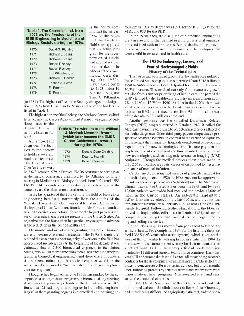
is the policy com-mitment that at least25% of the pagespublished be identi-fiable as applied,that an active pro-gram for the incor-poration of tutorialand applied reviewsbe undertaken.” Theeditors of the Trans-actions were, dur-ing the 1970s,David Geselowitz(to 1973), Hun H.Sun (to 1979), andTheo C. Pilkington
(to 1984). The highest office in the Society changed its designa-tion in 1973 from Chairman to President. The office holders arelisted in Table 4.
The highest honor of the Society, the Morlock Award, (whichlater became the Career Achievement Award), was granted onlythree times in thedecade. The win-ners are listed in Ta-ble 5.
An importantevent was the deci-sion by the Societyto hold its own an-nual conference.The First AnnualConference washeld 6-7 October 1979 in Denver. EMBS continued to participatein the annual conference organized by the Alliance for Engi-neering in Medicine and Biology. Indeed, from 1979 until 1985EMBS held its conference immediately preceding, and in thesame city as, the older annual conference.
In the last quarter of the 20th century the field of biomedicalengineering benefited enormously from the actions of theWhitaker Foundation, which was established in 1975 as part ofthe legacy of Uncas Whitaker, founder of AMP Inc., a manufac-turer of electrical connectors. It became the largest private spon-sor of biomedical engineering research in the United States. Anobjective that the foundation has particularly sought to advanceis the reduction in the cost of health care.
The number and size of degree-granting programs in biomed-ical engineering continued to increase in the 1970s, though it re-mained the case that the vast majority of workers in the field hadnot received such degrees. (At the beginning of the decade, it wasestimated that of 7,500 biomedical engineers in the UnitedStates, only 400 of them came from formal advanced-degree pro-grams in biomedical engineering.) And there was still concernthat someone trained as a biomedical engineer would, in theworkplace, be regarded as “neither fish nor fowl” (neither physi-cian nor engineer).
Though it had begun earlier, the 1970s was marked by the ac-ceptance of undergraduate programs in biomedical engineering.A survey of engineering schools in the United States in 1974found that 121 had programs or degrees in biomedical engineer-ing (49 of them granting degrees in biomedical engineering); en-
rollment in 1974 by degree was 1,530 for the B.S., 1,306 for theM.S., and 933 for the Ph.D.
In the 1970s, then, the discipline of biomedical engineeringgrew in size and further defined itself in professional organiza-tions and in educational programs. Behind the discipline growth,of course, were the many improvements in technologies thatwere useful in research and in health care.
The 1980s: Endoscopy, Lasers, andFear of Electromagnetic Fields
History of the TechnologiesThe 1980s saw continued growth for the health-care industry.
In the United States, expenditures increased from $248 billion in1980 to $666 billion in 1990. Adjusted for inflation, this was a70.7% increase. This resulted not only from economic growthbut also from a further prioritizing of health care: the part of theGNP claimed by the health-care industry increased from about9% in 1980 to 12.2% in 1990. And, as in the 1970s, there wasgreat concern over rising medical costs. Partly as a result, the en-rollment in HMOs continued its rise: from 9.1 million at the startof the decade to 38.6 million at the end.
Another response was the so-called Diagnostic RelatedGroups (DRG) program started in October 1983. It called forMedicare payments according to predetermined prices affixed toparticular diagnoses. Other third-party payers adopted such pro-spective payment systems, in contrast to the earlier cost-plus re-imbursement that meant that hospitals could count on recoupingexpenditures for new technologies. The flat-rate payment putemphasis on cost containment and thus retarded the adoption ofnew technologies, such as magnetic resonance imaging (MRI)equipment. Though the medical devices themselves made uponly 2.5% of health-care costs, critics often pointed to it as a ma-jor cause of medical inflation.
Cardiac medicine remained an area of particular interest forbiomedical engineers. In 1986 the FDA gave market approval tothe rate-responsive pacemaker (Activitrax) made by Medtronic.Clinical trials in the United States began in 1983, and by 198712,000 patients worldwide had received the device (7,000 ofthem in the United States). An automatic implantabledefibrillator was developed in the late 1970s, and the first wasimplanted in a human on 4 February 1980 at Johns Hopkins Uni-versity Hospital. Following further clinical trials, the FDA ap-proved the implantable defibrillator in October 1985, and severalcompanies, including Cardiac Pacemakers Inc., began produc-ing and selling the device.
In the 1980s emphasis moved from permanent to temporaryartificial hearts. For example, in 1984, for the first time the Stan-ford LVAS (left-ventricular assist system), which takes on thework of the left ventricle, was implanted in a patient in 1984. Itspurpose was to sustain a patient waiting for the transplantation ofa natural heart. In 1986 temporary artificial hearts were im-planted by 11 different surgical teams in five countries. Early thatyear NIH announced that it would cancel all outstanding researchcontracts for the development of an implantable artificial heart inorder to concentrate efforts on assist devices, but a few monthslater, following protests by senators from states where there weremajor artificial-heart programs, NIH reversed itself and rein-stated the cancelled contracts.
In 1980 Harold Swan and William Gantz introduced bal-loon-tipped catheters for clinical use (earlier Andreas Gruentzighad introduced a stiff-walled angioplasty catheter), and the open-
May/June 2002 IEEE ENGINEERING IN MEDICINE AND BIOLOGY 35
Table 4. The Chairmen and, from1973 on, the Presidents of the
IEEE Engineering in Medicine andBiology Society during the 1970s.
1970 David G. Fleming
1971 Richard J. Johns
1972 Richard J. Johns
1973 Robert Plonsey
1974 Robert Plonsey
1975 L.L. Wheeless, Jr.
1976 Richard J. Gowen
1977 Thelma A. Estrin
1978 Eli Fromm
1979 Eli Fromm
Table 5. The winners of the WilliamJ. Morlock Memorial Award
(which later became the EMBSCareer Achievement Award)
during the 1970s.
1973 Donald Gene Childers
1974 Dean L. Franklin
1979 Robert Plonsey

ing up of blood vessels using such catheters, known as balloonangioplasty, soon became common (some 300,000 being per-formed in the United States in 1991). The use of metallic stents tohold blood vessels open, though first reported experimentally byCharles Dotter in 1969, came into clinical use in the 1980s.
A major advance of the 1980s was endoscopy, sometimes calledkeyhole surgery, which came to be widely practiced. Tiny instru-ments could be passed through small tubes inserted in the body eitherthrough natural openings or incisions. Mechanical miniaturizationand fiber-optic techniques made it possible to replace a wide range oftraditional surgical procedures, both diagnostic and corrective, bynew minimally invasive ones. The U.S. market for endoscopy prod-ucts reached $533 million in sales in 1988.
Surgery benefited greatly from the use of lasers. At the begin-ning of the decade the applications were limited, and most em-
ployed one of three continuous-wave lasers. At the end of the de-cade there were a great many medical applications, and these useda variety of pulsed and continuous-wave lasers. Among the newapplications were pulse-dye lasers for fragmentation of kidneystones, excimer lasers for reshaping the cornea, pulse-dye lasersfor the treatment of port-wine stain, and argon-pumped pulse-dyelasers for treatment of cancer. Lasers could be used in endoscopy,such as the laser catheter for cleaning arteries of plaque made byTrimedyne Inc. that received FDA approval in 1987.
A momentous advance of the 1980s was the adoption ofMRI. In 1984 the FDA gave approval to four companies to mar-ket MRI machines. In October 1983 only 14 MRI machineswere in use in the United States; a year later the number wasmore than 90, with more than 140 installed worldwide. At thebeginning of the decade, superconducting solenoids, to achieve
36 IEEE ENGINEERING IN MEDICINE AND BIOLOGY May/June 2002
PH
OT
OS
CO
UR
TE
SY
OF
DR
.JA
YB
UT
TE
RM
AN
,FO
NA
RC
OR
P.
A major achievement of the 1980s was magnetic resonance imaging. In1983, only 14 machines were in use in the United States; however, in 1984there were more than 90, with 140 in use worldwide. Left: The team thatproduced the first whole body human image (above) via MRI in 1977:(from left to right) Dr.Damadian and graduatestudents Larry Minkoff(the subject used for theimage) and Michael Gold-smith.
The 1982 FONAR Beta 3000 perma-nent magnet (above), and for theclaustrophobic, the FONAR-360 from1998, where the MRI scanner is hid-den behind the landscape, 360 degreesopen to reduce the claustrophobia. Itcan also be used for MRI-guided sur-gical interventions.
ImagingExpands with
MRI
higher field strengths, were introduced, and in 1987 techniquesfor extremely rapid MRI made it possible to create a movie of abeating heart.
PET, which, as we saw, was introduced the previous decade,became more widely used. The technique was especially valu-able because it could chart metabolic processes. But the cost ofPET scanning remained so high that it was used almost exclu-sively for research. The use of smaller cyclotrons contributed tolowering the cost, which in 1988 was one-third of what was itwas five years earlier.
The decade also saw important improvements to ultrasoundimaging and X-ray computed tomography. For example, in 1985Douglas Boyd developed superfast CT, and in 1989 a spiral CTwas first marketed. (Instead of sequential scanning of consecu-tive slices, spiral CT is a mode for continuous volume scanning.)
Medical imaging became big business. In 1988 in Europesales of medical imaging equipment totaled $1.46 billion (X ray62%, ultrasound 20%, CT 8.5%, nuclear imagers 5%, and MRI3%). In the United States the ultrasound imaging market alonereached nearly $600 million in 1988. As these figures suggest, inthe 1980s biotechnology became a major sector of the economy,recognized as such by economists with careful measurements ofnational investments and outputs. Another example is providedby cardiac monitoring equipment, whose total sales in the UnitedStates reached the $2 billion level in 1990.
In 1963 Richard McFee and B.M. Baule created the firstmagnetocardiogram, a depiction of the magnetic field of theheart, using coils with several million turns of wire. Later DavidCohen used the superconducting quantum interference device(SQUID) to record both a magnetocardiogram and amagnetoencephalogram. Several investigators developed thetheory for the generation of these fields. Toward the end of the1980s there appeared commercial systems using SQUIDs to cre-ate images of biomagnetic fields: in 1989 both Siemens MedicalSystems and Biomedical Technologies announced suchbiomagnetic measurement systems. Used initially in neurologi-cal research, they gave information about overall brain activityand permitted the localization of brain activity within a few milli-meters. (In both respects, the electroencephalogram and themagnetoencephalogram provide complementary tools.)
The expanding capabilities of solid-state electronics hadmany other biomedical applications as well. For example, micro-chips transformed cardiac monitoring equipment: what had beenbulky, expensive, and confined to the laboratory or hospital be-came small, inexpensive, and usable by the individual. One suchmonitoring device was the pulse oximeter, which noninvasivelymeasured blood oxygen levels, giving warning of insufficientoxygenation. In many instances, solid-state transducers replacedlarger, handmade devices. In the late 1980s, for example, bio-medical pressure transducers were introduced that sold for 5% ofthe price of earlier devices, which made possible disposablepressure sensors for clinical applications.
The new capabilities expanded the range of animal electron-ics, which are the sensing and imaging technologies for researchor for use on farms or in animal- and food-processing plants.Electronic sensors were used, for example, to measure animalweight, to feed dairy cows based on their milk production, and todetect diseased animals. Zoological research, too, benefited. Forexample, in 1989 engineers at Oak Ridge National Laboratorydeveloped a tiny solar-powered transmitter that could be glued tothe thorax of a honeybee. (At the time the movement ofAfricanized honeybees was a cause of concern.)
Another advance in instrumentation during the 1980s was thefluorescence polarization instrument for measurement of serumdrug levels (the Abbot Laboratories TDx system), whichachieved widespread clinical use by 1985. Also noteworthy wasthe FDA approval in 1989 of the nitroprusside infusion control-ler; this was the first medical device that continuously monitoreda physiological parameter (blood pressure) and then used the in-formation to control the injection rate of a drug without humanintervention.
A breakthrough advance was the invention in 1981, by GerdBinnig and Heinrich Rohrer, of the scanning tunneling micro-scope. (In 1986 Binnig and Rohrer, along with Ernst Ruska, in-ventor of the electron microscope, won the Nobel Prize forPhysics.) This led to, among other things, the atomic force mi-croscope (AFM). In the mid 1990s use of AFM in the biologicalsciences increased rapidly, and in 1997 there was an issue ofIEEE Engineering in Medicine and Biology Magazine devotedto the subject.
In the 1980s there was a revival of interest in the study of hu-man metabolism. It had been vigorously pursued in the late 19thand early 20th centuries, but then interest in it waned, partly be-cause of lack of suitable instrumentation. In the 1980s there was arevival of interest in the topic among biomedical engineers, oneindication of which was a special issue of IEEE Engineering inMedicine and Biology Magazine on the topic.
Several other advances of the 1980s might be mentioned.Shock-wave lithotripsy (the use of acoustic waves to destroy kid-ney stones) reached the marketplace. The U.S. patent fortwo-photon excitation (TPE) fluorescence microscopy was filedin 1989. TPE had been first proposed in the 1970s, and it becamean extremely important technique in the 1990s. Between March1982 and January 1985, an implantable peristaltic pump devel-oped by Medtronic was placed in more than 150 people to delivermorphine, chemotherapy agents, or heparin (against blood clot-ting). At Johns Hopkins University, a team led by ChristopherSaudek developed the Programmable Implantable MedicationSystem (PIMS). The FDA gave approval for human implant inMay 1986. PIMS was used to deliver insulin automatically in di-abetes patients according to programming stored in its micropro-cessor.
The decade of the 1980s saw several advances in correctingdeafness. A joint venture of Ehime University and Sanyo Elec-tric Company developed an implantable middle-ear device thatachieved direct vibratory coupling to the ossicles. The cochlearimplant, which consists of an internal receiver embedded underthe skin behind the ear that is connected to electrodes in the co-chlea (part of the inner ear), received FDA market approval in1984 (for a single-electrode unipolar implant) and 1985 (for a22-electrode device). A related device called the centralelectroauditory prosthesis (CEP) was also developed; it is simi-
May/June 2002 IEEE ENGINEERING IN MEDICINE AND BIOLOGY 37
In the 1980s there was arevival of interest in the study
of human metabolism.

lar to the cochlear implant except that the electrode wiring isplaced directly on the brain stem rather than in the cochlea.
Another technology involving connection to the nervous sys-tem was functional electrical stimulation (FES). In 1983 pro-grams at several institutions, Case Western Reserve and IllinoisInstitute of Technology among them, demonstrated FES that en-abled sufferers of spinal-cord injuries to walk again and that re-stored grasping functions to the hands of quadriplegics. OtherFES devices restored bladder control in patients and assisted inthe treatment of scoliosis.
An important stimulus to the development of FES and othertypes of rehabilitation engineering in the United States had beenthe passage of the Rehabilitation Act of 1973. This act led to theestablishment of more than a dozen research-and-developmentcenters for rehabilitation engineering under the jurisdiction ofthe National Institute for Handicapped Research (later the Na-tional Institute on Disability and Rehabilitation Research). Theact also recognized various areas of rehabilitation engineering asparticularly important, and the different research centers focusedon different sets of problems.
The participation of people in wheelchairs in athletics, partic-ularly marathons and other races, increased markedly in the1980s. This led to improvements in wheelchair design and muchgreater public awareness of the needs of people who use wheel-chairs (one consequence of which was the passage of the Ameri-cans with Disabilities Act in 1990). A long-term program ofsystematic research and development in wheelchair design be-gan at the University of Virginia’s Rehabilitation EngineeringCenter in 1977, and in the 1980s there was drastic improvementin powered wheelchairs. The adoption in the late 1980s of volun-tary standards for manual and powered wheelchairs was impor-tant, as the standards enabled comparisons using objectivecriteria served as criteria for third-party payer acceptance.
A significant advance in the use of artificial materials inbiomedicine was the 1986 approval by the FDA of Gore-Tex (ex-panded polytetrafluoroethylene) as an artificial ligament.(Gore-Tex was already being used for sutures and blood-vesselgrafts, as well as in sportswear.) In 1987 the passive soft tissueimplant market in the United States totaled $234.4 million. Themost important products were vascular grafts, fabrics andmeshes, and vascular access devices. A recent innovation was asurface coating for Dacron vascular grafts to eliminate time-con-suming preclotting of the graft.
In the 1980s computers became ubiquitous in biomedical re-search and in hospitals, clinics, and doctor’s offices. These weremainly personal computers, which first became common in theearly 1980s. Researchers and hospitals used, in addition, mini-computers and mainframe computers. And in the 1980s the ap-plication of supercomputers in biomedicine attracted attention,as they were used to model complex systems (such as the cardio-
vascular system), in image processing (such as real-time CTscanning), in studies of macromolecular structures (such as genesequences), and in designing large-scale database systems.
Work on expert systems, mentioned in earlier sections, con-tinued in the 1980s. A frequent objective was an expert systemthat explains its behavior in useful ways, so that it is easily usedas an adviser rather than as a decision maker. There was workalso on procedures for reasoning under uncertainty; two new ap-proaches in the 1980s were the use of fuzzy logic and the applica-tion of the Dempster-Shaffer theory of evidence. Applications offuzzy logic in biomedicine gradually increased, and in 1994IEEE Engineering in Medicine and Biology Magazine devotedone issue to the topic, presenting 14 articles. Another notable ad-vance was an expert system on a chip: S.M. Weiss, C.A.Kulikowski, and R.S. Galen at Rutgers University developed theSPE/EXPERT system for serum protein electrophoresis analysis(designed to process signals from the scanning densitometer ofthe Cliniscan instrument).
Two other mathematical techniques received increased atten-tion. A 1982 paper by J.J. Hopfield revived interest in neural net-works. And chaos theory was applied to a few biomedical areas,such as cardiology (in modeling the beating of aggregates of em-bryonic heart cells). The first book devoted to the physiologicalaspects of chaos appeared in 1988.
History of the ProfessionAs in previous decades, biomedical engineering came to the
fore with certain public-health issues. In the 1980s the fear of mi-crowaves diminished, but concern over the use of video-displayterminals (VDTs) became extreme. In 1982 there was a Congres-sional hearing on VDTs and RF heat-sealers. COMAR issued aposition paper onVDTs and one onRF heat-sealers; thelater was critical ofindustry’s efforts tocontrol exposurenear the heat-seal-ers. Toward the endof the decade thepossible health ef-fects of the electricand magnetic fieldsfrom electric powerlines became a sub-ject of widespreadinterest, and therewere U.S. Congres-sional hearings on the subject on 6 October 1987 and much newresearch. This concern continued into the 1990s, and public fearswere gradually allayed.
Degree-granting programs continued to become more com-mon. At the beginning of the decade approximately 100 engineer-ing schools in the United States had a degree program inbiomedical engineering. It was reported toward the end of the de-cade that there was an approximate match between the number ofdegrees granted and the number of career opportunity available (incontrast to the early years of biomedical engineering programs,when there were fewer positions available than trained people).
The IEEE Engineering in Medicine and Biology Society ex-panded its already wide range of activities. Membership was
38 IEEE ENGINEERING IN MEDICINE AND BIOLOGY May/June 2002
Table 6. The Presidents of theIEEE Engineering in Medicine andBiology Society during the 1980s.
1980 Morton D. Schwartz
1981 Morton D. Schwartz
1982 Lee E. Ostrander
1983 Alfred R. Potvin
1984 Joseph D. Bronzino
1985 Joseph D. Bronzino
1986 Dov Jaron
1987 Dov Jaron
1988 Willis J. Tompkins
1989 Willis J. Tompkins
In 1982 the Society inaugurateda magazine “to provide
published material of broadmember interest,” the first issueappearing in March of that year.

fairly stable, declining slighting in the early 1980s but reachingat the end of the decade a membership total of 7,500. The numberof Chapters continued to increase; at the end of the decade therewere more than 30. The presidents of the Society are list in Table6. In 1988 the Society AdCom decided to authorize the hiring ofan Executive Officer, commenting “The person hired for this po-sition would help increase the visibility and viability of EMBSand would provide an interface between EMBS and IEEE,year-to-year continuity, conference management, and general
support of EMBS.”In 1982 the Soci-
ety inaugurated amagazine “to pro-vide published ma-ter ia l of broadmember interest,”the first issue ap-pearing in March ofthat year. Also in1982 the IEEE
Transactions on Medical Imaging made its debut. This cameabout through an agreement among four IEEE Technical Soci-eties in 1980. The EMBS and the Nuclear and IEEE Plasma Sci-ence Society each undertook a one-third interest in the venture,and the IEEE Signal Processing Society and the IEEEUltrasonics Society each undertook a one-sixth interest. TheEMBS Medical Imaging Committee had, as its primary activity,overseeing the operation of the IEEE Transactions on MedicalImaging. In 1989 the IEEE Publications Committee voted to ini-tiate the IEEE Transactions on Neural Networks, starting in 1990as a quarterly publication. EMBS was involved as one of the So-cieties of the IEEE Neural Networks Committee, the sponsoringorganization.
The highest honor of the society, the William J. Morlock Me-morial Award, was renamed the EMBS Career AchievementAward. It was granted only four times in the decade. The winnersare listed in Table 7. In 1983 Eli Fromme received special recog-nition for service to the Society (and therefore is regarded as thefirst recipient of the EMBS Service Award, which was formallyinstituted in 1990, as described in the following section).
In biomedical engineering, as in other branches of engineer-ing, the establishment of standards is an extremely important ac-tivity. For example, in 1980 the Medical Research Programme ofthe European Community started a project to establish standardsin quantitative ECG. The measurement database thus establishedbecame “an internationally recognized yardstick for evaluationand improvement of European, American, and Japanese ECGprogrammes” The EMBS Standards Committee was active,working to develop standards for a local-area network specifi-cally for use in the health-care environment, for a Medical Infor-mation Bus (MIB), and for a Medical Data Interchange(MEDIX).
As mentioned in the preceding section, EMBS had been amember of the Alliance for Engineering in Medicine and Biol-ogy (AEMB). AEMB organized an annual conference, and until1986 EMBS held its annual conference immediately precedingthe AEMB conference and in the same city. Leaders of EMBSfelt, however, that EMBS was not getting its share of the profitsfrom the joint endeavor, and beginning in 1986, EMBS held itsconference apart from that of AEMB. The separation was benefi-cial to the Society’s financial position, but it led to the demise of
the AEMB conference. (In 1988 the constituent societies ofAEMB decided to hold separate conferences and to assist withthe founding of the American Institute for Medical and Biologi-cal Engineering.) In 1987, EMBS, in connection with IEEE Edu-cational Activities, held a video conference on “NewTechnologies in Biomedical Engineering.”
The 1990s: The Human Genome Project,Robotics, and Internationalization
History of the TechnologiesThe Human Genome Project was perhaps the most prominent
scientific and technological effort of the 1990s. With the objec-tive of learning the entire sequence of human DNA, the project,which began in 1991, was funded by the U.S. National Institutesof Health, the U.S. Department of Energy, and the WelcomeTrust. Some of the engineering products that have been vital tothis effort are automatic sequencers, robotic liquid-handling de-vices, and software for databasing and sequence assembly. Amajor shift was occurring in bioengineering toward consider-ation of processes at the cellular and molecular level rather thanat the organ-system level.
Robotics, here and in countless other areas of biomedicine, at-tracted considerable attention. A survey at the beginning of the de-cade found approximately 400 applications of robotics tomedicine. One area was the laboratory. For example, a lab robot (amodified Tecan Robotic Sample Processor) was used to cultureand harvest bone marrow, lymph nodes, fibroblasts, and other tis-sues. The largest area for medical robotics was in rehabilitation:tactile sensors (as used in the Utah-MIT dextrous four-fingerhand), prosthetics (such as the artificial arm), orthotics, assistivedevices for the blind (as the Japanese (MITI) project MELDOG,designed to provide the functions of a guide dog), and systems forrehabilitation after surgery (as continuous-passive-motion ma-chines). In surgery there are some robots that assist surgeons (suchas for stereotactic neurosurgery) and some that perform surgerythemselves (such as a robot to perform prostatectomies). On 25March 1991 at Shaftesbury Hospital in London, a surgical robotSARP (Surgical Assistant Robot for Prostatectomy), under com-puter control, carried out an operation for the first time. And in1993 the FDA authorized clinical testing of a robotic surgical pro-cedure for implanting artificial hips.
There were many advances in implantable devices. For ex-ample, in 1997 the FDA gave approval to market a device thatcontrols tremor (such as tremor resulting from Parkinson’s dis-ease) with a pulse generator implanted in the chest that sendssignals to an electrode placed in the thalamus (a region of thebrain that controls bodily movement). This was the first timethe FDA approved an active device for implantation in thebrain. (Earlier-approved brain implants, such as shunts, to drainfluids, and clips, to seal off aneurysms, are passive devices.)Also in 1997, the FDA approved the Freehand System, devel-oped by P. Hunter Peckham at Case Western Reserve Univer-sity, which uses FES to restore function to a paralyzed hand of aquadriplegic. This was the first implantable FES system to re-ceive FDA approval for wide use. In early 2000 about 160 quad-riplegics were using Freehand. By the mid 1990s in the UnitedStates, two to three million patients a year were being treatedwith an artificial implant of some kind, including supportingdevices (such as pacemakers) and prostheses (such as cardiacvalves and hip joints).
May/June 2002 IEEE ENGINEERING IN MEDICINE AND BIOLOGY 39
Table 7. The winners of the EMBSCareer Achievement Award
(formerly the Morlock Award)during the 1980s.
1985 David B. Geselowitz
1986 Leslie A. Geddes
1987 Otto Schmitt
1988 R. Stuart Mackay

40 IEEE ENGINEERING IN MEDICINE AND BIOLOGY May/June 2002
Providing alternatives to artificial materials in therapy is theobjective of the newly emerging field of tissue engineering orcell therapy. Among the products under development in the1990s were the following: cell-based skin substitutes forwounds; corneal grafts to restore vision; transplanted cells forbone, cartilage, and ligament repair; transplants in or near thebrain and spinal cord for treating neurodegenerative disease;pancreas substitution systems for diabetics; liver assist devicesfor hepatic failure; and tubular structures for repair of blood ves-sels, ureters, and fallopian tubes. Such tissue-engineering prod-ucts were entering clinical trials in the late 1990s.
Medical imaging continued its vigorous expansion in the1990s. An indication of medical usefulness is provided by a 1997survey which found that there were 4.3 million MRI scans in theUnited States per year, 21 million CAT scans, and 208 millionuses of X rays. The oldest modality, X-ray imaging, began themove to digital techniques. For example, in 1998 the GeneralElectric digital detector Revolution made its medical debut in theFull Field Digital Mammography System introduced in Europe.Unlike other digital X-ray devices that pieced together a largeimage from small ones, the GE system created a single image atonce. Magnetic resonance imaging made important advances. Anumber of techniques were developed to decrease MRI acquisi-tion times, such as turboflash developed by Siemens and fastSPGR and fast spin-echo developed by GE; these methods madepossible complete scans in only a few minutes. A major newtechnique was introduced in 1991: using MRI to map humanbrain activity by observing changes in cortical blood oxygena-tion. Called functional MRI (fMRI), it has since become a majormethodology in brain science and other areas of research.
Interest in biomagnetic imaging continued to grow. (Abiomagnetometer, by sensing the tiny magnetic field near thesurface of the brain, provides a noninvasive indicator of local-
ized brain function, which is useful, for example, in locating epi-leptic foci.) In 1990, Biomagnetic Technologies of San Diegodelivered units of its 37-channel Magnes biomagnetometer toseveral hospitals, and Siemens installed a 37-channel unit calledthe Krenikon at the University of Nürnberg. A related technique,magnetic stereotaxis (a system for locating, in three dimensions,precise areas of the brain), entered clinical testing in 1997.
In 1991 PET images of the human brain as it performs the taskof recalling a word were made public for the first time. The im-ages showed that the recall process was going on in unexpectedparts of the brain. Stimulated by advances made by the militaryand NASA for night vision and space exploration, the 1990s sawrenewed interest in infrared imaging, especially for breast-can-cer screening. (At the 1994 EMBS conference, IR imaging wasone of the emerging technologies topics, and a special issue ofIEEE Engineering in Medicine and Biology Magazine was de-voted to the subject in 1998.) In 1995 Martin Nuss and co-work-ers at AT&T Bell Laboratories (now Lucent Technologies) firstdemonstrated terahertz-pulse imaging, in which laser pulses ofT-rays (electromagnetic waves that lie between infrared radia-tion and microwaves in the electromagnetic spectrum) are usedto create images. This technique is able to distinguish very simi-lar materials, such as burned and healthy tissues.
Surgical “Aids”
PH
OT
OS
CO
UR
TE
SY
OF
INT
UIT
IVE
SU
RG
ICA
L,IN
C.
One of the applications roboticshas found in biomedicine has beenassisting surgeons. Left: the daVinci Surgical System in action atSt. Pierre University in Brussels,Belgium. The surgeon is seated tothe left at a console from which heguides robotic instruments thatreplicate natural hand and wristmovements. From the console thesurgeon views a true-to-life 3-Dimage (below).

Innovations in imaging technology have been vital for ad-vances in endoscopic surgery. The computer-chip video camera,the 3-chip camera (which produces color images), and 3-D videosystems are important milestones. According to Roger Barr,“Catheter-based sensing and surgery, while still invasive, are re-markably less so. Through ingenious development and incredi-bly skilled practitioners, crude tools have been replaced bysmaller, more sophisticated devices that are remotely maneu-vered while carrying surgical tools, multiple electrodes, RFtransmitters, and imaging sensors.”
Medical imaging attained even more prominence through theVisible Human Project, a digital depiction of every millimeter ofthe cadaver of a human (“the first digital human being”), whichwent online 29 November 1994. This representation of anatomyhas contributed to highly realistic simulation systems used totrain physicians in bronchoscopy, arthroscopy, venipuncture,and other procedures.
Medical devices in general continued to improve. Some newproducts of the 1990s were the tympanic or ear thermometer, in-troduced in 1991, which measured infrared heat from the eardrumand surrounding tissue, and the intra-arterial blood-gas monitor-ing system. The latter, introduced by Puritan-Bennett Corporationin 1992, determined in real time the hydrogen-ion concentrationand partial pressures of oxygen and carbon dioxide (instead of de-termining these quantities from blood samples taken at infrequentintervals). Two other new devices that incorporated biosensors—and provided point-of-care diagnosis—were a handheld bloodanalyzer introduced by i-Stat Corporation and a device for testingthyroid disorders developed by Biocircuits Corporation. TheLasette laser finger perforator, which draws blood from the finger-tip for glucose testing, became the first medical laser devicecleared by the FDA for consumer use. Medical technology, ofcourse, had long since entered the realm of consumer electronics.In the 1990s, in the United States some 1.5 million hearing aidswere sold every year. Consumers were offered devices to measureheart rate and blood pressure; more than 750,000 heart-rate moni-tors are sold each year in the United States.
The use of computers in biomedicine became even more perva-sive than in the preceding decade. With much of the work both inresearch and in health care taking place in software design, it be-came much easier for individuals and groups from less wealthycountries to contribute to the field internationally (whereas, exper-imentation and equipment prototyping are much more expensiveactivities). Increased travel and, more importantly, improvedcommunication also facilitated wider participation in biomedicalresearch and development. The Internet became an especially ef-fective means of communication. For example, Medline, an elec-tronic gateway to medical literature, had become much used sinceits introduction in 1966. But use increased by an order of magni-tude in 1998 when the PubMed version of Medline became avail-able on the Internet as a free service.
As in earlier decades, computers facilitated mathematical mod-eling. With faster computers and better models, it became possiblein many areas to obtain quantitative estimates of the consequencesof specific interventions, the models being idealizations of the bio-logical mechanisms rather than statistical or empirical substitutes.Among the new techniques was fractals. The June 1992 issue ofIEEE Engineering in Medicine and Biology Magazine was de-voted to fractals in biomedical engineering. Among the articleswere ones on fractal mechanisms in the electrophysiology of theheart, on the fractal nature of ion channel kinetics, and on fractaldimension in the analysis of medical images.
In the early 1990s neural networks were a promising but un-proved technology in medicine and biology. At the end of the de-cade neural networks had come into routine use for certaindiagnoses. According to Evangelia Micheli-Tzanakou, “In thelater part of the 1990s, neural networks have been combined withfuzzy logic, wavelet analysis, and other feature-extraction meth-ods to yield greater confidence in predicting outcomes and inclassifying patterns of normal and abnormal situations. The fieldhas grown to the point of having several major journals dedicatedto neural networks.”
In mid decade the biomedical industry in the United Stateshad sales of $40 billion and was the fastest growing sector of theU.S. economy. But this success was decried by those whoblamed higher health-care costs on new technology. Many of theplans advanced in the 1990s to contain costs chose technology asan area for downsizing. Biomedical engineers pointed out thatthe most expensive medical equipment, diagnostic imagers, ac-counted for only half a percent of health-care spending and thatprices of comparable products had actually declined in recentyears (though utilization had expanded considerably). More andmore people belonged to HMOs; in 1997 three-fourths of U.S.workers with health benefits received coverage through anHMO. (In the United States, HMO enrollment increased fromabout six million in 1976 to about 50 million in 1995.) Amongthe ways HMOs worked to contain costs were discouraging ex-perimental procedures, controlling access to specialists and hos-pitals, and delaying acceptance of new technologies.
History of the ProfessionPublic-health issues were often in the news in the 1990s.
There was widespread concern that the use of cell phones mightcause brain cancer, and the telecommunications industry began a$20 million study to examine the potential health hazards ofwireless communications. In the 1960s and 1970s plastic sur-geons in the United States began to use silicone for breast en-largement. Dow Corning developed a silicone gel, which was putinto a plastic container to make a breast implant. By 1992 an esti-mated two million women had received silicone gel breast im-plants. Many problems associated with the implants werereported, and at the beginning of the 1990s the FDA instituted amoratorium on further implants. In the United States, regulationof medical devices became even stricter in 1990 with the passageof the Safe Medical Devices Act; one of its requirements is thatmanufacturers track where certain implantable products go.Some people believe that the regulatory climate in the UnitedStates is prompting U.S. companies to move development andmanufacturing to countries with less stringent oversight.
Public unease with biotechnology was reflected in the approvalon 17 May 1992 by Swiss voters of a referendum on biotechnol-ogy and reproductive medicine; this prompted the Federal Assem-
May/June 2002 IEEE ENGINEERING IN MEDICINE AND BIOLOGY 41
The Human Genome Project wasperhaps the most prominent
scientific and technological effortof the 1990s.
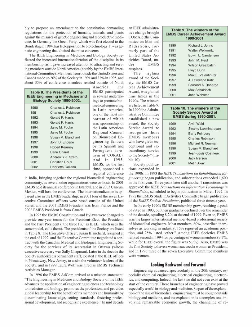
bly to propose an amendment to the constitution demandingregulations for the protection of humans, animals, and plantsagainst the misuses of genetic engineering and reproductive medi-cine. In Germany the Green Party, which was first elected to theBundestag in 1984, has led opposition to biotechnology. It was ge-netic engineering that elicited the most concerns.
The IEEE Engineering in Medicine and Biology Society re-flected the increased internationalization of the discipline in itsmembership, as it gave increased attention to attracting and serv-ing members outside North America (notably by the EMBS Inter-national Committee). Members from outside the United States andCanada made up 26% of the Society in 1991 and 32% in 1995, andabout 35% of conference attendees resided outside of North
America. TheEMBS participatedin several undertak-ings to promote bio-medical engineeringin Latin America,one of the most im-portant of whichwas sponsorship ofthe Latin AmericanRegional Councilon Biomedical En-gineering (knownby its Spanish andPortuguese acro-nym of CORAL).And in 1995,EMBS, for the firsttime, sponsored aregional conference
in India, bringing together the regional biomedical engineeringcommunity, as several other organizations were co-hosts. In 2001EMBS held its annual conference in Istanbul, and in 2003 Cancun,Mexico, will host the conference. The internationalization is ap-parent also in the EMBS leadership: in 2001 four of the seven Ex-ecutive Committee officers were based outside of the UnitedStates, and the 2001 EMBS President was from France and the2002 EMBS President is from Canada.
In 1995 the EMBS Constitution and Bylaws were changed toprovide one-year terms for the President-Elect, the President,and the Past President (“the three Ps,” as IEEE, which uses thesame model, calls them). The presidents of the Society are listedin Table 8. The Executive Officer, Susan Blanchard, resigned atthe end of 1992, and the Executive Committee negotiated a con-tract with the Canadian Medical and Biological Engineering So-ciety for the services of its secretariat in Ottawa (whoseexecutive secretary was Sally Chapman). Later in the decade theSociety authorized a permanent staff, located at the IEEE officesin Piscataway, New Jersey, to assist the volunteer leaders of theSociety, and in 1999 Laura Wolf was hired as EMBS TechnicalActivities Manager.
In 1996 the EMBS AdCom arrived at a mission statement:“The Engineering in Medicine and Biology Society of the IEEEadvances the application of engineering sciences and technologyto medicine and biology, promotes the profession, and providesglobal leadership for the benefit of its members and humanity bydisseminating knowledge, setting standards, fostering profes-sional development, and recognizing excellence.” In mid decade
an IEEE administra-tive change broughtCOMAR (the Com-mittee on Man andRadiat ion) , for-merly part of theUnited States Ac-tivities Board, un-der EMBSauspices.
The highestaward of the Soci-ety, the EMBS Ca-reer AchievementAward, was grantednine times in the1990s. The winnersare listed in Table 9.In 1990 the Admin-istrative Committeeestablished a newaward, the SocietyService Award “torecognize thoseEMBS memberswho have given ex-ceptional and ex-traordinary serviceto the Society” (Ta-ble 10).
Society publica-tions expanded inthe 1990s. In 1993 the IEEE Transactions on Rehabilitation En-gineering began publication, and subscriptions exceeded 1,600in the first year. Three years later still another Transactions wasapproved: the IEEE Transactions on Information Technology inBiomedicine, scheduled to begin publication in March 1997. In1992 the EMBS Student Activities Committee began publicationof the EMBS Student Newsletter, published three times a year.
In the early 1990s EMBS membership grew, reaching at peakof 9,426 in 1993, but then gradually declined over the remainderof the decade, equaling 8,204 at the end of 1999. Even so, EMBSwas the largest international member-based professional societyof biomedical engineers. Most members, 60%, described them-selves as working in industry; 15% reported an academic posi-tion, and 25% listed “other.” Among IEEE Societies EMBSranked second in 1994 for percentage of women members (9.7%,while for IEEE overall the figure was 5.7%). Also, EMBS wasthe first Society to have a woman succeed a woman as President,and in 1996 three of the seven Executive Committee memberswere women.
Looking Backward and ForwardEngineering advanced spectacularly in the 20th century, es-
pecially chemical engineering, electrical engineering, electron-ics, and computing. Indeed, the last two did not even exist at thestart of the century. These branches of engineering have provedespecially useful in biology and medicine. So part of the explana-tion of the rise of biomedical engineering must be sought outsidebiology and medicine, and the explanation is a complex one, in-volving remarkable economic growth, the channeling of re-
42 IEEE ENGINEERING IN MEDICINE AND BIOLOGY May/June 2002
Table 8. The Presidents of theIEEE Engineering in Medicine and
Biology Society 1990-2002.
1990 Charles J. Robinson
1991 Charles J. Robinson
1992 Gerald F. Harris
1993 Gerald F. Harris
1994 Janie M. Fouke
1995 Janie M. Fouke
1996 Susan M. Blanchard
1997 John D. Enderle
1998 Robert Kearney
1999 Banu Onaral
2000 Andrew Y.J. Szeto
2001 Christian Roux
2002 Henrietta Galiana
Table 9. The winners of theEMBS Career Achievement Award
1990-2001.
1990 Richard J. Johns
1991 Walter Welkowitz
1992 Edwin L. Carstensen
1993 John M. Reid
1994 Wilson Greatbatch
1995 Floyd Dunn
1996 Max E. Valentinuzzi
1997 J. Lawrence Katz
1999 Fernand A. Roberge
2000 Max Schaldach
2001 John Webster
Table 10. The winners of theSociety Service Award ofEMBS during 1990-2001.
1990 Alvin Wald
1992 Swamy Laxminarayan
1994 Barry Feinberg
1995 Charles Robinson
1996 Michael R. Neuman
1998 Susan M. Blanchard
1999 Jean-Louis Coatrieux
2000 Jack Iverson
2001 Metin Akay

sources toward science and engineering, and the opening up ofnew realms of engineering; that is, new sets of instrumentalities.
Coupled with the spectacular advances in engineering is theincrease in social resources devoted to health care. In the UnitedStates expenditures on health care increased from 5% of thegross domestic product (GDP) in 1950 to 13.4% of GDP in 1992.This almost threefold increase in resource allocation is multi-plied fourfold by the overall economic growth (corrected for in-flation) during these years, so the resources going into health carewere 12 times as great in the early 1990s.
Some of these resources have permitted, first, research intothe underlying mechanisms of disease and, second, developmentof new ways of preventing, curing, and alleviating disease. Engi-neering has played a huge role in both: new instruments and com-puters have transformed almost all types of scientific research inthe last half century, and most modes of intervention involve en-gineering.
The individual achievements of biomedical engineering havebeen astounding. Exploratory surgery is almost a thing of thepast, having been replaced by medical imaging. Tissue engineer-ing has made great advances, including the creation of syntheticstructures that the body recognizes as its own and the growth of apatient’s own tissue in vitro for later transplantation back into hisbody. Microelectronics has been used to restore some controlover paralyzed limbs. Cochlear implants allow the profoundlydeaf to hear well enough to carry on a conversation. Artificial sil-icon retinas have been implanted in the eyes of blind patients.Computer-based simulators are beginning to be used in surgicaltraining. And many other important achievements might benamed.
Controversy, too, has increased the prominence of medicaltechnology. It has been blamed for the huge increases inhealth-care costs. It has been criticized for dehumanizing medi-cal care, as patients confront machines in the hospitals and re-ceive computerized bills and health records. Some people haveobjected to the use of animals in research. And new technologieshave raised new ethical issues, such as what measures should betaken to prolong life.
In prospect are advances that will increase still further the roleof biomedical engineering. Natural organs may be regrown afterinjury or disease. Molecular nanotechnology may provide mi-croscopic means for targeted delivery of medications. An all-in-clusive lifelong health record (text, images, instrumentalreadings, and so on), under control of the patient, may be readilyaccessible. Precise understanding of genetic defects may permitmore effective treatment by conventional means, and gene trans-fer may alleviate or correct problems resulting from genetic de-fects. Treatment at a distance, especially treatment in the home,may become common.
At the end of the 1990s the EMBS Committee on EmergingTechnologies identified 11 technologies that appear to hold a po-tential for revolutionary impact in the first decade of the 21st cen-tury: advanced computer modeling and simulation ofphysiological systems; high-performance computing and dis-tributed systems; Web-based means for interacting and for dis-seminating information; advanced medical imaging modalities;biomedical informatics; computer hardware, smart biosensors,implantable devices, and novel instrumentation; human perfor-mance engineering; high-throughput methods for genetic engi-neering; artificial organs and assist devices; rehabilitationengineering; and health-care evaluation systems. The committee
went on to recommend specific ways the EMBS may take thelead in facilitating progress in these areas, many of which requireinterdisciplinary research.
As the work of this committee suggests, EMBS has broad-ened the range of its concerns and increased its efforts to collabo-rate with other professional organizations. Two notableexamples are the 1999 and 2002 annual conferences, which wereheld in conjunction with the Biomedical Engineering Society,and the 2000 annual conference, which was held in conjunctionwith the World Congress on Medical Physical and BiomedicalEngineering. One of the most important EMBS initiatives is thedevelopment of a Web-based infrastructure for biomedical engi-neering, facilitating interaction among all the people involved increating and effectively using biomedical technologies and mak-ing information resources readily available. An important part ofthis is the EMBS move toward electronic publishing, with con-cern for customizing electronic products and services.
Besides the activities of EMBS, the professional communityhas benefited enormously from the work of the Whitaker Foun-dation in promoting research and education in biomedical engi-neering, as well as support from the NIH and NSF. New materialsupport as well as greater prominence came from the establish-ment in 2000 of a new institute at the National Institutes ofHealth: the National Institute of Biomedical Imaging and Bioen-gineering.
A look back at the development of biomedical engineering re-veals countless individual achievements and a remarkablegrowth of the profession; there are many signs that this develop-ment will continue unabated in the new century.
Forecasting Progress in Biomedical Engineeringby J.W. Clark
Biomedical engineering (BME) is an interdisciplinary fieldwhose scope is exceptionally wide and ranges fromnanomedicine to space medicine, from molecular and cellularengineering to robotics applied in surgery, and fromneuromuscular systems to many devices such as mechanicalheart pumps and the artificial lung. Interest and growth in BMEhas increased dramatically in the past several years, and the com-mon expectation is that it will play a major partnership role inmedical and life science research and health-care delivery. Therecent establishment of the National Institute of Biomedical Im-aging and Bioengineering within the National Institutes ofHealth underscores the validity of this assumption, and to manyit indicates preparation for the future.
New technological developments traditionally drive ad-vances in BME. Some advances represent significant improve-ments in existing technologies. For example, cardiacballoon/stent angioplasty is an established technique for openingclogged coronary arteries, but coating the stent (wire coil forpropping open the artery) with a special new biomaterial contain-ing time-released immunosuppressant drugs can prevent ram-pant cell growth that reclogs the artery (a major problem inangioplasty). This is a perfect problem for an engineer seeking tobridge the gap between new discoveries in molecular biologyand gene therapy and clinical cardiology.
Other advances deal more directly with the development of newtechnology itself as well as its application in medicine. The field ofBME is virtually exploding with new discoveries and importantnew applications for medicine. Research into the basis of how cellsand tissues interact with photons is providing new image contrast
May/June 2002 IEEE ENGINEERING IN MEDICINE AND BIOLOGY 43

mechanisms, whereas technological advances such as micro-elec-tromechanical systems (MEMS) and point-spread function engi-neering are improving the performance of microscopes. Theseadvances in both contrast mechanisms and performance form thebasics of a new microscopy that will find ready use in tomorrow’sresearch. Pilot and early clinical studies using these new opticaltechnologies are already being reported. Many BMEs work in areasthat contribute significantly to a specific technology but where thwwork does not result in a product (as in the benchtop-to-bedside ex-amples cited above). This trend will continue and BME will providean umbrella for a wide variety of engineering activity.
Biomedical engineering leaders are currently challenged by thetask of better defining the infrastructure of biomedical engineering,its multidisciplinary nature, and the processes employed to educatenew engineers in this changing field. Clear to many is the fact thatmultidisciplinary skills and knowledge are necessary requisites forany student, and that fundamental system design should play a rolein the education of all BMEs. Curriculum design is an internationalproblem that will continue to receive a great deal of attention, asBME Programs search for teaching talent, meaningful ways inwhich technology can be put to work in providing an increased ef-fectiveness and efficiency in instruction, and new modes of instruc-tion (e.g., exploiting web-based learning). The objective is todetermine the optimal structure of the professional BME educa-tional curriculum, one that maintains quantitative rigor yet containsparallel educational paths designed to satisfy the particular educa-tional needs of different BME career paths.
It is an exciting time to be associated with a growing field thathas such potential and to have as a challenge the reformation anddesign of a new professional educational curriculum that willserve this discipline.
The Biomedical Engineers of TomorrowPredict the Future
by Elsie Fear and Faustina Hwang,with contributions from EMBS Student Members
EMBS student members were asked to give their thoughts on thefuture of engineering in medicine and biology. Here is a summary ofpredictions from the next generation of biomedical engineers.
Students foresee many developments in diagnostics: imagingat a cellular and sub-cellular level, for example, to diagnose can-cer without biopsies; improved signal and image processingmethods for early detection; new techniques for noninvasivemeasurements; and integration of genome information for bothdiagnosis and therapy. In the area of information technology, stu-dents predict that advances in telecommunications and mobilecomputing will play an increasingly important role, giving pro-fessionals ubiquitous access to important information. This willfacilitate the advancement of areas such as telemedicine, which
will then become more common and effective. We will seesignificant developments in micro- and nanotechnology, for ex-ample, in fusing “soft” organic materials with “hard” inorganicmaterials, which will lead to new implants with increasedbiocompatibility. We will also see increased efforts in the devel-opment of technologies for the early and rapid detection of dan-gerous pathogens as a defense against the threat of biologicalwarfare. Other hot areas include brain-machine interfaces, tissueand organ engineering, drug delivery, bioinformatics, and cellu-lar and physiological modeling.
With so many exciting developments predicted for the future,EMBS student members were also asked what challenges lieahead. One of the most important challenges is said to be suc-cessful integration of the talents of people in engineering, medi-cine, and biology. Students say that the divide between thedisciplines needs to be overcome—progress in this field requirescross-disciplinary understanding. Other challenges include deal-ing with economic constraints and resolving ethical issues aswell as improving the safety, reliability, and usability of biomed-ical technology.
What about the limitations? Very few technical limitationswere identified, rather the major stumbling blocks were seen tocome from other sources. Ever-present friction over social andmoral issues as well as rigorous certification requirements im-posed by government agencies were cited as factors that couldimpede rapid progress in this field. Other limitations would bedictated not by technology but rather by what patients would al-low. Some students offered a more optimistic outlook—we arelimited only by our imaginations.
Where Is Biomedical Engineering Going?By L.A. Geddes
I have been asked the question in the title many times. To an-swer the question it is appropriate to ask, “Where did biomedicalengineering come from?” Answering this second question maypermit forecasting the future more clearly.
In the 1800s physiologists and physicians, many of whomwere physicists, were making instruments to investigate the lawsof nature as well as to aid patients. A significant observation wasmade then by Oliver Wendell Holmes (1809-1881), physician,anatomist, attorney, dean of the Harvard Medical School and theman who coined the term “anesthesia,” who stated: “Medicineappropriates everything from every source that can be the slight-est use to anybody who is ailing in any way, or is likely to be ail-ing from any cause.”
An important step that involved physical scientists in medi-cine came when Roentgen (1845-1923) discovered X rays in1895, which gave birth to radiology and medical physics, a spe-cialty that is alive and well today.
The discovery that made possible many medical instrumentswas the invention of the vacuum tube by De Forest in 1907. Hecalled it the “audion,” which was first used as a detector of feebleradio signals; shortly later it was used as an amplifier and later asa generator of high-frequency alternating current. The audionfound its way into many medical devices; some were benefi-cial—others were without benefit.
The need for communications in World War I (1914-1918),and the radio amateur’s desire for communications equipmentbetter than the spark-gap, stimulated the production of vac-uum tubes. In fact, commercial radio, which became the larg-est market for vacuum tubes, was the outcome from amateur
44 IEEE ENGINEERING IN MEDICINE AND BIOLOGY May/June 2002
EMBS was the first Society tohave a woman succeed a
woman as President, and in1996 three of the seven
Executive Committee memberswere women.

radio. The operator of amateur radio station W8XK in 1920originated the first music broadcasts that gave rise to the firstcommercial radio station, which later became KDKA, Pitts-burgh; it is still on the air with the same call letters. All of theseevents produced a pool of technically trained electronics peo-ple and many new electromedical devices appeared. Radar inWorld War II (1939-1945) produced even more need for elec-tronics, and after the war more new medical devices were cre-ated. Invention of the transistor in 1947 made devices smallerand energy efficient, and most importantly, it gave birth to thefirst implanted electronic medical device, the cardiac pace-maker that paved the way for many other implanted and exter-nal medical devices. The appearance of the integrated circuitand the microprocessor made it possible to create very sophis-ticated automatic medical devices. The biomedical engineernow had a very large tool kit.
Medical Device TypesBiomedical and clinical engineers are concerned with a wide
variety of medical devices made possible by the large inventoryof technology, and it is useful to classify them because it can shedlight on what engineers working in the life and medical sciencesdo at present and will do in the future. Basically there are threetypes of medical devices: 1) diagnostic, 2) therapeutic, and 3)assistive or rehabilitative. The diagnostic device presents infor-mation to the human senses, familiar examples being thebioelectric recorders (ECG, EEG, EMG, etc.), imaging devices(X ray, CT scanner, MRI, ultrasonic imagers, etc.).
The latest imaging device approved by the FDA is aswallowable pill endoscope, which is a miniature telecamera thatacquires images as it travels through the gastrointestinal tract. Theimaging system consists of a swallowable capsule, a data recorder,and a workstation. The patient swallows the capsule, which mea-sures 26 mm in length and 11 mm in diameter and contains a cam-era, lights, transmitter, and battery. The capsule acquires two
images per second and transmits video signals to sensors that areattached to the patient’s body. The sensors also help estimate thecapsule’s location as it travels through the gastrointestinal tract,propelled by peristalsis. The wireless data recorder, which is wornon a waist belt, receives signals from the sensors and stores the im-age data. The patient’s normal activities are not restricted whilethe images are being recorded. The battery has an eight-hour ex-pected life, which is long enough to view the small intestine, butnot the large intestine. The body excretes the disposable capsulenaturally approximately eight to 72 hours after ingestion. Afterimages are recorded, the physician downloads the data from therecorder into the workstation, and software processes the data toproduce a video of the small intestine. After images are recorded,the physician can then view, edit, and archive the video as well assave individual images or video clips.
It is interesting to observe that swallowable-pill transmittersappeared in the late 1950s and early 1960s. Calledendoradiosondes, they transmitted environmental pressure, pH,and temperature to an external receiver as they passed throughthe gastrointestinal tract. Interestingly, Vladimir Zworkyn, thefather of electronic television, created and used an endoradio-sonde. However, he could not make it send pictures because thetechnology was not available then.
The diagnostic device is really an extender of the humansenses, providing new sensory modalities. For example, the eyecannot see X rays; nor can one feel the feeble bioelectric signalsthat appear on the body surface.
The therapeutic device is used by the physician or his dele-gated representative to cure or halt a disease process. The “pros-thetic” and “orthotic” devices are examples of such devices. Aprosthetic device is a substitute for a missing part. An orthesis is astraightening device; i.e., a brace. The most familiar therapeuticagent is a drug; however, the cardiac pacemaker, defibrillator,auditory and visual prosthesis, pain-suppression stimulator, thehigh-voltage X ray, the laser, and other devices that produce ion-
May/June 2002 IEEE ENGINEERING IN MEDICINE AND BIOLOGY 45
Today’s Technology, Tomorrow’s Challenges©
GIV
EN
IMA
GIN
G,L
TD
.
The swallowablepill endoscope is aminiaturetelecamera thatacquires imagesas it travelsthrough thegastrointestinaltract. Theimaging systemconsists of aswallowablecapsule (shown
here), a data recorder, and a workstation. Thepatient swallows the capsule, which contains acamera, lights, transmitter, and battery. Thecapsule acquires images and transmits video signalsto sensors that are attached to the patient’s body.The sensors also help estimate the capsule’slocation as it travels through the gastrointestinaltract, propelled by peristalsis.
Stem cells can be cultured in the lab and devel-oped into skin cells, muscle, blood cells, neu-rons and all the other cell types that comprisehuman tissue and organs. The cells can be usedto treat cell-based diseases. This is a 5X view ofa colony of undifferentiated human embryonicstems cells being studied in developmental biol-ogist James Thomson’s research lab at the Uni-versity of Wisconsin. The embryonic stem cellcolonies are the rounded, dense masses of cells.
©UNIVERSITY OF WISCONSINBOARD OF REGENTS
©D
IGIT
ALV
ISIO
N
If the challenges presented by genetic engineering andgene therapy can be met by future bioengineers, thentreatment or correction of genetic disorders and dis-eases could become reality. Due to advances in geneisolation, it has become possible to inject a single geneinto a single cell to alter its function. One such methodis electroporation, where a pore is created in the cellmembrane by the use of a high-voltage pulse. Elec-trode design, pulse intensity, duration, repetition rate,and cell membrane properties are aspects of this fieldthat will benefit from the expertise of the bioengineer.

izing radiation as well as high-energy ultrasound are therapeuticdevices. The surgeon uses many devices such as theelectrosurgical unit and endoscope, and the latter often includes avideo camera. Programmed drug infusion pumps are now widelyused. The new area of tissue engineering (see below) is starting toproduce new therapeutic modalities.
It is becoming increasingly popular to couple diagnostic andtherapeutic devices, resulting in automatically controlled thera-peutic systems. A familiar example is the demand cardiac pace-maker that monitors the heart continuously, and if apreprogrammed time elapses without a beat, it provides a stimu-lus. Likewise, the implanted cardioverter defibrillator monitorsthe ventricles continuously, and if fibrillation occurs, a therapeu-tic shock is delivered to restore normal beating. An even simplerexample is pressure-transducer control of a drug infusion tomaintain a constant blood pressure.
The assistive or rehabilitative device is used when a diseasehas been cured or halted, but there remains a deficit. Familiar ex-amples are eyeglasses, a cane, a brace or crutch, dentures, a hear-ing aid, an artificial larynx, a transcutaneous electrical nervestimulator (TENS), a respirator, etc. These devices are largelyused by the patient and can be life supporting and are designed toprovide the user with an improved quality of life and allowhim/her to be more independent.
Governmental RegulationsBiomedical engineers of the future will have to be more aware
of applicable governmental regulations, which set standards forefficacy and safety. After the mid 1940s an enormous variety ofmedical devices became available to physicians and a large num-ber of these made no claims regarding safety and efficacy. It isimportant to note that many of these devices played lifesavingroles in the hands of competent physicians. However, many de-vices had technological flaws and provided little or no benefit toa patient. For example, the Federal Register of 9 March 1976noted that a committee chaired by Dr. Theodore Cooper, then di-rector of the National Heart Institute, issued a report indicatingthat in the ten years prior to 1969, medical devices caused 10,000serious injuries and over 750 deaths. An intrauterine device, mar-keted in the early 1970s, was linked to 16 deaths and 25 miscar-riages. Significant defects in cardiac pacemakers had resulted in34voluntary recalls, involving 23,000 units.
Taking note of these facts, the lawmakers passed the MedicalDevice Legislation Amendment Bill of 1976 (HR-111124, Fed-eral Register, 9 March 1976), which was signed into law. TheBill amended the federal Food, Drug and Cosmetic Act to givethe FDA jurisdiction over the safety and effectiveness of medicaldevices intended for human use in the United States.
A special feature of the Bill allows the FDA to exempt a devicefrom the requirements if the device is intended solely for investi-gational use and if the proponent of the device submits a plan dem-onstrating that the testing of the device will be supervised by an
institutional review committee, ensures appropriate patientconsent, and maintains certain records and reports. The testingprotocol must be scientifically sound and the benefits and knowl-edge to be gained must outweigh the risk to the patient. This provi-sion thereby provides a means for the creation of new medicaldevices.
After enactment of the law, the FDA classified all medical de-vices into three classes: 1) not dangerous and good manufactur-ing procedure is all that is required; 2) devices for which efficacyand safety have been established; 3) devices for which there is noor inadequate efficacy and safety information; this class includesall life-supporting and life-sustaining devices. To sell such de-vices, the manufacturer must provide efficacy and safety studiescarried out under tightly controlled conditions. Veterinary prod-ucts are exempt.
Accidents do occur with medical devices; some are the resultof misuse; some are due to a manufacturing or design defect andsome are due to environmental factors. Every accident associ-ated with a medical device must be reported to the FDA in theform of a Medical Device Report (MDR), now a Manufacturerand User Device Experience (MAUDE) report. These reports arepublic documents and are available from the FDA. The biomedi-cal engineer of the future must be aware of the three interests ofthe FDA: 1) safety, 2) efficacy, and 3) incident-report filing.
Tissue EngineeringTissue engineering is one of the newest engineering special-
ties. It can be loosely defined as the use of engineering principles,facilities, and theory to design and create tissues and devices to re-place structures that have impaired function or have lost theirfunction. As such, this new specialty brings together engineers,cell biologists, histologists, and surgeons in a team to improve thequality of life and to lengthen it. The activities range from the cre-ation of new biocompatible devices, ranging from those madefrom nonbiological materials (typically polymers, ceramics, andmetal, all treated to be accepted by the host), to materials on whichspecially selected cells are grown on a scaffold matrix, some typesof which are biodegradable. The cell types are selected to providea function that is impaired or missing in the host.
The obvious desired characteristics of a tissue-engineeredmaterial are: 1) it should provide the impaired or missing func-tion, 2) it should be biocompatible without treating the host to ac-cept it, 3) it should not do more than is needed (i.e., it should notcontinue to promote unchecked growth, like cancer cells), 4) itshould not have any immune or toxic side effects, and 5) it shouldremodel to become host tissue, indistinguishable histologicallyfrom native adjacent host tissue.
A significant body of research has been conducted during thepast decade showing that tissue repair scaffolds, derived fromnative extracellular matrix (ECM), can induce constructive re-modeling of missing or severely damaged tissues. Use of thesescaffold materials is associated with tissue healing that includesdifferentiated cell and tissue types such as functional arteries andveins, innervated smooth muscle and skeletal muscle, ligament,cartilage, and specialized epithelial structures. These differenti-ated cells are highly organized and the remodeled tissue resem-bles native tissue by the end of the repair process. The source ofcells that contribute to this remodeling process has been the sub-ject of considerable investigation, which is ongoing.
One of these ECMs, derived from porcine small intestine sub-mucosa (SIS), has remodeled to become smooth, cardiac, and skele-
46 IEEE ENGINEERING IN MEDICINE AND BIOLOGY May/June 2002
Biomedical engineers of thefuture will have to be more
aware of applicablegovernmental regulations.

tal muscle and tendon, ligament, and bone. It has remodeled tobecome a vein, an artery, the cusp of a heart valve, and even a vocalcord. It is an excellent wound dressing for pressure and diabetic ul-cers. The active factors in SIS are under investigation and the FDAhas given approval for orthopedic use and soft-tissue repair. Tissueengineering is a fertile field for future biomedical engineers.
BionanotechnologyNanotechnology builds devices one atom at a time, seemingly
without limit to the type of device. Biosensors on chips are a real-ity. The term biochip is becoming popular and can be defined as amicroscale biosensor. It is possible to create sensors for specificproteins by attracting them to their antibodies on a chip and de-tecting their presence by electrical impedance or by fluores-cence. The term “laboratory-on-a-chip” is now becomingpopular for these nanosensors, which are examples of the diag-nostic instrument.
Microelectrode fabrication using nanotechnology techniquesis here and expanding. Such microelectrodes can be used to detectthe electrical activity of a single cell in a cell cluster. Typically, anarray of 10 × 10 or more micron-size electrodes are on a chip andeach electrode is connected to a FET follower deposited on theelectrode. Such arrays have been implanted into the brain.
Arrays of nanotechnology microelectrodes have been used tostimulate cortical nerve cells. They have also been implanted tostimulate peripheral nerves.
The engineering problems associated with bionano-technology electrodes are sufficient to challenge any biomedicalengineer. Such electrodes must produce a minimal inflammatoryresponse, must not be allergenic, must establish a stable electri-cal interface with the tissue and if used for stimulation, and mustnot decompose and liberate toxic ions when used for chronicstimulation.
Stem CellsStem cells are progenitor cells that have the ability to become
any type of cell. Fetal stem cells are the most numerous; but theycan be isolated from the adult with difficulty. Stem cells have theability to repair and even replace injured tissue. The multipleways of using stem cells is under investigation and future bio-medical engineers will certainly be involved with all phases ofstem-cell research and therapy.
Genetic EngineeringThe term “genetic engineering” has been around for a long
time and has been practiced for even longer, examples being inthe plant and animal kingdom to breed offspring with more desir-able characteristics. However, it is only recently, due to advancesin gene isolation, that it has become possible to inject a singlegene into a single cell to alter its function.
Although there are many ways of coaxing foreign genes intocells, two are popular: 1) the use of viruses and 2)electroporation. With the first method the selected gene is at-tached to a friendly virus that carries the gene through the cellmembrane. When in the cell, the foreign DNA competes with thecell’s own DNA to create a cell with different characteristics.
Electroporation is the creation of a pore in the cell membraneby the use of a high-voltage pulse. The interesting feature of thismethod is that the pore remains open for only a short time (sec-
onds to minutes). With electroporation (which is really con-trolled dielectric breakdown), the desired cell is placed in asolution containing the DNA that is to be inserted. A high-volt-age pulse, applied to electrodes in the solution in which the cellresides, produces pores. The DNA in the solution enters the celland the pores close, capturing the foreign DNA in the cell.
Electroporation can also be used to fuse two cells together. Thetwo cells are placed in contact with each other and a high-voltagepulse makes pores where the cells are in contact and the cells fuseto become a single large cell containing two types of DNA.
Incidentally, new cells created by genetic engineering arepatentable because they constitute a new form of life not createdby nature. They are the result of man’s handiwork and thereforepatentable.
Quantitating electroporation is a problem well suited to thetalents of a future biomedical engineer. Electrode design, pulseintensity, duration, repetition rate, and cell membrane propertiesare fodder for the guns of future biomedical engineers.
The clinical use of gene therapy has been put on hold by theFDA because of a single death. However, it is anticipated thatwhen the cause of this death has been established, gene therapywill resume at some time in the future.
Attributes of a Biomedical Engineer of the FutureIt is as difficult to predict the future of biomedical engineering
as it is to predict the life span of a person. Perhaps extrapolationfrom the past to the present and to the future provides the best pre-diction. However, no matter what the activities of a future biomed-ical engineer may be, he/she must have certain attributes. In 1726,Jacob Leuopold, a mechanic who made mechanical theaters in Eu-rope, stated the qualifications of future mechanics when he wrote:
A mechanic ought to be a person who not only understands welland thoroughly all handicrafts, such as wood, steel, iron, brass, sil-ver, gold, glass, and all such materials to be treated according to thearts, and who knows how to judge on physical principles, how fareach according to its nature and property is adequate or suitable towithstand and endure this or that, so that everything receives itsnecessary proportion, strength, and convenience, and neither toomuch nor too little is done in the matter; but he must also be able toarrange according to mechanical sciences or rules for any requiredproportion, or effect according to present or proposed force orload; for which purpose he must also have learned from geometryand arithmetic all that is necessary for calculation of the parts of themachine. And when he desires thoroughly to understand his profes-sion, he must have a complete grasp of all the arts and professionsfor which he will have to make and invent machines; for otherwisehe knows not what he is doing, and has also no power to improveanything, or invent anything new, such as is chiefly demanded of amechanic. But above all he has to be a born mechanic, so that heshall not only be skilled in invention by natural instinct, but shallalso grasp with little trouble all arts and sciences, in such a way thatit may be said of him: what his eyes see, that also his hands are ableto do; and that love of his art lets him avoid no trouble, labor, orcost, because throughout his whole life he has daily to learn some-thing new and to experiment.
If we substitute “biomedical engineer” for “mechanic” and“technology” for “handicrafts” and “mechanical science,” wehave established the credentials for biomedical engineers of thefuture, no matter what he or she may do.
May/June 2002 IEEE ENGINEERING IN MEDICINE AND BIOLOGY 47