52nd rocky mountain conference on analytical chemistry
TRANSCRIPT


52ND ROCKY MOUNTAIN CONFERENCE ON ANALYTICAL CHEMISTRYAugust 1–5, 2010
Snowmass Conference Center • Snowmass, Colorado
TABLE OF CONTENTSOrganizers and Chairpersons . . . . . . . . . . . . . . . . . . . . . . . . . . . . . . . . . . . . . . . . . . . . . . . . . . . . . . . . . . . . . . . . . . . . . . . . . . . . . .2
Exhibitors & Sponsors.. . . . . . . . . . . . . . . . . . . . . . . . . . . . . . . . . . . . . . . . . . . . . . . . . . . . . . . . . . . . . . . . . . . . . . . . . . . . . . . . . . . . . .2
Rocky Mountain Conference Information . . . . . . . . . . . . . . . . . . . . . . . . . . . . . . . . . . . . . . . . . . . . . . . . . . . . . . . . . . . . . . . . .3Registration
Exhibition Schedule
Altitude
Conference Lunch
Conference Reception
Cyber Lounge
Messages
52ND Rocky Mountain Conference-at-a-Glance . . . . . . . . . . . . . . . . . . . . . . . . . . . . . . . . . . . . . . . . . . . . . . . . . . . . . . . . . . . .4
Snowmass Meeting Spaces. . . . . . . . . . . . . . . . . . . . . . . . . . . . . . . . . . . . . . . . . . . . . . . . . . . . . . . . . . . . . . . . . . . . . . . . . . . . . . . . .4
RMCAC Technical Program Schedule
EPR.SYMPOSIUM. . . . . . . . . . . . . . . . . . . . . . . . . . . . . . . . . . . . . . . . . . . . . . . . . . . . . . . . . . . . . . . . . . . . . . . . . . . . . . . . . . . . . . . 5Monday Oral Sessions . . . . . . . . . . . . . . . . . . . . . . . . . . . . . . . . . . . . . . . . . . . . . . . . . . . . . . . . . . . . . . . . . . . . . . . . . . . . . . . . . . . . . . . . . . . . . . 6
Tuesday Oral Sessions . . . . . . . . . . . . . . . . . . . . . . . . . . . . . . . . . . . . . . . . . . . . . . . . . . . . . . . . . . . . . . . . . . . . . . . . . . . . . . . . . . . . . . . . . . . . . . 7
Wednesday Oral Sessions . . . . . . . . . . . . . . . . . . . . . . . . . . . . . . . . . . . . . . . . . . . . . . . . . . . . . . . . . . . . . . . . . . . . . . . . . . . . . . . . . . . . . . . . . . . 8
Thursday Oral Sessions . . . . . . . . . . . . . . . . . . . . . . . . . . . . . . . . . . . . . . . . . . . . . . . . . . . . . . . . . . . . . . . . . . . . . . . . . . . . . . . . . . . . . . . . . . . . . 9
EPR Poster Sessions . . . . . . . . . . . . . . . . . . . . . . . . . . . . . . . . . . . . . . . . . . . . . . . . . . . . . . . . . . . . . . . . . . . . . . . . . . . . . . . . . . . . . . . . . . . . . . . . . 9
SOLID-.STATE.NMR.SYMPOSIUM . . . . . . . . . . . . . . . . . . . . . . . . . . . . . . . . . . . . . . . . . . . . . . . . . . . . . . . . . . . . . . . . . . . . . . . 12Sunday Oral Sessions . . . . . . . . . . . . . . . . . . . . . . . . . . . . . . . . . . . . . . . . . . . . . . . . . . . . . . . . . . . . . . . . . . . . . . . . . . . . . . . . . . . . . . . . . . . . . .13
Monday Oral Sessions . . . . . . . . . . . . . . . . . . . . . . . . . . . . . . . . . . . . . . . . . . . . . . . . . . . . . . . . . . . . . . . . . . . . . . . . . . . . . . . . . . . . . . . . . . . . .13
Tuesday Oral Sessions . . . . . . . . . . . . . . . . . . . . . . . . . . . . . . . . . . . . . . . . . . . . . . . . . . . . . . . . . . . . . . . . . . . . . . . . . . . . . . . . . . . . . . . . . . . . .14
Wednesday Oral Sessions . . . . . . . . . . . . . . . . . . . . . . . . . . . . . . . . . . . . . . . . . . . . . . . . . . . . . . . . . . . . . . . . . . . . . . . . . . . . . . . . . . . . . . . . . .15
Thursday Oral Sessions . . . . . . . . . . . . . . . . . . . . . . . . . . . . . . . . . . . . . . . . . . . . . . . . . . . . . . . . . . . . . . . . . . . . . . . . . . . . . . . . . . . . . . . . . . . .16
NMR Poster Sessions . . . . . . . . . . . . . . . . . . . . . . . . . . . . . . . . . . . . . . . . . . . . . . . . . . . . . . . . . . . . . . . . . . . . . . . . . . . . . . . . . . . . . . . . . . . . . . .16
RMCAC Abstracts.. . . . . . . . . . . . . . . . . . . . . . . . . . . . . . . . . . . . . . . . . . . . . . . . . . . . . . . . . . . . . . . . . . . . . . . . . . . . . . . . . . . . 22–136.
Index of Presenters.. . . . . . . . . . . . . . . . . . . . . . . . . . . . . . . . . . . . . . . . . . . . . . . . . . . . . . . . . . . . . . . . . . . . . . . . . . . . . . . . . 136–140.
www.rockychem.com
Milestone Presentations, LLC • 4255 South Buckley Road, #118 • Aurora, CO 80013 Ph: 800-996-3233 or 303-690-3233 • Fax: 888-996-3296 or 303-690-3278
E-mail: [email protected] • Web: www.milestoneshows.com

2
EPR Scientific Committee:
Glenn.Millhauser.–.Chair.University of California Santa Cruz
Alex.Angerhofer.–.Co-chair.University of Florida
Christoph.Boehme.University of Utah
Gail.Fanucci.University of Florida
Malcom.Forbes.University of North Carolina
Gary.Gerfen.Albert Einstein College
Howard.Halpern.University of Chicago
David.Tierney.Miami University
SOLID-STATE NMR Scientific Committee:
Mei.Hong.–.Chair.Iowa State University
Robert.Schurko.–.Chair.Elect.University of Windsor
Philip.Grandinetti.–.Past.Chair.Ohio State University
Zhehong.Gan.National High Magnetic Field Lab
Gillian.Goward.McMaster University
Gerard.Harbison.University of Nebraska
Leonard.Mueller.University of California Riverside
Ulrich.Scheler.Leibniz Institue for Polymer Research, Dresden
ORGANIZERS AND CHAIRPERSONSENDORSED BY:
Colorado Section — American Chemical Society & Society for Applied Spectroscopy
CONFERENCE CHAIRKurt.W ..Zilm
Yale University, Department of Chemistry • PO Box 20817 • New Haven, CT 06520-8107Ph: 203-432-3956 • Fax: 203-432-6144 • [email protected]
SPECIAL THANKS TO THE FOLLOWING CONFERENCE-WIDE
SPONSORS:
Bruker.BioSpin
Revolution.NMR,.LLC
Varian,.Inc ...(now Agilent Technologies)
CONFERENCE SUPPORTERS & EXHIBITORS (As of July 23, 2010)
Agilent Technologies
American Elements
Bruker BioSpin
Communication Power Corp (CPC)
CoretecNet
Doty Scientific, Inc .
Environmental Molecular Sciences Laboratory/PNNL
Iowa State University
JEOL USA, Inc .
James and Karen Hyde Foundation
Jules Stein Professorship Endowment, UCLA
Medinox, Inc .
Molecular Specialties, Inc .
National High Magnetic Field Laboratory
Research Coordination Network for NMR of
Biological Solids
Resonance Instruments, Inc .
Revolution NMR, LLC
Sigma-Aldrich
Spectra Stable Isotopes
Tecmag
Varian, Inc . (now Agilent Technologies)
Virginia Diodes, Inc .
Wiley-Blackwell
Wilmad LabGlass
3
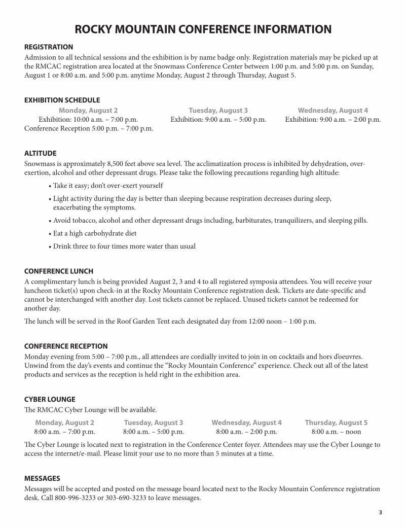
ROCKY MOUNTAIN CONFERENCE INFORMATIONREGISTRATIONAdmission to all technical sessions and the exhibition is by name badge only. Registration materials may be picked up at the RMCAC registration area located at the Snowmass Conference Center between 1:00 p.m. and 5:00 p.m. on Sunday, August 1 or 8:00 a.m. and 5:00 p.m. anytime Monday, August 2 through Thursday, August 5.
EXHIBITION SCHEDULE Monday, August 2 Tuesday, August 3 Wednesday, August 4 Exhibition: 10:00 a.m. – 7:00 p.m. Exhibition: 9:00 a.m. – 5:00 p.m. Exhibition: 9:00 a.m. – 2:00 p.m. Conference Reception 5:00 p.m. – 7:00 p.m.
ALTITUDESnowmass is approximately 8,500 feet above sea level. The acclimatization process is inhibited by dehydration, over-exertion, alcohol and other depressant drugs. Please take the following precautions regarding high altitude:
• Take it easy; don’t over-exert yourself
• Light activity during the day is better than sleeping because respiration decreases during sleep, exacerbating the symptoms.
• Avoid tobacco, alcohol and other depressant drugs including, barbiturates, tranquilizers, and sleeping pills.
• Eat a high carbohydrate diet
• Drink three to four times more water than usual
CONFERENCE LUNCHA complimentary lunch is being provided August 2, 3 and 4 to all registered symposia attendees. You will receive your luncheon ticket(s) upon check-in at the Rocky Mountain Conference registration desk. Tickets are date-specific and cannot be interchanged with another day. Lost tickets cannot be replaced. Unused tickets cannot be redeemed for another day.
The lunch will be served in the Roof Garden Tent each designated day from 12:00 noon – 1:00 p.m.
CONFERENCE RECEPTIONMonday evening from 5:00 – 7:00 p.m., all attendees are cordially invited to join in on cocktails and hors d’oeuvres. Unwind from the day’s events and continue the “Rocky Mountain Conference” experience. Check out all of the latest products and services as the reception is held right in the exhibition area.
CYBER LOUNGEThe RMCAC Cyber Lounge will be available.
Monday, August 2 Tuesday, August 3 Wednesday, August 4 Thursday, August 5 8:00 a.m. – 7:00 p.m. 8:00 a.m. – 5:00 p.m. 8:00 a.m. – 2:00 p.m. 8:00 a.m. – noon
The Cyber Lounge is located next to registration in the Conference Center foyer. Attendees may use the Cyber Lounge to access the internet/e-mail. Please limit your use to no more than 5 minutes at a time.
MESSAGESMessages will be accepted and posted on the message board located next to the Rocky Mountain Conference registration desk. Call 800-996-3233 or 303-690-3233 to leave messages.

4
CONFERENCE-AT-A-GLANCE
SNOWMASS MEETING SPACES
E VENT LOC ATIONSunday Monday Tuesday Wednesday Thursday
a.m. p.m. a.m. p.m. a.m. p.m. a.m. p.m. a.m. p.m.
EPR Lectures Hoaglund
EPR Posters Max Park / Campground
EXHIBITION Carroll / Erickson / Sinclair
NMR Lectures Anderson
NMR Posters Max Park / Campground
Speaker Prep Snobble

5
33RD INTERNATIONAL EPR SYMPOSIUMAugust 2–5, 2010
52nd Rocky Mountain Conference on Analytical ChemistryAugust 1-5, 2010
Snowmass Conference Center - Snowmass, Colorado
CONFERENCE CHAIRKurt W . Zilm
EPR SYMPOSIUM COMMITTEEGlenn Millhauser and Alex Angerhofer (Co-Chairs)
Christoph Boehme, Gail Fanucci, Malcom Forbes,
Gary Gerfen, Howard Halpern, David Tierney
EPR SYMPOSIUM SPONSORSBruker BioSpin, EPR Division
James and Karen Hyde Foundation
Jules Stein Professorship Endowment, UCLA
Medinox, Inc .
National High Magnetic Field Laboratory
Resonance Instruments, Inc .
REGISTRATIONRegister.at.www .rockychem .com
Admission to all technical sessions and the exhibition is by name badge only .
Registration.materials may be picked up at the RMCAC registration area located at the Snowmass Conference Center between 1:00 pm and 5:00 pm on Sunday, August 1 or 8:00 am and 5:00 pm anytime Monday, August 2 through Thursday, August 5 .
Complimentary.lunches are being provided August 2, 3 and 4 to all registered symposia attendees . You will receive your luncheon ticket(s) upon check-in at the Rocky Mountain Conference registration desk . Tickets are date-specific and cannot be interchanged
with another day . Lost tickets cannot be replaced . Unused tickets cannot be redeemed for another day . The lunch will be served each designated day from 12:00 noon until 1:00 pm .

6
EPR SYMPOSIUM Oral Sessions
MONDAY, AUGUST 2, 2010
Session I Pulse Techniques — Gary Gerfen, Chairing
8:20 a.m. Welcoming Remarks. Glenn Millhauser
8:25 a.m. Introduction to Session. Gary Gerfen
8:30 a.m. 101 ApplicationsofPulseDipolarESRtoSolvingStructuresofLargeProteinComplexes. Jack Freed, Cornell University
9:00 a.m. 102 IncreasingSensitivityofHighFieldPulseEPRbyPopulationTransfer,ParallelAcquisitionandNewSpinLabellingSchemes. Daniella Goldfarb, Weizmann Institute of Science
9:30 a.m. 103 DrivingElasticNetworkModelsofProteinsbyEPRDistanceConstraints. Gunnar Jeschke, ETH Zürich
10:00 a.m. Break
10:30 a.m. 104 ParameterEstimationasaProbleminStatisticalThermodynamics. Keith A. Earle, University at Albany
11:00 a.m. 105 RadicalReaction-ProteinDynamicsCouplinginB12EnzymeCatalysis. Kurt Warncke, Emory University
12:00 p.m. Lunch (included with registration)
Session II Proteins — Gail Fanucci, Chairing
1:30 p.m. 106 DEERDistanceMeasurementsBetweenaSpinLabelandNativeFADSemiquinoneinElectronTransferFlavoprotein.Sandra S. Eaton, University of Denver
2:00 p.m. 107 CluesIntoProtein-DNASpecificityDeterminantsbyPulsedESRDistanceMeasurements.Sunil Saxena, University of Pittsburgh
2:30 p.m. 108 ProbingtheStructureofMembraneProteinswithDEERandESEEMSpectroscopy.Gary A. Lorigan, Miami University
3:00 p.m. Break
3:30 p.m. 109 Inversion-recoveryFiltered(IRf)PELDOR:SimplifyingComplexDistanceDistributionsinaNativeMulti-CupricNitriteReductase.Fraser MacMillan, University of East Anglia
4:00 p.m. 110 ControloftheSpeciationandOxidationStatesofRutheniumAnticancerDrugsbyHumanSerumProteins. Charles J. Walsby, Simon Fraser University
4:30 p.m. 111 CharacterizationofIntermediatesBetweenSignalRecognitionParticleanditsReceptorinProteinTargetingPathway. Vinh Q. Lam, California Institute of Technology
5:00–7:00 p.m. Conference Reception
Session III Posters
7:30-9:30 p.m. Authors Present for Posters Labeled A

7
TUESDAY, AUGUST 3, 2010
Session IV Metalloproteins — Joint Session — Dave Tierney, Chairing
8:30 a.m. 112MicrocrystallineParamagneticProteins:Relaxation-OptimizedSequences,Ultra-FastMASandStructuralConstraintsintheSolid-state. Guido Pintacuda, CNRS / Université de Lyon
9:00 a.m. 113EPRSpectroscopyasPartofaCombinedSpectroscopicApproachtoUnderstandElectronicStructureContributionstoReactivityinPyranopterinMolybdenumEnzymesandModels.Martin L. Kirk, The University of New Mexico
9:30 a.m. 114 InvestigationofMetalCentersinProteinsviaCombinedSolid-stateNMRandQMMMMethods. Andrew S. Lipton, Battelle, PNNL
10:00 a.m. Break
10:30 a.m. 115 IntegratedParamagneticResonanceofHigh-SpinCo(II)inBiologicallyRelevantEnvironments. David L. Tierney, Miami University
11:00 a.m. 116 MagicAngleSpinningSolid-stateNMRStudiesofParamagneticProteins.Christopher P. Jaroniec, The Ohio State University
11:30 a.m. 117 MetalloenzymesStudiedbyMultifrequencyEPRandRelatedTechniques. Wolfgang Lubitz, Max-Planck-Institut fuer Bioanorganische Chemie
12:00 p.m. Lunch (included with registration)
Session V Transient Radicals — Malcolm Forbes, Chairing
1:30 p.m. 118 PulsedEPRofTrappedTransientRadicals:ExtendingtheLifetime.Michael K. Bowman, The University of Alabama
2:00 p.m. 119 AvianMagnetoreceptionUsingTransientRadicalsinProteins.Peter J. Hore, University of Oxford
2:30 p.m. Break
3:00 p.m. 120 ESRDetectionofTransientRadicalsusingImmobilizedEnzymes.Bradley E. Sturgeon, Monmouth College
3:30 p.m. 121 ApproachestoSpinTeleportationUsingPhotogeneratedTriradicals. Michael R. Wasielewski, Northwestern University
Session VI Award Lectures
6:30 p.m. PietteLecture. Gary Gerfen
Session VII Posters
7:30-9:30 p.m. Authors Present for Posters Labeled B

8
WEDNESDAY, AUGUST 4, 2010
Session VIII Materials — Christoph Boehme, Chairing
8:30 a.m. 123 OpticallyandElectricallyDetectedMagneticResonanceStudiesofπ-ConjugatedMaterialsandDevices. Joseph Shinar, Iowa State University
9:00 a.m. 124LocalNanoscopicHeterogeneitiesDuringtheThermalCollapseofThermoresponsiveDendronizedPolymersCharacterizedbyEPRSpectroscopy.Hans W. Spiess, Max Planck Institute for Polymer Research
9:30 a.m. 125 ScannedProbeDetectionofElectronSpinResonancefromOrganicRadicals.Eric W. Moore, Cornell University
9:50 a.m. Break
10:20 a.m. 126 EntanglingRemoteNuclearSpinsLinkedbyaChromophore. Brendon W. Lovett, University of Oxford
10:40 a.m. 127 MagneticResonanceinMetalOxideSemiconductorSystems. Patrick Lenahan, Penn State University
11:10 a.m. 128 VanishingofElectron-HoleAsymmetryinNano-SizedChargeOrderedManganitesasReflectedinEPR‘g’Parameters. Subray V. Bhat, Indian Institute of Science
11:40 a.m. 129 Free-ElectronLaser-BasedPulsedEPRat240GHzandBeyond.Mark S. Sherwin, University of California at Santa Barbara
12:00 p.m. Lunch (included w/registration)
1:10 p.m. 130 ObservationofanElectron-onlySpinDependentProcessinOLEDs.William Baker, University of Utah
Session IX Young Investigator — Glenn Millhauser, Chairing
1:30 p.m. 131MultifrequencyENDORSpectroscopyIdentifiesaUniqueIronSiteontheIron-sulphurClusterInvolvedinSubstrateReductionofHeterodisulfideReductase.Alistair J. Fielding, Max-Plank Institute for Biophysical Chemistry
1:45 p.m. 132 OverhauserEffectDynamicNuclearPolarizationfortheMeasurementofHydrationDynamics. John Franck, University of California
2:00 p.m. 133 PulsedElectronSpinResonanceResolvestheCoordinationSiteofCu+2ionsinα1-GlycineReceptor. Sharon Ruthstein, University of Pittsburgh
2:15 p.m. 134 ProbingFlexibilityinPorphyrin-BasedMolecularWiresusingDEER. Janet E. Lovett, University of Oxford
2:30 p.m. 135 EPR,UpCloseandfromAfar:ElucidatingtheMechanisticIntermediatesinCytochromec Oxidasefrom Paracoccus denitrificans. Jessica H. van Wonderen, University of East Anglia
2:45 p.m. Break
3:15 p.m. 136 T2Measurementsat240GHzforNuclearSpinBathEffectsandBiologicalDistanceMeasurement. Devin T. Edwards, University of California at Santa Barbara
3:30 p.m. 137 Spin-dependentProcessesinSilicon-richSilicon-nitrideThinFilmSolarCells.Sang-Yun Lee, University of Utah
3:45 p.m. 138 SpinIncoherenceofDonorElectronsNearc-Si(111)/SiO2InterfaceDefects. Seoyoung Paik, University of Utah
4:00 p.m. 139 AMulti-FrequencyEPRApproachforInvestigatingtheIntrinsicallyDisorderedProtein,IA3.Natasha L. Pirman, University of Florida
4:15 p.m. 140 OrganometallicMechanismsofAction,andInhititionofthe4Fe-4SProteinsGcpEandLytB:APulsedEPRInvestigation.Weixue Wang, University of Illinois at Urbana-Champaign

9
THURSDAY, AUGUST 5, 2010
Session X In Vivo EPR — Howard Halpern, Chairing
8:25 a.m. 141 ExploringtheLimitsofElectronSpinEchoin vivoOxygenImaging.Boris Epel, University of Chicago
8:55 a.m. 142 AProgrammablePulseGeneratorwithNanoSecondResolutionforPulsedEPRApplications.Nallathamby Devasahayam, National Cancer Institute
9:25 a.m. 143 NovelProbesandOpportunitiesforClinicalOximetry. Periannan Kuppusamy, The Ohio State University
9:55 a.m. Break
10:15 a.m. 144 NitroxidesasSensitiveO2ImagingAgentsfor in vivo ElectronParamagneticResonanceImaging.John M.Weaver, University of New Mexico
10:45 a.m. 145 ClinicalEPR:ChallengesandProgress.Harold M. Swartz, Dartmouth Medical School
11:15 a.m. 146FastEPRSpinTrappingofSuperoxideRadicalAnionbyCyclicNitrone-Calix[4]pyrroleConjugate:TheoreticalandExperimentalStudies. Frederick Villamena, The Ohio State University
11:45 a.m. Closing Remarks,Glenn Millhauser, Chair 2010 EPR Symposium.
EPR SYMPOSIUM Poster Sessions
MONDAY AUGUST 2, 20107:30–9:30 p.m. (Poster Session A)
TUESDAY, AUGUST 3, 20107:30–9:30 p.m. (Poster Session B)
A 155 TheSolvationofNitroxideRadicalsinIonicLiquidsStudiedbyHigh-FieldEPRSpectroscopy. Yasar Akdogan, Max Planck Institute for Polymer Research
B 156 ProteinStructureDeterminationfromSparseEPRData. Nathan Alexander, Vanderbilt University
A 157 AtomicHydrogenasHigh-PrecisionFieldStandardforHigh-FieldEPR. Alexander Angerhofer, University of Florida
B 158 IntegratedRefocusedVirtualESEEM:aDeadTimeFreeDetectionofFundamentalLines. Andrei V. Astashkin, University of Arizona
A 159 NewXenonSoftwareModulesforSpinCountingandIsotropicSimulation. David Barr, Bruker BioSpin Corp.
B 160EPRDetectedFreeRadicalFormationFollowingPhoto-ActivationofaCommercialHopProductusedintheBrewingIndustry. David Barr, Bruker BioSpin Corp.
A 161 NitroxylLinewidthsinAqueousSolutionat3Frequencies. Joshua R. Biller, University of Denver

10
B 162ElectricallyActiveDefectsinGateOxidesObservedThroughSpinDependentTrapAssistedTunneling. Brad Bittel, Penn State University
A 163 Site-directedSpin-labelingStudiesoftheCouplingofProteinMotionstoRadicalCatalysisinB12-DependentEthanolamineAmmonia-Lyase. Adonis Bovell, Emory University
B 164 Site-directedSpin-labelingEPRStudiesofMultiply-DisulfideBondedProteinsHIV-1ProteaseandGM2ActivatorProtein. Jeff D. Carter, University of Florida
A 165 EPRSpinProbeCharacterizationoftheCouplingofRadicalReactionChemistryandProteinandSolventMotionsinaB12Enzyme. Hanlin Chen, Emory University
B 166 EntropicParadoxintheProtein-ligandComplexObservedbyFreeze-hyperquenchEPR. Alexey V. Cherepanov, Goethe University
A 167 AdaptiveSignalAveragingTechniqueforEnhancingtheSensitivityofContinuousWaveMagneticResonanceExperimentsII. Corey Cochrane, Penn State University
B 168 HigherResolutionbySkewedProjectionofEchoDetectedEPRSpectra. Alex A. Cruce, University of Alabama
A 169ProbingtheEffectsofPrimaryandSecondaryMutationsontheConformationalSamplingofHumanImmunodeficiencyVirusType1ProteaseSubtypeBbyDoubleElectron-ElectronResonanceSpectroscopy. Ian Mitchelle S. De Vera, University of Florida
B 170 InvestigationoftheCu(II)-bindingPropertiesofAlpha-synuclein. Christopher G. Dudzik, University of California at Santa Cruz
A 171 SpecMan4EPR:AversatilecontrolsoftwareforpulseEPRspectrometers. Boris Epel, University of Chicago
B 172 DistanceMeasurementsinthePrionProteinbyUnnaturalAminoAcidSpin-labelingandDoubleElectron-ElectronResonance(DEER)Spectroscopy. Eric G.B. Evans, University of California, Santa Cruz
A 173 Cu(II)-imidazoleCoordinationStructureintheAmyloid-βProteinofAlzheimer’sDiseaseRevealedby14NESEEMSpectroscopy. William A. Gunderson, Emory University
B 174 InactivingD25NMutationinHIV-1ProteaseAltersProteaseStability,andFlapConformationsandFlexibility. Xi Huang, University of Florida
A 175 TheGlobalAnalysisofDEERData. Eric J. Hustedt, Vanderbilt University
B 176 EffectofGlucoseonSpinLabelEPRinbloodfromHealthyandDiabeticVeins. Asako Kawamori, AGAPE-Kabutoyama Institute of Medicine
A 177NitroxideLineshapeAnalysisWithX-BandPureAbsorptionRapidScan(PARS)ElectronParamagneticResonance(EPR). Aaron W. Kittell, Medical College of Wisconsin
B 178 TestingtheSiteofSubstrateBindingonNitrogenaseCofactorby95MoENDORSpectroscopy. Dmitriy Lukoyanov, Northwestern University
A 179NewApproachesinDiscriminationandCharacterizationofMembraneDomainsUsingEPRSpin-labelingmethods. Laxman Mainali, Medical College of Wisconsin
B 180Membranedomainsinsphingomyelin/cholesterolmembranes:TheirstructureandpropertiesusingEPRSpin-labelingMethods. Laxman Mainali, Medical College of Wisconsin
A 181 Out-of-PhasePELDOR. Andriy Marko, Goethe University
B 182Predictionofthe6,6'-dioxo-3,3'-biverdazylElectronicGroundStatebyDifferenceDedicatedMulti-ReferenceConfigurationInteractionandBrokenSymmetryTechniques. Saba M. Mattar, University of New Brunswick

11
A 183 EPRBasedStructuralBiologyatMiamiUniversity'sOhioAdvancedEPRLaboratory. Robert M. McCarrick, Miami University
B 184 ResonatorforOptimizationofLiquid-PhaseEPRConcentration-SensitivityforSpinLabelsatQ-Band. Richard R. Mett, Medical College of Wisconsin
A 185Physical,ChemicalandMineralogicalCharacterizationofTestMaterialsusedin28-Dayand90-DayIntratrachealInstillationToxicologyStudiesinRats. William J. Miles, Miles Industrial Mineral Research
B 186Understandingtheα-HelicalConformationoftheN-TerminusinIA3UsingSite-DirectedSpin-labelingandElectronParamagneticResonance. Eugene Milshteyn, University of Florida
A 187 ApplyingX-BandRapid-scanEPRtoMeasureShortRelaxationTimes. Deborah G. Mitchell, University of Denver
B 188 DevelopmentofHigh-fieldOverhauser-enhancedMRIwithCircularTransportTechnique. Yukio Mizuta, JEOL LTD.
A 189 EPRforEveryone.GeneratingtheInterest of6-12GradersforMagneticResonance. Reef Morse, Steppingstone MAgnetic Resonance Training (SMART) Center
B 190 IonizingRadiationTreatmentofChromium-DopedSyntheticForsteriteasStudiedbyMultifrequencyEPR. Laila V. Mosina, Kazan Physical-Technical Institute
A 191 StructuralInvestigationofStratumCorneumLipidUsingElectronParamagneticResonance. Kouichi Nakagawa, Fukushima Medical University
B 192DEERandfunctionalmutagenesisindicateaHydrophobicClusterintheForceGenerationRegionoftheMyosinHead,ImportantforMyosinFunction. Yuri Nesmelov, University of North Carolina Charlotte
A 193 ConformationalDistributionsattheN-peptide/boxBRNAInterfaceStudiedUsingSite-directedSpin-labeling. Peter Z. Qin, University of Southern California
B 194 LigandBindingPocketPropertiesofGM2APandSapBbyCWEPR. YongRan, University of Florida
A 195 CW-EPR,ESEEM,andDEERSpectroscopicMeasurementsoftheFullLengthHumanKCNE1MembraneProtein. Indra D. Sahu, Miami University
B 196 AdvancesinLoop-GapResonatorTechnologyforX-bandAqueousSamples. Jason W. Sidabras, Medical College of Wisconsin
A 197ConformationalFlexibilityofElectronTransferFlavoproteinProbedUsingDEERMeasurementsofDistancesBetweenSpinLabelsandaNativeFADSemiquinone. Michael A. Swanson, University of Denver
B 198 ConformationalChangesofSecBUponBindingtoaModelSubstrate–BPTI. Wolfgang E. Trommer, TU Kaiserslautern
A 199 GeneralMethodtoRecoverSlowScanSpectrafromSinusoidalRapidScans. MarkTseytlin, University of Denver
B 200MeasuringtheInfluenceofIronIonsontheMagneticPropertiesinMetal-DopedApatiteNanoparticles. Robert Usselman, NIST
A 201 FrequencyDependenceofSpin-latticeRelaxation. Johan van Tol, Florida State University
B 202 CharacterisationoftheSemiquinoneIntermediateinCytochromebc1Complex. Preethi R. Vennam, The University of Alabama
A 203NewInsightsintothe in-vivo BehaviorofRutheniumAnticancerCompoundsfromEPRMeasurementsofLigandExchangeProcessesandBiomoleculeInteractions. Charles J. Walsby, Simon Fraser University

12
SOLID-STATE NMR SYMPOSIUMAugust 1–5, 2010
52nd Rocky Mountain Conference on Analytical ChemistryAugust 1-5, 2010
Snowmass Conference Center - Snowmass, Colorado
CONFERENCE CHAIRKurt W . Zilm
SOLID-STATE NMR SYMPOSIUM COMMITTEEMei Hong (Chair), Robert Schurko (Chair Elect), Philip Grandinetti (Past Chair)
Zhehong Gan, Gillian Goward, Gerard Harbison, Leonard Mueller, Ulrich Scheler
SOLID-STATE NMR SYMPOSIUM SPONSORSAgilent Technologies
Bruker BioSpin CoretecNet
Environmental Molecular Sciences Laboratory/PNNL Iowa State University
National High Magnetic Field Laboratory Research Coordination Network for NMR of Biological Solids
Revolution NMR, LLC Sigma-Aldrich
Spectra Stable Isotopes Tecmag
Varian, Inc . (now Agilent Technologies) Wiley-Blackwell
REGISTRATIONRegister.at.www .rockychem .com
Admission to all technical sessions and the exhibition is by name badge only .
Registration.materials may be picked up at the RMCAC registration area located at the Snowmass Conference Center between 1:00 pm and 5:00 pm on Sunday, August 1 or 8:00 am and 5:00 pm anytime Monday, August 2 through Thursday, August 5 .
Complimentary.lunches are being provided August 2, 3 and 4 to all registered symposia attendees . You will receive your luncheon ticket(s) upon check-in at the Rocky Mountain Conference registration desk . Tickets are date-specific and cannot be interchanged
with another day . Lost tickets cannot be replaced . Unused tickets cannot be redeemed for another day . The lunch will be served each designated day from 12:00 noon until 1:00 pm .
SSNMR.Evening.Hors.D’oeuvre.Reception: A complimentary reception, sponsored by the SSNMR Vendors, will be held Tuesday Night (cash bar will be open) 5:20 – 7:20 pm in the Fanny Hill Tent .
Sunday.Bruker.Users.Meeting:.To register for the Bruker Users Meeting taking place on Sunday, August 1 access http://www .bruker-biospin .com/rmc2010_nmr .html
Sunday.Varian.Users.Meeting: To register for the Varian Users Meeting taking place on Sunday, August 1 access http://varianinc .com/cgi-bin/nav?/products/nmr/events/solids_2010/index

13
SOLID-STATE NMR SYMPOSIUM Oral Sessions
SUNDAY, AUGUST 1, 2010
Session I Gillian Goward, presiding
7:00 p.m. Opening Remarks, Mei Hong
7:10 p.m. 210 Solid-stateNMRofNanostructuredFunctionalMaterials.Hans W. Spiess,Max-Planck-Institut for Polymer Research
7:40 p.m. 2116Li2DExchangeand1DSelectiveInversionStudiesofSlowlyExchangingLithiumVanadiumFluorophosphates.Linda J.M. Davis, McMaster University
8:00 p.m. 212 DynamicPropertiesofHydrogenBondedPolymerComplexesandMultilayers.Linda Reven, McGill University
8:30 p.m. 213 CharacterizationofPharmaceuticalsUsingSolid-stateNMRSpectroscopy.Eric J. Munson, University of Kentucky
MONDAY, AUGUST 2, 2010
Session II Zhehong Gan, presiding
8:20 a.m. Opening Remarks
8:30 a.m. 214 NovelApproachestoDipolarRecouplingUsingMulitiple-OscillatingFieldandOptimalControlTechniques.Niels Chr. Nielsen, Aarhus University
9:00 a.m. 215 CrystalStructureofTypeIIRedPhosphorusfromFirstPrinciplesandSolid-stateNMR.Maria Baias, University College London
9:20 a.m. 2161HDouble-QuantumBuild-UpCurvesfromDQFiltered1H-13CCorrelationSpectraofIndomethacin-γ.Jonathan P. Bradley, University of Warwick
9:40 a.m. 217 High-ResolutionSolid-stateNMRImagingandMicroscopy.Alan Wong, CEA Saclay
10:00 a.m. Break
10:30 a.m. 218 EfficientDecouplingandRecouplingatVeryHighStaticFieldsandSpinningSpeeds.Piotr Tekely, Ecole Normale Superieure
11:00 a.m. 219 EfficientRotationalEchoDoubleResonanceRecouplingBetweenaSpin-1/2andaQuadrupolarSpinatHighSpinningRatesandWeakIrradiationFields.Amir Goldbourt, Tel Aviv University
11:20 a.m. 220 High-resolutionCryogenicDNP/MAS:InstrumentationandResultsonMembraneProteins.Alexander B. Barnes, Massachusetts Institute of Technology
11:40 a.m. 221 ResolutionandCalibrationofNMRStarkEffectsfromPOWERNMR.Jim Kempf, Rensselaer Polytechnic Institute
12:00 p.m. Lunch (included w/registration)
Session III Ulrich Scheler, presiding
1:30 p.m. 222 LocalStructureoftheOrganic-InorganicNanocompositeinBoneProbedbySolid-stateNMR.Klaus Schmidt-Rohr, Ames Laboratory / Iowa State University

14
2:00 p.m. 223 ProtonDetectionMethodsandApplicationstoMembraneProteinsandFibrils.Andrew J. Nieuwkoop, University of Illinois at Urbana-Champaign
2:20 p.m. 224 StructuralCharacterizationofaHuntingtinN-terminalFragmentinitsFibrillarStatebyMASSolid-stateNMR.Patrick C.A. van der Wel, University of Pittsburgh School of Medicine
2:50 p.m. Break
3:20 p.m. 225 MeasuringandUnderstandingInorganic-OrganicInterfacesUsingSolid-stateNMR.Brad F. Chmelka, University of California, Santa Barbara
3:50 p.m. 226 InteractionsofanAntifreezeProteinwithIceanditsHydrationShellStudiedbySolid-stateNMR.Ansgar B. Siemer, Columbia University
4:10 p.m. 227NMRCrystallographyinanEnzymeActiveSite:CharacterizingtheChemicalStructureofCatalyticIntermediatesinTryptophanSynthase.Leonard J Mueller, University of California, Riverside
4:30 p.m. 228 InvestigatingStructure,DisorderandBondinginInner-EarthMineralsusingMultinuclearSolid-stateNMRandFirst-PrinciplesCalculations.John M.Griffin, University of St Andrews
5:00-7:00 p.m. Conference Reception
Session IV SSNMR Poster Session A
7:30-9:30 p.m. Authors Present for Posters Labeled A
TUESDAY, AUGUST 3, 2010
Session V David Tierney and Gerard Harbison, presiding
8:30 a.m. 229 MicrocrystallineParamagneticProteins:Relaxation-OptimizedSequences,Ultra-FastMASandStructuralConstraintsintheSolid-state.Guido Pintacuda, CNRS / Université de Lyon
9:00 a.m. 230EPRSpectroscopyasPartofaCombinedSpectroscopicApproachtoUnderstandElectronicStructureContributionstoReactivityinPyranopterinMolybdenumEnzymesandModels.Martin L. Kirk, The University of New Mexico
9:30 a.m. 231 InvestigationofMetalCentersinProteinsviaCombinedSolid-stateNMRandQMMMMethods.Andrew S. Lipton,Battelle, PNNL
10:00 a.m. Break
10:30 a.m. 232 IntegratedParamagneticResonanceofHigh-SpinCo(II)inBiologicallyRelevantEnvironments.David L. Tierney, Miami University
11:00 a.m. 233 MagicAngleSpinningSolid-stateNMRStudiesofParamagneticProteins.Christopher P. Jaroniec, The Ohio State University
11:30 a.m. 234 MetalloenzymesStudiedbyMultifrequencyEPRandRelatedTechniques.Wolfgang Lubitz, Max-Planck-Institut fuer Bioanorganische Chemie
12:00 p.m. Lunch (included w/registration)
Session VI Mei Hong, presiding
1:20 p.m. Introduction by Mei Hong
1:30 p.m. 236 From57FeCurtaintoWorldWideWeb.Ago Samoson, Tallinn Warwick
2:20 p.m. 237 HowExceptionalisSolid-stateNMR.Karl T. Mueller, Penn State University

15
3:00 p.m. Break
3:20 p.m. 238 BiologicalSolid-stateNMRatLowTemperatures.Robert Tycko, National Institutes of Health
4:00 p.m. 239 HomonuclearDipolarDecouplingatUltra-FastMagicAngleSpinningFrequencies.Perunthiruthy K. Madhu, TIFR
4:40 p.m. 240 FromCryoMAStoWindFuels.F. David Doty, Doty Scientific
5:20–7:20 p.m. SSNMR Hors D'oeuvre Reception
Session VII SSNMR Poster Session B
7:30–9:30 p.m. Authors Present for Posters Labeled B
WEDNESDAY, AUGUST 4, 2010
Morning a.m. Free Time to Explore the Area
12:00 p.m. Lunch (included w/registration)
Session VIII Len Mueller, presiding
1:30 a.m. 241TheStructureofHumanaB-CrystallinbySolid-stateNMRandSmall-AngleX-ray-Scattering,andSomeExcitingAdventuresWithDNP.Hartmut Oschkinat, Leibniz-Institut für Molekulare Pharmakologie
2:00 p.m. 242 StructureandConformationalHeterogeneityoftheInfluenzaAM2ProtonChannelfromSolid-stateNMR.Sarah Cady, Iowa State University
2:20 p.m. 243Solid-stateNMRStudyoftheMechanismofActionofNovelAmphipathicCationicPeptidesinModelMembranes.Michele Auger, Universite Laval
2:50 PM Break
3:20 p.m. 244FunctionallyTailored,BiogenicInorganicMaterials:ASolid-stateNMRViewonHowNatureDoesIt.Asher Schmidt, Technion – Israel Institute of Technology
3:50 p.m. 245 NMRandEPRStudiesofLungSurfactantOrganization,Structure,andDynamics.Joanna R. Long, University of Florida
4:10 p.m. 246MagicAngleSpinningSolid-stateNMRStructuralStudiesofProteinsModifiedwithParamagneticTags.Ishita Sengupta, The Ohio State University
4:30 p.m. 247 MagicAngleSpinningStudiesofAlpha-SynucleinFibrils.Chad M. Rienstra, University of Illinois
5:00–6:30 p.m. Cortec Wine and Cheese
7:00 p.m. Rocky Mountain Conference Night at Snowmass Rodeo

16
THURSDAY, AUGUST 5, 2010
Session IX Robert Schurko, presiding
8:30 a.m. 248 ParamagneticPerovskites:OrderedNanostructuresinNd2/3-xLi3xTiO3.
Gina L. Hoatson, College of William and Mary
9:00 a.m. 249 Q(n)-speciesDistributionsinAlkaliandAlkalineEarthSilicateGlassesby29Si2DMAFand2DPASSNMR.Michael C. Davis, The Ohio State University
9:20 a.m. 250Solid-stateNMRCharacterizationoftheMorphologyandMotionalDynamicsofPolymericMaterialsforReverseOsmosisWaterPurification.Sungsool Wi, Virginia Polytechnic Institute and State University
9:40 a.m. 251Bromine-79/81andIodine-127Solid-stateNMR:UtilityinStructureRefinementsandObservationofHigher-OrderQuadrupolar-InducedShiftsinMetalHalidesandTheirHydrates.David L. Bryce, University of Ottawa
10:00 a.m. Break
10:30 a.m. 252 OrderandDisorderinSolidsfromHomonuclearandHeteronuclearMQExperiments. Dominique Massiot, CEMHTI – CNRS
11:00 a.m. 25395MoSolid-stateNMRStudyofTransitionMetalClusterCompounds:ASynergeticExperimental&ComputationalApproach.Jérôme Cuny, Ecole Nationale Supérieure de Chimie de Rennes
11:20 a.m. 254 SymmetryPathwaysinSolid-stateNMR.Phillip Grandinetti, The Ohio State University
11:50 a.m. 2011 Vaughan Lecturer Announcement
SOLID-STATE NMR SYMPOSIUM Poster Sessions
MONDAY AUGUST 2, 2010 7:30–9:30 p.m. (Poster Session A)
TUESDAY, AUGUST 3, 2010 7:45–9:45.p .m ..(Poster Session B)
A 265 InvestigatingGold-SulphurinteractioninL-Cysteine/L-CystineCoatedGoldNanoparticlesUsingSolidandLiquid-stateNMR. Anuji Abraham, West Virginia University
B 266 CorrelatingStructuralandElectronicChangesinaPhenalenyl-BasedNeutralRadicalConductorviaSolid-stateNMR. Arun Agarwal, University of California at Riverside
A 267 EvidencefortheCo-existenceofDistortedTetrahedralandTrigonalBipyramidalAluminiumSitesinSrAl12O19From27AlNMRStudies. T.G. Ajithkumar, National Chemical Laboratory
B 268 Solid-stateNMRandCrystallographicStudyofInteractionsinCocrystalsofPeptidesandDenaturants. Benjamin D. Altheimer, Oberlin College
A 269 SSNMRInvestigationofInfluenzaAM218-60. Loren B. Andreas, FBML and Massachusetts Institute of Technology
17
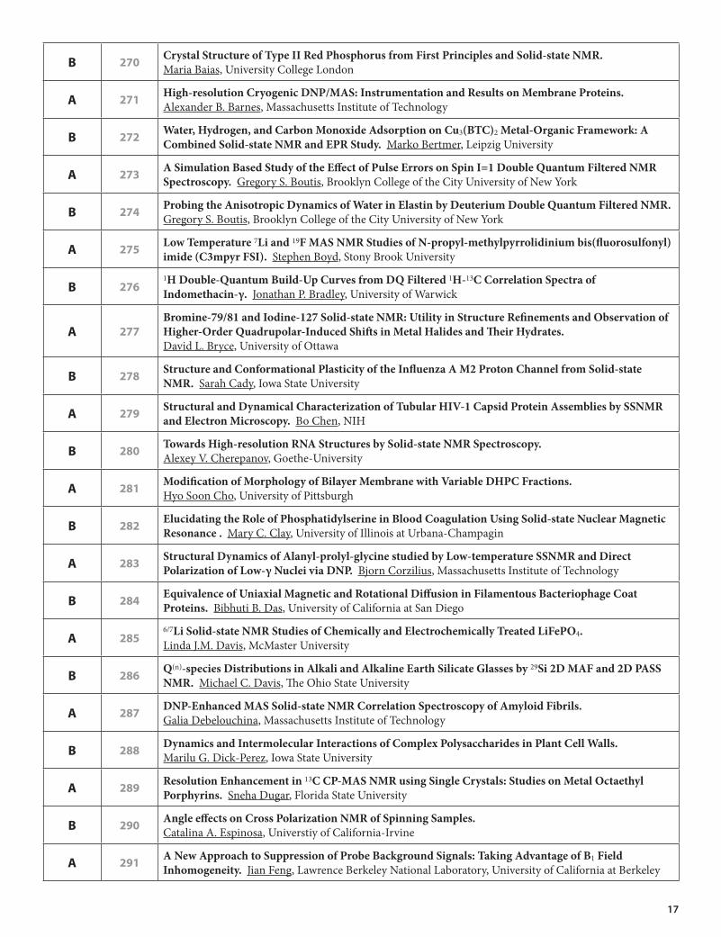
B 270 CrystalStructureofTypeIIRedPhosphorusfromFirstPrinciplesandSolid-stateNMR. Maria Baias, University College London
A 271 High-resolutionCryogenicDNP/MAS:InstrumentationandResultsonMembraneProteins. Alexander B. Barnes, Massachusetts Institute of Technology
B 272 Water,Hydrogen,andCarbonMonoxideAdsorptiononCu3(BTC)2Metal-OrganicFramework:ACombinedSolid-stateNMRandEPRStudy. Marko Bertmer, Leipzig University
A 273 ASimulationBasedStudyoftheEffectofPulseErrorsonSpinI=1DoubleQuantumFilteredNMRSpectroscopy. Gregory S. Boutis, Brooklyn College of the City University of New York
B 274 ProbingtheAnisotropicDynamicsofWaterinElastinbyDeuteriumDoubleQuantumFilteredNMR. Gregory S. Boutis, Brooklyn College of the City University of New York
A 275 LowTemperature7Liand19FMASNMRStudiesofN-propyl-methylpyrrolidiniumbis(fluorosulfonyl)imide(C3mpyrFSI). Stephen Boyd, Stony Brook University
B 2761HDouble-QuantumBuild-UpCurvesfromDQFiltered1H-13CCorrelationSpectraofIndomethacin-γ. Jonathan P. Bradley, University of Warwick
A 277Bromine-79/81andIodine-127Solid-stateNMR:UtilityinStructureRefinementsandObservationofHigher-OrderQuadrupolar-InducedShiftsinMetalHalidesandTheirHydrates. David L. Bryce, University of Ottawa
B 278 StructureandConformationalPlasticityoftheInfluenzaAM2ProtonChannelfromSolid-stateNMR. Sarah Cady, Iowa State University
A 279 StructuralandDynamicalCharacterizationofTubularHIV-1CapsidProteinAssembliesbySSNMRandElectronMicroscopy. Bo Chen, NIH
B 280 TowardsHigh-resolutionRNAStructuresbySolid-stateNMRSpectroscopy. Alexey V. Cherepanov, Goethe-University
A 281 ModificationofMorphologyofBilayerMembranewithVariableDHPCFractions. Hyo Soon Cho, University of Pittsburgh
B 282 ElucidatingtheRoleofPhosphatidylserineinBloodCoagulationUsingSolid-stateNuclearMagneticResonance. Mary C. Clay, University of Illinois at Urbana-Champagin
A 283 StructuralDynamicsofAlanyl-prolyl-glycinestudiedbyLow-temperatureSSNMRandDirectPolarizationofLow-γNucleiviaDNP. Bjorn Corzilius, Massachusetts Institute of Technology
B 284 EquivalenceofUniaxialMagneticandRotationalDiffusioninFilamentousBacteriophageCoatProteins. Bibhuti B. Das, University of California at San Diego
A 2856/7LiSolid-stateNMRStudiesofChemicallyandElectrochemicallyTreatedLiFePO4. Linda J.M. Davis, McMaster University
B 286 Q(n)-speciesDistributionsinAlkaliandAlkalineEarthSilicateGlassesby29Si2DMAFand2DPASSNMR. Michael C. Davis, The Ohio State University
A 287 DNP-EnhancedMASSolid-stateNMRCorrelationSpectroscopyofAmyloidFibrils. Galia Debelouchina, Massachusetts Institute of Technology
B 288 DynamicsandIntermolecularInteractionsofComplexPolysaccharidesinPlantCellWalls. Marilu G. Dick-Perez, Iowa State University
A 289 ResolutionEnhancementin13CCP-MASNMRusingSingleCrystals:StudiesonMetalOctaethylPorphyrins. Sneha Dugar, Florida State University
B 290 AngleeffectsonCrossPolarizationNMRofSpinningSamples. Catalina A. Espinosa, Universtiy of California-Irvine
A 291 ANewApproachtoSuppressionofProbeBackgroundSignals:TakingAdvantageofB1FieldInhomogeneity. Jian Feng, Lawrence Berkeley National Laboratory, University of California at Berkeley

18
B 292 ProbingtheStructureandDynamicsof2Hand13CLabelledPMAAWithinComplexesUsingssNMR. Blythe E. Fortier-McGill, McGill
A 293 DeterminationofChemicalShiftTensorOrientationofAlanine-andGlycine-ContainingTripeptidesUsingRotationalEchoDoubleResonance. Hannah A. Fuson, Oberlin College
B 294 MagicAngleSpinningSolid-stateNMRStudiesofAminoAcid-basedSelf-assembledNanostructures. Min Gao, Ohio State University
A 295 TrimethyltinFluoride:AHigh-Resolution119Sn,13C,and19FSolid-stateNMRStudy. James T. Goettel, University of Lethbridge
B 296 CharacterizationofPOPC/Cholesterol/BMP/GM1ModelMembranesUsing2HNMRand2DExchange31PMASNMR. Philip C. Goff, University of Florida
A 297 EfficientRotationalEchoDoubleResonanceRecouplingBetweenaSpin-1/2andaQuadrupolarSpinatHighSpinningRatesandWeakIrradiationFields. Amir Goldbourt, Tel Aviv University
B 298 MagicAngleSpinningNMRStudiesofClass-IandClass-IIIntactFilamentousBacteriophageViruses. Amir Goldbourt, Tel Aviv University
A 299 CompositeProton-ConductingIonicLiquidElectrolytesforFuelCells. Gillian R. Goward, McMaster University
B 3001HSolid-stateNMRInvestigationofStructureandDynamicsofAnhydrousProtonConductingPolymers. Robert Graf, Max-Planck Institute for Polymer Research
A 301 Solid-stateNMRProbesfortheStudyofMembraneProteinsinHydratedPhospholipidBilayers. Christopher Grant, UCSD
B 302 InvestigatingStructure,DisorderandBondinginInner-EarthMineralsusingMultinuclearSolid-stateNMRandFirst-PrinciplesCalculations. John M. Griffin, University of St Andrews
A 303 ProbingtheStructuralOriginsofVapochromisminPt(di-t-butyl-bipyridyl)(C≡C-C6H4-BMes2)2–a195Pt,13C,11B,2H,1HMultinuclearSolid-stateNMRStudy. Kristopher J. Harris, University of Windsor
B 304 MonitoringTopochemicalPhotochemistryintheSolid-stateinMolecularCrystalsandSupramolecularComplexes. Kimberly Hartstein, Washington University in St. Louis
A 30519FSolid-stateNMR InvestigationintotheStructureandDynamicsofβ-Cyclodextrin/PerfluorooctanoicAcidInclusionComplexes. Paul Hazendonk, University of Lethbridge
B 306 CombiningSolid-stateandHR-MASNMRMethodstoInvestigateConformationalStructureandMobilityofSpiderSilkProteins. Gregory P. Holland, Arizona State University
A 307DeterminationofRelativeTensorOrientationsbyγ-encodedChemicalShiftAnisotropy/HeteronuclearDipolarCoupling3DNMRSpectroscopyinBiologicalSolids. Guangjin Hou, University of Delaware
B 308 SpinDiffusionDrivenbyR-SymmetrySequences:ApplicationsonHomonuclearCorrelationSpectroscopyinMASSolid-stateNMR. Guangjin Hou, University of Delaware
A 309 MechanismofProtonConductionandGatinginInfluenzaAM2ProtonChannelfromSolid-stateNMR. Fanghao Hu, Iowa State University
B 310 CitrateBoundinBoneandtoBoneMineralIdentifiedandCharacterizedbyMultinuclearNMR. Yanyan Hu, Ames Lab, Iowa State University
A 311 DetectionofaTransientIntermediateinaRapidProteinFoldingProcessbySolid-stateNuclearMagneticResonance. Kan-Nian Hu, National Institutes of Health
B 312 NMRandMolecularDynamicsSimulationsCombinedtoCharacteriseMulti-ScaleDynamics. Andy J. Ilott, Durham University
A 313 Modelingthe13CChemicalShiftofPolymorphicPharmaceuticalCompoundswithDFTPlaneWaves. Robbie Iuliucci, Washington and Jefferson College

19
A 314 ThermalDecompositionofFlame-RetardedPolycarbonat/SiliconRubberBlends:ASolid-stateNMRInvestigation. Christian Jaeger, BAM Federal Institute for Materials Research and Testing
B 315 ThepolarphaseofNaNbO3:aCombinedStudybyPowderDiffraction,Solid-stateNMRandFirst-PrinciplesCalculations. Karen E. Johnston, University of St Andrews
A 316 ResolutionandCalibrationofNMRStarkEffectsfromPOWERNMR. Jim Kempf, Rensselaer Polytechnic Institute
B 317 SteadyState,NonlinearCalibration&OrientationDependenceofRFQuadrupolarNMRStarkSpectroscopy. Jim Kempf, Rensselaer Polytechnic Institute
A 318 EPRStudiesofAstaxanthinRadicalsandMetalComplexes. Lowell D. Kispert, The University of Alabama
B 31931PMASNMR-aMethodforAnalysisanDevelopmentofComplexBioceramicsbasedonCa10(K,Na)(PO4)7. Thoralf Krahl, BAM – Federal Institute of Materials Research and Testing
A 320 SurfaceNMRSpectroscopyEnhancedbyDynamicNuclearPolarization. Moreno Lelli, CNRS/ENS-LYON
B 321 EarlyonsetParkinson’sDiseaseMutantandWild-typea-synucleinFibrilsHaveaSimilarFibril-core. Luisel R. Lemkau, University of Illinois at Urbana Champaign
A 322 AnisotropicCollectiveMotionsinCrystallineProteins. Józef R. Lewandowski, Université de Lyon, CNRS / ENS-Lyon / UCB-Lyon 1, Centre de RMN à Très Hauts Champs
B 32313C-2HREDORDistanceMeasurementViaSolid-stateNMR. Wenjing Li, University of Missouri-Kansas City
A 32493Nb-NMRStudyofDion-JacobsonTypeLayeredNiobatesXLaNb2O7(X=Cs,Rb,K,H). Ting Liu, Clark University
B 325 NMRandEPRStudiesofLungSurfactantOrganization,Structure,andDynamics. Joanna R. Long, University of Florida
A 326 Amyloid-betaFibrilsStructureFromHumanAffectedbyAlzheimerDisease. Junxia Lu, NIDDK, National Institute of Health
B 327 OntheNatureofSilica-Bound(Pentafluorophenyl)Propyl:aSolid-stateNMRInvestigation. Kanmi Mao, U.S. DOE Ames Laboratory
A 328 SingleCrystalNMRofPhotoreactedCinnamicAcid:ProductFormation Investigation. Sarah Mattler, Washington University in St. Louis
B 329 Importanceof1H-1HHomonuclearDecouplingin2DNMRCharacterizationofOrganic-InorganicSolids. Robert J. Messinger, University of California, Santa Barbara
A 330 MagicAngleControlinaSwitchedAngleSampleSpinningProbe. Eugene Mihaliuk, West Virginia University
B 331 NMRStudiesofEnhancedNuclearPolarizationinInP. Joel B. Miller, Naval Research Laboratory
A 332 EffectsofElectricalandIonicConductivityonMAS-NMRofQuadrupolarNucleiinγ-CuprousIodide. Joel B. Miller, Naval Research Laboratory
B 333 InvestigatingDisorderinPyrochloreMaterialsbyMASNMRandFirstPrinciplesCalculations. Martin R. Mitchell, University of St. Andrews
A 334 ProbingMolecularInteractionsResponsiblefortheβ-hairpinStructureinβ-amyloidPeptideAssociatedwithAlzeimer’sDisease. Venus S. Mithu, Tata Institiute of Fundamental Research
B 335UnexpectedAluminiumandOxygenCoordinationinGlassyandCrystallineBaAl4O7Samples,EvidencedbyPowderDiffractionandHigh-resolutionNMRExperiments. Valerie Montouillout, CEMHTI-CNRS

20
A 33633SSolid-stateNMRandFirstPrinciplesCalculationsinInorganicSulfates. Igor L. Moudrakovski, National Research Council
B 337NMRCrystallographyinanEnzymeActiveSite:CharacterizingtheChemicalStructureofCatalyticIntermediatesinTryptophanSynthase. Leonard J Mueller, University of California, Riverside
A 338 SamplePreparationand2DSolid-stateNMRStudiesoftheFP-HairpinConstructofgp41. Matthew J. Nethercott, Michigan State University
B 339 Solid-stateNMRInvestigationsofParamagneticJarosites,KB3(SO4)2(OH)6);B=V(III),Cr(III),Fe(III). Ulla Gro Nielsen, University of Southern Denmark
A 340 ProtonDetectionMethodsandApplicationstoMembraneProteinsandFibrils. Andrew J. Nieuwkoop, University of Illinois at Urbana-Champaign
B 341125TeNMRofComplexTellurides. Bosiljka Njegic, Iowa State University – Ames Laboratory
A 342 Ultra-wideline14NNMRasaProbeofMolecularStructureandDynamics. Luke A. O’Dell, Steacie Institute for Molecular Sciences
B 343 DipolarDecouplinginSolid-stateNMR. Subhradip Paul, Tata Institute of Fundamental Research
A 344 DynamicNuclearPolarizationat263GHz:ExperimentalMethodsandApplications. Shane Pawsey, Bruker BioSpin
B 345 ApplicationofAdiabaticPulsestoParamagneticSolids. Andrew J. Pell, ENS-Lyon
A 346 TheEffectsofTemperatureontheDynamicsofaMicrocrystallineSH3DomainasObservedbyMASNMR. Alexey Potapov, National Institute of Diabetes and Digestive and Kidney Diseases
B 34733SSolid-stateNMRandFirstPrinciplesCalculations. Thomas Poumeyrol, CEMHTI – CNRS UPR3079
A 348 Solid-stateNMRInvestigationsofAluminaCatalystSupports. Sesh Prabhakar, UOP, a Honeywell Co.
B 349SelectiveFormation,MorphologicalCharacteristicsandArchitecturalFeatureofParallelandAnti-parallelβSheetStructuresforIowaMutantBeta-amyloidFibrils. Wei Qiang, National Institutes of Health
A 350 TwoInterfacialWaterLayersinBoneLocalizedbySpinDiffusion. Aditya Rawal, Ames laboratory- Iowa State University
B 351 HomonuclearDecouplingforHigh-ResolutionProtonSolid-stateNMRwithVeryFastMAS. Elodie Salager, Universite de Lyon
A 352 ChainPackinginGlassyPolymersbyNatural-Abundance13C-13CSpinDiffusionUsing2DCODEX. Jacob Schaefer, Washington University
B 353 PolymersUnderMechanicalStress—aLow-fieldNMRInvestigation. Ulrich Scheler, Leibniz Institut für Polymerforschung Dresden e.V.
A 354 CharacterizationofMicroencapsulatedScandiumComplexesbyMulti-nuclearSolid-stateNMR. Robert W. Schurko, University of Windsor
B 355 MagicAngleSpinningSolid-stateNMRStructuralStudiesofProteinsModifiedWithParamagneticTags. Ishita Sengupta, The Ohio State University
A 356 AStudyoftheAtomicMotionsofLiBH4inCarbonNanostructures. David T. Shane, Washington University in St. Louis
B 357 StructuralInsightsintotheMechanismforToxicityofTrichothecenesT-2andDeoxynivalenol. Roxanne A. Shank, University of Lethbridge

21
A 358 ThermalStabilizationofDMPC/DHPCBicellesbyAdditionofCholesterolSulfate. Rebecca A. Shapiro, University of California, Irvine
B 359 InteractionsofanAntifreezeProteinwithIceanditsHydrationShellStudiedbySolid-stateNMR. Ansgar B. Siemer, Columbia University
A 360 ExaminingDNPPolarizationTransferMechanismsviaEnhancementandBuildupTime. Albert A. Smith, Massachusetts Institute of Technology
B 361 MomentAnalysisofQuadrupolarSidebandPatterns. Luis J. Smith, Clark University
A 362TowardsaBetterUnderstandingofComplexGlassesStructures:ACombinationofMultinuclearSolid-stateNMRExperimentsandComputationalAnalysis. Anne Soleilhavoup, CEA Saclay
B 363 High-PotentialCathodeMaterialsforLithiumIonBatteries. Leigh Spencer, McMaster University
A 364 MagicAngleSpinningSolid-stateNMRStudiesofa41-kDaDsbA/DsbBMembraneProteinComplex. Lindsay J. Sperling, University of Illinois at Urbana-Champaign
B 365 StructuralStudiesofMammalianDynactinCAP-GlyDomainbySolid-stateNMR. Shangjin Sun, University of Delaware
A 366 SpectralEditingMethodsEmployingHomonuclear1HDecoupling:TowardsBetterCharacterisationofPharmaceuticalSolids. Andrew S. Tatton, University of Warwick
B 367Solid-state17ONMRStudyof Tris(4-methoxyphenyl)phosphineoxide-17Oand Indium(III)triiodidebis(tris(4-methoxyphenyl)phosphineoxide-17O. Rosha Teymoori, University of Alberta
A 368CarbonSequestrationMechanismsofAntigoriteandForsteriteinSupercriticalCO2andH2OStudiedbySolid-state13Cand29SiNMRSpectroscopy. Flaviu R.V. Turcu, Pacific Northwest National Laboratory
B 3691HCSAand1H-15NDipolarInteractionsofAmideNHGroupsMeasuredbySymmetry-basedMASPulseSequences. Alexander J. Vega, University of Delaware
A 370 DeterminationofWaterAscentVelocityinEmbolizedXylemVesselsofGrapevineStemsUsing1HNMRMicroscopy. Mingtao Wang, University of Alberta
B 371 OpticalPumpingPhenomenainsi-GaAs. Dustin Wheeler, Washington University in St. Louis
A 372Solid-stateNMRCharacterizationoftheMorphologyandMotionalDynamicsofPolymericMaterialsforReverseOsmosisWaterPurification. Sungsool Wi, Virginia Polytechnic Institute and State University
B 373 High-ResolutionSolid-stateNMRImagingandMicroscopy. Alan Wong, CEA Saclay
A 374 StructuralInvestigationofLead-BoroaluminateandBorogallateGlassesUsingMultinuclearMagneticResonance. John E.C. Wren, University of Manitoba
B 375 BackboneDynamicsStudiesofMammalianDynactinCAP-GlyDomainbySolid-stateNMR. Si Yan, University of Delaware
A 376 UsingDeuteriumMASNMRtoElucidatePlasticizationandBackboneDynamicsinSpiderSilk. Jeff Yarger, Arizona State University
B 377InternalStructureDeterminationoftheFibrilsFormedbyC-terminalFragmentsofBeta2MicroglobulinbyApplyingSSNMR. Chi Zhang, University of Missouri-Kansas City
A 378 SimpleAnalyticFormalismAccountsfor13Cand15N T1RelaxationofSolidProteinsandPeptidesUnderMAS. Kurt W. Zilm, Yale University

22
EPR SYMPOSIUM Oral Sessions
101 ApplicationsofPulseDipolarESRtoSolvingStructuresofLargeProteinComplexes. Jack Freed Cornell University
Pulsed dipolar ESR (PDS) is emerging as a technique in structural biology that bridges the resolution gap between x-ray crystallography/NMR and cyro-electron microscopy. We will demonstrate this with two examples and indicate the challenges to PDS. Bacterial chemotaxis refers to the mechanism of bacterial movement in response to gradients of nutrients and repellents. One of the fundamental questions of the complex assembly of proteins is the ternary structure of the signaling complex of the multi-domain CheA dimer, two CheW’s (the adaptor protein), and the receptor dimer. Whereas each individual sub-unit could be studied by crystallography or NMR, neither technique can address this six protein complex. However, we have succeeded for the first time in determining the structure of this complex. We have shown that the receptor binds and stabilizes the regulatory domains of CheA. Our direct distance measurements by PDS between the P3 domain of CheA and receptor have shown that the two interact with their helical axes running anti-parallel to each other. Alpha-synuclein (αS) is a presynaptic protein that participates in synaptic strength maintenance and dopamine homeostasis, but accumulation of αS amyloid fibrils is associated with Parkinson’s disease. In previous PDS distance measurements of αS bound to micelles we showed that it bends to form two linked helices to surround the micelles. We also used PDS to measure large distances (up to 8.7 nm) in αS bound to lipid vesicles, rod-like micelles, and isotropic lipid bicelles, all of which present the protein with a more extensive, less highly curved surface than spheroidal micelles. Distances measured for αS between labels are in close agreement with those expected for a single continuous helix, which argues strongly for a single, unbroken helix. Conditions which favor one or the other conformer will be discussed. Supported by NCRR Grant P41-RR016242 and NIBIB Grant 2R01EB003150.
EPRORALSESSIONJack H. Freed, Department of Chemistry and Chemical Biology and National Biomedical Center for Advanced ESR Technology (ACERT), Cornell University, Ithaca, NY 14853, USA Tel: 607-255-3647, E-mail: [email protected]
102 IncreasingSensitivityofHighFieldPulseEPRbyPopulationTransfer,ParallelAcquisitionandNewSpinLabellingSchemes. Daniella Goldfarb Weizmann Institute of Science
Limited sensitivity has always been an issue in pulse EPR and in many cases has prevented its applications, particularly in biological systems, Measurements at high fields, in principle, improve the absolute sensitivity. The extent of this improvement, however depends on the nature of the sample and the experiment. Here we describe three different approaches to sensitivity enhancement for pulse EPR at high fields, demonstrated on our W-band (95 GHz) home built spectrometer. The first concerns a new acquisition scheme that allows recording up to 10 orientation selective pulse EPR spectra in parallel by applying rapid field jumps within the relaxation delay between consecutive pulse sequences. The second is the use of Gd3+ (S=7/2) based spin labels for distance measurements in proteins. The third applies for half integer high spin system and involved populations transfer from low lying Ms states to the MS=-1/2 by adiabatic inversion of low lying transitions.
EPRORALSESSIONDaniella Goldfarb, Weizmann Institute of Science, Chemical Physics, Hertzl St., Rehovot, 76100, Israel E-mail: [email protected]
ABSTRACTS

23
103 DrivingElasticNetworkModelsofProteinsbyEPRDistanceConstraints. G. Jeschke and E. Bordignon,ETH Zürich, Laboratory for Physical Chemistry
Large-scale structural transitions are a key element of protein function, for instance in substrate uptake and release by enzymes or in substrate translocation by membrane transporters. Systematic experimental approaches for characterization of such transitions are lacking. In those cases where several structures of a protein or protein complexes in different states could be obtained,1 structural changes are highly collective, i.e. large groups of residues move as rigid or almost rigid domains or subdomains. This suggests that the effective number of degrees of freedom is small. Indeed, it was found that such transitions can be modeled rather well by only about 10 to 20 normal modes of simple elastic network models that are based on just the Cα coordinates of the structure in one state.2 It should be possible to quantify movement along such a small number of degrees of freedom by a similar number of distance constraints. Algorithms that suggest 10 pairs of residues for this quantification and for determining the transition pathway and final structure from the distances between Cα atoms of these residues exist.3 Closer examintaion shows that these distances generally fall in the range accessible by site-directed spin labeleing EPR, with most of them being accessible by pulsed EPR and a few by continuous-wave EPR. The contribution discusses extension of the algorithm to label-to-label distances, in silico tests, and preliminary results for concerted motion of the P2 loop and maltose binding protein in maltose ABC importer. Supported by SNF 200021_121579.
1. http://www.molmovdb.org/ 2. I. Bahar, T.R. Lezon, A. Bakan, I.H. Shrivastava, Chem. Rev., 2010, 110, 1463.3. W. Zeng, B.R. Brooks, Biophys. J., 2006, 90, 4327.
EPRORALSESSIONGunnar Jeschke, ETH Zürich, Lab. Phys. Chem., Wolfgang-Pauli-Strasse 10, Zürich, 8093, Switzerland E-mail: [email protected]
104 ParameterEstimationasaProbleminStatisticalThermodynamics. Keith A. Earle1 and David J. Schneider2
1. University at Albany, Physics Department, 1400 Washington Ave., Albany, NY 12222 2. USDA Agricultural Research Service and Dept. of Plant Pathology, Cornell University, Ithaca, NY 14853
In this work, we explore the connections between parameter fi tting and statistical thermodynamics using the maxent principle of Jaynes as a starting point. In particular, we show how signal averaging may be described by a suitable one particle partition function, modi ed for the case of a variable number of particles. These modifications lead to an entropy that is extensive in the number of measurements in the average. Systematic error may be interpreted as a departure from ideal gas ehavior. In addition, we show how to combine measurements from different experiments in an unbiased way in order to maximize the entropy of simultaneous parameter tting. We suggest how fit parameters may be interpreted as generalized coordinates and the forces conjugate to them may be derived from the system partition function. From this perspective, the parameter fitting problem may be interpreted as a process where the system (spectrum) does work against internal stresses (non-optimum model parameters) to achieve a state of minimum free energy/maximum entropy. We introduce a suitable definition of volume that allows one to defi ne compressibilities and thus obtain further insights into the fi tting process from classical thermodynamics. Finally, we show how the distribution function allows us to de ne a geometry on parameter space, building on previous work.1,2 This geometry has implications for error estimation and we outline a program for incorporating these geometrical insights into an automated parameter tting algorithm.
1. K.A. Earle, L. Mainali, I.D. Sahu, and D.J. Schneider. Magnetic Resonance Spectra and Statistical Geometry. Appl. Magn. Reson., 37:865--880, 2009.
2. K.A. Earle, L. Mainali, I.D. Sahu, and D.J. Schneider. Estimating the Parameter Sensitivity of Spectra via Analytical Derivatives. In preparation.
EPRORALSESSIONKeith A. Earle, University at Albany, Physics Department, 1400 Washington Ave, Albany, NY, 12222, USA Tel: 518-442-4521, E-mail: [email protected]

24
105 RadicalReaction-ProteinDynamicsCouplinginB12EnzymeCatalysis. Chen Zhu, Hanlin Chen, Adonis Bovell and Kurt WarnckeEmory University, Department of Physics
The modulation of adiabatic reaction chemistry by protein dynamics is addressed in the ethanolamine ammonia-lyase from Salmonella typhimurium by using techniques of EPR spectroscopy. The transient decay reaction kinetics of the cryotrapped CoII-substrate radical pair catalytic intermediate are measured by using time-resolved, full-spectrum X-band continuous-wave EPR spectroscopy in frozen bulk aqueous solution, upon annealing over the temperature range of 190-223 K.1 The decay kinetics for 190≤T≤207 K represent two isolated populations (fast decay population: normalized amplitude=0.57 ±0.04; observed rate constant, kobs,f=7.3×10⁻⁵ – 1.5×10⁻³ s-1; slow decay population: kobs,s=5.2×10⁻⁶ – 2.9×10⁻⁴ s-1). Substrate 1H/2H isotope effects show that the decay is rate-limited by the radical rearrangement.2 Thus, the measurements probe the core reaction of the enzyme. Electron-electron (EPR) and electron-nuclear (ESEEM) distance determinations show no evidence for significant structural differences among the nuclear centers of the reactants in the protein active site region, for the fast and slow decay populations. At 207<T<214 K, the slow phase decay rate merges with the fast phase rate, and effective activation parameters from the detailed temperature dependence suggest an origin in a protein dynamical transition. To further characterize the modulation of reactivity by the protein, measurements of the decay at T<190 K are being used to detect and characterize rate determination by protein dynamics. In parallel, Site-directed spin-labeling is being used to correlate protein motional properties with the decay reaction, over the full temperature range. Supported by NIDDK/NIH DK54514.
1. Zhu, C., Warncke, K., Biophys. J. 2008, 95, 5890.2. Zhu, C., Warncke, K., J. Am. Chem. Soc. 2010, submitted.
EPRORALSESSIONKurt Warncke, Emory University, Physics, N201 Mathematics and Science Center, 400 Dowman Dr., Atlanta, GA 30322, USA Tel: 404-727-2975, E-mail: [email protected]
106 DEERDistanceMeasurementsBetweenaSpinLabelandNativeFADSemiquinoneinElectronTransferFlavoprotein.Michael A. Swanson,1 Velavan Kathirvelu,1 Tomas Majtan,2 Frank E. Frerman,2 Gareth R. Eaton1 and Sandra S. Eaton1
1. Department of Chemistry and Biochemistry, University of Denver, Denver, CO 80208 2. Department of Pediatrics, University of Colorado School of Medicine, Aurora, CO 80045
The mitochondrial protein electron transfer flavoprotein (ETF) accepts electrons from at least 10 different flavoprotein dehydrogenases and transfer electrons to a single electron acceptor in the inner membrane. It has been proposed that mobility of the αII domain permits the promiscuous behavior of ETF with respect to a variety of redox partners. ETF contains a single redox center, FAD, in the αII domain. Cysteine mutations were introduced at A43 in domain I, A210 in domain II, or A111 in domain III and spin labeled with MTSL. In the as-isolated protein the FAD is diamagnetic. We have demonstrated that the FAD can be enzymatically reduced to semiquinone, without destroying the nitroxyl spin label.1
The distances between the spin label and the FAD semiquinone were measured by DEER at X-band and Q-band. The distributions of distances found for each of the labeling sites will be discussed.
1. M.A. Swanson, V. Kathirvelu, T. Majtan, F.E. Frerman, G.R. Eaton, and S.S. Eaton, J. Amer. Chem. Soc. 131, 15978-15979 (2009).
EPRORALSESSIONSandra S. Eaton, University of Denver, Chemistry and Biochemistry, 2101 E. Wesley Ave., Denver, CO, 80208, USA Tel: 303-871-3102, E-mail: [email protected]
107 CluesIntoProtein-DNASpecificityDeterminantsbyPulsedESRDistanceMeasurements.Zhongyu Yang1, Ming Ji1, Jessica Sarver1, Preeti Mehta2, J. Townsend2 L. Jen-Jacobson2 and Sunil Saxena1
1. Department of Chemistry, University of Pittsburgh, Pittsburgh, PA 15260 2. Department of Biological Sciences, University of Pittsburgh, Pittsburgh, PA 15260
Restriction endonuclease EcoRI binds to the specific DNA sequence GAATTC with an affinity that is 50,000-90,000-fold greater than that of a miscognate site that differs by only one base pair. Even lower binding affinity is also exhibited at non-specific binding sites which differ from the specific sequence by two or more base pairs. In the presence divalent metal ions, such as magnesium, EcoRI the specific sequence of viral DNA with a high specificity. Other ions, such as copper ions, do not support the catalysis by themselves. Nitroxide based distance measurements on several spin labeled EcoRI mutants bound to specific, miscognate, and non-specific sequences of DNA demonstrate that on average the arms of EcoRI, thought to play a major role in binding specificity, are similarly positioned. Additionally, noncognate (miscognate and non-specific)

25
complexes demonstrated broader distance distributions indicating that the flexibility of the arms plays a large role in binding specificity. In order to gain insight into the role of metal ions, pulsed ESR was used to deduce the coordination of copper ions in EcoRI1. The Electron Spin Echo Envelope Modulation (ESEEM) experiments revealed that copper is coordinated to one of the five histidine residues in EcoRI. In order to determine this copper binding histidine, copper ion based distance measurements were performed using Double Electron Electron Resonance (DEER). Molecular models were developed to extract the copper-copper and copper-nitroxide distances from the DEER data. This work established key aspects of paramagnetic metal-ion based distance measurements. A triangulation procedure based on the copper-copper and copper-nitroxide distances demonstrated that copper binds to histidine 114 in EcoRI. In support of a role for His114 in binding Cu2+, biochemical assays show that the mutant H114Y-DNA complex binds with 1600-fold lower affinity than the wt-DNA complex. Additionally, to our astonishment, we observed a 1600-fold enhancement of the Mg2+ (0.5mM)-catalyzed rate in the presence of a saturating concentration Cu2+. The novelty of these observations lies in the fact that the second metal is not coordinated to the scissile phosphate, and thus cannot act by facilitating protonation of the leaving group or by (directly) stabilizing the pentacovalent phosphate intermediate. In other words, we have discovered a novel accelerant chemistry for nuclease catalysis. This work is supported by NSF (MCB 0842956)
EPRORALSESSIONSunil Saxena, University of Pittsburgh, Chemistry, 219 Parkman Avenue, Pittsburgh, PA, 15260, USA Tel: 412-624-8680, E-mail: [email protected]
108 ProbingtheStructureofMembraneProteinswithDEERandESEEMSpectroscopy.Gary A. Lorigan, Dan Mayo, Hari Ghimire, Indra D. Sahu, Aaron Coey, and Robert McCarrick,Miami University, Department of Chemistry and Biochemistry
Currently, very limited structural and dynamic information on membrane proteins and peptides exist. New biophysical/structural biology methods are needed to probe these systems in a lipid bilayer. The Lorigan lab is applying unique hybrid NMR and spin-label EPR spectroscopic techniques to study membrane proteins. Magnetic resonance spectroscopic data of 15N-, 2H-labeled and/or spin-labeled membrane proteins incorporated into vesicles and bicelles will be presented. State-of-the-art pulsed EPR techniques such as Electron Spin Echo Envelope Modulation (ESEEM) spectroscopy, and Double Electron-Electron Resonance (DEER) spectroscopy will be used. The ESEEM technique can determine short to medium range distances (out to about 9 Å) between a site-specific nitroxide spin label and a nearby NMR-active isotopic labeled residue for a variety of different peptides and proteins which ultimately can be used to determine the difference between an α-helical and β-sheet secondary structure. DEER can be used to measure distances between 2 spin labels out to about 70 Å. We have shown a huge improvement is sensitivity with DEER measurements at Q-band when compared to X-band.1
1. Ghimire et al., Biochem., 2009, 48, 5782.
EPRORALSESSIONGary A. Lorigan, Miami University, Department of Chemistry and Biochemistry, 701 E. High St., Oxford, OH, 45056, USA Tel: 513-529-3338, E-mail: [email protected]
109 Inversion-recoveryFiltered(IRf)PELDOR:SimplifyingComplexDistanceDistributionsinaNativeMulti-CupricNitriteReductase.Jessica H. van Wonderen1, Doritz N. Kostrz2, Chris Dennison2 and Fraser MacMillan1
1. Henry Wellcome Unit for Biological EPR, School of Chemistry, University of East Anglia, Norwich, NR4 7TJ, UK 2. Institute for Cell and Molecular Biosciences, Newcastle University, UK
The copper-containing nitrite reductase (CuNIR) is a key enzyme in the respiratory pathway of denitrifying bacteria, in which nitrate is reduced to gaseous products (NO, N2O and N2)1. The CuNIR from Achromobacter xylosoxidans (AxNiR) is trimeric with each monomer consisting of two spectroscopically different copper atoms; a mononuclear electron transferring Type 1 copper site, known as a ‘blue’ copper due to its typical intensely blue colour, and a catalytic Type 2 copper site, where nitrite binds and is reduced to nitric oxide.
Pulsed electron electron double resonance (PELDOR) has been used to determine intra-molecular distances between both Type 1 and 2 Cu(II)s in this copper nitrite reductase. Intra-molecular distances of 3.0, 3.5, 4.0 and 4.3 nm were obtained which are comparable to those determined in the X-ray crystal structure and those predicted by MMM.
The Type 1 and Type 2 Cu(II)s in CuNIR have overlapping electron paramagnetic resonance (EPR) signals. However, inversion-recovery EPR experiments show that their T1 spin-lattice relaxation times are different enough to deconvolute these signals by using REFINE2. By measuring field-swept spectra at different filter times (TF) and comparing to simulated spectra, we can decide which TF times can remove the effect of either Type 1 or Type 2 copper. As a way of simplifying

26
complicated PELDOR spectra in large biomolecules, we have developed a technique called inversion-recovery filtered (IRf) PELDOR to remove distances one at a time. We demonstrate this method here using different TF times for Type 1 and 2 Cu(II)s to selectively remove distances in this complex system.
1. K. Sato, S.J. Firbank, C. Li, M.J. Banfield and C. Dennison, Chem. Eur. J. 2008, 14, 5820-58282. T. Maly, F. MacMillan, K. Zwicker, N. Kashani-Poor, U. Brandt and T. Prisner, Biochemistry, 2004, 43, 3969-3978
EPRORALSESSIONFraser MacMillan, Henry Wellcome Unit of Biological EPR, School of Chemistry, University of East Anglia, Norwich, NR4 7TJ, UK E-mail: [email protected]
110 ControloftheSpeciationandOxidationStatesofRutheniumAnticancerDrugsbyHumanSerumProteins. Michael I. Webb, Changhua Mu, Naniye Cetinbas, Joshua A. Dubland, Charles J. WalsbyDepartment of Chemistry, Simon Fraser University
Ruthenium (III) complexes are the most promising metal-based alternatives to platinum anticancer drugs, with two of these compounds currently in phase II clinical trials. Both of these compounds are members of the “Keppler-type” family of Ru(III) compounds, which are comprised of kinetically-inert azole ligands and exchangeable chlorides. Despite recent intensive research into the activity of these molecules, surprisingly little is known about their in vivo behavior. Using EPR methods, we have probed interactions of the two most promising drug candidates, indazolium [trans-RuCl4(1H-indazole)2] (KP1019) and imidazolium [trans-RuCl4(1H-imidazole)(DMSO-S)] (NAMI-A), and a number of their analogues, with human serum and have found that protein binding is critical to controlling both the oxidation-state of the ruthenium centers and ligand-exchange processes. Binding to human serum proteins such as albumin (hsA) and transferrin is thought to be a key factor in the efficient, selective delivery of these complexes to tumor cells. Our EPR studies demonstrate that the dominant species in serum are the aquated complexes, and also that binding to serum proteins occurs both through ligand exchange and via hydrophobic interactions mediated by the heterocyclic ligands found in these complexes. The latter process is strongly dependent on the identity of the azole ligands and their ability to interact with the hydrophobic binding domains of hsA. In the case of KP1019, the indazole ligands facilitate rapid binding to hsA, implicating this process in the low toxicity of this compound due to rapid sequestering of the complex after intravenous infusion. NAMI-A also shows a high tendency for protein binding, but in addition, we observe that binding to hsA limits reduction of the Ru(III), which has implications for the anticancer mechanism of this compound. These studies provide insight into the true active species present in human patients and are facilitating our design of new anticancer agents.
EPRORALSESSIONCharles J. Walsby, Simon Fraser University, Chemistry, 8888 University Drive, Burnaby, BC, V5A 1S6, Canada Tel: 778-782-4607, E-mail: [email protected]
111 CharacterizationofIntermediatesBetweenSignalRecognitionParticleanditsReceptorinProteinTargetingPathway. Vinh Q. Lam, Xin Zhang and Shu-ou Shan,California Institute of Technology, Division of Chemistry and Chemical Engineering
Analogous to the protein folding process that involves intermediates before reaching native structure, many protein-protein interactions involve the formation of transient intermediates before reaching the final complex. Elucidation of the properties of these intermediates is important for understanding how proteins interact with one another. Here, we characterized the structural and dynamic properties of an early intermediate during complex assembly between the signal recognition particle (SRP) and SRP receptor (SR), by using a combination of mutational analysis, time resolved fluorescence lifetime, and SDSL-EPR. We demonstrate that the early intermediate shares overlapping but not identical interaction surfaces with the final complex, has a broad conformational distribution, and its conformational dynamics can be further regulated by biological regulators of SRP. These results support a model in which intermediates during protein-protein interactions are analogous to molten globules during protein folding, with the binding partners sampling different conformations and relative orientations before achieving the final stable complex. Supported by NIH GM078024, the Packard foundation, and the Burroughs Welcome Travel Fund.
EPRORALSESSIONVinh Q. Lam, California Institute of Technology, Chemistry and Chemical Engineering, 1200 East California Blvd., Pasadena, CA, 91125, USA Tel: 626-395-4071, E-mail: [email protected]

27
112 MicrocrystallineParamagneticProteins:Relaxation-OptimizedSequences,Ultra-FastMASandStructuralConstraintsintheSolid-state. Guido Pintacuda, CNRS / Université de Lyon
We present our recent advances in the structural investigation by solid-state magic angle spinning (MAS) NMR of microcrystalline paramagnetic proteins.
First, we explore the impact of so-called ultrafast (>60 kHz) MAS in the characterization of biomolecular solids containing paramagnetic centers. We discuss a set of experiments based on low-power rf irradiation to observe and assign, in highly paramagnetic proteins (the dimeric Cu(II),Zn(II)-superoxide dismutase and the Co(II)-replaced catalytic domain of matrix metalloproteinase 12), 13C and 1H resonances from the residues coordinating the metal center. In addition, by exploiting the enhanced relaxation caused by the paramagnetic center, and the low power irradiation enabled by the fast MAS, this can be achieved in remarkably short times and at high-field, with only less than 1 mg of sample.1 Second, to gain access to crowded spectral regions, we describe the use of relaxation-optimized methods for 13C-13C spin-state selection, which remove the broadening due to the 13C-13C J couplings and lead to a considerable enhancement in both resolution and sensitivity in 2D and 3D dipolar and scalar correlations.2-3 Finally, we show how the quantitative evaluation of some of the paramagnetic effects can unveil precious structural information that integrate “traditional”, diamagnetic distance measurements in the full macromolecular structure determination.
1. I. Bertini, L. Emsley, C. Luchinat, M. Lelli, Y. Mao and G. Pintacuda “Ultra-Fast MAS Solid-state NMR Permits Extensive 13C and 1H Detection in Paramagnetic Metalloproteins”, J. Am. Chem. Soc., 2010, 132, 5558-5559.
2. S. Laage, R. Pierattelli, I.C. Felli, I. Bertini, A. Lesage, L. Emsley and G. Pintacuda “Transverse Dephasing Optimized Homonuclear J-Decoupling in Solid-state NMR Spectroscopy of Uniformly 13C-Labeled Protein”, J. Am. Chem. Soc., 2009, 131, 10816-10817.
3. I. Bertini, L. Emsley, I.C. Felli, S. Laage, A. Lesage, J.R. Lewandowski, R. Pierattelli and G. Pintacuda, submitted.
EPRORALSESSIONGuido Pintacuda, CNRS / Université de Lyon, Centre de RMN à Très Hauts Champs, 5 rue de la Doua, Villeurbanne, France, 69100, France Tel: (33) 4 26 23 3888, E-mail: [email protected]
113 EPRSpectroscopyasPartofaCombinedSpectroscopicApproachtoUnderstandElectronicStructureContributionstoReactivityinPyranopterinMolybdenumEnzymesandModels.Martin L. Kirk, Regina P. Mtei, Joseph Sempombe and Benjamin Stein,The University of New Mexico, Department of Chemistry and Chemical Biology
Pyranopterin molybdenum enzymes are essential to human life and are involved in the global carbon, nitrogen, and sulfur cycles. In humans, their significance is underscored by the increasing realization of their importance in purine and amino acid catabolism, pro-drug activation and drug metabolism, oxidative stress due to the production of reactive oxygen species (ROS) leading to postischemic reperfusion injury, and fatal diseases caused by molybdenum cofactor deficiency. Here we highlight how EPR spectroscopy has played a critical role in increasing our understanding of active site geometric structure, metal-ligand bond covalency, and the nature of the redox active molecular orbital. We have used EPR spectroscopy to determine spin-Hamiltonian parameters (g-tensor, metal and ligand hyperfine, non-coincidence angles) for pyranopterin molybdenum enzymes and their small molecule analogues. These ground state EPR studies have been augmented by excited state probes such as electronic absorption, magnetic circular dichroism, and resonance Raman spectroscopies in order to calibrate the results of bonding, transition state, and spectroscopic calculations. EPR spectroscopy, as part of a combined spectroscopic approach, has allowed us to understand how the unique electronic structure of pyranopterin molybdenum active sites facilitate oxygen atom transfer, substrate C-H bond cleavage, and electron transfer reactivity, providing detailed insight into the general nature of enzyme reaction coordinates. Supported by NIH (GM-057378) and NSF (NSF CHE-0616190) (New Mexico).
EPRORALSESSIONMartin L. Kirk, The University of New Mexico, Chemistry and Chemical Biology, MSC03 2060, 1 University of New Mexico, Albuquerque, NM, 87131-0001, USA Tel: 505-277-5992, E-mail: [email protected]

28
114 InvestigationofMetalCentersinProteinsviaCombinedSolid-stateNMRandQMMMMethods. Andrew S. Lipton, Jesse A. Sears, Robert W. Heck, Marat Valiev, Ping Yang, and Paul D. Ellis, Battelle, PNNL
The long-term aim of our research is an understanding of the biological role of metals, such as magnesium, manganese, calcium, and zinc. This presentation will illustrate by example the approach we have taken to overcome the sensitivity issues of detecting broad lines (up to several MHz) arising from a nuclide in a dilute matrix such as a protein. Interpretation of the spectroscopy with a combined quantum mechanical and molecular mechanics treatment is also demonstrated. Examples discussed range from determination of active site residue ionizations (67Zn NMR of LpxC) to questions of stoichiometry (25Mg NMR of APE1 and pol β). Challenges include non-specific binding, overlapping lineshapes, and generation of models for molecular theory. Also discussed will be the application of this method on defining the S1 state of photosystem II via 55Mn solid-state NMR.
EPRORALSESSIONAndrew S. Lipton, Battelle, PNNL, PO Box 999 MS K8-98, Richland, WA, 99354, USA Tel: 509-371-6533, E-mail: [email protected]
115 IntegratedParamagneticResonanceofHigh-SpinCo(II)inBiologicallyRelevantEnvironments. David L. Tierney Dept. of Chemistry and Biochemistry, Miami University
The use of divalent cobalt as a spectroscopic probe of biological zinc sites is a well-established protocol in metallobiochemistry. With increasing recognition of zinc’s importance to biology, this approach continues to gain importance, owing to Co(II)’s amenity to a number of magnetic spectroscopies, including NMR, EPR and ENDOR. While a significant library of magnetic resonance data on biological Co(II) and related small molecules exists in the literature, systematic studies are limited. An integrated approach to paramagnetic resonance (NMR, EPR and ENDOR) applied to a series of high-spin Co(II) complexes encompassing four-, five- and six-coordination with biologically relevant ligands will be presented. These studies provide benchmark measurements for similar studies on metalloproteins, while demonstrating the unique information available through a self-consistent, integrated approach, including exquisite sensitivity to dynamics and a simple means for separation of through-space and through-bond hyperfine couplings.
EPRORALSESSIONDavid L. Tierney, Miami University, Dept. of Chemistry and Biochemistry, 701 E. High St., Oxford, OH, 45056, USA Tel: 513-529-8234, E-mail: [email protected]
116 MagicAngleSpinningSolid-stateNMRStudiesofParamagneticProteins.Christopher P. Jaroniec, Jonathan J. Helmus, Min Gao, Philippe S. Nadaud and Ishita Sengupta,The Ohio State University
I will present our recent results on magic angle spinning (MAS) solid-state NMR (SSNMR) of natively diamagnetic proteins intentionally modified with paramagnetic tags, including nitroxide spin labels and transition metal ions. The primary aim of these studies is to use nuclear paramagnetic relaxation enhancements (PREs) in the solid phase to obtain long-range structural information that is not readily accessible via conventional SSNMR methods. Using paramagnetic analogues of the model protein, B1 immunoglobulin binding domain of protein G (GB1), we have previously demonstrated that longitudinal and transverse nuclear PREs can be detected by multidimensional MAS NMR techniques with significant effects observed for nuclei removed by up to ~20 Å from the paramagnetic center. Cu(II) modified proteins appear to be particularly promising for detailed structural studies due to the combination of small transverse and substantial longitudinal PREs, which enables the quantitative determination of longitudinal PREs and electron-nucleus distances for many protein sites by 3D SSNMR techniques. Initial attempts to incorporate such PRE restraints into protein structure refinement protocols will be described. I will also discuss preliminary data on the development of improved paramagnetic tags for SSNMR structural studies, and applications of condensed SSNMR data collection approaches that facilitate high resolution and sensitivity 2D and 3D SSNMR spectra to be recorded in as little as a few minutes to several hours, respectively, for samples containing ~100-200 nmol of 13C,15N-labeled protein by combining high MAS rates (~40 kHz), optimized low power pulse schemes and inherently short protein 1H spin-lattice relaxation times caused by the covalently bound paramagnetic tags.
EPRORALSESSIONChristopher P. Jaroniec, The Ohio State University, Dept. of Chemistry, 100 West 18th Ave., Columbus, OH, 43210, USA Tel: 614-247-4284, E-mail: [email protected]

29
117 MetalloenzymesStudiedbyMultifrequencyEPRandRelatedTechniques. Wolfgang Lubitz Max-Planck-Institut fuer Bioanorganische Chemie
The application of multifrequency cw and pulse EPR and related techniques to metal centers in proteins is described. The methods encompass field-swept-echo- (FSE-) and FID-detected EPR, relaxation measurements, ESEEM (HYSCORE), ENDOR, electron-nuclear-nuclear triple resonance, PELDOR/DEER and ELDOR-detected NMR. Examples are chosen from our investigations of various metalloenzymes, including water oxidase in photosystem II of oxygenic photosynthesis,1 [NiFe]- and [FeFe]- hydrogenase 2 and ribonucleotide reductase.3
1. L.V. Kulik, B. Epel, W. Lubitz and J. Messinger. Electronic Structure of the Mn4OxCa Cluster in the S0 and S2 States of the Oxygen-evolving Complex of Photosystem II Based on Pulse 55Mn-ENDOR and EPR Spectroscopy. J. Am. Chem. Soc., 2007, 129, 13421.
2. W. Lubitz, E. Reijerse and M. van Gastel. [NiFe] and [FeFe] Hydrogenases Studied by Advanced Magnetic Resonance Techniques. Chem. Rev. 2007, 107, 4331.
3. N. Cox, H. Ogata, P. Stolle, E. Reijerse, G. Auling and W. Lubitz. A Tyrosyl-Dimanganese Coupled Spin System is the Native Metalloradical Cofactor of the R2F Subunit of the Ribonucleotide Reductase of Corynebacterium ammoniagenes. J. Am. Chem. Soc., 2010, submitted.
EPRORALSESSIONWolfgang Lubitz, Max-Planck-Institut fuer Bioanorganische Chemie, Stiftstr. 34-36, Muelheim an der Ruhr, 45470, Germany E-mail: [email protected]
118 PulsedEPRofTrappedTransientRadicals:ExtendingtheLifetime.Michael K. Bowman1, Preethi Vennam1, Alex Cruce1, Quiang Xu2, and David M. Kramer2
1. The University of Alabama, Department of Chemisty, Tuscaloosa, AL 35487-0336 USA 2. Washington State University, Institute of Biological Chemistry, Pullman, WA, 99164 USA
Transient free radicals are key intermediates in many chemical, photochemical and enzymmatic reactions. EPR is used as a highly selective means of observing those transient radicals. One focus of the EPR studies is to identify the radicals and the reactions they participate in and to measure the reaction kinetics. These measurements can determine some of the properties of EPR silent states through CIDEP effects and the ‘spin memory’ of the radicals. Equally important aspects of transient free radicals, particularly in enzymes or on surfaces, are their surroundings and their interactions with the enzyme or surface which can be probed by relaxation, ENDOR and HYSCORE measurements. The lifetime of many transient radicals is too short or the relaxation properties are not suitable for such measurements. However, extending the lifetime of the transient radicals at low temperatures using photolysis or rapid freeze quench methods allows such measurements to be made. Examples will be shown of the trapping of quinone radicals by low temperatures and investigation of their location and interaction with the surroundings using pulsed EPR, HYSCORE and ENDOR.
EPRORALSESSIONMichael K. Bowman, The University of Alabama, Chemistry, Box 870336, Tuscaloosa, AL, 35487-0336, USA Tel: 205-348-7846, E-mail: [email protected]
119 AvianMagnetoreceptionUsingTransientRadicalsinProteins.Peter J. Hore, University of Oxford
Most chemists and physicists would probably treat with scepticism the suggestion that a chemical reaction could respond to a magnetic field as weak as the Earth’s. The energy of interaction of a molecule with a ~50 mT magnetic field is more than a million times smaller than the average thermal energy kBT at room temperature, which in turn is 10-100 times smaller than the strength of a chemical bond. It therefore seems inconceivable that the position of a chemical equilibrium or the rate of an activated chemical reaction could be significantly altered by such a minuscule perturbation. Nevertheless, it has been known since the 1970s that certain chemical reactions are magnetically sensitive. The key species are pairs of transient radicals created simultaneously, such that the two electron spins, one on each radical, are correlated. They have the unique properties that their chemical fate is largely controlled by weak (<< kBT) magnetic interactions, and that the electron spins remain far enough from thermal equilibrium for long enough that the kBT objection is irrelevant. In this talk, I will present evidence that radical pair chemistry forms the basis of the magnetic compass sense of migratory birds.
EPRORALSESSIONPeter J. Hore, Department of Chemistry, Physical Theoretical Chem Lab, University of Oxford, Oxford, 0X1 3QZ, UK Tel: 44 186-527-5415, E-mail: [email protected]

30
120 ESRDetectionofTransientRadicalsusingImmobilizedEnzymes.Bradley E. Sturgeon Monmouth College, Department of Chemistry
The ESR detection of transient radicals has offered many challenges. In general, the difficulty in detecting transient radicals comes from both the method of generation and the stability of the radical. Since the stability of the radical is an intrinsic property, focus has been placed on designing methods of generation. In a radical-generating enzymatic system the steady-state radical concentration is a result of the rate of formation and the rate of decay. This steady state concentration can be maintained as long as substrate is available. In a “beaker” experiment, where all reagents are mixed at time zero, the radical concentration will peak and then decay away as a result of a decrease in substrate concentration, assuming a substrate dependence. The method of spin trapping has been developed to prolong the life a radical-related spin adduct. Continuous-flow (or fast-flow) methods have been used to detect enzyme generated radicals by flowing enzyme and substrate solutions through the active region of the ESR spectrometer. Continuous-flow methods maintain a steady-state radical concentration by continually refreshing all components of the enzymatic system. In reality, the continuous-flow method is extremely wasteful of all reaction components and can only be applied to systems where reaction components are cost effective. In addition, the continuous-flow method does not fully utilize the catalytic nature of the enzyme. In a more optimized continuous-flow system, the consumed substrate is the only component that requires refreshing; hence the need to immobilize the enzyme. This presentation will present a variety of experiments using the method termed, immobilized-enzyme ESR.
EPRORALSESSIONBradley E. Sturgeon, Monmouth College, Chemistry, 700 E. Broadway, IL, 61462, USA Tel: 309-457-2368, E-mail: [email protected]
121 ApproachestoSpinTeleportationUsingPhotogeneratedTriradicals. Raanan Carmieli, Steven Karlen, Daniel Gardner, Mark A. Ratner and Michael R. Wasielewski Department of Chemistry, Northwestern University
Photoinduced ultra¬fast charge separation in both natural photosynthetic proteins and artificial biomimetic systems leads to a spin-correlated radical pairs (SCRPs), which have two spatially separated spins in a Bell state. Salikhov has suggested that using a π/2 microwave pulse to impose coherence on a stable spin followed by photogeneration of a SCRP that can chemically react with the stable spin will result in teleportation of the coherence to the single electron spin that remains following the chemical reaction. Spin echo detection can be used to read out the teleported spin. This scheme was first proposed for modified photosynthetic reaction center proteins, but we have been working to implement it using fixed distance electron donor-acceptor molecules, which we have demonstrated earlier produce SCRPs in high yield and have spin dynamics well suited to this application. The molecular design of a chemical system for spin teleportation remains challenging because of the many optical, electron transfer, spin dynamical, and magnetic resonance properties of the system that must be simultaneously optimized. The 2,6-di-t-butylphenoxyl radical (R) is used as the stable spin that undergoes reversible reduction to the corresponding anion. An electron Donor-Bridge-Acceptor-R• system was prepared where D = perylene, B = oligo(p-phenylene), and A = pyromellitimide. Photoexcitation of D generates the SCRP D+•-B-A-•-R•. The EPR spectra of the radical ions within the SCRP are chosen to have narrow spectral widths (aided by deuteration) with discernible g-factor differences. The rate of the electron transfer reaction: D+•-B-A-•-R• → D+•-B-A-R- must be faster than charge recombination of D+•-B-A-• as well as spin relaxation. We will present time-resolved EPR results on this system and related systems to illustrate the challenges inherent in achieving spin teleportation in a chemical system.
EPRORALSESSIONMichael R. Wasielewski, Northwestern University, Dept. of Chemistry, 2145 Sheridan Rd., Evanston, IL, 60208-3113, USA Tel: 847-467-1423, E-mail: [email protected]
123 OpticallyandElectricallyDetectedMagneticResonanceStudiesofπ-ConjugatedMaterialsandDevices. Joseph Shinar Iowa State University
Optically and electrically detected magnetic resonance (ODMR and EDMR, respectively) studies of luminescent p-conjugated thin films and OLEDs have provided striking insight into the nature and importance of the various excitations and photophysical processes.1 The salient feature in the photoluminescence (PL)-detected magnetic resonance (PLDMR) of luminescent p-conjugated polymers (e.g., polythiophenes, poly(p-phenylenevinylenes), ladder-type poly(p-phenylenes, etc.), and small molecules (e.g., tris(8-hydroxyquinoline) Al) is the positive (PL-enhancing) spin 1/2 polaron resonance (Fig. 1).2,3 Although its nature is still debated, there is very strong evidence that it results from reduced quenching of the

31
luminescent singlet excitons (SEs) by reduced populations of polarons and triplet excitons (TEs). The reduction in the populations of these species is due, in turn, to the well-known strongly spin-dependent quenching of TEs by polarons.4 This is also plausible due to the simple observation that, under typical steady-state conditions, the populations of these species overwhelms the SE population (by > 100 fold under photoexcitation, by > 104 fold under carrier injection).
Although the salient PLDMR in the films is the positive spin 1/2 resonance mentioned above, under sufficiently short wavelength excitation, or under carrier injection in OLEDs, the salient PLDMR and electroluminescence (EL)-detected magnetic resonance (ELDMR) are a negative spin 1/2 resonance, respectively (Fig. 2).3,5,6 In the latter case of OLEDs, a negative electrical current-detected magnetic resonance (EDMR) and photocurrent-detected magnetic resonance (PCDMR) is also observed (Fig. 2).3,5 Since these negative spin 1/2 resonances were first reported in 1992,5 they have been assigned to enhanced formation of bipolarons (or trions7). Yet their relative effect invariably decreases with increasing current,3,5 apparently due to saturation of the sites that induce their formation. Hence while these bipolarons and trions are clearly deleterious to the EL, current, and photocurrent in OLEDs, their effect decreases with increasing current.
The negative spin 1/2 EDMR was also observed in ITO/poly(phenyl-phenylene-vinylene) (PPPV)/Al photodiodes,8 yet it has not been adopted as a major tool in investigating OPVs. In light of recent results by Street, Heeger, and coworkers, which suggest an intrinsic “midgap” state between the highest occupied molecular orbital (HOMO) of poly(3-hexylthiophene) and the lowest unoccupied molecular orbital (LUMO) of [6,6]-phenyl-C60-butyric acid methyl ester (PCBM), the adoption of EDMR and PCDMR to study OPVs appears to be highly desirable.
Ames Laboratory is operated by Iowa State University for the US Department of Energy (USDOE) under Contract No. DE-AC 02-07CH11358. This work was supported by the Director for Energy Research, Office of Basic Energy Sciences, USDOE.
Fig. 1. The positive spin 1/2 PLDMR in Alq3 thin films.3 Fig. 2. The ELDMR (left panels) and EDMR (right panels) in Alq3 OLEDs with an AlOx (top panels) and a CsF (bottom panels) buffer layer.3
1. M.-K. Lee, M. Segal, Z.G. Soos, J. Shinar, and M.A. Baldo, “Yield of Singlet Excitons in Organic Light-Emitting Devices: A Double Modulation Photoluminescence-Detected Magnetic Resonance Study,” Phys. Rev. Lett. 94, 137403 (2005); M. Segal, M.A. Baldo, M.K. Lee, J. Shinar, and Z.G. Soos, “Frequency Response and Origin of the Spin-1/2 Photoluminescence-Detected Magnetic Resonance in a π-Conjugated Polymer,” Phys. Rev. B 71, 245201 (2005).
2. L.S. Swanson, J. Shinar, and K. Yoshino, “Optically Detected Magnetic Resonance Study of Polaron and Triplet Exciton Dynamics in Poly(3-hexylthiophene) and Poly(3-dodecylthiophene) Films and Solutions,” Phys. Rev. Lett. 65, 1140 (1990); L.S. Swanson, P.A. Lane, J. Shinar, and F. Wudl, “Polaron and Triplet Polaronic Excitons in Polyparaphenylenevinylene (PPV) and Substituted PPV: An Optically Detected Magnetic Resonance Study,” Phys. Rev. B 44, 10617 (1991); W. Graupner, J. Partee, J. Shinar, G. Leising, and U. Scherf, “Dynamics of Long-Lived Polarons in Poly(p-phenylene)-Type Ladder Polymers,” Phys. Rev. Lett. 77, 2033 (1996).
3. G. Li, C.-H. Kim, P.A. Lane, and J. Shinar, “Magnetic Resonance Studies of Tris-(8-hydroxyquinoline) Aluminum-Based Organic Light-Emitting Devices,” Phys. Rev. B 69, 165311 (2004); G. Li, J. Shinar, and G.E. Jabbour, “Electroluminescence-detected magnetic resonance studies of Pt octaethyl porphyrin-based phosphorescent organic light-emitting devices,” Phys. Rev. B 71, 235211 (2005).
4. M. Pope and C.E. Swenberg, Electronic Processes in Organic Crystals and Polymers, 2nd Edition, Oxford University Press, NY, 1999.
5. L.S. Swanson, J. Shinar, A.R. Brown, D.D.C. Bradley, R.H. Friend, P.L. Burn, A. Kraft, and A.B. Holmes,

32
“Electoluminescence-Detected Magnetic Resonance Study of Polyparaphenylene vinylene (PPV)-Based Light-Emitting Diodes,” Phys. Rev. B 46, 15072 (1992); N.C. Greenham, J. Shinar, J. Partee, P.A. Lane, O. Amir, F. Lu, and R.H. Friend, “Optically Detected Magnetic Resonance Study of Efficient Two-Layer Conjugated Polymer Light Emitting Diodes,” Phys. Rev. B 53, 13528 (1996).
6. J. Shinar, A.V. Smith, P.A. Lane, K. Yoshino, Y.W. Ding, and T.J. Barton, “UV-Excited Optically Detected Magnetic Resonance (UV-ODMR) Study of p-Conjugated Polymers,” Mol. Cryst. Liq. Cryst. 256, 691 (1994).
7. A. Kadashchuk, V.I. Arkhipov, C.-H. Kim, J. Shinar, D.-W. Lee, Y.-R. Hong, J.-I. Jin, P. Heremans, and H. Bässler, “Localized trions in conjugated polymers,” Phys. Rev. B 76, 235205 (2007).
8. V. Dyakonov, G. Rössler, M. Schwoerer, S. Blumstengel, and K. Lüders, “Magnetic Resonance in Films and Photodiodes Based on Poly(phenyl-phenylene-vinylene),” J. Appl. Phys. 79, 1556 (1996).
EPRORALSESSIONJoseph Shinar, Ames Laboratory – USDOE & Department of Physics & Astronomy, Iowa State University, Ames, IA 50011 Tel: 515-294-5442, E-mail: [email protected]
124 LocalNanoscopicHeterogeneitiesDuringtheThermalCollapseofThermoresponsiveDendronizedPolymersCharacterizedbyEPRSpectroscopy.D. Hinderberger,1 M.J.N. Junk,1 W. Li,2 A.D. Schlueter,2 A. Zhang,2 G. Wegner1 and H.W. Spiess1 1. Max Planck Institute for Polymer Research, Ackermannweg 10, 55128 Mainz (Germany) 2. Institut für Polymers, Dept. of Materials ETH Zurich, Wolfgang-Pauli-Straße 10, HCI G525, 8093 Zürich, Switzerland
Thermoresponsive polymeric materials are of great interest due to their potential use in fields such as actuation, drug delivery and surface modification.1 Ever since Wu’s discovery of the coil-globule transition of single poly(N-isopropylacrylamide) (PNiPAAm) chains near the lower critical solution temperature (LCST), the collapse mechanism and the formation of stable mesoglobules have been intense topics of research. Despite these efforts, a molecular scale picture of what happens when thermoresponsive polymers start to dehydrate at a certain temperature, subsequently collapse and assemble to mesoglobules, does not exist. This severely hampers rational materials design. In an exploratory research effort aiming at detecting unusual properties of dendronized polymers, we recently discovered that such systems based on oligoethyleneglycole (OEG) units exhibit fast and fully reversible phase transitions with a sharpness that is amongst the most extreme ever observed.2 In this report, the thermal transition of thermoresponsive dendronized polymers is characterized on a molecular scale by continuous wave EPR spectroscopy. It is found to be accompanied by dynamic structural heterogeneities on the nanoscale that trigger the aggregation of single polymer chains into mesoglobules. Interestingly, this is in sharp contrast to the nano-heterogeneities that we found in a similar study in PNiPAAm-hydrogels.3 There, the nano-heterogeneities were static over at least hours. While macroscopically a sharp phase transition, this study reveals that the dehydration of the dendronized polymer chains proceeds over a temperature interval of at least 30°C and is a case of a molecularly controlled non-equilibrium state. While the aggregation temperature mainly depends on the dendron periphery, the dehydration of the mesoglobule is governed by the hydrophobicity of the dendritic core.
1. H.G. Schild, Prog. Polym. Sci. 1992, 17, 163. 2. W. Li, A. Zhang, K. Feldman, P. Walde, A.D. Schlueter, Macromolecules 2008, 41, 3659. 3. M.J.N. Junk, U. Jonas, D. Hinderberger, Small 2008, 4, 1485.
EPRORALSESSIONHans W. Spiess, Max Planck Institute for Polymer Research, Ackermannweg 10, Mainz, 55128, Germany E-mail: [email protected]
125 ScannedProbeDetectionofElectronSpinResonanceFromOrganicRadicals.Eric W. Moore,1 SangGap Lee,1 Steven A. Hickman,1 Sarah J. Wright,1 Lee E. Harrell,2 Jonilyn G. Longenecker,1 Peter P. Borbat,1 Jack H. Freed,1 John A. Marohn1
1. Cornell University, Dept. of Chemistry and Chemical Biology, Ithaca, NY 14853 2. U.S. Military Academy, Dept. of Physics and Nuclear Engineering, West Point, NY 10996
Magnetic resonance force microscopy is a promising route to 3-dimensional nanoscale magnetic resonance imaging. Previously demonstrated techniques for mechanical detection of single electrons spins require samples with rotating-frame spin-lattice relaxation times of T1ρ ≥ 0.1 s to reach single spin sensitivity1. At low temperatures nitroxide spin labels are expected to have spin-lattice relaxation times in the range 1 s ≥ T1 ≥ 1 ms and T1ρ’s of only a few μs—necessitating a new, more general, approach to mechanical detection of single electron spins2. Here we report on our efforts toward single electron sensitivity on organic radicals using batch fabricated 100 nm nickel nanorod tipped ultrasensitive cantilevers.

33
1. Rugar et al., Nature, 2004, 430, 329-3322. Moore et al., Proc. Natl. Acad. Sci., 2009, 106, 22251
EPRORALSESSIONEric W. Moore, Cornell University, Dept. of Chemistry and Chemical Biology, 150 Baker Laboratory, Ithaca, NY, 14853, USA Tel: 607-254-4685, E-mail: [email protected]
126 EntanglingRemoteNuclearSpinsLinkedbyaChromophore. Brendon W. Lovett,1 Marcus Schaffry,1 Vasileia Filidou,1 Steven D. Karlen,1, 2 Erik M. Gauger,1 Simon C. Benjamin,1, 3 Harry L. Anderson,2 Arzhang Ardavan,4 G. Andrew D. Briggs,1 Kiminori Maeda,5, 6 Kevin B. Henbest,5, 6 Feliciano Giustino,1 and John J.L. Morton1,4 1. Department of Materials, University of Oxford, Parks Road, Oxford OX1 3PH, UK 2. CRL, Department of Chemistry, University of Oxford, Oxford OX1 3TA, UK 3. Centre for Quantum Technologies, National University of Singapore, 3 Science Drive 2, Singapore 117543 4. CAESR, The Clarendon Laboratory, Department of Physics, University of Oxford, OX1 3PU, UK 5. PTCL, Department of Chemistry, University of Oxford, South Parks Road, Oxford OX1 3QZ, UK 6. CAESR, ICL, Department of Chemistry, University of Oxford, South Parks Road, Oxford OX1 3QR, UK
Molecular nanostructures may constitute the fabric of future quantum technologies, if their degrees of freedom can be fully harnessed. Ideally one might use nuclear spins as low-decoherence qubits and optical excitations for fast controllable interactions. We will first present a method for entangling two nuclear spins through their mutual coupling to a transient optically-excited electron spin triplet. We will show that the resulting rate of entanglement generation depends crucially on the state of the triplet. This allows for control of nuclear spin entanglement by electron spin resonance, and also means that optical decay of the molecule does not necessarily degrade the coherence of the nuclear spins.1 We will go on to discuss a candidate molecule for testing the idea, which is a bis-diethyl malonate fullerene. The two nuclei are embodied by the 13C in the functional groups, and the fullerene cage can be optically excited. We will describe optical spin resonance experiments on the mono-adduct that probe the hyperfine coupling between the optically generated triplet and the nucleus. Density functional calculations indicate that the hyperfine coupling in mono and bis adducts are similar. The experiments also indicate hyperpolarization of the electron spin triplet. These observations suggest that electron spin controlled entanglement of nuclear spins may be possible in this kind of system.1
1. M. Schaffry et al. http://arxiv.org/abs/0911.5320, Phys. Rev. Lett. in press (2010)
EPRORALSESSIONBrendon W. Lovett, University of Oxford, Department of Materials, Parks Rd, Oxford, OX1 3PH, United Kingdom Tel: +44 1865 283341, E-mail: [email protected]
127 MagneticResonanceinMetalOxideSemiconductorSystems. Patrick Lenahan, Penn State University
Metal oxide semiconductor (MOS) systems have dominated solid state electronics for more than 30 years. The performance of the devices has, since the beginning, been strongly affected by point defects which can be observed with EPR. As anyone who utilizes computers knows, the power of solid state electronics has increased exponentially over this time span following Moore’s famous prediction. This increasing power has come as a result of a remarkable down scaling in device dimensions. During the past few years MOS devices have become so small that their performance has become constrained by fundamental physical limits, particularly electron tunneling through gate oxides which now have effective oxide thickness approaching one nanometer. In part, to overcome these physical limits, there is great interest in replacing the conventional silicon/silicon dioxide materials chemistry with new systems, replacing silicon dioxide with a higher dielectric constant material (hafnium oxide) and replacing silicon with a compound semiconductor. Devices based upon these new materials have numerous shortcomings. In this presentation I will first review earlier conventional EPR measurements on large SiO2/Si structures and then discuss much more recent electrically detected magnetic resonance (EDMR) studies involving both spin dependent recombination and spin dependent trap assisted tunneling (SDT). These EDMR techniques, particularly SDT, have the ability to directly link defects structure and energy levels. They also have the sensitivity to allow detection of very small numbers of paramagnetic defects in very small devices and they clearly “work” on at least some of the new MOS systems.
EPRORALSESSIONPatrick Lenahan, Penn State University, 212 EES Bldg, University Park, PA 16802 Tel: 814-863-4630, E-mail: [email protected]
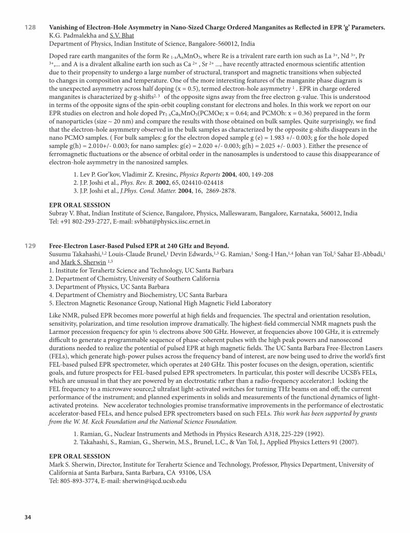
34
128 VanishingofElectron-HoleAsymmetryinNano-SizedChargeOrderedManganitesasReflectedinEPR‘g’Parameters. K.G. Padmalekha and S.V. BhatDepartment of Physics, Indian Institute of Science, Bangalore-560012, India
Doped rare earth manganites of the form Re 1-xAxMnO3, where Re is a trivalent rare earth ion such as La 3+, Nd 3+, Pr 3+,... and A is a divalent alkaline earth ion such as Ca 2+ , Sr 2+ ..., have recently attracted enormous scientific attention due to their propensity to undergo a large number of structural, transport and magnetic transitions when subjected to changes in composition and temperature. One of the more interesting features of the manganite phase diagram is the unexpected asymmetry across half doping (x = 0.5), termed electron-hole asymmetry 1 . EPR in charge ordered manganites is characterized by g-shifts2, 3 of the opposite signs away from the free electron g-value. This is understood in terms of the opposite signs of the spin-orbit coupling constant for electrons and holes. In this work we report on our EPR studies on electron and hole doped Pr1-xCaxMnO3(PCMOe; x = 0.64; and PCMOh: x = 0.36) prepared in the form of nanoparticles (size ~ 20 nm) and compare the results with those obtained on bulk samples. Quite surprisingly, we find that the electron-hole asymmetry observed in the bulk samples as characterized by the opposite g-shifts disappears in the nano PCMO samples. ( For bulk samples: g for the electron doped sample g (e) = 1.983 +/- 0.003; g for the hole doped sample g(h) = 2.010+/- 0.003; for nano samples: g(e) = 2.020 +/- 0.003; g(h) = 2.025 +/- 0.003 ). Either the presence of ferromagnetic fluctuations or the absence of orbital order in the nanosamples is understood to cause this disappearance of electron-hole asymmetry in the nanosized samples.
1. Lev P. Gor’kov, Vladimir Z. Kresinc, Physics Reports 2004, 400, 149-2082. J.P. Joshi et al., Phys. Rev. B. 2002, 65, 024410-0244183. J.P. Joshi et al., J.Phys. Cond. Matter. 2004, 16, 2869-2878.
EPRORALSESSIONSubray V. Bhat, Indian Institute of Science, Bangalore, Physics, Malleswaram, Bangalore, Karnataka, 560012, India Tel: +91 802-293-2727, E-mail: [email protected]
129 Free-ElectronLaser-BasedPulsedEPRat240GHzandBeyond.Susumu Takahashi,1,2 Louis-Claude Brunel,1 Devin Edwards,1,3 G. Ramian,1 Song-I Han,1,4 Johan van Tol,5 Sahar El-Abbadi,1 and Mark S. Sherwin 1,3
1. Institute for Terahertz Science and Technology, UC Santa Barbara 2. Department of Chemistry, University of Southern California 3. Department of Physics, UC Santa Barbara 4. Department of Chemistry and Biochemistry, UC Santa Barbara 5. Electron Magnetic Resonance Group, National High Magnetic Field Laboratory
Like NMR, pulsed EPR becomes more powerful at high fields and frequencies. The spectral and orientation resolution, sensitivity, polarization, and time resolution improve dramatically. The highest-field commercial NMR magnets push the Larmor precession frequency for spin ½ electrons above 500 GHz. However, at frequencies above 100 GHz, it is extremely difficult to generate a programmable sequence of phase-coherent pulses with the high peak powers and nanosecond durations needed to realize the potential of pulsed EPR at high magnetic fields. The UC Santa Barbara Free-Electron Lasers (FELs), which generate high-power pulses across the frequency band of interest, are now being used to drive the world’s first FEL-based pulsed EPR spectrometer, which operates at 240 GHz. This poster focuses on the design, operation, scientific goals, and future prospects for FEL-based pulsed EPR spectrometers. In particular, this poster will describe UCSB’s FELs, which are unusual in that they are powered by an electrostatic rather than a radio-frequency accelerator;1 locking the FEL frequency to a microwave source;2 ultrafast light-activated switches for turning THz beams on and off; the current performance of the instrument; and planned experiments in solids and measurements of the functional dynamics of light-activated proteins. New accelerator technologies promise transformative improvements in the performance of electrostatic accelerator-based FELs, and hence pulsed EPR spectrometers based on such FELs. This work has been supported by grants from the W. M. Keck Foundation and the National Science Foundation.
1. Ramian, G., Nuclear Instruments and Methods in Physics Research A318, 225-229 (1992). 2. Takahashi, S., Ramian, G., Sherwin, M.S., Brunel, L.C., & Van Tol, J., Applied Physics Letters 91 (2007).
EPRORALSESSIONMark S. Sherwin, Director, Institute for Terahertz Science and Technology, Professor, Physics Department, University of California at Santa Barbara, Santa Barbara, CA 93106, USA Tel: 805-893-3774, E-mail: [email protected]

35
130 ObservationofanElectron-onlySpinDependentProcessinOLEDs.William Baker, Dane R. McCamey, John M. Lupton and Christoph Boehme University of Utah, Dept of Physics and Astronomy
Low spin-orbit coupling imposes spin-selection rules on electronic transitions in organic materials. An example of one such spin-dependent process is the recombination of electrons (e) and holes (h), which depends on their relative spin orientation. When the spin (of either e or h or both) is manipulated with magnetic resonance, a change of the current through the material is observed. This work aims to test the hypothesis that, in the presence of majority charge carriers, unipolar spin-dependent processes (e.g. a bipolaron transport process) would become observable. To do this, we repeated our recent coherent spin nutation experiment on MEH-PPV OLEDs using devices without a PEDOT h-injection layer, anticipating that the imbalance between e (majority) and h (minority) polarons should allow observation of spin-dependent e-only processes. The on resonance dynamics of these signals was more complex than previously seen in bipolar devices, a signal inexplicable by a single spin dependent process.1 In order to verify that one of the processes is due to a unipolar transition between e-states, we performed spin Rabi nutation by monitoring the current as a function of the duration of magnetic resonance excitation. For both short and long times we observed a periodic modulation of the current consistent with the rotation of spin-½ particles. In addition, the long time signal exhibited a strong hyperfine-induced spin-beating component, as was previously seen in bipolar OLEDs2 and attributed to polaron pair recombination. In contrast, the short-time signal did not show this beating, consistent with a pure unipolar electron process. This observation provides evidence that there are at least two spin-dependent channels in MEH-PPV: electron-hole pair recombination, and an electron-electron process.
1. D.R. McCamey, et. al. Nature Mater. 7, 723, (2008) 2. D.R. McCamey, et. al. Phys. Rev. Lett. 104, 017601 (2010).
EPRORALSESSIONWilliam J. Baker, University of Utah, Physics, 115 S 1400 E, Salt Lake City, UT, 84112-0830, USA Tel: 801-783-6619, E-mail: [email protected]
131 MultifrequencyENDORSpectroscopyIdentifiesaUniqueIronSiteontheIron-sulphurClusterInvolvedinSubstrateReductionofHeterodisulfideReductase.Alistair J. Fielding,1 Kristian Parey,2 Ulrich Ermler,2 Bernhard Jaun,3 Silvan Scheller,3 and Marina Bennati1
1. Max-Plank Institute for Biophysical Chemistry, Göttingen, Germany 2. Max-Plank Institute for Biophysics, Frankfurt, Germany 3. Laboratory of Organic Chemistry, ETHZ, Switzerland
Heterodisulfide reductase (HDR) is a key enzyme in the energy metabolism of methanogenic archaea. The enzyme catalyzes the reversible reduction of the heterodisulfide (CoM-S-S-CoB) to the thiol coenzymes, coenzyme M (CoM-SH) and coenzyme B (CoB-SH) in the final step of methanogenesis. It employs an unusual [4Fe4S] cluster to carry out substrate chemistry. Previous studies have identified a mechanistic-based paramagnetic intermediate generated upon half-reaction of the oxidized enzyme with CoM-SH in the absence of CoB-SH.1 The unusual [4Fe4S]-cluster is bound in subunit HdrB of the Methanothermobacter marburgenis HdrABC holoenzyme within the C-terminal domain of two cysteine-rich sequence motifs (Cx31-39CCx35-36CxxC). EPR studies on the isolated subunit HdrB heterologously produced in E. coli have shown that the [4Fe4S] cluster can be observed after oxidation in the absence of substrate.2 Our previous electron nuclear double resonance (ENDOR) investigations on the cluster within the Hdr-CoM-SH complex have lead to the assignment of four distinct iron sites and to the determination of the full hyperfine tensor elements. However, we were unable to provide conclusive evidence of a unique iron site and it was postulated that the cluster could interact with the substrate at multiple sites.1 Recently, Mössbauer measurements on the CCG-domain-containing subunit SdhE of succinate:quinone oxidreductase from Sulfolobus sulfataricus P2 has unambiguously indicated the presence of a unique iron site in the reduced cluster.3 We report multifrequency 57Fe ENDOR spectroscopic measurements on the iron sulphur cluster in HdrABC, HdrB and SdhE. Specifically, we report on the first results at 34-GHz, which show enhanced resolution compared to previous measurements at 9- and 94-GHz arising from the absence of proton resonances and polarization effects. The combined results at different frequencies allow assignment of all four 57Fe resonances and provide evidence of a unique iron site. We also report first 13C ENDOR results of 13C labelled CoM-SH bound to the cluster.
1. Hedderich et al., Biological Chemistry, 2005, 386, 961. 2. Hamann et al., Biochemistry, 2007, 46, 12875.3. Hamann et al., J. Biol. Inorg. Chem. 2009, 14, 457-470.
EPRORALSESSIONAlistair J. Fielding, Max-Plank Institute for Biophysical Chemistry, 11 Am Fassburg, Göttingen, 37077, Germany Tel: 004 95 51 201 1362, E-mail: [email protected]

36
132 OverhauserEffectDynamicNuclearPolarizationfortheMeasurementofHydrationDynamics. John Franck,1 Anna Pavlova,1 Mingming Ma,2 Patricia Soto,3 Denis Bong,2 Joan Emma-Shea1 and Songi Han1
1. University of California, Department of Chemistry and Biochemistry, Santa Barbara, CA, 93106-9510 2. The Ohio State University, Department of Chemistry, Columbus, OH 43210-1185 3. Creighton University, Department of Physics, Omaha, NE 68178
In recent years, the Overhauser Effect in liquids (OE-DNP) has proven an effective method for measuring intermolecular translational motion with residue-specific locality. This exciting method offers the possibility of becoming a routine yet powerful tool in the quest for understanding the role of hydration dynamics in protein folding, coacervation, and the driving forces of the hydrophobic effect. Here, we highlight an application demonstrating changes in dynamics on the surface of vesicles that coalesce due to interactions between their head groups.1 These applications provide the motivation to transform OE-DNP into a quantitative, reproducible analytical method for determining the correlation time, τ, of water near a spin label. This work involves challenges in the model, the measurement, and the instrument. We find that the customary model2 for the spectral density function disagrees with MD simulations, even at lower frequencies; we tie this to discrepancies previously observed3 between field cycling relaxometry (FCR) and OE-DNP. By propagating the instrument noise, we are able to identify that variations in the E (NMR signal enhancement) values as they are extrapolated to infinite power are significant, and can thus affect the reproducibility of dynamics measurements. We validate a fact witnessed by other groups: the E vs. p curve does not always exhibit the asymptotic dependence, 1-E = (1-Emax)*Ap⁄(1-Emax+Ap) expected. We demonstrate that in previous studies, simultaneous variations in the NMR T1 relaxation masked this effect, which we attribute to sample heating, as evidenced by changes in the T1 of pure water. We present hardware, paired with adaptable and powerful processing libraries, that fully automates detection, processing, and error analysis of all our measurements. These advances will facilitate the dissemination of OE DNP as a useful tool applicable to the vesicle systems we demonstrate, the protein and DNA systems we are investigating, and reproducible measurements in other labs.
1. Y. Gong, M. Ma, Y. Luo, D. Bong, J. Am. Chem. Soc., 2008, 130 (19), pp 6196-62052. L.P. Hwang, J.H. Freed, J. Chem. Phys. 1975, 63, 40173. B.D. Armstrong, S. Han, J. Am. Chem. Soc., 2009, 131 (13), pp 4641-4647
EPRORALSESSIONJohn Franck, University of California, Chemistry and Biochemistry, Chemistry and Biochemistry 9510, Santa Barbara, CA, 93106-9510, USA E-mail: [email protected]
133 PulsedElectronSpinResonanceResolvestheCoordinationSiteofCu+2ionsinα1-GlycineReceptor. Sharon Ruthstein,1 Katherine M. Stone,1 Timothy F. Cunningham,1 Michael Cascio,2 and Sunil Saxena1
1. Department of Chemistry, University of Pittsburgh, Pittsburgh, PA, 15260. 2. Department of Chemistry and Biochemistry, Duquesne University, Pittsburgh, PA, 15282.
In this talk, we will discuss the coordination environment of copper ions (Cu+2) in human α1 glycine receptor (GlyR). GlyRs are members of the pentameric ligand-gated ion channel (pLGIC) superfamily that mediate fast signaling at synapses. Metal ions like Zn+2 and Cu+2 significantly modulate the activity of pLGICs, and in vivo results suggest that metal ion coordination might be essential for proper physiological postsynaptic inhibition by GlyR. In order to better understand the molecular basis of this effect we have used pulsed electron spin resonance (ESR) methods like electron spin echo envelope modulation (ESEEM) and double electron electron resonance (DEER) spectroscopy to directly examine Cu+2 coordination with GlyR. We show, that Cu+2 has one binding site per α1 subunit, and that five Cu+2 can be coordinated per GlyR. Cu+2 binds to GlyR with 40 μM apparent dissociation constant, and the metal ion are coordinated to E192 and H215 in each subunit. This is consistent with earlier functional measurements. However, the coordination site does not include residues around the agonist/antagonist binding site, such as T112, R131, R65, E157 that were previously suggested to have a roles in Cu+2 coordination by functional measurements on site-directed mutants. Intriguingly, the E192 and H215 are also known to bind Zn+2. The binding of Zn+2 at this site potentiates channel activity, whereas Cu+2 has an inhibitory effect. The opposing modulatory actions of these similar metal divalent cations at a shared binding site highlight the sensitive allosteric nature of GlyR. This work is supported by NIH (5R01NS053788). S.R. is supported by a long-term postdoctoral fellowship awarded by the EMBO.
EPRORALSESSIONSharon Ruthstein, University of Pittsburgh, Department of Chemistry, 219 Parkman Av., Pittsburgh, PA, 15260, USA Tel: 412-624-8123, E-mail: [email protected]

37
134 ProbingFlexibilityinPorphyrin-BasedMolecularWiresusingDEER.Janet E. Lovett,1 Markus Hoffmann,1 Arjen Cnossen,1 Alexander T.J. Shutter,1 Hannah J. Hogben,1 John E. Warren,2 Sofia I. Pascu,3 Christopher W.M. Kay,4 Christiane R. Timmel,1 Harry L. Anderson1
1. Centre for Advanced Electron Spin Resonance, Department of Chemistry, University of Oxford, UK 2. Synchrotron Radiation Source, Daresbury Laboratory, Warrington, UK 3. Department of Chemistry, University of Bath, UK 4. Institute of Structural and Molecular Biology and London Centre for Nanotechnology, University College London, UK
This presentation will show how DEER (double electron electron resonance, also known as pulsed electron double resonance, PELDOR) was used to probe a series of nitroxide spin-labelled butadiyne-linked zinc porphyrin oligomers to investigate their conformational flexibility.1 These π-conjugated oligomers are also known as molecular wires because of their ability to mediate electron transfer. The oligomers were found to adopt linear conformations with a distribution of distances following the worm-like chain model, consistent with molecular dynamics simulations. The worm-like chain model was fitted to the DEER time traces by adapting the methods available in DeerAnalysis20062. Despite measuring in frozen solutions, we found that changing the solvent and therefore the glass transition temperature led to small but measurable changes to the conformational flexibility of the molecules. DEER experiments were also used to show that the oligomers were able to self-assemble to form ladder-like structures in the presence of suitable templates and additionally, despite the apparent inflexibility of the oligomers, they were readily bent around multidentate ligands. The study demonstrates the scope of DEER for providing structural information about synthetic nanostructures.
1. Lovett et al. JACS, 2009, 131, 13852-138592. Jeschke et al. AMR, 2006, 30, 473-498
EPRORALSESSIONJanet E. Lovett, University of Oxford, CAESR, Inorganic Chemistry, South Parks Road, Oxford, OX1 3QR, UK Tel: 0777-589-5200, E-mail: [email protected]
135 EPR,UpCloseandFromAfar:ElucidatingtheMechanisticIntermediatesinCytochromec OxidasefromParacoccus denitrificans. Jessica H. van Wonderen,1 Iris von der Hocht,2 Hartmut Michel2 and Fraser MacMillan1
1. University of East Anglia, Norwich NR47TJ, United Kingdom 2. Max Planck Institute of Biophysics, D 60438 Frankfurt am Main, Germany
Cytochrome c Oxidase (CcO) is the terminal enzyme of the respiratory chain. The redox driven proton pump catalyses the four electron reduction of molecular oxygen to water. Electrons are delivered by cytochrome c to the bimetallic CuA centre and transferred via heme a into the binuclear heme a3 CuB centre where the reduction of oxygen takes place. Despite high resolution X-ray crystallographic structures, the properties of the catalytic redox states of the metal centres and their relation to protonation states within this class of enzyme still remain poorly understood. Modern EPR techniques (also in combination with magneto-optical studies) enable us to probe different catalytic intermediate states either directly or indirectly. From afar, pulsed ELDOR spectroscopy, a technique for accurately measuring inter-spin distances in the range 2-8 nm, is used to resolve subtle structural changes when applied to spin-labelled systems trapped in different intermediate states (e.g. P, R & F states) and which allows the study of local conformational changes in great detail. Using this technique conformational changes within the proton uptake channels are discussed. Up close, both EPR1 and Magnetic Circular Dichroism are used to address the nature of the metal ligands in the binuclear centre as well as transiently formed radical species from different intermediate states.
1. Interconversions of P and F Intermediates of Cytochrome c Oxidase from Paracoccus denitrificans, Iris von der Hocht, Jessica H. van Wonderen, Florian Hilbers, Heike Angerer Fraser MacMillan and Hartmut Michel, submitted to PNAS
EPRORALSESSIONJessica H. van Wonderen, University of East Anglia, Chemistry, Chancellor Drive, Norwich, Norfolk, NR4 7TJ, UK Tel: 0044-160-359-1422, E-mail: [email protected]

38
136 T2Measurementsat240GHzforNuclearSpinBathEffectsandBiologicalDistanceMeasurement. Devin T. Edwards1, Susumu Takahashi2, Songi Han3,4, Mark Sherwin1,3
1. University of California, Department of Physics, Santa Barbara, CA 93106 2. University of Southern California, Department of Chemistry, Los Angeles CA 90089 3. University of California, Institute for Terahertz Science & Technology, Santa Barbara, CA 93106 4. University of California, Department of Chemistry and Biochemistry, Santa Barbara, CA 93106
Spin decoherence is a fundamental and complex process which encodes a variety of useful information about a system. Recent work has demonstrated that spin decoherence due to the dipolar coupling can be varied and well-characterized with 240 GHz pulsed EPR by controlling temperature1. This characterization allows determination of the inter-electron dipolar coupling strength, which is strongly distance dependent. This distance dependence has been observed in various concentrations of a nitroxide spin label in deuterated water glass-allowing extraction of an average inter-spin distance from experiments. While a variety of advanced pulsed and cw techniques exist for studying spin-spin distances, this technique may offer advantages for measurement of long, broadly distributed distances, where existing methods remain challenging. Because we can not quench the nuclear spin bath even at low temperatures, its effects may limit application to biological systems if electron spin-spin dipolar coupling is not a dominant relaxation mechanism. Work presented will include extension of initial experiments to study effects of modifying the solvent deuteration and nitroxide protonation to gain insight into the effect of the nuclear spin bath. Further experimental work will examine spin-labeled, lipid vesicles with varying concentrations of spin-label, to observe distance dependence in a biologically relevant system. Spin-labeling of lipid headgroups allows the nitroxide to be solvent exposed, where deuteration of the solvent can increase relaxation times. This represents a test system for spin-labeling solvent exposed portions of bilayer constituents to investigate organization behavior of proteins and lipids in membranes. This work is supported by the NSF DMR05-20415 grant, and the W.M. Keck Foundation Award for Science and Engineering.
1. S. Takahashi et al., Phys. Rev. Lett. 101, 047601 (2008)
EPRORALSESSIONDevin T. Edwards, University of California at Santa Barbara, Building 557, Santa Barbara , CA, 93106-9510, Tel: 805-893-2032, E-mail: [email protected]
137 Spin-dependentProcessesinSilicon-richSilicon-nitrideThinFilmSolarCells.Sang-Yun Lee,1 S-.Y. Paik,1 D.R. McCamey,1 C. Boehme,1 J. Hu,2 F. Zhu,2 A. Madan2
1. University of Utah, Department of Physics and Astronomy, Salt Lake City, UT 84112 2. MV Systems, Inc., Golden, CO 80401
Silicon-rich silicon-nitride (SiNx:H) has recently attracted attention due to its relevance for band-gap tunable light emitting diodes1 and as a semiconductor electrode for photoelectrochemical (PEC) hydrogen production2. Charge transport and recombination in this material are key factors for its optoelectronic properties. Since SiNx:H is highly disordered and exhibits weak spin-orbit coupling, similar to amorphous silicon (a-Si:H), many of the electronic transitions in this material take place through localized electronic states and are governed by spin-selection rules. It is possible to study these processes using electrically detected magnetic resonance spectroscopy (EDMR). Using coherent, pulsed EDMR, we observed a variety of qualitatively different spin-dependent processes. We present a multidimensional pEDMR mapping of these processes which shows a variety of defect types and couplings strengths in this material. The qualitative nature of many of these processes is similar to previously studied mechanisms in a-Si:H as they involve a variety of different states (dangling bonds, tails states), with various spin coupling modes (exchange, dipolar coupling). However, unlike in a-Si:H3, many of the transitions between strongly spin-coupled pairs in SiNx:H influence the conductivity, and are therefore not due to geminate recombination processes. Dipolar coupled spin pairs also contribute to the conductivity in SiNx:H in contrast to those in a-Si:H. Also, in contrast to the dipolar coupled geminate pairs in a-Si:H, the average separation distance is 0.6 nm, much smaller than the 1.6 nm in a-Si:H4.
1. N.-M. Park, T.-S. Kim, and S.-J. Park, App. Phys. Let., 2001, 78, 25752. A. Madan, J. Hu, F. Zhu, I. Matulionis, and A. Kunrath, presented at DOE PEC Working Group meeting, San Francisco, April 17-18, 2009. 3. T. Herring, S.-Y. Lee, D.R. McCamey, P.C. Taylor, K. Lips, J. Hu, F. Zhu, A. Madan, and C. Boehme, Phys. Rev. B, 2009, 79, 195205.4. K. Lips, C. Boehme, and T. Ehara, J. Optoelectronics and advanced Mat., 2005, 7, 13
EPRORALSESSIONSang-Yun Lee, University of Utah, Dept. of Physics and Astronomy, 115 S. 1400 East, Salt Lake City, UT, 84112- 0830, USA E-mail: [email protected]

39
138 SpinIncoherenceofDonorElectronsNearc-Si(111)/SiO2InterfaceDefects.Seoyoung Paik, S.-Y. Lee, W.J. Baker, D.R. McCamey and C. Boehme University of Utah
Electron and nuclear spins of phosphorous (31P) donors in crystalline silicon have been investigated extensively in recent years as they both have extremely long coherence times. This makes them interesting candidates for quantum information and spin-electronics applications1. Existing silicon quantum computer concepts2 propose to use 31P qubits close to the silicon surface. We present here a study of how microscopic defects at the oxide layer of the silicon surface influence the spin coherence times (T1 and T2 times) of the 31P qubits. Using surface sensitive pulsed electrically detected magnetic resonance spectroscopy3 we show that, when the 31P qubits are near interface states, the extremely long bulk coherence times (>> 600ms ,T2)4 which have been observed previously are drastically shortened (~1 μs, T2)5. Our measurements of the 31P donor T2 times as a function of the interface defect density support predictions by De Sousa6 that this reduction is due to interface defect induced field noise.
1. J.J.L. Morton, et al., Nature 2008 455, 10852. B.E. Kane, Nature 1998 393, 133 3. A.R. Stegner, et al., Nature Physics 2006 2, 8354. A.M. Tyryshkin et al. Phys. Rev. B 2003 68, 1932075. S.-Y. Paik, et al.,Phys. Rev. B 2010 81, 075214 6. R. de Sousa, Phys. Rev. B 2007 76, 245306
EPRORALSESSIONSeoyoung Paik, University of Utah, Physics and astronomy , 115 South 1400 East, SLC, UT, 84112- 0830, USA Tel: 801-403-0633, E-mail: [email protected]
139 AMulti-FrequencyEPRApproachforInvestigatingtheIntrinsicallyDisorderedProtein,IA3.Natasha L. Pirman, Eugene Milshteyn and Gail E. Fanucci Department of Chemistry, University of Florida
Intrinsically disordered proteins (IDPs) contain little to no secondary or tertiary structure and are essential in biological systems. Many IDPs undergo a conformational change, where structure is induced upon binding to its target protein. Due to the inherent nature of IDPs, structural studies are often challenging. Here, we show that site-directed spin-labeling (SDSL) electron paramagnetic resonance (EPR) spectroscopy can be utilized to characterize the mobility and conformational changes of IDPs. We have applied this method to IA3, which is a 68 residue IDP whose unstructured-to-α-helical conformational transition has been extensively characterized by various biophysical techniques. Both X- and W-band frequencies were utilized to monitor the chemically induced α-helical conformation upon addition of 2,2,2-trifluoroethanol (TFE). Detailed analyses of the X-band EPR spectral line shapes reveal global correlation time changes consistent with a two-state model consisting of an unstructured system and the tumbling of a rigid helix; whereas, the W-band EPR spectral line shapes appear more sensitive to differences in site-specific mobility and conformational changes. Using IA3 as a model system, we are developing multi-frequency SDSL EPR approaches for application to IDP systems that are otherwise difficult to characterize.
EPRORALSESSIONNatasha L. Pirman, University of Florida, Chemistry, PO Box 117200, Gainesville, FL, 32611, USA E-mail: [email protected]
140 OrganometallicMechanismsofAction,andInhititionofthe4Fe-4SProteinsGcpEandLytB:APulsedEPRInvestigation.Weixue Wang1 and Eric Oldfield2
1. University of Illinois at Urbana-Champaign, Center for Computational Biology and Biophysics 2. University of Illinois at Urbana-Champaign, Department of Chemistry, Urbana, IL 61801
GcpE (also known as IspG) and LytB (also known as IspH) are the last two enzymes in the non-mevalonate isoprenoid biosynthesis pathway.1 They are essential for growth of most pathogenic bacteria and malaria parasites, but are not found in humans, so are potentially important anti-infective drug targets.2 Both GcpE and LytB contain a 4Fe-4S cluster with a unique 4th iron not liganded to the cysteine residue, and catalyze 2e-/2H+ reductions.3 We find that the catalytic mechanisms of both enzymes involve organometallic species. For the GcpE enzyme, we characterized a reaction intermediate using ENDOR and HYSCORE spectroscopy with 13C, 2H and 17O labeled samples. The results indicate formation of an Fe-C-H containing reactive intermediate, a ferraoxetane. This is then rapidly reduced to a “metallacycle”, in which the alkene product forms a π or π/σ, η2-alkenyl complex with the 4th iron in the 4Fe-4S cluster. In the LytB enzyme,

40
we also find formation of a similar “metallacycle” involving the 4th iron and its alkene substrate as a reaction intermediate. Based on this metallacycle concept, we have discovered the first potent GcpE and LytB inhibitors, alkynyl diphosphates (Ki ~300 nM in GcpE and 970 nM in LytB). These alkyne inhibitors form another “metallacycle” with the 4Fe-4S cluster in both enzymes, as evidenced by ~ 6 MHz 13C and ~ 10 MHz 1H hyperfine couplings in the ENDOR spectra. Overall, the results are of broad general interest since they provide new mechanistic insights into 4Fe-4S proteins, as well as their inhibition, with organometallic bond formation playing a key role in both cases. Supported by NIH grant GM65307 for EO.
1. Rohmer, Lipids, 2008, 43, 1095.2. Rohmer et al., Curr. Opin. Investig. Drugs, 2004, 5, 154.3. Seemann et al., J. Biol. Inorg. Chem., 2005, 10, 131.
EPRORALSESSIONWeixue Wang, University of Illinois at Urbana-Champaign, Center for Computational Biology and Biophysics, 600 S Mathews Ave, Urbana, IL, 61801, USA Tel: 217-333-8328, E-mail: [email protected]
141 ExploringtheLimitsofElectronSpinEcho in vivo OxygenImaging.Boris Epel, Subramanian V. Sundramoorthy, Payam Seifi, Jonathan Bryant, Gage Redler and Howard J. Halpern Center for EPR Imaging In Vivo Physiology, University of Chicago
In our previous work we have demonstrated that for in vivo oxymetry based on the injection of narrow line paramagnetic spin probe into an experimental animal, the Electron Spin Echo (ESE) imaging methodology outperforms continuous wave (CW), both in acquisition speed and precision. However instrumental limitations, particularly imager bandwidth, suggested that this methodology would be applicable for small animals only. We have developed a Multiple B0 imaging technology capable of acquiring images whose bandwidth requirements exceed the hardware bandwidth. We present images of a rat and rabbit leg and discuss approaches for larger objects imaging. In the second part of the presentation, we discuss reconstruction of ESE images during acquisition (“real time reconstruction”). Real time reconstruction provides the imager operator with a measure of trityl concentration in tissues of interest throughout image acquisition, allowing the operator to adjust the rate of spin probe administration to achieve a constant, desired concentration. In the future, real time acquisition will allow the operator to direct imaging to a particular region of interest based on early images reconstructed from a small number of projections. This is analogous to the use of a “pilot” image in MRI studies. For fast data processing we have applied hardware accelerated image reconstruction using CUDA toolbox for MATLAB.
This work is supported by NIH, grants number P41 EB002034 and R01 CA98575.
EPRORALSESSIONBoris Epel, University of Chicago, Department of Radiation and Cellular Oncology, 5841 S. Maryland Avenue, MC1105, Chicago, IL, 60637, USA E-mail: [email protected]
142 AProgrammablePulseGeneratorwithNanoSecondResolutionforPulsedEPRApplications.Nallathamby Devasahayam, Sankaran Subramanian and Murali C. Krishna Radiation Biology Branch, Center for Cancer Research, National Cancer Institute
A Pulse Programmer with nanosecond (ns) time resolution needed for time-domain EPR spectroscopic applications is described. This uses commercially available timing and I/O modules and control software developed in our laboratory. The pulse programmer is controlled by a PC. Through front panel graphic user interface (GUI) inputs to control pulse widths, delays and acquisition trigger timings can be entered. Based on these parameters, all other associated gate and trigger timings are internally generated without the need to enter them explicitly. The excitation pulses can have widths with ns resolution while all other gate pulses can be incremented in steps of 20 ns without compromising spectrometer performance. The current configuration of the pulse programmer permits generation of single pulse or multiple pulse sequences for EPR imaging with minimal data entry via the front panel GUI.
1. M.C. Krishna, S. Subramanian, P. Kuppusamy, and J.B. Mitchell, Semi.n Radiat. Oncol. 11, 58 (2001).2. N. Devasahayam, R. Murugesan, K. Yamada, K. Reijnders, J.B. Mitchell, S. Subramanian, M.C. Krishna, and J.A.
Cook, Rev. Sci. Instrum. 73, 3920 (2002).3. S. Subramanian, N. Devasahayam, R. Murugesan, K. Yamada, J. Cook, A. Taube, J.B. Mitchell, J.A.B. Lohman,
and M.C. Krishna, Magn. Reson. Med. 48, 370 (2002).4. H.J. Halpern, C. Yu, M. Peric, E. Barth, D.J. Grdina, and B.A. Teicher, Proc. Natl. Acad. Sci. (USA) 91, 13047
(1994).5. S. Subramanian, K.I. Matsumoto, J.B. Mitchell, and M.C. Krishna, Nmr in Biomedicine 17, 263 (2004).

41
EPRORALSESSIONNallathamby Devasahayam, Radiation Biology Branch, Center for Cancer Research, National Cancer Institute, NIH, Bethesda, MD 20892, USA Tel: 301-402-6320, Email:[email protected]
143 NovelProbesandOpportunitiesforClinicalOximetry. Guruguhan Meenakshisundaram, Ramasamy Padian and Periannan Kuppusamy The Ohio State University
Oxygen is a critical determinant in the prediction of treatment outcome of several disease including surgical interventions, cancer therapy, tissue graft, and cell therapy. There is a great need for methods capable of reliable noninvasive measurement and monitoring of oxygen concentration in tissues. Although several methods are utilized to measure oxygen concentration, a suitable technique for noninvasive and repeated measurements of oxygen in the same tissue or cells on a temporal scale is warranted. While electrode techniques have evolved as the standard methods for measurement of oxygen, they generate analytical artifacts during assay procedures at the freshly probed sites. Near-infrared and nuclear magnetic resonance techniques are noninvasive methods. However, they do not report the absolute values of oxygen concentration, and lack the resolution of oxygen measurements. Electron paramagnetic resonance (EPR) oximetry enables reliable and accurate measurements of concentrations of molecular oxygen. EPR oximetry can measure directly and at the actual site of interest. The ability of EPR oximetry to make repeated measurements from localized sites provides a very important capability that can enable critical aspects of a number of biomedical applications. We have developed innovative approaches using oxygen-sensing nano/microcrystalline probes to perform noninvasive oximetry/imaging in a variety of applications including myocardial ischemia/reperfusion injury, cellular cardiomyoplasty (cell therapy), tumor angiogenesis, cancer therapy, and wound healing. Current developments in in vivo EPR methodologies enable a number of potential applications that could be very significant additions to clinical medicine. The current developments of oximetry probes and designs for clinical oximetry will be presented. Supported by NIH grant R01 EB004031
EPRORALSESSIONPeriannan Kuppusamy, Davis Heart and Lung Research Institute, The Ohio State University, 420 W. 12th Ave, Columbus, OH 43210, USA Tel: 614-292-8998, E-mail: [email protected]
144 NitroxidesasSensitiveO2ImagingAgentsfor in vivo ElectronParamagneticResonanceImaging.John M.Weaver University of New Mexico
In vivo electron paramagnetic resonance imaging (EPRI) is an emergent imaging method for studying a number of important physiological parameters, such as visualizing O2 distribution in various tissues. However, this technique has been hindered by a lack of suitable imaging modalities. Recent developments in low-frequency EPR spectrometers that can detect free radicals in animals in real time makes it feasible to image paramagnetic oximetry probes such as nitroxides in brain tissue. Nitroxides exhibit many ideal qualities, including ease of preparation, chemical flexibility, and high stability at physiologic pH and temperature. Such imaging capabilities would allow O2 mapping in tumors, different brain regions following a hypoxic episode or administration of drugs of abuse. We have investigated several nitroxides including 3-acetoxymethoxycarbonyl-2,2,5,5-tetramethyl-1-pyrrolidinyloxyl (amctp) which can be entrapped in brain tissue as 3-carboxy-2,2,5,5-tetramethyl-1-pyrrolidinyloxyl (ctp) and be used to quantitate O2 concentrations in situ. To increase the sensitivity of O2 measurement using EPRI, we have investigated isotopic-substituted labile nitroxides as a next generation of promising pro-imaging agents. EPR spectroscopic measurements demonstrate that these doubly isotopic-labeled nitroxide markedly improves signal-to-noise ratio (SNR) and thus, detection limits. Additionally, these nitroxides are more sensitive to changes in local O2 concentration, which will enable more accurate O2 measurement in tissues. Taken together, these imaging agents and improved EPR techniques will offer novel and innovative EPR images of cerebral oxygenation and oxidative stress related to several areas of study including cerebral ischemia and drug abuse.
EPRPOSTERSESSIONJohn Weaver, University of New Mexico College of Pharmacy, Domenici Hall BRaIN Imaging Center, 1101 Yale, NE, Albuquerque, NM 87131 Tel: 505-272-6056, E-mail: [email protected]

42
145 ClinicalEPR:ChallengesandProgress.Harold M. Swartz, Benjamin B. Williams, Nadeem Khan, Ruhong Dong, Huagang Hou, Piotr Lesniewski, Maciej Kmiec, Jean LaRiviere, Tom Mathews, Tim Raynolds, Oleg Grinberg, Ildar Salikhov, Eugene Demidenko, Javier Nicolalde, Ann B. Flood, Alan Hartford, Bassem Zaki, Lesley Jarvis, Eunice Chen, David GladstoneDartmouth Medical School
The development and use of in vivo techniques for experimental applications in animals has been very successful and now has led to attractive clinical applications. This presentation provides an overview of the challenges, opportunities, and results as in vivo EPR is extended into use in human subjects.
The most widespread clinical use is oximetry, where EPR can make repeated and accurate measurements of pO2 in tissues, which provides clinicians with information that bears directly on diagnosis and therapy, especially for oncology, peripheral vascular disease, and wound healing. The other area of extensive utilization in human subjects is the ability of in vivo EPR to measure clinically significant exposures to ionizing radiation ‘after-the-fact’, due to accidents, terrorism, or nuclear war.
The unique capabilities of in vivo EPR to detect and characterize free radicals could be applied to measure free radical intermediates from drugs and oxidative process, including measurements of nitric oxide. These unique capabilities, combined with the sensitivity of EPR spectra to the immediate environment (e.g. pH, molecular motion, charge) have resulted in productive applications in animals, and may be used in humans in the future.
The challenges for achieving full implementation include having oximetric techniques that can be used routinely in human subjects, adapting the spectrometer for safe and comfortable measurements in human subjects by regular clinical personnel, achieving sufficient sensitivity for measurements at the sites of the pathophysiological processes, and establishing a consensus on the clinical value of the measurements.
This work was supported by NIH grants PO1 EB2180, U19 AI067733, R21 CA121593 and R21 DK072112 and contracts from DARPA (Defense Advanced Research Projects Agency), contact no. HR0011-08-C-0022 and HR0011-08-C-0023.
EPRORALSESSIONHarold M. Swartz, Dartmouth Medical School, HB 7785, Vail 702, Hanover, NH 03755 USA Tel: 603-650-1955, E-mail: [email protected]
146 FastEPRSpinTrappingofSuperoxideRadicalAnionbyCyclicNitrone-Calix[4]pyrroleConjugate:TheoreticalandExperimentalStudies. Shang-U Kim,1 Yangping Liu,1 Kevin M. Nash,1 Antal Rockenbauer2 and Frederick A. Villamena1
1. Dept. of Pharmacology, Heart and Lung Research Research Institute, The Ohio State University, Columbus, OH 2. Chemical Research Center, Institute of Structural Chemistry, Budapest, Hungary
Nitrone spin traps have been employed as probes for the identification of transient radical species in chemical and biological systems using electron paramagnetic resonance (EPR) spectroscopy. Since calix[4]pyrroles have been shown to exhibit high affinity to anions, a cyclic nitrone conjugate of calix[4]pyrrole (CalixMPO) was designed, synthesized, and characterized. Computational studies suggest a pendant-type linkage between the calix[4]pyrrole and the nitrone to be the most efficient design for spin trapping of a superoxide radical anion (O2•-), giving exoergic reaction enthalpies and free energies of ΔH298K,aq = -17 and ΔG298K,aq = -2.1 kcal/mol, respectively. 1D- and 2D-NMR studies revealed solvent-dependent conformational changes in CalixMPO leading to changes in electronic properties of the nitronyl group-the site of radical addition. CalixMPO spin trapping of O2•- exhibited distinctive EPR spectra and kinetic analysis of O2•- adduct formation and decay in aprotic polar solvents gave an exceptionally high rate constant of k = 682.3 ± 82.7 M-1 s-1 (in DMF) and a longer half-life t1/2 = 25 min (in DMSO) compared to other cyclic nitrones. The unusually high reactivity of CalixMPO to O2•- was rationalized to be due to the synergy between alpha and electrostatic effects by the calix[4]pyrrole moiety.
EPRORALSESSIONFrederick Villamena, The Ohio State University, Pharmacology, 460 W. 12th Ave., Columbus, OH, 43210, USA Tel: 614-292-8215, E-mail: [email protected]

43
EPR SYMPOSIUM Poster Sessions
155 TheSolvationofNitroxideRadicalsinIonicLiquidsStudiedbyHigh-FieldEPRSpectroscopy. Yasar Akdogan,1 Jeannine Heller,1 Herbert Zimmermann2 and Dariush Hinderberger1 1. Max Planck Institute for Polymer Research, Mainz, Germany 2. Max Planck Institute for Medical Research, Heidelberg, Germany
Ionic liquids (ILs) feature a variety of properties that make them a unique class of solvents. To gain a better understanding of how ILs solvate compounds of different chemical structure, we used pulsed high-field EPR spectroscopy at W-band and continuous wave EPR at X-band on three TEMPO-based spin probes with different substitutions at the 4-position with R=N(CH3)3+, Cat-1, R=COO-, Tempo-4-carboxylate, and R=OH, TEMPOL. The spin probes are dissolved in imidazolium based ILs with different alkyl chain lengths (-C2H5, -C4H9, -C6H13) and anions (BF4-, PF6-) and also in molecular solvents (methanol, water/glycerol). X-band EPR at RT shows that the reorientational motion of the charged spin probes in ILs is about fivefold slower than that of the TEMPOL. Moreover, anion variation from BF4- to PF6- in ILs most strongly slows down the rotational motion of Cat-1, followed by TEMPOL, while τr of Tempo-4-carboxylate is least affected. The EPR parameters gxx and Azz are sensitive to environmental effects and are only fully resolved at the high field used in this study. Changes of gxx and Azz values of the Cat-1 in ILs and methanol are very small especially compared to that of Tempo-4-carboxylate, indicating that Cat-1 is located in a polar region of the ILs resembling the situation in methanol. On the other hand, the gxx value of Tempo-4-carboxylate is sensitive to the length of alkyl group which shows that Tempo-4-carboxylate is close to nonpolar region of ILs. The small differences in the chemical substitution of the spin probes are sufficient for the molecules to reside in different domains of different dielectric properties in ILs. Our combined results are in good agreement with a picture of a nanophase separation, in which the charged cations and anions form polar regions and the hydrophobic alkyl chains of the IL cations form non-polar regions.
EPRPOSTERSESSIONYasar Akdogan, Max Planck Institute for Polymer Research, Ackermannweg 10, Mainz, Germany E-mail: [email protected]
156 ProteinStructureDeterminationfromSparseEPRData. Nathan Alexander1,2, Stephanie Hirst1,3, Hassane S. Mchaourab1,4 and Jens Meiler1,2
1. Center for Structural Biology,Vanderbilt University, Nashville, TN 37212 2. Department of Chemistry, Vanderbilt University, Nashville, TN 37212 3. Chemical and Physical Biology, Vanderbilt University, Nashville, TN 37212 4. Department of Molecular Physiology and Biophysics, Vanderbilt University, Nashville, TN 37212
Electron paramagnetic resonance in conjunction with site-directed spin-labeling can provide structural information for important biological targets unable to be investigated by classic structural biology techniques such as X-ray crystallography and nuclear magnetic resonance. EPR can routinely provide intra-protein distances of 50Å. The limitation with EPR is that the spin label projects into an unknown position in space, which creates the need to interpret measured spin label distances relative to the protein backbone. For using EPR distances to aid protein structure prediction algorithms in determining the overall topology of a protein, an implicit model of the spin label can be used. A knowledge-based potential was developed to determine the likelihood of observing a distance between β-carbons given a measured distance between spin labels. This EPR distance potential has been implemented into the Rosetta structure prediction algorithm and has been shown to outperform other scoring functions which are not tailored to account for the uncertainties in EPR distance measurements. Additionally, a full atom explicit representation of the methanethiosulfonate spin label (MTSSL) has been introduced into Rosetta as a rotamer library. During full atom structural refinement, this allows comparison between the experimental and model spin label distances at atomic detail. Using singly labeled T4-lysozyme structures available in the protein data bank, Rosetta has been shown to be able to recover experimentally observed spin label conformations. The MTSSL rotamer library also allows Rosetta to accurately recover experimental spin label distances measured in doubly labeled T4-lysozyme.
EPRPOSTERSESSIONNathan Alexander, Vanderbilt University, 5154G Biosci/MrbIII 465 21st Ave South, Nashville, TN, 37232, USA E-mail: [email protected]

44
157 AtomicHydrogenasHigh-PrecisionFieldStandardforHigh-FieldEPR. Stefan Stoll,1 Andrew Ozarowski,2 Jurek Krzystek,2 R. David Britt,1 Alexander Angerhofer3
1. Department of Chemistry, University of California, One Shields Ave, Davis, California 95616, USA 2. National High Magnetic Field Laboratory, Florida State University, 1800 East Paul Dirac Drive, Tallahassee, Florida
32310, USA 3. Department of Chemistry, University of Florida, Gainesville, Florida 32611, USA
We introduce atomic hydrogen trapped in an octaisobutylsilsesquioxane nanocage (H@iBuT8) as a new molecular high-precision magnetic field standard for high-field EPR spectroscopy of organic radicals and other systems with signals around g = 2. Its solid-state EPR spectrum consists of two narrow lines separated by about 51 mT and centered at g ≈ 2. The isotropic g factor is 2.00294(3) and essentially temperature independent. The isotopic 1H hyperfine coupling constant is 1416.8(2) MHz below 70 K and decreases slightly with increasing temperature to 1413.7(1) MHz at room temperature. The spectrum of the standard does not overlap with those of most organic radicals, and it can be easily prepared and is stable at room temperature.
EPRPOSTERSESSIONAlexander Angerhofer, University of Florida, Chemistry, 318A, Chemistry Lab Bldg, Gainesville, FL, 32611, USA E-mail: [email protected]
158 IntegratedRefocusedVirtualESEEM:aDeadTimeFreeDetectionofFundamentalLines. Andrei V. Astashkin, University of Arizona
The electron spin echo envelope modulation (ESEEM) investigations of complex systems usually require application of different types of techniques. Some spectroscopic parameters are more conveniently estimated from fundamental lines, and some – from combination lines. In some cases, obtaining 2D correlation spectra is necessary, while in other cases 1D spectra are sufficient. Accordingly, a range of ESEEM techniques has been developed over time to obtain spectra of fundamental lines only (stimulated ESEEM), fundamental and combination lines (primary ESEEM, integrated refocused primary and integrated four-pulse ESEEM), 2D correlation spectra (HYSCORE, refocused primary ESEEM). A desirable kind of spectra that cannot be obtained by the described techniques is the spectrum of fundamental lines corresponding to (nearly) zero dead time, with all of the harmonics being pure cos(wt) functions. The only technique partly answering these requirements is the τ-integrated stimulated ESEEM with pulse swapping, but it has relatively large dead time determined by the ringdown time of the resonator. In this work it is shown, both theoretically and experimentally, that the required kind of spectra can be obtained using refocused virtual ESEEM technique with integration over the first and third time intervals between the microwave pulses. Because of the integration, the spectrum obtained is not distorted by the blind spots, and because the refocused virtual echo is a pure signal (unlike, e.g., HYSCORE signal), the normalized ESEEM amplitude is meaningful and can be used to determine the number of contributing nuclei.
EPRPOSTERSESSIONAndrei V. Astashkin, University of Arizona, Chemistry and Biochemistry, 1306 E. University Blvd., Tucson, AZ, 85721, USA E-mail: [email protected]
159 NewXenonSoftwareModulesforSpinCountingandIsotropicSimulation. Peter Höfer,1 Patrick Carl,1 Christoph Albers,1 David Barr2 and Ralph Weber2
1. EPR Division Bruker BioSpin GmbH, 76287 Rheinstetten, Germany 2. EPR Division Bruker BioSpin Corporation, 44 Manning Road, Billerica, MA 01821-3931
In the last few years we have been working on a procedure for reference free spin counting. The basic idea was that it should be possible to calibrate all relevant elements of an EPR spectrometer (cavity, bridge, lock-in amplifier) to allow the conversion of the final measured signal voltage into the number of spins in the sample. By EPR imaging we have determined the microwave cavity’s spatial sensitivity profile. All relevant measuring parameters are recorded and saved in the data parameter file. In addition, the bridge transfer function has been established. The result of this development is SpinCountTM; a software module that determines the number of spins in a sample with just a few mouse clicks. A comparison of spectrophotometric analysis and EPR spin counting with a Tempol sample show an excellent agreement of both methods.
A challenging application for spin counting is the direct quantification of spin trapped radical adducts that display complex or mixed spectra representing several radical adducts. Many times these spectra can also be quite noisy and have baseline drift that do not provide for an accurate double integration. We can now provide precise quantification for such spectra by combining the SpinCountTM and SpinFitTM modules. SpinFit is a simulation module for the Xenon software platform with a fitting routine that disentangles the various spin adducts and calculates a highly accurate double integral for each

45
component from the simulated composite spectrum. Combined with the SpinCount, this double integral is converted into the number of spins (or concentration) for each adduct. SpinFit also includes a built-in spin trap data base which conveniently provides the starting input data for many radical adducts from various spin traps. Here we show several examples that demonstrate the effectiveness of using SpinCount and SpinFit for simulating and quantifying complex spin trap spectra.
EPRPOSTERSESSIONDavid Barr, Bruker BioSpin Corp., EPR Division, 44 Manning Rd, Billerica, MA, 01821, USA Tel: 978-663-7406, E-mail: [email protected]
160 EPRDetectedFreeRadicalFormationFollowingPhoto-ActivationofaCommercialHopProductusedintheBrewingIndustry. David Barr, Bruker BioSpin Corp.
Hops used in the brewing process contain a mixture of active components that include humulones, cohumulones, adhumulones, beta acids, iso-alpha acids and essential oils such as humulene. Some forms of these compounds are photo-reactive. For example, light exposure of beer often leads to the formation of a free radical that combines with sulfur compounds (e.g., cysteine) to form a mercaptan compound that is involved in the “skunking” of beer1.
The hop product studied here was described as a 9% aqueous alkaline solution of the potassium salts from tetrahydroiso-alpha-acids (THAA). It is used as a stabilizer for beer and to provide the bitterness and foaming properties in cases where the dry hopping process is not used. The hop product was exposed to UV/Visible light from 220-600 nm using Bruker’s ER 203UV lamp accessory. EPR spectra from light-induced free radicals were recorded both in the presence and absence of the spin trap reagent DMPO (5,5-Dimethyl-1-Pyroline-N-Oxide).
The direct EPR spectrum revealed at least seven lines but was somewhat broadened by light induced aggregation of the sample.Previous EPR spin trapping studies by the De Keukeliere group showed carbon centered free radical production from various isohumulones in the absence of oxygen and the presence of riboflavin2. In this report, DMPO adducts for the superoxide radical anion as well as two carbon centered radicals were detected in the presence of oxygen but without the addition of riboflavin. Phosphorescence (presumably due to triplet state THAA) was also observed upon irradiation of the sample. This light induced reactivity could provide one pathway for a pro-oxidant effect of hop components in beer.
1. C.S. Burns, A.Heyerick, D. De Keukeleire, M.D.E. Forbes. (2001) Chem. A Eur. J. vol 7, 4553-4561. 2. Huviere, K. et.al (2005) J. Agric Food Chem. 53, 1489-1494
EPRPOSTERSESSIONDavid Barr, Bruker BioSpin Corp., EPR Division, 44 Manning Rd., Billerica, MA, 01821, USA Tel: 978-663-7406, E-mail: [email protected]
161 NitroxylLinewidthsinAqueousSolutionat3Frequencies. Joshua R. Biller,1 Sandra S. Eaton,1 Gareth R. Eaton,1 and Gerald M. Rosen2
1. Department of Chemistry and Biochemistry, University of Denver, Denver, CO 80208 2. Department of Pharmaceutical Sciences, University of Maryland School of Pharmacy, Baltimore, MD 21201
CW spectra for three nitroxyl radicals in aqueous solution were measured at X-band (ca. 9.2 GHz), L-Band (ca. 1 GHz), and VHF (ca. 250 MHz) with the goal of guiding synthesis of improved nitroxyl radicals for in vivo imaging. Spectra were compared for natural abundance, perdeuterated, and 15N enriched isotopes. A goal of these experiments is to determine whether EPR lines are narrower, and thus more sensitive to oxygen concentration, in 14N or 15N nitroxyls. To determine the spin packet linewidths, spectral simulations included explicit hyperfine splitting for all protons/deuterons. Analysis of carefully degassed 50 to 200 μM samples in aqueous solution at X-band yielded spin packet linewidths close to 100 mG, corresponding to T2 relaxation of about 0.6 μs, for most of the radicals independent of isotopic composition. Comparison of spectra recorded at the three frequencies indicated that spin packet linewidths for the same nitroxyl increase as resonance frequency decreases. The spin packet linewidths determined in the simulations are in good agreement with values of spin echo dephasing time constants.
EPRPOSTERSESSIONJoshua R. Biller, University of Denver, Department of Chemistry and Biochemistry, 2101 E. Wesley Ave., Denver CO, 80208 Tel: 303-871-2642, E-mail: [email protected]
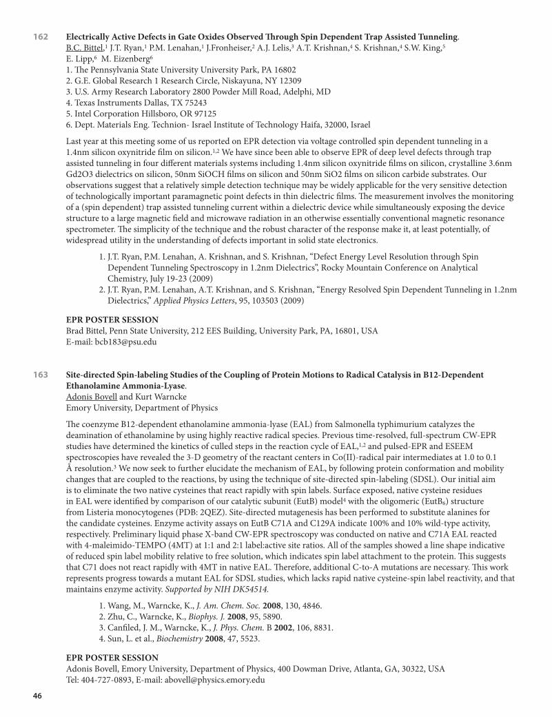
46
162 ElectricallyActiveDefectsinGateOxidesObservedThroughSpinDependentTrapAssistedTunneling. B.C. Bittel,1 J.T. Ryan,1 P.M. Lenahan,1 J.Fronheiser,2 A.J. Lelis,3 A.T. Krishnan,4 S. Krishnan,4 S.W. King,5 E. Lipp,6 M. Eizenberg6
1. The Pennsylvania State University University Park, PA 16802 2. G.E. Global Research 1 Research Circle, Niskayuna, NY 12309 3. U.S. Army Research Laboratory 2800 Powder Mill Road, Adelphi, MD 4. Texas Instruments Dallas, TX 75243 5. Intel Corporation Hillsboro, OR 97125 6. Dept. Materials Eng. Technion- Israel Institute of Technology Haifa, 32000, Israel
Last year at this meeting some of us reported on EPR detection via voltage controlled spin dependent tunneling in a 1.4nm silicon oxynitride film on silicon.1,2 We have since been able to observe EPR of deep level defects through trap assisted tunneling in four different materials systems including 1.4nm silicon oxynitride films on silicon, crystalline 3.6nm Gd2O3 dielectrics on silicon, 50nm SiOCH films on silicon and 50nm SiO2 films on silicon carbide substrates. Our observations suggest that a relatively simple detection technique may be widely applicable for the very sensitive detection of technologically important paramagnetic point defects in thin dielectric films. The measurement involves the monitoring of a (spin dependent) trap assisted tunneling current within a dielectric device while simultaneously exposing the device structure to a large magnetic field and microwave radiation in an otherwise essentially conventional magnetic resonance spectrometer. The simplicity of the technique and the robust character of the response make it, at least potentially, of widespread utility in the understanding of defects important in solid state electronics.
1. J.T. Ryan, P.M. Lenahan, A. Krishnan, and S. Krishnan, “Defect Energy Level Resolution through Spin Dependent Tunneling Spectroscopy in 1.2nm Dielectrics”, Rocky Mountain Conference on Analytical Chemistry, July 19-23 (2009)
2. J.T. Ryan, P.M. Lenahan, A.T. Krishnan, and S. Krishnan, “Energy Resolved Spin Dependent Tunneling in 1.2nm Dielectrics,” Applied Physics Letters, 95, 103503 (2009)
EPRPOSTERSESSIONBrad Bittel, Penn State University, 212 EES Building, University Park, PA, 16801, USA E-mail: [email protected]
163 Site-directedSpin-labelingStudiesoftheCouplingofProteinMotionstoRadicalCatalysisinB12-DependentEthanolamineAmmonia-Lyase. Adonis Bovell and Kurt WarnckeEmory University, Department of Physics
The coenzyme B12-dependent ethanolamine ammonia-lyase (EAL) from Salmonella typhimurium catalyzes the deamination of ethanolamine by using highly reactive radical species. Previous time-resolved, full-spectrum CW-EPR studies have determined the kinetics of culled steps in the reaction cycle of EAL,1,2 and pulsed-EPR and ESEEM spectroscopies have revealed the 3-D geometry of the reactant centers in Co(II)-radical pair intermediates at 1.0 to 0.1 Å resolution.3 We now seek to further elucidate the mechanism of EAL, by following protein conformation and mobility changes that are coupled to the reactions, by using the technique of site-directed spin-labeling (SDSL). Our initial aim is to eliminate the two native cysteines that react rapidly with spin labels. Surface exposed, native cysteine residues in EAL were identified by comparison of our catalytic subunit (EutB) model4 with the oligomeric (EutB6) structure from Listeria monocytogenes (PDB: 2QEZ). Site-directed mutagenesis has been performed to substitute alanines for the candidate cysteines. Enzyme activity assays on EutB C71A and C129A indicate 100% and 10% wild-type activity, respectively. Preliminary liquid phase X-band CW-EPR spectroscopy was conducted on native and C71A EAL reacted with 4-maleimido-TEMPO (4MT) at 1:1 and 2:1 label:active site ratios. All of the samples showed a line shape indicative of reduced spin label mobility relative to free solution, which indicates spin label attachment to the protein. This suggests that C71 does not react rapidly with 4MT in native EAL. Therefore, additional C-to-A mutations are necessary. This work represents progress towards a mutant EAL for SDSL studies, which lacks rapid native cysteine-spin label reactivity, and that maintains enzyme activity. Supported by NIH DK54514.
1. Wang, M., Warncke, K., J. Am. Chem. Soc. 2008, 130, 4846.2. Zhu, C., Warncke, K., Biophys. J. 2008, 95, 5890.3. Canfiled, J. M., Warncke, K., J. Phys. Chem. B 2002, 106, 8831.4. Sun, L. et al., Biochemistry 2008, 47, 5523.
EPRPOSTERSESSIONAdonis Bovell, Emory University, Department of Physics, 400 Dowman Drive, Atlanta, GA, 30322, USA Tel: 404-727-0893, E-mail: [email protected]

47
164 Site-directedSpin-labelingEPRStudiesofMultiply-DisulfideBondedProteinsHIV-1ProteaseandGM2ActivatorProtein. Jeffrey D. Carter, Jamie L. Kear, and Gail E. Fanucci; University of Florida, Department of Chemistry
Our lab deals with the expression, purification, and biophysical characterization of proteins with essential cysteine (CYS) residues that cannot be removed for proper in vivo function. Under consideration are GM2 activator protein (GM2AP) and human immunodeficiency virus type 1 protease (HIV-1PR); each of which play a critical role in health and disease. In this study, site-directed spin-labeling (SDSL) electron paramagnetic resonance spectroscopy (EPR) was utilized to examine the conformational changes and dynamics of the loop region of GM2AP and the flap region of HIV-1PR. SDSL-EPR was used on GM2AP in an attempt to monitor the conformational change in the loop region associated with ligand binding. The unnatural amino acid p-acetyl-L-phenylalanine was genetically incorporated into HIV-1PR in an attempt to examine the conformations of the flap regions of the Subtype B protease. Using CW and DEER EPR techniques, the distances between the selected sites can be measured, and distance profiles generated. We have shown previously, via traditional SDSL-EPR approaches, that the FDA-approved HIV-1PR inhibitors induce changes in the conformational sampling of the flap regions of the protease. However, traditional approaches require removal of the native CYS residues. By incorporating the unnatural amino acid p-acetyl-L-phenylalanine modified with the K1 spin label side chain, we can compare the distance profiles obtained for protease with these original results.
EPRPOSTERSESSIONJeff D. Carter, University of Florida, Chemistry, 214 Leigh Hall, Gainesville, FL, 32611, USA E-mail: [email protected]
165 EPRSpinProbeCharacterizationoftheCouplingofRadicalReactionChemistryandProteinandSolventMotionsinaB12Enzyme. Hanlin Chen and Kurt Warncke Emory University, Department of Physics
The kinetics of the substrate radical rearrangment step in the cryotrapped Co(II)-substrate radical pair in coenzyme B12-dependent ethanolamine ammonial-lyase (EAL) from Salmonella typhimurium have been measured by time-resolved, full-spectrum CW-EPR spectroscopy from 190-223 K in frozen aqueous solution.1,2 The proposed protein dynamical transition at ~210 K,1 which leads to a bifurcation of the monotonic reaction kinetics with decreasing temperature, and the effects of added sucrose on the reaction kinetics, evidence coupling of the core adiabatic reactions to protein and solvent motions. To gain further insight into this coupling, we have used CW-EPR and ESEEM spectroscopy of different TEMPO-derived spin probes to characterize the temperature dependent dynamics of water and water-sucrose solutions, and solute interactions, in the absence and presence of EAL. TEMPOL displays a mobility transition in water (decrease in outer line width from 74 to 36 Gauss) over a range of 200-220 K, which coincides with the proposed dynamical transition in EAL. Relatively low proportions of sucrose co-solvent shift the mobility transition to higher temperature, and induce slowing and polytonicity in the rearrangement reaction kinetics. The proximity of TEMPOL and 2H2-sucrose was probed by using 2H ESEEM, over the range of 0.5-75% (w/v) sucrose. The results reveal meso-domain co-localization of the solutes at low percent sucrose, as previously reported for glycerol solutions.2 Extension of the method to spin-labeled (4-maleimido-TEMPO) EAL indicates that the protein also resides in the meso-domain. The results calibrate the sucrose sensitivity of the substrate radical reaction kinetics, and provide an approach for correlating enzyme catalysis with protein and solvent motions. Supported by NIH DK54514.
1. Zhu, C., Warncke, K. Biophys. J. 2008, 95, 5890; J. Am. Chem. Soc. 2010, in press.2. Banerjee, D. and Bhat, S.V.J. Non-Cryst. Solids2009, 355, 2433.
EPRPOSTERSESSIONHanlin Chen, Emory University, Physics, 201 Dowman Drive, Atlanta, GA, 30322, USA E-mail: [email protected]
166 EntropicParadoxintheProtein-ligandComplexObservedbyFreeze-hyperquenchEPR. Alexey V. Cherepanov1,3 and Marat Gafurov1,2
1. Center for Biomolecular Magnetic Resonance 2. Institute for Physical & Theoretical Chemistry 3. Institute for Organic Chemistry & Chemical Biology, Goethe-University, Max-von-Laue-Str. 7, 60438 Frankfurt am Main,
Germany
An early intermediate state of the binding reaction between horse heart metmyoglobin and sodium azide was isolated by microsecond freeze-hyperquenching at pH=4.7 and 77K. The apparent sample-ageing time was 20-30μs. The sample

48
temperature was increased in 5K steps from 140 to 180K and the sample was incubated at fixed temperatures for <20-min intervals. In between the incubations, the sample was cooled to 14K for the X-band cw EPR measurements. EPR showed rapid binding of azide to the haem iron during the 1-3-min-long 5K temperature increase steps around 160-180K. The reaction rates at fixed temperatures in the same range were at least 10-50-fold slower. Could this rapid binding be explained by a collective motion of highly viscous liquid water, which might steer the protein conformational changes during crystallization of cubic ice at ~170K? Judging the low heat of ice crystallization (-0.31 kcal mol-1) and relatively high activation energy of azide binding (15.8 kcal mol-1), the reaction in this temperature range is expected to be both enthalpically and entropically slow.
Incubation of low-spin haem iron – azide complex at 220K leads to the formation of high-spin species with more than 2-fold broader derivative signal at 3g⊥ compared to the free metmyoglobin and the weaker signal at g⊥ shifted from 2.008 to 2.027. These species can be attributed to the metmyoglobin-hydrazoic acid complex MetMbFe3+-HN3.
Sample preparation. The reaction was triggered by a submicrosecond protein-ligand mixing. After mixing, a 18-20-μm-diameter liquid jet with nozzle velocity of 250 m s-1 was sprayed in the supercooled liquid methane rotating at 250 rpm at 77K. The freeze-quenched glassy reaction mixture contained >65% of non-reacted metmyoglobin as detected by X-band EPR and low-temperature optical spectroscopy. X-ray diffraction analysis showed that the sample phase consisted of >90% vitrified water. Formation of cubic ice was observed after warming to ~190K.
EPRPOSTERSESSIONAlexey V. Cherepanov, Goethe University, Max-von-Laue-Str. 7, Frankfurt am Main, Hessen, D-60438, Germany E-mail: [email protected]
167 AdaptiveSignalAveragingTechniqueforEnhancingtheSensitivityofContinuousWaveMagneticResonanceExperimentsII. C.J. Cochrane and P.M. Lenahan Pennsylvania State University, Department of Engineering Science
Last year, our group presented results of a signal averaging technique that greatly reduces the acquisition time in conventional electron spin resonance (ESR) and spin dependent recombination (SDR) on samples and devices with quite simple magnetic resonance spectra. More specifically, we utilized the exponentially weighted recursive least squares (EWRLS) algorithm in an adaptive linear prediction (ALP) scheme to enhance the signal to noise ratio (SNR) of individual magnetic resonance scans in ESR and SDR experiments. The filtered scans are then averaged separately and are shown to converge much faster than that of the conventional average. We have termed this technique adaptive signal averaging (ASA).1 In this study, we utilize the same technique on conventional continuous wave ESR measurements with somewhat more complex spectra to show that the filtering process can faithfully extract a more complex spectrum and compare our ASA results with those obtained via more conventional approaches. The filter was applied to measurements on the 3-carbamoyl-2,2,5,5-tetra-methyl-3-pyrrolene-1-yloxyl (CTPO) spin label. The ASA results were compared to those obtained through conventional signal averaging and the widely used Savitzky-Golay filter. We find that the ASA approach provides superior performance.
1. Cochrane and Lenahan, J. Magn. Res. 195, pp. 17-22, (2008).
EPRPOSTERSESSIONCorey Cochrane , Penn State University, 212 EES Building, University Park, PA, USA E-mail: [email protected]
168 HigherResolutionbySkewedProjectionofEchoDetectedEPRSpectra.Alex Cruce1, Preethi Vennam1, Ralph T. Weber2 and Michael K. Bowman1
1. Department of Chemistry, The University of Alabama, Box 870336, 35487-0336, Tuscaloosa, Alabama, USA 2. EPR Division, Bruker BioSpin Corporation, 44 Manning Road, Billerica, Massachusetts, USA
The ideal spectrum in EPR spectroscopy would consist of narrow peaks with high sensitivity, which leads to a good characterization of resonance frequencies and intensities. Resolution in pulsed EPR is often limited by rapid relaxation times, T2 and T2*, leading to broad lines and spectral overlap. The conventional field swept echo detected EPR spectra throws away sensitivity and resolution by effectively windowing the echo signal. To overcome these drawbacks, we describe a method that records the spin echo at different field steps to create a 2-D spectrum. Fourier Transformation of the echo followed by skewed projection produces a high resolution, high sensitivity spectrum. The projection allows spectral information from several magnetic fields to be summed, increasing signal to noise without reducing resolution. We have

49
been able to measure high resolution spectra over 1 GHz wide containing lines less than 1 MHz wide.
Acknowledgements: Work supported by NIH through GM061904 and HL095820. Thanks to Peter Höfer of Bruker-Biospin for technical advice.
EPRPOSTERSESSIONAlex A. Cruce, University of Alabama, Chemistry, Box 870336, Tuscaloosa, AL, 35487, USA Tel: 205-348-8457, E-mail: [email protected]
169 ProbingtheEffectsofPrimaryandSecondaryMutationsontheConformationalSamplingofHumanImmunodeficiencyVirusType1ProteaseSubtypeBbyDoubleElectron-ElectronResonanceSpectroscopy. Ian Mitchelle S. De Vera, 1 Adam N. Smith,1 Ben M. Dunn2 and Gail E. Fanucci.11. University of Florida, Department of Chemistry, Gainesville, FL 32611-7200 2. University of Florida College of Medicine, Department of Biochemistry and Molecular Biology, Gainesville, FL 32610
The development of drug resistance-associated mutations in the human immunodeficiency virus type 1 protease (HIV-1 PR) can lead to highly active antiretroviral therapy (HAART) failure.1 In this study, site-directed spin-labeling coupled with electron paramagnetic resonance (SDSL-EPR) was used to probe changes in the conformational sampling of subtype B HIV-1 PR attributed to the primary mutation D30N and the secondary mutations M36I and A71V. Double Electron-Electron Resonance (DEER) dipolar echo curves were converted to distance distribution profiles using Tikhonov regularization.2 The relative percentage of curled, tucked, closed, semi-open and wide-open conformations for each construct in the D30N, M36I and A71V mutation series were derived from Gaussian-shaped functions required to reconstruct the distance profile. A higher percentage of curled/tucked conformation of the HIV-1 PR was observed in constructs that have the M36I mutation. In the presence of D30N, adding the M36I and A71V mutations results to a higher percentage of wide-open conformation relative to wild-type. Changes in conformational sampling seen with DEER were correlated to the kinetic parameters for these constructs.3 The information gained from single-point mutation-induced changes in the conformational sampling of HIV-1 PR can provide key insights on the mechanism of drug resistance that is possibly useful in the development of new protease inhibitors.
1. Paolucci et al J. Antimicrob. Chemother., 2003, 51, 135-139. 2. Chiang et al J. Magn. Res., 2005, 172, 279-295.3. Clemente et al Biochemistry, 2003, 42, 15029-15035.
EPRPOSTERSESSIONIan Mitchelle S. De Vera, University of Florida, Chemistry, Leigh Hall, Gainesville, FL, 32611, USA Tel: 352-871-6807, E-mail: [email protected]
170 InvestigationoftheCu(II)-bindingPropertiesofAlpha-synuclein. Christopher G. Dudzik, Eric D. Walter and Glenn L. Millhauser University of California at Santa Cruz
Alpha Synuclein (AS) is a 140 residue, intrinsically disordered protein that is often discussed in the context of its role in neurological diseases such as Parkinson’s and Alzheimer’s disease. However, its true function and/or role in disease pathology remains the subject of debate. Sequence analysis of AS shows a series of 11-mer imperfect repeats from residues 1-90 that have been demonstrated to form an alpha-helix in the presence of lipid vesicles, similar to apolipoproteins. Indeed, further evidence has shown that AS associates with synaptic vesicles and plays a role in modulating vesicular trafficing to and fusion with the synaptic cleft membrane and protection of the vesicles from oxidative stress. Several studies have indicated that there is an abundance of stress on synaptic vesicles from Reactive Oxidative Species (ROS) and Cu(II) ions, particularly in the substantia nigra, which is strongly affected by Parkinson’s disease. Copper levels by mass/weight have been shown to be higher there than in other parts of the brain. In vitro assays show that Cu(II) decreases lag time in AS fibril formation and there have been numerous studies demonstrating the peptide’s affinity for Cu(II) ions. Perhaps then AS serves as a first line of defense against the ingress and damage of Cu(II) ions in the cell by sequestering these ions for later disposal. Elucidation of the mode(s) of AS-Cu(II) binding and/or their role in facilitation of fibril formation can therefore lead to further understanding of the natural function of AS. This work was generously funded by NIH grant GM65790.
EPRPOSTERSESSIONChristopher G. Dudzik, University of California at Santa Cruz, Chemistry, 1156 High Street, Santa Cruz, CA, 95060, USA E-mail: [email protected]

50
171 SpecMan4EPR:AversatilecontrolsoftwareforpulseEPRspectrometers. Boris Epel1 and Reef Morse2
1. University of Chicago, Department of Radiation Oncology, Chicago, IL 60637 2. Scientific Software Services, 39900 Stoneleigh St, Northville, MI 48167
The rapid development of modern state-of-the-art pulse EPR spectroscopy constantly creates new instrumental challenges. The scarcity of commercial solutions has motivated many research groups to develop their own instruments. Construction of pulse spectrometer requires a broad expertise both in microwave electronics and acquisition hardware. However, what turns the assembly of equipment into a robust and useful tool is its’ front-end, the control software. Often, home-built software has a lab lifetime of the student(s) who wrote it and usually does not benefit from the advantages of testing in different environments and the support of staff who understands and use the combined software and hardware. SpecMan4EPR is a comprehensive and inexpensive solution to this problem. SpecMan4EPR was introduced as a collaborative project between groups of Prof. Daniella Goldfarb, Weizmann Institute of Science and Prof. Arthur Schweiger, Swiss Federal Institute of Technology (ETH), Switzerland.1 The initial design of the software was carried out by BE, Dr. Stefan Stoll (currently Department of Chemistry, University of California, Davis, CA) and Dr. Igor Gromov (currently Bruker Biospin, Germany). The current version exhibits new generation of hardware engine, allows remote control, supports large data arrays for imaging and is in use in several laboratories across the world. A typical configuration of SpecMan4EPR implements a fast A/D converter (such as Agilent Acqiris board) and pulse generator (such as SpinCore PulseBlasterESR board or pattern generator from Chase Scientific) although many other instrumentation boards are supported. SpecMan4EPR implements a customizable pulse programming language and executes experiments with minimal overhead. Real time graphics provides visual verification of the acquisition process. SpecMan4EPR is a commercial product marketed by SSS.2 Full support and services are offered. Imaging features were developed in collaboration with the Center for EPR Imaging In Vivo Physiology, University of Chicago, NIH grants number P41 EB002034, R01 CA98575.
EPRPOSTERSESSIONBoris Epel, University of Chicago, Department of Radiation and Cellular Oncology, 5841 S. Maryland Avenue, MC1105, Chicago, IL, 60637, USA E-mail: [email protected]
172 DistanceMeasurementsinthePrionProteinbyUnnaturalAminoAcidSpin-labelingandDoubleElectron-ElectronResonance(DEER)Spectroscopy. Eric G.B. Evans and Glenn L. MillhauserUniversity of California, Santa Cruz, Department of Chemistry and Biochemistry
The prion protein (PrP) is a membrane-anchored glycoprotein present in the central nervous system of all avian and mammalian species. The conversion of the normal cellular isoform, termed PrPC, into a misfolded and infectious form, PrPSc, is implicated in the rare but fatal class of diseases known as the transmissible spongiform encephalopathies (TSEs). Despite the high conservation of PrP, the precise function of the protein in vivo is largely unknown. PrP binds Cu2+ in a multi-component fashion through its unstructured and highly flexible N-terminal domain, an action that is essential for PrP endocytosis and neuronal protection. Moreover, Cu2+ slows the rate of PrP fibril formation in vitro, although the mechanism of this inhibition is unknown. To probe a potential metal ion-mediated interaction between the copper binding domain and the structured C-terminal domain, site-directed spin-labeling (SDSL) and EPR spectroscopy can be used to determine inter-nitroxide distances. In the present study, doubly-labeled mouse PrP constructs were generated through the site specific genetic incorporation of the unnatural amino acid p-acetyl phenylalanine (pAcF).1 Unlike traditional cysteine-based SDSL, the addition of pAcF to the primary sequence of PrP does not interfere with the formation of the native disulfide bond during oxidative refolding. pAcF PrP mutants were then reacted with a nitroxide radical bearing an aminoxy functional group to generate the corresponding ketoxime-linked spin-labeled proteins.2 Inter-nitroxide distances were obtained using four-pulse double electron-electron resonance (DEER) EPR. Distances between labels placed in the structured C-terminus are in reasonable agreement with the known structure of the C-terminal domain. Expansion of this method to generate inter-domain distance constraints in PrP as a function of Cu2+ may help point to a physiological role for PrP and will likely provide insight into the role of Cu2+ in prion diseases.
1. Wang et al., Proc Natl Acad Sci USA, 2003, 100(1), 562. Fleissner et al., Proc Natl Acad Sci USA, 2009, 106(51), 21637
EPRPOSTERSESSIONEric G.B. Evans, University of California, Santa Cruz, Chemistry and Biochemistry, 1156 High Street, Santa Cruz, CA, 95064, USA E-mail: [email protected]

51
173 Cu(II)-imidazoleCoordinationStructureintheAmyloid-βProteinofAlzheimer’sDiseaseRevealedby14NESEEMSpectroscopy. William A. Gunderson,1 Jessica Hernandez-Guzman,1 Veronika A. Szalai2 and Kurt Warncke.11. Emory University, Department of Physics, Atlanta, GA 30322. 2. University of Maryland, Baltimore County, Department of Chemistry & Biochemistry, Baltimore, MD 21250
Aggregation of the amyloid-β (Aβ) protein is associated with the development of Alzheimer’s disease (AD). The transition metal ions, Cu(II), Zn(II) and Fe(II), are found at high concentrations in Aβ aggregates. However, roles of metals in Aβ toxicity and AD pathology remain unknown. Resolution of metal involvement in AD, and the development of therapeutic approaches, requires the characterization of the metal coordination environment(s) in Aβ. We are developing and applying techniques of 14N ESEEM spectroscopy of the non-coordinated (remote) imidazole ring nitrogen to determine the 3-D structure of multiple histidine coordination of S=1/2 Cu(II) in native [Aβ(1-40, 42)] and modified Aβ peptides, and related model complexes. The soluble truncated Aβ(1-16) peptide segment contains the Cu(II) binding domain, but lacks the hydrophobic domain, thus eliminating aggregation-induced sample heterogeneity. It has been previously proposed that either two or three histidines are coordinated to Cu(II), in both Aβ(1-16) and Aβ(1-40). Simulations of the 14N ESEEM from Cu(II)-Aβ(1-16), by using OPTESIM,1 are used to determine the histidine coordination number. Model complexes, including bis-cis and bis-trans imidazole Cu(II), and [tris-imidazole-carboxyaldehyde Cu(II)], are also examined. Analysis of the histidine imidazole remote 14N ΔmI=±2 “double quantum” feature and harmonics is used to determine the relative orientations of the imidazole ligands. The results also provide constraints on the Cu(II) interaction conformation assumed by Aβ.
1. Sun, et. al., J. Magn. Res., 2009,200, 21.
EPRPOSTERSESSIONWilliam A. Gunderson, Emory University, Physics, 400 Dowman Dr. NE, Atlanta, GA, 30322, USA Tel: 404-727-4287, E-mail: [email protected]
174 InactivingD25NMutationinHIV-1ProteaseAltersProteaseStability,andFlapConformationsandFlexibility. Xi Huang, Jamie L. Kear, Angelo M. Veloro, Mandy E. Blackburn and Gail E. Fanucci University of Florida, Department of Chemistry
Subtype-specific and drug-pressure selected polymorphisms among HIV-1 protease (HIV-1PR) variants have been shown to induce altered conformational sampling and flexibility of the flap regions of the protease. Here, we have utilized the pulsed EPR technique called double electron-electron resonance (DEER) to investigate the difference between flap conformations and flexibility in active (D25) and inactive (D25N) subtype F HIV-1PR constructs with each of the nine FDA-approved HV-1 protease inhibitors. Not surprisingly, results indicate that active constructs, when bound to inhibitor, have increasingly closed flap conformations when compared to inactive constructs. Furthermore, differential scanning calorimetry (DSC) was used to compare the stability of both the inactive and active HIV-1PR-inhibitor complexes. These data, which correlate with our DEER results, show that inactive constructs generally have low stability when compared to the active constructs.
EPRPOSTERSESSIONXi Huang, University of Florida, Department of Chemistry,, University of Florida, PO Box 117200, Gainesville, Florida 32611, Gainesville, FL, 32611, USA E-mail: [email protected]
175 TheGlobalAnalysisofDEERData. Eric J. Hustedt Vanderbilt University, Department of Molecular Physiology and Biophysics
Previously, global analysis has been successfully used to analyze both continuous wave and saturation transfer EPR data1-3. In the present work, algorithms have been developed for the global analysis of DEER data. Potential applications include the analysis of DEER data collected at multiple frequencies or multiple timescales. Careful analysis of DEER data from the soluble protein CDB3 (MW ≈ 90 kD) have shown that the background DEER signal is not well-fit by an exponential decay due to the large size of the CDB3 dimer. As a result, background correction with an exponential decay prior to analysis results in a poor fit to the data. An algorithm has been developed which explicitly fits the background signal with the radius of the molecule (assuming it is spherical) and the spin concentration as parameters. Using this approach, excellent fits to DEER data can be obtained without prior background correction. The global analysis of DEER data collected at multiple time scales can aid in the simultaneous analysis of the background signal and the distance distribution of interest. Also, DEER data can be globally analyzed to determine changes in the relative populations of components of the distance

52
distribution as a function of changing experimental conditions. For example, DEER has been previously used to study the structural effects of a proline to arginine mutation at residue 327 of CDB3. Intradimer distances in spin-labelled wild type CDB3 can be fit using a single component distance distribution4. The same measurements on P327R CDB3 indicate the mutation induces a second more disordered component in the distance distribution5. The global analysis of DEER data collected for multiple spin-labelling sites in both the WT and P327R background is being used to further test this two-component model. Supported by NIH GM 080513.
1. Hustedt and Beth, Biophysical Journal 69:1409-1423 (1995). 2. Hustedt et al., Biophysical Journal 64:614-621 (1993). 3. Hustedt et al., Biophysical Journal 72:1861-1877 (1997). 4. Zhou et al., Biochemistry 44:15115-15128 (2005). 5. Zhou et al., Biophysical Journal 90:528A (2006).
EPRPOSTERSESSIONEric J. Hustedt, Vanderbilt University, Department of Molecular Physiology and Biophysics, 735B Light Hall, Nashville, TN, 37232, USA Tel: 615-322-3181, E-mail: [email protected]
176 EffectofGlucoseonSpinLabelEPRinbloodfromHealthyandDiabeticVeins. Asako Kawamori, Hiroshi Kosugi and Wataru Hattori AGAPE-Kabutoyama Institute of Medicine
Glucose buffer solution of various concentration from 0 to 10 mM was added in blood from a healthy human vein. 0.2 mM TEMPO spin label was added and ESR signal decay has been observed. The rate of signal decay by reduction of TEMPO in blood increased until 20 % by addition of original value of blood glucose. Further addition of glucose decreased the reduction rate from the original value of 1000 ×10-3 to 300 ×10-3 for one example. These results are well understood that the rate of reduction from diabetic blood is higher for the value of 30 % high value of that in healthy blood and lower for the higher value of blood glucose than 50 %. The glucose effect on blood in mouse tail in vivo will be compared. Detailed results of EPR study of relation between blood glucose and diabetic disease will be presented.
EPRPOSTERSESSIONAsako Kawamori, AGAPE-Kabutoyama Institute of Medicine, Kabutoyama-cho 53-4, Nishinomiya, 662-0001, Japan Tel: 81-798-61-8402, E-mail: [email protected]
177 NitroxideLineshapeAnalysisWithX-BandPureAbsorptionRapidScan(PARS)ElectronParamagneticResonance(EPR). Aaron W. Kittell, Theodore G. Camenisch and James S. Hyde Medical College of Wisconsin, Department of Biophysics
Pure absorption rapid scan (PARS) electron paramagnetic resonance (EPR) involves rapidly sweeping the magnetic field (5-10 kHz) by driving the modulation coils to change the field. Direct detection of voltage changes allows for a pure absorption display. This methodology was initially developed at low frequencies (1-3 GHz) to improve the sensitivity of measuring small dipolar broadenings on a narrow line. However, it also has the distinct advantage of avoiding field modulation which is used in slow-scan continuous wave (CW) EPR to obtain phase-sensitive detection. Avoiding field modulation is desirable because it can potentially distort lineshapes and/or prevent the observation of small features. This becomes particularly important at X- and Q-band frequencies when g and A components overlap in slow tumbling nitroxide spectra. To probe this opportunity an X-band PARS EPR spectrometer has been developed. The three lines of 14N MTSL spectrum were analyzed individually using the PARS and CW EPR techniques over four orders of magnitude in rotational correlation time. Spectra were compared by integrating the first derivative spectrum obtained by the slow-scan technique.
EPRPOSTERSESSIONAaron W. Kittell, Medical College of Wisconsin, Biophysics Dept., 8701 Watertown Plank Road, Milwaukee, WI 53226, USA Tel: 414-456-4799, E-mail: [email protected]

53
178 TestingtheSiteofSubstrateBindingonNitrogenaseCofactorby95MoENDORSpectroscopy. Dmitriy Lukoyanov1 Zhi-Yong Yang,2 Dennis R. Dean,3 Lance C. Seefeldt,2 and Brian M. Hoffman1
1. Department of Chemistry, Northwestern University, Evanston, IL 60208 2. Department of Chemistry and Biochemistry, Utah State University, Logan, UT 84322 3. Department of Biochemistry, Virginia Tech, Blacksburg, VA 24061
Nitrogenase reduces dinitrogen gas to ammonia with the participation of eight protons and electrons. During this reaction substrate binds to the [7Fe, 9S, Mo, X] cluster – cofactor of the MoFe protein of nitrogenase and gets reduced with electrons supplied by the partner Fe protein. In our study we used ENDOR spectroscopy of 95Mo enriched nitrogenase to clarify the binding sites on cofactor for substrates in intermediates trapped with V70I mutant and WT proteins. Substitution of V70 with the larger amino acid Ile allowed trapping an intermediate state during reduction of protons. It has been shown that the intermediate is the four-electron reduced E4 state, and it contains two hydrides bound to cofactor. Checking the hyperfine interaction for the molybdenum site of 95Mo-enriched nitrogenase V70I protein under turnover conditions provides a test if binding involves the Mo atom. The other object of study were CO-inhibited forms of WT nitrogenase. It is known that CO is a strong inhibitor of all substrate reductions by nitrogenase, except for H+ reaction, and this makes the CO-intermediates good subjects for testing binding sites of substrates different from hydride. ENDOR study of 95Mo enriched nitrogenase proteins in the resting state and trapped during reaction showed that even though the molybdenum site of cofactor is clearly perturbed during turnover, 95Mo hyperfine coupling did not change significantly and is rather weak. These results do not support the idea of Mo as binding site of FeMo cofactor. Nevertheless, the observed alterations of the Mo coupling during reaction indicates that Mo is involved in modulation of properties of atoms at the binding FeS face.
EPRPOSTERSESSIONDmitriy Lukoyanov, Northwestern University, 2145 Sheridan Road, Evanston, IL, 60208, USA Tel: 847-491-4508, E-mail: [email protected]
179 NewApproachesinDiscriminationandCharacterizationofMembraneDomainsUsingEPRSpin-labelingmethods. Laxman Mainali, Marija Raguz and Witold K. Subczynski Department of Biophysics, Medical College of Wisconsin
EPR spin-labeling methods provide a unique approach to discriminating membrane domains and to determining several important membrane properties as a function of bilayer depth. One of us (WKS) was involved in the development of the discrimination by oxygen transport (DOT) method. This is a dual-probe, saturation-recovery EPR approach in which the observable parameter is the spin-lattice relaxation time (T1) of lipid spin labels, and the measured value is the bimolecular collision rate between molecular oxygen and the nitroxide moiety of spin labels. When located in two different membrane domains, the spin label alone most often cannot differentiate between these domains, giving similar conventional EPR spectra and similar T1 values. However, even small differences in lipid packing in these domains will affect oxygen partitioning and oxygen diffusion, which can be detected by observing the different T1s from spin labels in these two locations in the presence of oxygen. In the new approach, we changed the hydrophobic relaxation agent oxygen to the water soluble NiEDDA. This allowed discrimination of membrane domains under conditions in which the DOT method is ineffective. These experiments form the basis of a new discrimination method: the discrimination by relaxation agent accessibility (DRA) method. Both methods were used to detect and to characterize coexisting domains in phospholipid membranes, with cholesterol content changing from zero to values exceeding the cholesterol solubility threshold. These two approaches, with oxygen and NiEDDA as relaxation agents, can provide complementary information about membrane lateral organization and domain properties. Thus, the method of discriminating membrane domains that is based on saturation-recovery measurements can be significantly broadened and strengthened with the use of hydrophobic and hydrophilic relaxation agents. Advantages and limitations of both approaches will be discussed. The new DRA method includes the DOT approach in which the relaxation agent is already defined.
EPRPOSTERSESSIONLaxman Mainali, Medical College of Wisconsin, Department of Biophysics, 8701 Watertown Plank Road, Milwaukee, WI, 53226, USA Tel: 414-456-4933, E-mail: [email protected]

54
180 Membranedomainsinsphingomyelin/cholesterolmembranes:TheirstructureandpropertiesusingEPRSpin-labelingMethods. Laxman Mainali, Marija Raguz and Witold K. Subczynski Department of Biophysics, Medical College of Wisconsin
Conventional and saturation-recovery EPR spin-labeling methods were used to investigate the physical properties of fluid-phase membranes made from egg sphingomyelin (ESM) and cholesterol at a cholesterol/ESM mixing ratio of 0 to 3. This work focused on the properties of the phospholipid-cholesterol domain, which with increased cholesterol content consists of the liquid-disordered phase, the liquid-disordered plus the liquid-ordered phase, the liquid-ordered phase, and the liquid-ordered phase coexisting with the cholesterol crystalline domain. Profiles of the alkyl chain order, as well as membrane fluidity and hydrophobicity, were obtained for each region of the phase diagram. Interestingly, membrane properties change drastically when the cholesterol content increases up to 20 to 33 mol%. Further increase of the cholesterol content causes much smaller (almost negligible) changes to membrane properties. Major changes caused by cholesterol include increase of alkyl-chain order at all depths in the membrane, increase of membrane fluidity in the membrane center and decrease of membrane fluidity close to the membrane surface, increase of hydrophobicity in the membrane center, and decrease of hydrophobicity in the polar head-group region. These results indicate that the major changes in membrane properties occur during formation of the liquid-ordered phase. When the liquid-ordered phase is completely formed and the cholesterol content is 33 mol%, membrane properties “stabilize” and change very little if the cholesterol content in the membrane increases further, also beyond the cholesterol solubility threshold (66 mol% for sphingomyelin membranes). Because human eye lenses are abundant in sphingolipids (mainly, sphingomyelin and dihydrosphingomyelin that can account for 66% of total phospholipids) and contain high (saturating and over-saturating) amounts of cholesterol, this work will help in understanding cholesterol function in eye lens fiber-cell membranes. Supported by NIH grants EY0115526, TW008052, and EB001980.
EPRPOSTERSESSIONLaxman Mainali, Medical College of Wisconsin, Department of Biophysics, 8701 Watertown Plank Road, Milwaukee, WI, 53226, USA Tel: 414-456-4933, E-mail: [email protected]
181 Out-of-PhasePELDOR. Andriy Marko, Vasyl Denysenkov and Thomas Prisner Goethe University, Institute of Physical and Theoretical Chemistry
Pulsed Electron-electron Double Resonance (PELDOR) is a method frequently used to determine the structure of bio-macromolecule on nanometer scale.1 With this technique distances between native paramagnetic centers or introduced spin labels and their mutual orientation can be extracted from the experimental data. Usually PELDOR experiments are carried out in the high temperature limit, i. e., when the Bolzmann populations of spins oriented parallel and antiparallel to external magnetic field are almost equal. There are also well-developed theories describing PELDOR in this case. However, the high temperature limit conditions are no more fulfilled in the experiments done in high magnetic field (above 6 Tesla) at low temperature (below 5 K), when the Zeeman interaction energy of an electron spin becomes comparable with the thermal energy kBT. In this work we demonstrate that the PELDOR signals measured at low temperature and high field deviate from the signals measured in the high temperature limit. The signals recorded at low temperature contain standard in-phase component which is usually observed in PELDOR and additional out-of-phase component that disappears by increasing the temperature.2 In the rotating coordinate system, it means that we observe not only the modulation of the refocused transversal magnetization along a single axis but rather its precession in the x-y plane with dipolar frequency. For this effect we provide a qualitative explanation as well as a detailed analysis based on the density matrix formalism. We have also shown that the ratio of out-of-phase signal, which contains the same intermolecular relaxation term as in-phase signal, to in-phase signal can be utilized to determine distance distribution function without background correction of PELDOR time trace.
1. Milov A., Ponomarev A. and Tsvetkov Yu., Chem. Phys. Lett, 110, 67 – 72 (1984) 2. Thurnauer M., Clark C., Photochemistry and photobiology, 40, 381-386 (1984)
EPRPOSTERSESSIONAndriy Marko, Goethe University, Institute of Physical and Theoretical Chemistry, Max-von-Laue-Str.7, Fankfurt am Main, 60438, Germany E-mail: [email protected]

55
182 Predictionofthe6,6'-dioxo-3,3'-biverdazylElectronicGroundStatebyDifferenceDedicatedMulti-ReferenceConfigurationInteractionandBrokenSymmetryTechniques. Saba M. Mattar and Hisham M. DokainishUniversity of New Brunswick, Department of Chemistry and Centre for Laser, Atomic and Molecular Sciences,
The Heisenberg-Dirac vanVleck exchange parameter (J) and singlet-triplet energy differences (ΔEST) of 6,6’-dioxo-3,3’-biverdazyl (BVD) are determined by the broken symmetry hybrid density functional (BS-HDF) and multi-reference difference dedicated configuration interaction (MRDDCI) methods. Energy scans as a function of the dihedral angle between the two verdazyl rings (φN2C3C3’N2’) have been performed. The BS-HDF calculations show an antiferromagnetic ground state with a diradical index (RBS) of 97.5 – 99.9% . This indicates that the interactions between the two unpaired electrons are very small. To properly calculate and ΔEST, the multireference character introduced by these weak spin-spin interactions must be taken into account. Therefore the MRDDCI method is used. The σ, out-of-plane-π (OPπ) and in-plane-π (IPπ) configurations are incorporated in the CI expansions in a balanced way. This reveals that the OPπ-OPπ and OPπ-IPπ overlaps are the predominant factors that influence the J and ΔEST as a function of φN2C3C3’N2’. They cause the antiferromagnetic interactions to be minimal around 40° and 140°and the MRDDCI predicts a triplet ground state. At φN2C3C3’N2’=0, ΔEST[MRDDCI3(14,12)] is in excellent agreement with that of 1,1’,5,5’-tetramethyl-6,6’-dioxo-3,3’-biverdazyl determined experimentally. This corroborates the theory that the BS-HDF technique increases the singlet-triplet energy gap and favors the singlet state.
EPRPOSTERSESSIONSaba M. Mattar, University of New Brunswick, Chemistry, 30 Dineen Drive, Fredericton, NB, E3B 6E2, Canada Tel: 506-447-3091, E-mail: [email protected]
183 EPRBasedStructuralBiologyatMiamiUniversity'sOhioAdvancedEPRLaboratory.Robert M. McCarrick, Daniel Majo, Harishchandra Ghimire, Indra Sahu, Yunhaung Yang Michael A. Kennedy and Gary A. LoriganMiami University, Department of Chemistry
At the Ohio Advanced EPR Laboratory, we are focused on fulfilling the EPR needs of the Ohio area and the Midwest. Our facilities include a state of the art Bruker ELEXSYS E580 pulse spectrometer and two Bruker EMX continuous wave spectrometers. At Miami University, multiple faculty members are using the facilities to examine systems ranging from spin labelled solution and membrane bound protein systems, to metalloproteins and synthetic models to large RNA macromolecules. Novel methods for rapidly determining local protein secondary structure using ESEEM methods and tackling the difficult problem of NMR based structural determination of protein homodimers and oligomers using carefully chosen DEER measurements are currently being developed. The ability to conduct DEER measurements at Q-band, where the sensitivity is greatly increased, has allowed for the more rapid throughput of samples. We are currently awaiting a 10 W upgrade to the SuperQ-FT bridge on the pulse spectrometer which will allow for shorter pulses, yielding greater pulse bandwidth and further enhanced sensitivity. In addition, we will be receiving a continuous wave Q-band bridge for one of our EMX spectrometers with a dispersion mode arm that will add Q-band CW ENDOR capabilities.
EPRPOSTERSESSIONRobert M. McCarrick, Miami University, Department of Chemistry and Biochemistry, 701 E. High Street, Oxford, OH, 45056, USA Tel: 513-273-4993, E-mail: [email protected]
184 ResonatorforOptimizationofLiquid-PhaseEPRConcentration-SensitivityforSpinLabelsatQ-Band.Richard R. Mett,1,2 James R. Anderson,1 Jason W. Sidabras,1 and James S. Hyde1
1. Medical College of Wisconsin, Department of Biophysics, Milwaukee, WI 53226-0509 2. Milwaukee School of Engineering, Department of Physics and Chemistry, Milwaukee, WI 53202-3109
A novel resonator that optimizes liquid-phase concentration sensitivity at Q-band (35 GHz) is presented. The resonator has a coaxial TM020 mode, which is uniform in all but the radial direction. The 50 μl sample volume in the form of a thin (0.09 mm) cylindrical shell of an 8 mm radius is predicted to give a saturable EPR signal 21 times the standard 0.5 μl sample in the cylindrical TE011. The structure incorporates several recent innovations of this laboratory (uniform field,1 long slot iris,2 radial feed, cutoff cavity, and waveguide taper) and was designed using the finite-element computer program Ansoft HFSS (ver. 11, Pittsburg, PA) and Wolfram Mathematica (ver. 7, Champaign, IL). The resonator is coupled by a radially symmetric long-slot capacitive iris driven by a central cylindrical TM010 coupling cavity. The coupling cavity is driven by a TE10 mode in a narrow rectangular waveguide open on the large face. A tuning adjustment placed in a pyramidal taper between the standard rectangular WR-28 and the narrow rectangular waveguide provides a wide tuning range to accommodate changes in temperature and machining tolerances. With an aqueous sample, the device has a loaded Q-value of about 1500 and a resonator efficiency parameter of 0.72 G/W1/2. Convenient sample access on one end of the cavity is provided by a circular

56
cut on an rf current null. Design criteria and results of simulations are presented. Fabrication of the resonator in two parts and the Rexolite sample holder, also in two parts, is currently underway. Bench measurements will be presented. Sample filling and handling will be discussed.
1. R.R. Mett, W. Froncisz, and J.S. Hyde, Rev. Sci. Instrum., 2001, 72, 4188.2. R.R. Mett, J.W. Sidabras, and J.S. Hyde, Appl. Magn. Reson., 2008, 35, 285.
EPRPOSTERSESSIONRichard R. Mett, Medical College of Wisconsin, Biophysics, 8701 Watertown Plank Road, Milwaukee, WI, 53226-0509, USA Tel: 414-456-4024, E-mail: [email protected]
185 Physical,ChemicalandMineralogicalCharacterizationofTestMaterialsusedin28-Dayand90-DayIntratrachealInstillationToxicologyStudiesinRats. William J. Miles, W.F. Moll, R.D. Hamilton and R.K. Brown Miles Industrial Mineral Research
Two recent intratracheal instillation toxicology studies in rats clearly show that a naturally occurring quartz, with occluded crystal surfaces (quartz isolate), produced significantly less inflammatory response than a crushed reference quartz (DQ12). Respirable-size quartz isolate was isolated from bentonite parent rock, without crushing or the use of chemicals, to ensure that the surface properties of the quartz particles were unaltered. The isolation technique utilized gentle mechanical dispersion followed by sedimentation in an aqueous medium.
Extensive mineralogical and chemical characterizations were undertaken to define the physical and chemical properties of the test materials. The characterizations showed significant, measurable physical and chemical differences between the two quartz types. These differences may help to explain the difference in toxicological response associated with these materials. The evaluation methods and resulting data that characterized the chemical and physical properties of the instillation test materials are discussed. The data presented show that such characterizations are essential if meaningful correlations are to be made between test materials and their toxicological properties.
The physical and chemical evaluations include: bulk elemental chemistry; mineral composition and degree of crystallinity by x-ray diffraction analysis; surface characteristics, shape, diameter and particle size distribution by scanning electron microscopy; surface area by absorption of nitrogen or ethylene glycol monoethyl ether; thermal analysis by differential scanning calorimetry; zeta potential; and electon spin resonance (ESR/EPR).
EPRPOSTERSESSIONWilliam J. Miles, Miles Industrial Mineral Research, 1244 Columbine Street, Denver, CO, 80206, USA Tel: 303-355-5568, E-mail: [email protected]
186 Understandingtheα-HelicalConformationoftheN-TerminusinIA3UsingSite-DirectedSpin-labelingandElectronParamagneticResonance. Eugene Milshteyn, Natasha L. Pirman, and Gail E. Fanucci Department of Chemistry, University of Florida
Intrinsically disordered proteins (IDPs) comprise a class of proteins that are defined by largely unstructured domains under normal physiological conditions, but still have crucial roles in various biological processes. Many IDPs undergo conformational changes towards a structured state either upon binding to a target, or by chemical induction. In this study, we use site-directed spin-labeling and electron paramagnetic resonance (EPR) to monitor the conformational changes in the N-terminus of IA3, a 68 amino acid IDP that acts as an inhibitor of yeast proteinase A (YPRA) in Saccharomyces cerevisiae. Previously, IA3 has been used as a model to system to study the transition of IDPs toward a more structured state by other biophysical techniques such as nuclear magnetic resonance. Site-directed mutagenesis allows us to incorporate cysteine residues in various sites in the N-terminus, which can be chemically modified by IAP (3-(2-Iodoacetamido)-PROXYL), a sulfhydryl-specific nitroxide spin label. By using 2,2,2-trifluoroethanol (TFE) to induce IA3 into an α-helical conformation and collecting X-band EPR data, we can gather further qualitative and quantitative information on the unstructured-to-structured transition of the N-terminus of IA3. The results obtained from site-directed spin-labeling and EPR will be compared to those gathered from previous investigations of the N-terminus by NMR, circular dichroism and fluorescence.1,2,3 By providing new EPR analyses of the N-terminus, we can show how site-directed spin-labeling and EPR can be used on other IDPs to understand structural information in conformational changes.
1. Ganesh et al., Biochemistry, 2006, 45(45), 13585.2. Green et al., Biochemistry, 2004, 43(14), 4071. 3. Narayanan et al., JACS, 2008, 130(34), 11477.

57
EPRPOSTERSESSIONEugene Milshteyn, University of Florida, Chemistry, PO Box 117200, Gainesville, FL, 32611, USA Tel: 954-655-7844, E-mail: [email protected]
187 ApplyingX-BandRapid-scanEPRtoMeasureShortRelaxationTimes. Deborah G. Mitchell, Mark Tseytlin, Richard W. Quine, Gareth R. Eaton, and Sandra S. Eaton University of Denver
Rapid-scan EPR spectra of several samples -BDPA, nitroxyl radicals, DPPH, LiPc in oxygen, and LiPc in air-were collected at X-Band (9.8 GHz). The term “rapid” refers to a condition where the time that is spent passing through the absorption line is less than the relaxation time.1 Rapid magnetic field scans (rates ranging from 50 to 600 MG/s with scan widths up to 40 G) were generated with Bruker electron nuclear double resonance (ENDOR) coils and data were collected via an E580 Bruker spectrometer. The ENDOR coils are rotated 90o from their normal orientation such that the radio frequency (RF) field is parallel to the main magnetic field. These spectra were simulated using the Bloch equations2 to determine the relaxation times for LiPc in oxygen (T2 = 25 ns) and LiPc in air (T2 = 79 ns). Some of these spectra were also deconvolved to recover the slow scan lineshapes. The faster the scan rate and the narrower the EPR line, the larger the bandwidth that is required. In some cases the limiting bandwidth is the resonator Q.1
1. Joshi, J. P.; Eaton, G. R.; Eaton, S. S., Impact of resonator on direct-detected rapid-scan EPR at 9.8 GHz. Appl Magn Reson 2005, 28, 239-249.
2. Bloch, F.; Hansen, W. W.; Packard, M., The Nuclear Induction Experiment. Phys Rev 1946, 70, 474-485.
EPRPOSTERSESSIONDeborah G. Mitchell, University of Denver, Dept. of Chemistry and Biochemistry, 2101 E. Wesley Ave., CO, 80208, USA Tel: 303-871-2642, E-mail: [email protected]
188 DevelopmentofHigh-fieldOverhauser-enhancedMRIwithCircularTransportTechnique. Yukio Mizuta,1 Tatsuya Naganuma,2 Kazuhiro Ichikawa3 and Hideo Utsumi3
1. JEOL LTD, Analytical Instruments Division, Tokyo 196-8558, Japan 2. Japan Redox Corporation. Machinely Development Department, Fukuoka 812-0044, Japan 3. Innovation Centre for Medical Redox Navigation, Medical Redox Imaging Group, Kyushu University, Fukuoka 812-8582,
Japan
Overhauser-enhanced Magnetic Resonance Imaging (OMRI) is a free-radical imaging technique based on Overhauser effect. Overhauser effect is also known as a dynamic nuclear polarization, where NMR signal of proton interacting with free-radicals is modulated by polarization transfer from un-paired electron during EPR excitation. OMRI combined with Redox-sensitive nitroxyl radical (OMRI / spin probe technique) is a molecular imaging technique for in vivo Redox imaging. Electron excitation in OMRI / spin probe is carried out at low magnetic field (ca 20mT), because of the limitation of skin-depth penetration. Field-cycling technique was developed to improve image resolution on Low-EPR irradiation field. Field-cycle technique by use of switchable resistive magnet needs huge power to switch the field strength, and had a limitation of field strength upon 0.5T. Circular sample transport technique uses two different field strength stationary magnets for EPR excitation and NMR detection, and has “circular constant speed” sample transport mechanism. Circular sample transport technique has no limitation in magnetic field strength. This system is not influenced by the eddy current because of using stationary magnets. In this study, an OMRI imager was constructed by 1.5T permanent magnet for NMR detection and low field magnet (ca 20mT) for EPR excitation. OMRI resonator was constructed by solenoid MRI detection coil (64.14 MHz) and Alderman-Grants EPR excitation coil (565 MHz). The mouse-size phantom filled with 14N- or 15N- labeled Carbamoyl-PROXYL solution was used to evaluate the performance of our OMRI imager. In the obtained image with 1mm slice thickness, glass tube with 0.15mm wall thickness was clearly separated. This work was supported by the “Development of System and Technology for Advanced Measurement and Analysis Program” of the Japan Science and Technology Agency.
EPRPOSTERSESSIONYukio Mizuta, JEOL LTD., Analytical Instruments Division, 1-2 Musashino 3-Chome, Akishima, Tokyo, 196-8558, Japan Tel: +81-42-542-2241, E-mail: [email protected]

58
189 EPRforEveryone.GeneratingtheInterest of6-12GradersforMagneticResonance. Reef Morse, Kiyo A. Morse2, and Arthur Heiss3
1. Director, Steppingstone MAgnetic Resonance Training (SMART) Center, Farmington Hills, MI 48334 2. Head of School, Steppingstone Center for Gifted Education, Farmington Hills, MI 48334 3. Bruker BioSpin Corporation, Billarica, MA 01821-3991
Traditionally, magnetic resonance, especially EPR, is first introduced to highly advanced undergraduates, graduates, and post-docs. This usually occurs because a thorough understanding the instrument and the data it provides requires advanced mathematics and physics. The operation and success of the SMART Center shows that students as young as 6th grade can learn to operate a Bruker ESP300 EPR spectrometer and begin understand some of the basic spectral information they obtain in a 4 to 5-day course without advanced mathematics1. Such concepts as resonance and relaxation times can be taught using simple coupled pendula and students can apply that knowledge to interpret relaxation times and infer chemical structure from EPR spectra (for example, the difference in the charred product of burnt toast vs grilled meat). Originally, we thought students would be attracted to imaging, but, to our surprise, all were much more interested in measuring antioxidant properties of common beverages and free radical formation during food preparation2. Several science fair projects have resulted from these studies, one of which won best in subject (Chemistry) at the Detroit Science Fair. In summary, young students with no previous knowledge of magnetic resonance can use this methodology to do basic, but interesting, research.
1. Morse, Reef. Images, Newsletter of the Michigan Alliance of Gifted Education, Winter, 2009. 2. Morse, Reef. Journal of the Michigan Science Teachers Association, 2010, in press.
EPRPOSTERSESSIONReef Morse, Steppingstone MAgnetic Resonance Training (SMART) Center, 28555 Middlebelt, Farmington Hills, MI 48334, USA Tel: 248-539-1666, E-mail: [email protected]
190 IonizingRadiationTreatmentofChromium-DopedSyntheticForsteriteasStudiedbyMultifrequencyEPR. Dmitry A. Akhmetzyanov, Aleksei A. Konovalov, Laila V. Mosina ,Valery F. Tarasov and Evgeny R. Zhiteytsev Kazan Physical-Technical Institute, Russian Academy of Sciences
The effect of ionizing radiation on the structure and valence state of paramagnetic centers formed by impurity chromium ions in synthetic forsterite is studied. Samples were grown by the Czochralski method in the neutral argon or oxidizing atmosphere and irradiated with gamma-rays and high-energy electrons. X-band and tunable high-frequency EPR spectra were studied at 4.2 K. The use of the quasi-optical waveguide and backward wave oscillators as a source of the microwave radiation1 allowed us to obtain a two-dimensional pattern of magnetic field dependences of resonance frequencies for various transitions of chromium paramagnetic centers in the frequency range of 65 – 270 GHz at magnetic fields of 0 – 0.5 T. It is shown that in the non-irradiated samples there are single Cr2+, Cr3+ and Cr4+ ions and associates of Cr3+ ions with paramagnetic centers. It is established that upon ionizing radiation a small amount of Cr3+ ions transforms into Cr2+ ions. This results in the appearance of associates of Cr2+ ions with paramagnetic centers. The integral intensity of the EPR signals of these associates is significantly lower than that of the single Cr2+ ions. Both associates are non-Kramers centers with an integer electron spin, though the electron spin of the Cr3+ ion is half-integer and that of the Cr2+ ion is integer. Magnetic properties of these associates, their possible structure and the effect of ionizing radiation are discussed. The work is partially supported by the RFBR grant 09-07-97006 and the grant of the President of the Russian Federation NSh -6267.2010.2
1. Tarasov and Shakurov, Appl. Magn. Reson., 1991, 2, 571.
EPRPOSTERSESSIONLaila V. Mosina, Kazan Physical-Technical Institute, 10/7 Sibirsky trakt, Kazan, 420029, Russia E-mail: [email protected]
191 StructuralInvestigationofStratumCorneumLipidUsingElectronParamagneticResonance. Kouichi Nakagawa, Fukushima Medical University
Electron paramagnetic resonance (EPR) in conjunction with a slow-tumbling simulation was utilized for defining stratum corneum (SC) lipid structure. We found that ordering calculated from the simulation is an appropriate index for evaluating SC lipids structure.1, 2 The SC from two sites (mid-volar forearm and lower-leg) of human volunteers and cadaver was stripped consecutively from one to three times using a glass plate coated with a cyanoacrylate resin. Aliphatic spin probes, 5-doxylstearic acid (5-DSA) and 3β-doxyl-5α-cholestane (CHL), were used to monitor SC ordering. The SC samples were incubated in the probe aqueous 60 minutes. EPR spectrum of 5-DSA incorporated in the SC~solution at 37 ºC for demonstrated a characteristic peak for the first strip. However, EPR spectra of CHL in the SC did not show a clear difference

59
for each strip, except for the peak intensity. The results imply that CHL is not incorporated into the lipid phase as easily as is 5-DSA. A slow-tumbling simulation of the EPR spectrum was performed to analyze the lipid structure. The simulation results for 5-DSA show differences in values of the SC ordering. Thus, these results along with the simulation analysis provide detailed SC layer structure.
1. K. Nakagawa, et al., Spectrochimica Acta Part A, 63, 816 (2006).2. E. Yagi, K. Sakamoto, and K. Nakagawa, J. Invest. Dermatol., 127, 895 (2007).3. K. Nakagawa, Lipids, 45, 91 (2010).
EPRPOSTERSESSIONKouichi Nakagawa, Fukushima Medical University, 1 Hikarigaoka, Fukushima, 960-1295, Japan E-mail: [email protected]
192 DEERandfunctionalmutagenesisindicateaHydrophobicClusterintheForceGenerationRegionoftheMyosinHead,ImportantforMyosinFunction. Roman V. Agafonov,1 Sarah Blakely,2 Margaret A. Titus,2 David D. Thomas2 and Yuri E. Nesmelov3
1. University of North Carolina, Charlotte, NC 2. University of Minnesota, Minneapolis, MN 3. Brandeis University, Waltham, MA
We have used pulsed EPR spectroscopy (DEER) to determine the effect of point mutations on the structure of the myosin motor head. Wild type myosin and three mutants were analyzed. All three mutations are located in the force generating region of myosin head. As it was previously reported, F506A (D.discoideum sequence), located in the relay loop, disrupts communication between the nucleotide binding site and the force generation region in myosin. F692A and I499A, located on the interface with the converter domain, affect coordination of the converter domain relative to the myosin head. The mutations were introduced into A639C:K498C construct, labeled with maleimide spin probes, designed to access the conformation of myosin’s force generation region. Obtained spin echo decays were interpreted in terms of one or two Gaussian distance distributions, corresponding to one or two myosin structural states. Myosin in several biochemical states, mimicked with nucleotide analogs was studied. Two myosin conformations are usually present in one biochemical state, pre- and post-recovery stroke. We have found that the I499A mutation shifts equilibrium between structural states towards pre-recovery stroke state, and increases interprobe distance distribution, indicating disorder. This observation agrees with the proposed role of I499, responsible for the relay helix — converter domain interaction. I499A mutation disrupts the interaction, affecting the relay helix orientational stability. Both F506A and F692A have had similar effect, shifting equilibrium between structural states towards post-recovery stroke state, and significantly increasing interprobe distance distributions. Available atomic structures of myosin in different structural states indicate that both F506 and F692 may participate in a hydrophobic cluster formation, located in the force generating region. Observed similar structural effect of F506A and F692A mutations leads to a conclusion that proposed hydrophobic cluster in the force generation region is important for the force generation in myosin.
EPRPOSTERSESSIONYuri Nesmelov, University of North Carolina Charlotte, Physics, 306 Grigg Hall, 9201 University City Blvd, Charlotte, NC, 28223, USA Tel: 704-687-5886, E-mail: [email protected]
193 ConformationalDistributionsattheN-peptide/boxBRNAInterfaceStudiedUsingSite-directedSpin-labeling. Xiaojun Zhang,1 Sang Won Lee,2 Liang Zhao,2 Tianbing Xia,2 and Peter Z. Qin1
1. Department of Chemistry, University of Southern California, Los Angeles, California 90089, USA 2. Department of Molecular and Cell Biology, The University of Texas at Dallas, Richardson, Texas 75080-3021, USA
In bacteriophage lamda, the interaction of a 22-amino acid peptide (called the N-peptide) with a stem-loop RNA element (called boxB) has been shown to play a critical role in transcription anti-termination. The N-peptide/boxB complex has been extensively studied, and serves as a paradigm for understanding mechanisms of protein/RNA recognition. Particularly, ultrafast spectroscopy techniques have been applied to monitor picosecond fluorescence decay behaviors of 2-aminopurines (2AP) embedded at various positions of the boxB RNA. The studies have led to a dynamic two-state model, in which the N-peptide is in dynamic equilibrium between a stacked and unstacked state(s), and the function of the N-peptide/boxB complex is linked to the fraction of the stacked state. Here, the N-peptide/boxB system is studied using the site-directed spin-labeling (SDSL) technique, in which X-band electron paramagnetic resonance (EPR) spectroscopy is used to monitor nanosecond rotational behaviors of stable nitroxide radicals covalently attached to different positions of the N-peptide. The data revealed multiple N-peptide conformations within the complex, with the exchange rate between these states slower than 1 ns-1. The characteristics of these conformations are consistent with the proposed stacked and unstacked states, and

60
their distributions vary upon mutations with the N-peptide. These results suggest the dynamic two-state model, remains valid in the nanosecond regime, and represents a unique mode of function in the N-peptide/boxB RNA complex. It also demonstrates a connection between picosecond and nanosecond dynamics in a biological complex.
EPRPOSTERSESSIONPeter Z. Qin, University of Southern California, Department of Chemistry, LJS-251, 840 Downey Way, Los Angeles, CA, 90089-0744, USA Tel: 213-821-2461, E-mail: [email protected]
194 LigandBindingPocketPropertiesofGM2APandSapBbyCWEPR. Yong Ran and Gail E. Fanucci University of Florida, Department of Chemistry,
GM2 Activator Protein (GM2AP) and Saposin Activator Protein B (SapB) are two co-factor proteins required for sphingolipids degradation. X-band CW EPR spectroscopy of spin-labeled lipids in complex with the GM2AP and SapB, was utilized to characterize the hydrophobic binding pocket of these lipid transfer proteins. EPR line shapes reveal that the mobility of the labeled lipids within the binding pockets are more restricted than when in a lipid bilayer environment while the lipids in GM2AP are more restricted than in SapB. Power saturation experiments show that the accessibility to the lipid in complex with GM2AP is different from the complex with SapB. The results are consistent with the lipid binding models revealed by X-ray crystal structures. We also measured the kinetics of lipid extraction from multilamellar dispersions of each protein. We found that spin-labeled phospholipids have relatively higher affinity with GM2AP than with SapB. Competition assays for spin-labeled phospholipids and ganglioside lipids reveal that GM2AP also has a relatively higher affinity with ganglioside than with spin-labeled phospholipids.
EPRPOSTERSESSIONYong Ran, University of Florida, Chemistry Department, Gainesville, FL, 32611, USA E-mail: [email protected]
195 CW-EPR,ESEEM,andDEERSpectroscopicMeasurementsoftheFullLengthHumanKCNE1MembraneProtein. Indra D. Sahu,1 Aaron T. Coey,1 Kaylee R. Troxel,1 Thusitha S. Gunasekera,1 Jaclyn M. Hawn,1 Robert M. McCarrick,1 Congbao Kang,2 Richard Welch,2 Carlos G. Vanoye,2 Charles R. Sanders2 and Gary A. Lorigan1
1. Department of Chemistry and Biochemistry, Miami University, 45056, OH, USA, 2. Department of Biochemistry, Vanderbilt University, 37322, TN, USA.
The KCNE1 membrane protein plays an important role in regulating KCNQ1 which forms a potassium voltage gated channel in the human heart. Advanced EPR techniques such as Electron Spin Echo Envelope Modulation (ESEEM) and Double Electron Electron Resonance (DEER) coupled with site-directed spin-labeling can be used to obtain structural information both qualitatively and quantitatively by measuring distances. Small range distances between isotopically coupled nuclear spins and nitroxide electronic spin labels out to a distance of about 9Å with ESEEM, and long range distances of 20-70 Å between two nitroxide spin labels using DEER can be measured. Cysteine mutants were generated along the transmembrane domain (TMD) and extracellular region of KCNE1 and further modified by MTSL (S-(2, 2, 5, 5-tetramethyl-2, 5-dihydro-1H-pyrrol-3-yl) methyl methanesulfonothioate) nitroxide spin labels. The purified proteins were reconstituted into model membranes Fos-Cholin micelles and POPC/POPG bilayer vesicles. The insertion of KCNE1 into lipid bilayers was verified by using the method of CW-EPR Power Saturation performed on sites G60C (probe inside) and R32C (probe outside). ESEEM experiments were performed on i+1 to i+5 sites, where i represents the deuterium position V502H on the TMD, and DEER was performed on sites V47C-I66C and V50C-S68C to characterize the structural behavior of KCNE1. The distances extracted from ESEEM and DEER experiments are in good agreement with Nano-scale Molecular Dynamics (NAMD)/Visual Molecular Dynamics (VMD) modeling results.
EPRPOSTERSESSIONIndra D. Sahu, Miami University, Chemistry and Biochemistry, 501 East High Street, Oxford, OH, 45056, USA Tel: 513-529-4703, E-mail: [email protected]

61
196 AdvancesinLoop-GapResonatorTechnologyforX-bandAqueousSamples. Jason W. Sidabras1, Richard R. Mett1,2, and James S. Hyde1
1. Medical College of Wisconsin, Department of Biophysics 2. Milwaukee School of Engineering, Department of Physics and Chemistry, Milwaukee, Wisconsin 53202, USA
Finite-element modeling techniques have provided the methodology to create number of advances in loop-gap resonator (LGR) design over the last 10 years. Loop coupling has been replaced by coupling using a long capacitive iris1 directly to a WR-90 waveguide. The waveguide is side-wall coupled in order minimize cross-sectional footprint in order to fit into a 44 mm diameter dewer. Tuning was accomplished with a pill placed within the outside loop; this also reduces the footprint needed by integrating the matching structure in the resonator body. The outside loop was designed in such a way that the impedance looking in from the iris closely matches the characteristic impedance of the WR-90 waveguide. This novel configuration minimizes frequency pulling as the resonator is matched. The length of the resonator is approximately a free-space wavelength long leading to 2.5 times the sample volume and signal intensity.2 Due to an aspect ratio of 1 mm diameter by 33 mm height, the microwave uniformity is approximately 90% over the volume.
The new resonator was fabricated out of 0.999 pure silver and was machined by Electric Discharge Machining (EDM) techniques. EDM uses a wire or probe at a high voltage potential to arch away grounded conductive material in a water bath. This computer controlled fabrication process allows for positional tolerances of 0.001 mm and features down to 0.05 mm thickness. In order to allow 100 kHz field modulation to penetrate into the solid silver, modulation slots on the order of 0.1 mm are cut perpendicular to the sample axis. Microwave leakage is minimized due to the slots being parallel to the microwave currents.3 A graphite shield was also fabricated to load any residual microwave leakage and to provide protection to the field modulation slots. The resonator was characterized using a standard 10 μM TEMPO sample and compared to finite-element modeling simulations for agreement.
1. R.R. Mett, J.W. Sidabras, and J.S. Hyde, “Coupling of Waveguide and Resonator by Inductive and Capacitive Irises for EPR Spectroscopy.”, Appl. Magn Reson., 2009; 35(2):285-318.
2. R.R. Mett, J.W. Sidabras, and J.S. Hyde, “Uniform radio frequency fields in loop-gap resonators for EPR spectroscopy.”, Appl. Magn. Reson. 31: 571-587 (2007).
3. R.R. Mett, J.W. Sidabras, and J.S. Hyde, “Microwave leakage from field modulation slots in TE011 EPR cavities.”, Rev. Sci. Instrum. 76: 014702 (2005).
EPRPOSTERSESSIONJason W. Sidabras, Medical College of Wisconsin, Dept. of Biophysics, 8701 Watertown Plank Rd, Milwaukee, WI, 53226, E-mail: [email protected]
197 ConformationalFlexibilityofElectronTransferFlavoproteinProbedUsingDEERMeasurementsofDistancesBetweenSpinLabelsandaNativeFADSemiquinone.Michael A. Swanson,1 Velavan Kathirvelu,1 Gareth R. Eaton,1 Sandra S. Eaton,1 and Frank E. Frerman2
1. University of Denver, Department of Chemistry and Biochemistry, Denver, CO 2. University of Colorado School of Medicine, Department of Pediatrics, Aurora, CO
Electron transfer flavoprotein (ETF) is a soluble heterodimeric flavoprotein located in the mitochondrial matrix and is responsible for linking fatty acid β-oxidation to the main respiratory chain. It contains a flavin adenine dinucleotide (FAD) that is used to shuttle electrons from at least 10 different flavoprotein dehydrogenases to the membrane-bound electron transfer flavoprotein ubiquinone oxidoreductase (ETF-QO).1 It has been proposed that the αII domain of ETF is mobile allowing the promiscuous interactions with structurally different partners. Double electron-electron resonance (DEER) was used to measure the distance between spin labels at various sites and an enzymatically reduced FAD cofactor in Paracoccus denitrificans ETF.2 Two or three interspin distance distributions were observed for spin-labels in the αI (A43C) and βIII (A111C) domains, but only one is observed for a label in the αII (A210C) domain. This suggests that the αII domain adopts several stable conformations which may correspond to a closed/inactive conformation and an open/active conformation. An additional mutation, E162A, was introduced to increase the mobility of the αII domain. The E162A mutation doubled the activity compared to wild-type and caused the distance distributions to become wider. The DEER method has the potential to characterize conformational changes in ETF that occur when it interacts with various redox partners.
1. Toogood, H.S., Leys, D. and Scrutton, N.S. (2007), FEBS Journal 274, 5481-5504. 2. Swanson, M.A., Kathirvelu, V., Majtan, T., Frerman, F.E., Eaton, G.R. and Eaton, S.S. (2009) J. Am. Chem. Soc.
131, 15978-15979.
EPRPOSTERSESSIONMichael A. Swanson, University of Denver, Chemistry and Biochemistry, 2101 E. Wesley Ave., Denver, CO, 80208, USA Tel: 303-871-2978, E-mail: [email protected]

62
198 ConformationalChangesofSecBUponBindingtoaModelSubstrate-BPTI.Michaela M. Haimann1, Yasar Akdogan2, Raghavan Varadarajan3, Reinhard Philipp1, Dariush Hinderberger2 Wolfgang E. Trommer1
1. Department of Chemistry, TU Kaiserslautern, D-67653 Kaiserslautern, Germany 2. Max Planck Institute for Polymer Research, D-55128 Mainz, Germany 3. Molecular Biophysics Unit, Indian Institute of Science, 560012 Bangalore, India
SecB is a homotetrameric cytosolic chaperone that forms part of the protein translocation machinery in E. coli. Due to SecB, nascent polypeptides are maintained in an unfolded translocation-competent state devoid of tertiary structure and thus are guided to the translocon. In vitro SecB rapidly binds to a variety of ligands in a non-native state. We have previously investigated the bound state conformation of the model substrate bovine pancreatic trypsin inhibitor (BPTI) as well as the conformation of SecB itself by using proximity relationships based on site-directed spin-labeling and pyrene fluorecence methods. It was shown that SecB undergoes a conformational change during the process of substrate binding. In order to back up these results we generated SecB mutants containing but a single cysteine per subunit and could quantitatively label each cysteine with the methanethiosulfonate spin label (MTS) at positions C97 or E90C. Furthermore, we created a BPTI mutant wherein all cysteins were replaced by alanine in order to allow for a binding-competent unfolded structure devoid of disulfide bridges.
Highfield (W-band) EPR measurements revealed a slight decrease of the gxx-value upon addition of the model substrate to both SecB mutants indicating that with BPTI present the spin labels are in a more polar/hydrophilic environment. DEER measurements yielded information about the distances between the MTS-labeled cysteins which were in excellent agreement with distances obtained by molecular modeling. Binding of BPTI also led to a slight change in distances between labels at C97 but not at E90C. Whereas the shorter distance in the tetramer increases, the larger diagonal distance decreased. These findings can be explained by a widening of the tetrameric structure upon substrate binding much like the opening of two pairs of scissors.
EPRPOSTERSESSIONWolfgang E. Trommer, TU Kaiserslautern, Chemistry, P.O.Box 3049, Kaiserslautern, D-67653, Germany Tel: +49-631-205 2045, E-mail: [email protected]
199 GeneralMethodtoRecoverSlowScanSpectrafromSinusoidalRapidScans. Mark Tseytlin, George A. Rinard, Richard W. Quine, Gareth R. Eaton, and Sandra S. Eaton University of Denver
In rapid scan EPR, the magnetic field is scanned through the signal in a time that is short relative to relaxation times. Previously it has been shown that the slow scan lineshape could be recovered from triangular rapid scans by Fourier deconvolution.1 Larger currents, and therefore larger scan field amplitudes, can be achieved by resonating the magnetic field scan coils. Another advantage of sinusoidal magnetic field scans is that there is only one frequency, so the current in the coils does not include higher harmonics, which would require larger amplifier bandwidths. However, the mathematical algorithm that was used to deconvolve triangular scans is not entirely applicable to sinusoidal scans. Since the Fourier transform of the sinusoidal driving function did not appear to be integrable in closed form, a numerical method, which is applicable to any arbitrary current (field) waveform, was developed to do the deconvolution. Rapid scan data were acquired with a locally-designed coil driver system on our 250 MHz spectrometer. 2 The slow scan EPR lineshapes recovered from rapid triangular and sinusoidal scans are in excellent agreement for lithium phthalocyanine and for a trityl radical.
1. J.P. Joshi, J.R. Ballard, G.A. Rinard, R.W. Quine, S.S. Eaton, and G.R. Eaton, J. Magn. Reson. 175, 44-51 (2005). 2. R.W. Quine, T. Czechowski, and G.R. Eaton, Conc. Magn. Reson. B (Magn. Reson. Engin.) 35B, 44-58 (2009).
EPRPOSTERSESSIONMark Tseytlin, University of Denver, Chemistry and Biochemistry, 2101 E. Wesley Ave., Denver, CO, 80208, USA Tel: 303-871-2978, E-mail: Mark.Tseytlin
200 MeasuringtheInfluenceofIronIonsontheMagneticPropertiesinMetal-DopedApatiteNanoparticles. Robert Usselman,1 Michael T. Klem,2,3 Amy Nickel,2,3 Marissa Pedula,2,3 Rajendra Kumer,2,3 and Stephen Russek1
1. Magnetics Group, National Institute of Standards and Technology, Boulder, Colorado 80305, USA 2. Department of Chemistry and Geochemistry, Montana Tech of the University of Montana, Butte, Montana 59701, USA 3. Center for Advanced Supramolecular and Nano Systems, Montana Tech of the University of Montana, Butte, Montana 59701, USA
Multi-modal nanoparticles are of interest in bionanotechnology with applications in diagnostics, MRI, drug delivery, and

63
cellular targeting. There is also important fundamental issues for understanding emerging properties in nanoscale magnetic objects that are at the interface between quantum and classical systems. With this regard, hydroxyapatite nanoparticles (nHA, 10 nm) were synthesized with a range of multi-functional properties (magnetic and luminescence) via calcium site substitution with Fe(III) ions. EPR spectroscopy and SQUID magnetometry were used to monitor nHA magnetic properties as a function of doping levels, representing a system at the quantum/classical interface. The EPR spectra show composite line shapes indicative of paramagnet species superimposed onto a broad line width. The broad component progressively diminishes in amplitude and concomitantly broadens with increasing iron doping concentration. The magnetization data are indicative of a magnetic species present that saturates at high fields for low doping levels accompanied by an increasing antiferromagnetic component that does not saturate at higher doping levels. These results suggest an evolution of the magnetic properties of nHA multi-modal nanoparticles as a function of iron doping levels.
EPRPOSTERSESSIONRobert Usselman, NIST, Magnetics Group, 325 Broadway, Boulder, CO, 80303, USA E-mail: [email protected]
201 FrequencyDependenceofSpin-latticeRelaxation. Johan van Tol, Florida State University
New multifrequency pulsed EPR spectrometers1 at high fields provide a new tool for the study of various spin systems. Especially the large spin polarization at low temperatures provides an opportunity to study spin systems in new conditions, as decoherence due to e-e dipolar interactions can be almnost completely quenched2, and large nuclear polarizations can be induced3,4, important for ENDOR. Multi-frequency spectrometers also enables the direct measurement of the spin lattice relaxation time T1 at different frequencies4 in the high field regime. As can be expected from theory most spin systems show a strong frequency dependence at low temperatures, where the direct single phonon process becomes dominant. However, not all studied spin systems exhibit the ω2 or ω4 frequency dependence that is expected from theory. An overview of the obtained results will be given and suggestions concerning the origins of the deviations from the current theories are discussed. Supported by NSF DMR-0654118.
1. Morley at al. Rev. Sci. Instrum, 2008, 79, 0647032. Takahashi et al. Phys. Rev. Lett. 2008, 101, 0476013. McCamey et al. Phys. Rev. Lett. 2009, 102, 0276014. Van Tol et al. Appl. Magn. Reson. 2009, 36, 259
EPRPOSTERSESSIONJohan van Tol, Florida State University, National High Magnetic Field Laboratory, 1800 E Paul Dirac Dr, Tallahassee, FL, 32310, USA Tel: 850-644-1187, E-mail: [email protected]
202 CharacterisationoftheSemiquinoneIntermediateinCytochromebc1Complex. Preethi R. Vennam1, Xu Quiang2 , Jonathan L. Cape2, and David M. Kramer2, Michael K. Bowman1
1. Department of Chemistry, The University of Alabama 2. Institute of Biological Chemistry, Washington State University, 289 Clark Hall, Pullman, Washington 99164-6314
The cytochrome bc1 and related complexes are essential energy transduction components in the respiratory and photosynthetic electron transport chains of a wide range of organisms. They aid in storing energy in mitochondria by increasing the electrochemical proton gradient. The cyt bc1 catalyzes the ubiquinol oxidation by forming a semiquinone radical intermediate in close proximity to the hemes of the cyt b subunit and the Rieske FeS cluster. We have characterized this semiquinone intermediate by several pulsed EPR spectroscopic techniques. The semiquinone radical anion was trapped in cyt bc1 by rapid freeze quench technique in 7 ms which is detected by CW and pulsed EPR measurements. The slight difference in the g-tensor values of semiquinone trapped in cyt bc1 and chemically prepared semiquinone is due to the difference in the semiquinone environment. Mims ENDOR spectra of the semiquinone trapped in the enzyme matched well with the simulated spectra of the radical anion with rapidly rotating methyl groups, but differs from the radical anion in frozen solution. We observed an enhancement in both the echo decay rate and spin-lattice relaxation rate of the semiquinone trapped in cyt bc1 relative to that of semiquinone in solution at X-band. Electron spin echo decay measurement show that the semiquinone trapped in cyt bc1 is in close proximity to the paramagnetic Rieske FeS cluster and/or heme b.
This work is supported by NIH GM 061904.
EPRPOSTERSESSIONPreethi R. Vennam, The University of Alabama, Department of Chemistry, Box 870336, Tuscaloosa, AL, 35487, USA Tel: 205-348-8457, E-mail: [email protected]

64
203 NewInsightsintotheIn-vivoBehaviorofRutheniumAnticancerCompoundsfromEPRMeasurementsofLigandExchangeProcessesandBiomoleculeInteractions. Michael I. Webb, Changhua Mu, Naniye Cetinbas, and Charles J. Walsby Simon Fraser University, Department of Chemistr, Burnaby, BC, Canada
Six-coordinate Ru(III) complexes containing heterocyclic nitrogen donor ligands, equatorial chlorides and, in some cases, exchangeable axial ligands such as DMSO, are receiving increasing attention as anticancer compounds. Two of these compounds, indazolium [trans-RuCl4(1H-indazole)2] (KP1019) and imidazolium [trans-RuCl4(1H-imidazole)(DMSO-S)] (NAMI-A), are currently in phase II clinical trials. Despite intensive study using a variety of analytical methods, considerable controversy exists on the fundamental properties of these compounds under physiological conditions, and the origin of their activity. Particularly interesting is the observation of diversity in activity, in terms of cytotoxicity and antimetastatic properties, which is observed with changes to their axial ligands. Using EPR methods, we have studied the behavior of KP1019, NAMI-A and a number of their analogues in simple physiological buffer solutions, in solutions of serum proteins, and with whole human serum. Analysis of the resulting spectra have allowed us to characterize: (i) effects of axial ligands on hydrolysis mechanisms and rates, (ii) covalent and non-covalent interactions with human serum proteins such as albumin and transferrin, (iii) the predominant species in serum, (iv) prevention of reduction of the Ru(III) center by protein binding. These studies demonstrate that the parent compounds are essentially pro-drugs and the active species in vivo are likely the aquated complexes or oligomers. Furthermore, correlation of axial ligand choice to protein binding rates has revealed that the low toxicity observed for many of these compounds likely arises from rapid sequestering of complexes by albumin. Based on these considerations, we are designing new compounds for targeted interactions with biomolecules and evaluating the success of these strategies spectroscopically.
EPRPOSTERSESSIONCharles J. Walsby, Simon Fraser University, Chemistry, 8888 University Drive, Burnaby, BC, V5A 1S6, Canada Tel: 778-782-4607, E-mail: [email protected]
SOLID-STATE NMR SYMPOSIUM Oral Sessions
210 Solid-stateNMRofNanostructuredFunctionalMaterials.Hans W. SpiessMax-Planck-Institut for Polymer Research
Functional nanostructures are in the focus of current materials science. They occur in advanced synthetic as well as in biological systems through self-assembly of carefully chosen building blocks. Secondary interactions such as hydrogen bonding, aromatic pi-interactions, and electrostatic forces are of central importance. Here, high resolution solid state NMR provides unique and highly selective information on structure and dynamics of such systems, e.g., on hydrogen bond networks in the solid state,1 columnar stacking, and local as well as cooperative molecular motions of discotics.2,3 Solid state NMR is also very helpful in elucidating self-assembly, conformation and dynamics of polypeptides.4 For full structural and dynamic elucidation, the spectroscopic data have to be combined with other techniques, in particular X-ray scattering, microscopy, dielectric specroscopy and last, but not least, quantum chemical calculations. The findings will be related to the function of such materials, such as proton- and photo-conductivity.
1. Y.J. Lee, T. Murakhtina, D. Sebastiani, H.W. Spiess, J. Am. Chem. Soc., 2007, 129, 12406.2. M.M. Elmahdy, G. Floudas, M. Mondeshki, H.W. Spiess, X. Dou, K. Müllen, Phys. Rev. Lett. 2008, 100, 107801.3. M.R. Hansen, T. Schnitzler, W. Pisula, R. Graf, K. Müllen, H.W. Spiess, Angew. Chem. Int. Ed. 2009, 48, 4621.4. G. Floudas and H.W. Spiess, Macromol. Rapid Commun. 2009, 30, 278.
SSNMRORALSESSIONHans W. Spiess, Max-Planck-Institut for Polymer Research, Ackermanweg 10, Mainz, 55128, Germany Tel: +49-6131-379120, E-mail: [email protected]

65
211 6Li2DExchangeand1DSelectiveInversionStudiesofSlowlyExchangingLithiumVanadiumFluorophosphates.Linda J.M. Davis, Alex D. Bain and Gillian R. Goward,McMaster University, Department of Chemistry and Chemical Biology
Variable temperature 6Li selective inversion measurements were used to study lithium mobility in various novel lithium vanadium phosphate compounds used as cathode materials for Li-ion batteries. Previous work in our group has employed 2D EXSY experiments over variable mixing times and temperature ranges to determine rate constants and energy barriers for Li-ion mobility.1,2 Many of the cathode materials investigated however, are paramagnetic in nature and therefore short T1 relaxation times of the Li nuclei are observed (~ μs – ms). This limits the range of possible mixing times in Li-ion exchange studies where τc >> T1 and thus leads to incomplete first order exponential buildup curves yielding rate information with a considerably large error. Furthermore, these systems often also have short T2 times meaning smaller increments in the indirect dimension are needed in order to resolve crosspeaks between Li-sites. This leads to large datasets and long experimental times. Collection of 1D selective inversion experiments over a variable mixing time range yield data curves containing both kinetic and relaxation information in a much more timely fashion.3,4 This data is modeled using the CIFIT program5 where the kinetic and relaxation information can be reliably separated. This analysis has been performed over a variable temperature range for a series of lithium vanadium fluorophosphates which demonstrate slow exchange rates.
1. Cahill, L.S., et al., J. Phys. Chem. B., 110, 7171 (2006) 2. Davis, L.J.M., et al., Chem. Mater., 22, 769 (2010) 3. Grassi, M. et al., J. Magn. Reson., 69, 92, (1986)4. A.D. Bain, Prog. Nucl. Magn. Reson., 43, 63, (2003) 5. A.D. Bain and J.A. Cramer, J. Magn. Reson., 118 A, 21 (1996)
SSNMRORALSESSIONLinda J.M. Davis, McMaster University, Chemistry and Chemical Biology, 1280 Main St. W., Hamilton, ON, L8S4M1, Canada E-mail: [email protected]
212 DynamicPropertiesofHydrogenBondedPolymerComplexesandMultilayers.Blythe Fortier-McGill, Violeta Toader and Linda Reven,Centre for Self-Assembled Chemical Structures (CSACS/CRMAA), Department of Chemistry, McGill University
The structure and motional properties of a series of hydrogen bonded polymer complexes that have been used for layer-by-layer polymer films and hollow capsules were characterized by wideline and high resolution solid-state NMR spectroscopy. Complexes of 2H and 13C labelled polymethacrylic acid (PMAA) with six different polymers that can interact via hydrogen bonding (PEO, PAAM), electrostatic (chitosan) and with additional hydrophobic interactions (PVME, PVPon, PVCl) were studied. Deuterium NMR spectra confirmed that the chain dynamics of PMAA has a higher proportion of high frequency motion when hydrogen bonding is the dominant stabilizing interaction as compared to hydrophobic or electrostatic interactions. 1H DQ MAS was combined with 1H-13C HETCOR experiments to characterize the hydrogen bonding networks of the PEO and PVME complexes with PMAA. The exchange rates among the dimeric, complexed and/or free carbonyl groups of 13C labelled PMAA when complexed to PEO versus PVME were compared. In the case of the dried complexes, the exchange rate of PEO/PMAA complex between the dimer and complexed form is ~4x higher than the PVME /PMAA complex. Saturating the PVME/PMAA complex with acidic water leads to a four-fold increase of the exchange rate. These results are compared to a previous study of the PMAA hydrogel as well as multilayers of these same polymers deposited on silica colloids.
SSNMRORALSESSIONLinda Reven, McGill University, Chemistry, 801 Sherbrooke St. W., Montreal, QC, H3A3K6, Canada Tel: 514-398-8058, E-mail: [email protected]
213 CharacterizationofPharmaceuticalsUsingSolid-stateNMRSpectroscopy.Eric Munson, University of Kentucky, Department of Pharmaceutical Sciences
The ability to effectively deliver solid pharmaceuticals is directly related to the form of the drug in the solid state. This is important because more than 70% of all pharmaceuticals are formulated as solids. Drugs may be formulated in several different states, including amorphous, crystalline, or diluted with excipients. In addition, many drugs exhibit polymorphism, or the ability to exist in two or more crystalline phases that differ in the arrangement or conformation of the molecules in the crystal lattice. We are developing solid-state NMR spectroscopy as a technique for the analysis of pharmaceuticals. We are particularly interested in characterizing the effects of formulation on the properties of pharmaceutical solids.
In this talk new developments and applications of solid-state 13C NMR spectroscopy with cross polarization (CP)

66
and magic-angle spinning (MAS) to study pharmaceuticals will be presented. A new probe design that facilitates high-throughput solid-state NMR will be shown. The ability to quantify the amounts of multiple crystalline and amorphous forms present in formulations will be described. The effects of differences in relaxation parameters and cross polarization efficiencies on characterizing mixtures of forms will be addressed. The ability to study common pharmaceutical excipients will be presented. Finally, correlations of formulation parameters with line widths and relaxation times will be presented.
SSNMRORALSESSIONEric J. Munson, University of Kentucky, Department of Pharmaceutical Sciences, 789 South Limestone Street, Lexington, KY, 40536, USA Tel: 859-323-3107, E-mail: [email protected]
214 NovelApproachestoDipolarRecouplingUsingMulitiple-OscillatingFieldandOptimalControlTechniques.Anders Bodholt Nielsen,1 Lasse Arnt Strassø,1 Joachim Vinther,1 Cindie Kehlet,1 Andy Nieuwkoop,2 Chad Rienstra,2 Morten Bjerring,1 Zdenek Tosner,1,3 Navin Khaneja4 and Niels Chr. Nielsen1
1. Aarhus University, Interdisciplinary Nanoscience Center (iNANO), Aarhus, Denmark 2. University of Illinios, Department of Chemistry, Urbana, Illinios 3. Charles University, Department of Chemistry, Praque, Czech Republic 4. Harvard University, Division of Applied Sciences, Cambridge, Massachussetts
Novel approaches for systematic design of dipolar recoupling experiments is presented. The methods are designed either analytically using mutiple-oscillating-field techniques or numerically using optimal control methods, or using combinations of the two approaches. The first part of the talk will address the concepts of multiple-oscillating field methods, and demonstrate the versatility of such methods for combined demodulation and modulation of internal parts of the nuclear spin Hamiltonian to provide efficient re- and decoupling of selected nuclear spin interations. Examples will include dipolar recoupling without decoupling, homonuclear dipolar recoupling without dipolar truncation for measurement of long-range internuclear distances, and design of experiments for recoupling of native dipolar coupling Hamiltonians. The second part of the talk will address the use of optimal control theory with constraints to emphasize certain nuclear spin interactions or specific coherence/polarization transfers while suppressing others. Examples will include recoupling methods offering higher robustness towards instrumental errors, band-selective dipolar recoupling, and methods improving the resolution of multiple-dimensional solid-state NMR spectra. The various methods will be described analytically and verified numerically and by experiments on biological samples, including ubiquitin, GB1, and amyloid fibrils of hIAPP(20-29).
SSNMRORALSESSIONNiels Chr. Nielsen, Aarhus University, Interdisciplinary Nanoscience Center, Langelandsgade 140, Aarhus, DK-8000, Denmark E-mail: [email protected]
215 CrystalStructureofTypeIIRedPhosphorusfromFirstPrinciplesandSolid-stateNMR.Maria Baias,1 Richard A.L. Winchester,2 Milo S.P. Shaffer,2 Max Whitby,3 John M. Griffin,4 Sharon E. Ashbrook4 and Chris J. Pickard1
1. Department of Physics & Astronomy, University College London, London, WC1E 6BT, UK 2. Department of Chemistry, Imperial College, London, SW7 2AZ, UK 3. RGB Research Ltd, London, W3 0RF, UK 4. School of Chemistry, University of St Andrews, St Andrews, KY16 9ST, UK
Phosphorus is one of the basic ingredients of life, taking place in the organization and functioning of the human body. Elemental phosphorus is known to exist as several different allotropes referred to as white, black and red phosphorus. The crystal structures of white, black and red phosphorus of type IV (fibrous) and type V (Hittorf) are well characterized, however the structure of the so-called type II red phosphorus1 is still not resolved. Our aim is to predict the structure of type II red phosphorus combining first-principles calculations with solid-state NMR. For this purpose we applied the Ab Initio Random Structure Searching (AIRSS) technique, previously used for identifying new phases of silane2, hydrogen3 and ammonia.4 AIRSS has provided several energetically stable structures of elemental phosphorus and thus helped in predicting the possible structures for type II red P. In combination with crystal structure prediction we utilize 31P solid-state Nuclear Magnetic Resonance experiments performed both on the known as well as on the unknown phases of P. This is complemented by the prediction of NMR parameters, such as chemical shieldings and J-couplings, using the DFT methods implemented in the NMR-CASTEP5,6 code to identify and characterize the different phases of phosphorus produced experimentally.
1. Winchester et al, Angew. Chem., 2009, 121, 3670.2. Pickard and Needs, Phys. Rev. Lett., 2006, 97, 045504.3. Pickard and Needs, Nature Physics, 2007, 3, 473.

67
4. Pickard and Needs, Nature Materials, 2008, 7, 775.5. Pickard and Mauri, Phys. Rev. B, 2001, 63, 245101.6. Joyce et al., J. Chem. Phys., 2007, 127, 204107.
SSNMRORALSESSIONMaria Baias, University College London, Physics & Astronomy, Gower Street, London, WC1E 6BT, UK E-mail: [email protected]
216 1HDouble-QuantumBuild-UpCurvesfromDQFiltered1H-13CCorrelationSpectraofIndomethacin-γ.Jonathan P Bradley1, S.P. Velaga2, O. Antzutkin1,2 and Steven P. Brown1
1. Department of Physics, University of Warwick, UK 2. Luleå University, Sweden1H double-quantum (DQ) spectroscopy is a well established method for obtaining structural information regarding proton proximities in solids, the presence of peaks typically indicating a H-H proximity of up to 3.5 Å.1 We have recently shown that quantitative information about H-H proximities can be obtained from the build-up of DQ peak intensity in 1H DQ CRAMPS2 spectra recorded with increasing numbers of POST-C7 recoupling elements.3 These build-up curves allow the reliable determination of relative H-H distances, even in dense networks of many dipolar-coupled spins, such as are present in organic molecules. Solid-state NMR spectra have been recorded for the gamma polymorph of the active pharmaceutical ingredient (API), indomethacin. The 1H chemical shifts were assigned on the basis of 1H-13C MAS-J-INEPT4 correlation spectra and first-principles GIPAW5,6 calculations of the chemical shifts using the known crystal structures.7The 1H DQ CRAMPS spectrum of indomethacin-γ features many overlapping peaks, making the extraction of build-up curves impossible for the majority of the 1H nuclei in the system. A 1H DQ – 13C correlation experiment8 has been used to exploit the narrower lines and wider chemical shift range of a 13C spectrum. The resulting spectrum, recorded at natural abundance at the UK 850 MHz solid-state NMR facility based at Warwick, separates the DQ peaks and consequently allows the extraction of DQ build-up curves from regions of the spectrum that are too crowded in a standard 1H DQ CRAMPS spectrum. Experimental 1H DQ build-up curves are compared to those simulated for 8 spins by SPINEVOLUTION.9
1. S.P. Brown, Prog. NMR. Spect., 2007, 50, 1992. S.P. Brown, B. Elena, L. Emsley, A. Lesage, J. Am. Chem. Soc., 2004, 126, 132303. J.P Bradley, C. Tripon, C. Filip, S.P Brown, Phys. Chem. Chem. Phys, 2009, 11, 69414. B. Elena, A. Lesage, S. Steuernagel, A Böckmann, L. Emsley, J. Am. Chem. Soc., 2005, 127, 172965. C.J. Pickard, F. Mauri, Phys. Rev. B., 2001, 63, 2451016. J.R. Yates, C.J. Pickard, F. Mauri, Phys. Rev. B., 2007, 76, 244017. K. Masuda, S. Tabata, et al., Int.J.Pharm, 2006, 318, 1468. A.L. Webber, et al., Phys. Chem. Chem. Phys; in press.9. R.G. Griffin, M. Veshtort, J. Magn. Reson., 2006, 178, 248
SSNMRORALSESSIONJonathan P. Bradley, University of Warwick, Department of Physics, Gibbet Hill Road, Coventry, CV4 7AL, UK Tel: 0044 2476150811, E-mail: [email protected]
217 High-ResolutionSolid-stateNMRImagingandMicroscopy.Alan Wong, Pedro M. Aguiar and Dimitris SakellariouDSM/IRAMIS/SIS2M/LSDRM, CEA Saclay, 91191 Gif-sur-Yvette, France
A back-reconstruction projection of 1H NMR frequency in the presence of a magnetic field gradient is the fundamental basis for today’s MRI experiments. Field gradients along the x, y and z directions are necessary for encoding multidimensional k-spaces of a 3-dimensional body-frame. The most common approach is to use an elaborate tri-axial gradient system (i.e. pulsed-filed gradient) that generates gradients along the x, y and z directions, with magnitudes of no greater than 3 T/m in most clinical and research facilities. There is a constant strive to apply higher gradients because spatial resolution is directly dependent to their magnitude: the stronger the gradient, the higher the resolution. One way is by adopting the free accessible stray field gradient from a NMR magnet. Stray field imaging is by nature a 1D imaging technique. However, in 2005, Baltisberger et al.1 proposed an approach for acquiring multidimensional images by combining the stray field and MAS (STRAFI-MAS). Further development and application of STRAFI-MAS have not been reported since. We revisit the STRAFI-MAS imaging technique: exploring the possibility in both bio- and inorganic-materials;2 and developing contrast imaging, enhancement schemes, and microscopy. In traditional NMR microscopy the constant struggle for high spatial resolution is limited by gradient strength. Here we demonstrate a straightforward NMR microscopy technique, incorporating with magic-angle coil spinning,3 in a large-gradient regime (10T/m and larger) which

68
readily allows for high resolution (~1 µm) microscopy. Supported by FP7/2007-2013 (Europe), ERC #205119 (Europe) and IIF #237068 (Europe).
1. Baltisberger et al., J. Magn. Reson., 2005, 172, 79.2. Wong et al., J. Magn. Reson. Imag., 2010, in press.3. Sakellariou et al., Nature, 2007, 447, 694.
SSNMRORALSESSIONAlan Wong, CEA Saclay, Bat-125 , DSM / IRAMIS / SIS2M / LSDRM, Gif-sur-Yvette Cedex, F-91191, France Tel: +33 1 69 08 41 05, E-mail: [email protected]
218 EfficientDecouplingandRecouplingatVeryHighStaticFieldsandSpinningSpeeds.Piotr Tekely, Ecole Normale Superieure
New pulse sequences for efficient heteronuclear decoupling and homonuclear recoupling at magnetic fields up to 23.5 T (1 GHz) and spinning frequencies up to 64 kHz will be presented. A new heteronuclear decoupling method, dubbed PISSARRO (Phase Inverted Supercycled Sequence for Attenuation of Rotary ResOnance), was designed to quench deleterious rotary resonance recoupling effects that occur at high spinning frequencies. It proved to be more effective than XiX, TPPM, SPINAL-64 and CW decoupling methods in quenching rotary resonance effects and offers improved decoupling efficiency over a wide range of rf amplitudes. PISSARRO decoupling also remains very efficient in low rf-power regime at very high spinning speeds.
In the second part, new applications of recently developed Phase-Alternated Recoupling Irradiation Scheme (PARIS) will be discussed. PARIS recoupling offers an attractive alternative to the popular DARR scheme because of its inherent immunity to the inhomogeneity of the rf field, its ability to achieve recoupling with rf amplitudes well below the rotary resonance condition, and its capacity to promote efficient magnetization transfer even when the spinning frequency exceeds the difference of isotropic chemical shifts between spectrally distant carbons, so that the rotational resonance condition cannot be fulfilled. In particular, an extension of the basic PARIS approach to a phase-shifted version dubbed PARIS-xy allows efficient broadband magnetization transfer with moderate rf amplitudes even at very high spinning frequencies and static fields.
SSNMRORALSESSIONPiotr Tekely, Ecole Normale Superieure, Chemistry, 24 rue Lhomond, Paris, 75231, France Tel: (33) 1 44 32 33 44, E-mail: [email protected]
219 EfficientRotationalEchoDoubleResonanceRecouplingBetweenaSpin-1/2andaQuadrupolarSpinatHighSpinningRatesandWeakIrradiationFields.Evgeny Nimerovsky and Amir GoldbourtSchool of Chemistry, Sackler Faculty of Exact Sciences, Tel Aviv University
We present a modification of the rotational echo (adiabatic passage) double resonance experiments, which allows recoupling of the dipolar interaction between a spin-1/2 and a half integer quadrupolar spin at high spinning rates (νr), low radio-frequency (RF) irradiation fields (ν1), and high values of the quadrupolar interaction (νq). We demonstrate efficient and uniform recoupling when the value of α(=ν12/νq·νr), the adiabaticity parameter, is down to less than 10% of the traditional adiabaticity limit for a spin-5/2 (α=0.55). The low-alpha rotational echo double resonance curve is obtained when the pulse on the quadrupolar nucleus is extended to full two rotor periods and beyond. For protons (spin-1/2) and aluminum (spin-5/2) species in the zeolite SAPO-42, a dephasing curve, which is significantly better than the regular REAPDOR experiment (pulse length of Tr/3), is obtained for a spinning rate of 13kHz and RF fields down to 10 and even 6 kHz. Under these conditions, α is estimated to be approximately 0.05 based on an average quadrupolar coupling in zeolites. Extensive simulations support our observations suggesting the method to be robust under a large range of experimental values.
SSNMRORALSESSIONAmir Goldbourt, Tel Aviv University, Chemistry, Ramat Aviv, Tel Aviv, 69978, Israel E-mail: [email protected]

69
220 High-resolutionCryogenicDNP/MAS:InstrumentationandResultsonMembraneProteins.Alexander B. Barnes,1 Emilio Nanni,2 Björn Corzilius,1 Melody L. Mak-Jurkauskas,1,3 Yoh Matsuki,1,3 Jagadishwar R. Sirigiri,2 Richard J. Temkin,2 Judith Herzfeld,3 and Robert G. Griffin1
1. Department of Chemistry and Francis Bitter Magnet Laboratory, Massachusetts Institute of Technology, Cambridge, MA 2. Plasma Science and Fusion Center, Massachusetts Institute of Technology, Cambridge, MA 02139 3. Department of Chemistry, Brandeis University, Waltham, MA 02454
Excellent resolution is demonstrated in cryogenic magic angle spinning spectra of crystalline peptides and membrane proteins enhanced with dynamic nuclear polarization (DNP). Narrow 13C and 15N resonances (< 40 Hz) in model compounds are maintained between 75 K and 298 K and DNP enhanced correlation spectra of the active site of bacteriorhodopsin (bR) embedded in its native lipid bilayer yield a 13C linewidth of < 1 ppm at a moderate field strength of 9 Tesla. These results should alleviate a broader concern in the community regarding a detrimental loss of resolution associated with DNP experiments. This excellent sensitivity and resolution was utilized to measure 13C14-retinal to 13Cε-Lys distance in the active site of the two resting state conformations of bR with excellent precision (3.90 ± 0.08 Å and 3.11 ± 0.22 Å). The measurement of such high-quality distance constraints in cryogenically trapped bR photocycle intermediates can be used to elucidate the mechanism of proton transfer off of the Schiff base, which to this point is still a matter of contention due to poor and conflicting x-ray crystallography structures. We describe the instrumentation capable of recording such high-resolution DNP spectra including a robust cryogenic sample ejection system and the stable cryogenic magic angle spinning apparatus capable of continual data collection for several months. In addition, recent modeling of the microwave propagation has allowed us to optimize important geometries such as the sapphire rotor wall thickness and addition of a Teflon lens to maximize the microwave coupling into the sample as well as estimate the electron γeB1 field. A detailed description of the B1 field throughout the 60 μL of sample is given; the γeB1 values range from 0.1 MHz to 4 MHz with an average strength of 0.85 MHz (29.8 mT) in the sample.
SSNMRORALSESSIONAlexander B. Barnes, Massachusetts Institute of Technology, Chemistry, 170 Albany St., Cambridge, MA, 02139, USA E-mail: [email protected]
221 ResolutionandCalibrationofNMRStarkEffectsfromPOWERNMR.Matt Tarasek and Jim KempfRensselaer Polytechnic Institute
NMR has potential as a powerful probe of electrostatic features from device physics to biochemistry. Responsiveness of NMR parameters to E fields is well accepted. Yet, direct measures of NMR Stark effects are rare. Calibration with applied E fields would enable NMR as a versatile electrostatics probe in molecular and material systems. For example, measurements on small-molecule carbonyls could empower such functionality as a native probe moiety in proteins. Unfortunately, for similar groups and realizable applied fields, we predict hopelessly small NMR responses relative to static linewidths. MAS enhancements with applied fields would be, at best, a great technical challenge, and typically incompatible due to concurrent averaging of Stark effects to zero. Instead, we present results with POWER (perturbations observed with enhanced resolution) NMR to resolve quadrupolar Stark effects. The POWER concept was first proposed in 2000,1 and later realized using optical pulses to introduce small perturbations on lattice nuclei from hyperfine or quadrupole couplings modified by photoexcited electrons in GaAs.2 More recently, we provided apparatus and POWER NMR designs to measure a linear quadrupolar Stark effect (LQSE) proportional to the amplitude of an rf E field at 2ω0.3 Needed resolution results from a pulse sequence (CLSW-16) that averages internal Hamiltonians (dipolar, quadrupolar, chemical shift) to zero, leaving the switched LQSE dominant. We characterized the latter using a test case, 69Ga in GaAs. Each 2ω0 E-field pulse activates a normally nonsecular term in HQ. Synchronization with CLSW-16 further converts the LQSE from I±2 to the familiar Iz2. Observed features are consistent with quadrupole splittings. Additionally, tensorial Stark response was accessed at constant sample orientation by merely varying the phase of the 2ω0 field. Experiments using pulsed, dc E fields will also be discussed, along with designs to measure chemical-shift Stark effects. Prospective extension to CO moieties will also be covered.
1. Kempf, J.G. and Weitekamp, DP, J.Vac.Sci.Technol.B 18(4), 2255-62 (2000). 2. Kempf, J.G. and Weitekamp, DP, PNAS USA 105(51), 20124-29 (2008). 3. Tarasek, M.R. and Kempf, J.G., (in press, 2010).
SSNMRORALSESSIONJim Kempf, Rensselaer Polytechnic Institute, Chemistry & Chem. Biol., 110 8th Street, NY, 12180, USA Tel: 518-276-3951, E-mail: [email protected]

70
222 LocalStructureoftheOrganic-InorganicNanocompositeinBoneProbedbySolid-stateNMR.Klaus Schmidt-Rohr, Yan-Yan Hu and Aditya RawalAmes Laboratory and Department of Chemistry, Iowa State University
The load-bearing material in bone is an organic-inorganic nanocomposite whose stiffness is imparted by thin nanocrystals of carbonated apatite, a calcium phosphate, while toughness is provided by the organic (collagen) matrix. We have studied this nanocomposite comprehensively by multinuclear NMR spectroscopy and internuclear distance measurements, analyzing the composition and thickness of both the inorganic nanocrystals and the collagen matrix. Most interesting is the characterization of the organic-inorganic interface, which is not amenable to other characterization techniques. Disproving previously claimed interfacial polysaccharide layers, we show that the surface of the calcium phosphate nanocrystals in bone is studded with strongly bound citrate molecules, HOC(COO-)(CH2COO-)2, which account for ~4 wt% of the organic matter in bone. The signals of citrate, which dominate the 13C{31P} REDOR difference spectrum, have been identified unambiguously by comprehensive spectral editing. The density of citrate on apatite in bone and of 13C-labeled citrate bound to bone mineral is ~1 molecule per (2 nm)2. Bound citrate is highly conserved, being found in fish, reptile, and mammalian bone, which indicates its critical role in stabilizing the 3-nm (4 unit cells) thick apatite crystals. We have also shown that water with isotropic mobility, which accounts for about 7% of the total volume of the nancomposite, forms a monomolecular layer between apatite and collagen; its location at the interface is proved by 1H-31P and 1H-13C NMR with 1H spin diffusion and refocused detection. T1ρ relaxation times show that the rotational correlation time in this water layer is about five orders of magnitude longer than that of liquid water. We propose that this water layer can be considered as “viscous glue” that holds the components of the nanocomposite together, avoiding stress concentration and, by virtue of its flexible H-bonding, reducing the requirement of matched lock-and-key binding sites for collagen sidegroups on the apatite surface.
SSNMRORALSESSIONKlaus Schmidt-Rohr, Ames Laboratory / Iowa State University, Chemistry, Gilman Hall, Ames, IA, 50011, USA Tel: 515-294-6105, E-mail: [email protected]
223 ProtonDetectionMethodsandApplicationstoMembraneProteinsandFibrils.Andrew J. Nieuwkoop1 Donghua H. Zhou,1 Deborah A. Berthold,1 and Chad M. Rienstra1,2,3
1. Department of Chemistry, University of Illinois at Urbana-Champaign 2. Department of Biochemistry, University of Illinois at Urbana-Champaign 3. Center for Biophysics and Computational Biology, University of Illinois at Urbana-Champaign
Proton detection of solid proteins using fast MAS and high magnetic fields offers the potential for substantial gains in sensitivity and resolution versus 13C or 15N detection. Here we show that the combination of triply 2H13C15N labeled proteins (with the exchangeable protons back exchanged with 1H2O) and efficient solvent suppression enable the realization of these gains for 2D and 3D spectra of solid protein samples. With 40 kHz MAS and MISSISSIPPI solvent suppression,1 we consistently observe linewidths of <0.2 ppm in the detected 1H dimensions for spectra of DsbA nanocrystals (21 kDa), the membrane protein DsbB (20 kDa) and fibrils of AS (14 kDa). NH 2D spectra can now routinely be acquired in a few minutes to tens of minutes, and highly digitized 3D in several hours to a few days. Using these experiments, the backbone HN, N, CA, C’ chemical shifts could be assigned for DsbA and AS, using only a few mg of labeled martial in each case. In addition to assignments, these 3D experiments can be partnered with RFDR 1H mixing to produce long-range distance constraints.2 Further gains in resolution were obtained by back exchanging with 25% H2O, 75% D2O, diluting the 1H reservoir and thereby attenuating 1H-1H couplings. A sample of AS fibrils prepared in this manner shows 0.05 ppm 1H linewidths and is amenable to both NH 2D and CNH 3D experiments. Supported by the National Institutes of Health (R01GM073770 and R01GM075937).
1. “High-performance solvent suppression for proton-detected solid-state NMR”, Zhou, D.H.; Rienstra, C.M. J. Magn. Reson. (Communication) 2008, 192, 167-172.
2. “Solid-state protein structure determination with proton-detected triple resonance 3D magic-angle spinning NMR spectroscopy”, Zhou, D.H.; Shea, J.J.; Nieuwkoop, A.J.; Franks, W.T.; Wylie, B.J.; Mullen, C.; Sandoz, D.; Rienstra, C.M., Angew. Chem. 2007, 46, 8380-8383.
SSNMRORALSESSIONAndrew J. Nieuwkoop, University of Illinois at Urbana-Champaign , Chemistry, 600 S. Mathews , Urbana, IL, 61821, USA Tel: 217-333-7606, E-mail: [email protected]

71
224 StructuralCharacterizationofaHuntingtinN-terminalFragmentinitsFibrillarStatebyMASSolid-stateNMR.V.N. Sivanandam,1 Murali Jayaraman,1,2 Cody L. Hoop1, Ravindra Kodali1,2 Ronald Wetzel1,2 Patrick C.A. van der Wel1
1. University of Pittsburgh School of Medicine, Department of Structural Biology 2. University of Pittsburgh School of Medicine, Pittsburgh Institute for Neurodegenerative Diseases
Several human diseases involve amyloid formation due to the expansion of naturally occurring poly-glutamine domains. Huntington’s disease features aggregates containing huntingtin (HTT) protein with expansions of the polyQ domain near its N-terminus. It has become increasingly clear that, while the polyQ expansion may play a central role in the formation of the fibrillar aggregates, the regions flanking the polyQ domain can strongly influence the fibril formation process. Following detailed characterization of ‘simple’ polyQ peptides, recent work1 has therefore focused on HTT-related polypeptides reflecting the N-terminus of the protein: containing the 17-residue N-terminal flanking region, the polyQ domain, and proline-rich C-terminal flanking region. This work showed a remarkable effect of each of the flanking regions, where the presence of the N-terminal segment strongly accelerated fibril formation and changed the aggregation process to now be initiated through interactions of the N-terminal segments. This mechanism differs from the fibrillization process of ‘simple’ polyQ and seemingly resembles the behavior of the HTT protein. To investigate how the N-terminus causes these remarkable effects, and to enhance our understanding of the aggregation of HTT, we have studied the structure of this domain in the mature fibrils. This was done via magic-angle-spinning (MAS) solid-state NMR on fibrils prepared from an HTT N-terminal fragment with site-specifically isotopically (13C,15N) labeled residues. This has resulted in site-specific structural information on both the N-terminal segment and the polyQ domain. We have also probed the local dynamics and water-accessibility of the labeled sites, to reveal significant differences between sites along the primary sequence.
1. Thakur et al., Nat. Struct. Mol. Biol., 2009, 16, 380.
SSNMRORALSESSIONPatrick C.A. van der Wel, University of Pittsburgh School of Medicine, Department of Structural Biology, 3501 Fifth Avenue, BST3 2044, Pittsburgh, PA 15260 Tel: 412-383-9896, E-mail: [email protected]
225 MeasuringandUnderstandingInorganic-OrganicInterfacesUsingSolid-stateNMR.Brad F. Chmelka, S. Cadars, A. Rawal, J.D. Epping, G.L. Athens, D. Kim, J.P. Jahnke and B.J. SmithUniversity of California, Santa Barbara, Department of Chemical Engineering
Solid-state 2D NMR techniques provide insights on the molecular compositions and structures of complicated inorganic-organic solids, especially among interfacial species that often influence macroscopic material properties. Dipole-dipole and J couplings, in particular, can be exploited in 2D hetero- and homo-nuclear correlation spectra to establish the local proximities and interactions of organic molecules at inorganic surfaces. Technologically important examples include solid-state 2D 29Si{19F}, 27Al{19F}, 29Si{1H}, 27Al{1H}, 13C{1H}, and 1H{1H} correlation NMR spectra of inorganic-organic fuel-cell-membrane,1 photoresponsive nanocomposites,2 semiconductor nanocrystals,3 nano- or mesoporous catalysts,4 and cements.5 Understanding the properties of these heterogeneous materials at a molecular level has generally been limited by their lack of long-range molecular order, their complicated species distributions, and their complex interfacial structures. 2D NMR methods provide crucial selectivity and resolution enhancement that allow molecular interactions among the numerous inorganic-organic components to be measured, correlated, and understood. Resulting insights permit the compositions, structures, and interfacial species of inorganic-organic solids to be established and controlled, enabling molecular-level optimizations of material properties with commercial promise.
1. G.L. Athens, R. Shayib, B.F. Chmelka, “Functionalization of Mesostructured Inorganic-Organic and Porous Inorganic Materials,” Curr. Opin. Coll. Interf. Sci., 14, 281 (2009).
2. C.A. Steinbeck, M. Ernst, B.H. Meier, B.F. Chmelka, “Anisotropic Optical Properties and Structures of Block Copolymer/Silica Thin Films Containing Porphyrin J-Aggregates,” J. Phys. Chem. C, 112, 2565 (2008).
3. S. Cadars, B. Smith, J.D. Epping, N. Belman, Y. Golan, B.F. Chmelka, “Atomic Positional versus Electronic Order in Semiconducting ZnSe Nanoparticles,” Phys. Rev. Lett., 103, 136802 (2009).
4. D. Serrano, G. Morales, J.D. Epping, B.F. Chmelka, “Molecular, meso- and macroscopic properties of hierarchical nanocrystalline ZSM-5 zeolite prepared by seed silanization,” Chem. Mater., 21, 641 (2009).
5. A. Rawal, B.J. Smith, G.L. Athens, B.F. Chmelka, “Molecular Silicate and Aluminate Species in Anhydrous and Hydrated Cements,” J. Am. Chem. Soc., in press (2010).
SSNMRORALSESSIONBrad F. Chmelka, University of California, Santa Barbara, Dept. of Chemical Engineering, Santa Barbara, CA, 93106, USA Tel: 805-893-3673, E-mail: [email protected]

72
226 InteractionsofanAntifreezeProteinwithIceanditsHydrationShellStudiedbySolid-stateNMR.Ansgar B. Siemer, Kuo-Ying Huang and Ann E. McDermottColumbia University, Department of Chemistry, New York, NY 10027
Antifreeze proteins (AFPs) bind to ice crystals and thereby cause thermal hysteresis, i.e. lower the freezing point of a solution below its melting point. This ice affinity distinguishes AFPs from other soluble proteins, which are, even in the presence of ice, surrounded by a hydration shell that is crucial for their dynamics and function. Solid-state NMR is an ideal tool to study AFPs when they are bound to ice and we chose to study the ice binding properties of a type III AFP (AFP III) and compare our data to the non-ice binding protein ubiquitin. By comparing 13C liquid-state NMR chemical shifts of AFP III in solution with 13C chemical shifts obtained from high resolution solid-state NMR in frozen solution, we could identify the ice binding surface of AFPIII. The 13C chemical shifts of ubiquitin were virtually unperturbed upon freezing.
Measurements of 1H and 2H ice-protein cross relaxation as well as 1H cross saturation using a novel solid-state NMR approach showed that AFP III is in direct contact to ice in frozen solution whereas ubiquitin makes no contact with ice. Using HETCOR type 1H-13C pulse sequences, we were also able to show that the hydration shell of ubiquitin and at least parts of AFP III’s hydration shell are non-frozen at moderate freezing temperatures. We were also able to detect the hydration shell of ubiquitin using 2H NMR.
Our results show that AFPs are special in that part of their hydration shell is less stable than the hydration shell of non-ice binding proteins, which causes it to freeze together with the bulk solution establishing direct contact to ice.
SSNMRORALSESSIONAnsgar B. Siemer, Columbia University, Chemistry, 3000 Broadway, New York, NY, 10027, USA Tel: 917-496 3559, E-mail: [email protected]
227 NMRCrystallographyinanEnzymeActiveSite:CharacterizingtheChemicalStructureofCatalyticIntermediatesinTryptophanSynthase.Jinfeng Lai,1 Dimitri Niks,2 Yachong Wang,1 Ye Tian,1 Michael F. Dunn,2 and Leonard J. Mueller1
1. Department of Chemistry, University of California 2. Department of Biochemistry, University of California, Riverside, California 92521
Chemical level details such as protonation and hybridization states are critical for understanding enzymatic mechanism and function. Even under moderately high resolution, these are difficult to determine from X-ray crystallography alone. The chemical shift, however, is an extremely sensitive probe of chemical environment and here we make use of a combined solid-state NMR, X-ray crystallographic, and ab initio approach to determine chemically-rich crystal structures for three intermediates in the PLP-dependent enzyme, tryptophan synthase. In these experiments, the substrate 13C and 15N chemical shifts of the enzyme-bound species are measured in the crystalline state under conditions of active catalysis. Chemically-rich structural models are then developed using a synergistic approach in which the structure of the substrate is freely optimized in the presence of active-site sidechain residues fixed at their crystallographically determined coordinates. Various models of charge and protonation state for the substrate and nearby catalytic residues can be distinguished by their calculated effect on the chemical shifts, allowing us to choose a single chemical species for each intermediate. Our models support the canonical protonation states proposed for two of the intermediates, but suggest that a third has a hydrogen shift that we believe has mechanistic implications.
SSNMRORALSESSIONLeonard J Mueller, University of California, Riverside, Department of Chemistry, Riverside, CA, 92521, USA Tel: 951-827-3565, E-mail: [email protected]
228 InvestigatingStructure,DisorderandBondinginInner-EarthMineralsusingMultinuclearSolid-stateNMRandFirst-PrinciplesCalculations.John M. Griffin,1 Jonathan R. Yates,2 Andrew J. Berry,3 Stephen Wimperis,4 Sharon E. Ashbrook1
1. University of St Andrews, School of Chemistry and EaStCHEM, St Andrews, Fife, KY16 9ST, UK 2. University of Oxford, Department of Materials, Parks Road, Oxford, OX1 3PH, UK 3. Imperial College London, Department of Earth Sciences and Engineering, South Kensington, London, SW7 2AZ, UK 4. Department of Chemistry and WestCHEM, University of Glasgow, Glasgow, G12 8QQ, UK
It is thought that the Earth’s mantle may contain a vast amount of water in the form of hydrogen bound at defect sites within nominally anhydrous silicate minerals. Structural studies of silicates and their partially- or fully-hydrated counterparts

73
therefore play an important part in our understanding of the physical and chemical properties of the inner Earth. Solid-state NMR provides a element-specific probe of local chemical environment without any requirement for long-range order, making it ideal for this purpose. Furthermore, the recent development of DFT codes that utilise periodic boundary conditions has enabled the efficient calculation of NMR parameters in the solid state. This has proven to be a valuable tool in the assignment and interpretation of experimental solid-state NMR spectra. Here, we show how experimental solid-state NMR and first-principles calculations can be combined to understand local chemical environment in both synthetic and natural silicate minerals. We have investigated disorder of fluorine sites within clinohumite (Mg2SiO4.Mg(F,OH)2), which serves as a model for the incorporation of water within forsterite (β-Mg2SiO4), the most abundant silicate polymorph in the upper mantle. A high-resolution 19F MAS NMR spectrum of a synthetic sample identifies four distinct fluorine environments arising from a single crystallographic fluorine site, indicating considerable structural disorder. The four sites can be assigned using DFT calculations of 19F NMR parameters for several model structures with a range of fluorine environments. Two-dimensional through-space and through-bond correlation experiments confirm these environments exist in the same phase and also reveal unexpected 19F-19F J-coupling interactions. However, DFT calculations predict significant J-couplings that are in agreement with splittings measured in a 19F J-resolved experiment. Further experiments on a natural clinohumite sample reveal similar structural disorder and spin-spin interactions suggesting that sample preparation is not a contributing factor to the structure of the material.
SSNMRORALSESSIONJohn M. Griffin, University of St Andrews, School of Chemistry, North Haugh, St Andrews, Fife, KY16 9ST, UK E-mail: [email protected]
229 MicrocrystallineParamagneticProteins:Relaxation-OptimizedSequences,Ultra-FastMASandStructuralConstraintsintheSolid-state.Guido Pintacuda, Université de Lyon
We present our recent advances in the structural investigation by solid-state magic angle spinning (MAS) NMR of microcrystalline paramagnetic proteins.
First, we explore the impact of so-called ultrafast (>60 kHz) MAS in the characterization of biomolecular solids containing paramagnetic centers. We discuss a set of experiments based on low-power rf irradiation to observe and assign, in highly paramagnetic proteins (the dimeric Cu(II),Zn(II)-superoxide dismutase and the Co(II)-replaced catalytic domain of matrix metalloproteinase 12), 13C and 1H resonances from the residues coordinating the metal center. In addition, by exploiting the enhanced relaxation caused by the paramagnetic center, and the low power irradiation enabled by the fast MAS, this can be achieved in remarkably short times and at high-field, with only less than 1 mg of sample.1 Second, to gain access to crowded spectral regions, we describe the use of relaxation-optimized methods for 13C-13C spin-state selection, which remove the broadening due to the 13C-13C J couplings and lead to a considerable enhancement in both resolution and sensitivity in 2D and 3D dipolar and scalar correlations.2-3 Finally, we show how the quantitative evaluation of some of the paramagnetic effects can unveil precious structural information that integrate “traditional”, diamagnetic distance measurements in the full macromolecular structure determination.
1. I. Bertini, L. Emsley, C. Luchinat, M. Lelli, Y. Mao and G. Pintacuda “Ultra-Fast MAS Solid-state NMR Permits Extensive 13C and 1H Detection in Paramagnetic Metalloproteins”, J. Am. Chem. Soc., 2010, 132, 5558-5559.
2. S. Laage, R. Pierattelli, I.C. Felli, I. Bertini, A. Lesage, L. Emsley and G. Pintacuda “Transverse Dephasing Optimized Homonuclear J-Decoupling in Solid-state NMR Spectroscopy of Uniformly 13C-Labeled Protein”, J. Am. Chem. Soc., 2009, 131, 10816-10817.
3. I. Bertini, L. Emsley, I.C. Felli, S. Laage, A. Lesage, J.R. Lewandowski, R. Pierattelli and G. Pintacuda, submitted.
SSNMRORALSESSIONGuido Pintacuda, CNRS / Université de Lyon, Centre de RMN à Très Hauts Champs, 5 rue de la Doua, Villeurbanne, France, 69100, France Tel: +33 4 26 23 38 88, E-mail: [email protected]

74
230 EPRSpectroscopyasPartofaCombinedSpectroscopicApproachtoUnderstandElectronicStructureContributionstoReactivityinPyranopterinMolybdenumEnzymesandModels.Martin L. Kirk, Regina P. Mtei, Joseph Sempombe and Benjamin SteinThe University of New Mexico, Department of Chemistry and Chemical Biology
Pyranopterin molybdenum enzymes are essential to human life and are involved in the global carbon, nitrogen, and sulfur cycles. In humans, their significance is underscored by the increasing realization of their importance in purine and amino acid catabolism, pro-drug activation and drug metabolism, oxidative stress due to the production of reactive oxygen species (ROS) leading to postischemic reperfusion injury, and fatal diseases caused by molybdenum cofactor deficiency. Here we highlight how EPR spectroscopy has played a critical role in increasing our understanding of active site geometric structure, metal-ligand bond covalency, and the nature of the redox active molecular orbital. We have used EPR spectroscopy to determine spin-Hamiltonian parameters (g-tensor, metal and ligand hyperfine, non-coincidence angles) for pyranopterin molybdenum enzymes and their small molecule analogues. These ground state EPR studies have been augmented by excited state probes such as electronic absorption, magnetic circular dichroism, and resonance Raman spectroscopies in order to calibrate the results of bonding, transition state, and spectroscopic calculations. EPR spectroscopy, as part of a combined spectroscopic approach, has allowed us to understand how the unique electronic structure of pyranopterin molybdenum active sites facilitate oxygen atom transfer, substrate C-H bond cleavage, and electron transfer reactivity, providing detailed insight into the general nature of enzyme reaction coordinates. Supported by NIH (GM-057378) and NSF (NSF CHE-0616190) (New Mexico).
SSNMRORALSESSIONMartin L. Kirk, The University of New Mexico, Chemistry and Chemical Biology, MSC03 2060, 1 University of New Mexico, Albuquerque, NM, 87131-0001, USA Tel: 505-277-5992, E-mail: [email protected]
231 InvestigationofMetalCentersinProteinsviaCombinedSolid-stateNMRandQMMMMethods.Andrew S. Lipton, Jesse A. Sears, Robert W. Heck, Marat Valiev, Ping Yang and Paul D. EllisPacific Northwest National Laboratory
The long-term aim of our research is an understanding of the biological role of metals, such as magnesium, manganese, calcium, and zinc. This presentation will illustrate by example the approach we have taken to overcome the sensitivity issues of detecting broad lines (up to several MHz) arising from a nuclide in a dilute matrix such as a protein. Interpretation of the spectroscopy with a combined quantum mechanical and molecular mechanics treatment is also demonstrated. Examples discussed range from determination of active site residue ionizations (67Zn NMR of LpxC) to questions of stoichiometry (25Mg NMR of APE1 and pol β). Challenges include non-specific binding, overlapping lineshapes, and generation of models for molecular theory. Also discussed will be the application of this method on defining the S1 state of photosystem II via 55Mn solid-state NMR.
SSNMRORALSESSIONAndrew S. Lipton, Battelle, PNNL, PO Box 999 MS K8-98, Richland, WA, 99354, USA Tel: 509-371-6533, E-mail: [email protected]
232 IntegratedParamagneticResonanceofHigh-SpinCo(II)inBiologicallyRelevantEnvironments.David L. TierneyDept. of Chemistry and Biochemistry, Miami University
The use of divalent cobalt as a spectroscopic probe of biological zinc sites is a well-established protocol in metallobiochemistry. With increasing recognition of zinc’s importance to biology, this approach continues to gain importance, owing to Co(II)’s amenity to a number of magnetic spectroscopies, including NMR, EPR and ENDOR. While a significant library of magnetic resonance data on biological Co(II) and related small molecules exists in the literature, systematic studies are limited. An integrated approach to paramagnetic resonance (NMR, EPR and ENDOR) applied to a series of high-spin Co(II) complexes encompassing four-, five- and six-coordination with biologically relevant ligands will be presented. These studies provide benchmark measurements for similar studies on metalloproteins, while demonstrating the unique information available through a self-consistent, integrated approach, including exquisite sensitivity to dynamics and a simple means for separation of through-space and through-bond hyperfine couplings.
SSNMRORALSESSIONDavid L. Tierney, Miami University, Dept. of Chemistry and Biochemistry, 701 E. High St., Oxford, OH, 45056, USA Tel: 513-529-8234, E-mail: [email protected]

75
233 MagicAngleSpinningSolid-stateNMRStudiesofParamagneticProteins.Christopher P. Jaroniec, Jonathan J. Helmus, Min Gao, Philippe S. Nadaud and Ishita SenguptaDepartment of Chemistry, The Ohio State University, Columbus, OH 43210
I will present our recent results on magic angle spinning (MAS) solid-state NMR (SSNMR) of natively diamagnetic proteins intentionally modified with paramagnetic tags, including nitroxide spin labels and transition metal ions. The primary aim of these studies is to use nuclear paramagnetic relaxation enhancements (PREs) in the solid phase to obtain long-range structural information that is not readily accessible via conventional SSNMR methods. Using paramagnetic analogues of the model protein, B1 immunoglobulin binding domain of protein G (GB1), we have previously demonstrated that longitudinal and transverse nuclear PREs can be detected by multidimensional MAS NMR techniques with significant effects observed for nuclei removed by up to ~20 Å from the paramagnetic center. Cu(II) modified proteins appear to be particularly promising for detailed structural studies due to the combination of small transverse and substantial longitudinal PREs, which enables the quantitative determination of longitudinal PREs and electron-nucleus distances for many protein sites by 3D SSNMR techniques. Initial attempts to incorporate such PRE restraints into protein structure refinement protocols will be described. I will also discuss preliminary data on the development of improved paramagnetic tags for SSNMR structural studies, and applications of condensed SSNMR data collection approaches that facilitate high resolution and sensitivity 2D and 3D SSNMR spectra to be recorded in as little as a few minutes to several hours, respectively, for samples containing ~100-200 nmol of 13C,15N-labeled protein by combining high MAS rates (~40 kHz), optimized low power pulse schemes and inherently short protein 1H spin-lattice relaxation times caused by the covalently bound paramagnetic tags.
SSNMRORALSESSIONChristopher P. Jaroniec, The Ohio State University, Dept. of Chemistry, 100 West 18th Ave., Columbus, OH, 43210, USA Tel: 614 247 4284, E-mail: [email protected]
234 MetalloenzymesStudiedbyMultifrequencyEPRandRelatedTechniques.Wolfgang LubitzMax-Planck-Institut fuer Bioanorganische Chemie
The application of multifrequency cw and pulse EPR and related techniques to metal centers in proteins is described. The methods encompass field-swept-echo- (FSE-) and FID-detected EPR, relaxation measurements, ESEEM (HYSCORE), ENDOR, electron-nuclear-nuclear triple resonance, PELDOR/DEER and ELDOR-detected NMR. Examples are chosen from our investigations of various metalloenzymes, including water oxidase in photosystem II of oxygenic photosynthesis,1 [NiFe]- and [FeFe]- hydrogenase 2 and ribonucleotide reductase.3
1. L. . Kulik, B. Epel, W. Lubitz and J. Messinger. Electronic Structure of the Mn4OxCa Cluster in the S0 and S2 States of the Oxygen-evolving Complex of Photosystem II Based on Pulse 55Mn-ENDOR and EPR Spectroscopy. J. Am. Chem. Soc., 2007, 129, 13421.
2. W. Lubitz, E. Reijerse and M. van Gastel. [NiFe] and [FeFe] Hydrogenases Studied by Advanced Magnetic Resonance Techniques. Chem. Rev. 2007, 107, 4331.
3. N. Cox, H. Ogata, P. Stolle, E. Reijerse, G. Auling and W. Lubitz. A Tyrosyl-Dimanganese Coupled Spin System is the Native Metalloradical Cofactor of the R2F Subunit of the Ribonucleotide Reductase of Corynebacterium ammoniagenes. J. Am. Chem. Soc., 2010, submitted.
SSNMRORALSESSIONWolfgang Lubitz, Max-Planck-Institut fuer Bioanorganische Chemie, Stiftstr. 34-36, Muelheim an der Ruhr, 45470, Germany E-mail: [email protected]
236 From57FeCurtaintoWorldWideWeb.Ago SamosonTallinn Warwick
Despite all the potential and contribution to science, NMR is generally in a gray zone of public financing and appreciation. Considering the fact that NMR performance progress has outpaced even computer chips over past 30 years, reforms in community are required to preserve momentum and stay at survival level. I shall try to pull out some suggestions from space-time experience, combined with new developments in probe technology.
SSNMRORALSESSIONAgo Samoson, Tallinn-Warwick Tel: 44 (0) 24761 50807, E-mail: [email protected]

76
237 HowExceptionalisSolid-stateNMR.Karl T. Mueller, Penn State University, Department of Chemistry
Both the serious and casual practitioner of solid-state NMR would agree that NMR is a powerful tool for investigating complex systems when we are fortunate enough to have sensitivity, selectivity, resolution, and spectrometer time on our hands. However, we are not always so fortunate. When lacking the “luck” of a high-gamma nuclide in a symmetric environment undergoing simple dynamics on an accessible time-scale, we must resort to the exceptional determination and skill of many in our field. Standing on the shoulders of our friends and colleagues, we are able to take both simple and complicated NMR experiments and apply them to solve difficult problems. Recently, we have applied solid-state NMR studies to problems in materials and environmental sciences, especially focusing on the nature of reactive sites on surfaces. Where sensitivity concerns are present, the use of nuclides such as 31P and 19F (or even 13C in enriched probe molecules) and the employment of methods such as “surface-selective” cross-polarization have provided quantification and identification of reactive sites.1-3 These ideas and a number of additional NMR methods can then be used to probe structure, dynamics, and/or kinetics, and examples will be provided where the exceptional information content of NMR experiments proves critical in addressing and solving chemical problems.4-8
1. E.F. Rakiewicz et al., J. Phys. Chem. B, 1998, 102, 2890.2. R. Fry, N. Tsomaia, C.G. Pantano, and K.T. Mueller, J. Am. Chem. Soc., 2003, 125, 2378.3. N. Tsomaia et al., Am. Mineral., 2003, 88, 54.4. K.J. Sutovich et al., J. Catal., 1999, 183, 155.5. M.D. Karra, K. Sutovich, and K.T. Mueller, J. Am. Chem. Soc., 2002, 124, 902.6. R.A. Fry, K. Kwon, J.D. Kubicki, and K.T. Mueller, Langmuir, 2006, 22, 9281.7. N.M. Washton, S.L. Brantley, and K.T. Mueller, Geochim. Cosmochim. Acta, 2008, 72, 5949.8. R.L. Sanders, N.M. Washton, and K.T. Mueller, J. Phys. Chem. C, 2010, 114, 5491.
SSNMRORALSESSIONKarl T. Mueller, Penn State University, Chemistry, 104 Chemistry Building, University Park, Pennsylvania, 16802, USA Tel: 814-863-8674, E-mail: [email protected]
238 BiologicalSolid-stateNMRatLowTemperatures.Kent R. Thurber, Kan-Nian Hu, Alexey Potapov and Robert TyckoLaboratory of Chemical Physics, NIDDK
We are investigating several aspects of biological solid state NMR at low temperatures. This work is motivated by the fact that lower temperatures lead to higher signal-to-noise, and the fact that frozen samples (especially freeze-trapped, transient structural states of proteins) are often of inherent interest. I will describe our efforts to develop ultra-low-temperature magic-angle spinning equipment, to test dynamic nuclear polarization with relatively compact microwave equipment, to combine ultra-low-temperature MAS with DNP, and to apply the new equipment to scientific problems. These include structural studies of partially folded proteins and metastable precursors to amyloid formation.
SSNMRORALSESSIONRobert Tycko, National Institutes of Health, Laboratory of Chemical Physics, NIDDK, Building 5, Room 112, Bethesda, MD, 20892-0520, USA Tel: 301-402-8272, E-mail: [email protected]
239 HomonuclearDipolarDecouplingatUltra-FastMagicAngleSpinningFrequencies.Perunthiruthy K. Madhu, Department of Chemical Sciences, Tata Institute of Fundamental Research
Several pulse schemes have been introduced to achieve homonuclear dipolar decoupling under magic-angle spinning (MAS) in solid-state NMR to obtain high-resolution 1H spectra. We here present experimental and theoretical results with two sequences, supercycled phase-modulated Lee-Goldburg (PMLG)1 and those based on the symmetry of the internal spin interactions, RN2. Results will be presented for MAS frequencies in the range 15-65 kHz. A comparison will be made with two other schemes, namely, DUMBO3 and SAM4.
1. M. Leskes, P.K. Madhu, S. Vega, Chem. Phys. Lett. 447 (2007) 370.2. S. Paul, R.S. Thakur, P.K. Madhu, Chem. Phys. Lett. 456 (2008) 253.3. E. Salager, R.S. Stein, S. Steuernagl, A. Lesage, B. Elena, L. Emsley, Chem. Phys. Lett. 46 (2009) 3364. J.P. Amoureux, B. Hu, J. Trebosc, J. Magn. Reson. 193 (2008) 305
SSNMRORALSESSIONPerunthiruthy K. Madhu, TIFR, Chemical Sciences, Homi Bhabha Road, Colaba, Mumbai, 400005, India Tel: 0091 22 22782874, E-mail: [email protected]

77
240 FromCryoMAStoWindFuels.F. David Doty, George Entzminger and Laura HolteDoty Scientific
Our early results toward the development of a commercial-grade triple-resonance CryoMAS probe for use with samples at room temperature demonstrated about a factor of three increase in S/N compared to state-of-the-art conventional 3-mm MAS probes under low-decoupling conditions. However, the previous experiments showed serious limitations, including (1) RF field strengths limited to ~30 kHz before the onset of arcing noise, and (2) incomplete cold-circuit optimization.1 Preliminary tests of a major revision of the coil topology appears to have addressed the limitations of the previous approach. No decoupler noise was seen at 1H decoupler levels of 60 kHz. The engineering challenges in the development of a CryoMAS probe with full-range VT capability and automatic sample change have been enormous, but they appear to have largely been met. The process of solving these challenges helped prepare us to work on a seemingly unrelated project in which our approach also involves physics-based approaches to a wide range of engineering challenges – the efficient synthesis of standard hydrocarbon fuels, denoted WindFuels, from CO2 and H2O using off-peak wind energy.2 The first portion of the presentation will present an update on the CryoMAS probe, and the second portion will present a brief overview of the WindFuels process and progress toward experimental demonstrations, with particular reference to some of the commonalities in the engineering challenges.
1. F.D. Doty, G Entzminger, H Mehta, JB Spitzmesser, J Staab, “Preliminary Experimental Results from the Development of 1H/13C/15N CryoMAS Probe for Solids NMR at 500 MHz” presented at CryoNMR Symposium, Southampton, UK, 2009.
2. F.D. Doty, JP Staab, GN Doty, and LL Holte, “Toward Efficient Reduction of CO2 to CO for Renewable Fuels,” ASME Energy Sustainability Conference, paper ES2010-90362, Phoenix, 2010.
SSNMRORALSESSIONF. David Doty, Doty Scientific, 700 Clemson Rd, Columbia, SC, 29229, USA E-mail: [email protected]
241 TheStructureofHumanaB-CrystallinbySolid-stateNMRandSmall-AngleX-ray-Scattering,andSomeExcitingAdventuresWithDNP.Hartmut Oschkinat, 1 Stefan Jehle, 1 Barth van Rossum, 1 Stefan Markovic, 1 Victoria Higman, 1 Ponni Rajagopal, 2 Rachel Klevit, 2 Ümit Akbey, 1 Trent Franks, 1 Arne Linden, 1 Sascha Lange, 1 Robert G. Griffin, 3 Janet Zapke, 1 Bernd Bukau,4 and Nandhakishore Rajagopalan 4
1. Leibniz-Institut für Molekulare Pharmakologie 2. University of Washington 3. Francis Bitter National Magnet Laboratory, MIT 4. Zentrum für Molekulare Biologie Heidelberg
Imbalanced protein homeostasis is detrimental to a large variety of vital biological functions in higher organisms, accordingly regulating protein systems are of high pharmacological interest. An important example is the small heat shock protein (sHSP) αB-crystallin (αB) which acts as an ATP-independent chaperone. Dysfunctions of human αB are associated with the occurrence of cataracts in the eye lens, multiple sclerosis, cardiomyopathies, and Alzheimer’s disease. αB is a paradigm example of a polydisperse supramolecular complex whose inherent dynamics is connected to its temporal activation and inactivation. We present a mechanistic understanding of oligomer assembly and heterogeneity on a molecular and atomic level, applying small-angle X-ray scattering (SAXS) and solid-state NMR. As a basic building block we obtained a curved dimer. The C-terminal IXI motif and the N-terminal residues S59-W60-F61 interact with other dimers. We observe a pH-dependent modulation of the interaction of the IXI motif with β4/β8.
Dynamic nuclear polarization enables the investigations of new types of samples when a maximum of signal enhancement can be achieved. In this section of the presentation, first the effects of freezing on a number of samples will be discussed, and then strong enhancements observed on deuterated samples. Following this, applications to the detection of the nascent chain emerging from ribosomes and kinesins bound to microtubule will be presented, using concepts of differential labeling, including deuteration.
SSNMRORALSESSIONHartmut Oschkinat, Leibniz-Institut für Molekulare Pharmakologie, Robert-Rössle-Str. 10, 13125 Berlin, Germany E-mail: Oschkinat @fmp-berlin.de

78
242 StructureandConformationalHeterogeneityoftheInfluenzaAM2ProtonChannelfromSolid-stateNMR.Sarah Cady1, Wenbin Luo1, Fanghao Hu1, Klaus Schmidt-Rohr1, Jun Wang2, Cinque S. Soto2, William F. DeGrado2 and Mei Hong1
1. Department of Chemistry, Iowa State University, Ames, IA, 50011 2. Department of Biochemistry and Biophysics, University of Pennsylvania, Philadelphia, PA, 19104
The M2 protein of the influenza A virus forms an amantadine-sensitive proton channel important for the virus lifecycle. Two recent high-resolution structures of the M2 protein in detergents1,2 concluded drastically different binding sites and thus incompatible inhibition mechanisms. We have used 13C-2H multi-spin REDOR to determine the amantadine binding site of M2(22-46) in native lipid bilayers. The use of perdeuterated amantadine, with 15 deuterons, significantly speeded up REDOR dephasing. Two 13C-2H REDOR versions were conducted to obtain qualitative information on the drug binding site and quantitative distance constraints. The data unambiguously showed that amantadine binds to the N-terminal pore of the channel at the pharmacologically relevant drug concentration, consistent with the X-ray result 1 and mutagenesis data. Thus, M2 inhibition is by physical occlusion. Excess amantadine binds from the lipid side to a secondary site on the protein surface.2 2H quadrupolar spectra revealed different dynamics and orientation of amantadine at these two binding sites.3 We describe the multi-spin REDOR distance analysis under the condition of uniaxial rotation of the drug. The resulting distance constraints, together with other conformational constraints, led to a 0.3 Å-resolution solid-state NMR structure of the drug-complexed M2(22-46) in lipid bilayers.13C and 15N chemical shifts of M2 prepared under different conditions were measured using 2D correlation NMR. The data show that the M2 conformation is sensitive to environmental factors such as the membrane thickness, the presence or absence of drug, and pH. Interestingly, most chemical shift heterogeneity can be assigned to a helix kink in the middle of the transmembrane domain, and the different environmental factors appear to shift the conformational equilibrium between the kinked and straight helices.
1. Stouffer et al., Nature, 451, 596 (2008).2. Schnell and Chou. Nature, 451, 591 (2008).3. Cady et al., Nature, 463, 689 (2010).
SSNMRORALSESSIONSarah Cady, Iowa State University, Department of Chemistry, 0110 Gilman Hall, Ames, IA, 50014, USA Tel: 515-450-8689, E-mail: [email protected]
243 Solid-stateNMRStudyoftheMechanismofActionofNovelAmphipathicCationicPeptidesinModelMembranes.Michele Auger, Mathieu Noël, Aurélien Lorin, Geneviève Valois-Paillard, Raafa Manai, Marie-Ève Provencher and Normand Voyer Department of Chemistry, PROTEO, CERMA, Université Laval
A wide variety of organisms produce antimicrobial peptides as part of their first line of defense. These short cationic peptides are being considered as a new generation of antibiotics and represent great hopes against multiresistant-resistant bacteria which are an important clinical problem. Despite their diversity, the main target of antimicrobial peptides is the membrane(s) of pathogens. We have previously shown that a non-natural peptide composed of 14 residues (10 leucines and 4 phenylalanines modified with a crown ether) is able to disrupt lipid bilayers but is not selective towards bacterial membranes. To gain specificity against negatively charged membranes, several leucines of this 14-mer have been substituted by positively charged residues (lysine, arginine and histidine). Biological tests indicate that some peptides are active against E. coli but ineffective against human erythrocytes. Solid-state NMR experiments performed in model membranes were used to better characterize the mode of action of the charged peptides. The results suggest that the peptides arrange themselves preferentially near the bilayer interface perturbing the membrane by the formation of pores. The structure and orientation of labelled peptides has also been investigated in bilayers oriented between glass plates as well as in biphenyl bicelles. Simulations of pore formation in bilayers oriented between glass plates will also be presented.
SSNMRORALSESSIONMichele Auger, Universite Laval, Department of Chemistry, 1045 Avenue de la Medecine, Quebec, QC, G1V 0A6, Canada Tel: 418-656-3393, E-mail: [email protected]
244 FunctionallyTailored,BiogenicInorganicMaterials:ASolid-stateNMRViewonHowNatureDoesIt.Asher Schmidt,1 Ronen Gertman,1 Anat Akiva,1 Ira Ben Shir,1 Shifi Kababya,1 Lilach Glaser,2 Amir Berman,2 Amir Sagi,2 Boaz Pokroy3
1. Technion – Israel Institute of Technology, Schulich Faculty of Chemistry 2. Ben Gurion University, Faculty of Natural Sciences 3. Technion – Israel Institute of Technology, Department of Materials Engineering

79
Organisms finely tailor the materials they form by biomineralization depending on the intended function. Control includes the finest details of the polymorph formed: composition, size and macroscopic assembly. Despite a vast number of biochemical and structural studies of biominerals, the underlying factors that govern their structure and composition and the role of the bioorganic-inorganic interfaces, are not fully understood. In this study, biominerals from three different classes of organisms, each producing and stabilizing a different form of CaCO3, were investigated by solid state NMR. (1) Coccolithophores are unicellular algae that produce exotic calcite cell coverings (coccoliths). Intact coccoliths from three species were examined (E. huxlyei, G. oceanica, P. carterae). We identify controlled incorporation of minute quantities of phosphate and nitrate ions within the bulk crystalline calcite in the form of intracrystalline structural defects. This incorporation does not disrupt the calcite crystalline structure, yet leads to the formation of regions with reduced lattice rigidity.1 (2) Crustaceans, particularly the red claw crayfish (Cherax quadricarinatus), provide an example of substantial phosphate incorporation within CaCO3 reservoir organs called gastroliths. Within them, CaCO3 is stabilized in an amorphous form, thus readily accessible for deposition when producing a new exoskeleton. Our solid state NMR measurements of intact gastroliths show that they are composed of amorphous CaCO3, with short-range order similar to that of calcite, denoted ‘amorphous calcite’ (AC). Within the AC, for the first time, the incorporation of inorganic phosphate is identified as a solid solution. Its vast occurrence is therefore concluded to be a major factor in stabilizing the amorphous CaCO3. (3) Green mussels (Perna canaliculs) produce aragonite shells. We show that the bioorganics at the mineral interface is stabilizing this polymorph. For the three distinct classes, solid state NMR proves most powerful in unraveling diverse molecular level strategies employed through biomineralization to tailor functionality.
1. Gertman et al., J. Am. Chem. Soc. 2008, 130, 13425
SSNMRORALSESSIONAsher Schmidt, Technion – Israel Institute of Technology, Schulich Faculty of Chemistry, Technion City, Haifa, Israel E-mail: [email protected]
245 NMRandEPRStudiesofLungSurfactantOrganization,Structure,andDynamics.Joanna R. Long,1 R. Suzanne Farver,1 Anna Kuznetsova,2 Austin Turner,2 and Gail Fanucci2
1. Dept. of Biochemistry & Molecular Biology and McKnight Brain Institute, Univeristy of Florida 2. Department of Chemistry, University of Florida, Gainesville, FL 32611
Surfactant protein B, SP-B, is critical to lung function, particularly for trafficking of lipids within pulmonary surfactant and altering lipid properties at the air-water interface. SP-B is extremely hydrophobic and functions at very low concentrations; at higher concentrations it aggregates. The N- and C-terminal segments of SP-B and synthetic analogs retain many of the properties of full-length SP-B and have proven successful in treating respiratory distress at higher concentrations (50-100 lipids/peptide). We are developing and applying ssNMR and EPR techniques to study the interplay between peptide partitioning, lipid dynamics, peptide secondary structure and dynamics, lipid polymorphisms, and temperature, providing important insights into lung surfactant function and more generally the enthalpic and entropic contributions underlying amphipathic peptides interactions with and influence on phospholipid assemblies. Our methodologies and samples present unique challenges due to the fragility of the samples and the need for strong RF excitation fields. The development of magic angle spinning ssNMR probes utilizing a dual resonator (“low-E”) coils has significantly improved our sample throughput and experimental range.1 Our initial lung surfactant studies have focused on KL4, a peptide mimetic of the C-terminus of SP-B. Using ssNMR dipolar recoupling experiments coupled with EPR measurements and molecular dynamics simulations, we are developing a molecular level understanding of the varied structure and function of KL4.2-4 Our results highlight lipid-dependent structural plasticity and unusual amphipathic helical secondary structure may be important to KL4 function. Ongoing studies of the C-terminus of SP-B indicate it partitions similarly to KL4 and adopts unusual helical conformations which are lipid dependent. In contrast, the N-terminus retains a uniform structure but significantly alters the phase behavior of the lung surfactant phospholipids. Based on our studies, an understanding of the varying roles of the lung surfactant peptides is emerging.
1. McNeill et al, J. Magn Reson 2009 197, 1352. Long et al., Biochim Biophys Acta 2010, 1798, 216.3. Antharam et al, Biophys J 2009, 96, 4085.4. Mills et al, Biochemistry 2008 47, 8292
SSNMRORALSESSIONJoanna R. Long, University of Florida, Biochemistry & Molecular Biology, Box 100245, Gainesville, FL, 32610-0245, USA Tel: 352-846-1506, E-mail: [email protected]

80
246 MagicAngleSpinningSolid-stateNMRStructuralStudiesofProteinsModifiedwithParamagneticTags.Ishita Sengupta, Philippe S. Nadaud, Jonathan J. Helmus and Christopher P. JaroniecDepartment of Chemistry, The Ohio State University
Recent studies have demonstrated that pseudocontact shifts and paramagnetic relaxation enhancements (PREs) can be determined in site-specific fashion by magic angle spinning (MAS) solid-state NMR techniques applied to uniformly 13C,15N-enriched metalloproteins and natively diamagnetic proteins intentionally modified with paramagnetic tags.1,2 Such measurements can yield a multitude of structural restraints on ~20 Å length scales that are inaccessible to conventional dipolar-coupling based SSNMR methods. Here we present the most recent results of our studies aimed at the determination of the three-dimensional fold of the model 56 amino acid B1 immunoglobulin binding domain of protein G (GB1), based on the quantitative measurements of longitudinal PREs by 3D SSNMR methods for a series of paramagnetic GB1 mutants modified with Cu2+-containing tags at residues 8, 19, 28, 46 and 53. These measurements, which have been carried out at high MAS rates (40 kHz) on samples containing ~100-200 nmol of 13C,15N-labeled proteins, utilize optimized low power pulse schemes and intrinsically short 1H T1 times of the paramagnetic proteins to rapidly determine the longitudinal PREs with high resolution and sensitivity.
1. Balayssac, S.; Bertini, I.; Bhaumik, A.; Lelli, M.; Luchinat, C. Proc. Natl. Acad. Sci. USA 2008, 105, 17284-17289.2. Nadaud, P. S.; Helmus, J. J.; Kall, S. L.; Jaroniec, C.P. J. Am. Chem. Soc. 2009, 131, 8108-8120.
SSNMRORALSESSIONIshita Sengupta, The Ohio State University, Chemistry, 100 W 18th Avenue, Columbus, OH, 43210, USA Tel: 614-247-4285, E-mail: [email protected]
247 MagicAngleSpinningStudiesofAlpha-SynucleinFibrils.Gemma C. Comellas, Andrew J. Nieuwkoop, and Luisel R. Lemkau and Chad M. RienstraDepartment of Chemistry, Department of Biochemistry, and Center for Biophysics and Computational Biology, University of Illinois at Urbana-Champaign
We present new results pertaining to the resonance assignments and collection of structural restraints for fibrils of alpha-synuclein (AS), which are the major proteinaceous component of Lewy bodies, the pathological hallmark of Parkinson’s disease. We present detailed resonance assignments encompassing the majority of the backbone and sidechain sites from residues 30 to 120, consistent with a greater amount of beta-sheet secondary structure than previously reported. We also present a variety of restraints-including order parameters, vector angles, correlations to water, relative spectral intensities and distance restraints-and discuss the implications for tertiary and quaternary structure. Mutants of AS (A30P, E46K, A53T) that are associated with early onset Parkinson’s disease yield similar but not identical spectra, indicative of subtle yet significant differences in structure. Supported by the National Institutes of Health (R01GM073770).
SSNMRORALSESSIONChad M. Rienstra, U. of Illinois, Chemistry, 600 South Mathews Ave., Box 50-6, Urbana, IL, 61801, USA Tel: 217-244-4655, E-mail: [email protected]
248 ParamagneticPerovskites:OrderedNanostructuresinNd2/3-xLi3xTiO3. Gina L. Hoatson1, Christopher A. Maher,1 Robert L. Vold,2 Peter K. Davies3 and Beth S. Guiton4
1. College of William and Mary, Department of Physics, Williamsburg, VA 23187-8795 2. College of William and Mary, Department of Applied Science, Williamsburg, VA 23187-8795 3. University of Pennsylvania, Department of Materials Science & Engineering, Philadelphia, PA 19104-6272 4. Beth S. Guiton, Oak Ridge National Laboratory, Materials Science & Technology Division, Oak Ridge, TN 37831-6071
Electron microscopy, X-ray diffraction, and neutron diffraction studies of mixed perovskites with overall stoichiometry Nd2/3-xLi3xTiO3 show that with careful thermal annealing, these materials spontaneously form nanostructures in which the Li cations are arranged in highly ordered “nano-chessboard” patterns within alternate Li-rich and Li-deficient layers along the crystallographic c-axis. In efforts to confirm these conclusions and to obtain more detailed structural information, 6Li and 7Li MAS NMR spectra have been obtained as a function of composition ( x = 0.05, 0.083, 0.116 and 0.142), temperature (240-330K), and magnetic field (7.05 and 17.6T). Spectra of both isotopes are dominated by pseudocontact interactions with f3 Nd3+ B-site cations, and the 7Li spectra also provide information about the magnitude of the Li electric field gradient tensor and its orientation relative to the paramagnetic tensor. Spin echo experiments reveal that the MAS sidebands show significant inhomogeneous broadening, and are therefore not suitable for estimating transverse relaxation rates. Procedures for simulating these rather complicated line shapes will be described in detail. Supported in part by NSF Grant CHE0713819.
1. Guiton, B.S.; Davies, P.K. Nat. Mater. 6, 586, 2007. 2. Guiton, B.S.; Davies, P.K., J . Am. Chem. Soc. 130, 17168, 2008.

81
SSNMRORALSESSIONGina L. Hoatson, College of William and Mary, Physics, Small Hall, Williamsburg, VA, 23187, USA Tel: 757-221-3517, E-mail: [email protected]
249 Q(n)-speciesDistributionsinAlkaliandAlkalineEarthSilicateGlassesby29Si2DMAFand2DPASSNMR.Michael C. Davis,1 Krishna K. Dey,1 Derrick C. Kaseman,1 Kevin J. Sanders,1 Philip J. Grandinetti,1 Sabyasachi Sen,2 Pierre Florian,3 and Dominque Massiot3
1. Department of Chemistry, The Ohio State University, 120 W. 18th Avenue, Columbus, Ohio 43210-1173 2. Department of Chemical Engineering and Materials Science, University of California at Davis, Davis, California, 95616 3. CNRS, UPR3079 CEMHTI, 1D Avenue de la Recherche Scientifique, 45071 Orleans Cedex 2, France, and Universite d
Orleans, Avenue duParc Floral, BP 6749, 45067 Orleans Cedex 2, France
Two-dimensional magic angle flipping (MAF) NMR was employed to measure Q(n) distributions in 29Si-enriched alkali and alkaline earth silicate glasses. Using the thermodynamic model for Q(n) species disproportionation these relative concentrations yielded equilibrium constants close to zero for a K2O ⋅ 2SiO2 glass (k3 = 0.0103 +/- 0.0008), indicating that the Q(n) species distribution is close to binary. In contrast, larger values of kn were observed for MgO ⋅ SiO2 (k1 = 0.1454, k2 = 0.1625, and k3 = 0.2598) indicating a more random distribution. An increase in the disproportionation constant as a function of modifying cation potential is observed between glass compositions, consistent with previously reported trends for disproportionation equilibria by Stebbins et al.1 and Murdoch et al.2 This indicates an increased disorder as small highly charged cations seek to maintain the necessary charge balance within the silicate network. Trends in nuclear shielding anisotropy were also observed for Q(2) and Q(3) sites, consistent with previous studies by Grimmer et al.3,4 Results indicate that the silicon-non-bridging oxygen (Si-NBO) bond length determines the magnitude of the nuclear shielding. Since Si-NBO bond distance is primarily influenced by the potential of the modifying cation, as the cation potential increases, Si-NBO bond length increases, and the nuclear shielding decreases. A disadvantage of MAF is the need for specialized hardware capable of reorienting the rotor axis. To overcome this limitation the 2D Phase Adjusted Spinning Sideband (PASS) experiment has also been utilized to determine Q(n) distributions. A significant sensitivity enhancement is observed allowing silicate glasses to be studied in natural abundance.
1. Stebbins, J. Non Cryst. Solids, 1988, 106 359.2. Murdoch et al., Am. Mineral., 1985, 70, 332.3. Grimmer et al., Chem. Phys. Lett., 1981, 77 331.4. Grimmer, Chem. Phys. Lett., 1985, 119, 416.
SSNMRORALSESSIONMichael C. Davis, The Ohio State University, Chemistry, 120 W. 18th Avenue, Columbus, OH, 43210-1173, USA Tel: 614-292-8064, E-mail: [email protected]
250 Solid-stateNMRCharacterizationoftheMorphologyandMotionalDynamicsofPolymericMaterialsforReverseOsmosisWaterPurification. Sungsool Wi1, Justin Spano1, Chang-Hyun Lee2, and James E. McGrath2
1Department of Chemistry, Virginia Polytechnic Institute and State University, Blacksburg, VA 24061, USA 2Macromolecules and Interfaces Institute, Virginia Polytechnic Institute and State University, Blacksburg, VA 24061, USA.
A random disulfonated poly(arylene ether sulfone) copolymer (BPS-20) in the potassium salt (-SO3-K+) form was physicochemically tuned for reverse osmosis (RO) applications via pseudo-immobilization of hydroxyl-terminated poly(ethylene glycol)s (PEG) for controlling cation complexing capability and fouling resistance. Tuned BPS-20 membranes blended with PEG displayed improved water permeability that is about two times higher than that of pure BPS-20, while maintaining the intrinsic NaCl rejection and mechanical properties of BPS-20. Techniques available in solid-state NMR spectroscopy were utilized for investigating motional dynamics and morphological changes induced in these polymeric blends to correlate molecular parameters to their desired chemical and mechanical properties. 1H spin-lattice relaxation time (T1), 1H rotating frame spin-lattice relaxation time (T1ρ), and 13C spin-spin relaxation time (T2) measurements were carried out to characterized motions of different timescales. Additionally, local segmental motions of polymeric backbones were investigated by 13C -1H separated local field (SLF) experiments to monitor the motion of aromatic ring flips that can be correlated to the van der Waals volume of local molecular segments of polymer chains. Blending of PEG had provided a plasticization effect to the BPS-20 polymeric system, and therefore resulted in shorter proton T1 relaxation times. Interestingly, however, the trend was not linear according to the amount and the size of PEG molecules blended, and showed synergistic effects at certain amount and size of PEG. Localized ring flip motions in hydrophobic domains did not evidence any correlations to the extent of PEG blends. This result generally agrees well with other data that the oxyethylene (-CH2CH2O-) units in PEG molecules interact with K+ ions in ionic domains via ion-dipole interactions. Furthermore,

82
PEG molecules make physically cross-linked ion-selective domains composed of -SO3-K+ groups in BPS-20, which, in turn, increase free volume elements that allow faster water permeation.
SSNMRPOSTERSESSIONSungsool Wi, Virginia Polytechnic Institute and State University, Chemistry, 107A Davidson Hall, Blacksburg, VA, 24061, USA Tel: 540-231-3329, E-mail: [email protected]
251 Bromine-79/81andIodine-127Solid-stateNMR:UtilityinStructureRefinementsandObservationofHigher-OrderQuadrupolar-InducedShiftsinMetalHalidesandTheirHydrates.Cory M. Widdifield and David L. BryceDepartment of Chemistry, University of Ottawa
Bromine and iodine are ubiquitous elements in diverse materials, catalysts, and minerals. However, solid-state NMR spectroscopy of 79/81Br (I = 3/2) and 127I (I = 5/2) is not widespread, and is challenging due to the substantial nuclear electric quadrupole moments of these nuclei.1 Fortunately, other NMR properties of these nuclides are quite favorable, as they are present in high natural abundance and have relatively high Larmor frequencies. Here, a systematic SSNMR study of 79/81Br and 127I nuclei in a series of powdered metal halides and metal halide hydrates is presented. NMR spectra have been collected at fields ranging from 11.7 to 21.1 T, often using variable-offset data acquisition techniques, and analyzed to provide complete quadrupolar and chemical shift tensors, and the relative orientation of their principal axis systems. We show that the NMR interaction tensors are particularly sensitive to structure, symmetry, and hydration state.2 We next apply 79/81Br SSNMR to the determination of the composition of a mixed hydrate of unknown composition and to ‘NMR crystallography’ structure refinement with the aid of gauge-including-projector augmented wave DFT calculations.3 In the case of 127I, we have observed quadrupolar shift effects which are beyond second-order. These are identified with the aid of simulations based on exact theory, and range from -50 to 450 ppm at 21.1 T. Overall, the feasibility and utility of 79/81Br and 127I solid-state NMR spectroscopy for Br- and I- anions in non-cubic environments is demonstrated. The 21.1 T spectra were recorded at the National Ultrahigh-Field NMR Facility for Solids (www.nmr900.ca).
1. Widdifield, C. M.; Chapman, R. P.; Bryce, D. L., Annu. Rep. Nucl. Magn. Reson. Spectrosc., 2009, 66, 195.2. Widdifield, C. M.; Bryce, D. L., J. Phys. Chem. A, 2010, 114, 2102.3. Widdifield, C. M.; Bryce, D. L., Phys. Chem. Chem. Phys., 2009, 11, 7120.
SSNMRORALSESSIONDavid L. Bryce, University of Ottawa, Chemistry, 10 Marie Curie Private, Ottawa, ON, K1N 6N5, Canada Tel: 613-562-5800 ext 2018, E-mail: [email protected]
252 OrderandDisorderinSolidsfromHomonuclearandHeteronuclearMQExperiments. Dominique Massiot, Michael Deschamps, Franck Fayon, Sylvian Cadars, Valérie Montouillout, Nadia Pellerin, Pierre Florian, Laura Martel, Mounesha Garaga Nagendrachar CEMHTI CNRS
The occurrence of disorder in crystalline, amorphous or glassy materials translates into a broadening of the isotropic solid state lines of the observed nuclei due to distributions of isotropic chemical shifts (29Si, 31P) and to distributions of quadrupolar interaction (11B, 17O, 27Al...) when dealing with quadrupolar nuclei. These distributions lead to overlap and loss of resolution of the signatures of the different chemical motifs which are usually resolved in spectra of perfectly ordered compounds.
The chemical disorder (nature of neighbouring spins and types of chemical bonds) and geometrical disorder (bond distances and angles) can be disentangled by putting to work experiments based on the existence of unresolved but still usable indirect J-couplings1,2,3,4 or dipolar interactions in spin counting3 or spectral editing experiments applied to dipolar and quadrupolar nuclei5 that allows quantifying the different structural motifs and evidencing their individual geometrical disorders. Emphasis will be put to the development of new pulse sequences taking benefit of very high spinning rates and high field experiments.
1. D.Massiot, F.Fayon, M.Deschamps, S.Cadars, P.Florian, V.Montouillout, N.Pellerin, J.Hiet, A.Rakhmatullin, C.Bessada, Comptes Rendus Chimie, 13 117-129 (2010)
2. A.Bytchkov, F.Fayon, D.Massiot, L.Hennet, D.L.Price, Phys. Chem. Chem. Phys., 12 1535-1542 (2010) 3. J.Hiet, M.Deschamps, N.Pellerin, F.Fayon, D.Massiot, Phys. Chem. Chem. Phys., 11 6935-6940 (2009) 4. P.Florian, F.Fayon, D.Massiot, J. Phys. Chem. C, 113 2562-2572 (2009)5. S.K.Lee, M.Deschamps, J.Hiet, D.Massiot, S.Y.Park, J. Phys. Chem. B, 113 5162-5167 (2009) 6. M.Deschamps, S.Cadars, V.Montouillout, A.L.Rollet, M.Allix, F.Fayon, D.Massiot, ENC Conference 2010

83
SSNMRORALSESSIONDominique Massiot, CEMHTI – CNRS, 1D Ave Recherche Scientifique, Orléans cedex 2, 45071, France Tel: +33 238 25 55 18, E-mail: [email protected]
253 95MoSolid-stateNMRStudyofTransitionMetalClusterCompounds:ASynergeticExperimental&ComputationalApproach.Jérôme Cuny,1 Régis Gautier,1 Laurent Le Pollès,1 Julien Trebosc,2 Laurent Delevoye2 and Chris J. Pickard3
1. Sciences Chimiques de Rennes, UMR CNRS 6226, Ecole Nationale Superieure de Chimie de Rennes, Rennes, France 2. Université Lille 1, Unité de Catalyse et Chimie du Solide, Lille, France 3. University College, Department of Physics & Astronomy, London, United Kingdom
Transition metal clusters are chemical units that containing three or more metal atoms held together by metal-metal bonds. From structural and electronic points of view, these compounds stand on the threshold between molecular and bulk chemistry. Some of them exhibit interesting physical properties and potential applications – e.g., superconductivity, thermoelectricity, catalytic or redox intercalation properties.1 Numerous oxides, chalcogenides, and halogenides compounds presenting Mo clusters of diverse nuclearities and geometries have already been synthesised.1 During the last few years, jointly to the emergence of nanosciences, a new field of research has been developed concerning the elaboration by soft chemistry route of transition metal cluster materials and nanomaterials. Since they are often poorly crystalline, 95Mo solid-state NMR can provide relevant informations their structure and the interactions between cluster and matrix. In a first step, well crystallized precursors must be studied. We present 95Mo solid-state NMR analyses of some dimeric units as well as trimers, tetramers, and larger molybdenum clusters. To overcome difficulties due the low NMR sensitivity of the 95Mo nucleus, high fields spectrometer (18.6 T) and sensitivity enhancement techniques were employed (HS-QCPMG). To our knowledge, no experimental work dealing with 95Mo solid-state NMR on insulator cluster compounds have been reported in the literature. Quantum chemical calculations of chemical shift and quadrupolar interaction parameters have been performed using DFT calculations based on the PAW and GIPAW formalisms to complement the experimental results.2 We then describe the application of this approach to the structural description of several materials. Among them, the study of several octahedral based nanowires is shown.3 Despite the large range of applications of those fibres, their precise structures is still under discussion. Our results suggest the presence of both metallic and insulating wires that were both evidenced by NMR.
1. Clusters and Colloids, ed. G. Schmid, 1994, VCH, Weinheim. 2. C.J. Pickard, F. Mauri Phys. Rev. B 2001, 63, 245101.3. D. Mihailovic Prog. Mater. Sci. 2009, 54, 309
SSNMRORALSESSIONJérôme Cuny, Ecole Nationale Supérieure de Chimie de Rennes, Avenue du Général Leclerc, Rennes, 35708, France Tel: 02 23 23 80 31, E-mail: [email protected]
254 SymmetryPathwaysinSolid-stateNMRPhilip J. Grandinetti, Jason T. Ash and Nicole M. TreaseDepartment of Chemistry, Ohio State University
We present a new framework for designing NMR experiments that extends the concept of coherence transfer pathways. This approach starts with two main pathways called the spatial pathway and the spin transition pathway, which completely describe an NMR experiment. Given these two pathways a series of related symmetry pathways can be derived which show, at a glance, when and which frequency components for the system will refocus into echoes. Although these frequency components are classified according to familiar symmetries under the orthogonal rotation subgroup, (i.e., s, p, d, f, ...), the power of this framework is in providing insight behind many experiments even when internal couplings are much larger than the rf coupling and one can no longer rely on the symmetries under the orthogonal rotation subgroup as a guide to designing new experiments. Additionally, this framework provides a more physical picture behind the use of affine transformations when processing the multidimensional signals obtained in many solid-state NMR experiments, and also serves as a useful guide when designing multi-dimensional NMR experiments with pure absorption mode lineshapes. Finally, this framework not only provides a powerful tool for designing new NMR experiments, but can be a useful pedagogical tool for NMR, allowing students to quickly grasp a number of modern solid-state NMR experiments without the need to enter into a full density operator description of each experiment.
SSNMRORALSESSIONPhillip Grandinetti, The Ohio State University, Dept. of Chemistry, 100 West 18th Avenue, Columbus, OH, 43210, USA E-mail: [email protected]

84
SOLID-STATE NMR SYMPOSIUM Poster Sessions
265 InvestigatingGold-SulphurInteractioninL-Cysteine/L-CystineCoatedGoldNanoparticlesUsingSolidandLiquid-stateNMR.Anuji Abraham, Eugene Mihaliuk, Novruz Akhmedov and Terry GullionDepartment of Chemistry, West Virginia University
We are tying to understand surface structures of self assembled monolayers of cysteine on gold nanoparticles (GNP). Cysteine is a representative thiol linker, used in the functionalization of GNP for engineering more advanced structures with high degree of complexity and wide range of applications.1, 2 However, formation of this seemingly simple sulphur-gold bond involves a number of complexities and unexpected pitfalls. We use liquid and solid-state NMR, together with Density Functional Theory (DFT) calculations to analyse the outcomes of a reaction between 1-8 nanometer gold particles and L-Cysteine. We have observed using 13C-{1H} cross polarization MAS NMR the presence of four different carbon species on GNP. 13C-{15N} Rotational Echo Double Resonance (REDOR) 3 NMR unravelled their assignment as two different types of L-cysteine or L-cystine or as a combination of both. This indicates sulphide – disulphide interconversion and possible coexistance of physisorbed and chemisorbed species. The two carbon peaks which were shifted down field to about 15 ppm were assigned to chemisorbedL-cysteine on GNP.
The origin of the other two peaks is ambiguous as this could be L-cysteine or L-cystine physisorbed on GNP as 1H and 13C liquid-state NMR showed the formation of the disulphide dimer, L-cystine in the solution. At this point, periodic super cell DFT calculation can provide some insights in conjunction with the NMR results. The total surface coverage of L-cysteine/L-cystine on GNP was estimated by doing a quantitative 1H liquid NMR study. Particle size was estimated using Atomic Force Microscopy (AFM).
1. Marie-Christine Daniel and Didier Astruc, Chem. Rev. 2004, 104, 293-346.2. Christof M. Niemeyer, Angew. Chem. Int. Ed. 2001, 40, 4128-4158.3. Terry Gullion and Jacob Schaefer, J. Magn. Reson. 1989, 81, 196-200.
SSNMRPOSTERSESSIONAnuji Abraham, West Virginia University, Department of Chemsitry, Prospect Street, P.O. Box 6045, Morgantown, WV, 26506, USA Tel: +13046851602, E-mail: [email protected]
266 CorrelatingStructuralandElectronicChangesinaPhenalenyl-BasedNeutralRadicalConductorviaSolid-stateNMR.Arun Agarwal, Sushanta Pal, Robert Haddon and Leonard J. MuellerDepartment of Chemistry, University of California
The butyl-substituted spiro-biphenalenyl radical is a member of a new class of organic conductors based on neutral radicals. These compounds incorporate the phenalenyl system as the molecular building block and have lead to the highest conductivities of any neutral organic molecular solid 1, 2dimers in the solid state. At room temperature, this compound exists as a diamagnetic π-dimer (interplanar separation of ~3.1 Å), but then undergoes a phase transition to a paramagnetic π-dimer (interplanar separation of ~3.3 Å) at 340K. Electrical resistivity measurements show that the transition from the high temperature paramagnetic π-dimer form to the low temperature diamagnetic π-dimer structure is accompanied by an increase in conductivity by about two orders of magnitude. 13C chemical shift measurements allow us to track the transition between the diamagnetic and paramagnetic states, which we find builds in as a gradual increase in the spin-density at the aromatic sites and a decrease in the electron spin-spin coupling between adjacent radicals. These correlate with the change in the inter-plane separation as a function of temperature and can be qualitatively modeled using ab initio calculations of NMR chemical shifts and isotropic Fermi contact couplings.-. This compound is found to be a conducting face-to-face π-
1) Chi, X. et al, J. Am. Chem. Soc., 2001, 123, 4041-40482) Barclay, T.M. et al, J. Am. Chem. Soc., 1999, 121, 969-976
SSNMRPOSTERSESSIONArun Agarwal, University of California at Riverside, Chemistry, 501 Big Springs Road, Riverside, CA, 92507, USA Tel: 951-827-7365, E-mail: [email protected]

85
267 EvidencefortheCo-existenceofDistortedTetrahedralandTrigonalBipyramidalAluminiumSitesinSrAl12O19from27AlNMRStudies.K. Harindranath,1 K. Anusree Viswanath,1 Bindhu Baby,1 Vinod Chandran,2 Thomas Bräuniger,2 P.K. Madhu,3 P.A. Joy,4 and T.G. Ajithkumar1
1. Central NMR Facility, National Chemical Laboratory, Pune 411008, India 2. MPI for Solid State Research,70569 Stuttgart, Germany 3. Institute of Fundamental Research, Mumbai 400005, India 4. Physical & Materials Chemistry Division, National Chemical Laboratory, Pune 411008, India
Strontium hexaluminate (SrAl12O19) is a ceramic material having a hexagonal magnetoplumbite structure, similar to that strontium ferrite (SrFe12O19), the well known hard ferrite material. Strontium aluminate is used for a wide variety of applications in the field of ceramic composites, catalytic substrates, and photo luminescent and thermo luminescent materials and has multiple Al co-ordination environments. The aim of this study was to synthesize single phase SrAl12O19 and understand the coordination behavior of Al using 27Al solid- state NMR. An earlier low-field 27Al solid-state NMR study reported five different Al sites in this system: one AlO4, one AlO5, and three AlO6 sites;1 whereas a later high-field study showed that the AlO5 site is a distorted AlO4 site with a very large quadrupolar coupling constant (~20 MHz).2 This has been explained using the ‘‘split atom model’’ for that particular Aluminium site. Our aim was to resolve the issue on the coordination environment of the AlO5 site and find out the exact number of Al sites in this system and their coordination behavior using MAS and MQMAS NMR experiments. Single phase SrAl12O19, as evidenced from detailed powder XRD studies, was synthesized by a citric acid precursor method and by heating the calcined precursor at 1200°C. Our magic angle spinning (MAS) and 3-quantum magic angle spinning (3QMAS) NMR studies at 7.05 T unambiguously show evidence for an AlO5 site. However, evidence for the presence of a distorted AlO4 site is obtained from studies at high fields (16.4 and 17.6 T), in addition to the AlO5 site. Thus the present solid-state NMR studies give evidence for the simultaneous presence of both the five-coordinated and distorted four-coordinated sites in this system.3
1. Jansen, S.R.; Hintzen, H.T.; Metselaar, R.; de Haan, J.W.; van de Ven, L.J.M.; Kentgens, A.P.M.; and Nachtegaal, G.H. J. Phys. Chem. B. 1998, 102, 5969-5976.
2. Lin-Shu Du and Stebbins, J.F. J. Phys. Chem. B. 2004, 108 (12), 3681-3685.3. Harindranath, K.; Anusree Vishwanath, K.; , Vinod Chandran, Thomas Brauniger, Madhu, P. K.; Ajithkumar,
T.G.; Joy, P. A.; J. Phys. Chem. C. 2010, 150, 262-266.
SSNMRPOSTERSESSIONT.G. Ajithkumar, National Chemical Laboratory, Central NMR Facility, Dr. Homi Bhabha Road, Pune, Maharashtra, 411008, India Tel: +91-20-25902569, E-mail: [email protected]
268 Solid-StateNMRandCrystallographicStudyofInteractionsinCocrystalsofPeptidesandDenaturants.Benjamin D. Altheimer and Manish A. Mehta,Oberlin College, Department of Chemistry and Biochemistry
Denaturants promote the unfolding of protein structures in solution. They are significant in understanding the role of solvation in protein structure. The interactions through which denaturants, such as guanidinium hydrochloride and urea, act on proteins in the liquid phase remains uncertain. We investigate these cocrystals of these denaturants and small peptides as model systems using solid-state NMR and X-ray diffraction. The changes in carbon-13 and nitrogen-15 chemical shifts between the peptide crystal and its cocrystal analog are expected to yield information about the nature of the interaction.
SSNMRPOSTERSESSIONBenjamin D. Altheimer, Oberlin College, Department of Chemistry and Biochemistry, 119 Woodland St., Oberlin, OH, 44074, USA E-mail: [email protected]

86
269 SSNMRInvestigationofInfluenzaAM218-60.Loren B. Andreas,1 Matthew T. Eddy,1 Rafal M. Pielak,2 James J. Chou,2 and Rober G. Griffin1
1. Francis Bitter Magnet Laboratory and Massachusetts Institute of Technology, Dept. of Chemistry, Cambridge, MA, 02139 2. Harvard Medical School, Department of Biological Chemistry and Molecular Pharmacology, Boston, MA, 02115
The tetrameric M2 proton channel from influenza A virus conducts protons at low pH and is inhibited by aminoadamantyl drugs such as amantadine and rimantadine. MAS-NMR spectra were recorded of POPC and DPhPC membrane embedded M218-60 both in apo and in the presence of rimantadine. Similar linewdiths in spectra of apo and bound M2 indicate that rimantadine does not have a significant impact on the dynamics or conformational heterogeneity of this construct. Substantial chemical shift changes in the transmembrane region informs of the mechanism of inhibition. A rimantadine titration reveals information on the stoichiometry of binding and further uncovers details of the binding mechanism. Supported by NIH EB001960 and EB002026.
SSNMRPOSTERSESSIONLoren B. Andreas, FBML and Massachusetts Institute of Technology, Chemistry, 170 Albany St, Cambridge, MA, 02139, USA E-mail: [email protected]
270 CrystalStructureofTypeIIRedPhosphorusfromFirstPrinciplesandSolid-stateNMR.Maria Baias,1 Richard A.L. Winchester,2 Milo S.P. Shaffer,2 Max Whitby,3 John M. Griffin,4 Sharon E. Ashbrook4 and Chris J. Pickard1
1. Department of Physics & Astronomy, University College London, London, WC1E 6BT, UK 2. Department of Chemistry, Imperial College, London, SW7 2AZ, UK 3. RGB Research Ltd, London, W3 0RF, UK 4. School of Chemistry, University of St Andrews, St Andrews, KY16 9ST, UK
Phosphorus is one of the basic ingredients of life, taking place in the organization and functioning of the human body. Elemental phosphorus is known to exist as several different allotropes referred to as white, black and red phosphorus. The crystal structures of white, black and red phosphorus of type IV (fibrous) and type V (Hittorf) are well characterized, however the structure of the so-called type II red phosphorus1 is still not resolved. Our aim is to predict the structure of type II red phosphorus combining first-principles calculations with solid-state NMR. For this purpose we applied the Ab Initio Random Structure Searching (AIRSS) technique, previously used for identifying new phases of silane2, hydrogen3 and ammonia.4 AIRSS has provided several energetically stable structures of elemental phosphorus and thus helped in predicting the possible structures for type II red P. In combination with crystal structure prediction we utilize 31P solid-state Nuclear Magnetic Resonance experiments performed both on the known as well as on the unknown phases of P. This is complemented by the prediction of NMR parameters, such as chemical shieldings and J-couplings, using the DFT methods implemented in the NMR-CASTEP5,6 code to identify and characterize the different phases of phosphorus produced experimentally.
1. Winchester et al, Angew. Chem., 2009, 121, 3670.2. Pickard and Needs, Phys. Rev. Lett., 2006, 97, 045504.3. Pickard and Needs, Nature Physics, 2007, 3, 473.4. Pickard and Needs, Nature Materials, 2008, 7, 775.5. Pickard and Mauri, Phys. Rev. B, 2001, 63, 245101.6. Joyce et al., J. Chem. Phys., 2007, 127, 204107.
SSNMRORALSESSIONMaria Baias, University College London, Physics & Astronomy, Gower Street, London, WC1E 6BT, United Kingdom E-mail: [email protected]
271 High-resolutionCryogenicDNP/MAS:InstrumentationandResultsonMembraneProteins.Alexander B. Barnes,1 Emilio Nanni,2 Björn Corzilius,1 Melody L. Mak-Jurkauskas,1,3 Yoh Matsuki,1,3 Jagadishwar R. Sirigiri,2 Richard J. Temkin,2 Judith Herzfeld,3 and Robert G. Griffin1
1. Department of Chemistry and Francis Bitter Magnet Laboratory, Massachusetts Institute of Technology, Cambridge, MA 2. Plasma Science and Fusion Center, Massachusetts Institute of Technology, Cambridge, MA 02139 3. Department of Chemistry, Brandeis University, Waltham, MA 02454
Excellent resolution is demonstrated in cryogenic magic angle spinning spectra of crystalline peptides and membrane proteins enhanced with dynamic nuclear polarization (DNP). Narrow 13C and 15N resonances (< 40 Hz) in model compounds are maintained between 75 K and 298 K and DNP enhanced correlation spectra of the active site of bacteriorhodopsin (bR) embedded in its native lipid bilayer yield a 13C linewidth of < 1 ppm at a moderate field strength

87
of 9 Tesla. These results should alleviate a broader concern in the community regarding a detrimental loss of resolution associated with DNP experiments. This excellent sensitivity and resolution was utilized to measure 13C14-retinal to 13Cε-Lys distance in the active site of the two resting state conformations of bR with excellent precision (3.90 ± 0.08 Å and 3.11 ± 0.22 Å). The measurement of such high-quality distance constraints in cryogenically trapped bR photocycle intermediates can be used to elucidate the mechanism of proton transfer off of the Schiff base, which to this point is still a matter of contention due to poor and conflicting x-ray crystallography structures. We describe the instrumentation capable of recording such high-resolution DNP spectra including a robust cryogenic sample ejection system and the stable cryogenic magic angle spinning apparatus capable of continual data collection for several months. In addition, recent modeling of the microwave propagation has allowed us to optimize important geometries such as the sapphire rotor wall thickness and addition of a Teflon lens to maximize the microwave coupling into the sample as well as estimate the electron γeB1 field. A detailed description of the B1 field throughout the 60 μL of sample is given; the γeB1 values range from 0.1 MHz to 4 MHz with an average strength of 0.85 MHz (29.8 mT) in the sample.
SSNMRORALSESSIONAlexander B. Barnes, Massachusetts Institute of Technology, Chemistry, 170 Albany St., Cambridge, MA, 02139, USA E-mail: [email protected]
272 Water,Hydrogen,andCarbonMonoxideAdsorptiononCu3(BTC)2Metal-OrganicFramework:ACombinedSolid-stateNMRandEPRStudy. Marko Bertmer1, Farhana Gul-E-Noor1, Bettina Jee1, Andreas Poeppl1, Martin Hartmann2
1. Leipzig University, Faculty of Physics and Geosciences, Institute of Experimental Physics II 2. Erlangen Catalysis Resource Center, Egerlandstr. 3, 91058 Erlangen, Germany
Metal-organic frameworks (MOFs) are promising materials for various applications such as separation, catalysis, and hydrogen storage. One member of this category is Cu3(BTC)2. Here, the presence of magnetic copper makes it also accessible for EPR and complicates NMR characterization. Nevertheless, it offers very interesting features. In this contribution, we present 1H and 13C NMR data of pure copper and mixed copper-zinc MOFs. Of interest is the variation of chemical shift as a function of zinc content. Samples treated with defined amount of water adsorbed lets us follow the stages of water adsorption in these samples which can be directly followed by their changes of the chemical shift. Furthermore, hydrogen and carbon monoxide adsorption is investigated and in the latter case compared to quantum-chemical predictions. In combination with EPR data we are aiming at obtaining precise information on the process of gas adsorption and in general to obtain an understanding of the 13C (and 1H) chemical shifts in paramagnetic systems. The work is supported by the german research foundation (DFG) under the priority program 1362 ‘porous metal-organic frameworks’.
SSNMRPOSTERSESSIONMarko Bertmer, Leipzig University, Physics and Geosciences, Linnestr. 5, Leipzig, Saxonia, 04103, Germany Tel: +493419732617, E-mail: [email protected]
273 ASimulationBasedStudyoftheEffectofPulseErrorsonSpinI=1DoubleQuantumFilteredNMRSpectroscopy. Cheng Sun and Gregory S. BoutisDepartment of Physics Brooklyn College of CUNY
Deuterium double quantum filtered (DQF) NMR spectroscopy is a well-known experimental scheme for characterizing anisotropic motion of nuclear spins resulting from local ordering. 1 The method has been implemented with great success in a variety of paradigms to probe structure in biological systems. 2, 3 In this work we investigate the dependence of the double quantum filtered (DQF) NMR pulse sequence on various experimental artifacts such as the magnetic field homogeneity and RF transients, by simulation. The results show how various experimental parameters result in artifacts in the DQF spectra, as well as a reduction in signal intensity.
1. Navon, G., H. Shinar, U. Eliav, and Y. Seo. 2001. Multiquantum filters and order in tissues. NMR in Biomed. 14: 112-132.
2. Sharf, Y., U. Eliav, H. Shinar, and G. Navon. 1995. Detection of anisotroscopy in cartilage using 2H double-quantum-filtered NMR-spectroscopy. J. Magn. Reson. Ser B. 107: 60-767.
3. Shinar, H., Y. Seo, and G. Navon. 1997. Discrimination between the different compartments in sciatic nerve by 2H double-quantum-filtered NMR. J. of Magn. Reson. 129: 98-104.
SSNMRPOSTERSESSIONGregory S. Boutis, Brooklyn College of CUNY, Physics, 2900 Bedford Avenue, Brooklyn, NY, 11210, USA Tel: 7189515000x2873, E-mail: [email protected]

88
274 ProbingtheAnisotropicDynamicsofWaterinElastinbyDeuteriumDoubleQuantumFilteredNMR. Cheng Sun and Gregory S. BoutisDepartment of Physics Brooklyn College of CUNY, Brooklyn New York 11210
Solvent dynamics, polarity and the degree of solvation are known to play a crucial role in the elasticity and thermal properties of elastin.1,2 It is also well known that over the temperature range of 20C to 40C elastin undergoes a hydrophobic collapse;3 a process by which the hydration of nonpolar solutes is opposed by the entropy of water ordering near 25C, and that the protein folding is driven by hydrophobicity.4 The anisotropic motion of tightly bound waters of hydration in bovine nuchal ligament elastin has been studied by deuterium double quantum filtered (DQF) NMR.5 The experiments have allowed for a direct measurement of the degree of anisotropy within pores of elastin over a time scale ranging from 100μs to 30ms, corresponding to a spatial displacement ranging from 0.2 to 7μm. We studied the anisotropic motion of deuterium nuclei in D2O hydrated elastin over a temperature of -15C to 37C and in solvents with varying dielectric constants. Our experimental measurements of the residual quadrupolar interaction as a function of temperature support an already existing notion of hydrophobic collapse near 20C.
1. Lillie, M.A., and J.M. Gosline. 1993. The effects of polar solutes on the viscoelastic behavoir of elastin. Biorheology. 30: 229-242.
2. Lyerla, J.R. Jr., and D.A. Torchia. 1975. Molecular mobility and structure of elastin deduced from the solvent and temperature dependence of 13C magnetic resonance relaxation data. Biochem. 14: 5175-5183.
3. Urry, D.W. 1988. Entropic elastic processes in protein mechanisms. I. Elastic structure due to an inverse temperature transition and elasticity due to internal chain dynamics. J. Protein. Chem. 7: 1-34.
4. Dill, K.A., P.L. Privalov, S.J. Gill, and K.P. Murphy. 1990. The meaning of hydropobicity. Science. 250: 297-298.5. Navon, G., H. Shinar, U. Eliav, and Y. Seo. 2001. Multiquantum filters and order in tissues. NMR in Biomed. 14:
112-132.
SSNMRPOSTERSESSIONGregory S. Boutis, Brooklyn College of the City University of New York, Physics, 2900 Bedford Avenue, Brooklyn, NY, 11210, USA Tel: 7189515000x2873, E-mail: [email protected]
275 LowTemperature7Liand19FMASNMRStudiesofN-propyl-methylpyrrolidiniumbis(fluorosulfonyl)imide(C3mpyrFSI). Stephen Boyd1, Adam Best2, Robert Rees4 and Clare Grey1,3
1. Stony Brook University, Department of Chemistry, Stony Brook, NY 11794-3400 2. CSIRO | Energy Technology Bayview Ave, Clayton, Victoria 3168 AUSTRALIA 3. Northeastern Center for Chemical Energy Storage, Geoffrey Moorhouse Gibson Professor of Chemistry, Cambridge
University, UK. 4. Energy Storage Technologies CSIRO – Energy Technology Bayview Avenue, Clayton, VIC. 3168 / Box 312, Clayton
South, Vic. 3169, Australia
Room Temperature Ionic Liquids or Ionic Liquids (ILs) are of interest for a range of applications, particularly in electrochemical devices, such as batteries and solar cells. In order to improve the performance of the electrolytes used in these devices, a better understanding of how ions are transported in the solid and liquid state are required. The ionic liquid, N-propyl-methylpyrrolidinium bis(fluorosulfonyl)imide (C3mpyr FSI) has been investigated with two lithium salt concentrations (0.25, 0.5 mol/kg LiFSI) using both solid state NMR and ab-initio molecular dynamics simulations at varying levels of theory, using the 6-311+G(3df) basis set. Differential scanning calorimetry (DSC) curves depict the phase transitions as a function of temperature (12°C to -110°C), and were used as a guide to probe the temperature dependence of structural transitions in the neat salted electrolyte. We have examined the structural properties of the materials using magic-angle spinning nuclear magnetic resonance (MAS NMR) of two nuclei, fluorine (19F) and lithium (7Li). Indirect indicators of ionic motion, spin-lattice and spin-spin relaxation times (T1, T2, respectively), have also been obtained. REDOR experiments have been performed to investigate Li-F dipolar interactions, and thus structure/dynamics.
SSNMRPOSTERSESSIONStephen Boyd, Stony Brook University, 100 Nichols Road, Stony Brook, AL, 11794-3400, USA E-mail: [email protected]

89
276 1HDouble-QuantumBuild-UpCurvesfromDQFiltered1H-13CCorrelationSpectraofIndomethacin-γ.Jonathan P Bradley1, S.P. Velaga2, O. Antzutkin1,2 and Steven P. Brown1
1. Department of Physics, University of Warwick, UK 2. Luleå University, Sweden1H double-quantum (DQ) spectroscopy is a well established method for obtaining structural information regarding proton proximities in solids, the presence of peaks typically indicating a H-H proximity of up to 3.5 Å.1 We have recently shown that quantitative information about H-H proximities can be obtained from the build-up of DQ peak intensity in 1H DQ CRAMPS2 spectra recorded with increasing numbers of POST-C7 recoupling elements.3 These build-up curves allow the reliable determination of relative H-H distances, even in dense networks of many dipolar-coupled spins, such as are present in organic molecules.
Solid-state NMR spectra have been recorded for the gamma polymorph of the active pharmaceutical ingredient (API), indomethacin. The 1H chemical shifts were assigned on the basis of 1H-13C MAS-J-INEPT4 correlation spectra and first-principles GIPAW5,6 calculations of the chemical shifts using the known crystal structures.7The 1H DQ CRAMPS spectrum of indomethacin-γ features many overlapping peaks, making the extraction of build-up curves impossible for the majority of the 1H nuclei in the system. A 1H DQ – 13C correlation experiment8 has been used to exploit the narrower lines and wider chemical shift range of a 13C spectrum. The resulting spectrum, recorded at natural abundance at the UK 850 MHz solid-state NMR facility based at Warwick, separates the DQ peaks and consequently allows the extraction of DQ build-up curves from regions of the spectrum that are too crowded in a standard 1H DQ CRAMPS spectrum. Experimental 1H DQ build-up curves are compared to those simulated for 8 spins by SPINEVOLUTION.9
1. S.P. Brown, Prog. NMR. Spect., 2007, 50, 1992. S.P. Brown, B. Elena, L. Emsley, A. Lesage, J. Am. Chem. Soc., 2004, 126, 132303. J.P Bradley, C. Tripon, C. Filip, S.P Brown, Phys. Chem. Chem. Phys, 2009, 11, 69414. B. Elena, A. Lesage, S. Steuernagel, A Böckmann, L. Emsley, J. Am. Chem. Soc., 2005, 127, 172965. C.J. Pickard, F. Mauri, Phys. Rev. B., 2001, 63, 2451016. J.R. Yates, C.J. Pickard, F. Mauri, Phys. Rev. B., 2007, 76, 244017. K. Masuda, S. Tabata, et al., Int.J.Pharm, 2006, 318, 1468. A.L. Webber, et al., Phys. Chem. Chem. Phys; in press. 9. R.G. Griffin, M. Veshtort, J. Magn. Reson., 2006, 178, 248
SSNMRORALSESSIONJonathan P. Bradley, University of Warwick, Department of Physics, Gibbet Hill Road, Coventry, CV4 7AL, UK Tel: 0044 2476150811, E-mail: [email protected]
277 Bromine-79/81andIodine-127Solid-StateNMR:UtilityinStructureRefinementsandObservationofHigher-OrderQuadrupolar-InducedShiftsinMetalHalidesandTheirHydrates.Cory M. Widdifield and David L. Bryce, Department of Chemistry, University of Ottawa
Bromine and iodine are ubiquitous elements in diverse materials, catalysts, and minerals. However, solid-state NMR spectroscopy of 79/81Br (I = 3/2) and 127I (I = 5/2) is not widespread, and is challenging due to the substantial nuclear electric quadrupole moments of these nuclei.1 Fortunately, other NMR properties of these nuclides are quite favorable, as they are present in high natural abundance and have relatively high Larmor frequencies. Here, a systematic SSNMR study of 79/81Br and 127I nuclei in a series of powdered metal halides and metal halide hydrates is presented. NMR spectra have been collected at fields ranging from 11.7 to 21.1 T, often using variable-offset data acquisition techniques, and analyzed to provide complete quadrupolar and chemical shift tensors, and the relative orientation of their principal axis systems. We show that the NMR interaction tensors are particularly sensitive to structure, symmetry, and hydration state.2 We next apply 79/81Br SSNMR to the determination of the composition of a mixed hydrate of unknown composition and to ‘NMR crystallography’ structure refinement with the aid of gauge-including-projector augmented wave DFT calculations.3 In the case of 127I, we have observed quadrupolar shift effects which are beyond second-order. These are identified with the aid of simulations based on exact theory, and range from -50 to 450 ppm at 21.1 T. Overall, the feasibility and utility of 79/81Br and 127I solid-state NMR spectroscopy for Br- and I- anions in non-cubic environments is demonstrated. The 21.1 T spectra were recorded at the National Ultrahigh-Field NMR Facility for Solids (www.nmr900.ca).
1. Widdifield, C. M.; Chapman, R. P.; Bryce, D. L., Annu. Rep. Nucl. Magn. Reson. Spectrosc., 2009, 66, 195. 2. Widdifield, C. M.; Bryce, D. L., J. Phys. Chem. A, 2010, 114, 2102. 3. Widdifield, C. M.; Bryce, D. L., Phys. Chem. Chem. Phys., 2009, 11, 7120.
SSNMRPOSTERSESSIONDavid L. Bryce, University of Ottawa, Chemistry, 10 Marie Curie Private, Ottawa, ON, K1N 6N5, Canada Tel: 613-562-5800 ext 2018, E-mail: [email protected]

90
278 StructureandConformationalHeterogeneityoftheInfluenzaAM2ProtonChannelfromSolid-stateNMR.Sarah Cady1, Wenbin Luo1, Fanghao Hu1, Klaus Schmidt-Rohr1, Jun Wang2, Cinque S. Soto2, William F. DeGrado2 and Mei Hong1
1. Department of Chemistry, Iowa State University, Ames, IA, 50011 2. Department of Biochemistry and Biophysics, University of Pennsylvania, Philadelphia, PA, 19104
The M2 protein of the influenza A virus forms an amantadine-sensitive proton channel important for the virus lifecycle. Two recent high-resolution structures of the M2 protein in detergents1,2 concluded drastically different binding sites and thus incompatible inhibition mechanisms. We have used 13C-2H multi-spin REDOR to determine the amantadine binding site of M2(22-46) in native lipid bilayers. The use of perdeuterated amantadine, with 15 deuterons, significantly speeded up REDOR dephasing. Two 13C-2H REDOR versions were conducted to obtain qualitative information on the drug binding site and quantitative distance constraints. The data unambiguously showed that amantadine binds to the N-terminal pore of the channel at the pharmacologically relevant drug concentration, consistent with the X-ray result 1 and mutagenesis data. Thus, M2 inhibition is by physical occlusion. Excess amantadine binds from the lipid side to a secondary site on the protein surface.2 2H quadrupolar spectra revealed different dynamics and orientation of amantadine at these two binding sites.3 We describe the multi-spin REDOR distance analysis under the condition of uniaxial rotation of the drug. The resulting distance constraints, together with other conformational constraints, led to a 0.3 Å-resolution solid-state NMR structure of the drug-complexed M2(22-46) in lipid bilayers.13C and 15N chemical shifts of M2 prepared under different conditions were measured using 2D correlation NMR. The data show that the M2 conformation is sensitive to environmental factors such as the membrane thickness, the presence or absence of drug, and pH. Interestingly, most chemical shift heterogeneity can be assigned to a helix kink in the middle of the transmembrane domain, and the different environmental factors appear to shift the conformational equilibrium between the kinked and straight helices.
1. Stouffer et al., Nature, 451, 596 (2008).2. Schnell and Chou. Nature, 451, 591 (2008).3. Cady et al., Nature, 463, 689 (2010).
SSNMRORALSESSIONSarah Cady, Iowa State University, Department of Chemistry, 0110 Gilman Hall, Ames, IA, 50014, USA Tel: 515-450-8689, E-mail: [email protected]
279 StructuralandDynamicalCharacterizationofTubularHIV-1CapsidProteinAssembliesbySSNMRandElectronMicroscopy.Bo Chen and Robert TyckoLaboratory of Chemical Physics, NIDDK, National Institutes of Health, Bethesda, MD 20892
The wild-type (WT) HIV-1 capsid protein (CA) self-assembles in vitro into tubular structures at high ionic strength. We report the results of solid state nuclear magnetic resonance (SSNMR) and electron microscopy (EM) measurements on these tubular CA assemblies, which are believed to contain a triangular lattice of hexameric CA proteins that is similar or identical to the lattice of capsids in intact HIV-1. Mass-per-length (MPL) values of CA assemblies determined by dark-field transmission electron microscopy (TEM) indicate a variety of structures, ranging from single-wall tubes to multi-wall tubes that approximate solid rods. Two-dimensional (2D) solid state 13C-13C and 15N-13C NMR spectra of CA assemblies prepared from uniformly 15N,13C-labeled CA are highly congested, as expected for spectra of a 25.6 kDa protein in which nearly the entire amino acid sequence is immobilized. SSNMR spectra of assemblies prepared from partially-labeled CA, expressed in 1,3-13C2-glycerol medium, are better resolved, allowing the identification of individual solid state 13C NMR signals with linewidths below 1 ppm. Comparison of crosspeak patterns in the experimental 2D spectra with simulated patterns based on the reported NMR chemical shifts of the individual N-terminal (NTD) and C-terminal (CTD) domains of CA in solution1,2 indicates that NTD and CTD retain their individual structures upon self-assembly of full-length CA into tubes. 2D 1H-13C NMR spectra of CA assemblies recorded under solution NMR conditions show a relatively small number of signals, which can be assigned to segments that link the alpha-helices of NTD and CTD and to non-helical segments at the N- and C-terminal ends of the polypeptide chains. Taken together, the data support the idea that CA assemblies contain a highly ordered 2D protein lattice in which the NTD and CTD structures are retained and largely immobilized.
SSNMRPOSTERSESSIONBo Chen, NIH, LCP, 9000 Rockville Pike,Bldg 5, Rm 406, Bethesda, MD, 20892, USA Tel: 301-402-4687, E-mail: [email protected]

91
280 TowardsHigh-resolutionRNAStructuresbySolid-stateNMRSpectroscopy.Alexey V. Cherepanov1,3, Clemens Glaubitz1,2 and Harald Schwalbe1,3
1. Center for Biomolecular Magnetic Resonance 2. Institute for Biophysical Chemistry, Max-von-Laue-Str. 9 3. Institute for Organic Chemistry and Chemical Biology, Max-von-Laue-Str. 7, Johann Wolfgang Goethe-University, 60438
Frankfurt am Main, Germany
The nuclear magnetic resonance (NMR) assignment and conformational analysis of a uniformly labeled ribonucleic acid oligonucleotide (RNA) has been performed by high-resolution solid-state MAS NMR spectroscopy. A 14-mer RNA hairpin containing the very stable cUUCGg tetraloop was studied in frozen aqueous solution at 258 K. All ribose and most of the nucleobase carbon resonances, in total 88%, could be assigned in 13C 2D dipolar recoupling experiments. 93% of the solid-state chemical shifts were found identical to those in solution within an average line width of 0.3 ppm. Analysis of ribose 13C chemical shifts using an improved canonical equation model showed that sugar puckering modes and the exocyclic torsion angle conformers of the hairpin in ice are highly similar to that in solution. Minor modulation of the structure is attributed to a partial dehydration of RNA, binding of Na+ ions and hydrogen bonding to water molecules at the ice interface. The results show that biologically-relevant RNAs can undergo the water/ice phase transition without significant structural changes and critical loss of NMR resolution and sensitivity. Use of uniformly labeled RNA is feasible because correlation experiments reveal remarkably sharp signals and sufficient chemical shift dispersion. Our findings pioneer the freeze-trapping studies of RNA structure-function in folding, ligand recognition and catalysis, form the basis and open new exciting possibilities for molecular analysis of RNAs and their complexes, advocating solid-state NMR to a broad RNA community.
SSNMRPOSTERSESSIONAlexey V. Cherepanov, Goethe-University, Max-von-Laue-str. 7, Frankfurt am Main, Hessen, D-60438, Germany E-mail: [email protected]
281 ModificationofMorphologyofBilayerMembranewithFariableDHPCFractions.Hyo Soon Cho1 and Megan M. Spence2
1. University of Pittsburgh, Department of Chemistry, Pittsburgh, PA 15260 2. University of Pittsburgh, Department of Chemistry, Pittsburgh, PA 15260
We made a bicelle system capable of forming lipid domains, by combining equal amounts of long chain saturated (DMPC) and unsaturated (POPC) lipid, cholesterol, and detergent (DHPC). We observed the formation of domains by using pulsed field gradient NMR to measure the time dependent lateral diffusion of lipids. Within a domain, the measured diffusion constant decreases with diffusion time due to the lipid encountering the boundaries of the domain. When domains are absent, the diffusion constant is time-independent and lipid displacement varies linearly with the square root of diffusion time. Surprisingly vesicles of similar composition containing asymmetric unsaturated lipid POPC do not form micron-scale domains1. To understand why POPC forms domains in bicelles but not in vesicles, we are examining the role of DHPC in domain formation. DHPC is responsible for the formation of perforations on the bicelle plane and is also slightly soluble in the bilayer, creating the two major differences between the bicelle and vesicle membranes. We studied the relationship between the partitioning of DHPC (ε) into the bilayer and the formation of domain on the bilayer plane. We made three different molar ratios of bilayer membrane (q=3,4 and 5, q=[long chain lipids]/[short chain lipids]) with non- hydrolyzable DHPC to increase the sample stability2. By measuring 31P spectra for each sample, we calculated ε in the overlapped bilayer alignment range of three membranes in order to see how much DHPC partitions onto membrane plane respectively.
1. Veatch, S.L.; Keller, S.L. Biophys. J., 2003, 85, 3074.2. De Angelis, A.A.; Opella, S. J. Nat. Protoc., 2007, 2, 2332.
SSNMRPOSTERSESSIONHyo Soon Cho, University of Pittsburgh, Chevron Science Center, 219 Parkman Ave, Pittsburgh, PA, 15260, USA E-mail: [email protected]

92
282 ElucidatingtheRoleofPhosphatidylserineinBloodCoagulationUsingSolidStateNuclearMagneticResonance.Mary C. Clay,1 John M. Boettcher,1 Rebecca L. Davis-Harrison,2 Andrew J. Nieuwkoop,1 Y. Zenmei Ohkubo,4 James H. Morrissey,2,3 Emad Tajkhorshid,3,4 and Chad M. Rienstra1,2,4 1. University of Illinois at Urbana-Champaign, Department of Chemistry, Urbana, IL 61801 2. University of Illinois at Urbana-Champaign, Department of Biochemistry, Urbana, IL 61801 3. University of Illinois at Urbana-Champaign, College of Medicine, Urbana IL 61801 4. University of Illinois at Urbana-Champaign, Center for Biophysics and Computational Biology, Urbana, IL 61801
The enzymatic activity of blood coagulation proteins (such as Factor VII) is triggered by the insertion of their γ-carboxyglutamate-rich (GLA) domains in phosphatidylserine (PS) rich regions of the membrane surface. It is widely accepted that calcium induces clustering of PS headgroups that foster the activation of coagulation proteins; this process is further synergized by phosphatidylethanolamine (PE). However, currently there are no published molecular explanations of PS clustering or synergy with PE. We propose the “Anything But Choline” hypothesis, which states that there are six to seven phospholipid headgroups interacting with the GLA domain, at least one of which is a PS-specific interaction involving the carboxylate group of the serine. Any phospholipid headgroup but choline, due to its bulky and highly hydrated headgroup, can synergize the PS-specific interaction by supplying the phosphate specific interactions. In this study, solid-state NMR (SSNMR) was used to investigate the assembly of blood coagulation proteins on PS-containing nanoscale bilayers using mixed lipid POPS and POPC Nanodiscs as a membrane mimetic. We demonstrate the lipid properties of Nanodiscs, measured by 1H T2 and 13C-13C correlation spectra in comparison with liposomes, are analogous to physiological membrane bilayers. We show that Ca2+ coordination induces two conformations of PS within five angstroms of each other, and we present additional experimental data leading towards the development of an atomic resolution model of the Ca2+ coordinated PS conformers. Finally, SSNMR studies of PS-enriched Nanodiscs containing bovine prothrombin fragment 1 show a third up field shifted PS headgroup spin system upon binding of GLA domain. The relative intensity of this third PS spin system is consistent with the one PS-specific interaction proposed in the ABC hypothesis.
1. Huang et al., Nat. Struct. Biol., 2003, 10, 751.2. Jaroniec et al., J. Am. Chem. Soc., 2002, 124, 10728.3. Jaroniec et al., J. Am. Chem. Soc., 2001, 123, 3507.4. Ohkubo, Y.Z.; Tajkhorshid, E., Structure, 2008, 16, 72.
SSNMRPOSTERSESSIONMary C. Clay, University of Illinois at Urbana-Champagin , Chemistry, 600 S. Mathews Ave., Urbana, IL, 61801, USA Tel: 217-244-1583, E-mail: [email protected]
283 Structuraldynamicsofalanyl-prolyl-glycinestudiedbylow-temperatureSSNMRanddirectpolarizationoflow-γnucleiviaDNP.Björn Corzilius,1 Alexander B. Barnes,1 Thorsten Maly,1 Evgeny Markhasin,1 Ta-Chung Ong,1 Loren B. Andreas,1 Alexey V. Markin,2 Natalia N. Smirnova2 and Robert G. Griffin1
1. Department of Chemistry and Francis Bitter Magnet Laboratory, Massachusetts Institute of Technology 2. Research Institute of Chemistry, N.I. Lobachevsky State University of Nizhny Novgorod, Nizhny Novgorod, Russia
Tripeptides have proven to be worthy model systems for far more complicated protein systems in various ways. However, due to the complexity of the required instrumentation for cryo-SSNMR experiments, these compounds are still poorly characterized at low temperatures by means of magnetic resonance. In this paper we present recent cryo-SSNMR studies on crystalline alanyl-prolyl-glycine (APG). These studies were performed between 75 and 295 K and reveal several intriguing dynamic properties of APG. First, the three-fold methyl group reorientation leads to a broadening of the Ala-Cβ resonance at temperatures between 90 and 180 K due to interference with the 1H decoupling field. Although this effect is efficient enough to render the 13C peak totally undetectable, it can be recovered at temperatures below 90 K in a pseudo-rigid case. At ~80 K the linewidth of the resonance approaches the corresponding width at room temperature and allows for the detection of correlation peaks. Similar observations were made for the reorientation of the alanine NH3-group. We also discovered a polymorphic phase transition of APG at a temperature of ~250 K. The high temperature form can be supercooled which could be confirmed by calorimetry and XRD. Up to now, most DNP experiments in biological systems utilize the polarization of protons with a subsequent cross polarization (CP) step to the nuclei observed (e.g. 13C). By directly polarizing low-γ nuclei, a much higher enhancement can be achieved. However, this gain in intensity is accompanied by a significantly slower build up of polarization. Whereas for polarization of 1H biradicals based on two TEMPO moieties have proven to be the most effective polarizing agent, radicals with a smaller g-anisotropy like trityl performs much better when polarizing low-γ nuclei. In the case of 2H polarization we have been able to achieve an enhancement of ~700 with trityl as polarizing agent. Furthermore, direct polarization of 17O is currently being investigated in our lab.
SSNMRPOSTERSESSIONBjorn Corzilius, Massachusetts Institute of Technology, Francis Bitter Magnet Laboratory, 77 Massachusetts Avenue, Cambridge, MA, 02139, USA Tel: 617-253-5586, E-mail: [email protected]

93
284 EquivalenceofUniaxialMagneticandRotationalDiffusioninFilamentousBacteriophageCoatProteins.Bibhuti B. Das and Stanley J. OpellaDepartment of Chemistry and Biochemistry, University of California San Diego
Uniaxial rotational diffusion faster than the relevant NMR spin interaction time scale averages powder line shapes in dinstinctive and predictable ways. The parallel edge of the resulting axially symmetric powder pattern has the same frequency as the single resonance line in an aligned sample, as long as the axis of motion is colinear with that of the alignment. Rapid rotational diffusion along the long axis of filamentous bacteriophage particles occurs in both concentrated unoriented (powder) and relatively dilute magnetically aligned samples, although the motional averaging can be stopped at low temperatures. We demonstrate that angular measurements obtained from 1H-15N dipolar couplings and 15N chemical shift anisotropy are equivalent, whether made from unoriented samples in stationary or magic angle spinning (MAS) solid-state experiments, or from magnetically aligned samples using oriented sample (OS) solid-state NMR methods.
SSNMRPOSTERSESSIONBibhuti B. Das, University of California at San Diego, Chemistry Biochemistry, 9500 Gilmann Drive, San Diego, CA, 92093 Tel: 8588225931, E-mail: [email protected]
285 6/7LiSolid-stateNMRStudiesofChemicallyandElectrochemicallyTreatedLiFePO4.Linda J.M. Davis,1 Ivo Heinmaa,2 Danielle L. Smiley,1 Jerry Kurian,1 and Gillian R. Goward1
1. McMaster University, Department of Chemistry, Hamilton, ON L8S 4M1 2. National Institute of Chemical Physics and Biophysics, Akadeemia tee 23, 12618 Tallinn, Estonia
Details of Li-environments and mobility in the olivine phosphate family of Li- intercalation materials were elucidated using solid-state 6/7Li and 31P solid state NMR under fast MAS conditions. Our study focuses on olivine LixMnPO4 and LixFePO4(x ≤ 1.0), which despite being iso-structural, show remarkably different 7Li and 31P MAS spectra due to the different electronic states of the paramagnetic center.1,2 For LiMnPO4 the higher 7Li MAS paramagnetic shift (65 ppm) and narrowed isotropic resonance (FWHM ~ 500 Hz) is attributed to an additional unpaired electron in the t2g orbital (LiFePO4, δiso = -11 ppm, FWHM = 9500 Hz). Only the delithiated phase FePO4 is iso-electronic and iso-structural with LiMnPO4. This similarity is readily observed in 7Li MAS spectra of partially delithiated samples of LixFePO4 where Li sitting near Fe in an oxidation state higher than 2+ takes on spectral features reminiscent of LiMnPO4. Overall, this characteristic allows for better understanding of the chemical and electrochemical (de)lithiation mechanisms of LiFePO4 and the Li-environments generated upon cycling. This analysis is performed where conditions such as LiFePO4 particle size, cycling rate, cycle lifetime are varied. Li-ion dynamics are investigated using 6Li{31P} REDOR under variable temperature conditions. This study has been performed on nano-dimension particles of the parent LiFePO4 as well as biphasic Li0.5FePO4.
1. Wilcke, S.L. et al. Appl. Magn. Reson. 2007, 32, 547.2. Grey, C.P. et al. Chem. Rev. 2004, 104, 4493.
SSNMRPOSTERSESSIONLinda J.M. Davis, McMaster University, Chemistry and Chemical Biology, 1280 Main St. W., Hamilton, ON, L8S !a1, Canada Tel: 905 525 9140 ext 26317, E-mail: [email protected]
286 Q(n)-speciesDistributionsinAlkaliandAlkalineEarthSilicateGlassesby29Si2DMAFand2DPASSNMR.Michael C. Davis,1 Krishna K. Dey,1 Derrick C. Kaseman,1 Kevin J. Sanders,1 Philip J. Grandinetti,1 Sabyasachi Sen,2 Pierre Florian,3 and Dominque Massiot3
1. Department of Chemistry, The Ohio State University, 120 W. 18th Avenue, Columbus, Ohio 43210-1173 2. Department of Chemical Engineering and Materials Science, University of California at Davis, Davis, California, 95616 3. CNRS, UPR3079 CEMHTI, 1D Avenue de la Recherche Scientifique, 45071 Orleans Cedex 2, France, and Universite d
Orleans, Avenue duParc Floral, BP 6749, 45067 Orleans Cedex 2, France
Two-dimensional magic angle flipping (MAF) NMR was employed to measure Q(n) distributions in 29Si-enriched alkali and alkaline earth silicate glasses. Using the thermodynamic model for Q(n) species disproportionation these relative concentrations yielded equilibrium constants close to zero for a K2O ⋅ 2SiO2 glass (k3 = 0.0103 +/- 0.0008), indicating that the Q(n) species distribution is close to binary. In contrast, larger values of kn were observed for MgO ⋅ SiO2 (k1 = 0.1454, k2 = 0.1625, and k3 = 0.2598) indicating a more random distribution. An increase in the disproportionation constant as a function of modifying cation potential is observed between glass compositions, consistent with previously reported trends for disproportionation equilibria by Stebbins et al.1 and Murdoch et al.2 This indicates an increased disorder as small highly charged cations seek to maintain the necessary charge balance within the silicate network. Trends in nuclear shielding anisotropy were also observed for Q(2) and Q(3) sites, consistent with previous studies by Grimmer et al.3,4 Results indicate that the silicon-non-bridging oxygen (Si-NBO) bond length determines the magnitude of the nuclear shielding. Since

94
Si-NBO bond distance is primarily influenced by the potential of the modifying cation, as the cation potential increases, Si-NBO bond length increases, and the nuclear shielding decreases. A disadvantage of MAF is the need for specialized hardware capable of reorienting the rotor axis. To overcome this limitation the 2D Phase Adjusted Spinning Sideband (PASS) experiment has also been utilized to determine Q(n) distributions. A significant sensitivity enhancement is observed allowing silicate glasses to be studied in natural abundance.
1. Stebbins, J. Non Cryst. Solids, 1988, 106 359. 2. Murdoch et al., Am. Mineral., 1985, 70, 332. 3. Grimmer et al., Chem. Phys. Lett., 1981, 77 331.4. Grimmer, Chem. Phys. Lett., 1985, 119, 416.
SSNMRORALSESSIONMichael C. Davis, The Ohio State University, Chemistry, 120 W. 18th Avenue, Columbus, OH, 43210-1173, USA Tel: 614-292-8064, E-mail: [email protected]
287 DNP-EnhancedMASSolid-StateNMRCorrelationSpectroscopyofAmyloidFibrils.Galia Debelouchina,1 Marvin Bayro,1 Melanie Rosay,2 Anthony Fitzpatrick,3 Christopher Dobson,3 Geoffrey Platt,4 Sheena Radford,4 Werner Maas,2 Robert Griffin1
1. Massachusetts Intstitute of Technology, Department of Chemistry, Cambridge, MA, USA 2. Bruker BioSpin Corp., Billerica, MA, USA 3. University of Cambridge, Department of Chemistry, Cambridge, UK 4. Astbury Centre for Structural Molecular Biology, University of Leeds, Leeds, UK
Dynamic Nuclear Polarization (DNP) allows the collection of MAS solid-state NMR spectra with unprecedented sensitivity and time efficiency.1,2 In previous work, we have demonstrated its application to the amyloid fibrils formed by the small peptide GNNQQNY and have shown that well resolved correlation spectra can be obtained at low temperature, allowing the collection of high quality structural data.3 Here, we extend this work to two other amyloid fibril systems, TTR(105-115) and full-length β2-microglobulin, and discuss the effects of temperature on the spectral resolution, molecular conformation and dynamics exhibited by these two systems. General trends in the application of DNP to the structure determination of amyloid fibrils will be presented and ways to improve on issues like sample preparation will be discussed.
1. Barnes et al., Appl. Mag. Reson. 2008, 34, 237.2. Maly et al., J. Chem. Phys. 2008, 128, 052211.3. Debelouchina et al., Phys. Chem. Chem. Phys., 2010, DOI:10.1039/c003661g.
SSNMRPOSTERSESSIONGalia Debelouchina, Massachusetts Institute of Technology, Department of Chemistry, 170 Albany St, Cambridge, MA, 02139, USA E-mail: [email protected]
288 Dynamicsandintermolecularinteractionsofcomplexpolysaccharidesinplantcellwalls.Marilu Dick-Perez,1 Yuan Zhang,1 Shenhui Li,1 Olga Zabotina,2 and Mei Hong1
1. Department of Chemistry and Ames Laboratory, Iowa State University, Ames, IA 50011 2. Department of Biochemistry, Biophysics, and Molecular Biology, Iowa State University, Ames, IA 50011
Understanding how polysaccharides are packed in the plant cell wall is important for fundamental understanding of plant biochemistry and for developing more efficient ways of extracting energy-rich materials. We present the first high-resolution multidimensional solid-state NMR study of the intact primary cell wall of the model plant, Arabidopsis thaliana, with uniform 13C labeling. Using 2D DARR and double-quantum filtered correlation experiments, we assigned most 13C chemical shifts of cellulose, pectin and hemicelluloses in the isolated cell wall. Genetic mutations that suppress the expression of enzymes responsible for hemicellulose biosynthesis further facilitated the assignment of low-level xyloglucan. 3D CCC DARR correlation experiments provided enhanced site-resolution and valuable information about the proximity of various cell wall carbohydrates in space. Intermolecular contacts between ordered and disordered cellulose and between cellulose and pectin are observed at long spin diffusion mixing times. To characterize the mobility of the polysaccharides, we carried out site-resolved dipolar coupling experiments in 2D and 3D. In addition to observing the anticipated enhanced mobility of pectin relative to cellulose and hemicellulose, we found evidence that the xyloglucan-deficient mutant is more rigid than the wild-type plant, shedding light on the role of hemicellulose in the organization of the cell wall. 13C T1 measurements indicate that the mutant plant appears to have a larger rate of motion. Finally, we investigated the water contact of the different carbohydrates using 1H/2H exchanged facilitated rotational-echo double-resonance experiments.

95
These results provide detailed structural insights into the three-dimensional structure and intermolecular packing of polysaccharides in the complex plant cell wall.
SSNMRPOSTERSESSIONMarilu G. Dick-Perez, Iowa State University, Chemistry, 1605 Gilman Hall, Ames, IA, 50011, USA Tel: 515-294-3036, E-mail: [email protected]
289 ResolutionEnhancementin13CCP-MASNMRusingSingleCrystals:StudiesonMetalOctaethylPorphyrins.Sneha Dugar1,2, Riqiang Fu2, Naresh S. Dalal1,2
1. Florida State University, Department of Chemisty and Biochemistry, Tallahassee, FL32306, USA 2. National High Magnetic Field Laboratory, Tallahassee, FL32310, USA
High resolution solid state 13C NMR spectra of octaethyl porphyrin (H2OEP) and its Ni(II) and Zn(II) analogs have been obtained by using CP-MAS at 300 and 600 MHz (protons). An important result is a significant resolution enhancement by using single crystals. In all cases we were able to resolve the different CH2 and CH3 groups, in contrast to earlier reports1,2. The alpha and beta carbons are also individually observed. For NiOEP, the powder spectra taken at 300 (or 600) MHz show two distinct peaks for the CH3 and CH2 groups, but not separate peaks from the alpha and beta groups. However, the use of a single crystal in the CP-MAS spectra also show additional splittings for these as well. Dipolar diffusion measurements have been used to identify the CH3 and CH2 groups. Similar results are obtained for ZnOEP and H2OEP. These results clearly indicate an enhancement in resolution using single crystals compared to powder, as reported previously for squaric acid3.
1. M. Okazaki and C.A. McDowell, J. Am. Chem. Soc. 1984, 106, 3185-31902. Lucio Frydman et.al, J. Am. Chem. Soc. 1988, 110, 5651-56613. N.S. Dalal et al, Phys. Rev. Lett. 1998, 81, 5924-5927.
SSNMRPOSTERSESSIONSneha Dugar, Florida State University, Chemistry, 95, Chieftan Way, Tallahassee, FL, 32306, USA E-mail: [email protected]
290 AngleeffectsonCrossPolarizationNMRofSpinningSamples.Catalina Espinosa, Rebecca Shapiro and Rachel Martin,University of California- Irvine, Department of Chemistry
Switched angle spinning (SAS)1 solid state NMR (ssNMR) is a technique with great potential in the structural studies of oriented biological samples. SAS takes the advantages of magic angle spinning (MAS), which attenuates the structural information provided by heteronuclear and homonuclear dipolar couplings and chemical shift anisotrophy (CSA), to give a higher resolution NMR spectrum and also reintroduces the structural information at a different angle. In two-dimensional SAS, the sample is spun at the magic angle in one Fourier dimension of the experiment and off the magic angle during for another; thus the final spectrum provides isotropic chemical shifts in one dimension and anisotropic information in the second dimension that can be used to obtain additional structural constraints2. Cross polarization (CP) allows the abundant nuclei, I, to transfer magnetization through dipolar couplings to less abundant nuclei3,4, S, and is more efficient than INEPT for solids. Under MAS, the match condition for maximum transfer efficiency is the Hartmann-Hahn condition (ωIrf=ωSrf ) +/- ωrotor; however, the match condition varies with the rotor angle. In order to obtain higher sensitivity in our SAS NMR experiments for samples where the dipolar couplings are attenuated by molecular motion, such as liquid crystals, the angular dependence of CP transfer efficiency is investigated through simulations and experiments.
1. Kuwahara, D.; Imashiro, F.; Terao, T. Chemical Physics Letters1993, 204, 533-537.2. Terao, T.; Miura, H.; Saika, A. Journal of Chemical Physics 1986, 85, 3816-3826.3. Pines, A.; Gibby, M.; Waugh, J. Journal of Chemical Physics 1973, 59, 569-590.4. Lowe, I. Physical Review Letters 1959, 2, 285-287.
SSNMRPOSTERSESSIONCatalina A. Espinosa, Universtiy of California-Irvine, Chemistry, 1403 Natural Sciences I, Irvine, CA, 92697-467, USA Tel: 949-824-1160, E-mail: [email protected]

96
291 ANewApproachtoSuppressionofProbeBackgroundSignals:TakingAdvantageofB1FieldInhomogeneity. Jian Feng,1,2 Lutgard C. De Jonghe1,3 and Jeffrey A. Reimer1,2
1. Lawrence Berkeley National Laboratory, Division of Materials Science, Berkeley, CA 94720 USA 2. University of California at Berkeley, Department of Chemical Engineering, Berkeley, CA 94720 USA 3. University of California at Berkeley, Department of Materials Science & Engineering, Berkeley, CA 94720 USA
Applying a long initial excitation pulse in the DEPTH sequence 1,2, instead of a π/2 pulse, greatly improves the efficiency of probe background suppression. The B1 fields outside the coil are largely inhomogeneous, and can be considered forming an irregular transverse pulse field gradient. The initial long pulse makes use of this B1 field inhomogeneity and functions to dephase in nutation frame (i.e., xz or yz plane) the spins outside the coil. Combining the long excitation pulse with two following consecutive EXORCYCLE 3 π pulses proves most effective in removing probe background signals. Experimentally, length of the first long pulse can be optimized around certain odd multiple of π/2 pulse (e.g., 9π/2 or 11π/2 pulse), depending on the individual probe designs, so as to reserve signals inside the coil while minimizing those from probe stator or spinning tips. This method extends the application of DEPTH sequence to probes with modest differences in B1 fields between the inside and outside of the coil, and can readily combine with the well-developed double resonance experiments for quantitative measurement, such as TRAPDOR 4,5 or REDOR 6,7. Weakly dipolar coupled spin systems are required to attain uniform excitation during the initial long pulse. Supported by DOE DE-AC02-05CH11231.
1. Bendall and Gordon, J. Magn. Reson., 1983, 53, 365.2. Cory and Ritchey, J. Magn. Reson., 1988, 80, 128.3. Bodenhausen et al., J. Magn. Reson., 1977, 27, 511.4. van Eck et al., Chem. Phys. Lett., 1990, 174, 428.5. Grey and Vega, J. Am. Chem. Soc., 1995, 117, 8232.6. Gullion and Schaefer, J. Magn. Reson., 1989, 81, 196.7. Gullion and Schaefer, Adv. Magn. Reson., Warren WD (Ed.), 1989, 13, 57. Academic Press: New York.
SSNMRPOSTERSESSIONJian Feng, Lawrence Berkeley National Laboratory, University of California at Berkeley, Chemical Engineering, University of California at Berkeley, Berkeley, CA, 94720, USA Tel: 510-643-3073, E-mail: [email protected]
292 ProbingtheStructureandDynamicsof2Hand13CLabelledPMAAWithinComplexesUsingssNMR. Blythe E. Fortier-McGill and L. RevenMcGill University, Department of Chemistry
The structure and dynamics of 2H and 13C labelled PMAA within complexes that are dominated either by hydrogen bonding, hydrophobic interactions or electrostatic interactions was analysed by ssNMR. 2H NMR shows that in a water saturated environment, PMAA has the highest proportion of high frequency motion when involved in complexes that are dominated by hydrogen bonding interactions compared with hydrophobic interaction or electrostatic interactions. Using 1H DQ MAS in combination with 1H-13C HETCOR, we show that PMAA exists in three chemically distinct environments including the complexed, dimerized and free acid forms; similar to what was found for the PMAA hydrogel1 and the PAA-PEO complex2. When comparing the 1H DQ MAS spectra, it isolates protons that are relativily rigid and in close proximity, thus making it clear that the dimeric form of the hydrophobic complex are weaker or further apart. This illustrates that only the PMAA complex with the highest degree of mobility induces order similar to that found in the PMAA hydrogel.
1. E. Diez-Pena, I. Quijada-Garrido, J.M. Barrales-Rienda, I. Schnell, H.W. Spiess, Macromolecular Chemistry and Physics 2004, 205, 430
2. T. Miyoshi, K. Takegoshi, T. Terao, Macromolecules 1999, 32, 8914
SSNMRPOSTERSESSIONBlythe E Fortier-McGill, McGill, Chemistry, 801 Sherbrooke Ouest, Montreal, QC, H3A 2K6, Canada Tel: 514-398-8228, E-mail: [email protected]

97
293 DeterminationofChemicalShiftTensorOrientationofAlanine-andGlycine-ContainingTripeptidesUsingRotationalEchoDoubleResonance.Hannah A. Fuson,1 Anil K. Mehta,2 and Manish A. Mehta1
1. Oberlin College, Department of Chemistry and Biochemistry, Oberlin, OH 44074 2. Emory University, Department of Chemistry, Atlanta, GA 30322
Valuable information about the secondary structure of biological solids is contained in the chemical shift tensor orientation constraints. Rotational-echo double-resonance (REDOR) experiments are common for measuring distances between heteronuclei. However, further information about the orientation of the internuclear dipole vector can be extracted from these experiments by examining the dephasing of the individual spinning sidebands.1 The newly developed DANTE-REDOR experiment allows for these measurements to be made on uniformly labeled samples, allowing for extraction of tensor orientation information from multiple spin systems. We report a study of a series of small glycine- and alanine-containing tripeptides, spanning a range of secondary geometries, where we determine the orientation of carbon-13 shift tensor in the molecular frame using REDOR and DANTE-REDOR.
1. Goetz and Schaefer, J. Magn. Reson., 1997, 129, 222-223.
SSNMRPOSTERSESSIONHannah A. Fuson, Oberlin College, Department of Chemistry, 119 Woodland St., Oberlin, OH, 44074, USA E-mail: [email protected]
294 MagicAngleSpinningSolid-stateNMRStudiesofAminoAcid-basedSelf-assembledNanostructures. Min Gao, Hui Shao, Jon Parquette and Christopher P. JaroniecOhio State University, Department of Chemistry
Bolaamphiphilic derivatives of the amino acid lysine, containing the naphthalene diimide (NDI) moiety, readily self-assemble into well-ordered nanotube arrays in aqueous solution, and are currently being explored for potential applications as organic semiconductors. Here we present our initial structural studies of these lysine-NDI-based nanotube arrays using transmission electron microscopy, atomic force microscopy, and magic-angle spinning solid-state NMR (SSNMR) spectroscopy. Solid-state NMR spectra recorded using unlabeled and uniformly 13C,15N-enriched nanotubes indicate a very high-degree of molecular order of the lysine-NDI monomers within the nanotubes. This has enabled us to perform a series of 2D and 3D dipolar-chemical shift SSNMR experiments, designed to probe intra- and intermolecular dipolar coupling restraints that can be used to generate an atomic level structural model of the nanotube arrays.
SSNMRPOSTERSESSIONMin Gao, Ohio State University, Department of Chemistry, 100 West 18th Avenue, Columbus, OH, 43210, USA Tel: 6142474285, E-mail: [email protected]
295 TrimethyltinFluoride:AHigh-Resolution119Sn,13C,and19FSolid-stateNMRStudy. Praveen Chaudhary, Michael Gerken, James T. Goettel and Paul HazendonkUniversity of Lethbridge, Department of Chemistry and Biochemistry
Trimethyltin Fluoride (Me3SnF) is a useful fluorinating agent in organic and organometallic chemistry. Its solid-state structure has been investigated by X-ray crystallography showing a polymeric fluorine-bridged structure. Disorder, however, precluded the accurate refinement of all structural parameters. From the X-ray crystallography structure one Sn-F distance was estimated to 2.1 Å and the second Sn-F bond distance was estimated in between 2.2 and 2.6 Å. 1 In order to obtain accurate structural information, trimethyltin fluoride was reinvestigated using 13C, 19F, and 119Sn solid-state NMR spectroscopy using a four-channel HFXY capability in our laboratory. The 119Sn{1H} solid-state NMR spectrum agrees with pentacoordination about Sn in this compound. The high-resolution 119Sn{19F, 1H}, 13C{1H,19F} and 19F{1H} NMR spectra offer unambiguous determination of 1J(119Sn-19F) and 1J(119Sn-13C) coupling constants. Furthermore, the analysis of the 119Sn{19F, 1H}, 119Sn{1H}, and 19F{1H} MAS spectra as a function of spinning speed allowed for the determination of the 119Sn CSA and J anisotropy, as well as the 119Sn-19F dipolar couplings. These were determined via SIMPSON simulations of the 13C, 19F, and 119Sn NMR spectra. Finally the 119Sn{19F, 1H} revealed fine structure as the result of 119Sn-117Sn and 119Sn-119Sn two bond J couplings, seen here for the first time.
1. Clark, H. C.; O’Brien, R. J.; Trotter J., J. Chem. Soc. 1964, 2332-2336.2. Bai H.; Harris, R.K., J. Magn. Res. 1992, 96, 24-30.
SSNMRPOSTERSESSIONJames T. Goettel, University of Lethbridge, Chemistry and Biochemistry, 4401 University Drive W., Lethbridge, AB, T1K 3M4, Canada Tel: 403-329-2137, E-mail: [email protected]

98
296 CharacterizationofPOPC/Cholesterol/BMP/GM1ModelMembranesUsing2HNMRand2DExchange31PMASNMR. Philip C. Goff,1 Thomas E. Frederick,1 Joanna R. Long2 and Gail E. Fanucci.11. University of Florida, Department of Chemistry, Gainesville, FL 32611-7200 2. University of Florida, Department of Biochemistry and Molecular Biology, Gainesville, FL 32610-0245
Solid-state 2H NMR was employed to characterize the phase behavior of model membranes comprised of ternary mixtures of POPC/Cholesterol/Bis(monoacylglycero)phosphate (BMP) and quaternary mixtures of POPC/Cholesterol/BMP/GM1 by monitoring the acyl chain dynamics of POPC-d31. Additionally, two-dimensional exchange 31P MAS NMR was used to characterize the lateral phase behavior of these model membranes by monitoring the PC/BMP cross-peaks that indicate proximity between the two phospholipids as a function of both cholesterol and GM1 composition. BMP is an anionic phospholipid found predominantly in the internal membranes of the lysosome and late endosome. Unlike typical phospholipids, BMP possesses two glycerol moieties each with a single oleoyl acyl chain as well as an unusual sn-1:sn-1’ stereoconfiguration, differing from the sn-3 stereoconfiguration exhibited by most phospholipids. In the cholesterol storage disease, Niemann-Pick type C (NPC), BMP is accumulated in large quantities and it has also been implicated in regulating endosomal cholesterol homeostasis.1 Degradation of ganglioside GM2 was shown to increase significantly in the presence of BMP. As a result of these findings, BMP is suggested to play a role in the structural integrity of the late endosome/lysosome, lipid and protein trafficking, as well as glycosphingolipid degradation.2 Recent negative-stain TEM studies suggested that BMP and ganglioside GM1 tend to form small homogeneous vesicles between 20-30 mol% GM1, indicating a favorable interaction between BMP and GM1 at these compositions. Furthermore, TEM results of quaternary mixtures of POPC/Cholesterol/BMP/GM1 revealed highly structured heterogeneous dispersions only present upon addition of BMP. These results combined may indicate that GM1 interacts more favorably with BMP than cholesterol at the acidic pH of the late endosome such that a phase separation yielding regions of high BMP/GM1 composition and high POPC/Cholesterol composition occurs.3
1. Hullin-Matsuda et al, PLEFA., 2009, 81, 313.2. Kolter and Sandhoff, FEBSLett., 2010, 584, 1700.3. Frederick et al, BBRC., 2010, 394, 509.
SSNMRPOSTERSESSIONPhilip C. Goff, University of Florida, Chemistry, PO Box 117200, Gainesville, FL, 32611, USA Tel: 352-294-1416, E-mail: [email protected]
297 EfficientRotationalEchoDoubleResonanceRecouplingBetweenaSpin-1/2andaQuadrupolarSpinatHighSpinningRatesandWeakIrradiationFields.Evgeny Nimerovsky and Amir Goldbourt,School of Chemistry, Sackler Faculty of Exact Sciences, Tel Aviv University
We present a modification of the rotational echo (adiabatic passage) double resonance experiments, which allows recoupling of the dipolar interaction between a spin-1/2 and a half integer quadrupolar spin at high spinning rates (νr), low radio-frequency (RF) irradiation fields (ν1), and high values of the quadrupolar interaction (νq). We demonstrate efficient and uniform recoupling when the value of α(=ν12/νq·νr), the adiabaticity parameter, is down to less than 10% of the traditional adiabaticity limit for a spin-5/2 (α=0.55). The low-alpha rotational echo double resonance curve is obtained when the pulse on the quadrupolar nucleus is extended to full two rotor periods and beyond. For protons (spin-1/2) and aluminum (spin-5/2) species in the zeolite SAPO-42, a dephasing curve, which is significantly better than the regular REAPDOR experiment (pulse length of Tr/3), is obtained for a spinning rate of 13kHz and RF fields down to 10 and even 6 kHz. Under these conditions, α is estimated to be approximately 0.05 based on an average quadrupolar coupling in zeolites. Extensive simulations support our observations suggesting the method to be robust under a large range of experimental values.
SSNMRPOSTERSESSIONAmir Goldbourt, Tel Aviv University, Chemistry, Ramat Aviv, Tel Aviv, 69978, Israel E-mail: [email protected]

99
298 MagicAngleSpinningNMRStudiesofClass-IandClass-IIIntactFilamentousBacteriophageViruses. Amir Goldbourt School of Chemistry, Sackler Faculty of Exact Sciences, Tel Aviv University
Filamentous bacteriophages are viruses that infect specific bacterial hosts. They all have a rod-like shape and are comprised of a circular ssDNA, wrapped by several thousands of copies of a major coat protein, each made of approximately 50 amino acids. The phages are divided into two structural groups according to their capsid symmetry. Pf1 bacteriophage (Class-II) and fd (Class-I) have been studied extensively by fiber diffraction and static solid-state NMR of aligned samples, and by cryo-EM (fd). Our magic-angle spinning NMR studies on infectious, wild-type, intact viruses reveal new information on these systems. For Pf1, which undergoes a structural phase transition at ~10oC, the residues driving the transition are identified and related to the hydrophobic surface connecting the capsid proteins. These studies were based on careful analysis of chemical shift changes between the two forms of the virus. For the fd phage we show that the capsid is highly symmetric despite all prior data indicating that a Y21M mutation is necessary for structural homogeneity. Despite highly overlapping spectra of the helical coat protein, we manage to identify and assign many amino acids in the sequence using multi-dimensional NMR experiments, and correlate them to secondary structure elements. For these types of helical systems, homonuclear recoupling sequences that favor reduced linewidths are superior, even on the expense of reduced signal-to-noise. Various types of such sequences are discussed, and their effects on the resulting two-dimensional linewidths are demonstrated.
SSNMRPOSTERSESSIONAmir Goldbourt, Tel Aviv University, Chemistry, Ramat Aviv, Tel Aviv, 69978, Israel E-mail: [email protected]
299 CompositeProton-ConductingIonicLiquidElectrolytesforFuelCells. Zhejia Yan, Chuan-Yu Ma, Jason W. Traer and Gillian R. Goward McMaster University, Department of Chemistry
Candidates for proton exchange membrane fuel cells (PEM-FCs) rely on proton transport, which is mediated by hydrogen-bonding. Solid-state NMR is an excellent tool for molecular-level investigations, where 1H-1H Double Quantum Spectroscopy, and Pulse-Field Gradient NMR methods provide quantitative measures of the proton dynamics. Proton-conducting ionic liquids have attracted attention as candidates for anhydrous PEM-FC devices. Their incorporation into host polymers such as Teflon™ may achieve the increase in high-temperature performance that is held as a holy grail in the community. The structures of these amorphous materials have been investigated through multi-nuclear solid state NMR. In particular, measurements of the 1H-1H dipolar couplings have been utilized to characterize the hydrogen bonding networks,1 including quantitative measurements of dipolar couplings. Centerband Only Detection of Exchange2 spectroscopy was applied to measure the geometry and rate of reorientation of the ions. The phosphonate reorientation is predicted to be rate-determining in the long-range proton transport. The rate of reorientation has been determined for both benzimidazole methane phosphonate and imidazole methane phosphonate.3 The strength of the hydrogen bonding to cation is shown to govern the reorientation rate and ultimately the conduction of the protons in the solid state.
1. Traer, J.W., Montoneri, E., Samoson, A., Past, J., Tuherm, T., Goward, G.R. Chem. Mater., 2006, 18, 4747 -4754. 2. deAzevedo, E.R., Hu, W.G., Bonagamba, T.J., Schmidt-Rohr, K., J. Am. Chem. Soc.1999, 121, 8411-8412.3. Traer, J.W., Goward, G.R. 2010 Physical Chemistry Chemical Physics, 12, 263-273.
SSNMRPOSTERSESSIONGillian R. Goward, McMaster University, Chemistry, 1280 Main St. West, Hamilton, ON, L8S 4M1, Canada E-mail: [email protected]
300 1HSolid-stateNMRInvestigationofStructureandDynamicsofAnhydrousProtonConductingPolymers. Robert Graf1, Ümit Akbey1, Bryan Coughlin2, and Hans W. Spiess1
1. Max-Planck Institute for Polymer Research, Ackermannweg 10, 55128 Mainz, Germany 2. University of Massachusetts, Department of Polymer Science and Engineering, Amherst, MA 01003, USA
Fuel cells are one of the most attractive approaches to mobile, renewable alternative energy sources, due to their high flexibility and their easy handling. Their operation above 100°C, however, requires a membrane with a proton conduction mechanism which does not rely on the diffusion of small evaporating molecules but rather on structural diffusion via proton exchange processes of proton accepting and donating sites.
Here NMR results obtained on a triazole-functionalized siloxane polymer with very promising proton conductivity values are presented. 2D 1H double quantum correlation experiments under fast MAS at 230 K provides the local packing of

100
the hydrogen bonding triazole rings in the glassy state. 1H and 2H variable temperature MAS experiments can be used for a site selective study of 1H exchange processes to follow structural changes at the glass transition as well as the proton hopping dynamics in the high temperature regime. From a careful analysis of the exchange processes reaction enthalpies for the glass transition and the proton hopping processes can be determined, as well as proton hopping and other molecular reorientation rates in the high temperature regime can be obtained. The latter relies on the analysis of the line shape and the line width and is based on extension of the well know two site exchange process to random exchange processes in broad distributions of packing arrangements via numerical simulation.
Combining all the information gathered with VT MAS NMR a detailed picture of the proton conduction mechanism of the material is obtained. Although, the dynamics processes on the molecular level are qualitatively in good agreement with the findings from macroscopic conductivity measurements, temperature dependent factors on mesoscopic scales beyond the local molecular mobility accessible to NMR measurements influence the macroscopic conductivity and hamper a quantitative interpretation of the conductivity data.
Reference: Ü. Akbey, S. Granados-Focil, B. Coughlin, R. Graf, H.W. Spiess, J. Phys. Chem. B, 2009, 113, p. 9151-9160
SSNMRPOSTERSESSIONRobert Graf, Max-Planck Institute for Polymer Research, Ackermannweg 10, Mainz, 55128, Germany Tel: +49 6131 379-240, E-mail: [email protected]
301 Solid-stateNMRProbesfortheStudyofMembraneProteinsinHydratedPhospholipidBilayers. Christopher V. Grant, Chin H. Wu, Yuan Yang and Stanley J. OpellaUniversity of California, San Diego
The development of solid-state NMR probes for static oriented samples of membrane proteins in hydrated phospholipid bilayers will be presented. NMR probe development is essential because the lossy nature of these samples adversely effects the performance of the NMR probe, particularly at high magnetic fields. Double and triple resonance probes will be presented based on two technologies, the first uses a Modified Alderman-Grant Coil (MAGC) as the proton resonator in a cross coil configuration. The low inductance MAGC coil leads to a significant reduction in sample heating and the separate high inductance low frequency coil provides good sensitivity. The second technology is a Strip-Shield probe, in which a Faraday shield is used to reduce sample heating and improve the properties of a double- or triple- tuned single solenoid coil. The performance of these probes will be illustrated. The Resource for NMR Molecular Imaging of Proteins is supported by the National Institute of Biomedical Imaging and Bioengineering (P41EB002031).
SSNMRPOSTERSESSIONChristopher Grant, UCSD, 9500 Gilman Dr M/C 0127, La Jolla, CA, USA E-mail: [email protected]
302 InvestigatingStructure,DisorderandBondinginInner-EarthMineralsusingMultinuclearSolid-stateNMRandFirst-PrinciplesCalculations.John M. Griffin,1 Jonathan R. Yates,2 Andrew J. Berry,3 Stephen Wimperis,4 Sharon E. Ashbrook1
1. University of St Andrews, School of Chemistry and EaStCHEM, St Andrews, Fife, KY16 9ST, UK 2. University of Oxford, Department of Materials, Parks Road, Oxford, OX1 3PH, UK 3. Imperial College London, Department of Earth Sciences and Engineering, South Kensington, London, SW7 2AZ, UK 4. Department of Chemistry and WestCHEM, University of Glasgow, Glasgow, G12 8QQ, UK
It is thought that the Earth’s mantle may contain a vast amount of water in the form of hydrogen bound at defect sites within nominally anhydrous silicate minerals. Structural studies of silicates and their partially- or fully-hydrated counterparts therefore play an important part in our understanding of the physical and chemical properties of the inner Earth. Solid-state NMR provides a element-specific probe of local chemical environment without any requirement for long-range order, making it ideal for this purpose. Furthermore, the recent development of DFT codes that utilise periodic boundary conditions has enabled the efficient calculation of NMR parameters in the solid state. This has proven to be a valuable tool in the assignment and interpretation of experimental solid-state NMR spectra. Here, we show how experimental solid-state NMR and first-principles calculations can be combined to understand local chemical environment in both synthetic and natural silicate minerals. We have investigated disorder of fluorine sites within clinohumite (Mg2SiO4.Mg(F,OH)2), which serves as a model for the incorporation of water within forsterite (β-Mg2SiO4), the most abundant silicate polymorph in the upper mantle. A high-resolution 19F MAS NMR spectrum of a synthetic sample identifies four distinct fluorine environments arising from a single crystallographic fluorine site, indicating considerable structural disorder. The four sites can be assigned using DFT calculations of 19F NMR parameters for several model structures with a range of fluorine

101
environments. Two-dimensional through-space and through-bond correlation experiments confirm these environments exist in the same phase and also reveal unexpected 19F-19F J-coupling interactions. However, DFT calculations predict significant J-couplings that are in agreement with splittings measured in a 19F J-resolved experiment. Further experiments on a natural clinohumite sample reveal similar structural disorder and spin-spin interactions suggesting that sample preparation is not a contributing factor to the structure of the material.
SSNMRPOSTERSESSIONJohn M. Griffin, University of St Andrews, School of Chemistry, North Haugh, St Andrews, Fife, KY16 9ST, UK E-mail: [email protected]
303 ProbingtheStructuralOriginsofVapochromisminPt(di-t-butyl-bipyridyl)(C≡C-C6H4-BMes2)2—a195Pt,13C,11B,2H,1HMultinuclearSolid-stateNMRStudy. Kristopher J. Harris,1 Bryan E.G. Lucier,1 Zachary M. Hudson,2 Christina Sun,2 Suning Wang,2 and Robert W. Schurko.11. University of Windsor, Department of Chemistry, Windsor ON, N9B 3P4. 2. Queens University, Department of Chemistry, Kingston ON, K7L 3N6.
In compounds containing Pt(II), interesting photophysical properties can be obtained from excitation of metal-centered electrons to an adjacent π* system. In such systems, the colour of light absorbed and/or emitted is often extremely sensitive to changes in intermolecular interactions. The colour of light absorbed and/or emitted can therefore be sensitive to changes in, e.,g., temperature (thermochromism), or solvent absorption (vapochromism). In addition to being of fundamental interest, such materials hold promise for technological applications.1 The compound reported on here, a new bipyridyl Pt(II) acetylide complex, contains large pores due to the sterically bulky t-butyl and triarylboron groups substituted at opposing ends of the molecule’s square-planar Pt(II) centre. Absorption of various volatile organic compounds, VOCs, into these pores leads to dramatic colour changes; however, mechanistic details underlying these changes are unclear. Because poor sample crystallinity hindered our attempts at following the structural changes using single-crystal diffraction methods, we employed multinuclear solid-state NMR and powder X-ray diffraction to investigate the structural changes. Acquisition of 195Pt SSNMR spectra of the material, both dry and exposed to several VOCs, allow us to rule out the most common mechanism of structure-dependent colour changes in such Pt(II) compounds, namely, changes in Pt-Pt stacking. We also followed the process of vapour-induced colour changes using 13C, 11B and 1H SSNMR spectra to study changes in both the Pt(II) complex and the absorbed solvent. Dynamics of the VOCs incorporated in the material were also studied using variable-temperature 2H SSNMR spectroscopy. Finally, attempts at using 2D MAS SSNMR techniques to determine locations of the VOC molecules with respect to the Pt(II) complex are reported.
1. Bariáin, C.; Matías, I. R.; Romeo, I.; Garrido, J.; Laguna, M. Appl. Phys. Lett. 2000, 77, 2274-2276.
SSNMRPOSTERSESSIONKristopher J. Harris, University of Windsor, Chemistry, 401 Sunset Avenue , Windsor, ON, N9B 3P4, Canada E-mail: [email protected]
304 MonitoringTopochemicalPhotochemistryintheSolid-stateinMolecularCrystalsandSupramolecularComplexes.Kimberly Hartstein,1 Sarah Mattler,1 Leonard MacGillivray,2 Marko Bertmer,3 and Sophia E. Hayes.11. Washington University in St. Louis, Department of Chemistry, St. Louis, MO 63130 2. University of Iowa, Department of Chemistry, Iowa City, IA 52242 3. University of Leipzig, Institute for Experimental Physics II, Leipzig, 04103 Germany
We have studied the [2+2] photocycloaddition in the solid state of cinnamic acid and its derivatives. The kinetics of this reaction can be determined using 13C NMR based on spectral differences between reactants and products. By using NMR for molecular crystal characterization, we have observed a polymorphic phase change with increasing conversion of the parent crystal. Single crystal NMR reveals crystallographically distinct sites in the truxillic acid photoproduct. We have since started examining the [2+2] photoreaction of a particular supramolecular complex known as 2(resorcinol)-2(4,4’-bipyridyl ethylene). This photodimerization has been studied using broadband light irradiation, and we have performed wavelength specific irradiations in order to determine the photon energy dependence and kinetics of the photoreaction. These irradiation schemes may be generalized to additional classes of materials to generate solid-state photoproducts.
SSNMRPOSTERSESSIONKimberly Hartstein, Washington University in St. Louis, Chemistry, One Brookings Drive, St. Louis, MO, 63130, USA E-mail: [email protected]

102
305 19FSolid-stateNMR InvestigationintotheStructureandDynamicsofβ-Cyclodextrin/PerfluorooctanoicAcidInclusionComplexes. A. Karoyo,1 A. Borisov,2 Paul Hazendonk2 and L.D. Wilson1
1. Department of Chemistry, University of Saskatchewan, Saskatoon, Saskatchewan, S7N 5C9. Canada 2. Department of Chemistry and Biochemistry, University of Lethbridge, Lethbridge, Alberta, T1K 3M4. Canada
The objective of this work was to prepare a series of inclusion complexes of β-cyclodextrin with perfluorinated guest molecules. Inclusion complexes of β-cyclodextrin have seen vast application in the pharmaceuticals, water filtration, and in environmental remediation, among other areas. Perfluorinated compounds are recognized as one of the emerging candidates for a new class of persistent organic pollutants and pose serious human health and environmental dangers due to their toxicity and high stability in the environment. Understanding the structure and dynamics of these systems is crucial in the rational design of sorbent materials with tailored properties. This study is therefore expected to provide an ideal model for the design of cyclodextrin supramolecular sorbent materials with optimum sorption properties. β-cyclodextrin has been shown to form stable inclusion complexes with perfluorinated alkanes due to a good ‘size-fit’ match and complimentarity of the host and guest, respectively. Solid state complexes of β-CD/PFOA were prepared in ratios of 1:1 and 2:1 via dissolution or slow cool methodologies and were characterized using FT-IR, TGA, DSC, PXRD and 1H and 19F NMR spectroscopy in solution- and solid-state. 19F{1H} MAS NMR spectra were obtained using 1H to19F Cross Polarization, over a range of temperatures above and below ambient. The the 19F spectra change dramatically between PFOA and the 1:1 complexes, and also between the complexes, especially when comparing the two preparation methods. The lineshapes were composed of several components corresponding to various binding configurations differing in mobility. FTIR, PXRD, DSC and TGA measurments support these observation.
SSNMRPOSTERSESSIONPaul Hazendonk, University of Lethbridge, Chemistry and Biochemistry, 4401 University Dr, Lethbridge, AB, T1K 3M4, Canada Tel: 4033292657, E-mail: [email protected]
306 CombiningSolid-stateandHR-MASNMRMethodstoInvestigateConformationalStructureandMobilityofSpiderSilkProteins. Gregory P. Holland, Janelle E. Jenkins, and Jeffery L. YargerArizona State University, Magnetic Resonance Research Center, Department of Chemistry and Biochemistry
Spider silk is a remarkable biopolymer that is produced from an aqueous spinning dope containing two proteins. We have been studying the conformational structure of the spider silk proteins in the viscous spinning dope and the silk fiber. This involves isotopically (13C/15N) labeling the proteins and implementing a combination of high-resolution magic angle spinning (HR-MAS) and multi-dimensional solid-state MAS NMR to study the proteins in the two states. In the silk fiber, we find that plasticizing with water introduces some of the dynamic features present in the spinning dope. This increase in chain dynamics provides a considerable resolution enhancement and allows us to use 1H-13C and 1H-15N INEPT J-transfer experiments with proton detection to study silk fibers. The performance of these indirect-detection HSQC experiments under very fast MAS conditions and high magnetic field will be discussed.
SSNMRPOSTERSESSIONGregory P. Holland, Arizona State University, Department of Chemistry and Biochemistry, PO Box 871604, Tempe, AZ, 85287-1604, USA Tel: 480-965-3613, E-mail: [email protected]
307 DeterminationofRelativeTensorOrientationsbyγ-encodedChemicalShiftAnisotropy/HeteronuclearDipolarCoupling3DNMRSpectroscopyinBiologicalSolids. Guangjin Hou,1,2 Sivakumar Paramasivam,1 In-Ja L. Byeon,2,3 Angela M. Gronenborn,2,3 Tatyana Polenova1,2
1. Department of Chemistry and Biochemistry, University of Delaware, Newark, Delaware 19716, USA 2. Pittsburgh Center for HIV Protein Interactions, University of Pittsburgh School of Medicine, 1051 Biomedical Science
Tower 3, 3501 Fifth Ave., Pittsburgh, PA 15261, USA 3. Department of Structural Biology, University of Pittsburgh School of Medicine, 1051 Biomedical Science Tower 3, 3501
Fifth Ave., Pittsburgh, PA 15261, USA
Solid-state nuclear magnetic resonance (SSNMR) is emerging as an integral technique for structural elucidation of biological systems, and is particularly powerful for studies of noncrystalline and insoluble biopolymers, such as membrane proteins, amyloid fibrils, intact viruses, and protein assemblies. The knowledge of the magnitude and orientation of the 15N chemical shift anisotropy (CSA) tensor is critical in biological solids because it provides important information on the primary/secondary structure, electrostatics, hydrogen bonding, solvation, and dynamics.1,2 In this work, we introduce

103
3D chemical shift anisotropy (CSA) / dipolar coupling correlation experiments, which are based on γ-encoding R-type symmetry sequences.3,4 The γ-encoded correlation spectra are very sensitive to the relative orientation of the CSA and dipolar tensors, and can provide important structural and dynamic information in peptides and proteins. We show that the first-order and second-order Hamiltonians in the R-symmetry recoupling sequences give rise to different correlation patterns due to their different dependencies on the crystallite orientation. The relative orientation between CSA and dipolar tensors can be measured by fitting the corresponding correlation patterns, and the orientation of 15N CSA tensor in the quasi-molecular frame can be determined by the relative Euler angles, αNH and βNH when the combined symmetry schemes are applied for orientational studies on 1H-15N dipolar and 15N CSA tensors. The correlation experiments introduced here work at moderate magic angle spinning frequencies (10-20 kHz) and allow for simultaneous measurement of multiple sites of interest. The results are demonstrated on [15N]-N-acetyl-valine (NAV) and N-formyl-Met-Leu-Phe (MLF) tripeptide.
1. A.C. DeDios, J.G. Pearson and E. Oldfield, Science, 1993, 260, 1491-1496. 2. J.R. Brender, D.M. Taylor and A. Ramamoorthy, J. Am. Chem. Soc., 2001, 123, 914-922. 3. A. Brinkmann and M.H. Levitt, J. Chem. Phys., 2001, 115, 357-384.4. M. Carravetta, M. Eden, X. Zhao, A. Brinkmann and M.H. Levitt, Chem. Phys. Lett., 2000, 321, 205-215.
SSNMRPOSTERSESSIONGuangjin Hou, University of Delaware, 041 Brown Lab University of Delaware, Newark, Delaware, 19716, USA Tel: 302-8318624, E-mail: [email protected]
308 SpinDiffusionDrivenbyR-SymmetrySequences:ApplicationsonHomonuclearCorrelationSpectroscopyinMASSolid-stateNMR. Guangjin Hou,1,2 In-Ja L. Byeon,2,3 Angela M. Gronenborn2,3 and Tatyana Polenova1,2
1. Department of Chemistry and Biochemistry, University of Delaware, Newark, Delaware 19716, USA 2. Pittsburgh Center for HIV Protein Interactions, University of Pittsburgh School of Medicine, 1051 Biomedical Science
Tower 3, 3501 Fifth Ave., Pittsburgh, PA 15261, USA 3. Department of Structural Biology, University of Pittsburgh School of Medicine, 1051 Biomedical Science Tower 3, 3501
Fifth Ave., Pittsburgh, PA 15261, USA
We present a series of spin diffusion experiments where the magnetization transfer is driven by rotor-synchronized R2nv symmetry sequences. We demonstrate that these experiments exhibit efficient homonuclear recoupling at various MAS rates, most importantly, under fast-MAS conditions with spinning frequencies of 40 kHz, where PDSD and DARR sequences fail. During the mixing time, an rf field of constant amplitude and alternating phase is applied on the protons, with the amplitude and phase shifts being chosen using the general design principles for the symmetry sequences established by Levitt and coworkers.1-3 Every pairwise combination of n = 1, 2 and v = 1, 2 was utilized to design a R2nv symmetry sequence, specifically R211, R212, R221, and R222 symmetry sequences. The pulse element of the R2 symmetry sequences can be either the basic π pulse or a composite pulse. These R2-driven spin diffusion (RDSD) schemes exhibit similar transfer efficiencies at moderate MAS rates (<20 kHz) except that R211 and POST R221 are more efficient for recoupling the coupled spins possessing small chemical shift differences. At fast MAS rates, due to the different resonance offset-dependence of the individual R2 irradiation schemes, a specific R2 sequence has to be chosen for a given correlation experiment, e.g., CACB vs. CACO. Detailed analysis of the spin magnetization transfer by these RDSD schemes is demonstrated by solid-state NMR experiments and numerical simulations, and applications are presented for the HIV-1 CA protein assemblies.
1. M. Eden and M.H. Levitt, J. Chem. Phys., 1999, 111, 1511-1519.2. A. Brinkmann and M.H. Levitt, J. Chem. Phys., 2001, 115, 357-384.3. M. Carravetta, M. Eden, X. Zhao, A. Brinkmann and M.H. Levitt, Chem. Phys. Lett., 2000, 321, 205-215.
SSNMRPOSTERSESSIONGuangjin Hou, University of Delaware, 041 Brown Lab, University of Delaware, Newark, DE, 19716, USA Tel: 302-8318624, E-mail: [email protected]
104
309 MechanismofProtonConductionandGatinginInfluenzaAM2ProtonChannelfromSolid-stateNMR. Fanghao Hu, Wenbin Luo and Mei HongIowa State University, Department of Chemistry
The M2 protein of influenza A virus forms a tetrameric proton channel that plays an important role in the virus replication cycle by regulating the viral interior pH. Histidine 37 is the proton-selective and channel-activation residue. We have used solid-state NMR spectroscopy to investigate the conformation, dynamics and hydrogen bonding of His37 as a function of pH to elucidate how it activates the proton channel. The 13C and 15N chemical shifts of histidine are extremely sensitive to the protonation state and tautomeric structure, and were measured using 2D correlation experiments. We found that all four His37 residues are protonated at pH 4.5, but are neutral at pH 8.5 with both τ and π tautomers present. 15N-1H and 13C-1H dipolar couplings measured at 243 K, where the sidechain is immobile, indicate increased hydrogen bond formation of the imidazolium ring at low pH than at high pH, consistent with increased water accessibility of the channel in the open state.1 Intramolecular 13C-15N distances measured using selective REDOR showed that His37 adopts the tt rotameric conformation at both low and high pH. Furthermore, we have investigated the dynamics of the His37 sidechain at physiological temperature by measuring several dipolar order parameters. We found that His37 is rigid at high pH, but undergoes restricted two-site jumps at low pH. The rate and activation energy of the two-site jump are estimated from the temperature dependence of the dipolar couplings. These structural and dynamical constraints, combined with the backbone structure of the protein and the proton conductivities of the channel2,3, led to a novel proposal for how the M2 channel is gated at high pH and open at low pH, where aromatic π-stacking interactions and ring dynamics play essential roles.
1. Luo et al., J. Amer. Chem. Soc., 2010, 132, 2378.2. Stouffer et al., Nature, 2008, 451, 96.3. Cady et al., Nature, 2010, 463, 689.
SSNMRPOSTERSESSIONFanghao Hu, Iowa State University, Chemistry, 0116 Gliman Hall, Ames, IA, 50011-3111, USA E-mail: [email protected]
310 CitrateBoundinBoneandtoBoneMineralIdentifiedandCharacterizedbyMultinuclearNMR. Yanyan Hu, A. Rawal and K. Schmidt-Rohr,Ames Laboratory and Departments of Chemistry, Iowa State University
The properties of nanocomposites are strongly influenced by the interfacial structure between the inorganic nanoparticles and the organic matrix. NMR is unique in its ability to study such buried interfaces, in particular with phosphate nanoparticles. We have studied the interface in the load-bearing protein (collagen) – calcium phosphate (apatite) nanocomposite in bone, using 13C{31P} REDOR, 13C{1H} dipolar-dephasing, 13C CSA dephasing, 13C{1H} and 13C{2H} spectral editing, as well as 1H-13C HetCor NMR. We show that citrate molecules, HOC(COO-)(CH2COO-)2, which account for ~5 wt% of the organic matter in bone, are strongly bound to the surface of the apatite nanocrystals. The signals of citrate, which dominate the 13C{31P} REDOR difference spectrum, have been identified unambiguously by comprehensive spectral editing and modeling. The quaternary alkyl carbon resonating at 76 ppm is particularly characteristic of the citrate molecule. These data disprove the previous assignment of the 76-ppm peak to interfacial polysaccharide layers. To study the geometry of citrate bound to bone apatite in more details, we have prepared samples of 13C-labeled citrate adsorbed to neat bone mineral (deproteinized bone) surface and determined distances between 31P and five carbon sites in citrate by 13C{31P} REDOR, which show that the citrate molecules lie almost flat on the apatite surface. Further, we have estimated the typical nearest distance between (13C1,13C5)-labeled citrate molecules by 13C CODEX NMR. The density of citrate on apatite in bone and of 13C-labeled citrate bound to bone mineral calculated from composition data is ~1 molecule per (2 nm)2. Modeling of 13C CODEX NMR with exchange between ~600 13C spins confirms this density and indicates moderate positional ordering of citrate on the apatite surfaces. The presence of citrate in fish, reptile, and mammalian bone suggests a role in stabilizing the 3-nm (4 unit cells) thick apatite crystals.
SSNMRPOSTERSESSIONYanYan Hu, Ames Lab, Iowa State University, Chemistry, 0114 Gilman Building, Iowa State University, Ames, IA, 50011, USA Tel: 5152943048, E-mail: [email protected]

105
311 DetectionofaTransientIntermediateinaRapidProteinFoldingProcessbySolid-stateNuclearMagneticResonance.Kan-Nian Hu, Wai-Ming Yau and Robert TyckoLaboratory of Chemical Physics, National Institute of Diabetes and Digestive and Kidney Diseases, National Institutes of Health
Rapid freezing of biological samples brought new applications of biomolecular solid state nuclear magnetic resonance (NMR) to the structural characterization of a kinetically trapped intermediate state in a protein folding process. A new apparatus was developed to permit rapid freeze-quenching of a protein solution, initially at an elevated temperature up to 90 °C, and subsequent transfer of the frozen solution to a solid state NMR spectrometer, where structural measurements can be carried out at < -120 °C for many days. Experiments were performed on a helical protein HP35 (35-residue villin headpiece subdomain), a model system that has been the subject of numerous experimental and theoretical studies by other groups because of its small size, high stability (70 °C unfolding temperature), and rapid folding rate (10 μs time scale). A 13C-labeled HP35 solution in glycerol/water was cooled from 90 °C to -120 °C in approximately 20 μs by spraying the hot solution through a 20 micron-diameter nozzle into a cold isopentane bath, generating slurry of frozen protein solution with ~10 micron particle sizes. One- and two-dimensional solid state NMR spectra show 13C NMR signals arising from both partially folded and unfolded protein components, in approximately equal proportions for some sites. Detailed analysis of the NMR signals indicates that the HP35 folding process is not cooperative for several selectively labeled amino acid residues. Moreover, the tertiary contacts and backbone ordering at the same residue do not form simultaneously through folding.
SSNMRPOSTERSESSIONKan-Nian Hu, National Institutes of Health, NIDDK, 9000 Rockville Pike, Bldg. 5, Rm. 406, Bethesda, MD, 20892, USA Tel: 301-402-4687, E-mail: [email protected]
312 NMRandMolecularDynamicsSimulationsCombinedtoCharacteriseMulti-ScaleDynamics. Andy J. Ilott,1 Sebastian Palucha,1 Mark R. Wilson1 and Paul Hodgkinson1
1. Durham University, Department of Chemistry, Durham, DH1 3LE, UK
Although NMR is a sensitive and versatile technique for investigating dynamics in the solid state, it cannot tell us directly about the physical process being observed. We present a case study on octafluoronaphthalene1, a molecular solid in which two distinct dynamic processes are observed via measurements of 19F relaxation times; a fast process associated with T1 relaxation and a correlation time of the order of nanoseconds at ambient temperature and a second, slower process occurring on a time scale of μs from T1ρ relaxation measurements. From the NMR alone it is not possible to connect the motions to molecular-level behavior. However, molecular dynamics (MD) simulation allows us to ‘see’ directly what is happening in the system. It allows us to connect the faster of the two processes to a jump of the molecules between two different orientations. Although such simulations are typically limited to the nanosecond regime, combining the statistics across the equivalent positions in the simulation cell allowed rare jumps into more extreme molecular orientations to be observed. Although these jumps do not in themselves account for the T1ρ measurements, they do provide a mechanism to enable the full rotation of the molecules, which does explain the T1ρ results, as well as previous wide-line 19F NMR studies. The simulations can also be used to explore subtle features of the system that are not readily accessed experimentally, such as the correlation between the orientations of neighbouring molecules. We will demonstrate the strong complementarity between MD and NMR in cases where relaxation times are being used to probe dynamics in the solid state.
1. Ilott et al., J. Am. Chem. Soc., 2010, 132, 5179-5185.
SSNMRPOSTERSESSIONAndy J. Ilott, Durham University, Department of Chemistry, South Road, Durham, County Durham, DH13LE, UK Tel: 00441913342056, E-mail: [email protected]

106
313 Modelingthe13CChemicalShiftofPolymorphicPharmaceuticalCompoundswithDFTPlaneWaves. Robbie Iuliucci, Matthew N. Srnec, Sean T. Holmes, Jacob A. Nagy and Mario J. Nigro,Washington and Jefferson College, Department of Chemistry, Washington, PA, 15301
Proper characterization of polymorphic forms of drugs is essential for patent proposals by pharmaceutical companies. Many drugs consist of a flexible carbon chain that enables dihedral angle changes to occur in the formation of various polymorphs. The carbon chemical shift is sensitive to such changes in this angle leading to dramatic shifts in spectral peaks. Thus, solid-state NMR has become a leading tool to characterize polymorphs. Here, using DFT plane-wave shielding calculations, we model the 13C chemical shift and chemical-shift anisotropy of pharmaceutical drugs that exhibit polymorphism. The polymorphs of cimetidine and ranitidine, both histamine H2-receptor antagonists, are model drugs for solid-state NMR studies because crystallization of samples suitable for diffraction studies proves difficult. Spectral peak assignments are established by magnetic shielding calculations. DFT methods that utilize plane waves (GIPAW) efficiently incorporate the full lattice structure of crystalline systems, allowing accurate predictions of chemical shifts. The shielding anisotropy of the linking carbon of the imidazole ring of cimetidine and the furan ring of ranitidine is large and can be used to monitor changes in dihedral angle. Shielding calculations will be used to quantify the anisotropy in terms of structural changes. The use of magnetic shielding surfaces to visually describe the changes to the chemical shift will also be demonstrated.
SSNMRPOSTERSESSIONRobbie Iuliucci, Washington and Jefferson College, 60 South Lincoln St., Washington, PA, 15301, USA Tel: 724-223-6132, E-mail: [email protected]
314 ThermalDecompositionofFlame-RetardedPolycarbonat/SiliconRubberBlends:ASolid-stateNMRInvestigation.Christian Jaeger,1 Andrea Karrasch,1 Eliza Wawrzyn2 and Bernhard Schartel2
1. BAM Federal Institute for Materials Research and Testing, Richard-Willstaetter Str. 11, D-12489 Berlin, Germany 2. BAM Federal Institute for Materials Research and Testing, Unter den Eichen 87, D-12205 Berlin, Germany
Flame retardancy of polymer systems is often achieved adding halogen-containing compounds. Due to their toxicity and environmental impact, such compounds are increasingly substituted, e.g by phosphorus-based flame-redardants (FRs). Phosphorus-based FRs affect the flame retardancy mechanism in the condensed phase. In order to understand the interaction between FRs and polymer matrix, solid-state NMR was used for a detailed structural characterization of solid residues after thermal treatment for bisphenol-A based polycarbonate (PC) with silicon rubber (SiR, main component polydimethyl siloxane) and bisphenol-A bis(diphenylphosphate) (BDP) as FRs and a second system that contains additionally zinc borate (ZnB). TG measurements reveal that both systems decompose in two steps. The structural changes taking place in these two steps were monitored by 13C, 31P and 29Si NMR investigations of thermal residues treated at different set-point temperatures. The first step is related to the SiR decomposition. Besides formation of volatile decomposition products part of the SiR forms D units hinting on a reaction between SiR and PC/BPD. In the second decomposition step these D groups are transformed first to T and later to Q groups forming an amorphous silicate network. Furthermore, decomposition of PC and BDP takes place. PC is finally converted to carbonaceous char, whereas BDP reacts via mixed methyl-phenyl phosphate esters to amorphous orthophosphates and phosphonates. 1H-13C{31P} CP REDOR and 31P{1H} REDOR data reveal that amorphous phosphates and phosphonates are in contact with the carbonaceous char. This interaction is a plausible reason for the enhanced char formation in PC/BDP blends compared to pure PC. If ZnB is added, an amorphous borate network is formed upon thermal decomposition. The formation of a possible borosilicate network was excluded by 11B{29Si} REDOR MAS NMR experiments. Both borate network and silicate networks are, therefore, separated and can act independently as protective layers on the char.
SSNMRPOSTERSESSIONChristian Jaeger, BAM Federal Institute for Materials Research and Testing, R. Willstaetter Str. 12, Berlin, Germany E-mail: [email protected]
315 ThepolarphaseofNaNbO3:aCombinedStudyByPowderDiffraction,Solid-stateNMRandFirst-principlesCalculations.Karen E. Johnston,1 Maria Baias,2 Chris J. Pickard,2 Philip Lightfoot1 and Sharon E. Ashbrook1
1. School of Chemistry and EaStCHEM, University of St Andrews, UK 2. Department of Physics & Astronomy, University College London, UK
Perovskites, ABX3, are an important class of materials owing to their compositional and structural flexibility. The alkaline niobates, NaNbO3, KNbO3 and, in particular, the solid-solution KxNa1-xNbO3 (KNN) are of considerable interest at present owing to recent reports of exceptional piezoelectric responses, believed to be comparable to those of the most widely used

107
piezoelectric ceramic, Pb(ZrxTi1-x)O3 (PZT). The work presented here focuses on the room temperature phases of NaNbO3, which remain a subject of considerable discussion. Published crystallographic data suggests NaNbO3 adopts a √2 × √2 × 4 supercell of the basic cubic perovskite subcell in space group Pbcm, thereby possessing two crystallographically distinct Na sites. Whilst some materials appear in agreement (notably one of two commercially purchased samples) many of our synthesised samples routinely comprise a mix of two very closely related phases, now believed to be the antiferroelectric Pbcm1 and polar P21ma2 polymorphs of NaNbO3. The polar nature of the P21ma polymorph has been confirmed using second harmonic generation (SHG) measurements. The relative quantities of the two phases present has been shown to vary considerably depending on the precise synthesis conditions used. Data will therefore be presented comparing various synthetic methods, including solid-state,3 molten salt,4 and sol-gel5 approaches. Both phases have been successfully characterised using a variety of techniques including high-resolution powder diffraction, solid-state NMR and density functional theory (DFT) calculations. In addition, ab initio random structure searching (AIRSS) has been utilised to predict the most stable phases of NaNbO3. To date, AIRSS has successfully identified new phases of silane6 and ammonia,7 and we are attempting to extend this method to the prediction of perovskite structures. To date, searches have been successfully completed for 2, 4, 6 and 8 formula units. In addition the NMR parameters calculated (using CASTEP8) for the most stable phases found will also be presented.
1. A.C. Sakowski-Cowley, K. Lukaszewicz, H.D. Megaw, Acta Cryst., 1969, B25, 851. 2. V.A. Shuvaeva, M. Yu. Antipin, S.V. Lindeman, O.E. Fesenko, V.G. Smotrakov and Yu. T. Struchkov,
Ferroelectrics, 1993, 141, 307.3. A. Castro, B. Jimenez and T. Hungria, J. Eur. Ceram. Soc., 2004, 24, 941.4. R.H. Arendt and J.H. Rosolowski, US Patent 4234557, Molten salt synthesis of alkaline niobate powders. 5. K. Tanaka, K. Kakimoto and H. Ohsato, J. Eur. Ceram. Soc., 2007, 27, 3591.6. C.J. Pickard and R.J. Needs, Phys. Rev. Lett., 2006, 97, 045504.7. C J. Pickard and R.J. Needs, Nature Materials, 2008, 7, 775.8. S.J. Clark, M.D. Segall, C.J. Pickard, P.J. Hasnip, M.J. Probert, K. Refson and M.C. Payne, Zeitschrift fuer
Krystallographie, 2005, 220, 567.
SSNMRPOSTERSESSIONKaren E. Johnston, University of St Andrews, Chemistry, Purdie Building, North Haugh, St Andrews, KY16 9ST, UK E-mail: [email protected]
316 ResolutionandCalibrationofNMRStarkEffectsfromPOWERNMR. Matt Tarasek and James G. Kempf Rensselaer Polytechnic Institute
NMR has potential as a powerful probe of electrostatic features from device physics to biochemistry. Responsiveness of NMR parameters to E fields is well accepted. Yet, direct measures of NMR Stark effects are rare. Calibration with applied E fields would enable NMR as a versatile electrostatics probe in molecular and material systems. For example, measurements on small-molecule carbonyls could empower such functionality as a native probe moiety in proteins. Unfortunately, for similar groups and realizable applied fields, we predict hopelessly small NMR responses relative to static linewidths. MAS enhancements with applied fields would be, at best, a great technical challenge, and typically incompatible due to concurrent averaging of Stark effects to zero. Instead, we present results with POWER (perturbations observed with enhanced resolution) NMR to resolve quadrupolar Stark effects. The POWER concept was first proposed in 2000,1 and later realized using optical pulses to introduce small perturbations on lattice nuclei from hyperfine or quadrupole couplings modified by photoexcited electrons in GaAs.2 More recently, we provided apparatus and POWER NMR designs to measure a linear quadrupolar Stark effect (LQSE) proportional to the amplitude of an rf E field at 2ω0.3 Needed resolution results from a pulse sequence (CLSW-16) that averages internal Hamiltonians (dipolar, quadrupolar, chemical shift) to zero, leaving the switched LQSE dominant. We characterized the latter using a test case, 69Ga in GaAs. Each 2ω0 E-field pulse activates a normally nonsecular term in HQ. Synchronization with CLSW-16 further converts the LQSE from I±2 to the familiar Iz2. Observed features are consistent with quadrupole splittings. Additionally, tensorial Stark response was accessed at constant sample orientation by merely varying the phase of the 2ω0 field. Experiments using pulsed, dc E fields will also be discussed, along with designs to measure chemical-shift Stark effects. Prospective extension to CO moieties will also be covered.
1. Kempf, J.G. and Weitekamp, DP, J.Vac.Sci.Technol.B 18(4), 2255-62 (2000).2. Kempf, J.G. and Weitekamp, DP, PNAS USA 105(51), 20124-29 (2008).3. Tarasek, M.R. and Kempf, J.G., (in press, 2010).
SSNMRPOSTERSESSIONJim Kempf, Rensselaer Polytechnic Institute, Chemistry & Chem. Biol., 110 8th Street, Troy, NY, 12180, USA Tel: 518-276-3951, E-mail: [email protected]

108
317 SteadyState,NonlinearCalibration&OrientationDependenceofRFQuadrupolarNMRStarkSpectroscopy.Matt Tarasek and James G. Kempf, Rensselaer Polytechnic Institute
Radiofrequency E fields at 2w0 can induce DQ transitions in quadrupolar nuclei. Exploration of these effects is of interest to aid understanding of electrical response in NMR parameters. This in turn may be used to characterize internal E fields in molecular or material contexts. The mechanism of the DQ transitions is a quadrupolar Stark effect that results when an E field distorts the local electronic environment. This alters components (V2,q) of the field-gradient tensor. When the E field oscillates at 2w0, the V2,q follow suit, rendering otherwise nonsecular quadrupolar terms as static 1st-order observables. Here, we present room-temperature, high-field (14.1 T) experiments to calibrate the 2w0 Stark response in crystalline GaAs. This material was chosen as a well-defined test case. Early experiments by Bloembergen1 and by Brun, et al.2 characterized quadrupolar responses to E fields that were static or oscillating at 2w0, respectively. Those experiments were at 77 K in low-field (500-900 mT), low-resolution apparatus of the day. Our results provide updated calibration of the quadrupolar Stark tuning rate (βQ) for 69Ga in this material, revealing a larger value than historical results. We also uncovered a previously unobserved double-quantum coherence due to steady state 2w0 Stark excitation. This appears as a completely separable dispersive signal component in our quadrature-detected presaturation spectra vs offset from 2w0. The new component should afford an independent route to calibrating βQ. Finally, we demonstrated the value of the steady state technique for defining tensorial Stark response, finding exceptional agreement with theory in our studies vs sample orientation. Both the calibration and orientation-dependent studies demonstrate the steady state technique for eventual extension as a probe of electrical responses in other materials or molecules.
1. Gill, D.; Bloembergen, N. Phys. Rev., 129, 2398, (1963).2. Brun, E.; Mahler, R.J.; Mahon, H.; Pierce, W.L. Phys. Rev., 129, 1965, (1963).
SSNMRPOSTERSESSIONJim Kempf, Rensselaer Polytechnic Institute, Chemistry & Chem. Biol., 110 8th Street, Troy, NY, 12180, USA Tel: 518-276-3951, E-mail: [email protected]
318 EPRStudiesofAstaxanthinRadicalsandMetalComplexes. Lowell D. Kispert1 Nikolay E. Polyakov,2 A. Ligia Focsan,1 Michael K. Bowman,1 and Peter Molnar3
1. Department of Chemistry, The University of Alabama, Tuscaloosa, AL 35487-0336, US 2. Institute of Chemical Kinetics & Combustion, Novosibirsk, 630090, Russia 3. Department of Pharmacognosy, University of Pecs, H-7624 Pecs, Rokus u. 2, Pecs, Hungary
Carotenoids (Car) and carotenoid radicals serving as quenching agents are indispensible to plants for protection against photooxidative damage by excessive light. Femtosecond spectroscopy has shown that the zeaxanthin radical cations are generated in LHC II by forming a charge transfer complex with chlorophyll.1 Previous ENDOR and DFT calculations have shown that neutral radicals are formed by proton loss from zeaxanthin and lutein radical cations. The arrangement of these carotenoids in LHC II with the terminal rings oriented towards the hydrophilic lumen and stroma regions would facilitate this proton loss.2 However, astaxanthin a carotenoid similar to zeaxanthin except for the addition of a keto group to both ends of the OH substituted cyclohexene rings is known to accumulate in some unicellular green algae under photooxidative and salt stress.3 It is not found in LHC II. The reasons for these large differences in properties may be the ability to form complexes with metal ions and the different behavior of astaxanthin radicals. Therefore, EPR and ENDOR measurements were carried out to establish the critical properties that produce such large variation in behavior. Supported by the U. S. Department of Energy grant DE-FG02-86ER-13465.
1. Holt et al., Science 2005, 307, 433.2. Focsan et al., J. Chem. Phys. B 2009, 113, 6078.3. Wang, et al., J. Phycol. 2003, 39, 1116.
SSNMRPOSTERSESSIONLowell D. Kispert, The University of Alabama, Chemistry, 250 Hackberry Ln., 1007 D Shelby Hall, Tuscaloosa, AL 35487, USA Tel: 205-348-7134, E-mail: [email protected]

109
319 31PMASNMR–aMethodforAnalysisanDevelopmentofComplexBioceramicsbasedonCa10(K,Na)(PO4)7. Thoralf Krahl,1 R. Gildenhaar,2 H. Kenning,2 G. Berger2 and C. Jäger1
BAM Federal Institute for Materials Research and Testing Berlin, Germany 1 Division I.3, Richard-Willstätter-Str. 11, 12489 Berlin, Germany 2 Division V.4, Unter den Eichen 44-46, 12203 Berlin, Germany
Bioceramics based on the alkali metal containing calcium phosphates Ca2KNa(PO4)2 (CKNP) and Ca10(K,Na)(PO4)7 (Ca10) are of main importance as bone substituent in different application form like sponges or cements. The possess enhanced resobability and surface activity compared to alkali free ceramics based on Ca3(PO4)2 (TCP) and Ca5(PO4)3OH (HAP). It is known that XRD can not distinguish between alkali-containing and alkali-free whitlockity phases like TCXP and Ca10, but 31P MAS NMR can do easily.1Interest in bioceramics with tuneable properties is growing rapidly. This can be achieved by the variation of the ratios of calcium, alkali metal and phosphorus. A series of such samples was synthesized by melting of CKNP, TCP, CDP and HAP in different fractions (CDP = Ca2P2O7).2
The 31P MAS NMR spectra are complex and significantly different from those of the reactants. The can be roughly divided into several Q regions. Quantitative evaluation of the spectra, and hence, analysis of the phase composition, can be achieved by simulation of the lines and comparison with reference samples. Reaction pathways and stability regions of the main constituents can be directly concluded from stoichiometric considerations. The whitlockite phase Ca10 is the preferably formed orthophosphate in these systems. As long as the amount of CKNP is between 10% and 50%, the reactants CKNP and TCP are consumed completely and all orthophosphate is transformed into Ca10. Additionally, new diphosphates and metaphosphates are formed.
1. L. Obadia, P. Deniard, B. Alonso, T. Rouillon, S. Jobic, J. Guicheux, M. Julien, D. Massiot, B. Bujoli, J.-M. Bouler, Chem. Mater. 2006, 18, 1425-14332. G. Berger, R. Gildenhaar, U. Ploska, Biomaterials 1995, 16, 1241-1248
SSNMRPOSTERSESSIONThoralf Krahl, BAM – Federal Institute of Materials Research and Testing, Division I.3, Richard-Willstätter-Str. 11, Berlin, Berlin, 12489, Germany Tel: +49 30 8104 5869, E-mail: [email protected]
320 SurfaceNMRSpectroscopyEnhancedbyDynamicNuclearPolarization. Moreno Lelli,1 Anne Lesage,1 David Gajan,2 Marc A. Caporini,3 Veronika Vitzthum,3 Pascal Miéville,3 Johan Alauzun,4 Arthur Roussey,2 Chloé Thieuleux,2 Ahmad Medhi,4 Geoffey Bodenhausen,3 Christophe Copéret,2 and Lyndon Emsley1
1. Centre de RMN à Très Hauts Champs, Université de Lyon, 69100 Villeurbanne, France,([email protected]) 2. Institut de Chimie de Lyon, Université de Lyon, C2P2, ESCPE Lyon, 69100, Villeurbanne, France 3. Institut des Sciences et Ingénierie Chimiques, Ecole Polytechnique Fédérale de Lausanne (EPFL), 1015 Lausanne, Switzerland 4. Institut Charles Gerhardt, Université Montpellier 2, 34095 Montpellier, France
Solid-state NMR is a powerful technique for the structural characterization of inorganic and hybrid materials, offering the possibility to directly investigate both the bulk, and the surface functionalities (adsorbates, grafted compounds or incorporated organic fragments for example). However the concentration of the NMR active nuclei often remains relatively low. This strongly limits the characterization power of solid-state NMR in surface chemistry. Here we show how high-field Dynamic Nuclear Polarization (DNP)1 can be implemented to yield a significant increase (up to a factor fifty) in the NMR sensitivity of molecular organic functionalities of hybrid nanoporous materials. For 13C CPMAS spectra, DNP enhancements between 10 and 50 could be observed for the NMR signals of the organic fragments, depending on the concentration and nature of the radical (TEMPO or TOTAPOL) dissolved in a 90:10 D2O/H2O solution. Only minor line broadening was observed at the optimum carbon-13 enhancements. In order to extend the applicability of the method to water sensitive materials, the use of non-protic solvent was investigated. Enhancement up to a factor 10 was observed using Toluene. 2D 1H-13C correlation spectra acquired on surface organic fragments at natural abundance will be presented. They provide essential information for the detailed characterization of these mesostructured materials.
1. Barnes, A.B.; De Paepe, G.; van der Wel, P.C.A.; Hu, K.N.; Joo, C.G.; Bajaj, V.S.; Mak-Jurkauskas, M.L.; Sirigiri, J.R.; Herzfeld, J.; Temkin, R.J.; Griffin, R.G., Appl. Magn. Reson., 34, 237-263 (2008).
SSNMRPOSTERSESSIONMoreno Lelli, CNRS/ENS-LYON, 5, Rue de la Doua, Villeurbanne, 69100, France Tel: 003 342 623 3874, E-mail: [email protected]

110
321 EarlyOnsetParkinson’sDiseaseMutantandWild-typea-synucleinFibrilsHaveaSimilarFibril-core.Luisel R. Lemkau,1 Gemma Comellas,2 Andrew J. Nieuwkoop,1 Kathryn D. Kloepper,5 Shin W. Lee,1 Lars K. Rikardson,1 Wendy S. Woods,3 Julia M. George, 3 and Chad M. Rienstra1,2,3,4
1. Department of Chemistry, University of Illinois at Urbana-Champaign, 600 South Mathews Avenue, Urbana, IL 61801 2. Center for Biophysics and Computational Biology, ibid 3. Department of Molecular and Integrative Physiology, ibid 4. Department of Biochemistry, 5. Department of Chemistry, Mercer University, 1400 Coleman Avenue, Macon, GA 31207
Inclusions in brain, mainly composed of aggregated protein termed a-synuclein (AS) fibrils, are proposed to cause Parkinson’s disease (PD), multiple system atrophy and other neurodegenerative diseases. These fibrils are proposed to start as unfolded protein, undergo an intermediate termed protofibrils, which further assemble into these mature fibrils. Although the detailed mechanism of this process is not well understood, three AS point mutations (A30P, E46K and A53T) have been identified in the familial form of PD. In the current investigation, the resultant fibril morphology caused by mutant modification is compared to that of the wild-type. Utilizing multidimensional solid-state NMR spectroscopy, site-specific chemical shift assignments facilitated the identification of perturbations in secondary structure between the fibrils. We observe the fibril core is conserved in all three mutants and the significant differences lie in the termini. Locating the structural anomalies induced by these mutations will lead to a more complete model of AS fibril formation important to understanding the pathogenesis of Parkinson’s disease.
SSNMRPOSTERSESSIONLuisel R. Lemkau, University of Illinois at Urbana Champaign, Chemistry, 600 S Mathews Avenue, Urbana, IL, 61822, USA E-mail: [email protected]
322 AnisotropicCollectiveMotionsinCrystallineProteins.Józef R. Lewandowski,1 Loïc Salomon,2 Hans Jürgen Sass,3 Julien Sein,1 Guillaume Bouvignies,2 Stephan Grzesiek,3 Martin Blackledge2 and Lyndon Emsley1
1. Université de Lyon, CNRS / ENS-Lyon / UCB-Lyon 1, Centre de RMN à Très Hauts Champs, 5 rue de la Doua, 69100 Villeurbanne, France
2. Protein Dynamics and Flexibility, Institut de Biologie Structurale Jean Pierre Ebel, UMR 5075,CNRS/CEA/UJF, 41 Rue Jules Horowitz, 38027 Grenoble, France
3. Biozentrum, University Basel, Klingelbergstrasse 70, 4056 Basel, Switzerland.
Absence of overall isotropic rotational diffusion in solids poses solid-state NMR as a powerful complementary approach to solution NMR for studying protein (or biopolymer in general) dynamics. In particular, it promises easier access to collective motions occurring on a relaxation active timescale, be it small-amplitude overall/domain motions or concerted motions of secondary structure elements. Note that such motions are of particular interest as they are often linked to functional dynamics of biopolymers in processes such as enzymatic catalysis, molecular recognition, signaling, ligand binding, and protein folding.1 Recently, we have introduced a 3-Dimensional Gaussian Axial Fluctuation (3D GAF)2 based model for treating Anisotropic Collective Motions (ACM) in crystalline proteins. On the example of a Crh dimer we illustrated that a large portion of the 15N spin-lattice relaxation rates can be reproduced by invoking small-amplitude domain motions consistent with a number of different coarse-grained physical models.3 However, in order to further evaluate the viability of such model larger number of relaxation probes is required. In this spirit we present an approach for measurement of site-specific 13C spin-lattice relaxation in uniformly 13C and 15N labeled proteins. Moreover, we discuss ACM analysis of protein GB1 based on 15N and 13C spin-lattice relaxation data at multiple magnetic fields. Supported in part by a grant from the Agence National de la Recherche (ANR PCV 2007 Protein Motion) and the Access to Research Infrastructures activity in the sixth Framework Program of the EC (RII3-026145, EU-NMR). J.R.L. was supported by an EU Marie Curie IRG (PIRG03-GA-2008-231026).
1. Berendsen, H.J.C.; Hayward, S. Curr. Opin. Struc. Biol., 2000, 10, 165.2. Bremi, T.; Brüschweiler, R. J. Am. Chem. Soc., 1997, 119, 6672.3. Lewandowski, J. R.; Sein, J.; Blackledge, M.; Emsley, L. J. Am. Chem. Soc., 2010, 132, 1246.
SSNMRPOSTERSESSIONJózef R. Lewandowski, Université de Lyon, CNRS / ENS-Lyon / UCB-Lyon 1, Centre de RMN à Très Hauts Champs, 5 rue de la Doua, Lyon, France, 69100 Tel: +33 4 26 23 38 79, E-mail: [email protected]

111
323 13C-2HREDORDistanceMeasurementViaSolid-stateNMR.Wenjing Li and Nathan A. OylerUniversity of Missouri-Kansas City
The solid-state NMR technique REDOR has been used to measure long-range heteronuclear distances quantitatively in many molecular structure determination projects. To our knowledge 13C-2H REDOR has only been applied to adjacent nuclei for torsion angle measurements. In this research, we aim to design and synthesize a series of distance standards in the range of 5~7 angstroms in order to extend the applicability of the technique to longer distances. Both single pulse variant REDOR and xy8 REDOR will be used for each 13C-2H distance measurement for results comparison. Here we present the process of the standards synthesis, and the synthetically labeled 13C-2H REDOR data to demonstrate the feasibility of the project and our ability measure 13C-2H distances.
SSNMRPOSTERSESSIONWenjing Li, University of Missouri-Kansas City, Chemistry, FH203, 5100 Rockhill Rd, Kansas City, MO, 64110, USA Tel: 816-235-2270, E-mail: [email protected]
324 93Nb-NMRStudyofDion-JacobsonTypeLayeredNiobatesXLaNb2O7(X=Cs,Rb,K,H).Ting Liu, Hui Yang and Luis J. Smith,Clark University, Gustaf H. School of Chemistry, Worcester, MA 01610
Double layered perovskites XLaNb2O7 (X=Cs, Rb, K) show excellent ion-exchange properties. Also the acid form shows strong Brönsted acidity and can react with organic bases,1 which makes it possible to exfoliate the stacked negatively charged slabs, resulting a new class of materials – layered perovskite nanosheets. They have attracted great attention recently as noval building blocks for advanced materials.2 It is important to study the local structures of these materials to understand the structural alteration upon ion-exchange, acid exchange and exfoliation from the perspective of the niobium metal center. The series of compounds were synthesized using microwave heating, molten exchange/acid exchange and exfoliation. According to the crystallographic structures, only a single niobium environment should be observed, which is confirmed by XRD and multiple quantum magic angle spinning (MQMAS) experiments. However, a second niobium environment was observed in both quadrupolar phased adjusted spinning sidebands (QPASS) experiments and static Wide-band, Uniform Rate, and Smooth Truncation (WURST) echo experiments. For the minor second environment, the electric field gradient (EFG) and chemical shift anisotropy (CSA) stay almost the same while the population increases as the X cation size decreases from Cs to K and may be due to site disorder or hydration. For the first environment, as the alkali cation size decreases from Cs to proton (except for Rb form), the quadrupole coupling constant (CQ) decreases from 34.2MHz to 26.6MHz with a wider distribution of EFG as CQ decreases, whereas the CSA does not change too much. Only the CSA of the acid form decreases by more than 200ppm, possibly because of the covalent bonding between the protons and perovskite sheets, compared with the ionic bonding in the other alkali forms.
1. F. Lichtenberg et al., Prog. Solid State Chem., 2008, 36, 2532. M.A. Bizeto et al., J. Mater. Chem., 2009, 19, 2512
SSNMRPOSTERSESSIONTing Liu, Clark University, Gustaf H. School of Chemistry , 950 Main ST, wocester, MA, 01610, USA E-mail: [email protected]
325 NMRandEPRStudiesofLungSurfactantOrganization,Structure,andDynamics. Joanna R. Long1, R. Suzanne Farver1, Anna Kuznetsova2, Austin Turner2, and Gail Fanucci2
1. Dept. of Biochemistry & Molecular Biology and McKnight Brain Institute, Univeristy of Florida, Gainesville, FL 32610 2. Department of Chemistry, University of Florida, Gainesville, FL 32611
Surfactant protein B, SP-B, is critical to lung function, particularly for trafficking of lipids within pulmonary surfactant and altering lipid properties at the air-water interface. SP-B is extremely hydrophobic and functions at very low concentrations; at higher concentrations it aggregates. The N- and C-terminal segments of SP-B and synthetic analogs retain many of the properties of full-length SP-B and have proven successful in treating respiratory distress at higher concentrations (50-100 lipids/peptide). We are developing and applying ssNMR and EPR techniques to study the interplay between peptide partitioning, lipid dynamics, peptide secondary structure and dynamics, lipid polymorphisms, and temperature, providing important insights into lung surfactant function and more generally the enthalpic and entropic contributions underlying amphipathic peptides interactions with and influence on phospholipid assemblies. Our methodologies and samples present unique challenges due to the fragility of the samples and the need for strong RF excitation fields. The development of magic angle spinning ssNMR probes utilizing a dual resonator (“low-E”) coils has significantly improved our sample throughput and experimental range.1 Our initial lung surfactant studies have focused on KL4, a peptide mimetic of the C-terminus of

112
SP-B. Using ssNMR dipolar recoupling experiments coupled with EPR measurements and molecular dynamics simulations, we are developing a molecular level understanding of the varied structure and function of KL4.2-4 Our results highlight lipid-dependent structural plasticity and unusual amphipathic helical secondary structure may be important to KL4 function. Ongoing studies of the C-terminus of SP-B indicate it partitions similarly to KL4 and adopts unusual helical conformations which are lipid dependent. In contrast, the N-terminus retains a uniform structure but significantly alters the phase behavior of the lung surfactant phospholipids. Based on our studies, an understanding of the varying roles of the lung surfactant peptides is emerging.
1. McNeill et al, J. Magn Reson 2009 197, 1352. Long et al., Biochim Biophys Acta 2010, 1798, 216.3. Antharam et al, Biophys J 2009, 96, 4085.4. Mills et al, Biochemistry 2008 47, 8292
SSNMRPOSTERSESSIONJoanna R. Long, University of Florida, Biochemistry & Molecular Biology, Box 100245, Gainesville, FL, 32610-0245, USA Tel: 3520846-1506, E-mail: [email protected]
326 Amyloid-betaFibrilsStructureFromHumanAffectedbyAlzheimerDisease. Jun-Xia Lu1, Robert Tycko 1, Kan-Nian Hu1, Stephen Meredith2 and Anders Olofsson3
1. Laboratory of Chemical Physics, NIDDK, National Institutes of Health, Bethesda, MD 20892 2. Department of Pathology, The university of Chicago, Chicago, Illinois, 60637 3. Umea Center for Molecular Pathogenesis, Umea University, SE-901 87 Umea, Sweden
The temporal and occipital lobe sections of brain from a patient affected by Alzheimer disease was dissected and amyloid fibrils were extracted using several steps of sucrose gradient centrifugation steps. The crude extraction product contains fibril structures, which is clearly shown by electronic microscopy images. The crude extraction product was used as the seeds to seed synthetic abeta-40 peptide labeled at seven specific sites and expressed uniformly labeled abeta-40. Solid-state NMR 13C-13C correlation spectra indicated only one major fibril conformation. NCACX and NCOCA correlation spectra enabled the assignment of most amino acid sites. 15N-15N PITHIRD experiment gave distance constraints. The final structure model was proposed using the solid-state NMR data combined with dark-field electronic microscopy images. A comparison between this amyloid-beta fibril structure and previous amyloid-beta structures were presented.
SSNMRPOSTERSESSIONJunxia Lu, NIDDK, Nnational Institute of Health, Building 5, Room 406, 9000 Rockville Pike, Bethesda, MD, USA E-mail: [email protected]
327 OntheNatureofSilica-Bound(Pentafluorophenyl)Propyl:aSolid-stateNMRInvestigation.Kanmi Mao1 Takeshi Kobayashi,1 Jerzy W. Wiench,1 Hung-Ting Chen,1,2 Chih-Hsiang Tsai,2 Victor S.-Y. Lin,1,2 and Marek Pruski1,2
1. U.S. DOE Ames Laboratory, Iowa State University, Ames, IA 50011, USA 2. Department of Chemistry, Iowa State University, Ames, IA 50011, USA
The (pentafluorophenyl)propyl (PFP) functionalities attached to mesoporous silica nanoparticles (MSN) are studied by using solid-state NMR combined with theoretical calculations.1 The 13C-19F HETCOR spectrum indicates that each 13C resonance in the aromatic ring is involved in double cross-peaks along the 19F dimension, which represent fluorine sites with similar but discernible chemical shifts. The presence of two conformations of PFP species (called PFP-u and PFP-p) is verified by the 2D 19F-19F DQMAS measurement. The 29Si-19F HETCOR spectrum indicates that all 19F nuclei in PFP-p correlate with the Q3 and Q4 surface silicon sites. Based on the 19F-29Si CP efficiency,2 the PFP-p groups are estimated to be located within ~0.45 nm from the MSN surface. To propose a model of the geometry of the PFP species inside the MSNs, ab initio calculations were carried out for C6F6-silanol and C6F6-siloxane models. In both cases, the center of the C6F6 molecule acts as a lone-pair acceptor and interacts with the oxygen atom of the model surface.3 In the optimized geometry of the C6F6-siloxane model, the C6F6 molecule lies flat right above the siloxane oxygen at a distance of 0.31 nm (interaction energy, ΔE = -11.0 kJ/mol). When the aromatic ring of PFP functionality resides in the similar geometry with the optimized C6F6-siloxane model, the geometry is consistent with above NMR results for the PFP-p (prone) species. The PFP-u species reside in the upright position, as further corroborated by the chemical shift calculation. Introduction of PFP significantly enhanced the catalytic performance of a Brønsted acidic diarylammonium triflate (DAT) functionality for the esterification reaction.4
Supported by the Ames Laboratory by the U.S. DOE, Office of Basic Energy Sciences, under Contract No. DE-AC02-07CH11358.

113
1. K. Mao et al., J. Am. Chem. Soc., submitted, 2010.2. Amoureux and Pruski, Mol. Phys., 2002, 100, 1595. 3. Danten et al., J. Phys. Chem. A, 1999, 103, 3530. 4. C.-H. Tsai et al., J. Am. Chem. Soc., submitted, 2010.
SSNMRPOSTERSESSIONKanmi Mao, U.S. DOE Ames Laboratory, Iowa State University, Ames, IA, 50011, USA E-mail: [email protected]
328 SingleCrystalNMRofPhotoreactedCinnamicAcid:ProductFormation Investigation. Sarah Mattler1, Ryan C. Nieuwendaal, Marko Bertmer2, Sophia E. Hayes1
1. Department of Chemistry, Washington University in St. Louis, St. Louis, MO 63130 2. Institute for Experimental Physics, University of Leipzig, 04103 Germany
The photoreaction of cinnamic acid to truxillic acid is a widely studied model system that has potential as a functional material. Cinnamic acid is incorporated into the side chains of polymers which then can act as polarization filters, non-linear optical materials, as well as materials for photolithography. Though the underlying photoreaction has been widely studied, it is still not fully understood, especially in single crystals, which can undergo a single-crystal-to-single-crystal (SC-SC) transformation with irradiation in the tail of the absorption band1. We have been performing studies on isotopically labelled 13C single crystals of cinnamic acid using a homebuilt transmission line goniometer probe to gain insight into how the product of the reaction begins to develop and the effect this has within the single crystal. There is very little movement of the cinnamic acid as the photoreaction proceeds, which can be detected spectroscopically. Rotation experiments coupled with simulations determine the orientation of cinnamic acid within a photoreacted crystal. The chemical shift tensor of one of the vinyl sites in cinnamic acid has been determined, and work is underway to determine the related chemical shift tensor of the truxillic acid. The comparison of the angle of the cinnamic acid in the crystal with the angle of truxillic acid that is forming gives us orientational information about how the truxillic acid is forming with respect to the reactant.
1. Enkelmann and Wegner, J .Amer. Chem. Soc., 1993, 115, 10390.
SSNMRPOSTERSESSIONSarah J. Mattler, Washigton University in St. Louis, Chemistry, 1 Brookings Dr. Box 1134, St. Louis, MO, 63139, USA Tel: 314-935-5031, E-mail: [email protected]
329 Importanceof1H-1HHomonuclearDecouplingin2DNMRCharacterizationofOrganic-InorganicSolids.Robert J. Messinger and Brad F. ChmelkaUniversity of California, Santa Barbara, Department of Chemical Engineering
Molecular-level characterization of heterogeneous organic-inorganic solids by nuclear magnetic resonance (NMR) spectroscopy is challenging, as strong 1H-1H dipolar couplings and broad distributions of local electronic environments significantly reduce spectral resolution. Homonuclear decoupling, such as provided by continuously phase-modulated eDUMBO1,2, used in conjunction with 2D solid-state HETeronuclear chemical shift CORrelation (HETCOR) NMR techniques, greatly improves the resolution and information content of 2D spectra acquired for organic-inorganic hybrid solids. Significantly enhanced spectral resolution and sensitivity enable the characterization of local compositions and interfacial structures, which are often not possible to establish by 2D HETCOR spectra acquired without 1H-1H decoupling. Examples will be presented for meso- or microporous organosilicas or nanoporous aluminosilicates, which result in new molecular-level insights on the interactions and distributions of the organic and inorganic species and how they influence material properties over diverse length scales.
1. Paepe, G., Hodgkinson, P., Emsley, L., Chem. Phys. Lett., 376, 259 (2003). 2. Elena, B., Paepe, G., Emsley, L., Chem. Phys. Lett., 293, 532 (2004).
SSNMRPOSTERSESSIONRobert J. Messinger, University of California, Santa Barbara, Dept. of Chemical Engineering, Santa Barbara, CA, 93106, USA E-mail: [email protected]

114
330 MagicAngleControlinaSwitchedAngleSampleSpinningProbe.Eugene Mihaliuk and Terry Gullion Department of Chemistry, West Virginia University
We present the details of a novel rotor orientation sensing system and a microprocessor based servo system for control of rotor tilt in a switched angle spinning probe. The probe is used to implement dipolar recoupling experiments where the dipolar evolution of the spin system occurs with the sample spinning axis parallel magnetic field; the sample is then quickly reoriented to the magic angle for the detection of the signal. This allows the use of simple Hahn echo pulse sequences instead of rotor-synchronized pulse trains. Additionally, it allows investigation of nuclei with very weak dipolar couplings due to the absence of spatial averaging of the dipolar interaction.
SSNMRPOSTERSESSIONEugene Mihaliuk, West Virginia University, Dept. of Chemistry, Clark Hall, Prospect St, Morgantown, WV, 26506-6045, USA Tel: 304-293-3435 x 6207, E-mail: [email protected]
331 NMRStudiesofEnhancedNuclearPolarizationinInP. C.A. Klug, K.L. Sauer, J.B. Miller and J.P. YesinowskiNaval Research Laboratory
Magnetic resonance techniques are well known for their powerful discrimination capabilities in analytical and sensor applications, however the techniques’ signal sensitivity is often insufficient with small numbers of nuclei. Weak nuclear polarization in most materials, typically on the order of 10-5 at room temperature, is the leading cause of the insufficient sensitivity. In some semiconductors high nuclear polarizations (>10-1) can be obtained by first optically spin-polarizing electrons. We would like to use these high nuclear polarizations as a source of polarization for other materials. In order to efficiently transfer polarization from one material to another the semiconductor must have a high nuclear polarization near the surface. We will report on recent experiments aimed at measuring the absolute nuclear polarization in optically pumped InP. These experiments are an important step in characterizing the InP system, which will allow us to optimize polarization transfer to other materials.
SSNMRPOSTERSESSIONJoel B. Miller, Naval Research Laboratory, Code 6120, 4555 Overlook Ave SW, Washington, DC, 20375, USA Tel: 202-767-2337, E-mail: [email protected]
332 EffectsofElectricalandIonicConductivityonMAS-NMRofQuadrupolarNucleiinγ-CuprousIodide.James P. Yesinowski, Harold D. Ladouceur, Andrew P. Purdy, and Joel B. MillerChemistry Division, Naval Research Laboratory
We have used variable-temperature MAS-NMR to investigate γ-CuI, a Cu+-ion conductor at elevated temperatures as well as a wide bandgap semiconductor. Puzzling anomalies are seen in the 63Cu, 65Cu and 127I MAS-NMR of γ-CuI, whose chemical shifts depend strongly upon the square of the spinning-speed as well as the particular sample studied.1,2 By using the 207Pb resonance of lead nitrate mixed with the γ-CuI as an internal chemical shift thermometer we show that frictional heating effects of the rotor cannot alone account for the observations. Instead, we find that spinning the electrically-conductive (unintentionally doped) p-type semiconductor in a magnetic field generates electric currents over the entire rotor that can resistively heat the sample by over 200° C. These induced currents and their associated heating effects are disrupted in samples containing inert filler material. A theoretical analysis and simulation accounting for these heating effects will be presented.
In addition to the dramatic consequences of electrical conductivity in the sample, ionic conductivity also influences the spectra. All three nuclei exhibit quadrupolar satellite transitions extending over several hundred kilohertz that reflect defects perturbing the cubic symmetry of the zincblende lattice. Broadening of these satellite transitions with increasing temperature arises from Cu+ ion motion modulating the electric field gradients and thus interfering with the formation of rotational echoes. This broadening can be quantitatively analyzed using a simple model to yield an activation barrier for the Cu+ ion dynamics.
1. Hayashi, S. and Hayamizu, K., J. Chem. Phys., 2818–2827 (1990).2. Jochum, M., Werner-Zwanziger, U. and Zwanziger J.W., J. Chem. Phys., 052304-1 – 052304-7.
SSNMRPOSTERSESSIONJoel B. Miller, Naval Research Laboratory, Code 6120, 4555 Overlook Ave SW, Washington, DC, 20375, USA E-mail: [email protected]

115
333 InvestigatingDisorderinPyrochloreMaterialsbyMASNMRandFirstPrinciplesCalculations. Martin R. Mitchell,1 Simon W. Reader,1 Karen E. Johnston,1 Diego Carnevale,1 Chris J. Pickard2 and Sharon E. Ashbrook1
1. School of Chemistry and EaStCHEM, University of St. Andrews, UK 2. Department of Physics & Astronomy, University College London, UK
NMR spectroscopy provides an element-specific probe of local structure and dynamics in solids, without any requirement for long-range order. Whilst techniques such as magic-angle spinning (MAS) remove the anisotropic interactions which broaden solid-state NMR spectra, and achieves high-resolution spectra in many cases, for disordered systems we typically see a distribution of NMR parameters and corresponding broadening or splittings in the spectrum, hindering analysis. There has been considerable recent progress in the calculation of NMR parameters from “first principles” in periodic systems (through use of the NMR-CASTEP code1), aiding both spectral assignment and interpretation. Here, we combine high-resolution NMR experiments with DFT calculations to investigate disorder in pyrochlore (A2B2O7) ceramics. These materials are of particular interest for their application in the long-term storage of radioactive waste. The chemical shift anisotropy (CSA) can also be used alongside isotropic chemical shifts, and similarly compared to calculations to provide an additional aid to spectral assignment. In complex materials it is often difficult to measure the CSA (using slow MAS or static experiments), owing to spectral overlap or strong dipolar interactions. Various solutions to this problem have been proposed, including 2D CSA-amplified PASS,2 a two-dimensional experiment where the CSA is reintroduced and measured in the indirect dimension, whilst retaining the practical advantages of faster MAS. Here, disorder in the pyrochlore solid solution Y2Ti2-xSnxO7 is investigated using 89Y and 119Sn NMR, combining experimental measurements with first-principles calculations. Using 89Y NMR, it is possible to resolve sites with different numbers of Sn next nearest neighbours. Through the assignment and integration of these peaks, the distribution of Sn and Ti in the pyrochlore B-sites surrounding the 89Y can be compared to the predicted population distributions.3-4 The 119Sn NMR spectra, appear less resolved and calculations are required in order to interpret the complex lineshapes which result.
1. C.J. Pickard, F. Mauri, Phys. Rev. B, 2001, 63, 2451012. R.M. Orr, M.J. Duer, S.E. Ashbrook, J. Magn. Reson. 2005, 174, 301-3093. S.E. Ashbrook, K.R. Whittle, G.R. Lumpkin, I. Farnan, J. Phys. Chem., 2006, 110, 10358-103644. S.W. Reader, M.R. Mitchell, K.E. Johnston, C.J. Pickard, K.R. Whittle, S.E. Ashbrook, J. Phys. Chem., 2009, 113,
18874-18883
SSNMRPOSTERSESSIONMartin R. Mitchell, University of St. Andrews, School of Chemistry, North Haugh, St. Andrews, KY16 9ST, United Kingdom E-mail: [email protected]
334 ProbingMolecularInteractionsResponsiblefortheβ-hairpinStructureinβ-amyloidPeptideAssociatedwithAlzeimer’sDisease. Venus S. Mithu1, C. Muralidharan1, S. Maiti1 and P.K. Madhu1
1. Department of Chemical Sciences, Tata Institute of Fundamental Research
Amyloid diseases are a group of progressive disorders including Alzheimer’s disease, Parkinson’s disease and prion diseases. These amyloid diseases are commonly characterized by misfolding of disease-specific amyloid proteins, leading to self-assembled fibrillar aggregates. These amyloid fibrils have a cross-β structure that contains parallel, in register β-sheets running perpendicular to the fibril growth axis. In case of fibrils formed by the 40-residue β-amyloid peptide associated with Alzheimer’s disease, each monomer unit of fibril consisits a 180° β-turn around residues 25-29. The occurrence of β-turn is generally attributed to these factors (i) presence of the glycine residue (GLY 25), (ii) a salt bridge between the side chains of the residues D23 and K28, and (iii) apparent stabilization provided by hydrophobic interactions between the side chains of the residues L17, F19, I32, L34 and V36. In order to gauge the impact of these factors on the molecular structure of amyloid fibrilss, three peptides [Aβ18-35 (S), Aβ18-24+26-35 (S’), and Aβ26-35+18-25 (P)] were prepared. These peptides consist of residues belonging to the core region of full length Aβ (V18 to M35) in different combinations. Under appropriate conditions, these peptides aggregate to form amyloid fibrils much like those formed by the full length Aβ. Solid-state NMR methods were used to gain insights into the secondary level structure of these peptides. 13C chemical shift assignments were obtained with 2D DARR 13C-13C chemical shift correlation spectra recorded at various mixing times. Sequential assignment was obtained by analysis of the 13C-13C Proton-driven spin diffusion (PDSD) spectra and confirmed by the sequential backbone spectra from the experiments NCA and N(CO)CA. PDSD spectra obtained using longer mixing times were used to observe the long range inter-residue interations. Frequency-selective rotational echo double resonance (fsREDOR) data was used to measure intra-residue and inter-sheet distances in these amyloid fibrils.
SSNMRPOSTERSESSIONVenus S. Mithu, Tata Institiute of Fundamental Research, Department of Chameical Sciences, Homi Bhaba Road, Colaba, Mumbai, Maharashtra, 400 005, India Tel: +91-02222782789, E-mail: [email protected]

116
335 UnexpectedAluminiumandOxygenCoordinationinGlassyandCrystallineBaAl4O7Samples,EvidencedbyPowderDiffractionandHigh-resolutionNMRExperiments. V. Montouillout, S. Alahraché, M. Allix, G. Matzen, P. Florian, M. Licheron, F. Millot and D. Massiot,CEMHTI CNRS UPR3079
Recent studies in aluminate and aluminosilicate melts suggest that because viscosity maxima are displaced from the expected ideal charge compensation join, excess non-bridging oxygen atoms (NBO) should be present. To maintain charge balance, either high coordinated aluminium must be present and/or oxygen tricluster µ3 coordination OAl3 must form. If AlV and AlVI are commonly observed and described in peraluminous aluminate and aluminosilicate glasses, oxygen with three highly charged tetrahedral cations as first neighbours are unusual in minerals and very little direct spectroscopic evidence has been presented for their existence. We present the complete characterization of glassy and crystalline barium dialuminate BaAl4O7. 27Al MAS and MQ-MAS spectra of BaAl4O7 show the presence of both penta- and hexa-coordinated aluminium in addition to the expected AlO4 species. We solved the BaAl4O7 structure from synchrotron (APS-11BM) and neutron (ILL-D2B) powder diffraction data using a powder charge-flipping algorithm. On the contrary to CaAl4O7, this aluminate crystallizes in an orthorhombic cell with Pm21n symmetry (a=12.77Å, b=9.19Å and c=5.55Å).This structure involves four different tetrahedral aluminium sites, and eight oxygen sites, including six with the usual µ2 coordination OAl2 and two with a tricluster µ3 coordination OAl3. Accurate NMR 27Al MAS and MQ-MAS spectra, acquired at two different magnetic fields (B0= 9.4 and 17,6T) show clearly two different signals, indicating a quasi-magnetic equivalence of couples of four-fold aluminium sites. This interpretation is confirmed by preliminary periodic DFT quantum mechanical calculations of NMR observables, indicating that two couples of AlIV sites present quite similar isotropic chemical shift and quadrupolar coupling constants. The presence of oxygen triclusters is also confirmed by 17O MAS NMR experiments.
SSNMRPOSTERSESSIONValerie Montouillout, CEMHTI-CNRS, 1D Avenue de la Recherche Scientifique, Orleans 45071, France E-mail: [email protected]
336 33SSolid-stateNMRandFirstPrinciplesCalculationsinInorganicSulfates. P.J. Pallister,1,2 J.A. Ripmeester,1,2 G.D. Enright,2 I.L. Moudrakovski2
1. Department of Chemistry, Carleton University, Ottawa, Ontario, K1S 5B6 2. Steacie Institute for Molecular Sciences, NRC, Ottawa, Ontario, K1A 0R6
Inorganic sulfates are of significant importance in numerous practical applications. One may expect that 33S solid state NMR can contribute substantially into our understanding of the chemistry of sulfates. However, due to the difficulties of observing this quadrupolar nucleus (S=3/2, Q = -6.78•10-26m2 ) with low Larmor frequency of 3.27MHz/T and natural abundance of only 0.75%, 33S solid state (SS) NMR studies remain relatively difficult and infrequent. Recent developments in high magnetic field instrumentation and signal-enhancing techniques such as QCPMG, RAPT, and DFS, make the study of 33S increasingly feasible. The use of Gauge Including Projector Augmented Wave (GIPAW) plane wave-based DFT calculations for shielding and EFG parameters in comparison to experimental results offers further insight into the nature of sulfur sites in various sulfate systems. In this study we performed natural abundance 33S SS NMR at 21T in a series of anhydrous sulfates with known structures. The experimental data are compared to results of the first principles calculations (CASTEP) and the accuracy of predicted NMR parameters is evaluated. The efficacy of relating structural parameters and NMR parameters and the possibility of structural optimization on the basis of calculated and experimental results are discussed. The correlation between calculated absolute isotropic chemical shielding and experimentally determined isotropic chemical shift, as well as the correlation between calculated and experimental quadrupolar coupling constants will be shown. In several instances we have also observed substantial contributions of the chemical shift anisotropy with values up to 30 ppm. We demonstrate that 33S SS NMR can be used effectively in validating the correctness of the crystal structures of sulfates.
SSNMRPOSTERSESSIONIgor L. Moudrakovski, National Research Council, Steacie Institute for Molecular Science, 100 Sussex Drive, Ottawa, ON, K1A 0R6, Canada Tel: 613-9935638, E-mail: [email protected]

117
337 NMRCrystallographyinanEnzymeActiveSite:CharacterizingtheChemicalStructureofCatalyticIntermediatesinTryptophanSynthase.Jinfeng Lai,1 Dimitri Niks,2 Yachong Wang,1 Ye Tian,1 Michael F. Dunn,2 and Leonard J. Mueller1
1. Department of Chemistry, University of California, Riverside, California 92521 2. Department of Biochemistry, University of California, Riverside, California 92521
Chemical level details such as protonation and hybridization states are critical for understanding enzymatic mechanism and function. Even under moderately high resolution, these are difficult to determine from X-ray crystallography alone. The chemical shift, however, is an extremely sensitive probe of chemical environment and here we make use of a combined solid-state NMR, X-ray crystallographic, and ab initio approach to determine chemically-rich crystal structures for three intermediates in the PLP-dependent enzyme, tryptophan synthase. In these experiments, the substrate 13C and 15N chemical shifts of the enzyme-bound species are measured in the crystalline state under conditions of active catalysis. Chemically-rich structural models are then developed using a synergistic approach in which the structure of the substrate is freely optimized in the presence of active-site sidechain residues fixed at their crystallographically determined coordinates. Various models of charge and protonation state for the substrate and nearby catalytic residues can be distinguished by their calculated effect on the chemical shifts, allowing us to choose a single chemical species for each intermediate. Our models support the canonical protonation states proposed for two of the intermediates, but suggest that a third has a hydrogen shift that we believe has mechanistic implications.
SSNMRPOSTERSESSIONLeonard J Mueller, University of California, Riverside, Department of Chemistry, Riverside, CA, 92521, USA Tel: 951-827-3565, E-mail: [email protected]
338 SamplePreparationand2DSolid-stateNMRStudiesoftheFP-HairpinConstructofgp41. Matthew J. Nethercott, Kelly Sackett, Douglas R. Kindra and David P. WelikyMichigan State University, Department of Chemistry
As of 2008, there are ~1.4 million people living with HIV in the US, and approximately 15,000 deaths per year associated from this disease.1,2 Currently there are no vaccines for HIV, and there is also a lack of high resolution structures for functionally relevant parts of the HIV gp41 protein, which directs fusion with the host cell. The talk will discuss how use of the Native Chemical Ligation (NCL) made it possible to engineer a 115 residue FP-Hairpin construct of gp41 that contains functionally relevant fusion peptide (FP) and six helix bundle regions which folds into the final gp41 conformation.3,4 The NCL approach allows for site specific labeling with uniform 13C, 15N at positions Ala-6 and Gly-10 in the FP region. This talk will highlight our advancements in use of the NCL to make isotopically labeled FP-Hairpin in milligram quantities needed for NMR structural studies. The second focus will be on the solid state NMR experiments that were performed at different protein loading conditions, and in membranes that either contained or lacked biologically relevant amounts of cholesterol. Results from working at 400 and 900 MHz field strength will also be highlighted.
1. http://www.cdc.gov/hiv/topics/surveillance/basic.htm#hivaidscases. 2. http://www.slideshare.net/UNAIDS/global-summary-of-the-aids-epidemic-2008-2572046. 3. Sackett, K., Nethercott, M.J., Shai, Y. & Weliky, D. . (2009). Biochemistry 48, 2714-2722, PMCID: 2782608.4. Sackett, K., Nethercott, M.J., Epand, R.F., Epand, R.M., Kindra, D.R., Shai, Y. & Weliky, D.P. (2010). J. Mol. Biol. 397, 301-315, PMCID: 2830311.
SSNMRPOSTERSESSIONMatthew J. Nethercott, Michigan State University, Chemistry, 42 Chemistry Building, East Lansing, MI, 48824, USA Tel: 315-271-8434, E-mail: [email protected]
339 Solid-stateNMRInvestigationsofParamagneticJarosites,KB3(SO4)2(OH)6);B=V(III),Cr(III),Fe(III). Elisabeth Grube1, Torsten Sendler 1, Christoph Eichenseer 1, Per Morgen1, Clare Grey2, Ago Samoson3 and Ulla Gro Nielsen1 1. Department of Physics and Chemistry, University of Southern Denmark, 5230 Odense M, Denmark 2. Department of Chemistry, Stony Brook University, Stony Brook, NY 11794, USA 3. Tallinn University of Technology, Tallinn, Estonia
Solid-state NMR (SSNMR) studies of paramagnetic samples give detailed insight into the local environment around the NMR nuclei investigated. This can be used to obtain information about how structural defects affect the magnetic properties and the nature of binding. Thus, a wide range of systems can be investigated from the interactions between molecules and a paramagnetic surface to binding of substrates to e.g., metalloorganic complexes and biomolecules. However, interpretation of the paramagnetic shift is not straightforward and the modelling of these open shell systems is a challenge. The isostructural series of jarosites with V(III), Cr(III), and Fe(III), have a very similar local environment around the B ion.

118
Thus, they represent an excellent model system for studies of how the electronic structure affects the SSNMR spectrum. The main difference between the three jarosites is their d-electron configuration, which is 3d1, 3d2, and 3d5 for V(III), Cr(III), and Fe(III), respectively resulting in quite different magnetic properties. We have characterized a series of jarosites with SSNMR, electron microscopy, and susceptibility measurements. This work focuses on interpretation of the 2H MAS NMR spectra of these jarosites and relating them to the electron configuration of the transition metal as well as local structural and magnetic properties. Variable temperature 2H MAS NMR studies reveals a large variation in paramagnetic shifts (800+ ppm) and temperature dependence of the isotropic shift is observed for the three types of jarosites. The individual orbital contributions to the paramagnetic shifts have been estimated from these studies.
SSNMRPOSTERSESSIONUlla Gro Nielsen, University of Southern Denmark, Dept/ of Physics and Chemistry, Campusvej 55, Odense M, 5230, Denmark Tel: +45 6550 4401, E-mail: [email protected]
340 ProtonDetectionMethodsandApplicationstoMembraneProteinsandFibrils. Andrew J. Nieuwkoop,1 Donghua H. Zhou,1 Deborah A. Berthold,1 and Chad M. Rienstra1,2,3
1. Department of Chemistry, University of Illinois at Urbana-Champaign, Urbana, IL 61801 2. Department of Biochemistry, University of Illinois at Urbana-Champaign, Urbana, IL 61801 3. Center for Biophysics and Computational Biology, University of Illinois at Urbana-Champaign, Urbana, IL 61801
Proton detection of solid proteins using fast MAS and high magnetic fields offers the potential for substantial gains in sensitivity and resolution versus 13C or 15N detection. Here we show that the combination of triply 2H13C15N labeled proteins (with the exchangeable protons back exchanged with 1H2O) and efficient solvent suppression enable the realization of these gains for 2D and 3D spectra of solid protein samples. With 40 kHz MAS and MISSISSIPPI solvent suppression,1 we consistently observe linewidths of <0.2 ppm in the detected 1H dimensions for spectra of DsbA nanocrystals (21 kDa), the membrane protein DsbB (20 kDa) and fibrils of AS (14 kDa). NH 2D spectra can now routinely be acquired in a few minutes to tens of minutes, and highly digitized 3D in several hours to a few days. Using these experiments, the backbone HN, N, CA, C’ chemical shifts could be assigned for DsbA and AS, using only a few mg of labeled martial in each case. In addition to assignments, these 3D experiments can be partnered with RFDR 1H mixing to produce long-range distance constraints.2 Further gains in resolution were obtained by back exchanging with 25% H2O, 75% D2O, diluting the 1H reservoir and thereby attenuating 1H-1H couplings. A sample of AS fibrils prepared in this manner shows 0.05 ppm 1H linewidths and is amenable to both NH 2D and CNH 3D experiments. Supported by the National Institutes of Health (R01GM073770 and R01GM075937).
1. “High-performance solvent suppression for proton-detected solid-state NMR”, Zhou, D.H.; Rienstra, C.M. J. Magn. Reson. (Communication) 2008, 192, 167-172.
2. “Solid-state protein structure determination with proton-detected triple resonance 3D magic-angle spinning NMR spectroscopy”, Zhou, D.H.; Shea, J.J.; Nieuwkoop, A.J.; Franks, W.T.; Wylie, B J.; Mullen, C.; Sandoz, D.; Rienstra, C.M., Angew. Chem. 2007, 46, 8380-8383.
SSNMRPOSTERSESSIONAndrew J. Nieuwkoop, University of Illinois at Urbana-Champaign , Chemistry, 600 S. Mathews , Urbana, IL, 61821, USA Tel: 217-333-7606, E-mail: [email protected]
341 125TeNMRofComplexTellurides.Y.-Y. Hu, B. Njegic, E.M. Levin, and K. Schmidt-RohrAmes Laboratory, Iowa State University, Ames, Iowa 50011, USA
Some of the best thermoelectric materials known are doped tellurides based on PbTe and GeTe. We use magic-angle spinning 125Te (spin-1/2) NMR to determine the presence, composition, local symmetry, and carrier concentration of various phases formed in these tellurides, and try to correlate our findings with the thermoelectric figure of merit. In Ag1-ySbPbnTen+2 (“LAST-n”) materials, 125Te NMR can detect Sb- and Ag-rich phases, which are invisible to X-ray diffraction for the n=18 materials with the best thermoelectric properties. Composition and thermal treatment affect the amounts of these minor phases as well as thermoelectric properties, and NMR can be used to establish structure-property relations. In PbTe doped with GeTe or with PbS, a series of clear 125Te NMR peaks are observed for dopant concentrations between 1 and 15%. These signals can be attributed to 0, 1, 2, and 3 dopant atoms bonded to Te, which enables NMR to characterize the composition of PbTe-rich phases after phase separation at higher concentrations of the other component. Defects in crystals due to vacancies or dopants result in changes in the crystal lattice, but the details of these changes are not fully understood. We use 125Te chemical-shift anisotropy (CSA) measurements based on simple recoupling to probe the distortion from local cubic symmetry at the Te sites next to the defects and throughout the lattice. While sharp Bragg

119
peaks suggest a regular cubic lattice, CSAs of 10 – 200 ppm show pervasive local distortions, which are related to the change in lattice parameter with dopant concentration (“Vegard’s law”). By means of DFT calculations, we try to correlate the measured CSAs with bond-length gradients and other features of the distorted lattice. Vacancies produce larger distortions per defect than dopants. Generally, the average distortion per defect appears to correlate inversely with the maximum stable concentration of the defect.
SSNMRPOSTERSESSIONBosiljka Njegic, Iowa State University – Ames Laboratory, Chemistry, 0114 Gilman Hall, Ames, IA, 50011, USA E-mail: [email protected]
342 Ultra-wideline14NNMRasaProbeofMolecularStructureandDynamics.Luke A. O’Dell1, Robert W. Schurko2, Kristopher J. Harris2, Jochen Autschbach3 and Christopher I. Ratcliffe1
1. Steacie Institute for Molecular Sciences, National Research Council, Ottawa, K1A 0R6, Ontario, Canada 2. Department of Chemistry, University of Windsor, Windsor, N9B 3P4, Ontario, Canada 3. Department of Chemistry, 312 Natural Sciences Complex, State University of New York at Buffalo, Buffalo, NY 14260-
3000, USA
Despite its high natural abundance and ubiquity, the 14N isotope has seldom been studied by solid-state NMR. This is due primarily to its large quadrupole moment and integer spin number I = 1, which means that the frequencies of both Zeeman transitions are broadened anisotropically by the first-order quadrupolar interaction, commonly resulting in powder patterns that are many MHz in width. Traditionally, such patterns have been considered as being beyond the detection limits of NMR.
We show that the recently introduced WURST-QCPMG pulse sequence1, combined with a high magnetic field strength (21.1 T), can allow 14N powder patterns to be recorded from samples with very large quadrupolar coupling constants in relatively short timeframes. This method allows for fast and accurate characterization of the electric field gradient tensor at the nitrogen site2-4, which is highly sensitive to the local electronic environment. We outline the aquisition strategy and discuss the importance of various factors such as spectral resolution, relaxation effects, and insight from DFT calculations.
In addition, this approach can also be used to extract quantitative information on molecular dynamics occurring on the μs timescale in a way that is directly analogous to more commonly used 2H echo experiments. We have used ultra-wideline 14N NMR and the EXPRESS software5 to measure motional correlation times in crystalline urea at various temperatures6, and also to gain insight into the proton conduction mechanism in imidazole.
1. O’Dell L.A. and Schurko R.W., Chem. Phys. Lett. 464 (2008) 972. O’Dell L.A. and Schurko R.W., J. Am. Chem. Soc. 131 (2009) 66583. O’Dell L.A. and Schurko R.W., Phys. Chem. Chem. Phys. 11 (2009) 70694. O’Dell L.A., Schurko R.W., Harris K.J., Autschbach J. and Ratcliffe C.I., In preparation 5. Vold R.L. and Hoatson G.L., J. Magn. Reson. 198 (2009) 576. O’Dell L.A. and Ratcliffe C.I., In preparation
SSNMRPOSTERSESSIONLuke A. O’Dell, Steacie Institute for Molecular Sciences, National Research Council, 100 Sussex Drive, Ottawa, ON, K1A 0R6, Canada Tel: 613-990-1566, E-mail: [email protected]
343 DipolarDecouplinginSolid-stateNMR. Subhradip Paul1, P.K. Madhu1, and N.D. Kurur2
1. Dept. of Chemical Sciences, Tata Institute of Fundamental Research, Homi Bhabha Road, Colaba, Mumbai 400 005 2. Department of Chemistry, Indian Institute of Technology Delhi, Hauz Khas, New Delhi 110 016
Homonuclear decoupling High-resolution solid-state NMR spectroscopy of 1H spins is normally rendered difficult due to the strong homonuclear dipolar coupling among the proton spins. This broadens the spectral lines besides obscuring the chemical-shift information. Whilst there has been a large number of such pulse sequence introduced, PMLG and DUMBO are the two sequences that have been successfully applied up to a spinning frequency of 65 kHz, but it has been seen that they require very high radio-frequency (RF) power. We demonstrate here the use of rotor-synchronized symmetry based sequences , to obtain high resolution spectra of protons using optimum RF power. Spectra of model compounds at moderate to fast MAS frequencies will be shown. These could have potential applications in both homo- and hetero-correlation spectroscopy.
Heteronuclear decoupling Efficient heteronuclear decoupling is an absolute necessity in order to achieve high-resolution spectra of rare nuclei like

120
13C and 15N. A combination of magic angle spinning and efficient RF pulse schemes have been tried to remove the heteronuclear dipolar couplings between the rare nuclei and the abundant spins like 1H. PISSARRO is a recently introduced scheme which is efficient in heteronuclear dipolar decoupling when the decoupler frequency is an integral multiple of the MAS frequency, a condition known as the rotary-resonance condition (RR) (ν1 = nνr, n = 1, 2, ...). With the advent of high spinning frequency MAS probes and growing interest in decoupling at low RF powers, the n=1 and n=2 conditions, which are broad in nature become very difficult to avoid. We have introduced a simple modified version of TPPM which delivers much superior performance compared to PISSARRO. Results and simulations on model compounds will be shown. A comparison of the performance of aforementioned decoupling sequences will be also presented over a wide range of power levels and MAS frequencies.
SSNMRPOSTERSESSIONSubhradip Paul, Tata Institute of Fundamental Research, Department of Chemical Sciences, Homi Bhabha Road, Navy Nagar, Mumbai, Maharashtra, 400005, India Tel: +91-222 278 2789, E-mail: [email protected]
344 DynamicNuclearPolarizationat263GHz:ExperimentalMethodsandApplications.Shane Pawsey,1 Melanie Rosay,1 Ralph T. Weber,1 Richard J. Temkin,2 Robert G. Griffin2 and Werner E. Maas1
1. Bruker BioSpin Corporation, 15 Fortune Drive, Billerica, MA 01821 2. Massachusetts Institute of Technology, 77 Massachusetts Avenue, Cambridge, MA 02139
Dynamic Nuclear Polarization (DNP) can be used to substantially increase the sensitivity of NMR by transferring higher Boltzmann polarization of unpaired electron spins to nuclear spins. To accomplish the polarization transfer unpaired electrons are irradiated with microwaves at or near the electron Larmor frequency. We have developed a spectrometer for DNP experiments of solids at 263 GHz microwave frequency, 400 MHz 1H frequency, and have recorded signal enhancements up to a factor of 80 at 100 K using the biradical TOTAPOL.1 A high power gyrotron is used to generate microwaves, which are transmitted to the NMR probe via a corrugated waveguide, and then irradiated onto the sample in a 3.2 mm rotor for magic angle spinning DNP-NMR experiments. This contribution focuses on DNP transfer efficiency and applications to biological solids and materials. DNP signal enhancements have been measured as a function of sample temperature, microwave power, and sample preparation parameters. The nuclear and electron relaxation times have also been investigated for insight into the dependence of DNP efficiency as a function of temperature. Additionally, a range of samples have been successfully polarized including small peptides, soluble proteins, membrane proteins, and large biological complexes.
1. Song, C., et al J. Am. Chem. Soc., 2006, 128, 11385-11390.
SSNMRPOSTERSESSIONShane Pawsey, Bruker BioSpin, 15 Fortune Drive, Billerica, MA, 01821, USA E-mail: [email protected]
345 ApplicationofAdiabaticPulsestoParamagneticSolids. Andrew J. Pell1, Gwendal Kervern2, Michael Deschamps3, Dominique Massiot3, Philip J. Grandinetti4, Lyndon Emsley1 and Guido Pintacuda1
1. Université de Lyon, CNRS/ENS-Lyon/UCB Lyon 1, Centre de RMN à Très Hauts Champs, 69100 Villeurbanne, France 2. University of California – Berkeley, Chemistry Department, Berkeley, CA 94720, USA 3. CNRS/CEMHTI, 45071 Orleans Cedex 2, France 4. Department of Chemistry, Ohio State University, Columbus, OH 43210, USA
One key barrier to further progress in the studies of paramagnetic systems is the very large dipolar interaction between the nuclear spins and the electron magnetic moment. This causes a shift in the resonances of the nuclei with a second-rank orientation dependence which has the same form as a very large chemical shift anisotropy, which can take values in the range 500-1000 ppm, depending on the nature of the paramagnetic centre. Such values are too large for the RF powers that are currently available, and conventional RF pulses can deliver neither efficient population inversion, nor acceptable broadband heteronuclear decoupling.
A solution to achieving broadband inversion is to use swept-frequency adiabatic pulses, which are widely used in solution-state experiments and in MRI1. These pulses give very high bandwidths in relation to the RF power used, and have been shown to be very tolerant of instrumental imperfections such as spatial inhomogeneity of the RF field. Their application to solid-state NMR is, however, more challenging since the time modulation of the shift anisotropy can interfere with the adiabatic inversion process.

121
In the present paper, we present our progress on the development of adiabatic excitation in solids with very large shift anisotropies under very fast MAS. We have recently developed unusual so-called “Short, High-powered Adiabatic Pulses” (SHAPs)2 capable of providing constant NMR responses over hundreds of kHz, even in the presence of fast MAS and large anisotropies. Here, we present an alternative method based on the use of selective pulses,3 showing that efficient inversion of an entire sideband family of several hundred kHz can be achieved using low-power, sideband-selective adiabatic pulses. We will illustrate how both these methods are readily applicable to the accurate measurement of site-specific dipolar shift anisotropies in paramagnetic compounds and to the problem of heteronuclear decoupling.
1. M. Garwood, L. DelaBarre, J. Magn. Reson., 2001, 153, 155.2. G. Kervern, G. Pintacuda, L. Emsley, Chem. Phys. Lett., 2007, 435, 157.3. P. Caravatti, G. Bodenhausen, R.R. Ernst, J. Magn. Reson., 1983, 55, 88.
SSNMRPOSTERSESSIONAndrew J. Pell, ENS-Lyon, Centre de RMN à Très Hauts Champs, 5 rue de la Doua, Lyon, 69100, France E-mail: [email protected]
346 TheEffectsofTemperatureontheDynamicsofaMicrocrystallineSH3DomainasObservedbyMASNMR. Alexey Potapov, Kan-Nian Hu, Wai-Ming Yau and Robert TyckoLaboratory of Chemical Physics, National Institute of Diabetes and Digestive and Kidney Diseases, Natl. Institutes of Health
The protein dynamics in solution is dominated by the overall tumbling and therefore the inner motions of the protein cannot be detected using liquid state NMR methods. In contrast tumbling is absent for a protein in a microcrystalline state thereby enabling the studies of inner protein motions using solid state NMR tools. The number of structural studies of the microcrystalline proteins has appeared in the recent years, and the low temperature measurements are sometimes deemed to be advantageous due to the increased Boltzmann polarization, lower noise level from the RF circuit and possibility of DNP (dynamic nuclear polarization). However the inner protein dynamics and especially its low-temperature behavior and manifestations in the ssNMR spectra are yet to be understood.
In this preliminary work we use the MAS NMR to investigate the temperature related changes in the protein conformations, molecular dynamics that occur to the model protein SH3 domain in the temperature range of 30-300 K. The SH3 domain was selectively labeled at 6 positions therefore the changes at individual sites could be followed. The 2D PDSD (proton-driven spin diffusion) spectra exhibit significant line broadening at 30 K which most probably arises due to a static disrorder in the protein. However the assignment was partially carried out and compared to the room temperature results. The cross-polarization is characterized by a significantly shorter build-up time at low temperatures probably due to the absence of partial averaging of the dipolar interactions present at room temperatures. At the temperatures as low as 100 K some microsecond timescale motion was detected.
SSNMRPOSTERSESSIONAlexey Potapov, National Institute of Diabetes and Digestive and Kidney Diseases, Laboratory of Chemical Physics, 9000 Rockville Pike, Bethesda, MD, 20892, USA Tel: 301-402-4687, E-mail: [email protected]
347 33SSolid-stateNMRandFirstPrinciplesCalculations. Thomas Poumeyrol, Franck Fayon, Sylvian Cadars and Dominique MassiotCEMHTI CNRS UPR3079
Sulfur atoms are widely present in many compounds and materials of the chemical and biochemical industries. In spite of its broad range of application, very few NMR studies have been dedicated to the investigation of the local environment of sulfur in the solid state. This is due to the very low NMR receptivity of the only NMR active isotope of sulfur which make it very difficult to be studied by NMR: sulfur-33 is a 3/2-spin nucleus with a low gyromagnetic ratio (3.272 MHz/T), a low natural abundance (0.76%) and a relatively large quadrupolar moment (-0.06780 barn) giving rise to a significant quadrupolar broadening of the 33S resonances.
We performed natural abundance 33S solid state NMR experiments on several crystalline sulfates and on dimethyl sulfone at magnetic fields of 9.4 T and 17.6 T. The use of both a high magnetic field (17.6 T) and the QCPMG1 sequence, which gives rise to significant enhancement of the 33S signal, allowed us to record 33S solid state NMR spectra with good signal to noise ratio for experimental time ranging for 4 to 12 hours. At lower magnetic field (9.4 T), we have used the Double Frequency Sweeps technique2 in addition to the QCPMG sequence to further enhance the 33S NMR signal. For the studied compounds, NMR parameters such as isotropic chemical shifts, chemical shift anisotropies and quadrupolar coupling constants were measured from static and MAS NMR spectra recorded at two magnetic fields. The parameters determined experimentally

122
were then compared to those obtained from first principle calculations using the Density Functional Theory and GIPAW3 method included in the CASTEP software.
1. T. Vosegaard, F.H. Larsen, H.J .Jakobsen, P.D. Ellis, N.C. Nielsen, J. Am. Chem. Soc. 1997, 119, 9055; F.H. Larsen, H.J. Jakobsen, P.D. Ellis, N.C. Nielsen, J. Magn. Reson. 1998, 131, 144.
2. D. Iuga, H. Schäfer, R. Verhagen, A.P.M. Kentgens, J. Magn. Reson. 2000, 147, 192.3. C.J. Pickard, F. Mauri, Phys. Rev. B 2001, 63, 245101; M. Profeta, M. Benoit, F. Mauri, C J. Pickard,
J. Am. Chem. Soc. 2004, 126, 12628.
SSNMRPOSTERSESSIONThomas Poumeyrol, CEMHTI – CNRS UPR3079, 1D avenue de la Recherche Scientifique, Orléans, 45071, France Tel: 00 33 2 38 25 76 79 , E-mail: [email protected]
348 Solid-stateNMRInvestigationsofAluminaCatalystSupports. Sesh Prabhakar, Linda Laipert and Colleen Costello Materials Characterization, UOP – a Honeywell Company
Multinuclear Magic Angle Spinning (MAS) NMR spectroscopy has been routinely employed to understand the local structure and bonding in aluminas. The local structure of alumina is very sensitive to the source of the material, temperature, calcination environment etc. A variety of commercial alumina supports have been investigated by high speed 27Al and 27Al MQMAS NMR as a function of calcination time, temperature and calcination environment. Spectra obtained at a spinning speed of 32 KHz resolve multiple four and six coordinate aluminum sites. Some of the transitional aluminas investigated show complex second-order quadrupolar linshapes, and were resolved in to multiple aluminum sites by MQMAS NMR. Results on aluminas obtained from variety of sources, and heat treatments will be reported.
SSNMRPOSTERSESSIONSesh Prabhakar, UOP – a Honeywell Co., Materials Characterization, 25 East Algonquin Road, Des Plaines, IL 60017, USA Tel: 847-391-1308, E-mail: [email protected]
349 SelectiveFormation,MorphologicalCharacteristicsandArchitecturalFeatureofParallelandAnti-parallelβSheetStructuresforIowaMutantBeta-amyloidFibrils. Wei Qiang and Robert TyckoLaboratory of Chemical Physics, National Institute of Diabetes and Digestive and Kidney Disease, National Institutes of Health
Recent Studies on the amyloid fibril structures formed by a 40-residue Asp23-to-Asn mutated amyloid beta peptide (known as Iowa mutant) revealed the existence of the antiparallel β sheet architecture in the hydrophobic core region. This antiparallel architecture was novel for fibrils formed by full length Aβ peptides, and was potentially associated with the unusual vasculotropic clinical symptom of the early on-set, familiar Alzheimer’s Disease and cerebral amyloid angiopathy. The present study shows that the predominant architecture for the fibrils formed in vitro from monomeric Aβ is affected by the incubation conditions. Measurements of the 13C-13C dipolar coupling using solid-state NMR experiments illustrates that the incubation condition that eliminates electrostatic interaction increases the fraction of parallel architecture, while the condition that eliminates hydrophobic interaction increases the population of antiparallel architecture. Using generation seeding procedures, one can selectively produce fibrils with either parallel architecture or predominant antiparallel architecture. The two types of fibrils are morphologically different on the electron microscope images. Fibrils with parallel architecture usually present as relatively long, straight and individual filaments with either “twisting” or “flat” morphologies. Fibrils with antiparallel architecture exhibits as short, highly curvy and aggregated filaments which will not converted into straight fibers over time. The 13C linewidths of the residues in both N and C-terminal β strands in the antiparallel fibrils are significantly broader than the residues in the parallel fibrils. For the antiparallel fibrils, two dimensional 13C-13C correlation experiments provide insights into the detailed structures such as the extent of the C-terminal β strand, the residue alignments in the N and C-terminal β strands and the long distance interaction between the two strands.
SSNMRPOSTERSESSIONWei Qiang, National Institutes of Health, 9000 Rockville Pike, Building 5, Room 406, Bethesda, MD, 20892, USA E-mail: [email protected]

123
350 TwoInterfacialWaterLayersinBoneLocalizedbySpinDiffusion. Aditya Rawal, Y.-Y. Hu and K. Schmidt-RohrAmes Laboratory and Iowa State University
Two types of water at the interface of apatite nanocrystals in bone are identified and localized, by various 1H-31P experiments with spin diffusion. A monomolecular layer of viscous water, with a motionally narrowed 1H NMR peak at 5.3 ppm, is identified at the organic-inorganic interface while bound water in the apatite produces a dominant 1H peak at 6.5 ppm observed in 1H-31P HETCOR spectra. The bound water is confirmed by a diagonal spectrum in 1H double-quantum NMR that proves the presence of two equivalent 1H-O in close proximity, characteristic of water. A spinning-sideband Pake pattern at 5-kHz MAS shows that these H2O molecules do not undergo tumbling motions and must therefore be tightly bound, which is confirmed by very slow exchange of this H2O with liquid D2O. 3D 1H-1H-31P NMR with homonuclear decoupling, and 1H spin diffusion reveals close proximity of bound H2O, HPO4, and the organic matrix, while the OH- groups of apatite do not participate in spin diffusion on the 3 ms time scale. Various bound H2O molecules are also seen to be in close proximity with one another. At the same time, 31P spin diffusion shows that bound H2O and HPO4 are part of the apatite nanocrystals. Bound H2O and HPO4 are at the same distance from the ordered apatite interior, as shown by 2D 1H-(31P)-31P experiments with 31P spin diffusion, calibrated by 31P CODEX NMR. These results prove that bound H2O and HPO4 form a thin surface layer of the apatite nanocrystals. OH- near the surface is displaced by H2O, which accounts for part of the hydroxide deficiency of bone apatite. These findings disprove various previous claims regarding water in bone, such as the water of crystallization resonating at 5 ppm and dispersed throughout the calcium phosphate crystallites, or the idea of a separate hydrated phase.
SSNMRPOSTERSESSIONAditya Rawal, Ames laboratory- Iowa State University, Chemistry, 0114 Gilman Hall, Ames, IA, 50011, USA Tel: 515-294-3048, E-mail: [email protected]
351 HomonuclearDecouplingforHigh-ResolutionProtonSolid-stateNMRwithVeryFastMAS. Elodie Salager1, Jean-Nicolas Dumez1, Robin S. Stein1,2, Stefan Steuernagel3, Anne Lesage1, Bénédicte Elena-Herrmann1 and Lyndon Emsley1
1. Université de Lyon, Centre de RMN à très hauts champs, CNRS/ENS Lyon/UCBL, 69100 Villeurbanne, France 2. Current address : Bruker BioSpin Ltd., CV4 9GH Coventry, UK 3. Bruker BioSpin GmbH, 76287 Rheinstetten, Germany
Protons are the nucleus of choice for Nuclear Magnetic Resonance studies due to their high natural abundance and high magnetogyric ratio. Yet, the dense dipolar-coupled network of protons results in spectral line broadening in most solids, obliterating the possibility of obtaining high-resolution chemical shift spectra. Radio-frequency field irradiation sequences have been specifically designed to remove the proton homonuclear dipolar couplings. Combined with Magic Angle Spinning (CRAMPS), these decoupling sequences are efficient for rigid crystalline samples. However, there is still a need for better proton homonuclear dipolar decoupling sequences to make use of the 1H NMR information for larger molecules. We show that the high spinning rates commercially available today (70 kHz) can be successfully combined with decoupling sequences designed in a static framework (DUMBO) or at moderate MAS rates (eDUMBO-122). We also present a new decoupling scheme, which was developed by screening random sequences, and experimentally optimizing the best candidates, directly on 1H NMR spectra using windowed acquisition and a very fast MAS rate of 65 kHz. It provides linewidths of 150 Hz (0.3 ppm at 500 MHz) and efficient decoupling for 1.3 mm MAS probes on different spectrometers (500, 800 and 1000 MHz). Experiment and calculations support the hypothesis of a joint radio-frequency and MAS averaging regime, where the large scaling factor contributes significantly to the overall performance of the decoupling sequence.
SSNMRPOSTERSESSIONElodie Salager, Universite de Lyon, Centre de RMN à très hauts champs, 5 rue de La Doua, Villeurbanne, 69100, France Tel: +33 426 233 878, E-mail: [email protected]

124
352 ChainPackinginGlassyPolymersbyNatural-Abundance13C-13CSpinDiffusionUsing2DCODEX. Manmilan Singh and Jacob SchaeferDepartment of Chemistry, Washington University
The proximities of specific subgroups of nearest-neighbor chains in glassy polymers are revealed by distance-dependent 13C-13C dipolar couplings and spin diffusion. In this poster we show that measuring such proximities is practical even with natural-abundance levels of 13C. The experiment utilizes a 2D version of centerband-only detection of exchange (CODEX). One-dimensional CODEX was first introduced by Schmidt-Rohr and co-workers ten years ago. Two-dimensional CODEX is a constant-time experiment that avoids variations in T1(C)’s due to dynamic site heterogeneity in the glass. Isotropic chemical shifts are encoded in the t1 preparation time prior to mixing, and variations in T2’s compensated by an S0 reference (no mixing). Data acquisition involves “REDOR-like” strategies to achieve stability and accuracy over long accumulation times. The model system to calibrate spin diffusion is the polymer itself. For a mixing time of 200 ms, only 13C-13C pairs separated by one or two bonds (2.5 Å) show cross peaks, which therefore identify reference intrachain proximities. For a mixing time of 1200 ms, 5-Å interchain proximities appear. We illustrate how these cross peaks can be used in a simple and direct way by comparing chain packing (and dynamics) for two commercial polycarbonates with decidedly different mechanical properties.
SSNMRPOSTERSESSIONJacob Schaefer, Washington University, Chemistry, One Brookings Drive, St. Louis, MO, 63130, USA Tel: 314-935-6844, E-mail: [email protected]
353 PolymersUnderMechanicalStress–aLow-fieldNMRInvestigation. Ute Böhme1, Bo Xu2, Johannes Leisen2, Haskell Beckham2 and Ulrich Scheler1
1. Leibniz Institut für Polymerforschung Dresden e.V., Dresden, Germany 2. Georgia Institute of Technology, Atlanta, GA
Polymers under mechanic stress exhibit partial chain ordering. The local order and the molecular dynamics in polymers under mechanical stress is studied by low-field NMR. Low-field NMR magnets have a rather confined stray field and permit the application of NMR in a stretching apparatus and a rheometer. The major drawback of low-field NMR, the lack of chemical shift resolution, is not a problem, because in the study of known materials properties other than their chemical composition are of interest. A Halbach magnet of 0.75 T has been used resulting in a Larmor frequency of 32 MHz for protons. The crystalline and amorphous fractions of semicrystalline polymers are distinguished by their transverse relaxation times. Under mechanical load there is a significant shortening of the transverse relaxation time, which partially relaxes with time, when the load is kept constant. This nicely correlates with the relaxation of the mechanical stress as a result of the rearrangement of the polymer chains. Mechanical load on elastomers results in partial chain ordering and consequently reduced chain mobility. The resulting stronger residual dipolar couplings show up in both the stronger buildup of double quantum coherences and a shortening of the slower component of the transverse relaxation time. The interaction with paramagnetic moieties in the fillers in polymer nanocomposites has a strong impact on the longitudinal relaxation time. Delaminating filler particles under mechanical stress results in a shorter T1 of the protons in the polymer, because the contact area between the filler and the polymer increases.
SSNMRPOSTERSESSIONUlrich Scheler, Leibniz Institut für Polymerforschung Dresden e.V., Hohe Str. 6, Dresden, 01069, Germany Tel: +49 351 4658 275, E-mail: [email protected]
354 CharacterizationofMicroencapsulatedScandiumComplexesbyMulti-nuclearSolid-stateNMR. A.J. Rossini,1 M.P. Hildebrand,1 R.W. Schurko1 and P.W. Hazendonk2
1. University of Windsor, Department of Chemistry and Biochemistry, Windsor, ON, Canada, N9B 3P4 2. University of Lethbridge, Department of Chemistry, Lethbridge, AB, Canada, T1K 3M4
Scandium(III) triflate [Sc(OTf)3] is extensively used in organic synthesis to catalyze a wide variety of reactions in aqueous media. It has previously been demonstrated that it is possible to immobilize Sc(OTf)3 in polystyrene and form microencapsulated (ME) Sc(OTf)3. The activity of ME Sc(OTf)3 as a catalyst in carbon-carbon bond forming reactions does not decrease appreciably with usage in comparison to microcrystalline Sc(OTf)3, while at the same time affording a catalyst that is more easily separated from reaction mixtures and minimizes Sc leaching into solution. Aside from some preliminary X-ray imaging studies and solution 45Sc NMR studies,1 there is little information available about the molecular structure of the ME catalyst; therefore, it is of particular interest to characterize its molecular level structure in order to understand the role of the polymer in catalyst immobilization and the retention of high catalyst activities. In this regard, we have initiated a solid-state 45Sc NMR investigation on the Sc environments in microcrystalline and ME forms of scandium(III) acetate [Sc(OAc)3], Sc(OTf)3 and a hydrated form of scandium triflate, Sc(OTf)3•9H2O. 1H-45Sc TRAPDOR NMR experiments

125
are utilized to demonstrate that the polymer and Sc domains are spatially proximate. Additional solid-state 1H, 19F and 13C NMR spectra, along with powder X-ray diffraction data, provide complimentary information which allows for structural models of ME complexes to be constructed. Various 2H labeled ME Sc(OTf)3 samples have also been prepared and characterized by solid-state 2H NMR. The principal finding is that Sc(OTf)3 is hydrated upon microencapsulation and is incorporated into the polystyrene as nano-crystalline domains of Sc(OTf)3•9H2O. Solvent molecules employed in the synthesis of ME Sc(OTf)3 are also observed to be adsorbed within the polymer and proximate to the Sc(OTf)3•9H2O domains.
1. A.J. Rossini and R.W. Schurko, J. Am. Chem. Soc., 2006, 128, 10391-10402.
SSNMRPOSTERSESSIONRobert W. Schurko, University of Windsor, Dept. of Chemistry and Biochemistry, 401 Sunset, Windsor, ON, N9B 3P4, Canada Tel: 519-253-3000 ext 4241, E-mail: [email protected]
355 MagicAngleSpinningSolid-stateNMRStructuralStudiesofProteinsModifiedWithParamagneticTags.Ishita Sengupta, Philippe S. Nadaud, Jonathan J. Helmus and Christopher P. Jaroniec,Department of Chemistry, The Ohio State University
Recent studies have demonstrated that pseudocontact shifts and paramagnetic relaxation enhancements (PREs) can be determined in site-specific fashion by magic angle spinning (MAS) solid-state NMR techniques applied to uniformly 13C,15N-enriched metalloproteins and natively diamagnetic proteins intentionally modified with paramagnetic tags.1,2 Such measurements can yield a multitude of structural restraints on ~20 Å length scales that are inaccessible to conventional dipolar-coupling based SSNMR methods. Here we present the most recent results of our studies aimed at the determination of the three-dimensional fold of the model 56 amino acid B1 immunoglobulin binding domain of protein G (GB1), based on the quantitative measurements of longitudinal PREs by 3D SSNMR methods for a series of paramagnetic GB1 mutants modified with Cu2+-containing tags at residues 8, 19, 28, 46 and 53. These measurements, which have been carried out at high MAS rates (40 kHz) on samples containing ~100-200 nmol of 13C,15N-labeled proteins, utilize optimized low power pulse schemes and intrinsically short 1H T1 times of the paramagnetic proteins to rapidly determine the longitudinal PREs with high resolution and sensitivity.
1. Balayssac, S.; Bertini, I.; Bhaumik, A.; Lelli, M.; Luchinat, C. Proc. Natl. Acad. Sci. USA 2008, 105, 17284-17289.2. Nadaud, P.S.; Helmus, J.J.; Kall, S.L.; Jaroniec, C.P. J. Am. Chem. Soc. 2009, 131, 8108-8120.
SSNMRPOSTERSESSIONIshita Sengupta, The Ohio State University, Chemistry, 100 W 18th Avenue, Columbus, OH, 43210, USA Tel: 614-247-4285, E-mail: [email protected]
356 AStudyoftheAtomicMotionsofLiBH4inCarbonNanostructures. David T. Shane,1 Mark S. Conradi,1 Robert L. Corey,2 Robert C. Bowman, Jr.,3 Laura H. Rayhel,1 Charlie McIntosh,1 Son-Jong Hwang,4 John J. Vajo,5 Adam F. Gross,5 Matthew Wellons,6 Joseph Teprovich6 and Ragaiy Zidan6
1. Washington University, Department of Physics, Saint Louis, MO 63130 2. South Dakota School of Mines and Technology, Department of Physics, Rapid City, SD 57701 3. RCB Hydrides LLC, Franklin, Ohio 45005 4. California Institute of Technology, Division of Chemistry and Chemical Engineering, Pasadena, California 91125 5. HRL Laboratories, LLC, Malibu, California 90265 6. Savannah River National Laboratory, Energy Security Directorate, Aiken, South Carolina 29808
After first studying motions in bulk LiBH4 by 1H and 7Li NMR, we turned our attention to how carbon nanostructures, including aerogels of varying pore sizes and C60, modified the behavior. We found that encapsulation in an aerogel resulted in more rapid motion of a fraction of the hydrogen atoms, with the effect increasing as pore sizes became smaller. In the bulk, lithium motion increases greatly at a solid-solid phase transition near 110°C, but in the aerogel the observation of a continous T1 suggests that some rapid lithium motion is still occurring below the phase transition temperature. We were also able to use hole burning to verify the close spatial proximity between the rapid and slow moving hydrogen atoms, and confirm that the LiBH4 in neighboring pores are connected. Our studies of LiBH4 with mixed-in C60 showed similar results, but only after the material was heated, suggesting that this resulted in the in situ formation of a nanostructure.
SSNMRPOSTERSESSIONDavid T. Shane, Washington University in St. Louis, Physics, 1 Brookings Drive, Campus Box 1105, Saint Louis, MO, 63130, USA Tel: 314-935-6292, E-mail: [email protected]

126
357 StructuralInsightsintotheMechanismforToxicityofTrichothecenesT-2andDeoxynivalenol. Roxanne A. Shank1,2, Nora Foroud1, Praveen Chaudhary2, James T. Goettel2, Tony Montina2, Paul Hazendonk2, François Eudes1
1. Agriculture and Agri-Food Canada; 5403 1 Ave S, Lethbridge, Alberta, T1J 4B1 2. University of Lethbridge; 4401 University Dr W, Lethbridge, Alberta, T1K 3M4
Fungal toxins, such as those produced by the Fusareum genus, are secondary metabolites which can infect and destroy wheat and cereal crops in the field. Among the most toxic are the members of the trichothecene family, T-2 toxin and deoxynivalenol (DON) toxin. These toxins have been shown to stall protein synthesis through their interaction with the Peptidyl Transferase Center (PTC) of the Ribosome. Trichothecenes have a characteristic epoxide ring, which is essential for toxicity. The current proposed mechanism suggests a nucleophilic attack to the epoxide ring from water, introducing a covalent bond to a nearby amino acid. However, without little is known about the internal dynamics of these molecules, and DFT calculations suggest that the epoxide is partially obscured. Solution and Solid State NMR refinements of the structure were able to determine the rigidity of the ring system, as well as the hydrogen bonding interactions present in these toxins, not only internally, but to bound water molecules. We demonstrate differences in the interaction to water when in the solid state as compared to when the molecule is in solution, which is highly suggestive that the configurations are slightly different. Furthermore, we provide confirmation of Crystallographic data for T-2 toxin, and DFT structural assignments for DON. The refinement of the three-dimensional structure suggests that the current proposed mechanism for toxicity is incorrect. These preliminary studies have provided some insight into the mechanism for toxicity within plant cells, and the interaction of these toxins with the ribosome.
SSNMRPOSTERSESSIONR.A. Shank, University of Lethbridge, Chemistry & Biochemistry, 4401 University Dr. W., Lethbridge, AB, T1K 3M4, Canada E-mail: [email protected]
358 ThermalStabilizationofDMPC/DHPCBicellesbyAdditionofCholesterolSulfate.Rebecca A. Shapiro,1 Amanda J. Brindley2 and Rachel W. Martin2,3
1. University of California, Department of Physics, Irvine, CA 92697-4575 2. University of California, Department of Chemistry, Irvine, CA 92697-2025 3. University of California, Department of Molecular Biology and Biochemistry, Irvine, CA 92697-3900
Bicelles made from a mixture of short- and long-chain phospholipids are an important orienting medium in NMR studies of biomolecules. In the present study, doping DMPC/DHPC bicelles with cholesterol sulfate is found to increase the temperature range over which stable alignment occurs. Cholesterol sulfate, a minor component of mammalian membranes, appears to combine the advantages of adding cholesterol to phospholipid bicelles to those of adding charged amphiphiles: it lowers the gel-to-liquid crystal phase transition temperature of the hydrocarbon chains and introduces repulsive interactions that prevent adjacent bicelles from adhering and precipitating. Therefore this bicelle composition allows NMR data for RDC and CSA protein structure constraints to be acquired at or below room temperature, an obvious advantage for solid-state and solution studies of heat-sensitive proteins. Furthermore, cholesterol sulfate is found to extend the alignment-temperature range without requiring samples with very low bicelle concentrations, which are easily disrupted by introducing solutes. This makes the DMPC/DHPC/cholesterol sulfate particularly promising for membrane protein studies, where concentrated bicelle samples are necessary to achieve sufficient protein concentration. Extensions to relevant experiments and important biological samples will be discussed. Supported by NSF CHE-0847375.
SSNMRPOSTERSESSIONRebecca A. Shapiro, University of California, Irvine, Physics, 4129 Frederick Reines Hall, Irvine, CA, 92697-4575, USA Tel: 949-824-1160, E-mail: [email protected]
359 InteractionsofanAntifreezeProteinwithIceanditsHydrationShellStudiedbySolid-stateNMR. Ansgar B. Siemer, Kuo-Ying Huang and Ann E. McDermottColumbia University, Department of Chemistry
Antifreeze proteins (AFPs) bind to ice crystals and thereby cause thermal hysteresis, i.e. lower the freezing point of a solution below its melting point. This ice affinity distinguishes AFPs from other soluble proteins, which are, even in the presence of ice, surrounded by a hydration shell that is crucial for their dynamics and function.
Solid-state NMR is an ideal tool to study AFPs when they are bound to ice and we chose to study the ice binding properties of a type III AFP (AFP III) and compare our data to the non-ice binding protein ubiquitin. By comparing 13C liquid-state NMR chemical shifts of AFP III in solution with 13C chemical shifts obtained from high resolution solid-state NMR in

127
frozen solution, we could identify the ice binding surface of AFPIII. The 13C chemical shifts of ubiquitin were virtually unperturbed upon freezing.
Measurements of 1H and 2H ice-protein cross relaxation as well as 1H cross saturation using a novel solid-state NMR approach showed that AFP III is in direct contact to ice in frozen solution whereas ubiquitin makes no contact with ice. Using HETCOR type 1H-13C pulse sequences, we were also able to show that the hydration shell of ubiquitin and at least parts of AFP III’s hydration shell are non-frozen at moderate freezing temperatures. We were also able to detect the hydration shell of ubiquitin using 2H NMR.
Our results show that AFPs are special in that part of their hydration shell is less stable than the hydration shell of non-ice binding proteins, which causes it to freeze together with the bulk solution establishing direct contact to ice.
SSNMRPOSTERSESSIONAnsgar B. Siemer, Columbia University, Chemistry, 3000 Broadway, New York, NY, 10027, USA Tel: 917-496 3559, E-mail: [email protected]
360 ExaminingDNPPolarizationTransferMechanismsviaEnhancementandBuildupTime. Albert A. Smith,1 Bjorn Corzilius,1 and Robert Griffin1
1. Massachusetts Institute of Technology, Department of Chemistry, Cambridge, MA 02139-4307
Polarization of nuclei via DNP is a powerful technique in NMR because of its ability to greatly reduce experimental times, and to allow experiments that would otherwise be impossible due to limited sensitivity. However in most DNP experiments, information obtained about the DNP mechanism itself is limited to an enhancement number and an exponential DNP buildup time, which is often close or equal to the nuclear T1.1 We have recently obtained DNP enhancements of 80 on glycerol in a TE011 coiled resonator2 via the solid effect at 5 T. Additionally, we find that the DNP buildup time is no longer equal to the nuclear T1, but instead depends on the microwave power. We examine the buildup times and enhancements and discuss what further can be learned about the DNP transfer from electrons to nearby nuclei, and to the bulk nuclei. Finally, the enhancements and buildup times for the solid effect are compared to those of the cross effect in order to contrast the DNP transfer mechanisms.
1. Song, C.; Hu, K.-N.; Swager, T. M.; Griffin, R. G., J. Am Chem. Soc 2006, 128, 11385-90.2. Weis, V.; Bennati, M.; Bryant, J. A.; Griffin, R. G., J. Magn. Reson. 1999, 140, 293-299.
SSNMRPOSTERSESSIONAlbert A. Smith, Massachusetts Institute of Technology, Chemistry, 77 Massachusetts Ave., Cambridge, MA, 02139, USA Tel: 617-253-5541, E-mail: [email protected]
361 MomentAnalysisofQuadrupolarSidebandPatterns. Luis J. SmithClark University, Carlson School of Chemistry and Biochemistry
QPASS1 and doubly sheared rotor-synchronized MQMAS2,3 experiments yield 2D data sets in which an ideally infinite spinning rate quadrupolar MAS powder pattern can be constructed. While the resulting powder pattern can be easily fit to yield EFG (electric field gradient) information for the site, the experiments lose all CSA (chemical shift anisotropy) information. Under MAS rate conditions in which multiple spinning sidebands are present in the spectra, CSA information is present in the intensities of the sidebands. Projections of the 2D data set along chemical shift or isotropic dimension yield a sideband spectrum based on both EFG and CSA tensors. One simple way to analyze the resulting pattern is through moment analysis. The equations for the second and third moments for these patterns based on the EFG and CSA tensors and Euler angles between them have been calculated. The equations and the effects of the Euler angles on the values of the moments will be presented.
1. D. Massiot, et. al Chem. Phys. Lett. 272 (1997) 295.2. X. Wang, et. al J. Phys. Chem. C 113, (2009) 17548.3. J.D. Gehman, et. al J. Magn. Reson. 200 (2009) 167.
SSNMRPOSTERSESSIONLuis J. Smith, Clark University, Carlson School of Chemistry and Biochemistry, 950 Main St., Worcester, MA, 01610, USA Tel: 508-793-7753, E-mail: [email protected]

128
362 TowardsaBetterUnderstandingofComplexGlassesStructures:ACombinationofMultinuclearSolid-stateNMRExperimentsandComputationalAnalysis. A.Soleilhavoup1, J.-M. Delaye2, F. Angeli3, P. Jollivet4, T. Charpentier1
1. CEA Saclay, IRAMIS/SIS2M, Gif-sur-Yvette, France 2. CEA Marcoule, LMPA, SECM, Bagnols-sur-Cèze, France 3. CEA Marcoule, LCLT, SECM, Bagnols-sur-Cèze, France 4. CEA, DTCD/SECM/LECLT, Bagnols sur Cèze Cedex, France
Glasses and amorphous structures are known to present interesting properties in many material fields but their local atomic structures are not yet fully understood. Although experimental Solid-State NMR has already proven to overcome some of the interrogations on these structures, the large continuous distributions of chemical shifts often limit the detailed interpretation of their NMR data. A new approach for complementing experimental data with computational methods is investigated. In our approach, a glass structure is pictured as a succession of large boxes (200 atoms) with identical stoechiometry but different atomic arrangements. These boxes are generated by classical Molecular Dynamics (MD) and relaxed using PBE-GGA approximation level of theory. Their NMR parameters are estimated by performing GIPAW DFT calculations and the large number of out coming data is used for mapping parameters distributions and correlations. We will show here how the combination of computational results with multinuclear experimental NMR data can potentially be used to refine the structural understanding and description of glasses.
SSNMRPOSTERSESSIONAnne Soleilhavoup, CEA Saclay, IRAMIS/SIS2M/LSDRM, Gif-sur-Yvette, France E-mail: [email protected]
363 High-PotentialCathodeMaterialsforLithiumIonBatteries. Leigh Spencer1, V. Thangadurai2 and Gillian R. Goward1
1. McMaster University, Department of Chemistry, 1280 Main St. W. Hamilton, ON, L8S 4M1 2. University of Calgary, Department of Chemistry, 2500 University Drive NW, Calgary, AB T2N 1N4, Canada
Garnet-like structures and spinel structures have attracted great interest as candidates for cathodes for lithium ion batteries. It has been shown recently that 6/7Li NMR is a powerful tool for investigating Li+ dynamics in garnet structures such as Li5La3Nb2O12.1 The garnet-like structure La32Li16Fe6.4O67 exhibits high ionic conductivity and is being considered as a high-potential cathode material. This material had been previously investigated using powder X-ray diffraction and studies, and the results showed only one crystallographically unique lithium site.2 7Li MAS NMR, however, showed that this material contains multiple lithium sites, which are distributed in the sample along with iron and a vacant site. This complex structural distribution is supported by 139La static NMR collected at 21.1 T. The distribution can be used to predict which arrangement of the distribution is most probably for each sample, and thus give an idea about the preferred lithium environments in these samples.
We have also have begun looking at a collection of materials with spinel structures, also known to have good electronic and ionic conductivity, which offer a high operating voltage, 5 V, as well as economic and environmental advantages.3 In addition to the parent material Li2FeMn3O8, we have studied Li2Co0.5Fe0.5Mn3O8 and Li2CoMn3O8 using 7Li solid-state MAS NMR. Here we study the changes in the NMR spectra as the composition of the materials is varied.
1. van Wullen, L.; Echelmeyer, T.; Meyer, H.W.; Wilmer, D. Physical Chemistry Chemical Physics 2007, 9, 3298-3303.2. Mazza, D.; Abbatista, F.; Vallino, M.; Ivaldi, G. Journal of the Less-Common Metals 1985, 106, 277-285.3. Kawai, H.; Nagata, M.; Tukamoto, H.; West, A.R. Journal of Materials Chemistry 1998, 8, 837-839.
SSNMRPOSTERSESSIONLeigh Spencer, McMaster University, Chemistry, 1280 Main St. West, Hamilton, ON, L8S4M1, Canada Tel: 905-525-9140 ext. 26317, E-mail: [email protected]
364 MagicAngleSpinningSolid-stateNMRStudiesofa41-kDaDsbA/DsbBMembraneProteinComplex. Lindsay J. Sperling,1 Ming Tang,1 Myat Tun Lin,3 Anna E. Nesbitt,1 Deborah A. Berthold,1 Robert B. Gennis1,2,3 and Chad M. Rienstra1,2,3
1. University of Illinois at Urbana-Champaign, Department of Chemistry Urbana, IL 61801, USA 2. University of Illinois at Urbana-Champaign, Department of Biochemistry Urbana, IL 61801, USA 3. University of Illinois at Urbana-Champaign, Center for Biophysics and Computational Biology, Urbana, IL 61801, USA
Solving atomic-resolution structures to gain functional and mechanistic understanding of membrane proteins and

129
membrane protein complexes remains a great challenge in the structural biology field. We present strategies for making chemical shift assignments, the first step toward solving full structures of proteins by magic-angle spinning (MAS) solid-state NMR (SSNMR), using the 21-kDa disulfide bond forming enzyme DsbA as a prototype. This enzyme participates in a disulfide rearrangement involving DsbA/DsbB, a 41 kDa membrane protein complex whose function is to create disulfide bonds in target proteins within the periplasm of E. coli. The exact mechanistic details of electron flow from DsbA to DsbB to ubiquinone are the subject of intense interest. Atomic-resolution structural information combined with studies of functional dynamics using MAS SSNMR techniques will help to elucidate mechanistic features of this disulfide transfer process. Assignments by amino acid type were facilitated by particular combinations of pulse sequences and isotopic labeling; for example, TEDOR experiments enhanced sensitivity for Pro and Gly residues, 2-13C-glycerol labeling clarified Val, Ile and Leu assignments and 3D NCACX experiments on 2-13C-glycerol samples provided unique sets of aromatic correlations. Together with high sensitivity CANCOCA 4D and CANCOCX 3D experiments, unambiguous backbone walks could be performed throughout the majority of the sequence.1 These assignments have facilitated the study of the covalent complex of DsbA with the membrane protein DsbB. This strategy of pairing specific isotopic labeling schemes with advanced spectroscopy also allows the study of even larger membrane proteins in a site-specific manner. Supported by the NIH (NIGMS/Roadmap, GM075937-01) and Molecular Biophysics Training Grant, Dr. Glen Ullyot and the Department of Chemistry at the University of Illinois for fellowship support (to LJS).
1. Sperling, Berthold, Sasser, Jeisy and Rienstra, J. Mol. Biol., in press.
SSNMRPOSTERSESSIONLindsay J. Sperling, University of Illinois at Urbana-Champaign, Chemistry, 600 S. Mathews Ave, Box 89-6, Urbana, IL, 61801, USA E-mail: [email protected]
365 StructuralStudiesofMammalianDynactinCAP-GlyDomainbySolid-stateNMR. Si Yan,1 Shangjin Sun,1 Guangjin Hou,1 Sivakumar Paramasivam,1 Shubbir Ahmed,2 Amanda E. Siglin.2 John C. Williams,2
and Tatyana Polenova1
1. Department of Chemistry and Biochemistry, University of Delaware, Newark, Delaware 19716, USA 2. Department of Molecular Medicine, Beckman Research Institute of City of Hope, 1500 East Duarte Road, Duarte, CA
91010, USA
Microtubules and their associated proteins play essential roles in a broad range of physiological functions, including cell migration, mitosis, polarization, differentiation, and vesicle and organelle transport. Dynactin is a multicomponent microtubule-associated protein complex functioning in retrograde transport. Mutations in the p150Glued subunit of the CAP-Gly domain of dynactin have been implicated in various diseases, ranging from motor neuron and neurodegenerative disorders, to neoplasia and viral infections. The exact mechanism of pathogenicity of the CAP-Gly mutants is unknown due to the lack of atomic-resolution information of the interactions between the wild type and mutant proteins with microtubules. We present our recent progress in structural studies of CAP-Gly domain of dynactin, and its neurologically important mutants by solid-state and by solution NMR spectroscopy. We demonstrate that numerous medium- and long-range distance restraints for subsequent structure calculation can be acquired from the DARR and R211 spectra of the sparsely enriched CAP-Gly produced from the minimal media containing 15NH4Cl and 1,3-13C or 2-13C glycerol. These restraints are based on the CAP-Gly resonance assignments obtained by us previously.1 By solution NMR, we verify that the neurologically important CAP-Gly mutants, G71A, G71R, G71E, T72P, and Q74P, while folded, exhibit multiple chemical shift perturbations, indicating global structural rearrangements as the result of point mutations. Biochemical and cosedimentation studies indicate that binding of these mutants to microtubules is not affected by a large extent, but binding to EB1 is abolished.2 This work is supported by the National Institutes of Health, grants 1R01GM085306-01A1 (T.P. and J.C.W.) from NIGMS and 5P20RR017716-07 from NCRR (T.P.)
1. S. Sun, A. Siglin, J.C. Williams, T. Polenova (2009) J. Am. Chem. Soc., 131 (29), pp. 10113-10126.2. S. Ahmed, S. Sun, A.E. Siglin, T. Polenova, J.C. Williams (2010) Biochemistry, Articles ASAP
SSNMRPOSTERSESSIONShangjin Sun, University of Delaware, Department of Chemistry and Biochemistry, Department of Chemistry and Biochemistry, 041 Brown Lab, Newark, DE, 19716, USA Tel: 302-831-8624, E-mail: [email protected]

130
366 SpectralEditingMethodsEmployingHomonuclear1HDecoupling:TowardsBetterCharacterisationofPharmaceuticalSolids. Andrew S. Tatton1, Tran N.Pham2 and Steven P. Brown1
1. Physics Department, University of Warwick, Coventry, CV4 7AL, UK 2. Chemical Development, Analytical Sciences, GlaxoSmithKline PLC, Gunnels Wood Road, Stevenage, SG1 2NY, UK.
In CPMAS solid-state NMR of moderately sized organic solids, such as those used as active pharmaceutical ingredients, it is important to be able to assign the distinct observed resonances according to their proton multiplicity (i.e., CH3, CH2, CH or quaternary). Moreover, in the case of a crowded spectrum, editing methods can also simplify the spectrum, thus allowing the interpretation of spectra for solid phases.
We present here a systematic investigation of the spectral editing approach presented by Lesage et al. 1. A 13C-1H CP MAS heteronuclear spin-echo pulse sequence is employed, using homonuclear 1H decoupling during the spin-echo evolution periods, such that the 13C magnetisation evolves only under the (scaled) 13C-1H J coupling(s). Spectral editing is achieved since zero crossings only occur for CH and CH3 multiplicities. In addition, different rates of dephasing are evident depending on the effect of the C-H dipolar couplings for the different moieties. Furthermore the viability of the editing technique for application to 15N-1H J couplings is presented for simple organic molecules using the same pulse sequence.
Multi-spin density-matrix simulations are used to understand the factors affecting optimum homonuclear 1H decoupling performance as observed experimentally on model compounds. Specifically, the dependence on rf nutation frequency as well as the role of homonuclear and heteronuclear dipolar couplings is considered.
1. Lesage, A., Steuernagel, S., Emsley L., J. Am. Chem. Soc., 1998, 120, 7095-7100.
SSNMRPOSTERSESSIONAndrew S. Tatton, University of Warwick, Physics, Gibbet Hill Road, Warwick, CV4 7AL, UK Tel: +44 2476150811, E-mail: [email protected]
367 Solid-state17ONMRStudyof Tris(4-methoxyphenyl)phosphineoxide-17Oand Indium(III)triiodidebis(tris(4-methoxyphenyl)phosphineoxide-17O. Roshanak Teymoori, Guibin Ma and Roderick E. Wasylishen,University of Alberta, Department of Chemistry, Edmonton, AB, Canada, T6G 2G2
Many of the fundamental questions concerning solid-state chemistry and biomolecular chemistry require an understanding of oxygen bonding. The electric field gradient (EFG) and chemical shift tensors for 17O (I = 5/2, NA= 0.037 %) are very useful probes to study oxygen bonding; however, obtaining 17O NMR spectra is difficult due to its low natural abundance. In this study, 17O NMR spectra of stationary and MAS powder samples of 17O labelled OP(p-Anis)3 and I3In[OP(p-Anis)3]2 have been obtained at 7.05 and 11.75 T to investigate the influence of metal coordination on the 17O shielding and EFG tensors. Single-crystal X-ray diffraction data 1,2 indicate two sites in the sample of 17OP(p-Anis)3 and one unique indium site with D3 molecular symmetry and a linear P-O-In-O-P fragment coincident with C3 axis for I3In[17OP(p-Anis)3]2. NMR experimental results confirm the presence of two sites in the phosphine oxide ligand. Preliminary 17O NMR measurements of the phosphine-oxide indium complex, I3In[17OP(p-Anis)3]2, indicate that one must consider the second-order quadrupolar interaction, anisotropic magnetic shielding, and spin-spin interactions to both 31P and 115In. To support the experimental data, computation of oxygen NMR parameters via the zeroth-order regular approximation DFT( ZORA DFT) method were undertaken using the Amsterdam Density Functional (ADF) package.3
1. See et al., J. Chem.Crystallog., 1998, 28, 893. 2. Chen et al., Chem. Commun., 2008, 5933.3. ADF, 2006.01, http://www.scm.com.
SSNMRPOSTERSESSIONRosha Teymoori, University of Alberta , Chemistry, 11227 Saskatchewan Drive – University of Alberta , Edmonton, AB, T6G 2G2, Canada Tel: 780 935 1976, E-mail: [email protected]
131
368 CarbonSequestrationMechanismsofAntigoriteandForsteriteinSupercriticalCO2andH2OStudiedbySolid-state13Cand29SiNMRSpectroscopy. Flaviu R.V. Turcu, Ja Hun Kwak, Jesse A. Sears, Kevin M. Rosso, Andrew R. Felmy, David H. Hoyt, Jian Zhi HuPacific Northwest National Laboratory
In this work, 13C and 29Si MAS NMR were used to study fundamental mineral carbonation processes relevant to geologic carbon sequestration (GCS). Two minerals, i.e., Forsterite and Antigorite, were used. Run conditions were 80°C at 96 atm – scCO2 and 50°C at 96 atm – scCO2 respectively. The role of H2O was investigated by precisely controlling the amount of H2O from below saturation in scCO2 to well above saturation in scCO2. For forsterite, with H2O but without CO2, 29Si MAS NMR reveals that the reaction products contain only two peaks of similar intensities located at about -84.8 and -91.8 ppm, corresponding to surface Q1 and Q2 species, respectively. Using scCO2 without H2O, no reaction is observed within 7 days. Using both scCO2 and H2O, the surface reaction products for silica are mainly Q4 species (-111.6 ppm) accompanied by a lesser amount of Q3 (-102 ppm). No detectable surface Q1 species were detected, indicating the carbonic acid formation and magnesite (MgCO3) precipitation reactions are faster than forsterite hydrolysis process. 29Si NMR further reveal that reaction is a surface reaction with the Mg2SiO4 crystallite in the core and with condensed Q2, Q3 and Q4 species forming highly porous amorphous surface layers. 13C NMR on samples that were reacted with H2O level at or below saturated level in scCO2 identified several possible reaction intermediates, consisting of hydromagnesites (3MgCO3•Mg(OH)2•3H2O), dypingite (4MgCO3•Mg(OH)2•5H2O) and nesquehonite (MgCO3•3H2O). These intermediates disappear in spectra acquired on samples that were prepared with H2O content above the saturated level in scCO2. Beside these findings, the detalied role of H2O in catalyzing the metal carbonation reaction in forsterite will be discussed. In parallel with forsterite studies, the magnesite precipitation in antigorite reacted with scCO2 and varied amount of H2O is also investigated and the results will be reported.
SSNMRPOSTERSESSIONFlaviu R.V. Turcu, Pacific Northwest National Laboratory, 902 Battelle Boulevard, Richland, WA, 99352, USA Tel: 509 372 4998, E-mail: [email protected]
369 1HCSAand1H-15NDipolarInteractionsofAmideNHGroupsMeasuredbySymmetry-basedMASPulseSequences. Sivakumar Paramasivam, Guangjin Hou, Alexander J. Vega, and Tatyana Polenova,Department of Chemistry and Biochemistry, University of Delaware
Previous work has shown that the symmetry-based pulse sequence RNnν applied to protons in a protein can be used to estimate the 1H-15N dipolar coupling constants of amide NH groups by monitoring the evolution of the 15N spin state.1 Besides the dipolar interaction, which provides insight into the local dynamics of the backbone, the spectra are to a lesser degree also influenced by the 1H CSA, which has important structural significance as its size is related to the hydrogen bonding of the amide group. We found, however, that the effects of the CSA on the spectra are not sufficiently pronounced to extract unambiguous values of its parameters. We now show the results of a different approach whereby the evolution of the proton spin state under RNnν irradiation is monitored by detection of the 15N signal generated by selective cross-polarization. Simultaneous curve fitting of spectra obtained with and without 15N decoupling during proton evolution provides CSA values with higher accuracies. The spectra are also influenced by proton relaxation during the multiple pulse irradiation. A modified pulse sequence to independently measure the relaxation rate has been developed. This work was supported by the National Institutes of Health (NIH Grants P50GM082251-03 from NIGMS, P20RR015588-10 and 5P20RR017716-07 from the NCRR).
1. J. Yang, M.L. Tasayco, and T. Polenova, J. Amer. Chem. Soc. 2009, 131, 13690-13702.
SSNMRPOSTERSESSIONAlexander J. Vega, University of Delaware, Department of Chemistry and Biochemistry, Newark, DE, 19716, USA E-mail: [email protected]

132
370 DeterminationofWaterAscentVelocityinEmbolizedXylemVesselsofGrapevineStemsUsing1HNMRMicroscopy. Mingtao Wang and Roderick E. Wasylishen Department of Chemistry, University of Alberta,
Information about the rate of water replacing air and thereby ascending in embolized xylem vessels is important for understanding embolism repair in plant stems. High-resolution multiple-slice multiple-echo spin-echo (MSME) 1H NMR microscopy, capable of resolving most individual xylem vessels, was used to estimate the water ascent velocity in embolized xylem vessels of grapevine stems at two different applied xylem pressures. In the MSME 1H NMR imaging experiments, a slice package containing four 2-mm thick axial slices with a interslice gap of 0.5 mm was used to track the progress of the water meniscus in embolized vessels; the experiments were performed every 10 min with the stems under a continuous water refilling condition. The relationships between the water ascent velocity and vessel diameter, and between the ascent velocity and vessel position in the cross section of the stems were also examined.
SSNMRPOSTERSESSIONMingtao Wang, University of Alberta, Chemistry, 87 Avenue, 114 Street, Edmonton, AB, Canada E-mail: [email protected]
371 OpticalPumpingPhenomenainsi-GaAs. Erika Sesti1, Katherine M. Wentz1, Dustin Wheeler1, Christopher Stanton2, Sophia E. Hayes1
1. Washington University in Saint Louis, Department of Chemistry, St. Louis, MO 63130 2. University of Florida, Department of Physics, Gainesville, FL 32611
Most recent optically pumped NMR (OPNMR) research has been focused on direct-gap semiconductors, in particular III-V semiconductors like GaAs. In the presence of a magnetic field, their relatively simple band structure splits into a series of quantized levels known as Landau levels. This quantization alters the optical excitation selection rules normally in effect for these structures, and in some cases it may change the overall polarization of electrons in the conduction band. Using experimental and theoretical magnetoabsorption data, we have successfully modeled some of the fine structure features in OPNMR signal intensity for energies above the band edge of GaAs. The next step is to use theoretical optical absorption data as an input to our model to discern fine structure in the high photon energy regime of the OPNMR signal profile, arising from spin splitting of the valence band. Magnetoabsorption, seen as the sum of spin-up and spin-down electrons in the conduction band, is a valuable tool in capturing the major features in the profile, but data from the Stanton group suggests that the difference between conduction band electron spins may capture many of the finer features that the current version of the model misses. Our goal is to use these tools to create a generalized model that can be applied to many different semiconductor samples in order to predict the coupling between OPNMR phenomena and electronic states in the material.
SSNMRPOSTERSESSIONDustin Wheeler, Washington University in St. Louis, Department of Chemistry, 1 Brookings Drive, Box 1134, Saint Louis, MO, 63130, USA Tel: 314-935-5031, E-mail: [email protected]
372 Solid-stateNMRCharacterizationoftheMorphologyandMotionalDynamicsofPolymericMaterialsforReverseOsmosisWaterPurification. Sungsool Wi1, Justin Spano1, Chang-Hyun Lee2, and James E. McGrath2
1. Department of Chemistry, Virginia Polytechnic Institute and State University, Blacksburg, VA 24061, USA 2. Macromolecules and Interfaces Institute, Virginia Polytechnic Institute and State University, Blacksburg, VA 24061, USA.
A random disulfonated poly(arylene ether sulfone) copolymer (BPS-20) in the potassium salt (-SO3-K+) form was physicochemically tuned for reverse osmosis (RO) applications via pseudo-immobilization of hydroxyl-terminated poly(ethylene glycol)s (PEG) for controlling cation complexing capability and fouling resistance. Tuned BPS-20 membranes blended with PEG displayed improved water permeability that is about two times higher than that of pure BPS-20, while maintaining the intrinsic NaCl rejection and mechanical properties of BPS-20. Techniques available in solid-state NMR spectroscopy were utilized for investigating motional dynamics and morphological changes induced in these polymeric blends to correlate molecular parameters to their desired chemical and mechanical properties. 1H spin-lattice relaxation time (T1), 1H rotating frame spin-lattice relaxation time (T1ρ), and 13C spin-spin relaxation time (T2) measurements were carried out to characterized motions of different timescales. Additionally, local segmental motions of polymeric backbones were investigated by 13C -1H separated local field (SLF) experiments to monitor the motion of aromatic ring flips that can be correlated to the van der Waals volume of local molecular segments of polymer chains. Blending of PEG had provided a plasticization effect to the BPS-20 polymeric system, and therefore resulted in shorter proton T1 relaxation times. Interestingly, however, the trend was not linear according to the amount and the size of PEG molecules blended, and showed synergistic effects at certain amount and size of PEG. Localized ring flip motions in hydrophobic domains did not

133
evidence any correlations to the extent of PEG blends. This result generally agrees well with other data that the oxyethylene (-CH2CH2O-) units in PEG molecules interact with K+ ions in ionic domains via ion-dipole interactions. Furthermore, PEG molecules make physically cross-linked ion-selective domains composed of -SO3-K+ groups in BPS-20, which, in turn, increase free volume elements that allow faster water permeation.
SSNMRPOSTERSESSIONSungsool Wi, Virginia Polytechnic Institute and State University, Chemistry, 107A Davidson Hall, Blacksburg, VA, 24061, USA Tel: 540-231-3329, E-mail: [email protected]
373 High-ResolutionSolid-stateNMRImagingandMicroscopy.Alan Wong, Pedro M. Aguiar and Dimitris SakellariouDSM/IRAMIS/SIS2M/LSDRM, CEA Saclay, 91191 Gif-sur-Yvette, France
A back-reconstruction projection of 1H NMR frequency in the presence of a magnetic field gradient is the fundamental basis for today’s MRI experiments. Field gradients along the x, y and z directions are necessary for encoding multidimensional k-spaces of a 3-dimensional body-frame. The most common approach is to use an elaborate tri-axial gradient system (i.e. pulsed-filed gradient) that generates gradients along the x, y and z directions, with magnitudes of no greater than 3 T/m in most clinical and research facilities. There is a constant strive to apply higher gradients because spatial resolution is directly dependent to their magnitude: the stronger the gradient, the higher the resolution. One way is by adopting the free accessible stray field gradient from a NMR magnet. Stray field imaging is by nature a 1D imaging technique. However, in 2005, Baltisberger et al.1 proposed an approach for acquiring multidimensional images by combining the stray field and MAS (STRAFI-MAS). Further development and application of STRAFI-MAS have not been reported since. We revisit the STRAFI-MAS imaging technique: exploring the possibility in both bio- and inorganic-materials;2 and developing contrast imaging, enhancement schemes, and microscopy. In traditional NMR microscopy the constant struggle for high spatial resolution is limited by gradient strength. Here we demonstrate a straightforward NMR microscopy technique, incorporating with magic-angle coil spinning,3 in a large-gradient regime (10T/m and larger) which readily allows for high resolution (~1 µm) microscopy. Supported by FP7/2007-2013 (Europe), ERC #205119 (Europe) and IIF #237068 (Europe).
1. Baltisberger et al., J. Magn. Reson., 2005, 172, 79.2. Wong et al., J. Magn. Reson. Imag., 2010, in press.3. Sakellariou et al., Nature, 2007, 447, 694.
SSNMRPOSTERSESSIONAlan Wong, CEA Saclay, Bat-125 , DSM / IRAMIS / SIS2M / LSDRM, Gif-sur-Yvette Cedex, F-91191, France Tel: +33 1 69 08 41 05, E-mail: [email protected]
374 StructuralInvestigationofLead-BoroaluminateandBorogallateGlassesUsingMultinuclearMagneticResonance. John E.C. Wren, Vladimir K. Michaelis and Scott Kroeker University of Manitoba, Department of Chemistry
Lead borate glasses are widely used for various applications in electronic and optical technologies, as non-linear optical and magneto-optical materials. Their properties are related to the coordination of boron within the glass network, with specific importance lying in the formation of four-coordinate boron. Doping borate glasses with Al2O3 and Ga2O3 has been shown to provide benefits such as increased hardness, increased viscosity and surface tension of the melt, as well as improvements in the optical properties.Although little work has been done on lead boroaluminates and borogallates, analogous effects are anticipated. We have prepared a series of lead boroaluminate and lead borogallate glasses to study the local coordination environments of the network forming cations using 27Al, 71Ga and 11B MAS-NMR. Unlike in alkali boroaluminosilicates where the Al is primarily four-coordinate, our results indicate the presence of multiple coordination environments. The use of ultra-fast MAS NMR (62.5 kHz) of both 27Al and 71Ga at high field (21 T) indicates corresponding ratios of the three coordination environments in the glass network. In conjunction with low-field 207Pb NMR spectra, a glass network can be proposed which characterizes each of the individual network species and their structural functions in the glass.
SSNMRPOSTERSESSIONJohn E.C. Wren, University of Manitoba, Chemistry, 455 Parker Building, winnipeg, MB, R3T 2N2, Canada Tel: 474-9335, E-mail: [email protected]

134
Name. Abstract.No . Name. Abstract.No . Name. Abstract.No .
INDEX OF PRESENTERS
134 134
375 BackboneDynamicsStudiesofMammalianDynactinCAP-GlyDomainbySolid-stateNMR.Si Yan,1 Shangjin Sun,1 Guangjin Hou,1 Sivakumar Paramasivam,1 Shubbir Ahmed,2 Amanda E. Siglin,2 John C. Williams,2 and Tatyana Polenova1
1. Department of Chemistry and Biochemistry, University of Delaware, Newark, Delaware 19716, USA 2. Department of Molecular Medicine, Beckman Research Institute of City of Hope, 1500 East Duarte Road, Duarte, CA
91010, USA
Dynactin is a multisubunit microtubule-associated protein complex functioning in retrograde transport, and the CAP-Gly domain of its p150Glued subunit is responsible for binding to microtubules. We have recently reported solid-state NMR investigations of CAP-Gly free and in complex with microtubules.1 Mutations in the CAP-Gly domain of the p150Glued subunit of dynactin are associated with neurological disorders, and little is known about what gives rise to the pathogenicity associated with these mutations. We have discovered recently that several neurologically related mutants, surprisingly, do not show altered binding to microtubules while their global fold and stability are perturbed with respect to the wild type protein.2 We hypothesize that backbone dynamics of CAP-Gly domain of dynactin may be an important determinant of its interaction with microtubules, and therefore understanding the internal motions in CAP-Gly and its mutants free and in complex with microtubules may provide insights into the mechanisms of regulation of CAP-Gly function. Here, we present site-specific measurements of 15N-1H dipolar and 15N CSA lineshapes of CAP-Gly by solid-state NMR spectroscopy, using R1817 and ROCSA recoupling sequences. The CSA and dipolar order parameters derived from these lineshapes by numerical simulations indicate different backbone mobility for the different residues of CAP-Gly.
This work is supported by the National Institutes of Health, grants 1R01GM085306-01A1 (T.P. and J.C.W.) from NIGMS and 5P20RR017716-07 from NCRR (T.P.)
1. S. Sun, A. Siglin, J.C. Williams, T. Polenova (2009) J. Am. Chem. Soc., 131 (29), pp. 10113-10126.2. S. Ahmed, S. Sun, A.E. Siglin, T. Polenova, J.C. Williams (2010) Biochemistry, Articles ASAP.
SSNMRPOSTERSESSIONSi Yan, University of Delaware, Department of Chemistry and Biochemistry, Department of Chemistry and Biochemistry, 041 Brown Lab, Newark, DE, 19716, USA Tel: 302-831-8624, E-mail: [email protected]
376 UsingDeuteriumMASNMRtoElucidatePlasticizationandBackboneDynamicsinSpiderSilk. Jeff Yarger, Greg Holland, Janelle Jenkins, Xiangyan Shi and Dingjie WangArizona State University, Magnetic Resonance Research Center, Department of Chemistry and Biochemistry
The mechanical properties of specific dragline spider silks can be drastically altered by the addition of water. Our research group has used solid-state 13C, 15N and 2H NMR to study spider silk fibers, so as to address the molecular origins of plasticization in wet silk. Using 13C and 15N NMR, we study backbone dynamics and demonstrate that, when in contact with water, a substantial fraction of the glycine, glutamine, tyrosine, serine, and proline residues in the protein backbone show dramatic increases in the rate of large-amplitude reorientation. Magic angle spinning (MAS) deuterium (2H) NMR techniques are presented to better elucidate specific molecular motions in individual amino acid motifs under enhanced resolution conditions.
* Jenkins, Janelle E.; Creager, M.S.; Lewis, R.V.; Holland, Gregory P.; Yarger, Jeffery L., Quantitative correlation Between the Protein Primary Sequences and Secondary Structures in Spider Dragline Silks. Biomacromolecules 11, 192-200 (2010).
* Izdebski, Thomas; Akenblit, Paul; Jenkins, Janelle E.; Yarger, Jeffery L.; Holland, Gregory P., Structure and Dynamics of Aromatic Residues in Spider Silk: Two-Dimensional Carbon Correlation NMR of Dragline Fibers. Biomacromolecules 11, 168-174 (2010).
* Holland, G.P.; Lewis, R.V.; Yarger, J.L., WISE NMR Characterization of Nanoscale Heterogeneity and Mobility in Supercontracted Nephila Clavipes Spider Dragline Silk J. Am. Chem. Soc. 126, 5867 (2004).
SSNMRPOSTERSESSIONJeff Yarger, Arizona State University, Dept. of Chemistry and Biochemistry, PO Box 871604, Tempe, AZ, 85287-1604, USA Tel: 480-965-0673, E-mail: [email protected]

135
Name. Abstract.No . Name. Abstract.No . Name. Abstract.No .
INDEX OF PRESENTERS377 InternalStructureDeterminationoftheFibrilsFormedbyC-terminalFragmentsofBeta2MicroglobulinbyApplyingSSNMR.Chi Zhang, Mei Zhu, Wenjing Li and Nathan OylerChemistry Department, University of Missouri-Kansas City
Long-term hemodialysis can cause dialysis-related amyloidosis (DRA) in which one type of small serum protein called beta-2 microglobulin builds up in the blood and joints and forms aggregates (amyloid fibrils) that can damage the surrounding tissues. The long term purpose of this project is to determine the internal structure of beta-2 microglobulin fibrils. In pursuit of that goal, we also studied fibrils formed by the C-terminal fragment (P72-M99). CT has been implicated as an important region for initiating and propagating the formation of fibrils. According to our CT-fpRFDR results applied to selectively labeled CT fragments, the CT fibrils consist of in register parallel beta sheets. These results and current progress towards producing full length labeled beta-2 microglobulin by e-coli expression are presented.
SSNMRPOSTERSESSIONChi Zhang, University of Missouri-Kansas City, Chemistry, 5100 Rockhill Road, Kansas, MO, 64110, USA E-mail: [email protected]
378 SimpleAnalyticFormalismAccountsfor13Cand15N T1RelaxationofSolidProteinsandPeptidesUnderMAS. Elizabeth Fry, Suvrajit Sengupta, Van Phan and Kurt W. Zilm Yale University
An analytic formalism has been developed for extracting dynamics parameters for proteins in the solid state from 15N and 13C T1 measurements under MAS. The method has been validated by studying methyl group 13C relaxation where the rotational correlation time is well known to closely follow an Arrhenius temperature dependence. In application to ubiquitin, the dispersion in amide 15N T1s is found to be a result of the variability of 15N-1H librations. The average correlation time tau is found to be 30 ps, with a range of 10 ps to 60 ps observed.
SSNMRPOSTERSESSIONKurt W. Zilm, Yale University, Chemistry, P.O. Box 208107, New Haven, CT 06520, USA E-mail: [email protected]

136
Name. Abstract.No . Name. Abstract.No . Name. Abstract.No .
INDEX OF PRESENTERS
136
Name. Abstract.No . Name. Abstract.No . Name. Abstract.No .
Abraham, Anuji . . . . . . . . . . . . . . . . . . . . . . . . . . . . . . . . . . . . . . . . . . . . . . . . . . . . . . . . . . . . . . . . . . . 265
Agafonov, Roman V . . . . . . . . . . . . . . . . . . . . . . . . . . . . . . . . . . . . . . . . . . . . . . . . . . . . . . . . . . . 192
Agarwal, Arun . . . . . . . . . . . . . . . . . . . . . . . . . . . . . . . . . . . . . . . . . . . . . . . . . . . . . . . . . . . . . . . . . . . . . 266
Aguiar, Pedro M . . . . . . . . . . . . . . . . . . . . . . . . . . . . . . . . . . . . . . . . . . . . . . . . . . . . . . . . 217, 373
Ahmed, Shubbir . . . . . . . . . . . . . . . . . . . . . . . . . . . . . . . . . . . . . . . . . . . . . . . . . . . . . . . 365, 375
Ajithkumar, T .G . . . . . . . . . . . . . . . . . . . . . . . . . . . . . . . . . . . . . . . . . . . . . . . . . . . . . . . . . . . . . . . . . . . 267
Akbey, Ümit . . . . . . . . . . . . . . . . . . . . . . . . . . . . . . . . . . . . . . . . . . . . . . . . . . . . . . . . . . . . . . . 241, 300
Akdogan, Yasar . . . . . . . . . . . . . . . . . . . . . . . . . . . . . . . . . . . . . . . . . . . . . . . . . . . . . . . . . 155, 198
Akhmedov, Novruz . . . . . . . . . . . . . . . . . . . . . . . . . . . . . . . . . . . . . . . . . . . . . . . . . . . . . . . . . . . 265
Akhmetzyanov, Dmitry A . . . . . . . . . . . . . . . . . . . . . . . . . . . . . . . . . . . . . . . . . . . . . . . 190
Akiva, Anat . . . . . . . . . . . . . . . . . . . . . . . . . . . . . . . . . . . . . . . . . . . . . . . . . . . . . . . . . . . . . . . . . . . . . . . . . . . 244
Alahraché, S . . . . . . . . . . . . . . . . . . . . . . . . . . . . . . . . . . . . . . . . . . . . . . . . . . . . . . . . . . . . . . . . . . . . . . . . . 335
Alauzun, Johan . . . . . . . . . . . . . . . . . . . . . . . . . . . . . . . . . . . . . . . . . . . . . . . . . . . . . . . . . . . . . . . . . . . 320
Albers, Christoph . . . . . . . . . . . . . . . . . . . . . . . . . . . . . . . . . . . . . . . . . . . . . . . . . . . . . . . . . . . . . . . 159
Alexander, Nathan . . . . . . . . . . . . . . . . . . . . . . . . . . . . . . . . . . . . . . . . . . . . . . . . . . . . . . . . . . . . . 156
Allix, M . . . . . . . . . . . . . . . . . . . . . . . . . . . . . . . . . . . . . . . . . . . . . . . . . . . . . . . . . . . . . . . . . . . . . . . . . . . . . . . . . . . 335
Altheimer, Benjamin D . . . . . . . . . . . . . . . . . . . . . . . . . . . . . . . . . . . . . . . . . . . . . . . . . . . . . 268
Anderson, Harry L . . . . . . . . . . . . . . . . . . . . . . . . . . . . . . . . . . . . . . . . . . . . . . . . . . . . 126, 134
Anderson, James R . . . . . . . . . . . . . . . . . . . . . . . . . . . . . . . . . . . . . . . . . . . . . . . . . . . . . . . . . . . . 184
Andreas, Loren B . . . . . . . . . . . . . . . . . . . . . . . . . . . . . . . . . . . . . . . . . . . . . . . . . . . . . . 269, 283
Andrew, G . . . . . . . . . . . . . . . . . . . . . . . . . . . . . . . . . . . . . . . . . . . . . . . . . . . . . . . . . . . . . . . . . . . . . . . . . . . . . 126
Angeli, F . . . . . . . . . . . . . . . . . . . . . . . . . . . . . . . . . . . . . . . . . . . . . . . . . . . . . . . . . . . . . . . . . . . . . . . . . . . . . . . . . 362
Angerhofer, Alexander . . . . . . . . . . . . . . . . . . . . . . . . . . . . . . . . . . . . . . . . . . . . . . . . . . . . 157
Antzutkin, O . . . . . . . . . . . . . . . . . . . . . . . . . . . . . . . . . . . . . . . . . . . . . . . . . . . . . . . . . . . . . . . 216, 276
Ardavan, Arzhang . . . . . . . . . . . . . . . . . . . . . . . . . . . . . . . . . . . . . . . . . . . . . . . . . . . . . . . . . . . . . . 126
Ash, Jason T . . . . . . . . . . . . . . . . . . . . . . . . . . . . . . . . . . . . . . . . . . . . . . . . . . . . . . . . . . . . . . . . . . . . . . . . . . 254
Ashbrook, Sharon . . . . . . . . . . . . . . 215, 228, 270, 302, 315, 333
Astashkin, Andrei V . . . . . . . . . . . . . . . . . . . . . . . . . . . . . . . . . . . . . . . . . . . . . . . . . . . . . . . . . . . 158
Athens, G .L . . . . . . . . . . . . . . . . . . . . . . . . . . . . . . . . . . . . . . . . . . . . . . . . . . . . . . . . . . . . . . . . . . . . . . . . . . . 225
Auger, Michele . . . . . . . . . . . . . . . . . . . . . . . . . . . . . . . . . . . . . . . . . . . . . . . . . . . . . . . . . . . . . . . . . . . . 243
Autschbach, Jochen . . . . . . . . . . . . . . . . . . . . . . . . . . . . . . . . . . . . . . . . . . . . . . . . . . . . . . . . . 342
Baby, Bindhu . . . . . . . . . . . . . . . . . . . . . . . . . . . . . . . . . . . . . . . . . . . . . . . . . . . . . . . . . . . . . . . . . . . . . . . 267
Baias, Maria . . . . . . . . . . . . . . . . . . . . . . . . . . . . . . . . . . . . . . . . . . . . . . . . . . . . . . . 215, 270, 315
Bain, Alex D . . . . . . . . . . . . . . . . . . . . . . . . . . . . . . . . . . . . . . . . . . . . . . . . . . . . . . . . . . . . . . . . . . . . . . . . . . 211
Baker, William . . . . . . . . . . . . . . . . . . . . . . . . . . . . . . . . . . . . . . . . . . . . . . . . . . . . . . . . . . . . 130, 138
Barnes, Alexander B . . . . . . . . . . . . . . . . . . . . . . . . . . . . . . . . . . . . . . . 220, 271, 283
Barr, David . . . . . . . . . . . . . . . . . . . . . . . . . . . . . . . . . . . . . . . . . . . . . . . . . . . . . . . . . . . . . . . . . . 159, 160
Bayro, Marvin . . . . . . . . . . . . . . . . . . . . . . . . . . . . . . . . . . . . . . . . . . . . . . . . . . . . . . . . . . . . . . . . . . . . . . 287
Beckham, Haskell . . . . . . . . . . . . . . . . . . . . . . . . . . . . . . . . . . . . . . . . . . . . . . . . . . . . . . . . . . . . . . . 353
Ben Shir, Ira . . . . . . . . . . . . . . . . . . . . . . . . . . . . . . . . . . . . . . . . . . . . . . . . . . . . . . . . . . . . . . . . . . . . . . . . . . 244
Benjamin, Simon C . . . . . . . . . . . . . . . . . . . . . . . . . . . . . . . . . . . . . . . . . . . . . . . . . . . . . . . . . . . . 126
Bennati, Marina . . . . . . . . . . . . . . . . . . . . . . . . . . . . . . . . . . . . . . . . . . . . . . . . . . . . . . . . . . . . . . . . . . 131
Berger, G . . . . . . . . . . . . . . . . . . . . . . . . . . . . . . . . . . . . . . . . . . . . . . . . . . . . . . . . . . . . . . . . . . . . . . . . . . . . . . . 319
Berman, Amir . . . . . . . . . . . . . . . . . . . . . . . . . . . . . . . . . . . . . . . . . . . . . . . . . . . . . . . . . . . . . . . . . . . . . . 244
Berry, Andrew J . . . . . . . . . . . . . . . . . . . . . . . . . . . . . . . . . . . . . . . . . . . . . . . . . . . . . . . . 228, 302
Berthold, Deborah A . . . . . . . . . . . . . . . . . . . . . . . . . . . . . . . . . . . . . 223, 340, 364
Bertmer, Marko . . . . . . . . . . . . . . . . . . . . . . . . . . . . . . . . . . . . . . . . . . . . . . . . 272, 304, 328
Best, Adam . . . . . . . . . . . . . . . . . . . . . . . . . . . . . . . . . . . . . . . . . . . . . . . . . . . . . . . . . . . . . . . . . . . . . . . . . . . 275
Bhat, S .V . . . . . . . . . . . . . . . . . . . . . . . . . . . . . . . . . . . . . . . . . . . . . . . . . . . . . . . . . . . . . . . . . . . . . . . . . . . . . . . . . 128
Biller, Joshua R . . . . . . . . . . . . . . . . . . . . . . . . . . . . . . . . . . . . . . . . . . . . . . . . . . . . . . . . . . . . . . . . . . . . 161
Bittel, B .C . . . . . . . . . . . . . . . . . . . . . . . . . . . . . . . . . . . . . . . . . . . . . . . . . . . . . . . . . . . . . . . . . . . . . . . . . . . . . . . 162
Bjerring, Morten . . . . . . . . . . . . . . . . . . . . . . . . . . . . . . . . . . . . . . . . . . . . . . . . . . . . . . . . . . . . . . . . . 214
Blackburn, Mandy E . . . . . . . . . . . . . . . . . . . . . . . . . . . . . . . . . . . . . . . . . . . . . . . . . . . . . . . . . . 174
Blackledge, Martin . . . . . . . . . . . . . . . . . . . . . . . . . . . . . . . . . . . . . . . . . . . . . . . . . . . . . . . . . . . . 322
Blakely, Sarah . . . . . . . . . . . . . . . . . . . . . . . . . . . . . . . . . . . . . . . . . . . . . . . . . . . . . . . . . . . . . . . . . . . . . . 192
Bodenhausen, Geoffey . . . . . . . . . . . . . . . . . . . . . . . . . . . . . . . . . . . . . . . . . . . . . . . . . . . . 320
Boehme, Christoph . . . . . . . . . . . . . . . . . . . . . . . . . . . . . . . . . . . . . . . . 130, 137, 138
Boettcher, John M . . . . . . . . . . . . . . . . . . . . . . . . . . . . . . . . . . . . . . . . . . . . . . . . . . . . . . . . . . . . . 282
Böhme, Ute . . . . . . . . . . . . . . . . . . . . . . . . . . . . . . . . . . . . . . . . . . . . . . . . . . . . . . . . . . . . . . . . . . . . . . . . . . 353
Bong, Denis . . . . . . . . . . . . . . . . . . . . . . . . . . . . . . . . . . . . . . . . . . . . . . . . . . . . . . . . . . . . . . . . . . . . . . . . . . 132
Borbat, Peter P . . . . . . . . . . . . . . . . . . . . . . . . . . . . . . . . . . . . . . . . . . . . . . . . . . . . . . . . . . . . . . . . . . . . . 125
Bordignon, E . . . . . . . . . . . . . . . . . . . . . . . . . . . . . . . . . . . . . . . . . . . . . . . . . . . . . . . . . . . . . . . . . . . . . . . . 103
Borisov, A . . . . . . . . . . . . . . . . . . . . . . . . . . . . . . . . . . . . . . . . . . . . . . . . . . . . . . . . . . . . . . . . . . . . . . . . . . . . . . 305
Boutis, Gregory S . . . . . . . . . . . . . . . . . . . . . . . . . . . . . . . . . . . . . . . . . . . . . . . . . . . . . 273, 274
Bouvignies, Guillaume . . . . . . . . . . . . . . . . . . . . . . . . . . . . . . . . . . . . . . . . . . . . . . . . . . . . . 322
Bovell, Adonis . . . . . . . . . . . . . . . . . . . . . . . . . . . . . . . . . . . . . . . . . . . . . . . . . . . . . . . . . . . 105, 163
Bowman, Michael K . . . . . . . . . . . . . . . . . . . . . . . . . . . . . 118, 168, 202, 318
Bowman, Jr ., Robert C . . . . . . . . . . . . . . . . . . . . . . . . . . . . . . . . . . . . . . . . . . . . . . . . . . . . . . 356
Boyd, Stephen . . . . . . . . . . . . . . . . . . . . . . . . . . . . . . . . . . . . . . . . . . . . . . . . . . . . . . . . . . . . . . . . . . . . 275
Bradley, Jonathan P . . . . . . . . . . . . . . . . . . . . . . . . . . . . . . . . . . . . . . . . . . . . . . . . . 216, 276
Bräuniger, Thomas . . . . . . . . . . . . . . . . . . . . . . . . . . . . . . . . . . . . . . . . . . . . . . . . . . . . . . . . . . . . 267
Briggs, D . . . . . . . . . . . . . . . . . . . . . . . . . . . . . . . . . . . . . . . . . . . . . . . . . . . . . . . . . . . . . . . . . . . . . . . . . . . . . . . . 126
Brindley, Amanda J . . . . . . . . . . . . . . . . . . . . . . . . . . . . . . . . . . . . . . . . . . . . . . . . . . . . . . . . . . . 358
Britt, R . David . . . . . . . . . . . . . . . . . . . . . . . . . . . . . . . . . . . . . . . . . . . . . . . . . . . . . . . . . . . . . . . . . . . . . . . 157
Brown, R .K . . . . . . . . . . . . . . . . . . . . . . . . . . . . . . . . . . . . . . . . . . . . . . . . . . . . . . . . . . . . . . . . . . . . . . . . . . . . 185
Brown, Steven P . . . . . . . . . . . . . . . . . . . . . . . . . . . . . . . . . . . . . . . . . . . . . . . 216, 276, 366
Brunel, Louis-Claude . . . . . . . . . . . . . . . . . . . . . . . . . . . . . . . . . . . . . . . . . . . . . . . . . . . . . . . . 129
Bryant, Jonathan . . . . . . . . . . . . . . . . . . . . . . . . . . . . . . . . . . . . . . . . . . . . . . . . . . . . . . . . . . . . . . . . 141
Bryce, David L . . . . . . . . . . . . . . . . . . . . . . . . . . . . . . . . . . . . . . . . . . . . . . . . . . . . . . . . . . . 251, 277
Bukau, Bernd . . . . . . . . . . . . . . . . . . . . . . . . . . . . . . . . . . . . . . . . . . . . . . . . . . . . . . . . . . . . . . . . . . . . . . . 241
Byeon, In-Ja L . . . . . . . . . . . . . . . . . . . . . . . . . . . . . . . . . . . . . . . . . . . . . . . . . . . . . . . . . . . . 307, 308
Cadars, Sylvian . . . . . . . . . . . . . . . . . . . . . . . . . . . . . . . . . . . . . . . . . . . . . . . . . 225, 252, 347
Cady, Sarah . . . . . . . . . . . . . . . . . . . . . . . . . . . . . . . . . . . . . . . . . . . . . . . . . . . . . . . . . . . . . . . . 242, 278
Camenisch, Theodore G . . . . . . . . . . . . . . . . . . . . . . . . . . . . . . . . . . . . . . . . . . . . . . . . . . 177
Cape, Jonathan L . . . . . . . . . . . . . . . . . . . . . . . . . . . . . . . . . . . . . . . . . . . . . . . . . . . . . . . . . . . . . . . 202
Caporini, Marc A . . . . . . . . . . . . . . . . . . . . . . . . . . . . . . . . . . . . . . . . . . . . . . . . . . . . . . . . . . . . . . . . . 320
Carl, Patrick . . . . . . . . . . . . . . . . . . . . . . . . . . . . . . . . . . . . . . . . . . . . . . . . . . . . . . . . . . . . . . . . . . . . . . . . . . 159
Carmieli, Raanan . . . . . . . . . . . . . . . . . . . . . . . . . . . . . . . . . . . . . . . . . . . . . . . . . . . . . . . . . . . . . . . . 121
Carnevale, Diego . . . . . . . . . . . . . . . . . . . . . . . . . . . . . . . . . . . . . . . . . . . . . . . . . . . . . . . . . . . . . . . 333
Carter, Jeffrey D . . . . . . . . . . . . . . . . . . . . . . . . . . . . . . . . . . . . . . . . . . . . . . . . . . . . . . . . . . . . . . . . . . 164
Cascio, Michael . . . . . . . . . . . . . . . . . . . . . . . . . . . . . . . . . . . . . . . . . . . . . . . . . . . . . . . . . . . . . . . . . . . 133
Cetinbas, Naniye . . . . . . . . . . . . . . . . . . . . . . . . . . . . . . . . . . . . . . . . . . . . . . . . . . . . . . 110, 203
Chandran, Vinod . . . . . . . . . . . . . . . . . . . . . . . . . . . . . . . . . . . . . . . . . . . . . . . . . . . . . . . . . . . . . . . . 267
Charpentier, T . . . . . . . . . . . . . . . . . . . . . . . . . . . . . . . . . . . . . . . . . . . . . . . . . . . . . . . . . . . . . . . . . . . . . . 362
Chaudhary, Praveen . . . . . . . . . . . . . . . . . . . . . . . . . . . . . . . . . . . . . . . . . . . . . . . 295, 357
Chen, Bo . . . . . . . . . . . . . . . . . . . . . . . . . . . . . . . . . . . . . . . . . . . . . . . . . . . . . . . . . . . . . . . . . . . . . . . . . . . . . . . . 279
Chen, Eunice . . . . . . . . . . . . . . . . . . . . . . . . . . . . . . . . . . . . . . . . . . . . . . . . . . . . . . . . . . . . . . . . . . . . . . . . 145
Chen, Hanlin . . . . . . . . . . . . . . . . . . . . . . . . . . . . . . . . . . . . . . . . . . . . . . . . . . . . . . . . . . . . . . 105, 165
Chen, Hung-Ting . . . . . . . . . . . . . . . . . . . . . . . . . . . . . . . . . . . . . . . . . . . . . . . . . . . . . . . . . . . . . . . . 327
Cherepanov, Alexey V . . . . . . . . . . . . . . . . . . . . . . . . . . . . . . . . . . . . . . . . . . . . . 166, 280
Chmelka, Brad F . . . . . . . . . . . . . . . . . . . . . . . . . . . . . . . . . . . . . . . . . . . . . . . . . . . . . . . . 225, 329
Cho, Hyo Soon . . . . . . . . . . . . . . . . . . . . . . . . . . . . . . . . . . . . . . . . . . . . . . . . . . . . . . . . . . . . . . . . . . . . 281
Chou, James J . . . . . . . . . . . . . . . . . . . . . . . . . . . . . . . . . . . . . . . . . . . . . . . . . . . . . . . . . . . . . . . . . . . . . . 269
Clay, Mary C . . . . . . . . . . . . . . . . . . . . . . . . . . . . . . . . . . . . . . . . . . . . . . . . . . . . . . . . . . . . . . . . . . . . . . . . . 282
Cnossen, Arjen . . . . . . . . . . . . . . . . . . . . . . . . . . . . . . . . . . . . . . . . . . . . . . . . . . . . . . . . . . . . . . . . . . . . 134
Cochrane, C .J . . . . . . . . . . . . . . . . . . . . . . . . . . . . . . . . . . . . . . . . . . . . . . . . . . . . . . . . . . . . . . . . . . . . . . . 167
Coey, Aaron . . . . . . . . . . . . . . . . . . . . . . . . . . . . . . . . . . . . . . . . . . . . . . . . . . . . . . . . . . . . . . . 108, 195
Comellas, Gemma C . . . . . . . . . . . . . . . . . . . . . . . . . . . . . . . . . . . . . . . . . . . . . . . 247, 321
Conradi, Mark S . . . . . . . . . . . . . . . . . . . . . . . . . . . . . . . . . . . . . . . . . . . . . . . . . . . . . . . . . . . . . . . . . . 356
Copéret, Christophe . . . . . . . . . . . . . . . . . . . . . . . . . . . . . . . . . . . . . . . . . . . . . . . . . . . . . . . . . 320
Corey, Robert L . . . . . . . . . . . . . . . . . . . . . . . . . . . . . . . . . . . . . . . . . . . . . . . . . . . . . . . . . . . . . . . . . . . 356
Corzilius, Björn . . . . . . . . . . . . . . . . . . . . . . . . . . . . . . . . . . . . . . . 220, 271, 283, 360
Costello, Colleen . . . . . . . . . . . . . . . . . . . . . . . . . . . . . . . . . . . . . . . . . . . . . . . . . . . . . . . . . . . . . . . . 348
Coughlin, Bryan . . . . . . . . . . . . . . . . . . . . . . . . . . . . . . . . . . . . . . . . . . . . . . . . . . . . . . . . . . . . . . . . . . 300
Cruce, Alex . . . . . . . . . . . . . . . . . . . . . . . . . . . . . . . . . . . . . . . . . . . . . . . . . . . . . . . . . . . . . . . . . 118, 168
Cunningham, Timothy F . . . . . . . . . . . . . . . . . . . . . . . . . . . . . . . . . . . . . . . . . . . . . . . . . . 133
Cuny, Jérôme . . . . . . . . . . . . . . . . . . . . . . . . . . . . . . . . . . . . . . . . . . . . . . . . . . . . . . . . . . . . . . . . . . . . . . . 253
Dalal, Naresh S . . . . . . . . . . . . . . . . . . . . . . . . . . . . . . . . . . . . . . . . . . . . . . . . . . . . . . . . . . . . . . . . . . . . 289
Das, Bibhuti B . . . . . . . . . . . . . . . . . . . . . . . . . . . . . . . . . . . . . . . . . . . . . . . . . . . . . . . . . . . . . . . . . . . . . . 284
Davies, Peter K . . . . . . . . . . . . . . . . . . . . . . . . . . . . . . . . . . . . . . . . . . . . . . . . . . . . . . . . . . . . . . . . . . . . 248
Davis, Linda J .M . . . . . . . . . . . . . . . . . . . . . . . . . . . . . . . . . . . . . . . . . . . . . . . . . . . . . . . . 211, 285
Davis, Michael C . . . . . . . . . . . . . . . . . . . . . . . . . . . . . . . . . . . . . . . . . . . . . . . . . . . . . . . 249, 286
Davis-Harrison, Rebecca L . . . . . . . . . . . . . . . . . . . . . . . . . . . . . . . . . . . . . . . . . . . . . . 282
De Jonghe, Lutgard C . . . . . . . . . . . . . . . . . . . . . . . . . . . . . . . . . . . . . . . . . . . . . . . . . . . . . . 291
De Vera, Ian Mitchelle S . . . . . . . . . . . . . . . . . . . . . . . . . . . . . . . . . . . . . . . . . . . . . . . . . . . 169
Dean, Dennis R . . . . . . . . . . . . . . . . . . . . . . . . . . . . . . . . . . . . . . . . . . . . . . . . . . . . . . . . . . . . . . . . . . . 178
Debelouchina, Galia . . . . . . . . . . . . . . . . . . . . . . . . . . . . . . . . . . . . . . . . . . . . . . . . . . . . . . . . . 287
DeGrado, William F . . . . . . . . . . . . . . . . . . . . . . . . . . . . . . . . . . . . . . . . . . . . . . . . . . 242, 278
Delaye, J .-M . . . . . . . . . . . . . . . . . . . . . . . . . . . . . . . . . . . . . . . . . . . . . . . . . . . . . . . . . . . . . . . . . . . . . . . . . . 362
Delevoye, Laurent . . . . . . . . . . . . . . . . . . . . . . . . . . . . . . . . . . . . . . . . . . . . . . . . . . . . . . . . . . . . . 253
Demidenko, Eugene . . . . . . . . . . . . . . . . . . . . . . . . . . . . . . . . . . . . . . . . . . . . . . . . . . . . . . . . . 145
Dennison, Chris . . . . . . . . . . . . . . . . . . . . . . . . . . . . . . . . . . . . . . . . . . . . . . . . . . . . . . . . . . . . . . . . . . 109
Denysenkov, Vasyl . . . . . . . . . . . . . . . . . . . . . . . . . . . . . . . . . . . . . . . . . . . . . . . . . . . . . . . . . . . . . 181
Deschamps, Michael . . . . . . . . . . . . . . . . . . . . . . . . . . . . . . . . . . . . . . . . . . . . . . 252, 345
Devasahayam, Nallathamby . . . . . . . . . . . . . . . . . . . . . . . . . . . . . . . . . . . . . . . . . . 142
Dey, Krishna K . . . . . . . . . . . . . . . . . . . . . . . . . . . . . . . . . . . . . . . . . . . . . . . . . . . . . . . . . . . 249, 286
Dick-Perez, Marilu . . . . . . . . . . . . . . . . . . . . . . . . . . . . . . . . . . . . . . . . . . . . . . . . . . . . . . . . . . . . . . 288
Dobson, Christopher . . . . . . . . . . . . . . . . . . . . . . . . . . . . . . . . . . . . . . . . . . . . . . . . . . . . . . . . 287
Dokainish, Hisham M . . . . . . . . . . . . . . . . . . . . . . . . . . . . . . . . . . . . . . . . . . . . . . . . . . . . . . . 182
Dong, Ruhong . . . . . . . . . . . . . . . . . . . . . . . . . . . . . . . . . . . . . . . . . . . . . . . . . . . . . . . . . . . . . . . . . . . . 145
Doty, F . David . . . . . . . . . . . . . . . . . . . . . . . . . . . . . . . . . . . . . . . . . . . . . . . . . . . . . . . . . . . . . . . . . . . . . . 240
Dubland, Joshua A . . . . . . . . . . . . . . . . . . . . . . . . . . . . . . . . . . . . . . . . . . . . . . . . . . . . . . . . . . . . 110
Dudzik, Christopher G . . . . . . . . . . . . . . . . . . . . . . . . . . . . . . . . . . . . . . . . . . . . . . . . . . . . . 170
Dugar, Sneha . . . . . . . . . . . . . . . . . . . . . . . . . . . . . . . . . . . . . . . . . . . . . . . . . . . . . . . . . . . . . . . . . . . . . . . 289
Dumez, Jean-Nicolas . . . . . . . . . . . . . . . . . . . . . . . . . . . . . . . . . . . . . . . . . . . . . . . . . . . . . . . . 351
Dunn, Ben M . . . . . . . . . . . . . . . . . . . . . . . . . . . . . . . . . . . . . . . . . . . . . . . . . . . . . . . . . . . . . . . . . . . . . . . . 169
Dunn, Michael F . . . . . . . . . . . . . . . . . . . . . . . . . . . . . . . . . . . . . . . . . . . . . . . . . . . . . . . . 227, 337
Earle, Keith A . . . . . . . . . . . . . . . . . . . . . . . . . . . . . . . . . . . . . . . . . . . . . . . . . . . . . . . . . . . . . . . . . . . . . . . 104
Eaton, Gareth R . . . . . . . . . . . . . . . . . . . . . . . . . . . . . 106, 161, 187, 197, 199
Eaton, Sandra S . . . . . . . . . . . . . . . . . . . . . . . . . . . . . 106, 161, 187, 197, 199
Eddy, Matthew T . . . . . . . . . . . . . . . . . . . . . . . . . . . . . . . . . . . . . . . . . . . . . . . . . . . . . . . . . . . . . . . . . 269
Edwards, Devin . . . . . . . . . . . . . . . . . . . . . . . . . . . . . . . . . . . . . . . . . . . . . . . . . . . . . . . . . 129, 136
Eichenseer, Christoph . . . . . . . . . . . . . . . . . . . . . . . . . . . . . . . . . . . . . . . . . . . . . . . . . . . . . . 339
Eizenberg, M . . . . . . . . . . . . . . . . . . . . . . . . . . . . . . . . . . . . . . . . . . . . . . . . . . . . . . . . . . . . . . . . . . . . . . . . 162
El-Abbadi, Sahar . . . . . . . . . . . . . . . . . . . . . . . . . . . . . . . . . . . . . . . . . . . . . . . . . . . . . . . . . . . . . . . . . 129
Elena-Herrmann, Bénédicte . . . . . . . . . . . . . . . . . . . . . . . . . . . . . . . . . . . . . . . . . . 351

137
Name. Abstract.No . Name. Abstract.No . Name. Abstract.No .
INDEX OF PRESENTERS
Ellis, Paul D . . . . . . . . . . . . . . . . . . . . . . . . . . . . . . . . . . . . . . . . . . . . . . . . . . . . . . . . . . . . . . . . . 114, 231
Emma-Shea, Joan . . . . . . . . . . . . . . . . . . . . . . . . . . . . . . . . . . . . . . . . . . . . . . . . . . . . . . . . . . . . . . 132
Emsley, Lyndon . . . . . . . . . . . . . . . . . . . . . . . . . . . . . . . . . . . . . 320, 322, 345, 351
Enright, G .D . . . . . . . . . . . . . . . . . . . . . . . . . . . . . . . . . . . . . . . . . . . . . . . . . . . . . . . . . . . . . . . . . . . . . . . . . . 336
Entzminger, George . . . . . . . . . . . . . . . . . . . . . . . . . . . . . . . . . . . . . . . . . . . . . . . . . . . . . . . . . . 240
Epel, Boris . . . . . . . . . . . . . . . . . . . . . . . . . . . . . . . . . . . . . . . . . . . . . . . . . . . . . . . . . . . . . . . . . . . 141, 171
Epping, J .D . . . . . . . . . . . . . . . . . . . . . . . . . . . . . . . . . . . . . . . . . . . . . . . . . . . . . . . . . . . . . . . . . . . . . . . . . . . 225
Ermler, Ulrich . . . . . . . . . . . . . . . . . . . . . . . . . . . . . . . . . . . . . . . . . . . . . . . . . . . . . . . . . . . . . . . . . . . . . . . 131
Espinosa, Catalina . . . . . . . . . . . . . . . . . . . . . . . . . . . . . . . . . . . . . . . . . . . . . . . . . . . . . . . . . . . . . 290
Eudes, François . . . . . . . . . . . . . . . . . . . . . . . . . . . . . . . . . . . . . . . . . . . . . . . . . . . . . . . . . . . . . . . . . . . 357
Evans, Eric G .B . . . . . . . . . . . . . . . . . . . . . . . . . . . . . . . . . . . . . . . . . . . . . . . . . . . . . . . . . . . . . . . . . . . . . 172
Fanucci, Gail E . . . . . . . . . . . . . . . . . . . . . . . .139, 164, 169, 174, 186, 194, 245, 296, 325
Farver, R . Suzanne . . . . . . . . . . . . . . . . . . . . . . . . . . . . . . . . . . . . . . . . . . . . . . . . . . . 245, 325
Fayon, Franck . . . . . . . . . . . . . . . . . . . . . . . . . . . . . . . . . . . . . . . . . . . . . . . . . . . . . . . . . . . . 252, 347
Felmy, Andrew R . . . . . . . . . . . . . . . . . . . . . . . . . . . . . . . . . . . . . . . . . . . . . . . . . . . . . . . . . . . . . . . . 368
Feng, Jian . . . . . . . . . . . . . . . . . . . . . . . . . . . . . . . . . . . . . . . . . . . . . . . . . . . . . . . . . . . . . . . . . . . . . . . . . . . . . . 291
Fielding, Alistair J . . . . . . . . . . . . . . . . . . . . . . . . . . . . . . . . . . . . . . . . . . . . . . . . . . . . . . . . . . . . . . . 131
Filidou, Vasileia . . . . . . . . . . . . . . . . . . . . . . . . . . . . . . . . . . . . . . . . . . . . . . . . . . . . . . . . . . . . . . . . . . . 126
Fitzpatrick, Anthony . . . . . . . . . . . . . . . . . . . . . . . . . . . . . . . . . . . . . . . . . . . . . . . . . . . . . . . . . 287
Flood, Ann B . . . . . . . . . . . . . . . . . . . . . . . . . . . . . . . . . . . . . . . . . . . . . . . . . . . . . . . . . . . . . . . . . . . . . . . . 145
Florian, Pierre . . . . . . . . . . . . . . . . . . . . . . . . . . . . . . . . . . . . . . . . . 249, 252, 286, 335
Focsan, A . Ligia . . . . . . . . . . . . . . . . . . . . . . . . . . . . . . . . . . . . . . . . . . . . . . . . . . . . . . . . . . . . . . . . . . . 318
Foroud, Nora . . . . . . . . . . . . . . . . . . . . . . . . . . . . . . . . . . . . . . . . . . . . . . . . . . . . . . . . . . . . . . . . . . . . . . . 357
Fortier-McGill, Blythe . . . . . . . . . . . . . . . . . . . . . . . . . . . . . . . . . . . . . . . . . . . . . 212, 292
Franck, John . . . . . . . . . . . . . . . . . . . . . . . . . . . . . . . . . . . . . . . . . . . . . . . . . . . . . . . . . . . . . . . . . . . . . . . . 132
Franks, Trent . . . . . . . . . . . . . . . . . . . . . . . . . . . . . . . . . . . . . . . . . . . . . . . . . . . . . . . . . . . . . . . . . . . . . . . . 241
Frederick, Thomas E . . . . . . . . . . . . . . . . . . . . . . . . . . . . . . . . . . . . . . . . . . . . . . . . . . . . . . . . . . 296
Freed, Jack . . . . . . . . . . . . . . . . . . . . . . . . . . . . . . . . . . . . . . . . . . . . . . . . . . . . . . . . . . . . . . . . . . 101, 125
Frerman, Frank E . . . . . . . . . . . . . . . . . . . . . . . . . . . . . . . . . . . . . . . . . . . . . . . . . . . . . . 106, 197
Fronheiser, J . . . . . . . . . . . . . . . . . . . . . . . . . . . . . . . . . . . . . . . . . . . . . . . . . . . . . . . . . . . . . . . . . . . . . . . . . 162
Fry, Elizabeth . . . . . . . . . . . . . . . . . . . . . . . . . . . . . . . . . . . . . . . . . . . . . . . . . . . . . . . . . . . . . . . . . . . . . . . 378
Fu, Riqiang . . . . . . . . . . . . . . . . . . . . . . . . . . . . . . . . . . . . . . . . . . . . . . . . . . . . . . . . . . . . . . . . . . . . . . . . . . . 289
Fuson, Hannah A . . . . . . . . . . . . . . . . . . . . . . . . . . . . . . . . . . . . . . . . . . . . . . . . . . . . . . . . . . . . . . . . 293
Gafurov, Marat . . . . . . . . . . . . . . . . . . . . . . . . . . . . . . . . . . . . . . . . . . . . . . . . . . . . . . . . . . . . . . . . . . . . 166
Gajan, David . . . . . . . . . . . . . . . . . . . . . . . . . . . . . . . . . . . . . . . . . . . . . . . . . . . . . . . . . . . . . . . . . . . . . . . . 320
Gao, Min . . . . . . . . . . . . . . . . . . . . . . . . . . . . . . . . . . . . . . . . . . . . . . . . . . . . . . . . . . . . . 116, 233, 294
Gardner, Daniel . . . . . . . . . . . . . . . . . . . . . . . . . . . . . . . . . . . . . . . . . . . . . . . . . . . . . . . . . . . . . . . . . . 121
Gauger, Erik M . . . . . . . . . . . . . . . . . . . . . . . . . . . . . . . . . . . . . . . . . . . . . . . . . . . . . . . . . . . . . . . . . . . . . 126
Gautier, Régis . . . . . . . . . . . . . . . . . . . . . . . . . . . . . . . . . . . . . . . . . . . . . . . . . . . . . . . . . . . . . . . . . . . . . . 253
Gennis, Robert B . . . . . . . . . . . . . . . . . . . . . . . . . . . . . . . . . . . . . . . . . . . . . . . . . . . . . . . . . . . . . . . . 364
George, Julia M . . . . . . . . . . . . . . . . . . . . . . . . . . . . . . . . . . . . . . . . . . . . . . . . . . . . . . . . . . . . . . . . . . . 321
Gerken, Michael . . . . . . . . . . . . . . . . . . . . . . . . . . . . . . . . . . . . . . . . . . . . . . . . . . . . . . . . . . . . . . . . . 295
Gertman, Ronen . . . . . . . . . . . . . . . . . . . . . . . . . . . . . . . . . . . . . . . . . . . . . . . . . . . . . . . . . . . . . . . . . 244
Ghimire, Hari . . . . . . . . . . . . . . . . . . . . . . . . . . . . . . . . . . . . . . . . . . . . . . . . . . . . . . . . . . . . . 108, 183
Gildenhaar, R . . . . . . . . . . . . . . . . . . . . . . . . . . . . . . . . . . . . . . . . . . . . . . . . . . . . . . . . . . . . . . . . . . . . . . . 319
Giustino, Feliciano . . . . . . . . . . . . . . . . . . . . . . . . . . . . . . . . . . . . . . . . . . . . . . . . . . . . . . . . . . . . . 126
Gladstone, David . . . . . . . . . . . . . . . . . . . . . . . . . . . . . . . . . . . . . . . . . . . . . . . . . . . . . . . . . . . . . . . 145
Glaser, Lilach . . . . . . . . . . . . . . . . . . . . . . . . . . . . . . . . . . . . . . . . . . . . . . . . . . . . . . . . . . . . . . . . . . . . . . . . 244
Glaubitz, Clemens . . . . . . . . . . . . . . . . . . . . . . . . . . . . . . . . . . . . . . . . . . . . . . . . . . . . . . . . . . . . . 280
Goettel, James T . . . . . . . . . . . . . . . . . . . . . . . . . . . . . . . . . . . . . . . . . . . . . . . . . . . . . . . 295, 357
Goff, Philip C . . . . . . . . . . . . . . . . . . . . . . . . . . . . . . . . . . . . . . . . . . . . . . . . . . . . . . . . . . . . . . . . . . . . . . . . 296
Goldbourt, Amir . . . . . . . . . . . . . . . . . . . . . . . . . . . . . . . . . . . . . . . . . . . . . . 219, 298, 297
Goldfarb, Daniella . . . . . . . . . . . . . . . . . . . . . . . . . . . . . . . . . . . . . . . . . . . . . . . . . . . . . . . . . . . . . 102
Goward, Gillian R . . . . . . . . . . . . . . . . . . . . . . . . . . . . . . . . . . 211, 285, 299, 363
Graf, Robert . . . . . . . . . . . . . . . . . . . . . . . . . . . . . . . . . . . . . . . . . . . . . . . . . . . . . . . . . . . . . . . . . . . . . . . . . . 300
Grandinetti, Philip J . . . . . . . . . . . . . . . . . . . . . . . . . . . . . 249, 254, 286, 345
Grant, Christopher V . . . . . . . . . . . . . . . . . . . . . . . . . . . . . . . . . . . . . . . . . . . . . . . . . . . . . . . . . 301
Grey, Clare . . . . . . . . . . . . . . . . . . . . . . . . . . . . . . . . . . . . . . . . . . . . . . . . . . . . . . . . . . . . . . . . . . 339, 275
Griffin, John M . . . . . . . . . . . . . . . . . . . . . . . . . . . . . . . . . . . . . . . 215, 228, 270, 302
Griffin, Robert G .
. . . . . . . . . . . . . . . . . . . . . . . . . . . . . . . . . 220, 241, 269, 271, 283, 287, 344, 360
Grinberg, Oleg . . . . . . . . . . . . . . . . . . . . . . . . . . . . . . . . . . . . . . . . . . . . . . . . . . . . . . . . . . . . . . . . . . . . 145
Gronenborn, Angela M . . . . . . . . . . . . . . . . . . . . . . . . . . . . . . . . . . . . . . . . . . 307, 308
Gross, Adam F . . . . . . . . . . . . . . . . . . . . . . . . . . . . . . . . . . . . . . . . . . . . . . . . . . . . . . . . . . . . . . . . . . . . . . 356
Grube, Elisabeth . . . . . . . . . . . . . . . . . . . . . . . . . . . . . . . . . . . . . . . . . . . . . . . . . . . . . . . . . . . . . . . . . 339
Grzesiek, Stephan . . . . . . . . . . . . . . . . . . . . . . . . . . . . . . . . . . . . . . . . . . . . . . . . . . . . . . . . . . . . . . 322
Guiton, Beth S . . . . . . . . . . . . . . . . . . . . . . . . . . . . . . . . . . . . . . . . . . . . . . . . . . . . . . . . . . . . . . . . . . . . . 248
Gul-E-Noor, Farhana . . . . . . . . . . . . . . . . . . . . . . . . . . . . . . . . . . . . . . . . . . . . . . . . . . . . . . . . . 272
Gullion, Terry . . . . . . . . . . . . . . . . . . . . . . . . . . . . . . . . . . . . . . . . . . . . . . . . . . . . . . . . . . . . . 265, 330
Gunasekera, Thusitha S . . . . . . . . . . . . . . . . . . . . . . . . . . . . . . . . . . . . . . . . . . . . . . . . . . . 195
Gunderson, William A . . . . . . . . . . . . . . . . . . . . . . . . . . . . . . . . . . . . . . . . . . . . . . . . . . . . . . 173
Haddon, Robert . . . . . . . . . . . . . . . . . . . . . . . . . . . . . . . . . . . . . . . . . . . . . . . . . . . . . . . . . . . . . . . . . . 266
Haimann, Michaela M . . . . . . . . . . . . . . . . . . . . . . . . . . . . . . . . . . . . . . . . . . . . . . . . . . . . . . 198
Halpern, Howard J . . . . . . . . . . . . . . . . . . . . . . . . . . . . . . . . . . . . . . . . . . . . . . . . . . . . . . . . . . . . . 141
Hamilton, R .D . . . . . . . . . . . . . . . . . . . . . . . . . . . . . . . . . . . . . . . . . . . . . . . . . . . . . . . . . . . . . . . . . . . . . . 185
Han, Song-I . . . . . . . . . . . . . . . . . . . . . . . . . . . . . . . . . . . . . . . . . . . . . . . . . . . . . . . 129, 132, 136
Harindranath, K . . . . . . . . . . . . . . . . . . . . . . . . . . . . . . . . . . . . . . . . . . . . . . . . . . . . . . . . . . . . . . . . . . 267
Harrell, Lee E . . . . . . . . . . . . . . . . . . . . . . . . . . . . . . . . . . . . . . . . . . . . . . . . . . . . . . . . . . . . . . . . . . . . . . . . 125
Harris, Kristopher J . . . . . . . . . . . . . . . . . . . . . . . . . . . . . . . . . . . . . . . . . . . . . . . . . . 303, 342
Hartford, Alan . . . . . . . . . . . . . . . . . . . . . . . . . . . . . . . . . . . . . . . . . . . . . . . . . . . . . . . . . . . . . . . . . . . . . 145
Hartmann, Martin . . . . . . . . . . . . . . . . . . . . . . . . . . . . . . . . . . . . . . . . . . . . . . . . . . . . . . . . . . . . . . 272
Hartstein, Kimberly . . . . . . . . . . . . . . . . . . . . . . . . . . . . . . . . . . . . . . . . . . . . . . . . . . . . . . . . . . . 304
Hattori, Wataru . . . . . . . . . . . . . . . . . . . . . . . . . . . . . . . . . . . . . . . . . . . . . . . . . . . . . . . . . . . . . . . . . . . 176
Hawn, Jaclyn M . . . . . . . . . . . . . . . . . . . . . . . . . . . . . . . . . . . . . . . . . . . . . . . . . . . . . . . . . . . . . . . . . . . 195
Hayes, Sophia E . . . . . . . . . . . . . . . . . . . . . . . . . . . . . . . . . . . . . . . . . . . . . . . 328, 371, 304
Hazendonk, Paul . . . . . . . . . . . . . . . . . . . . . . . . . . . . . . . . . . . 295, 305, 354, 357
Heck, Robert W . . . . . . . . . . . . . . . . . . . . . . . . . . . . . . . . . . . . . . . . . . . . . . . . . . . . . . . . . 114, 231
Heinmaa, Ivo . . . . . . . . . . . . . . . . . . . . . . . . . . . . . . . . . . . . . . . . . . . . . . . . . . . . . . . . . . . . . . . . . . . . . . . 285
Heiss, Arthur . . . . . . . . . . . . . . . . . . . . . . . . . . . . . . . . . . . . . . . . . . . . . . . . . . . . . . . . . . . . . . . . . . . . . . . . 189
Heller, Jeannine . . . . . . . . . . . . . . . . . . . . . . . . . . . . . . . . . . . . . . . . . . . . . . . . . . . . . . . . . . . . . . . . . . 155
Helmus, Jonathan J . . . . . . . . . . . . . . . . . . . . . . . . . . . . . . 116, 233, 246, 355
Henbest, Kevin B . . . . . . . . . . . . . . . . . . . . . . . . . . . . . . . . . . . . . . . . . . . . . . . . . . . . . . . . . . . . . . . . 126
Hernandez-Guzman, Jessica . . . . . . . . . . . . . . . . . . . . . . . . . . . . . . . . . . . . . . . . . 173
Herzfeld, Judith . . . . . . . . . . . . . . . . . . . . . . . . . . . . . . . . . . . . . . . . . . . . . . . . . . . . . . . . 220, 271
Hickman, Steven A . . . . . . . . . . . . . . . . . . . . . . . . . . . . . . . . . . . . . . . . . . . . . . . . . . . . . . . . . . . . 125
Higman, Victoria . . . . . . . . . . . . . . . . . . . . . . . . . . . . . . . . . . . . . . . . . . . . . . . . . . . . . . . . . . . . . . . . 241
Hildebrand, M .P . . . . . . . . . . . . . . . . . . . . . . . . . . . . . . . . . . . . . . . . . . . . . . . . . . . . . . . . . . . . . . . . . . 354
Hinderberger, Dariush . . . . . . . . . . . . . . . . . . . . . . . . . . . . . . . . . . .124, 155, 198
Hirst, Stephanie . . . . . . . . . . . . . . . . . . . . . . . . . . . . . . . . . . . . . . . . . . . . . . . . . . . . . . . . . . . . . . . . . . 156
Hoatson, Gina L . . . . . . . . . . . . . . . . . . . . . . . . . . . . . . . . . . . . . . . . . . . . . . . . . . . . . . . . . . . . . . . . . . 248
Hodgkinson, Paul . . . . . . . . . . . . . . . . . . . . . . . . . . . . . . . . . . . . . . . . . . . . . . . . . . . . . . . . . . . . . . 312
Höfer, Peter . . . . . . . . . . . . . . . . . . . . . . . . . . . . . . . . . . . . . . . . . . . . . . . . . . . . . . . . . . . . . . . . . . . . . . . . . . 159
Hoffman, Brian M . . . . . . . . . . . . . . . . . . . . . . . . . . . . . . . . . . . . . . . . . . . . . . . . . . . . . . . . . . . . . . . 178
Hoffmann, Markus . . . . . . . . . . . . . . . . . . . . . . . . . . . . . . . . . . . . . . . . . . . . . . . . . . . . . . . . . . . . 134
Hogben, Hannah J . . . . . . . . . . . . . . . . . . . . . . . . . . . . . . . . . . . . . . . . . . . . . . . . . . . . . . . . . . . . . 134
Holland, Gregory P . . . . . . . . . . . . . . . . . . . . . . . . . . . . . . . . . . . . . . . . . . . . . . . . . . 306, 376
Holmes, Sean T . . . . . . . . . . . . . . . . . . . . . . . . . . . . . . . . . . . . . . . . . . . . . . . . . . . . . . . . . . . . . . . . . . . 313
Holte, Laura . . . . . . . . . . . . . . . . . . . . . . . . . . . . . . . . . . . . . . . . . . . . . . . . . . . . . . . . . . . . . . . . . . . . . . . . . 240
Hong, Mei . . . . . . . . . . . . . . . . . . . . . . . . . . . . . . . . . . . . . . . . . . . . . . . . 242, 278, 288, 309
Hoop, Cody L . . . . . . . . . . . . . . . . . . . . . . . . . . . . . . . . . . . . . . . . . . . . . . . . . . . . . . . . . . . . . . . . . . . . . . . 224
Hore, Peter J . . . . . . . . . . . . . . . . . . . . . . . . . . . . . . . . . . . . . . . . . . . . . . . . . . . . . . . . . . . . . . . . . . . . . . . . . 119
Hou, Guangjin . . . . . . . . . . . . . . . . . . . . . . . . . . . . . . . 307, 308, 365, 369, 375
Hou, Huagang . . . . . . . . . . . . . . . . . . . . . . . . . . . . . . . . . . . . . . . . . . . . . . . . . . . . . . . . . . . . . . . . . . . . . 145
Hoyt, David H . . . . . . . . . . . . . . . . . . . . . . . . . . . . . . . . . . . . . . . . . . . . . . . . . . . . . . . . . . . . . . . . . . . . . . 368
Hu, Fanghao . . . . . . . . . . . . . . . . . . . . . . . . . . . . . . . . . . . . . . . . . . . . . . . . . . . . . 242, 278, 309
Hu, J . . . . . . . . . . . . . . . . . . . . . . . . . . . . . . . . . . . . . . . . . . . . . . . . . . . . . . . . . . . . . . . . . . . . . . . . . . . . . . . . . . . . . . . . 137
Hu, Jian Zhi . . . . . . . . . . . . . . . . . . . . . . . . . . . . . . . . . . . . . . . . . . . . . . . . . . . . . . . . . . . . . . . . . . . . . . . . . . 368
Hu, Kan-Nian . . . . . . . . . . . . . . . . . . . . . . . . . . . . . . . . . . . . . . . . . . 238, 311, 326, 346
Hu, Yan-Yan . . . . . . . . . . . . . . . . . . . . . . . . . . . . . . . . . . . . . . . . . . . . . 222, 310, 341, 350
Huang, Kuo-Ying . . . . . . . . . . . . . . . . . . . . . . . . . . . . . . . . . . . . . . . . . . . . . . . . . . . . . . 226, 359
Huang, Xi . . . . . . . . . . . . . . . . . . . . . . . . . . . . . . . . . . . . . . . . . . . . . . . . . . . . . . . . . . . . . . . . . . . . . . . . . . . . . . 174
Hudson, Zachary M . . . . . . . . . . . . . . . . . . . . . . . . . . . . . . . . . . . . . . . . . . . . . . . . . . . . . . . . . . . 303
Hustedt, Eric J . . . . . . . . . . . . . . . . . . . . . . . . . . . . . . . . . . . . . . . . . . . . . . . . . . . . . . . . . . . . . . . . . . . . . 175
Hwang, Son-Jong . . . . . . . . . . . . . . . . . . . . . . . . . . . . . . . . . . . . . . . . . . . . . . . . . . . . . . . . . . . . . . 356
Hyde, James S . . . . . . . . . . . . . . . . . . . . . . . . . . . . . . . . . . . . . . . . . . . . . . . . . . 177, 184, 196
Ichikawa, Kazuhiro . . . . . . . . . . . . . . . . . . . . . . . . . . . . . . . . . . . . . . . . . . . . . . . . . . . . . . . . . . . . 188
Ilott, Andy J . . . . . . . . . . . . . . . . . . . . . . . . . . . . . . . . . . . . . . . . . . . . . . . . . . . . . . . . . . . . . . . . . . . . . . . . . . 312
Iuliucci, Robbie . . . . . . . . . . . . . . . . . . . . . . . . . . . . . . . . . . . . . . . . . . . . . . . . . . . . . . . . . . . . . . . . . . . 313
Jaeger, Christian . . . . . . . . . . . . . . . . . . . . . . . . . . . . . . . . . . . . . . . . . . . . . . . . . . . . . . . . . . . . . . . . . 314
Jäger, C . . . . . . . . . . . . . . . . . . . . . . . . . . . . . . . . . . . . . . . . . . . . . . . . . . . . . . . . . . . . . . . . . . . . . . . . . . . . . . . . . . 319
Jahnke, J .P . . . . . . . . . . . . . . . . . . . . . . . . . . . . . . . . . . . . . . . . . . . . . . . . . . . . . . . . . . . . . . . . . . . . . . . . . . . . . 225
Jaroniec, Christopher P . . . . . . . . . . . . . . .116, 233, 246, 294, 355
Jarvis, Lesley . . . . . . . . . . . . . . . . . . . . . . . . . . . . . . . . . . . . . . . . . . . . . . . . . . . . . . . . . . . . . . . . . . . . . . . . 145
Jaun, Bernhard . . . . . . . . . . . . . . . . . . . . . . . . . . . . . . . . . . . . . . . . . . . . . . . . . . . . . . . . . . . . . . . . . . . 131
Jayaraman, Murali . . . . . . . . . . . . . . . . . . . . . . . . . . . . . . . . . . . . . . . . . . . . . . . . . . . . . . . . . . . . . 224
Jee, Bettina . . . . . . . . . . . . . . . . . . . . . . . . . . . . . . . . . . . . . . . . . . . . . . . . . . . . . . . . . . . . . . . . . . . . . . . . . . . 272
Jehle, Stefan . . . . . . . . . . . . . . . . . . . . . . . . . . . . . . . . . . . . . . . . . . . . . . . . . . . . . . . . . . . . . . . . . . . . . . . . 241
Jen-Jacobson, L . . . . . . . . . . . . . . . . . . . . . . . . . . . . . . . . . . . . . . . . . . . . . . . . . . . . . . . . . . . . . . . . . . 107
Jenkins, Janelle E . . . . . . . . . . . . . . . . . . . . . . . . . . . . . . . . . . . . . . . . . . . . . . . . . . . . . 306, 376
Jeschke, G . . . . . . . . . . . . . . . . . . . . . . . . . . . . . . . . . . . . . . . . . . . . . . . . . . . . . . . . . . . . . . . . . . . . . . . . . . . . . 103
Ji, Ming . . . . . . . . . . . . . . . . . . . . . . . . . . . . . . . . . . . . . . . . . . . . . . . . . . . . . . . . . . . . . . . . . . . . . . . . . . . . . . . . . . 107
Johnston, Karen E . . . . . . . . . . . . . . . . . . . . . . . . . . . . . . . . . . . . . . . . . . . . . . . . . . . . 315, 333
Jollivet, P . . . . . . . . . . . . . . . . . . . . . . . . . . . . . . . . . . . . . . . . . . . . . . . . . . . . . . . . . . . . . . . . . . . . . . . . . . . . . . . 362
Joy, P .A . . . . . . . . . . . . . . . . . . . . . . . . . . . . . . . . . . . . . . . . . . . . . . . . . . . . . . . . . . . . . . . . . . . . . . . . . . . . . . . . . . . 267
Junk, M .J .N . . . . . . . . . . . . . . . . . . . . . . . . . . . . . . . . . . . . . . . . . . . . . . . . . . . . . . . . . . . . . . . . . . . . . . . . . . . 124
Kababya, Shifi . . . . . . . . . . . . . . . . . . . . . . . . . . . . . . . . . . . . . . . . . . . . . . . . . . . . . . . . . . . . . . . . . . . . . 244
Kang, Congbao . . . . . . . . . . . . . . . . . . . . . . . . . . . . . . . . . . . . . . . . . . . . . . . . . . . . . . . . . . . . . . . . . . . 195
Karlen, Steven . . . . . . . . . . . . . . . . . . . . . . . . . . . . . . . . . . . . . . . . . . . . . . . . . . . . . . . . . . . 121, 126
Karoyo, A . . . . . . . . . . . . . . . . . . . . . . . . . . . . . . . . . . . . . . . . . . . . . . . . . . . . . . . . . . . . . . . . . . . . . . . . . . . . . . . 305
Karrasch, Andrea . . . . . . . . . . . . . . . . . . . . . . . . . . . . . . . . . . . . . . . . . . . . . . . . . . . . . . . . . . . . . . . . 314
Kaseman, Derrick C . . . . . . . . . . . . . . . . . . . . . . . . . . . . . . . . . . . . . . . . . . . . . . . . . 249, 286
Kathirvelu, Velavan . . . . . . . . . . . . . . . . . . . . . . . . . . . . . . . . . . . . . . . . . . . . . . . . . 106, 197
Kawamori, Asako . . . . . . . . . . . . . . . . . . . . . . . . . . . . . . . . . . . . . . . . . . . . . . . . . . . . . . . . . . . . . . . 176
Kay, Christopher W .M . . . . . . . . . . . . . . . . . . . . . . . . . . . . . . . . . . . . . . . . . . . . . . . . . . . . . . . 134
Kear, Jamie L . . . . . . . . . . . . . . . . . . . . . . . . . . . . . . . . . . . . . . . . . . . . . . . . . . . . . . . . . . . . . . 164, 174

138
Name. Abstract.No . Name. Abstract.No . Name. Abstract.No .
INDEX OF PRESENTERS
Kehlet, Cindie . . . . . . . . . . . . . . . . . . . . . . . . . . . . . . . . . . . . . . . . . . . . . . . . . . . . . . . . . . . . . . . . . . . . . . 214
Kempf, James G . . . . . . . . . . . . . . . . . . . . . . . . . . . . . . . . . . . . . . . . . . . . . . . .221, 316, 317
Kennedy, Michael A . . . . . . . . . . . . . . . . . . . . . . . . . . . . . . . . . . . . . . . . . . . . . . . . . . . . . . . . . . 183
Kenning, H . . . . . . . . . . . . . . . . . . . . . . . . . . . . . . . . . . . . . . . . . . . . . . . . . . . . . . . . . . . . . . . . . . . . . . . . . . . . 319
Kervern, Gwendal . . . . . . . . . . . . . . . . . . . . . . . . . . . . . . . . . . . . . . . . . . . . . . . . . . . . . . . . . . . . . . 345
Khan, Nadeem . . . . . . . . . . . . . . . . . . . . . . . . . . . . . . . . . . . . . . . . . . . . . . . . . . . . . . . . . . . . . . . . . . . . 145
Khaneja, Navin . . . . . . . . . . . . . . . . . . . . . . . . . . . . . . . . . . . . . . . . . . . . . . . . . . . . . . . . . . . . . . . . . . . . 214
Kim, D . . . . . . . . . . . . . . . . . . . . . . . . . . . . . . . . . . . . . . . . . . . . . . . . . . . . . . . . . . . . . . . . . . . . . . . . . . . . . . . . . . . . . 225
Kim, Shang-U . . . . . . . . . . . . . . . . . . . . . . . . . . . . . . . . . . . . . . . . . . . . . . . . . . . . . . . . . . . . . . . . . . . . . . 146
Kindra, Douglas R . . . . . . . . . . . . . . . . . . . . . . . . . . . . . . . . . . . . . . . . . . . . . . . . . . . . . . . . . . . . . . 338
King, S .W . . . . . . . . . . . . . . . . . . . . . . . . . . . . . . . . . . . . . . . . . . . . . . . . . . . . . . . . . . . . . . . . . . . . . . . . . . . . . . . 162
Kirk, Martin L . . . . . . . . . . . . . . . . . . . . . . . . . . . . . . . . . . . . . . . . . . . . . . . . . . . . . . . . . . . . . 113, 230
Kispert, Lowell D . . . . . . . . . . . . . . . . . . . . . . . . . . . . . . . . . . . . . . . . . . . . . . . . . . . . . . . . . . . . . . . . 318
Kittell, Aaron W . . . . . . . . . . . . . . . . . . . . . . . . . . . . . . . . . . . . . . . . . . . . . . . . . . . . . . . . . . . . . . . . . . . 177
Klem, Michael T . . . . . . . . . . . . . . . . . . . . . . . . . . . . . . . . . . . . . . . . . . . . . . . . . . . . . . . . . . . . . . . . . . . 200
Klevit, Rachel . . . . . . . . . . . . . . . . . . . . . . . . . . . . . . . . . . . . . . . . . . . . . . . . . . . . . . . . . . . . . . . . . . . . . . . 241
Kloepper, Kathryn D . . . . . . . . . . . . . . . . . . . . . . . . . . . . . . . . . . . . . . . . . . . . . . . . . . . . . . . . . 321
Klug, C .A . . . . . . . . . . . . . . . . . . . . . . . . . . . . . . . . . . . . . . . . . . . . . . . . . . . . . . . . . . . . . . . . . . . . . . . . . . . . . . . . 331
Kmiec, Maciej . . . . . . . . . . . . . . . . . . . . . . . . . . . . . . . . . . . . . . . . . . . . . . . . . . . . . . . . . . . . . . . . . . . . . . 145
Kobayashi, Takeshi . . . . . . . . . . . . . . . . . . . . . . . . . . . . . . . . . . . . . . . . . . . . . . . . . . . . . . . . . . . . 327
Kodali, Ravindra . . . . . . . . . . . . . . . . . . . . . . . . . . . . . . . . . . . . . . . . . . . . . . . . . . . . . . . . . . . . . . . . . 224
Konovalov, Aleksei A . . . . . . . . . . . . . . . . . . . . . . . . . . . . . . . . . . . . . . . . . . . . . . . . . . . . . . . . 190
Kostrz, Doritz N . . . . . . . . . . . . . . . . . . . . . . . . . . . . . . . . . . . . . . . . . . . . . . . . . . . . . . . . . . . . . . . . . . . 109
Kosugi, Hiroshi . . . . . . . . . . . . . . . . . . . . . . . . . . . . . . . . . . . . . . . . . . . . . . . . . . . . . . . . . . . . . . . . . . . . 176
Krahl, Thoralf . . . . . . . . . . . . . . . . . . . . . . . . . . . . . . . . . . . . . . . . . . . . . . . . . . . . . . . . . . . . . . . . . . . . . . . 319
Kramer, David M . . . . . . . . . . . . . . . . . . . . . . . . . . . . . . . . . . . . . . . . . . . . . . . . . . . . . . . 118, 202
Krishna, Murali C . . . . . . . . . . . . . . . . . . . . . . . . . . . . . . . . . . . . . . . . . . . . . . . . . . . . . . . . . . . . . . . . 142
Krishnan, A .T . . . . . . . . . . . . . . . . . . . . . . . . . . . . . . . . . . . . . . . . . . . . . . . . . . . . . . . . . . . . . . . . . . . . . . . . 162
Krishnan, S . . . . . . . . . . . . . . . . . . . . . . . . . . . . . . . . . . . . . . . . . . . . . . . . . . . . . . . . . . . . . . . . . . . . . . . . . . . 162
Kroeker, Scott . . . . . . . . . . . . . . . . . . . . . . . . . . . . . . . . . . . . . . . . . . . . . . . . . . . . . . . . . . . . . . . . . . . . . . 374
Krzystek, Jurek . . . . . . . . . . . . . . . . . . . . . . . . . . . . . . . . . . . . . . . . . . . . . . . . . . . . . . . . . . . . . . . . . . . . 157
Kumer, Rajendra . . . . . . . . . . . . . . . . . . . . . . . . . . . . . . . . . . . . . . . . . . . . . . . . . . . . . . . . . . . . . . . . . 200
Kuppusamy, Periannan . . . . . . . . . . . . . . . . . . . . . . . . . . . . . . . . . . . . . . . . . . . . . . . . . . . . 143
Kurian, Jerry . . . . . . . . . . . . . . . . . . . . . . . . . . . . . . . . . . . . . . . . . . . . . . . . . . . . . . . . . . . . . . . . . . . . . . . . . 285
Kurur, N .D . . . . . . . . . . . . . . . . . . . . . . . . . . . . . . . . . . . . . . . . . . . . . . . . . . . . . . . . . . . . . . . . . . . . . . . . . . . . . 343
Kuznetsova, Anna . . . . . . . . . . . . . . . . . . . . . . . . . . . . . . . . . . . . . . . . . . . . . . . . . . . . 245, 325
Kwak, Ja Hun . . . . . . . . . . . . . . . . . . . . . . . . . . . . . . . . . . . . . . . . . . . . . . . . . . . . . . . . . . . . . . . . . . . . . . . 368
Ladouceur, Harold D . . . . . . . . . . . . . . . . . . . . . . . . . . . . . . . . . . . . . . . . . . . . . . . . . . . . . . . . . 332
Lai, Jinfeng . . . . . . . . . . . . . . . . . . . . . . . . . . . . . . . . . . . . . . . . . . . . . . . . . . . . . . . . . . . . . . . . . 227, 337
Laipert, Linda . . . . . . . . . . . . . . . . . . . . . . . . . . . . . . . . . . . . . . . . . . . . . . . . . . . . . . . . . . . . . . . . . . . . . . 348
Lam, Vinh Q . . . . . . . . . . . . . . . . . . . . . . . . . . . . . . . . . . . . . . . . . . . . . . . . . . . . . . . . . . . . . . . . . . . . . . . . . . 111
Lange, Sascha . . . . . . . . . . . . . . . . . . . . . . . . . . . . . . . . . . . . . . . . . . . . . . . . . . . . . . . . . . . . . . . . . . . . . 241
LaRiviere, Jean . . . . . . . . . . . . . . . . . . . . . . . . . . . . . . . . . . . . . . . . . . . . . . . . . . . . . . . . . . . . . . . . . . . . 145
Le Pollès, Laurent . . . . . . . . . . . . . . . . . . . . . . . . . . . . . . . . . . . . . . . . . . . . . . . . . . . . . . . . . . . . . . . 253
Lee, Chang-Hyun . . . . . . . . . . . . . . . . . . . . . . . . . . . . . . . . . . . . . . . . . . . . . . . . . . . . . . . . . . . . . . . 372
Lee, Sang Gap . . . . . . . . . . . . . . . . . . . . . . . . . . . . . . . . . . . . . . . . . . . . . . . . . . . . . . . . . . . . . . . . . . . . . 125
Lee, Sang Won . . . . . . . . . . . . . . . . . . . . . . . . . . . . . . . . . . . . . . . . . . . . . . . . . . . . . . . . . . . . . . . . . . . . 193
Lee, Sang-Yun . . . . . . . . . . . . . . . . . . . . . . . . . . . . . . . . . . . . . . . . . . . . . . . . . . . . . . . . . . . 137, 138
Lee, Shin W . . . . . . . . . . . . . . . . . . . . . . . . . . . . . . . . . . . . . . . . . . . . . . . . . . . . . . . . . . . . . . . . . . . . . . . . . . . 321
Leisen, Johannes . . . . . . . . . . . . . . . . . . . . . . . . . . . . . . . . . . . . . . . . . . . . . . . . . . . . . . . . . . . . . . . . 353
Lelis, A .J . . . . . . . . . . . . . . . . . . . . . . . . . . . . . . . . . . . . . . . . . . . . . . . . . . . . . . . . . . . . . . . . . . . . . . . . . . . . . . . . . 162
Lelli, Moreno . . . . . . . . . . . . . . . . . . . . . . . . . . . . . . . . . . . . . . . . . . . . . . . . . . . . . . . . . . . . . . . . . . . . . . . . 320
Lemkau, Luisel R . . . . . . . . . . . . . . . . . . . . . . . . . . . . . . . . . . . . . . . . . . . . . . . . . . . . . . 247, 321
Lenahan, Patrick . . . . . . . . . . . . . . . . . . . . . . . . . . . . . . . . . . . . . . . . . . . . . . 127, 162, 167
Lesage, Anne . . . . . . . . . . . . . . . . . . . . . . . . . . . . . . . . . . . . . . . . . . . . . . . . . . . . . . . . . . . . . 320, 351
Lesniewski, Piotr . . . . . . . . . . . . . . . . . . . . . . . . . . . . . . . . . . . . . . . . . . . . . . . . . . . . . . . . . . . . . . . . 145
Levin, E .M . . . . . . . . . . . . . . . . . . . . . . . . . . . . . . . . . . . . . . . . . . . . . . . . . . . . . . . . . . . . . . . . . . . . . . . . . . . . . . 341
Lewandowski, Józef R . . . . . . . . . . . . . . . . . . . . . . . . . . . . . . . . . . . . . . . . . . . . . . . . . . . . . . 322
Li, Shenhui . . . . . . . . . . . . . . . . . . . . . . . . . . . . . . . . . . . . . . . . . . . . . . . . . . . . . . . . . . . . . . . . . . . . . . . . . . . 288
Li, W . . . . . . . . . . . . . . . . . . . . . . . . . . . . . . . . . . . . . . . . . . . . . . . . . . . . . . . . . . . . . . . . . . . . . . . . . . . . . . . . . . . . . . . . 124
Li, Wenjing . . . . . . . . . . . . . . . . . . . . . . . . . . . . . . . . . . . . . . . . . . . . . . . . . . . . . . . . . . . . . . . . . . 323, 377
Licheron, M . . . . . . . . . . . . . . . . . . . . . . . . . . . . . . . . . . . . . . . . . . . . . . . . . . . . . . . . . . . . . . . . . . . . . . . . . . 335
Lightfoot, Philip . . . . . . . . . . . . . . . . . . . . . . . . . . . . . . . . . . . . . . . . . . . . . . . . . . . . . . . . . . . . . . . . . . 315
Lin, Myat Tun . . . . . . . . . . . . . . . . . . . . . . . . . . . . . . . . . . . . . . . . . . . . . . . . . . . . . . . . . . . . . . . . . . . . . . . 364
Lin, Victor S .-Y . . . . . . . . . . . . . . . . . . . . . . . . . . . . . . . . . . . . . . . . . . . . . . . . . . . . . . . . . . . . . . . . . . . . . . 327
Linden, Arne . . . . . . . . . . . . . . . . . . . . . . . . . . . . . . . . . . . . . . . . . . . . . . . . . . . . . . . . . . . . . . . . . . . . . . . . 241
Lipp, E . . . . . . . . . . . . . . . . . . . . . . . . . . . . . . . . . . . . . . . . . . . . . . . . . . . . . . . . . . . . . . . . . . . . . . . . . . . . . . . . . . . . 162
Lipton, Andrew S . . . . . . . . . . . . . . . . . . . . . . . . . . . . . . . . . . . . . . . . . . . . . . . . . . . . . 114, 231
Liu, Ting . . . . . . . . . . . . . . . . . . . . . . . . . . . . . . . . . . . . . . . . . . . . . . . . . . . . . . . . . . . . . . . . . . . . . . . . . . . . . . . . . 324
Liu, Yangping . . . . . . . . . . . . . . . . . . . . . . . . . . . . . . . . . . . . . . . . . . . . . . . . . . . . . . . . . . . . . . . . . . . . . . 146
Long, Joanna R . . . . . . . . . . . . . . . . . . . . . . . . . . . . . . . . . . . . . . . . . . . . . . . . 245, 296, 325
Longenecker, Jonilyn G . . . . . . . . . . . . . . . . . . . . . . . . . . . . . . . . . . . . . . . . . . . . . . . . . . . 125
Lorigan, Gary A . . . . . . . . . . . . . . . . . . . . . . . . . . . . . . . . . . . . . . . . . . . . . . . . 108, 183, 195
Lorin, Aurélien . . . . . . . . . . . . . . . . . . . . . . . . . . . . . . . . . . . . . . . . . . . . . . . . . . . . . . . . . . . . . . . . . . . . 243
Lovett, Brendon W . . . . . . . . . . . . . . . . . . . . . . . . . . . . . . . . . . . . . . . . . . . . . . . . . . . . . . . . . . . . . 126
Lovett, Janet E . . . . . . . . . . . . . . . . . . . . . . . . . . . . . . . . . . . . . . . . . . . . . . . . . . . . . . . . . . . . . . . . . . . . . 134
Lu, Jun-Xia . . . . . . . . . . . . . . . . . . . . . . . . . . . . . . . . . . . . . . . . . . . . . . . . . . . . . . . . . . . . . . . . . . . . . . . . . . . . 326
Lubitz, Wolfgang . . . . . . . . . . . . . . . . . . . . . . . . . . . . . . . . . . . . . . . . . . . . . . . . . . . . . . 117, 234
Lucier, Bryan E .G . . . . . . . . . . . . . . . . . . . . . . . . . . . . . . . . . . . . . . . . . . . . . . . . . . . . . . . . . . . . . . . . . 303
Lukoyanov, Dmitriy . . . . . . . . . . . . . . . . . . . . . . . . . . . . . . . . . . . . . . . . . . . . . . . . . . . . . . . . . . . 178
Luo, Wenbin . . . . . . . . . . . . . . . . . . . . . . . . . . . . . . . . . . . . . . . . . . . . . . . . . . . . . . 242, 278, 309
Lupton, John M . . . . . . . . . . . . . . . . . . . . . . . . . . . . . . . . . . . . . . . . . . . . . . . . . . . . . . . . . . . . . . . . . . 130
Ma, Chuan-Yu . . . . . . . . . . . . . . . . . . . . . . . . . . . . . . . . . . . . . . . . . . . . . . . . . . . . . . . . . . . . . . . . . . . . . . 299
Ma, Guibin . . . . . . . . . . . . . . . . . . . . . . . . . . . . . . . . . . . . . . . . . . . . . . . . . . . . . . . . . . . . . . . . . . . . . . . . . . . . 367
Ma, Mingming . . . . . . . . . . . . . . . . . . . . . . . . . . . . . . . . . . . . . . . . . . . . . . . . . . . . . . . . . . . . . . . . . . . . 132
Maas, Werner . . . . . . . . . . . . . . . . . . . . . . . . . . . . . . . . . . . . . . . . . . . . . . . . . . . . . . . . . . . . . 287, 344
MacGillivray, Leonard . . . . . . . . . . . . . . . . . . . . . . . . . . . . . . . . . . . . . . . . . . . . . . . . . . . . . . . 304
MacMillan, Fraser . . . . . . . . . . . . . . . . . . . . . . . . . . . . . . . . . . . . . . . . . . . . . . . . . . . . . 109, 135
Madan, A . . . . . . . . . . . . . . . . . . . . . . . . . . . . . . . . . . . . . . . . . . . . . . . . . . . . . . . . . . . . . . . . . . . . . . . . . . . . . . . 137
Madhu, Perunthiruthy K . . . . . . . . . . . . . . . . . . . . . 239, 267, 334, 343
Maeda, Kiminori . . . . . . . . . . . . . . . . . . . . . . . . . . . . . . . . . . . . . . . . . . . . . . . . . . . . . . . . . . . . . . . . . 126
Maher, Christopher A . . . . . . . . . . . . . . . . . . . . . . . . . . . . . . . . . . . . . . . . . . . . . . . . . . . . . . . 248
Mainali, Laxman . . . . . . . . . . . . . . . . . . . . . . . . . . . . . . . . . . . . . . . . . . . . . . . . . . . . . . . 179, 180
Maiti, S . . . . . . . . . . . . . . . . . . . . . . . . . . . . . . . . . . . . . . . . . . . . . . . . . . . . . . . . . . . . . . . . . . . . . . . . . . . . . . . . . . . 334
Majo, Daniel . . . . . . . . . . . . . . . . . . . . . . . . . . . . . . . . . . . . . . . . . . . . . . . . . . . . . . . . . . . . . . . . . . . . . . . . . 183
Majtan, Tomas . . . . . . . . . . . . . . . . . . . . . . . . . . . . . . . . . . . . . . . . . . . . . . . . . . . . . . . . . . . . . . . . . . . . . 106
Mak-Jurkauskas, Melody L . . . . . . . . . . . . . . . . . . . . . . . . . . . . . . . . . . . 220, 271
Maly, Thorsten . . . . . . . . . . . . . . . . . . . . . . . . . . . . . . . . . . . . . . . . . . . . . . . . . . . . . . . . . . . . . . . . . . . . 283
Manai, Raafa . . . . . . . . . . . . . . . . . . . . . . . . . . . . . . . . . . . . . . . . . . . . . . . . . . . . . . . . . . . . . . . . . . . . . . . . 243
Mao, Kanmi . . . . . . . . . . . . . . . . . . . . . . . . . . . . . . . . . . . . . . . . . . . . . . . . . . . . . . . . . . . . . . . . . . . . . . . . . . 327
Markhasin, Evgeny . . . . . . . . . . . . . . . . . . . . . . . . . . . . . . . . . . . . . . . . . . . . . . . . . . . . . . . . . . . . 283
Markin, Alexey V . . . . . . . . . . . . . . . . . . . . . . . . . . . . . . . . . . . . . . . . . . . . . . . . . . . . . . . . . . . . . . . . . 283
Marko, Andriy . . . . . . . . . . . . . . . . . . . . . . . . . . . . . . . . . . . . . . . . . . . . . . . . . . . . . . . . . . . . . . . . . . . . . . 181
Markovic, Stefan . . . . . . . . . . . . . . . . . . . . . . . . . . . . . . . . . . . . . . . . . . . . . . . . . . . . . . . . . . . . . . . . 241
Marohn, John A . . . . . . . . . . . . . . . . . . . . . . . . . . . . . . . . . . . . . . . . . . . . . . . . . . . . . . . . . . . . . . . . . . 125
Martel, Laura . . . . . . . . . . . . . . . . . . . . . . . . . . . . . . . . . . . . . . . . . . . . . . . . . . . . . . . . . . . . . . . . . . . . . . . 252
Martin, Rachel . . . . . . . . . . . . . . . . . . . . . . . . . . . . . . . . . . . . . . . . . . . . . . . . . . . . . . . . . . . 290, 358
Massiot, Dominque . . . . . . . . . . . 249, 252, 286, 345, 347, 335
Mathews, Tom . . . . . . . . . . . . . . . . . . . . . . . . . . . . . . . . . . . . . . . . . . . . . . . . . . . . . . . . . . . . . . . . . . . . . 145
Matsuki, Yoh . . . . . . . . . . . . . . . . . . . . . . . . . . . . . . . . . . . . . . . . . . . . . . . . . . . . . . . . . . . . . . 220, 271
Mattar, Saba M . . . . . . . . . . . . . . . . . . . . . . . . . . . . . . . . . . . . . . . . . . . . . . . . . . . . . . . . . . . . . . . . . . . . 182
Mattler, Sarah . . . . . . . . . . . . . . . . . . . . . . . . . . . . . . . . . . . . . . . . . . . . . . . . . . . . . . . . . . . . 304, 328
Matzen, G . . . . . . . . . . . . . . . . . . . . . . . . . . . . . . . . . . . . . . . . . . . . . . . . . . . . . . . . . . . . . . . . . . . . . . . . . . . . . 335
Mayo, Dan . . . . . . . . . . . . . . . . . . . . . . . . . . . . . . . . . . . . . . . . . . . . . . . . . . . . . . . . . . . . . . . . . . . . . . . . . . . . 108
McCamey, Dane R . . . . . . . . . . . . . . . . . . . . . . . . . . . . . . . . . . . . . . . . . . . 130, 137, 138
McCarrick, Robert . . . . . . . . . . . . . . . . . . . . . . . . . . . . . . . . . . . . . . . . . . . 108, 183, 195
McDermott, Ann E . . . . . . . . . . . . . . . . . . . . . . . . . . . . . . . . . . . . . . . . . . . . . . . . . . . 226, 359
McGrath, James E . . . . . . . . . . . . . . . . . . . . . . . . . . . . . . . . . . . . . . . . . . . . . . . . . . . . . . . . . . . . . . 372
Mchaourab, Hassane S . . . . . . . . . . . . . . . . . . . . . . . . . . . . . . . . . . . . . . . . . . . . . . . . . . . . 156
McIntosh, Charlie . . . . . . . . . . . . . . . . . . . . . . . . . . . . . . . . . . . . . . . . . . . . . . . . . . . . . . . . . . . . . . . 356
Medhi, Ahmad . . . . . . . . . . . . . . . . . . . . . . . . . . . . . . . . . . . . . . . . . . . . . . . . . . . . . . . . . . . . . . . . . . . . 320
Meenakshisundaram, G . . . . . . . . . . . . . . . . . . . . . . . . . . . . . . . . . . . . . . . . . . . . . . . . . . 143
Mehta, Preeti . . . . . . . . . . . . . . . . . . . . . . . . . . . . . . . . . . . . . . . . . . . . . . . . . . . . . . . . . . . . . . . . . . . . . . . 107
Mehta, Anil K . . . . . . . . . . . . . . . . . . . . . . . . . . . . . . . . . . . . . . . . . . . . . . . . . . . . . . . . . . . . . . . . . . . . . . . 293
Mehta, Manish A . . . . . . . . . . . . . . . . . . . . . . . . . . . . . . . . . . . . . . . . . . . . . . . . . . . . . . 293, 268
Meiler, Jens . . . . . . . . . . . . . . . . . . . . . . . . . . . . . . . . . . . . . . . . . . . . . . . . . . . . . . . . . . . . . . . . . . . . . . . . . . 156
Meredith, Stephen . . . . . . . . . . . . . . . . . . . . . . . . . . . . . . . . . . . . . . . . . . . . . . . . . . . . . . . . . . . . 326
Messinger, Robert J . . . . . . . . . . . . . . . . . . . . . . . . . . . . . . . . . . . . . . . . . . . . . . . . . . . . . . . . . . . 329
Mett, Richard R . . . . . . . . . . . . . . . . . . . . . . . . . . . . . . . . . . . . . . . . . . . . . . . . . . . . . . . . . 184, 196
Michaelis, Vladimir K . . . . . . . . . . . . . . . . . . . . . . . . . . . . . . . . . . . . . . . . . . . . . . . . . . . . . . . . 374
Michel, Hartmut . . . . . . . . . . . . . . . . . . . . . . . . . . . . . . . . . . . . . . . . . . . . . . . . . . . . . . . . . . . . . . . . . 135
Miéville, Pascal . . . . . . . . . . . . . . . . . . . . . . . . . . . . . . . . . . . . . . . . . . . . . . . . . . . . . . . . . . . . . . . . . . . . 320
Mihaliuk, Eugene . . . . . . . . . . . . . . . . . . . . . . . . . . . . . . . . . . . . . . . . . . . . . . . . . . . . . 265, 330
Miles, William J . . . . . . . . . . . . . . . . . . . . . . . . . . . . . . . . . . . . . . . . . . . . . . . . . . . . . . . . . . . . . . . . . . . . 185
Miller, J .B . . . . . . . . . . . . . . . . . . . . . . . . . . . . . . . . . . . . . . . . . . . . . . . . . . . . . . . . . . . . . . . . . . . . . 331, 332
Millhauser, Glenn L . . . . . . . . . . . . . . . . . . . . . . . . . . . . . . . . . . . . . . . . . . . . . . . . . 170, 172
Millot, F . . . . . . . . . . . . . . . . . . . . . . . . . . . . . . . . . . . . . . . . . . . . . . . . . . . . . . . . . . . . . . . . . . . . . . . . . . . . . . . . . . 335
Milshteyn, Eugene . . . . . . . . . . . . . . . . . . . . . . . . . . . . . . . . . . . . . . . . . . . . . . . . . . . 139, 186
Mitchell, Deborah G . . . . . . . . . . . . . . . . . . . . . . . . . . . . . . . . . . . . . . . . . . . . . . . . . . . . . . . . . . 187
Mitchell, Martin R . . . . . . . . . . . . . . . . . . . . . . . . . . . . . . . . . . . . . . . . . . . . . . . . . . . . . . . . . . . . . . 333
Mithu, Venus S . . . . . . . . . . . . . . . . . . . . . . . . . . . . . . . . . . . . . . . . . . . . . . . . . . . . . . . . . . . . . . . . . . . . 334
Mizuta, Yukio . . . . . . . . . . . . . . . . . . . . . . . . . . . . . . . . . . . . . . . . . . . . . . . . . . . . . . . . . . . . . . . . . . . . . . . 188
Moll, W .F . . . . . . . . . . . . . . . . . . . . . . . . . . . . . . . . . . . . . . . . . . . . . . . . . . . . . . . . . . . . . . . . . . . . . . . . . . . . . . . . 185
Molnar, Peter . . . . . . . . . . . . . . . . . . . . . . . . . . . . . . . . . . . . . . . . . . . . . . . . . . . . . . . . . . . . . . . . . . . . . . . 318
Montina, Tony . . . . . . . . . . . . . . . . . . . . . . . . . . . . . . . . . . . . . . . . . . . . . . . . . . . . . . . . . . . . . . . . . . . . . 357
Montouillout, Valérie . . . . . . . . . . . . . . . . . . . . . . . . . . . . . . . . . . . . . . . . . . . . . . 252, 335
Moore, Eric W . . . . . . . . . . . . . . . . . . . . . . . . . . . . . . . . . . . . . . . . . . . . . . . . . . . . . . . . . . . . . . . . . . . . . . 125
Morgen, Per . . . . . . . . . . . . . . . . . . . . . . . . . . . . . . . . . . . . . . . . . . . . . . . . . . . . . . . . . . . . . . . . . . . . . . . . . 339
Morrissey, James H . . . . . . . . . . . . . . . . . . . . . . . . . . . . . . . . . . . . . . . . . . . . . . . . . . . . . . . . . . . . 282
Morse, Reef . . . . . . . . . . . . . . . . . . . . . . . . . . . . . . . . . . . . . . . . . . . . . . . . . . . . . . . . . . . . . . . . 171, 189
Morse, Kiyo A . . . . . . . . . . . . . . . . . . . . . . . . . . . . . . . . . . . . . . . . . . . . . . . . . . . . . . . . . . . . . . . . . . . . . . 189
Morton, John J .L . . . . . . . . . . . . . . . . . . . . . . . . . . . . . . . . . . . . . . . . . . . . . . . . . . . . . . . . . . . . . . . . . 126
Mosina, Laila V . . . . . . . . . . . . . . . . . . . . . . . . . . . . . . . . . . . . . . . . . . . . . . . . . . . . . . . . . . . . . . . . . . . . 190
Moudrakovski, I .L . . . . . . . . . . . . . . . . . . . . . . . . . . . . . . . . . . . . . . . . . . . . . . . . . . . . . . . . . . . . . . . 336
Mtei, Regina P . . . . . . . . . . . . . . . . . . . . . . . . . . . . . . . . . . . . . . . . . . . . . . . . . . . . . . . . . . . . 113, 230
Mu, Changhua . . . . . . . . . . . . . . . . . . . . . . . . . . . . . . . . . . . . . . . . . . . . . . . . . . . . . . . . . . 110, 203

139
Name. Abstract.No . Name. Abstract.No . Name. Abstract.No .
INDEX OF PRESENTERS
Mueller, Karl T . . . . . . . . . . . . . . . . . . . . . . . . . . . . . . . . . . . . . . . . . . . . . . . . . . . . . . . . . . . . . . . . . . . . . . 237
Mueller, Leonard J . . . . . . . . . . . . . . . . . . . . . . . . . . . . . . . . . . . . . . . . . . 227, 266, 337
Munson, Eric . . . . . . . . . . . . . . . . . . . . . . . . . . . . . . . . . . . . . . . . . . . . . . . . . . . . . . . . . . . . . . . . . . . . . . . . 213
Muralidharan, C . . . . . . . . . . . . . . . . . . . . . . . . . . . . . . . . . . . . . . . . . . . . . . . . . . . . . . . . . . . . . . . . . . 334
Nadaud, Philippe S . . . . . . . . . . . . . . . . . . . . . . . . . . . . . . . 116, 233, 246, 355
Naganuma, Tatsuya . . . . . . . . . . . . . . . . . . . . . . . . . . . . . . . . . . . . . . . . . . . . . . . . . . . . . . . . . . 188
Nagendrachar, M .G . . . . . . . . . . . . . . . . . . . . . . . . . . . . . . . . . . . . . . . . . . . . . . . . . . . . . . . . . . . 252
Nagy, Jacob A . . . . . . . . . . . . . . . . . . . . . . . . . . . . . . . . . . . . . . . . . . . . . . . . . . . . . . . . . . . . . . . . . . . . . . 313
Nakagawa, Kouichi . . . . . . . . . . . . . . . . . . . . . . . . . . . . . . . . . . . . . . . . . . . . . . . . . . . . . . . . . . . 191
Nanni, Emilio . . . . . . . . . . . . . . . . . . . . . . . . . . . . . . . . . . . . . . . . . . . . . . . . . . . . . . . . . . . . . 220, 271
Nash, Kevin M . . . . . . . . . . . . . . . . . . . . . . . . . . . . . . . . . . . . . . . . . . . . . . . . . . . . . . . . . . . . . . . . . . . . . 146
Nesbitt, Anna E . . . . . . . . . . . . . . . . . . . . . . . . . . . . . . . . . . . . . . . . . . . . . . . . . . . . . . . . . . . . . . . . . . . 364
Nesmelov, Yuri E . . . . . . . . . . . . . . . . . . . . . . . . . . . . . . . . . . . . . . . . . . . . . . . . . . . . . . . . . . . . . . . . . 192
Nethercott, Matthew J . . . . . . . . . . . . . . . . . . . . . . . . . . . . . . . . . . . . . . . . . . . . . . . . . . . . . 338
Nickel, Amy . . . . . . . . . . . . . . . . . . . . . . . . . . . . . . . . . . . . . . . . . . . . . . . . . . . . . . . . . . . . . . . . . . . . . . . . . . 200
Nicolalde, Javier . . . . . . . . . . . . . . . . . . . . . . . . . . . . . . . . . . . . . . . . . . . . . . . . . . . . . . . . . . . . . . . . . 145
Nielsen, Anders Bodholt . . . . . . . . . . . . . . . . . . . . . . . . . . . . . . . . . . . . . . . . . . . . . . . . . 214
Nielsen, Niels Chr . . . . . . . . . . . . . . . . . . . . . . . . . . . . . . . . . . . . . . . . . . . . . . . . . . . . . . . . . . . . . . . 214
Nielsen, Ulla Gro . . . . . . . . . . . . . . . . . . . . . . . . . . . . . . . . . . . . . . . . . . . . . . . . . . . . . . . . . . . . . . . . . 339
Nieuwendaal, Ryan C . . . . . . . . . . . . . . . . . . . . . . . . . . . . . . . . . . . . . . . . . . . . . . . . . . . . . . . 328
Nieuwkoop, Andrew J . . . . . . . 214, 223, 247, 282, 321, 340
Nigro, Mario J . . . . . . . . . . . . . . . . . . . . . . . . . . . . . . . . . . . . . . . . . . . . . . . . . . . . . . . . . . . . . . . . . . . . . . 313
Niks, Dimitri . . . . . . . . . . . . . . . . . . . . . . . . . . . . . . . . . . . . . . . . . . . . . . . . . . . . . . . . . . . . . . . 227, 337
Nimerovsky, Evgeny . . . . . . . . . . . . . . . . . . . . . . . . . . . . . . . . . . . . . . . . . . . . . . . 219, 297
Njegic, B . . . . . . . . . . . . . . . . . . . . . . . . . . . . . . . . . . . . . . . . . . . . . . . . . . . . . . . . . . . . . . . . . . . . . . . . . . . . . . . . 341
Noël, Mathieu . . . . . . . . . . . . . . . . . . . . . . . . . . . . . . . . . . . . . . . . . . . . . . . . . . . . . . . . . . . . . . . . . . . . . 243
O’Dell, Luke A . . . . . . . . . . . . . . . . . . . . . . . . . . . . . . . . . . . . . . . . . . . . . . . . . . . . . . . . . . . . . . . . . . . . . . 342
Ohkubo, Y . Zenmei . . . . . . . . . . . . . . . . . . . . . . . . . . . . . . . . . . . . . . . . . . . . . . . . . . . . . . . . . . . . 282
Oldfield, Eric . . . . . . . . . . . . . . . . . . . . . . . . . . . . . . . . . . . . . . . . . . . . . . . . . . . . . . . . . . . . . . . . . . . . . . . . 140
Olofsson, Anders . . . . . . . . . . . . . . . . . . . . . . . . . . . . . . . . . . . . . . . . . . . . . . . . . . . . . . . . . . . . . . . . 326
Ong, Ta-Chung . . . . . . . . . . . . . . . . . . . . . . . . . . . . . . . . . . . . . . . . . . . . . . . . . . . . . . . . . . . . . . . . . . . . 283
Opella, Stanley J . . . . . . . . . . . . . . . . . . . . . . . . . . . . . . . . . . . . . . . . . . . . . . . . . . . . . . . 284, 301
Oschkinat, Hartmut . . . . . . . . . . . . . . . . . . . . . . . . . . . . . . . . . . . . . . . . . . . . . . . . . . . . . . . . . . 241
Oyler, Nathan A . . . . . . . . . . . . . . . . . . . . . . . . . . . . . . . . . . . . . . . . . . . . . . . . . . . . . . . . 323, 377
Ozarowski, Andrew . . . . . . . . . . . . . . . . . . . . . . . . . . . . . . . . . . . . . . . . . . . . . . . . . . . . . . . . . . . 157
Padian, Ramasamy . . . . . . . . . . . . . . . . . . . . . . . . . . . . . . . . . . . . . . . . . . . . . . . . . . . . . . . . . . . . 143
Padmalekha, K .G . . . . . . . . . . . . . . . . . . . . . . . . . . . . . . . . . . . . . . . . . . . . . . . . . . . . . . . . . . . . . . . . 128
Paik, S- .Y . . . . . . . . . . . . . . . . . . . . . . . . . . . . . . . . . . . . . . . . . . . . . . . . . . . . . . . . . . . . . . . . . . . . . . 137, 138
Pal, Sushanta . . . . . . . . . . . . . . . . . . . . . . . . . . . . . . . . . . . . . . . . . . . . . . . . . . . . . . . . . . . . . . . . . . . . . . . 266
Pallister, P .J . . . . . . . . . . . . . . . . . . . . . . . . . . . . . . . . . . . . . . . . . . . . . . . . . . . . . . . . . . . . . . . . . . . . . . . . . . . 336
Palucha, Sebastian . . . . . . . . . . . . . . . . . . . . . . . . . . . . . . . . . . . . . . . . . . . . . . . . . . . . . . . . . . . . 312
Paramasivam, Sivakumar . . . . . . . . . . . . . . . . . . . 307, 365, 369, 375
Parey, Kristian . . . . . . . . . . . . . . . . . . . . . . . . . . . . . . . . . . . . . . . . . . . . . . . . . . . . . . . . . . . . . . . . . . . . . . 131
Parquette, Jon . . . . . . . . . . . . . . . . . . . . . . . . . . . . . . . . . . . . . . . . . . . . . . . . . . . . . . . . . . . . . . . . . . . . . 294
Pascu, Sofia I . . . . . . . . . . . . . . . . . . . . . . . . . . . . . . . . . . . . . . . . . . . . . . . . . . . . . . . . . . . . . . . . . . . . . . . . 134
Paul, Subhradip . . . . . . . . . . . . . . . . . . . . . . . . . . . . . . . . . . . . . . . . . . . . . . . . . . . . . . . . . . . . . . . . . . 343
Pavlova, Anna . . . . . . . . . . . . . . . . . . . . . . . . . . . . . . . . . . . . . . . . . . . . . . . . . . . . . . . . . . . . . . . . . . . . . 132
Pawsey, Shane . . . . . . . . . . . . . . . . . . . . . . . . . . . . . . . . . . . . . . . . . . . . . . . . . . . . . . . . . . . . . . . . . . . . 344
Pedula, Marissa . . . . . . . . . . . . . . . . . . . . . . . . . . . . . . . . . . . . . . . . . . . . . . . . . . . . . . . . . . . . . . . . . . . 200
Pell, Andrew J . . . . . . . . . . . . . . . . . . . . . . . . . . . . . . . . . . . . . . . . . . . . . . . . . . . . . . . . . . . . . . . . . . . . . . 345
Pellerin, Nadia . . . . . . . . . . . . . . . . . . . . . . . . . . . . . . . . . . . . . . . . . . . . . . . . . . . . . . . . . . . . . . . . . . . . . 252
Pham, Tran N . . . . . . . . . . . . . . . . . . . . . . . . . . . . . . . . . . . . . . . . . . . . . . . . . . . . . . . . . . . . . . . . . . . . . . . 366
Phan, Van . . . . . . . . . . . . . . . . . . . . . . . . . . . . . . . . . . . . . . . . . . . . . . . . . . . . . . . . . . . . . . . . . . . . . . . . . . . . . . 378
Philipp, Reinhard . . . . . . . . . . . . . . . . . . . . . . . . . . . . . . . . . . . . . . . . . . . . . . . . . . . . . . . . . . . . . . . 198
Pickard, Chris J . . . . . . . . . . . . . . . . . . . . . . . . . . . . . . 215, 253, 270, 315, 333
Pielak, Rafal M . . . . . . . . . . . . . . . . . . . . . . . . . . . . . . . . . . . . . . . . . . . . . . . . . . . . . . . . . . . . . . . . . . . . . 269
Pintacuda, Guido . . . . . . . . . . . . . . . . . . . . . . . . . . . . . . . . . . . . . . . . . . . . 112, 229, 345
Pirman, Natasha L . . . . . . . . . . . . . . . . . . . . . . . . . . . . . . . . . . . . . . . . . . . . . . . . . . . . 139, 186
Platt, Geoffrey . . . . . . . . . . . . . . . . . . . . . . . . . . . . . . . . . . . . . . . . . . . . . . . . . . . . . . . . . . . . . . . . . . . . . 287
Poeppl, Andreas . . . . . . . . . . . . . . . . . . . . . . . . . . . . . . . . . . . . . . . . . . . . . . . . . . . . . . . . . . . . . . . . . 272
Pokroy, Boaz . . . . . . . . . . . . . . . . . . . . . . . . . . . . . . . . . . . . . . . . . . . . . . . . . . . . . . . . . . . . . . . . . . . . . . . . 244
Polenova, Tatyana . . . . . . . . . . . . . . . . . . . . . . . 365, 375, 307, 308, 369
Polyakov, Nikolay E . . . . . . . . . . . . . . . . . . . . . . . . . . . . . . . . . . . . . . . . . . . . . . . . . . . . . . . . . . . 318
Potapov, Alexey . . . . . . . . . . . . . . . . . . . . . . . . . . . . . . . . . . . . . . . . . . . . . . . . . . . . . . . . 238, 346
Poumeyrol, Thomas . . . . . . . . . . . . . . . . . . . . . . . . . . . . . . . . . . . . . . . . . . . . . . . . . . . . . . . . . . 347
Prabhakar, Sesh . . . . . . . . . . . . . . . . . . . . . . . . . . . . . . . . . . . . . . . . . . . . . . . . . . . . . . . . . . . . . . . . . . 348
Prisner, Thomas . . . . . . . . . . . . . . . . . . . . . . . . . . . . . . . . . . . . . . . . . . . . . . . . . . . . . . . . . . . . . . . . . . 181
Provencher, Marie-Ève . . . . . . . . . . . . . . . . . . . . . . . . . . . . . . . . . . . . . . . . . . . . . . . . . . . . . 243
Pruski, Marek . . . . . . . . . . . . . . . . . . . . . . . . . . . . . . . . . . . . . . . . . . . . . . . . . . . . . . . . . . . . . . . . . . . . . . . 327
Purdy, Andrew P . . . . . . . . . . . . . . . . . . . . . . . . . . . . . . . . . . . . . . . . . . . . . . . . . . . . . . . . . . . . . . . . . 332
Qiang, Wei . . . . . . . . . . . . . . . . . . . . . . . . . . . . . . . . . . . . . . . . . . . . . . . . . . . . . . . . . . . . . . . . . . . . . . . . . . . . 349
Qin, Peter Z . . . . . . . . . . . . . . . . . . . . . . . . . . . . . . . . . . . . . . . . . . . . . . . . . . . . . . . . . . . . . . . . . . . . . . . . . . 193
Quiang, Xu . . . . . . . . . . . . . . . . . . . . . . . . . . . . . . . . . . . . . . . . . . . . . . . . . . . . . . . . . . . . . . . . . . . . . . . . . . . 202
Quine, Richard W . . . . . . . . . . . . . . . . . . . . . . . . . . . . . . . . . . . . . . . . . . . . . . . . . . . . . 187, 199
Radford, Sheena . . . . . . . . . . . . . . . . . . . . . . . . . . . . . . . . . . . . . . . . . . . . . . . . . . . . . . . . . . . . . . . . . 287
Raguz, Marija . . . . . . . . . . . . . . . . . . . . . . . . . . . . . . . . . . . . . . . . . . . . . . . . . . . . . . . . . . . . . 179, 180
Rajagopal, Ponni . . . . . . . . . . . . . . . . . . . . . . . . . . . . . . . . . . . . . . . . . . . . . . . . . . . . . . . . . . . . . . . . 241
Rajagopalan, N . . . . . . . . . . . . . . . . . . . . . . . . . . . . . . . . . . . . . . . . . . . . . . . . . . . . . . . . . . . . . . . . . . . 241
Ramian, G . . . . . . . . . . . . . . . . . . . . . . . . . . . . . . . . . . . . . . . . . . . . . . . . . . . . . . . . . . . . . . . . . . . . . . . . . . . . . 129
Ran, Yong . . . . . . . . . . . . . . . . . . . . . . . . . . . . . . . . . . . . . . . . . . . . . . . . . . . . . . . . . . . . . . . . . . . . . . . . . . . . . . 194
Ratcliffe, Christopher I . . . . . . . . . . . . . . . . . . . . . . . . . . . . . . . . . . . . . . . . . . . . . . . . . . . . . 342
Ratner, Mark A . . . . . . . . . . . . . . . . . . . . . . . . . . . . . . . . . . . . . . . . . . . . . . . . . . . . . . . . . . . . . . . . . . . . 121
Rawal, Aditya . . . . . . . . . . . . . . . . . . . . . . . . . . . . . . . . . . . . . . . . . . 222, 225, 310, 350
Rayhel, Laura H . . . . . . . . . . . . . . . . . . . . . . . . . . . . . . . . . . . . . . . . . . . . . . . . . . . . . . . . . . . . . . . . . . . 356
Raynolds, Tim . . . . . . . . . . . . . . . . . . . . . . . . . . . . . . . . . . . . . . . . . . . . . . . . . . . . . . . . . . . . . . . . . . . . . . 145
Reader, Simon W . . . . . . . . . . . . . . . . . . . . . . . . . . . . . . . . . . . . . . . . . . . . . . . . . . . . . . . . . . . . . . . . 333
Redler, Gage . . . . . . . . . . . . . . . . . . . . . . . . . . . . . . . . . . . . . . . . . . . . . . . . . . . . . . . . . . . . . . . . . . . . . . . . 141
Rees, Robert . . . . . . . . . . . . . . . . . . . . . . . . . . . . . . . . . . . . . . . . . . . . . . . . . . . . . . . . . . . . . . . . . . . . . . . . 275
Reimer, Jeffrey A . . . . . . . . . . . . . . . . . . . . . . . . . . . . . . . . . . . . . . . . . . . . . . . . . . . . . . . . . . . . . . . . 291
Reven, L . . . . . . . . . . . . . . . . . . . . . . . . . . . . . . . . . . . . . . . . . . . . . . . . . . . . . . . . . . . . . . . . . . . . . . . . . . . . . . . . . 292
Reven, Linda . . . . . . . . . . . . . . . . . . . . . . . . . . . . . . . . . . . . . . . . . . . . . . . . . . . . . . . . . . . . . . . . . . . . . . . . 212
Rienstra, Chad . . . . . . . . . . . . .214, 247, 223, 282, 321, 340, 364
Rikardson, Lars K . . . . . . . . . . . . . . . . . . . . . . . . . . . . . . . . . . . . . . . . . . . . . . . . . . . . . . . . . . . . . . . . 321
Rinard, George A . . . . . . . . . . . . . . . . . . . . . . . . . . . . . . . . . . . . . . . . . . . . . . . . . . . . . . . . . . . . . . . . 199
Ripmeester, J .A . . . . . . . . . . . . . . . . . . . . . . . . . . . . . . . . . . . . . . . . . . . . . . . . . . . . . . . . . . . . . . . . . . . 336
Rockenbauer, Antal . . . . . . . . . . . . . . . . . . . . . . . . . . . . . . . . . . . . . . . . . . . . . . . . . . . . . . . . . . . 146
Rosay, Melanie . . . . . . . . . . . . . . . . . . . . . . . . . . . . . . . . . . . . . . . . . . . . . . . . . . . . . . . . . . 287, 344
Rosen, Gerald M . . . . . . . . . . . . . . . . . . . . . . . . . . . . . . . . . . . . . . . . . . . . . . . . . . . . . . . . . . . . . . . . . 161
Rossini, A .J . . . . . . . . . . . . . . . . . . . . . . . . . . . . . . . . . . . . . . . . . . . . . . . . . . . . . . . . . . . . . . . . . . . . . . . . . . . . 354
Rosso, Kevin M . . . . . . . . . . . . . . . . . . . . . . . . . . . . . . . . . . . . . . . . . . . . . . . . . . . . . . . . . . . . . . . . . . . . 368
Roussey, Arthur . . . . . . . . . . . . . . . . . . . . . . . . . . . . . . . . . . . . . . . . . . . . . . . . . . . . . . . . . . . . . . . . . . 320
Russek, Stephen . . . . . . . . . . . . . . . . . . . . . . . . . . . . . . . . . . . . . . . . . . . . . . . . . . . . . . . . . . . . . . . . . 200
Ruthstein, Sharon . . . . . . . . . . . . . . . . . . . . . . . . . . . . . . . . . . . . . . . . . . . . . . . . . . . . . . . . . . . . . . 133
Ryan, J .T . . . . . . . . . . . . . . . . . . . . . . . . . . . . . . . . . . . . . . . . . . . . . . . . . . . . . . . . . . . . . . . . . . . . . . . . . . . . . . . . . 162
Sackett, Kelly . . . . . . . . . . . . . . . . . . . . . . . . . . . . . . . . . . . . . . . . . . . . . . . . . . . . . . . . . . . . . . . . . . . . . . . 338
Sagi, Amir . . . . . . . . . . . . . . . . . . . . . . . . . . . . . . . . . . . . . . . . . . . . . . . . . . . . . . . . . . . . . . . . . . . . . . . . . . . . . 244
Sahu, Indra D . . . . . . . . . . . . . . . . . . . . . . . . . . . . . . . . . . . . . . . . . . . . . . . . . . . . 108, 183, 195
Sakellariou, Dimitris . . . . . . . . . . . . . . . . . . . . . . . . . . . . . . . . . . . . . . . . . . . . . . . . 217, 373
Salager, Elodie . . . . . . . . . . . . . . . . . . . . . . . . . . . . . . . . . . . . . . . . . . . . . . . . . . . . . . . . . . . . . . . . . . . . 351
Salikhov, Ildar . . . . . . . . . . . . . . . . . . . . . . . . . . . . . . . . . . . . . . . . . . . . . . . . . . . . . . . . . . . . . . . . . . . . . . 145
Salomon, Loïc . . . . . . . . . . . . . . . . . . . . . . . . . . . . . . . . . . . . . . . . . . . . . . . . . . . . . . . . . . . . . . . . . . . . . 322
Samoson, Ago . . . . . . . . . . . . . . . . . . . . . . . . . . . . . . . . . . . . . . . . . . . . . . . . . . . . . . . . . . . 236, 339
Sanders, Charles R . . . . . . . . . . . . . . . . . . . . . . . . . . . . . . . . . . . . . . . . . . . . . . . . . . . . . . . . . . . . . 195
Sanders, Kevin J . . . . . . . . . . . . . . . . . . . . . . . . . . . . . . . . . . . . . . . . . . . . . . . . . . . . . . . . 249, 286
Sarver, Jessica . . . . . . . . . . . . . . . . . . . . . . . . . . . . . . . . . . . . . . . . . . . . . . . . . . . . . . . . . . . . . . . . . . . . . 107
Sass, Hans Jürgen . . . . . . . . . . . . . . . . . . . . . . . . . . . . . . . . . . . . . . . . . . . . . . . . . . . . . . . . . . . . . . 322
Sauer, K .L . . . . . . . . . . . . . . . . . . . . . . . . . . . . . . . . . . . . . . . . . . . . . . . . . . . . . . . . . . . . . . . . . . . . . . . . . . . . . . 331
Saxena, Sunil . . . . . . . . . . . . . . . . . . . . . . . . . . . . . . . . . . . . . . . . . . . . . . . . . . . . . . . . . . . . . 133, 107
Schaefer, Jacob . . . . . . . . . . . . . . . . . . . . . . . . . . . . . . . . . . . . . . . . . . . . . . . . . . . . . . . . . . . . . . . . . . . 352
Schaffry, Marcus . . . . . . . . . . . . . . . . . . . . . . . . . . . . . . . . . . . . . . . . . . . . . . . . . . . . . . . . . . . . . . . . . 126
Schartel, Bernhard . . . . . . . . . . . . . . . . . . . . . . . . . . . . . . . . . . . . . . . . . . . . . . . . . . . . . . . . . . . . . 314
Scheler, Ulrich . . . . . . . . . . . . . . . . . . . . . . . . . . . . . . . . . . . . . . . . . . . . . . . . . . . . . . . . . . . . . . . . . . . . . 353
Scheller, Silvan . . . . . . . . . . . . . . . . . . . . . . . . . . . . . . . . . . . . . . . . . . . . . . . . . . . . . . . . . . . . . . . . . . . . 131
Schlueter, A .D . . . . . . . . . . . . . . . . . . . . . . . . . . . . . . . . . . . . . . . . . . . . . . . . . . . . . . . . . . . . . . . . . . . . . . 124
Schmidt, Asher . . . . . . . . . . . . . . . . . . . . . . . . . . . . . . . . . . . . . . . . . . . . . . . . . . . . . . . . . . . . . . . . . . . 244
Schmidt-Rohr, Klaus . . . . . . . . . . 222, 242, 278, 341, 350, 310
Schneider, David J . . . . . . . . . . . . . . . . . . . . . . . . . . . . . . . . . . . . . . . . . . . . . . . . . . . . . . . . . . . . . 104
Schurko, Robert W . . . . . . . . . . . . . . . . . . . . . . . . . . . . . . . . . . . . . . . . . . 342, 354, 303
Schwalbe, Harald . . . . . . . . . . . . . . . . . . . . . . . . . . . . . . . . . . . . . . . . . . . . . . . . . . . . . . . . . . . . . . . 280
Sears, Jesse A . . . . . . . . . . . . . . . . . . . . . . . . . . . . . . . . . . . . . . . . . . . . . . . . . . . 114, 231, 368
Seefeldt, Lance C . . . . . . . . . . . . . . . . . . . . . . . . . . . . . . . . . . . . . . . . . . . . . . . . . . . . . . . . . . . . . . . 178
Seifi, Payam . . . . . . . . . . . . . . . . . . . . . . . . . . . . . . . . . . . . . . . . . . . . . . . . . . . . . . . . . . . . . . . . . . . . . . . . . . 141
Sein, Julien . . . . . . . . . . . . . . . . . . . . . . . . . . . . . . . . . . . . . . . . . . . . . . . . . . . . . . . . . . . . . . . . . . . . . . . . . . . 322
Sempombe, Joseph . . . . . . . . . . . . . . . . . . . . . . . . . . . . . . . . . . . . . . . . . . . . . . . . 113, 230
Sen, Sabyasachi . . . . . . . . . . . . . . . . . . . . . . . . . . . . . . . . . . . . . . . . . . . . . . . . . . . . . . . . 249, 286
Sendler, Torsten . . . . . . . . . . . . . . . . . . . . . . . . . . . . . . . . . . . . . . . . . . . . . . . . . . . . . . . . . . . . . . . . . . 339
Sengupta, Ishita . . . . . . . . . . . . . . . . . . . . . . . . . . . . . . . . . . . 116, 233, 246, 355
Sengupta, Suvrajit . . . . . . . . . . . . . . . . . . . . . . . . . . . . . . . . . . . . . . . . . . . . . . . . . . . . . . . . . . . . . 378
Sesti, Erika . . . . . . . . . . . . . . . . . . . . . . . . . . . . . . . . . . . . . . . . . . . . . . . . . . . . . . . . . . . . . . . . . . . . . . . . . . . . 371
Shaffer, Milo S .P . . . . . . . . . . . . . . . . . . . . . . . . . . . . . . . . . . . . . . . . . . . . . . . . . . . . . . . . 215, 270
Shan, Shu-ou . . . . . . . . . . . . . . . . . . . . . . . . . . . . . . . . . . . . . . . . . . . . . . . . . . . . . . . . . . . . . . . . . . . . . . . 111
Shane, David T . . . . . . . . . . . . . . . . . . . . . . . . . . . . . . . . . . . . . . . . . . . . . . . . . . . . . . . . . . . . . . . . . . . . 356
Shank, Roxanne A . . . . . . . . . . . . . . . . . . . . . . . . . . . . . . . . . . . . . . . . . . . . . . . . . . . . . . . . . . . . . . 357
Shao, Hui . . . . . . . . . . . . . . . . . . . . . . . . . . . . . . . . . . . . . . . . . . . . . . . . . . . . . . . . . . . . . . . . . . . . . . . . . . . . . . . 294
Shapiro, Rebecca . . . . . . . . . . . . . . . . . . . . . . . . . . . . . . . . . . . . . . . . . . . . . . . . . . . . . 290, 358
Sherwin, Mark S . . . . . . . . . . . . . . . . . . . . . . . . . . . . . . . . . . . . . . . . . . . . . . . . . . . . . . . 129, 136
Shi, Xiangyan . . . . . . . . . . . . . . . . . . . . . . . . . . . . . . . . . . . . . . . . . . . . . . . . . . . . . . . . . . . . . . . . . . . . . . 376
Shinar, Joseph . . . . . . . . . . . . . . . . . . . . . . . . . . . . . . . . . . . . . . . . . . . . . . . . . . . . . . . . . . . . . . . . . . . . . 123
Shutter, Alexander T .J . . . . . . . . . . . . . . . . . . . . . . . . . . . . . . . . . . . . . . . . . . . . . . . . . . . . . . . 134
Sidabras, Jason W . . . . . . . . . . . . . . . . . . . . . . . . . . . . . . . . . . . . . . . . . . . . . . . . . . . . 184, 196
Siemer, Ansgar B . . . . . . . . . . . . . . . . . . . . . . . . . . . . . . . . . . . . . . . . . . . . . . . . . . . . . . 226, 359
Siglin, Amanda E . . . . . . . . . . . . . . . . . . . . . . . . . . . . . . . . . . . . . . . . . . . . . . . . . . . . . . 365, 375
Singh, Manmilan . . . . . . . . . . . . . . . . . . . . . . . . . . . . . . . . . . . . . . . . . . . . . . . . . . . . . . . . . . . . . . . . 352
Sirigiri, Jagadishwar R . . . . . . . . . . . . . . . . . . . . . . . . . . . . . . . . . . . . . . . . . . . . 220, 271
Sivanandam, V .N . . . . . . . . . . . . . . . . . . . . . . . . . . . . . . . . . . . . . . . . . . . . . . . . . . . . . . . . . . . . . . . . 224
Smiley, Danielle L . . . . . . . . . . . . . . . . . . . . . . . . . . . . . . . . . . . . . . . . . . . . . . . . . . . . . . . . . . . . . . . 285

140
Name. Abstract.No . Name. Abstract.No . Name. Abstract.No .
INDEX OF PRESENTERS
Smirnova, Natalia N . . . . . . . . . . . . . . . . . . . . . . . . . . . . . . . . . . . . . . . . . . . . . . . . . . . . . . . . . . 283
Smith, Adam N . . . . . . . . . . . . . . . . . . . . . . . . . . . . . . . . . . . . . . . . . . . . . . . . . . . . . . . . . . . . . . . . . . . . 169
Smith, Albert A . . . . . . . . . . . . . . . . . . . . . . . . . . . . . . . . . . . . . . . . . . . . . . . . . . . . . . . . . . . . . . . . . . . 360
Smith, B .J . . . . . . . . . . . . . . . . . . . . . . . . . . . . . . . . . . . . . . . . . . . . . . . . . . . . . . . . . . . . . . . . . . . . . . . . . . . . . . 225
Smith, Luis J . . . . . . . . . . . . . . . . . . . . . . . . . . . . . . . . . . . . . . . . . . . . . . . . . . . . . . . . . . . . . . . 361, 324
Soleilhavoup, A . . . . . . . . . . . . . . . . . . . . . . . . . . . . . . . . . . . . . . . . . . . . . . . . . . . . . . . . . . . . . . . . . . 362
Soto, Cinque S . . . . . . . . . . . . . . . . . . . . . . . . . . . . . . . . . . . . . . . . . . . . . . . . . . . . . . . . . . . 242, 278
Soto, Patricia . . . . . . . . . . . . . . . . . . . . . . . . . . . . . . . . . . . . . . . . . . . . . . . . . . . . . . . . . . . . . . . . . . . . . . . . 132
Spano, Justin . . . . . . . . . . . . . . . . . . . . . . . . . . . . . . . . . . . . . . . . . . . . . . . . . . . . . . . . . . . . . . . . . . . . . . . 372
Spencer, Leigh . . . . . . . . . . . . . . . . . . . . . . . . . . . . . . . . . . . . . . . . . . . . . . . . . . . . . . . . . . . . . . . . . . . . 363
Spence, Megan M . . . . . . . . . . . . . . . . . . . . . . . . . . . . . . . . . . . . . . . . . . . . . . . . . . . . . . . . . . . . . . 281
Sperling, Lindsay J . . . . . . . . . . . . . . . . . . . . . . . . . . . . . . . . . . . . . . . . . . . . . . . . . . . . . . . . . . . . . 364
Spiess, Hans W . . . . . . . . . . . . . . . . . . . . . . . . . . . . . . . . . . . . . . . . . . . . . . . . . .124, 210, 300
Srnec, Matthew N . . . . . . . . . . . . . . . . . . . . . . . . . . . . . . . . . . . . . . . . . . . . . . . . . . . . . . . . . . . . . . 313
Stanton, Christopher . . . . . . . . . . . . . . . . . . . . . . . . . . . . . . . . . . . . . . . . . . . . . . . . . . . . . . . . 371
Stein, Benjamin . . . . . . . . . . . . . . . . . . . . . . . . . . . . . . . . . . . . . . . . . . . . . . . . . . . . . . . . 113, 230
Stein, Robin S . . . . . . . . . . . . . . . . . . . . . . . . . . . . . . . . . . . . . . . . . . . . . . . . . . . . . . . . . . . . . . . . . . . . . . 351
Steuernagel, Stefan . . . . . . . . . . . . . . . . . . . . . . . . . . . . . . . . . . . . . . . . . . . . . . . . . . . . . . . . . . 351
Stoll, Stefan . . . . . . . . . . . . . . . . . . . . . . . . . . . . . . . . . . . . . . . . . . . . . . . . . . . . . . . . . . . . . . . . . . . . . . . . . . 157
Stone, Katherine M . . . . . . . . . . . . . . . . . . . . . . . . . . . . . . . . . . . . . . . . . . . . . . . . . . . . . . . . . . . 133
Strassø, Lasse Arnt . . . . . . . . . . . . . . . . . . . . . . . . . . . . . . . . . . . . . . . . . . . . . . . . . . . . . . . . . . . . . 214
Sturgeon, Bradley E . . . . . . . . . . . . . . . . . . . . . . . . . . . . . . . . . . . . . . . . . . . . . . . . . . . . . . . . . . 120
Subczynski, Witold K . . . . . . . . . . . . . . . . . . . . . . . . . . . . . . . . . . . . . . . . . . . . . . 179, 180
Subramanian, Sankaran . . . . . . . . . . . . . . . . . . . . . . . . . . . . . . . . . . . . . . . . . . . . . . . . . . 142
Sun, Cheng . . . . . . . . . . . . . . . . . . . . . . . . . . . . . . . . . . . . . . . . . . . . . . . . . . . . . . . . . . . . . . . . . 273, 274
Sun, Christina . . . . . . . . . . . . . . . . . . . . . . . . . . . . . . . . . . . . . . . . . . . . . . . . . . . . . . . . . . . . . . . . . . . . . . 303
Sun, Shangjin . . . . . . . . . . . . . . . . . . . . . . . . . . . . . . . . . . . . . . . . . . . . . . . . . . . . . . . . . . . . 365, 375
Sundramoorthy, S .V . . . . . . . . . . . . . . . . . . . . . . . . . . . . . . . . . . . . . . . . . . . . . . . . . . . . . . . . . . 141
Swanson, Michael A . . . . . . . . . . . . . . . . . . . . . . . . . . . . . . . . . . . . . . . . . . . . . . . . 106, 197
Swartz, Harold M . . . . . . . . . . . . . . . . . . . . . . . . . . . . . . . . . . . . . . . . . . . . . . . . . . . . . . . . . . . . . . . . 145
Szalai, Veronika A . . . . . . . . . . . . . . . . . . . . . . . . . . . . . . . . . . . . . . . . . . . . . . . . . . . . . . . . . . . . . . . 173
Tajkhorshid, Emad . . . . . . . . . . . . . . . . . . . . . . . . . . . . . . . . . . . . . . . . . . . . . . . . . . . . . . . . . . . . . 282
Takahashi, Susumu . . . . . . . . . . . . . . . . . . . . . . . . . . . . . . . . . . . . . . . . . . . . . . . . . 129, 136
Tang, Ming . . . . . . . . . . . . . . . . . . . . . . . . . . . . . . . . . . . . . . . . . . . . . . . . . . . . . . . . . . . . . . . . . . . . . . . . . . . 364
Tarasek, Matt . . . . . . . . . . . . . . . . . . . . . . . . . . . . . . . . . . . . . . . . . . . . . . . . . . . . 221, 316, 317
Tarasov, Valery F . . . . . . . . . . . . . . . . . . . . . . . . . . . . . . . . . . . . . . . . . . . . . . . . . . . . . . . . . . . . . . . . . 190
Tatton, Andrew S . . . . . . . . . . . . . . . . . . . . . . . . . . . . . . . . . . . . . . . . . . . . . . . . . . . . . . . . . . . . . . . 366
Tekely, Piotr . . . . . . . . . . . . . . . . . . . . . . . . . . . . . . . . . . . . . . . . . . . . . . . . . . . . . . . . . . . . . . . . . . . . . . . . . . 218
Temkin, Richard J . . . . . . . . . . . . . . . . . . . . . . . . . . . . . . . . . . . . . . . . . . . . 220, 271, 344
Teprovich, Joseph . . . . . . . . . . . . . . . . . . . . . . . . . . . . . . . . . . . . . . . . . . . . . . . . . . . . . . . . . . . . . . 356
Teymoori, Roshanak . . . . . . . . . . . . . . . . . . . . . . . . . . . . . . . . . . . . . . . . . . . . . . . . . . . . . . . . . 367
Thangadurai, V . . . . . . . . . . . . . . . . . . . . . . . . . . . . . . . . . . . . . . . . . . . . . . . . . . . . . . . . . . . . . . . . . . . . 363
Thieuleux, Chloé . . . . . . . . . . . . . . . . . . . . . . . . . . . . . . . . . . . . . . . . . . . . . . . . . . . . . . . . . . . . . . . . 320
Thomas, David D . . . . . . . . . . . . . . . . . . . . . . . . . . . . . . . . . . . . . . . . . . . . . . . . . . . . . . . . . . . . . . . . 192
Thurber, Kent R . . . . . . . . . . . . . . . . . . . . . . . . . . . . . . . . . . . . . . . . . . . . . . . . . . . . . . . . . . . . . . . . . . . 238
Tian, Ye . . . . . . . . . . . . . . . . . . . . . . . . . . . . . . . . . . . . . . . . . . . . . . . . . . . . . . . . . . . . . . . . . . . . . . . . . 227, 337
Tierney, David L . . . . . . . . . . . . . . . . . . . . . . . . . . . . . . . . . . . . . . . . . . . . . . . . . . . . . . . . 115, 232
Timmel, Christiane R . . . . . . . . . . . . . . . . . . . . . . . . . . . . . . . . . . . . . . . . . . . . . . . . . . . . . . . . . 134
Titus, Margaret A . . . . . . . . . . . . . . . . . . . . . . . . . . . . . . . . . . . . . . . . . . . . . . . . . . . . . . . . . . . . . . . . 192
Toader, Violeta . . . . . . . . . . . . . . . . . . . . . . . . . . . . . . . . . . . . . . . . . . . . . . . . . . . . . . . . . . . . . . . . . . . . 212
Tosner, Zdenek . . . . . . . . . . . . . . . . . . . . . . . . . . . . . . . . . . . . . . . . . . . . . . . . . . . . . . . . . . . . . . . . . . . 214
Townsend, J . . . . . . . . . . . . . . . . . . . . . . . . . . . . . . . . . . . . . . . . . . . . . . . . . . . . . . . . . . . . . . . . . . . . . . . . . 107
Traer, Jason W . . . . . . . . . . . . . . . . . . . . . . . . . . . . . . . . . . . . . . . . . . . . . . . . . . . . . . . . . . . . . . . . . . . . . . 299
Trease, Nicole M . . . . . . . . . . . . . . . . . . . . . . . . . . . . . . . . . . . . . . . . . . . . . . . . . . . . . . . . . . . . . . . . . 254
Trebosc, Julien . . . . . . . . . . . . . . . . . . . . . . . . . . . . . . . . . . . . . . . . . . . . . . . . . . . . . . . . . . . . . . . . . . . . 253
Trommer, Wolfgang E . . . . . . . . . . . . . . . . . . . . . . . . . . . . . . . . . . . . . . . . . . . . . . . . . . . . . . . 198
Troxel, Kaylee R . . . . . . . . . . . . . . . . . . . . . . . . . . . . . . . . . . . . . . . . . . . . . . . . . . . . . . . . . . . . . . . . . . . 195
Tsai, Chih-Hsiang . . . . . . . . . . . . . . . . . . . . . . . . . . . . . . . . . . . . . . . . . . . . . . . . . . . . . . . . . . . . . . . 327
Tseytlin, Mark . . . . . . . . . . . . . . . . . . . . . . . . . . . . . . . . . . . . . . . . . . . . . . . . . . . . . . . . . . . . 187, 199
Turcu, Flaviu R .V . . . . . . . . . . . . . . . . . . . . . . . . . . . . . . . . . . . . . . . . . . . . . . . . . . . . . . . . . . . . . . . . . 368
Turner, Austin . . . . . . . . . . . . . . . . . . . . . . . . . . . . . . . . . . . . . . . . . . . . . . . . . . . . . . . . . . . . 245, 325
Tycko, Robert . . . . . . . . . . . . . . . . . . . . . . 238, 279, 311, 326, 346, 349
Usselman, Robert . . . . . . . . . . . . . . . . . . . . . . . . . . . . . . . . . . . . . . . . . . . . . . . . . . . . . . . . . . . . . . 200
Utsumi, Hideo . . . . . . . . . . . . . . . . . . . . . . . . . . . . . . . . . . . . . . . . . . . . . . . . . . . . . . . . . . . . . . . . . . . . . 188
Vajo, John J . . . . . . . . . . . . . . . . . . . . . . . . . . . . . . . . . . . . . . . . . . . . . . . . . . . . . . . . . . . . . . . . . . . . . . . . . . 356
Valiev, Marat . . . . . . . . . . . . . . . . . . . . . . . . . . . . . . . . . . . . . . . . . . . . . . . . . . . . . . . . . . . . . . 114, 231
Valois-Paillard, Geneviève . . . . . . . . . . . . . . . . . . . . . . . . . . . . . . . . . . . . . . . . . . . . . . 243
van der Wel, Patrick C .A . . . . . . . . . . . . . . . . . . . . . . . . . . . . . . . . . . . . . . . . . . . . . . . . . . . 224
van Rossum, Barth . . . . . . . . . . . . . . . . . . . . . . . . . . . . . . . . . . . . . . . . . . . . . . . . . . . . . . . . . . . . 241
van Tol, Johan . . . . . . . . . . . . . . . . . . . . . . . . . . . . . . . . . . . . . . . . . . . . . . . . . . . . . . . . . . . 129, 201
van Wonderen, Jessica H . . . . . . . . . . . . . . . . . . . . . . . . . . . . . . . . . . . . . . 109, 135
Vanoye, Carlos G . . . . . . . . . . . . . . . . . . . . . . . . . . . . . . . . . . . . . . . . . . . . . . . . . . . . . . . . . . . . . . . . 195
Varadarajan, Raghavan . . . . . . . . . . . . . . . . . . . . . . . . . . . . . . . . . . . . . . . . . . . . . . . . . . . . 198
Vega, Alexander J . . . . . . . . . . . . . . . . . . . . . . . . . . . . . . . . . . . . . . . . . . . . . . . . . . . . . . . . . . . . . . 369
Velaga, S .P . . . . . . . . . . . . . . . . . . . . . . . . . . . . . . . . . . . . . . . . . . . . . . . . . . . . . . . . . . . . . . . . . . . 216, 276
Veloro, Angelo M . . . . . . . . . . . . . . . . . . . . . . . . . . . . . . . . . . . . . . . . . . . . . . . . . . . . . . . . . . . . . . . 174
Vennam, Preethi . . . . . . . . . . . . . . . . . . . . . . . . . . . . . . . . . . . . . . . . . . . . . . 118, 168, 202
Villamena, Frederick A . . . . . . . . . . . . . . . . . . . . . . . . . . . . . . . . . . . . . . . . . . . . . . . . . . . . . 146
Vinther, Joachim . . . . . . . . . . . . . . . . . . . . . . . . . . . . . . . . . . . . . . . . . . . . . . . . . . . . . . . . . . . . . . . . 214
Viswanath, K . Anusree . . . . . . . . . . . . . . . . . . . . . . . . . . . . . . . . . . . . . . . . . . . . . . . . . . . . . 267
Vitzthum, Veronika . . . . . . . . . . . . . . . . . . . . . . . . . . . . . . . . . . . . . . . . . . . . . . . . . . . . . . . . . . . . 320
Vold, Robert L . . . . . . . . . . . . . . . . . . . . . . . . . . . . . . . . . . . . . . . . . . . . . . . . . . . . . . . . . . . . . . . . . . . . . 248
von der Hocht, Iris . . . . . . . . . . . . . . . . . . . . . . . . . . . . . . . . . . . . . . . . . . . . . . . . . . . . . . . . . . . . . 135
Voyer, Normand . . . . . . . . . . . . . . . . . . . . . . . . . . . . . . . . . . . . . . . . . . . . . . . . . . . . . . . . . . . . . . . . . 243
Walsby, Charles J . . . . . . . . . . . . . . . . . . . . . . . . . . . . . . . . . . . . . . . . . . . . . . . . . . . . . . 110, 203
Walter, Eric D . . . . . . . . . . . . . . . . . . . . . . . . . . . . . . . . . . . . . . . . . . . . . . . . . . . . . . . . . . . . . . . . . . . . . . . 170
Wang, Dingjie . . . . . . . . . . . . . . . . . . . . . . . . . . . . . . . . . . . . . . . . . . . . . . . . . . . . . . . . . . . . . . . . . . . . . 376
Wang, Jun . . . . . . . . . . . . . . . . . . . . . . . . . . . . . . . . . . . . . . . . . . . . . . . . . . . . . . . . . . . . . . . . . . . 242, 278
Wang, Mingtao . . . . . . . . . . . . . . . . . . . . . . . . . . . . . . . . . . . . . . . . . . . . . . . . . . . . . . . . . . . . . . . . . . . 370
Wang, Suning . . . . . . . . . . . . . . . . . . . . . . . . . . . . . . . . . . . . . . . . . . . . . . . . . . . . . . . . . . . . . . . . . . . . . . 303
Wang, Weixue . . . . . . . . . . . . . . . . . . . . . . . . . . . . . . . . . . . . . . . . . . . . . . . . . . . . . . . . . . . . . . . . . . . . . 140
Wang, Yachong . . . . . . . . . . . . . . . . . . . . . . . . . . . . . . . . . . . . . . . . . . . . . . . . . . . . . . . . . 227, 337
Warncke, Kurt . . . . . . . . . . . . . . . . . . . . . . . . . . . . . . . . . . . . . . . . . . 105, 163, 165, 173
Warren, John E . . . . . . . . . . . . . . . . . . . . . . . . . . . . . . . . . . . . . . . . . . . . . . . . . . . . . . . . . . . . . . . . . . . . 134
Wasielewski, Michael R . . . . . . . . . . . . . . . . . . . . . . . . . . . . . . . . . . . . . . . . . . . . . . . . . . . . 121
Wasylishen, Roderick E . . . . . . . . . . . . . . . . . . . . . . . . . . . . . . . . . . . . . . . . . . 370, 367
Wawrzyn, Eliza . . . . . . . . . . . . . . . . . . . . . . . . . . . . . . . . . . . . . . . . . . . . . . . . . . . . . . . . . . . . . . . . . . . . 314
Weaver, John M . . . . . . . . . . . . . . . . . . . . . . . . . . . . . . . . . . . . . . . . . . . . . . . . . . . . . . . . . . . . . . . . . . 144
Webb, Michael I . . . . . . . . . . . . . . . . . . . . . . . . . . . . . . . . . . . . . . . . . . . . . . . . . . . . . . . . 110, 203
Weber, Ralph . . . . . . . . . . . . . . . . . . . . . . . . . . . . . . . . . . . . . . . . . . . . . . . . . . . . 159, 168, 344
Wegner, G . . . . . . . . . . . . . . . . . . . . . . . . . . . . . . . . . . . . . . . . . . . . . . . . . . . . . . . . . . . . . . . . . . . . . . . . . . . . . 124
Welch, Richard . . . . . . . . . . . . . . . . . . . . . . . . . . . . . . . . . . . . . . . . . . . . . . . . . . . . . . . . . . . . . . . . . . . . 195
Weliky, David P . . . . . . . . . . . . . . . . . . . . . . . . . . . . . . . . . . . . . . . . . . . . . . . . . . . . . . . . . . . . . . . . . . . . 338
Wellons, Matthew . . . . . . . . . . . . . . . . . . . . . . . . . . . . . . . . . . . . . . . . . . . . . . . . . . . . . . . . . . . . . . 356
Wentz, Katherine M . . . . . . . . . . . . . . . . . . . . . . . . . . . . . . . . . . . . . . . . . . . . . . . . . . . . . . . . . . 371
Wetzel, Ronald . . . . . . . . . . . . . . . . . . . . . . . . . . . . . . . . . . . . . . . . . . . . . . . . . . . . . . . . . . . . . . . . . . . . 224
Wheeler, Dustin . . . . . . . . . . . . . . . . . . . . . . . . . . . . . . . . . . . . . . . . . . . . . . . . . . . . . . . . . . . . . . . . . . 371
Whitby, Max . . . . . . . . . . . . . . . . . . . . . . . . . . . . . . . . . . . . . . . . . . . . . . . . . . . . . . . . . . . . . . . 215, 270
Wi, Sungsool . . . . . . . . . . . . . . . . . . . . . . . . . . . . . . . . . . . . . . . . . . . . . . . . . . . . . . . . . . . . . . 250, 372
Widdifield, Cory M . . . . . . . . . . . . . . . . . . . . . . . . . . . . . . . . . . . . . . . . . . . . . . . . . . . 251, 277
Wiench, Jerzy W . . . . . . . . . . . . . . . . . . . . . . . . . . . . . . . . . . . . . . . . . . . . . . . . . . . . . . . . . . . . . . . . . 327
Williams, Benjamin B . . . . . . . . . . . . . . . . . . . . . . . . . . . . . . . . . . . . . . . . . . . . . . . . . . . . . . . . 145
Williams, John C . . . . . . . . . . . . . . . . . . . . . . . . . . . . . . . . . . . . . . . . . . . . . . . . . . . . . . . 365, 375
Wilson, L .D . . . . . . . . . . . . . . . . . . . . . . . . . . . . . . . . . . . . . . . . . . . . . . . . . . . . . . . . . . . . . . . . . . . . . . . . . . . . 305
Wilson, Mark R . . . . . . . . . . . . . . . . . . . . . . . . . . . . . . . . . . . . . . . . . . . . . . . . . . . . . . . . . . . . . . . . . . . . 312
Wimperis, Stephen . . . . . . . . . . . . . . . . . . . . . . . . . . . . . . . . . . . . . . . . . . . . . . . . . . 228, 302
Winchester, Richard A .L . . . . . . . . . . . . . . . . . . . . . . . . . . . . . . . . . . . . . . . . . 215, 270
Wong, Alan . . . . . . . . . . . . . . . . . . . . . . . . . . . . . . . . . . . . . . . . . . . . . . . . . . . . . . . . . . . . . . . . . 217, 373
Woods, Wendy S . . . . . . . . . . . . . . . . . . . . . . . . . . . . . . . . . . . . . . . . . . . . . . . . . . . . . . . . . . . . . . . . 321
Wren, John E .C . . . . . . . . . . . . . . . . . . . . . . . . . . . . . . . . . . . . . . . . . . . . . . . . . . . . . . . . . . . . . . . . . . . . 374
Wright, Sarah J . . . . . . . . . . . . . . . . . . . . . . . . . . . . . . . . . . . . . . . . . . . . . . . . . . . . . . . . . . . . . . . . . . . . 125
Wu, Chin H . . . . . . . . . . . . . . . . . . . . . . . . . . . . . . . . . . . . . . . . . . . . . . . . . . . . . . . . . . . . . . . . . . . . . . . . . . . 301
Xia, Tianbing . . . . . . . . . . . . . . . . . . . . . . . . . . . . . . . . . . . . . . . . . . . . . . . . . . . . . . . . . . . . . . . . . . . . . . . . 193
Xu, Bo . . . . . . . . . . . . . . . . . . . . . . . . . . . . . . . . . . . . . . . . . . . . . . . . . . . . . . . . . . . . . . . . . . . . . . . . . . . . . . . . . . . . . 353
Xu, Quiang . . . . . . . . . . . . . . . . . . . . . . . . . . . . . . . . . . . . . . . . . . . . . . . . . . . . . . . . . . . . . . . . . . . . . . . . . . . 118
Yan, Zhejia . . . . . . . . . . . . . . . . . . . . . . . . . . . . . . . . . . . . . . . . . . . . . . . . . . . . . . . . . . . . . . . . . . . . . . . . . . . . 299
Yan, Si . . . . . . . . . . . . . . . . . . . . . . . . . . . . . . . . . . . . . . . . . . . . . . . . . . . . . . . . . . . . . . . . . . . . . . . . . . . 365, 375
Yang, Hui . . . . . . . . . . . . . . . . . . . . . . . . . . . . . . . . . . . . . . . . . . . . . . . . . . . . . . . . . . . . . . . . . . . . . . . . . . . . . . . 324
Yang, Ping . . . . . . . . . . . . . . . . . . . . . . . . . . . . . . . . . . . . . . . . . . . . . . . . . . . . . . . . . . . . . . . . . . . 114, 231
Yang, Yunhaung . . . . . . . . . . . . . . . . . . . . . . . . . . . . . . . . . . . . . . . . . . . . . . . . . . . . . . . . . . . . . . . . . 183
Yang, Yuan . . . . . . . . . . . . . . . . . . . . . . . . . . . . . . . . . . . . . . . . . . . . . . . . . . . . . . . . . . . . . . . . . . . . . . . . . . . . 301
Yang, Zhi-Yong . . . . . . . . . . . . . . . . . . . . . . . . . . . . . . . . . . . . . . . . . . . . . . . . . . . . . . . . . . . . . . . . . . . . 178
Yang, Zhongyu . . . . . . . . . . . . . . . . . . . . . . . . . . . . . . . . . . . . . . . . . . . . . . . . . . . . . . . . . . . . . . . . . . . 107
Yarger, Jeffery L . . . . . . . . . . . . . . . . . . . . . . . . . . . . . . . . . . . . . . . . . . . . . . . . . . . . . . . . 306, 376
Yates, Jonathan R . . . . . . . . . . . . . . . . . . . . . . . . . . . . . . . . . . . . . . . . . . . . . . . . . . . . . 228, 302
Yau, Wai-Ming . . . . . . . . . . . . . . . . . . . . . . . . . . . . . . . . . . . . . . . . . . . . . . . . . . . . . . . . . . . 311, 346
Yesinowski, J .P . . . . . . . . . . . . . . . . . . . . . . . . . . . . . . . . . . . . . . . . . . . . . . . . . . . . . . . . . . . 331, 332
Zabotina, Olga . . . . . . . . . . . . . . . . . . . . . . . . . . . . . . . . . . . . . . . . . . . . . . . . . . . . . . . . . . . . . . . . . . . . 288
Zaki, Bassem . . . . . . . . . . . . . . . . . . . . . . . . . . . . . . . . . . . . . . . . . . . . . . . . . . . . . . . . . . . . . . . . . . . . . . . . 145
Zapke, Janet . . . . . . . . . . . . . . . . . . . . . . . . . . . . . . . . . . . . . . . . . . . . . . . . . . . . . . . . . . . . . . . . . . . . . . . . 241
Zhang, A . . . . . . . . . . . . . . . . . . . . . . . . . . . . . . . . . . . . . . . . . . . . . . . . . . . . . . . . . . . . . . . . . . . . . . . . . . . . . . . . 124
Zhang, Chi . . . . . . . . . . . . . . . . . . . . . . . . . . . . . . . . . . . . . . . . . . . . . . . . . . . . . . . . . . . . . . . . . . . . . . . . . . . . 377
Zhang, Xiaojun . . . . . . . . . . . . . . . . . . . . . . . . . . . . . . . . . . . . . . . . . . . . . . . . . . . . . . . . . . . . . . . . . . . 193
Zhang, Xin . . . . . . . . . . . . . . . . . . . . . . . . . . . . . . . . . . . . . . . . . . . . . . . . . . . . . . . . . . . . . . . . . . . . . . . . . . . . 111
Zhang, Yuan . . . . . . . . . . . . . . . . . . . . . . . . . . . . . . . . . . . . . . . . . . . . . . . . . . . . . . . . . . . . . . . . . . . . . . . . . 288
Zhao, Liang . . . . . . . . . . . . . . . . . . . . . . . . . . . . . . . . . . . . . . . . . . . . . . . . . . . . . . . . . . . . . . . . . . . . . . . . . . 193
Zhiteytsev, Evgeny R . . . . . . . . . . . . . . . . . . . . . . . . . . . . . . . . . . . . . . . . . . . . . . . . . . . . . . . . 190
Zhou, Donghua H . . . . . . . . . . . . . . . . . . . . . . . . . . . . . . . . . . . . . . . . . . . . . . . . . . . . 223, 340
Zhu, Chen . . . . . . . . . . . . . . . . . . . . . . . . . . . . . . . . . . . . . . . . . . . . . . . . . . . . . . . . . . . . . . . . . . . . . . . . . . . . . 105
Zhu, F . . . . . . . . . . . . . . . . . . . . . . . . . . . . . . . . . . . . . . . . . . . . . . . . . . . . . . . . . . . . . . . . . . . . . . . . . . . . . . . . . . . . . . 137
Zhu, Mei . . . . . . . . . . . . . . . . . . . . . . . . . . . . . . . . . . . . . . . . . . . . . . . . . . . . . . . . . . . . . . . . . . . . . . . . . . . . . . . . 377
Zidan, Ragaiy . . . . . . . . . . . . . . . . . . . . . . . . . . . . . . . . . . . . . . . . . . . . . . . . . . . . . . . . . . . . . . . . . . . . . . . 356
Zilm, Kurt W . . . . . . . . . . . . . . . . . . . . . . . . . . . . . . . . . . . . . . . . . . . . . . . . . . . . . . . . . . . . . . . . . . . . . . . . . 378
Zimmermann, Herbert . . . . . . . . . . . . . . . . . . . . . . . . . . . . . . . . . . . . . . . . . . . . . . . . . . . . 155
