document
TRANSCRIPT

letters to nature
432 NATURE | VOL 413 | 27 SEPTEMBER 2001 | www.nature.com
Technology Corporation for support. This work was also funded in part by the RalphHochstetter Medical Research Fund.
Correspondence and requests for materials should be addressed to F.S.(e-mail: [email protected]).
.................................................................The RNA component of telomerase ismutated in autosomal dominantdyskeratosis congenitaTom Vulliamy*, Anna Marrone*, Frederick Goldman², Andrew Dearlove³,Monica Bessler§, Philip J. Mason* & Inderjeet Dokal*
* Department of Haematology, Division of Investigative Science,
Faculty of Medicine, Imperial College School of Science, Technology and Medicine,
Hammersmith Hospital, Ducane Road, London W12 ONN, UK² Department of Pediatrics, The University of Iowa Hospitals and Clinics,
Iowa City, Iowa 52242-1083, USA³ MRC UK, HGMP Resource Centre, Hinxton Cambridge, CB10 1SB, UK§ Division of Hematology, Washington University School of Medicine, St. Louis,
Missouri 63110, USA
..............................................................................................................................................
Dyskeratosis congenita is a progressive bone-marrow failuresyndrome that is characterized by abnormal skin pigmentation,leukoplakia and nail dystrophy1,2. X-linked, autosomal recessiveand autosomal dominant inheritance have been found in differentpedigrees. The X-linked form of the disease is due to mutations inthe gene DKC1 in band 2, sub-band 8 of the long arm of the Xchromosome (ref. 3). The affected protein, dyskerin, is a nucleolar
protein that is found associated with the H/ACA class of smallnucleolar RNAs and is involved in pseudo-uridylation of speci®cresidues of ribosomal RNA4. Dyskerin is also associated withtelomerase RNA (hTR)5, which contains a H/ACA consensussequence6,7. Here we map the gene responsible for dyskeratosiscongenita in a large pedigree with autosomal dominant inheri-tance. Affected members of this family have an 821-base-pairdeletion on chromosome 3q that removes the 39 74 bases ofhTR. Mutations in hTR were found in two other families withautosomal dominant dyskeratosis congenita.
Three other proteins, GAR1, NHP2 and NOP10, are known to bepresent along with dyskerin in the nucleolar ribonucleoproteincomplex and in the telomerase complex5,8,9. Telomerase is anRNA±protein complex that is essential for maintaining the nucleo-protein caps at the ends (telomeres) of eukaryotic chromosomes10,11.The principal components of telomerase are hTR6 and a specializedreverse transcriptase (hTERT)12. Dyskeratosis congenita is a multi-system disease that affects tissues such as skin, gut and bonemarrow, all of which require constant renewal that is dependenton stem-cell activity, and thus may be due to a defect in stem-cellturn over or proliferative capacity2,13. Defects in rRNA synthesisand/or in telomere maintenance might affect stem-cell function14.Dyskeratosis congenita patients have markedly shorter telomeresthan normal individuals and this is apparent from an early age15.The relative importance of rRNA processing and telomere main-tenance in the pathophysiology of dyskeratosis congenita may beclari®ed by the nature of the genetic loci causing the autosomalform(s) of the disease. Our ®nding of mutations in the telomeraseRNA component (hTR) in three separate autosomal dominantpedigrees suggests that dyskeratosis congenita is due to defectivetelomerase activity.
Among the families on the dyskeratosis congenita registry at theHammersmith Hospital is a large family from Iowa, USA, with amild form of dyskeratosis congenita and autosomal dominantinheritance (DCR101; see Supplementary Information for the full
1 2 3 4 5
M NII
I1 2
1
NII
I1 2
1 2M
TCAATTCCTTAGGATCATCTGGGG
Mutant
Normal
CCGCAGGAAGAGGATCATCTGGGG
CCGCAGGAAGAGGAACGGAGCGAGH box
HTRF2 HTRR1 HTRR2 HTRR3
100 bp
HTRR5HTRR4HTRF1
a
c
b
d
e5'
Pseudoknot domain
Hyp
erva
riab
lep
aire
d r
egio
n
CR
4–C
R5
dom
ain
CR7 domain Box
H/A
CA
dom
ain
Deleted
GGGA
CCCUG410
420
AAAGUCAGC
UUUCAGUCG
U
GA110
180
f
I1 2 3 4 5 6 8 9 10
+ N B
II1 2 3 4 5 6 7 10 11 12 13 14 15 16
III2 3 4 5 6 7 8 14 15 16 17 18 19 ·
·
·
·
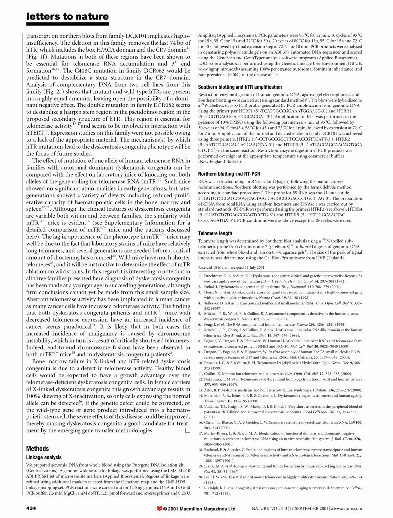
Figure 1 Identi®cation and location of hTR mutations. a, Southern blot analysis using an
hTR probe to genomic DNA digested with Pst I (lanes 1 and 2) and Taq I (lanes 3±5) of an
affected member from family DCR101 (lanes 1 and 4) and unrelated normal individuals
(lanes 2, 3 and 5). Abnormal fragments are indicated by arrowheads; normal fragments
are indicated with a dash. b, Diagram of an 821-bp deletion that removes the 39 end of
the hTR gene. The location, orientation and names of oligonucleotides used in various
PCR reactions are given. c, Segregation of the deletion. A subset of members of family
DCR101 are shown above the appropriate lanes. The larger and smaller fragments are
from the normal and mutant alleles, respectively. Shading indicates that individuals were
either too young for diagnosis or showed features that were suggestive but not diagnostic
of dyskeratosis congenita. Affected individuals are shown in black; normal individuals in
white. Plus, mutant control; N, normal control; B, blank control. d, The segregation of an
hTR C408G substitution in family DCR063, which results in the loss of a T® I site. M,
molecular mass marker (pEMBL8 plasmid cut with Taq I and Pvu II). e, The presence of a
GC to AG double substitution at nucleotides 107±108 of hTR in family DCR082 is
con®rmed by the creation of a Dde I site in affected individuals. f, Model of human hTR16.
Dotted boxes show the location of these point mutations and the extent of the deletion
observed in family DCR101.
© 2001 Macmillan Magazines Ltd

letters to nature
NATURE | VOL 413 | 27 SEPTEMBER 2001 | www.nature.com 433
pedigree). In addition to the diagnostic mucocutaneous features,some affected family members had premature greying, early dentalloss, bone marrow failure, liver cirrhosis, pulmonary disease andskin cancer (see Supplementary Information). A genome-wide scanwas performed using 400 microsatellite markers on DNA from sixaffected and two unaffected individuals. After the initial stage ofthe analysis, LOD scores (log10 of the odds ratio in favour of linkage)ranged from -100 to 1.5 with only one region in chromosome 3qshowing consistent, adjacent high LOD scores. The addition ofother DCR101 family members and more markers narrowed downthe region of interest to a segment of approximately 30 centimor-gans, the highest LOD score of 1.8 being achieved with the D3S3725marker. Of all the genes in this region our attention was drawn to thegene encoding the RNA component of telomerase, which maps to3q21±3q28, as this molecule had already been shown to be asso-ciated with dyskerin5.
The entire coding region of the gene was ampli®ed from anaffected individual (II.6) using a pair of ¯anking primers. Althoughthe sequence of this fragment was normal, Southern blotting ofDNA from affected and normal individuals showed clear differencesin the pattern obtained (Fig. 1a), consistent with the existence of asmall deletion in the DNA of patients. Further ampli®cation withprimersÐmore distal than the original 39 primerÐshowed thatone allele in the patients' DNA had 821 base pairs (bp) deleted,including the 74 39 base pairs of the coding region (Fig. 1b). Thedeleted region is ¯anked by a 4-bp direct repeat (AGGA). Usingprimers speci®c for the deletion and for the normal allele, DNAfrom all members of the pedigree was tested. The results show thatall available affected individuals in generation II were heterozygousfor the deletion and all unaffected members had two normal alleles(Fig. 1c). Out of the two individuals in generation II (II:7 and II:13)with undetermined clinical status, one (II:13) has the deletion.Individual II:13 was 37 years of age when examined, while affectedmembers of this family from generation II were diagnosed at ages29, 40, 46 and 48. In generation III all members diagnosed as beingaffected had the deletion. Some family members were too young toshow signs of the disease. Of these, three (III:15, III:16 and III:19)were heterozygous and ®ve (III:5, III:6, III:14, III:17 and III:18) werenormal. The three children who were heterozygous were 3, 9 and 13years of age, whereas the two individuals in this generation who hadthe disease were diagnosed at 17 and 22 years. The presence of thedeletion in these unaffected members (II:13, III:15, III:16 and III:19)re¯ects the variability in the severity and age of onset in this mildform of dyskeratosis congenita.
Lymphocyte cell lines were derived from affected individuals bytransformation with the Epstein Barr Virus (EBV). These cell lineswere examined by northern blotting using an hTR probe. Nodifference was found between cell lines from affected and unaffectedindividuals (Fig. 2a). All cell lines showed only the normal 451-bptelomerase RNA. Polymerase chain reaction with reverse transcrip-tion (RT-PCR) experiments, using a mutant-speci®c 39 primerabutting the point of the deletion, showed that the mutant tran-script is barely detectable (Fig. 2b). Similarly no signi®cant differ-ence was found in the telomerase activity levels measured by a TRAPassay in the EBV cell lines (data not shown). Telomere length inaffected family members was measured with a chromosome-7-speci®c subtelomeric probe. As with our previous observationstelomere length in these dyskeratosis congenita patients is signi®-cantly shorter than in normal patients15, even in young children(Fig. 2d).
Having found a lesion in the hTR gene in one family we examinedthis gene in two other families with autosomal dominant dysker-atosis congenita, as well as in one family with an unknown patternof inheritance. None of these families showed alteration whenanalysed with a Southern blot. However, sequence analysis of thehTR gene revealed affected members of one family (DCR063) wereheterozygous for a single point mutation at position 408 (C408G).
In another family (DCR082) a double point mutation (GC to AG) atpositions 107±108 was observed in affected members. The presenceof these mutations and their co-segregation with the disease wascon®rmed in the respective families by detection of restrictionenzyme sites lost (DCR063; loss of T®I, Fig. 1d) or created(DCR082; new DdeI, Fig. 1e) by the mutations. In family DCR063the father (I:2; age 39 years) had only mild haematological abnorm-alities, whereas his children (II:1and II:2; 10 and 12 years) had severebone marrow failure in association with other somatic abnormal-ities. In family DCR082 both I:1 and II:1 showed severe disease withages at diagnosis of 52 and 30 years, respectively. The genetic lesionsfound in all three families were absent in a panel of 50 unrelatedindividuals that we tested. No abnormality of the hTR gene wasdetected in the remaining family. This family had a more severeform of the disease than the other three families, with both thefather and daughter developing aplastic anaemia by the age of5 years.
Our results show that in three families, mutations in one allele ofthe RNA component of telomerase are suf®cient to cause dyskera-tosis congenita. This could be due to haplo-insuf®ciency or to adominant negative effect. The fact that we do not detect an aberrant
10
15
20
0 20 40
Age (years)
Telo
mer
e le
ngth
(kb
)
a
N AM N A N A
DNA RNA Blankb
1 2
M NII
I
1D BU
c
1 2 3 43.02.0
1.0
0.5
0.2
N A
d
Figure 2 Expression of hTR mutations. a, Northern blot analysis of two normal individuals
(N, lanes 1 and 2) and two affected members of family DCR101 (A, lanes 3 and 4). The
lower panel shows hybridization of a 5S rRNA probe to the same ®lter. The position and
size (kilobases) of molecular mass markers are shown. b, PCR and RT-PCR ampli®cation
from genomic DNA and primary mononuclear cell RNA as indicated from a normal (N) and
an affected (A) individual from family DCR101. No products are seen from cDNA prepared
without reverse transcription (blank). c, RT-PCR products from EBV lines from members of
family DCR063 (drawn above the appropriate lanes) digested with T® I. M, Molecular mass
marker; N, normal individual; U, undigested PCR product; D, ampli®cation from genomic
DNA of an affected individual; B, ampli®cation from a cDNA sample prepared without
reverse transcription. d, The telomere lengths of affected individuals from family DCR101
(®lled circles), DCR063 (®lled diamonds) and DCR086 (®lled triangles) are compared with
age-matched normal controls (open circles). Telomere lengths are signi®cantly smaller
(P , 0.001, Student's t-test), with short telomeres seen also in the younger, affected
individuals.
© 2001 Macmillan Magazines Ltd
letters to nature
434 NATURE | VOL 413 | 27 SEPTEMBER 2001 | www.nature.com
transcript on northern blots from family DCR101 implicates haplo-insuf®ciency. The deletion in this family removes the last 74 bp ofhTR, which includes the box H/ACA domain and the CR7 domain16
(Fig. 1f). Mutations in both of these regions have been shown tobe essential for telomerase RNA accumulation and 39 endformation16,17. The G408C mutation in family DCR063 would bepredicted to destabilize a stem structure in the CR7 domain.Analysis of complementary DNA from two cell lines from thisfamily (Fig. 2c) shows that mutant and wild-type hTRs are presentin roughly equal amounts, leaving open the possibility of a domi-nant negative effect. The double mutation in family DCR082 seemsto destabilize a hairpin stem region in the pseudoknot region in theproposed secondary structure of hTR. This region is essential fortelomerase activity16,17and seems to be involved in interaction withhTERT18. Expression studies on this family were not possible owingto a lack of the appropriate material. The mechanism(s) by whichhTR mutations lead to the dyskeratosis congenita phenotype will bethe focus of future studies.
The effect of mutation of one allele of human telomerase RNA infamilies with autosomal dominant dyskeratosis congenita can becompared with the effect on laboratory mice of knocking out bothalleles of the gene coding for telomerase RNA (mTR)19. Such miceshowed no signi®cant abnormalities in early generations, but latergenerations showed a variety of defects including reduced prolif-erative capacity of haematopoietic cells in the bone marrow andspleen20,21. Although the clinical features of dyskeratosis congenitaare variable both within and between families, the similarity withmTR-/- mice is evident14 (see Supplementary Information for adetailed comparison of mTR-/- mice and the patients discussedhere). The lag in appearence of the phenotype in mTR-/- mice maywell be due to the fact that laboratory strains of mice have relativelylong telomeres, and several generations are needed before a criticalamount of shortening has occurred22. Wild mice have much shortertelomeres23, and it will be instructive to determine the effect of mTRablation on wild strains. In this regard it is interesting to note that inall three families presented here diagnosis of dyskeratosis congenitahas been made at a younger age in succeeding generations, although®rm conclusions cannot yet be made from this small sample size.Aberrant telomerase activity has been implicated in human canceras many cancer cells have increased telomerase activity. The ®ndingthat both dyskeratosis congenita patients and mTR-/- mice withdecreased telomerase expression have an increased incidence ofcancer seems paradoxical24. It is likely that in both cases theincreased incidence of malignancy is caused by chromosomeinstability, which in turn is a result of critically shortened telomeres.Indeed, end-to-end chromosome fusions have been observed inboth mTR-/- mice25 and in dyskeratosis congenita patients2.
Bone marrow failure in X-linked and hTR-related dyskeratosiscongenita is due to a defect in telomerase activity. Healthy bloodcells would be expected to have a growth advantage over thetelomerase-de®cient dyskeratosis congenita cells. In female carriersof X-linked dyskeratosis congenita this growth advantage results in100% skewing of X-inactivation, so only cells expressing the normalallele can be detected26. If the genetic defect could be corrected, orthe wild-type gene or gene product introduced into a haemato-poietic stem cell, the severe effects of this disease could be improved,thereby making dyskeratosis congenita a good candidate for treat-ment by the emerging gene transfer methodologies. M
MethodsLinkage analysis
We prepared genomic DNA from whole blood using the Puregene DNA isolation kit(Gentra systems). A genome-wide search for linkage was performed using the LMS-MD10ABI PRISM set of microsatellite markers (Applied Biosystems). Regions of linkage werere®ned using additional markers selected from the Genethon map and the LMS-HD5linkage mapping set. PCR reactions were carried out on 12.5 ng genomic DNA in 1´ GoldPCR buffer, 2.5 mM MgCl2, 1mM dNTP, 1.25 pmol forward and reverse primer and 0.25 U
Amplitaq (Applied Biosystems). PCR parameters were 95 8C for 12 min, 10 cycles of 95 8Cfor 15 s, 55 8C for 15 s and 72 8C for 30 s, 20 cycles of 89 8C for 15 s, 55 8C for 15 s and 72 8Cfor 30 s, followed by a ®nal extension step at 72 8C for 10 min. PCR products were analysedin denaturing polyacrylamide gels on an ABI 377 automated DNA sequencer and scoredusing the GeneScan and GenoTyper analysis software programs (Applied Biosystems).LOD score analysis was performed using the Genetic Linkage User Environment (GLUE,www.hgmp.mrc.ac.uk) assuming 100% penetrance, autosomal dominant inheritance, andrare prevalence (0.001) of the disease allele.
Southern blotting and hTR ampli®cation
Restriction enzyme digestion of human genomic DNA, agarose gel electrophoresis andSouthern blotting were carried out using standard methods27. The blots were hybridized toa 32P-labelled, 653-bp hTR probe, generated by PCR ampli®cation from genomic DNAusing the primer pair HTRF1 (59-TCATGGCCGGAAATGGAACT-39) and HTRR1(59-GGGTGACGGATGCGCACGAT-39). Ampli®cation of hTR was performed in thepresence of 10% DMSO using the following parameters: 7 min at 95 8C, followed by30 cycles of 94 8C for 45 s, 58 8C for 45 s and 72 8C for 1 min, followed by extension at 72 8Cfor 7 min. Ampli®cation of the normal and deleted alleles in family DCR101 was achievedusing three primers, HTRF2 (59-CCTGCCGCCTTCCACCGTTCATT-3'), HTRR2(59-AATCTGCAGAGCAGGAACTAA-39) and HTRR3 (59-CATTACCAGCAACAGTGGACTCT-39) in the same reaction. Restriction enzyme digestion of PCR products wasperformed overnight at the appropriate temperature using commercial buffers(New England Biolabs).
Northern blotting and RT-PCR
RNA was extracted using an RNeasy kit (Qiagen) following the manufacturersrecommendations. Northern blotting was performed by the formaldehyde methodaccording to standard procedures27. The probe for 5S RNA was the 41-nucleotide59-GGTCTCCCATCCAAGTACTAACCAGGCCCGACCCTGCTTAG-39. The preparationof cDNA from total RNA using random hexamers and DNAse 1 was carried out bystandard methods. RT-PCR was performed using the primers HTRF2 (see above), HTRR4(59-GCATGTGTGAGCCGAGTCCTG-39) and HTRR5 (59-TCTTGGCAACTACCCCCAGATGA-39). PCR conditions were as above except that 26 cycles were used.
Telomere length
Telomere length was determined by Southern blot analysis using a 32P-labelled sub-telomeric probe from chromosome 7 (pTelBam8)28 to BamHI digests of genomic DNAextracted from whole blood and run on 0.8% agarose gels29. The size of the peak of signalintensity was determined using the Gel Blot-Pro software from UVP (Upland).
Received 15 March; accepted 11 July 2001.
1. Drachtman, R. A. & Alter, B. P. Dyskeratosis congenita: clinical and genetic heterogeneity. Report of a
new case and review of the literature. Am. J. Pediatr. Hematol. Oncol. 14, 297±304 (1992).
2. Dokal, I. Dyskeratosis congenita in all its forms. Br. J. Haematol. 110, 768±779 (2000).
3. Heiss, N. S. et al. X-linked dyskeratosis congenita is caused by mutations in a highly conserved gene
with putative nucleolar functions. Nature Genet. 19, 32±38 (1998).
4. Tollervey, D. & Kiss, T. Function and synthesis of small nucleolar RNAs. Curr. Opin. Cell. Biol. 9, 337±
342 (1997).
5. Mitchell, J. R., Wood, E. & Collins, K. A telomerase component is defective in the human disease
dyskeratosis congenita. Nature 402, 551±555 (1999).
6. Feng, J. et al. The RNA component of human telomerase. Science 269, 1236±1241 (1995).
7. Mitchell, J. R., Cheng, J. & Collins, K. A box H/ACA small nucleolar RNA-like domain at the human
telomerase RNA 39 end. Mol. Cell. Biol. 19, 567±576 (1999).
8. Pogacic, V., Dragon, F. & Filipowicz, W. Human H/ACA small nucleolar RNPs and telomerase share
evolutionarily conserved proteins NHP2 and NOP10. Mol. Cell. Biol. 20, 9028±9040 (2000).
9. Dragon, F., Pogacic, V. & Filipowicz, W. In vitro assembly of human H/ACA small nucleolar RNPs
reveals unique features of U17 and telomerase RNAs. Mol. Cell. Biol. 20, 3037±3048 (2000).
10. Prescott, J. C. & Blackburn, E. H. Telomerase: Dr Jekyll or Mr Hyde? Curr. Opin. Genet. Dev. 9, 368±
373 (1999).
11. Collins, K. Mammalian telomeres and telomerase. Curr. Opin. Cell. Biol. 12, 378±383 (2000).
12. Nakamura, T. M. et al. Telomerase catalytic subunit homologs from ®ssion yeast and human. Science
277, 955±959 (1997).
13. Alter, B. P. Molecular medicine and bone marrow failure syndromes. J. Pediatr. 136, 275±276 (2000).
14. Marciniak, R. A., Johnson, F. B. & Guarente, L. Dyskeratosis congenita, telomeres and human ageing.
Trends. Genet. 16, 193±195 (2000).
15. Vulliamy, T. J., Knight, S. W., Mason, P. J. & Dokal, I. Very short telomeres in the peripheral blood of
patients with X-linked and autosomal dyskeratosis congenita. Blood Cells Mol. Dis. 27, 353±357
(2001).
16. Chen, J. L., Blasco, M. A. & Greider, C. W. Secondary structure of vertebrate telomerase RNA. Cell 100,
503±514 (2000).
17. Martin-Rivera, L. & Blasco, M. A. Identi®cation of functional domains and dominant negative
mutations in vertebrate telomerase RNA using an in vivo reconstitution system. J. Biol. Chem. 276,
5856±5865 (2001).
18. Bachand, F. & Autexier, C. Functional regions of human telomerase reverse transcriptase and human
telomerase RNA required for telomerase activity and RNA-protein interactions. Mol. Cell. Biol. 21,
1888±1897 (2001).
19. Blasco, M. A. et al. Telomere shortening and tumor formation by mouse cells lacking telomerase RNA.
Cell 91, 25±34 (1997).
20. Lee, H. W. et al. Essential role of mouse telomerase in highly proliferative organs. Nature 392, 569±574
(1998).
21. Rudolph, K. L. et al. Longevity, stress response, and cancer in aging telomerase-de®cient mice. Cell 96,
701±712 (1999).
© 2001 Macmillan Magazines Ltd

letters to nature
NATURE | VOL 413 | 27 SEPTEMBER 2001 | www.nature.com 435
22. Herrera, E. et al. Disease states associated with telomerase de®ciency appear earlier in mice with short
telomeres. EMBO J. 18, 2950±2960 (1999).
23. Hemann, M. T. & Greider, C. W. Wild-derived inbred mouse strains have short telomeres. Nucleic
Acids Res. 28, 4474±4478 (2000).
24. Artandi, S. E. & DePinho, R. A. A critical role for telomeres in suppressing and facilitating
carcinogenesis. Curr. Opin. Genet. Dev. 10, 39±46 (2000).
25. Artandi, S. E. et al. Telomere dysfunction promotes non-reciprocal translocations and epithelial
cancers in mice. Nature 406, 641±645 (2000).
26. Vulliamy, T. J., Knight, S. W., Dokal, I. & Mason, P. J. Skewed X-inactivation in carriers of X-linked
dyskeratosis congenita. Blood 90, 2213±2216 (1997).
27. Sambrook, J., Fritsch, E. & Maniatis, T. Molecular Cloning: A Laboratory Manual. (Cold Spring
Harbor, New York, 1989).
28. Brown, W. R. et al. Structure and polymorphism of human telomere-associated DNA. Cell 63, 119±
132 (1990).
29. Notaro, R., Cimmino, A., Tabarini, D., Rotoli, B. & Luzzatto, L. In vivo telomere dynamics of human
hematopoietic stem cells. Proc. Natl Acad. Sci. USA 94, 13782±13785 (1997).
Supplementary information is available on Nature's World-Wide Web site(http://www.nature.com) or as paper copy from the London editorial of®ce of Nature.
Acknowledgements
We thank B. Alter and E. Gluckman for facilitating recruitment to the dyskeratosiscongenita registry, and the dyskeratosis congenita families and their clinicians whoprovided us with the samples. We also thank Z. Amoura, R. Gruppo and J. Miller. Wethank D. Stevens for technical assistance and D. Smyth for help with the linkage. We aregrateful to S. Knight, W. Watkins, J. Clarke, J. O'Donnel and S. Reiss for help anddiscussions, and J. Goldman and L. Luzzatto for their continued support of thedyskeratosis congenita project. The genome screen was performed at the Linkage HotelMRC UK HGMP Resource Centre. This work was supported by the Wellcome Trust.
Correspondence and requests for materials should be addressed to P.J.M.(e-mail: [email protected]).
.................................................................DNA topoisomerase IIa is required forRNA polymerase II transcription onchromatin templatesNeelima Mondal & Jeffrey D. Parvin
Department of Pathology, Brigham and Women's Hospital,Harvard Medical School, Boston, Massachusetts 02115, USA
..............................................................................................................................................
In the nucleus of the cell, core RNA polymerase II (pol II) isassociated with a large complex called the pol II holoenzyme(holo-pol)1,2. Transcription by core pol II in vitro on nucleosomaltemplates is repressed compared with that on templates ofhistone-free naked DNA3±5. We found that the transcriptionalactivity of holo-pol, in contrast to that of core pol II, is notmarkedly repressed on chromatin templates. We refer to thisproperty of holo-pol as chromatin-dependent coactivation(CDC). Here we show that DNA topoisomerase IIa is associatedwith the holo-pol and is a required component of CDC. Etoposideand ICRF-193, speci®c inhibitors of topoisomerase II, blockedtranscription on chromatin templates, but did not affect tran-scription on naked templates. Addition of puri®ed topoisomeraseIIa reconstituted CDC activity in reactions with core pol II. These®ndings suggest that transcription on chromatin templatesresults in the accumulation of superhelical tension, making therelaxation activity of topoisomerase II essential for productiveRNA synthesis on nucleosomal DNA.
Biochemically puri®ed core RNA polymerase II (pol II) contains12 subunits6, but, in the nucleus of the cell, pol II is associated withholo-pol1,2. Holo-pol also contains basal transcription factors,suppressors of RNA polymerase B mutations (SRB proteins),chromatin remodelling factors and other regulatory proteins7,8. Toidentify conditions in which the holo-pol is functionally distinct
from the core pol II enzyme, we compared these factors intranscription under various conditions. All reactions included thepuri®ed basal factors, the coactivator PC4 and one of the pol IIpreparations. Every reaction contained two templates that encodeda 210-nucleotide basal control transcript and a 380-nucleotidetranscript, the synthesis of which was stimulated by GAL4-fusedactivators. Both templates were digested with restriction endonu-cleases and thus had linear topology.
Core pol II was puri®ed from calf thymus9 and holo-pol from asucrose gradient fraction enriched for this complex10. These two polII preparations were normalized by basal polymerase activity onnaked templates. Under these conditions, 0.2 ml (40 ng) of core polII was equivalent in basal transcription activity to 1.5 ml of holo-pol(Fig. 1a). In subsequent experiments, these matched amounts ofeach pol II preparation were used for standard comparison. Whencomparing these normalized pol II activities for mass of polymeraseby western blot, it is apparent that the amount of pol II in the core
1110987654321
Core Holo
380 nt
210 nt
2.52.01.51.00.50.50.20.150.10.050
4321
Core Holo
Stim.
Basal
GAL4-VP16 – + – +
Chromatin
GAL4-VP16 – + – + – + – +
Stim.
Basal
87654321
87654321 9
GAL4-VP16 – + – + + +
Stim.
Basal
+ + +
a b
c d
e
Holo
ChromatinCore
EF H Cor
e
Hol
o
Naked DNA
HoloCore0.50.2 1.5 3.0
Core Holo
1.5
0.2
0.4
0.8
3.0
6.0
654321
IB: 210K Pol II
Figure 1 Pol II holoenzyme is qualitatively different from core pol II for transcription on
chromatin templates. a, Basal transcription reactions with naked DNA templates were
used to normalize core pol II (Core) and holo-pol (Holo) preparations. The indicated
volumes (in ml) of each polymerase were added. nt, nucleotide. b, Western blot of core pol
II and holo-pol. The indicated volumes of core and holoenzyme were subjected to SDS±
PAGE and immunoblotted (IB) for the pol II subunit with relative molecular mass 210,000
(Mr 210K). c, Activated transcription with naked DNA templates. The volumes (in ml) of
core pol II and holo-pol included in each reaction were varied as indicated. The
transcriptional activator GAL4-VP16 was included in half of the reactions, as indicated.
The 380-nucleotide GAL4-responsive transcript (Stim.) and the 210-nucleotide basal
control transcript (Basal) are indicated. d, Transcription of reconstituted chromatin
templates. Core pol II (40 ng) and holo-pol (1.5 ml), matched for basal activity as in a,
were assayed for transcription on chromatin templates in the absence or presence of the
activator GAL4-VP16. e, The CDC activity is not due to a basal factor. Reactions with
chromatin templates were supplemented by doubling the amount of the indicated factors.
Eight nanograms of TFIIE (E, lane 5), 200 ng of TFIIF (F, lane 6), 1 ml of TFIIH (H, lane 7)
and 80 ng of core pol II (lane 8) were included in the indicated reactions. Along with 40 ng
of core pol II, 1.5 ml of holoenzyme was included in lane 9. GAL4-VP16 was included as
indicated.
© 2001 Macmillan Magazines Ltd