6.1: skin and its tissues
TRANSCRIPT

9/27/2016
1
1Copyright © McGraw-Hill Education. Permission required for reproduction or display.
Chapter 06Lecture Outline
See separate PowerPoint slides for all figures and tables pre-inserted into PowerPoint without notes.
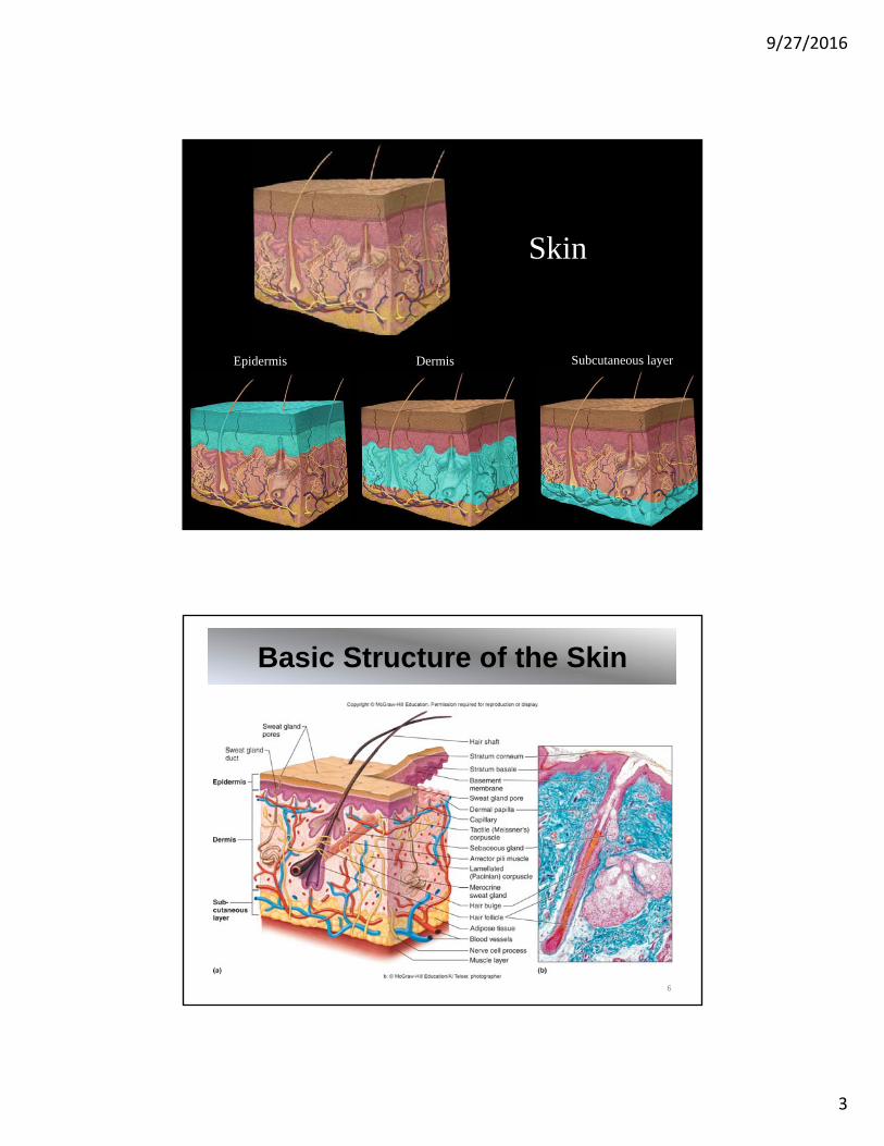
6.1: Skin and Its Tissues
2
• Two or more types of tissues grouped together and performing specialized functions constitute an organ
• Skin is the largest organ in body by weight
• Skin is also called the cutaneous membrane
• The skin and its accessory structures (hair, nails, glands, sensory receptors) make up the integumentary system
• Composed of several tissue types
• Contains 2 layers: epithelial tissue overlying connective tissue
• Outer layer is the epidermis
• Inner layer is the dermis

9/27/2016
2
Skin of Finger
Epidermis DermisThin Skin
Thick Skin
Epidermis:• Outer layer• Stratified squamous epithelium• Basement membrane between
epidermis and dermis
Dermis:• Inner layer• Thicker of the 2 layers of the skin• Connective tissue• Contains collagenous and elastic fibers
Subcutaneous layer (hypodermis):• Beneath dermis; insulating layer• Areolar and adipose connective tissue• Not considered part of the skin• Contains blood vessels that supply skin
Skin and Its Tissues
4
9/27/2016
3
Skin
Epidermis Dermis Subcutaneous layer
Basic Structure of the Skin
6

9/27/2016
4
Epidermis
Thick Skin Thin Skin
9/27/2016
5
Thick SkinLow Magnification
Epidermis
Dermis
Dermal papillae
Duct of merocrine sweat gland
Stratum corneum
Stratum granulosum
Stratum spinosum
Stratum basale
Thick SkinHigh Magnification
Epidermis
Dermis
Stratumbasale
Stratumspinosum
Stratumgranulosum
Stratumlucidum
Stratumcorneum

9/27/2016
6
Thin SkinLow MagnificationEpidermis
DermisHair follicle
Sebaceous gland
Duct of Sebaceous
gland
Hair
Thin SkinHigh Magnification
Epidermis
DermisStratumbasale
Stratumspinosum
Stratumgranulosum
Stratumcorneum

9/27/2016
7
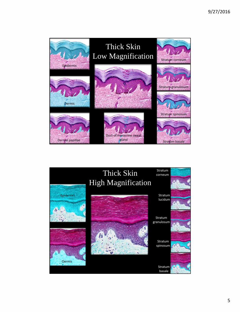
Epidermis:
• Stratified squamous epithelium; rests on basement membrane
• Lacks blood vessels
• Deepest layer, stratum basale, nourished by blood vessels in dermis
• As cells grow, they migrate toward free surface, away from nutrient supply
• As they migrate, older cells, keratinocytes, begin to flatten and die
• Keratinization: Process of hardening, dehydration, and keratin accumulation that occurs in epidermal cells as they migrate outward
• Keratin: tough, fibrous, waterproof protein made and stored in the cells
• As cells reach outer surface, become tightly packed, develop desmosomes, form outer layer, stratum corneum
• Stratum corneum cells are eventually shed from skin surface
• Thickest on palms and soles (0.8-1.4mm)
• Most of body has thinner epidermis, 0.07 – 0.12 mm
Epidermis
13
5 Layers of Epidermis:
• Stratum corneum: outermost layer; dead, keratinized cells
• Stratum lucidum: only in thick skin – palms, soles
• Stratum granulosum
• Stratum spinosum
• Stratum basale/germinativum: deepest, mitotic layer
Functions of epidermis: Protects against water loss, harmful chemicals, mechanical injury, pathogens
Epidermis
14

9/27/2016
8
Layers of the Epidermis
15
• Melanocytes located in the stratum basale produce the dark pigment melanin
• Absorbs UV light from sunlight and provides skin color
• Melanin is distributed into keratinocytes, to protect skin cells from damaging effects of UV light (DNA damage, fibroblast damage, skin cancer)
Epidermis: Melanocytes
16

9/27/2016
9
Factors Affecting Skin Color:• Hereditary Factors:
• All people have same number of melanocytes, but vary in amount of melanin produced (this is under genetic control)
• Varying distribution and size of melanin granules• Albinos inherit mutation in melanin genes; lack melanin
• Environmental Factors:• Sunlight• UV light from sunlamps• X-rays
• Physiological Factors:• Oxygenation in blood of dermal blood vessels: pinkish, cyanosis• Vasodilation/vasoconstriction of dermal blood vessels• Accumulation of carotene pigment from diet• Jaundice
Epidermis: Skin Color
17
Indoor Tanning and Skin Cancer• Exposure to sunlight or a tanning bed causes melanocytes to
produce more melanin, and skin darkens
• Tanning bed uses doses of UV radiation that can overwhelm body’s natural protective responses against skin cancer
• Basal cell carcinoma and squamous cell carcinoma arise from epithelial cells in skin
• Melanomas arise from melanocytes
• Melanomas are least common (4%) skin cancers, but cause 80% of skin cancer deaths
Clinical Application 6.1
18

9/27/2016
10
Dermis:• Inner layer of skin• Average of 1-2 mm thick• Contains dermal papillae
between epidermal ridges• Binds epidermis to underlying
tissues• Connective tissue layer• Contains muscle fibers• Nerve cell processes• Dermal blood vessels supply
nutrients to all skin cells• Hair follicles, sweat & sebaceous glands • Sensory receptors: Lamellated (Pacinian)
corpuscles for pressure, Tactile (Meissner’s) corpuscles for light touch
Dermis
19
Dermis

9/27/2016
11
Dermis and Dermal Papillae
Dermis Dermal papillae
The dermis consists of 2 layers:
Papillary layer:
• Superficial layer
• Areolar connective tissue
• Thinner of the 2 layers
• Location of dermal papillae
Reticular layer:
• Deeper layer
• Dense irregular connective
tissue
• Thicker of 2 layers
Dermis
22
9/27/2016
12
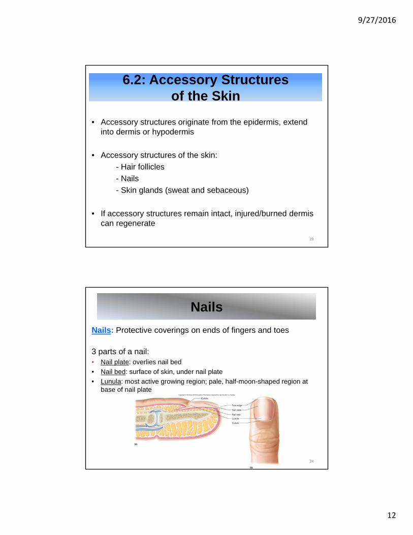
6.2: Accessory Structures of the Skin
23
• Accessory structures originate from the epidermis, extend into dermis or hypodermis
• Accessory structures of the skin:
- Hair follicles
- Nails
- Skin glands (sweat and sebaceous)
• If accessory structures remain intact, injured/burned dermis can regenerate
Nails: Protective coverings on ends of fingers and toes
3 parts of a nail:• Nail plate: overlies nail bed
• Nail bed: surface of skin, under nail plate
• Lunula: most active growing region; pale, half-moon-shaped region at base of nail plate
Nails
24

9/27/2016
13
Nails
Free edge Nail body Eponychium
Nail root Nail matrix
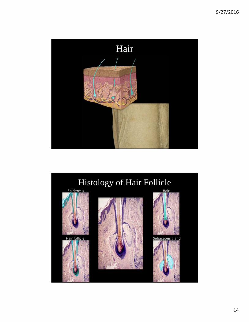
• Hair is present on all surfaces of skin except palms, soles, lips, nipples, parts of external reproductive organs
• Hair follicle: Tube-like depressionof epidermal cells from which hairdevelops
• Extends into dermis or thesubcutaneous layer
• 3 parts of hair:• Hair bulb (dividing cells)• Hair root• Hair shaft (dead, epidermal cells)
• Hair papilla contains blood vessels to nourish hair
• Hair color is due to type and amount of melanin
• Arrector pili muscle (goosebumps)
Hair Follicles
26
9/27/2016
14
Hair
Histology of Hair FollicleEpidermis
Hair follicle
Hair
Sebaceous gland

9/27/2016
15
HairHair shaft
Keratinocytes
Hair FollicleMedium Magnification
Cortex of hair
Differentiationzone of hair
Hair matrix
Connectivetissuesheath
Glassymembrane
External root
sheath
Internal root
sheath
9/27/2016
16
Dermis
Connective tissue sheath
Glassy membrane
External root sheath
Internal root
sheath
Hair matrix
Dermal papilla
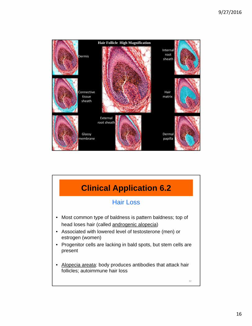
Hair Follicle High Magnification
Hair Loss
• Most common type of baldness is pattern baldness; top of
head loses hair (called androgenic alopecia)
• Associated with lowered level of testosterone (men) or estrogen (women)
• Progenitor cells are lacking in bald spots, but stem cells are present
• Alopecia areata: body produces antibodies that attack hair follicles; autoimmune hair loss
Clinical Application 6.2
32

9/27/2016
17
Sebaceous Glands:
• Holocrine glands
• Usually associated with
hair follicles
• Produce sebum, which
consists of fatty material
and cellular debris
• Sebum keeps hair and skin
soft and waterproof
• Excess sebum can result
in acne
• Absent on palms and soles
Skin Glands: Sebaceous Glands
33
Sebaceous GlandLow MagnificationEpidermis
Hair follicle
Arrector pili m. Sebaceous gland and duct
Sebaceous gland
Epidermis
Sweat gland

9/27/2016
18
Sebaceous Gland High Magnification
Sebaceous gland
Duct of sebaceous gland
Hair follicle
Basal cell of sebaceous gland
Secretory cells
Acne• Acne vulgaris is a disorder of sebaceous glands
• Common at puberty, because sebaceous glands are excessively responsive to androgens
• Sebaceous glands become clogged with extra sebum and epithelial cells
• Clogged glands provide good environment for anaerobic bacteria; infection results in inflammation
• Affects 80% of people between 11 and 30 years of age
• Treated best with Vitamin A derivatives, systemic antibiotics, salicylic acid, benzoyl peroxide
Clinical Application 6.3
36

9/27/2016
19
Sweat Glands:• Also called sudoriferous glands• Widespread in skin• Originate in deeper dermis or
hypodermis as ball-shaped coils• Eccrine (merocrine) glands:
- most numerous- respond to elevated body
temperature• Apocrine sweat glands:
- axillary and groin areas- secrete by exocytosis- respond to emotions, pain
• Ceruminous glands—ear wax• Mammary glands—milk
Skin Glands: Sweat Glands
37
Eccrine Sweat GlandLow MagnificationEpidermis
Dermis
Hypodermis
Eccrine sweat gland and duct
Eccrine sweat gland
Duct of eccrine sweat gland

9/27/2016
20
Eccrine Sweat GlandHigh Magnification
Eccrine sweat gland
Lumen of sweat gland
Epithelial cells of sweat gland
Myoepithelial cells
Basal lamina of eccrine sweat gland
Duct of Sweat GlandHigh Magnification Epidermis
Dermis
Duct of sweat gland

9/27/2016
21
Apocrine Sweat GlandLow Magnification
Apocrine sweat gland
Epithelium
Lumen
Apocrine Sweat GlandHigh Magnification
Apocrine sweat gland Lumen Epithelial cells Myoepthelial cells

9/27/2016
22
Summary of Skin Glands
43
6.3: Skin Functions
44
Skin is versatile, and vital for homeostasis.
Functions of the skin:• Protective covering, barrier against harmful substances and
microorganisms
• Prevents some water loss
• Contains sensory receptors
• Excretes some wastes
• Helps produce Vitamin D
• Helps regulate body temperature

9/27/2016
23
• Important to regulate body temperature; slight shift can disrupt rates of metabolic reactions
• Set point is monitored by Hypothalamus
• Deep body temperature stays close to set point of 37oC or 98.6oF
• Skin plays key role in homeostatic mechanisms that regulate body temperature
Regulation of Body Temperature
45
• Heat is a product of cellular metabolism
• The most active body cells are major heat producers: Skeletal muscle, cardiac muscle, cells of the liver
• When body is too warm, body responds with vasodilation of dermal blood vessels and vasoconstriction of deep blood vessels. Heat can escape through skin.
• Methods of heat loss:
1. Radiation: Primary method, infrared heat rays escape
2. Conduction: Heat moves from skin to cooler objects
3. Convection: Heat loss into circulating air currents
4. Evaporation: Sweat changes into a gas, carries heat away
Heat Production and Loss
46

9/27/2016
24
When body temperature rises:• Thermoreceptors signal
hypothalamus
• Vasodilation of dermal blood
vessels
• Sweat glands are activated
When body temperature falls:• Thermoreceptors signal
hypothalamus
• Vasoconstriction of dermal blood
vessels
• Sweat glands are inactive
• Muscles contract involuntarily
(shivering)
Body Temperature Regulation
47
Hyperthermia: abnormally high body temperature• Can occur on hot, humid day, when sweat cannot evaporate
• When air temperature is high, radiation is less effective
• Body may gain heat from hotter air
• Skin becomes dry, person gets weak, dizzy, nauseous, with headache, rapid pulse
Hypothermia: abnormally low body temperature• Can result from prolonged exposure to cold, or illness
• Shivering is involuntary skeletal muscle contraction, caused by hypothalamus
• Progresses to confusion, lethargy, loss of reflexes and consciousness
• Without treatment, organs shut down
Problems in Temperature Regulation
48

9/27/2016
25
Elevated Body TemperatureLoss of ability of homeostatic temperature control mechanism to function in an extremely hot environment:
• Exposure to very high heat can overwhelm temperature control mechanisms, leading to hyperthermia
• If body heat builds up faster than heat can be lost from body, body temperature will rise, even when set point is normal
• Extreme vasodilation can collapse cardiovascular system; can be fatal
Fever:
• Set point is elevated by the immune system, to fight infection
• Phagocytes release pyrogens in response to presence of bacteria, viruses; hypothalamus increases set point and raises body temperature
• Elevated body temperature helps destroy pathogens
Clinical Application 6.4
49
6.4: Healing of Wounds and Burns
50
• Inflammation is a normal response to injury or stress
• Inflammation is body’s attempt to restrict spread of infection
• Blood vessels in affected tissues dilate and become more permeable, allowing fluids to leak into the damaged tissues
• Inflamed skin may become:• Reddened • Swollen • Warm • Painful

9/27/2016
26
• A shallow cut, which affects only the epidermis, results in epidermal cells along its margin dividing more rapidly than usual, to fill gap
• A deep cut, reaching dermis or subcutaneous layer, results in blood vessels breaking; released blood forms a clot
• Clot consists of fibrin, blood cells and platelets
• Clot and dried tissue fluid form scab
• Epithelial cells reproduce, fill in the wound
• Fibroblasts secrete collagen fibers to bind wound together
• Growth factors stimulate new tissue formation
• Phagocytic cells remove dead cells and debris, scab sloughs off
• Excess collagenous fibers may form elevated mass called a scar
Cuts
51
Healing of a Wound
52
9/27/2016
27
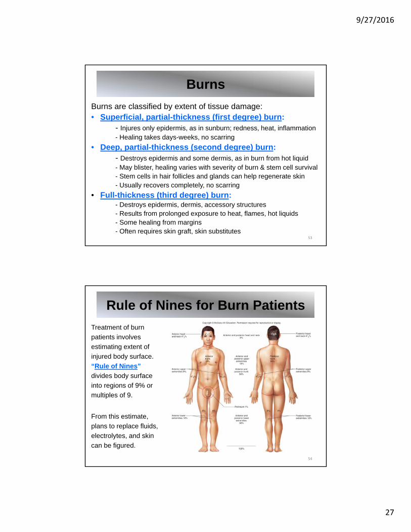
Burns are classified by extent of tissue damage:• Superficial, partial-thickness (first degree) burn:
- Injures only epidermis, as in sunburn; redness, heat, inflammation- Healing takes days-weeks, no scarring
• Deep, partial-thickness (second degree) burn:- Destroys epidermis and some dermis, as in burn from hot liquid - May blister, healing varies with severity of burn & stem cell survival- Stem cells in hair follicles and glands can help regenerate skin- Usually recovers completely, no scarring
• Full-thickness (third degree) burn:- Destroys epidermis, dermis, accessory structures - Results from prolonged exposure to heat, flames, hot liquids- Some healing from margins- Often requires skin graft, skin substitutes
Burns
53
Treatment of burn
patients involves
estimating extent of
injured body surface.
“Rule of Nines”
divides body surface
into regions of 9% or
multiples of 9.
From this estimate,
plans to replace fluids,
electrolytes, and skin
can be figured.
Rule of Nines for Burn Patients
54

9/27/2016
28
6.5: Life-Span Changes
55
• Cell cycle slows, skin becomes scaly, age spots appear
• Epidermis and dermis become thinner
• Loss of fat in subcutaneous layer; person feels cold
• Wrinkling, sagging of skin occur
• Sebaceous glands secrete less oil; skin becomes dry
• Melanin production slows; hair whitens
• Hair thins
• Number of hair follicles decreases
• Nail growth becomes impaired
• Sensory receptors decline
• Body temperature regulation becomes less effective
• Diminished ability to produce Vitamin D