644 painless dissections of the aorta presenting as acute...
TRANSCRIPT

644 Painless Dissections of the Aorta Presenting as AcuteNeurologic Syndromes
O D E D GERBER, M . D . , ERIC J. HEYER, M.D. , P H . D . , * AND ULRICH VIEUX, M.D.
SUMMARY We describe three patients who had painless dissections of the aorta which resulted inneurologic syndromes at the time of presentation. Two patients had acute hemimotor and sensory findings.In one of these cases progression to paraplegia occurred. In a third patient, acute weakness and ischemia ofa leg occurred at presentation. We review previously described painless aortic dissections. Such aorticdissections may be suspected in the setting of an acute neurologic event by abnormalities in the examinationof the peripheral pulses and the heart and by attention to characteristic chest x-ray changes.
Stroke Vol 17, No 4, 1986
IN 1941 Willius and Cragg1 cited four reasons forfailure to diagnose dissecting aneurysms of the aorta.These were: 1) the relative infrequency of the condi-tion; 2) the variation in its clinical manifestations andthe absence of a characteristic syndrome; 3) the limita-tion of special diagnostic adjuncts; and 4) the lack ofuniversal clinical suspicion. More than thirty yearslater, we have encountered three patients in whom thediagnosis of dissection of the aorta (DA) was over-looked at initial presentation. These patients exhibitedvariations from the usual clinical manifestations ofDA. They did not demonstrate the features of what hasbecome known as the characteristic syndrome of DA,which has generally included severe chest, back orabdominal pain. Our patients had painless dissectionsof the aorta resulting in acute neurologic syndromes.This presentation has not been previously reported.
Case 1A 63 year old woman with a history of hypertension
developed left sided weakness while walking. Therewas no loss of consciousness. She had a past history ofbilateral corneal ulceration, alcoholism, multiple sui-cide attempts, erosive gastritis, and a "heart attack"three years earlier. One month prior to admission shehad been seen at another hospital for dizziness, head-ache and hearing loss, and was studied with a cardiacHolter monitor, electroencephalography (EEG) and acomputerized tomographic (CT) scan of the head.Physical examination and all studies were normal.
On admission, blood pressure in the upper extrem-ities was unobtainable; her pulse was 68 beats perminute. Cardiac and abdominal examinations werenormal. Her right leg was cool and the right toes weremottled. She had no right carotid pulse, no right fem-oral pulse nor any pulse in the upper extremities. Thepatient was alert and oriented. Her speech was fluentbut slow and dysarthric. Because of her longstandingcorneal ulceration she had no vision in the right eye
From the Departments of Neurology, and Radiology, Mount SinaiServices at Elmhurst and The Mount Sinai School of Medicine of theCity University of New York.
•Supported in part by NINCDS Teacher-Investigator DevelopmentAward (NS 00657).
Address correspondence to: Dr. Oded Gerber, Department of Neurol-ogy, Mount Sinai Services, City Hospital Center at Elmhurst, 79-01Broadway, Elmhurst, New York 11373.
Received November 21, 1984; revision # 2 accepted November 5,1985.
and minimal vision in the left eye. Visual fields couldnot be determined. Left facial weakness was present.A dense left hemiplegia was present. She had absentpin, vibration and joint position sensation on the leftside. Chest x-ray on admission demonstrated a wid-ened mediastinum. The electrocardiogram showednon-specific ST wave changes and poor R wave pro-gression. An aortogram demonstrated a Type 1 dis-secting aortic aneurysm involving the aortic arch, de-scending aorta and the right iliac artery. There wasobstruction of the right brachiocephalic and right iliacarteries by the intimal flap. There was hypoperfusionof the right kidney. The left subclavian and left carotidarteries filled normally. During the subsequent eightmonths of hospitalization her neurologic examinationremained essentially unchanged except for bouts ofconfusion and lethargy. In addition, she had severalepisodes of sepsis as a result of urinary tract infectionsand pneumonia. She expired after this time. Permis-sion for post mortem examination was refused.
Case 2A 69 year old woman with a past history of hypothy-
roidism and hypertension was well until she noted "afunny feeling" in her throat. Fifteen minutes later shelost consciousness briefly. When she awakened, shehad left sided weakness.
On admission her blood pressure was 200/80 mmHg, and her pulse 45 beats per minute. A left homony-mous hemianopsia, left hemiparesis and left Babinskisign were present. The right carotid pulse was weak.Brachial, radial and femoral pulses were absent on theright. No cardiac murmurs were heard. The abdominalexamination was normal. A neurological consultantfound an alert patient who followed commands, wasoriented, performed simple calculations and named thelast several presidents accurately. There was neglect ofher left side. A left homonymous hemianopia waspresent. Her head and eyes were deviated to the right atrest; however, she was able to move her eyes in alldirections on command. She had left central facialweakness and drift of the left upper extremity. Shewould not cooperate with formal motor testing of theleft extremities. There was extinction of touch and pinon the left side. Twelve hours after admission, whenboth lower extremities became painful below theknees, a vascular consultant diagnosed chronic vascu-lar insufficiency. Twenty-four hours after admission,the patient developed a flaccid paraplegia. The neglect
by guest on July 3, 2018http://stroke.ahajournals.org/
Dow
nloaded from

PAINLESS AORTIC DISSECTION/Gerber et al 645
syndrome and the left arm weakness had improved bythen. A sensory level was found at Tl 1 as well as atT5. Her mental status remained normal. A chest x-raydemonstrated elongation, tortuosity and calcificationof the thoracic aorta. An electrocardiogram demon-strated sinus bradycardia. Several hours after the onsetof her paraplegia an aortic arch study and abdominalaortogram were performed. These showed a Type 1dissecting aortic aneurysm arising from the root of theaorta, (fig. 1) There was complete obstruction of theright brachiocephalic artery by the false lumen as wellas narrowing of the true lumen of the aortic arch andthe distal descending aorta, (fig. 2) The other majorbranches of the aortic arch were patent. Complete ob-struction of the abdominal aorta below the level of therenal arteries by the false lumen had occurred. Twohours later she became comatose and died. Permissionfor post mortem examination was refused.
Case 3A 78 year old man was admitted to the hospital
because of left leg weakness. He was well until the dayof admission, when he noticed the sudden onset ofweakness and numbness of the left leg. In the emer-gency room this leg was cold and pulseless. He deniedchest, back or leg pain. The admitting diagnosis wasacute thrombotic or embolic occlusion in the left leg.His past medical history was negative except for ahistory of hypertension.
On admission the patient was afebrile. A grade 2
FIOURE 1. Patient 2. Thoracic aortogram in the right posteri-or oblique projection shows a dissection of the aorta startingfrom the ascending aorta. The intimalfiap (solid arrows) can beseen in the ascending aorta. The false lumen is compressing andoccluding the right brachiocephalic artery (open arrow). Theleft carotid and the left subclavian arteries fed by the true lumenwere opacified in previous films. The intimal flap between thefalse and true lumens is well demonstrated in the descendingaorta (small arrows).
FIGURE 2. Patient 2. The true lumen of the abdominal aorta isseen giving off both renal arteries and the superior mesentericartery. There is an aneurysmal dilatation of the false lumencharacterized by splaying of the branches of the superior me-senteric artery and occlusion of the true lumen (arrow).
diastolic murmur was heard loudest at the third inter-costal space on the left. His abdomen was soft, non-tender with normal bowel sounds present and no or-ganomegaly. The left leg was cold and no pulses werefound at the popliteal, posterior tibial or dorsalis pedisareas. Except for decreased strength and sensation inthe left leg, the neurological examination was normal.The chest x-ray showed enlargement of the mediasti-num as a result of the widening and tortuosity of thedescending aorta, (fig. 3) An abdominal sonogram
FIGURE 3. Patient 3. A frontal chest radiograph shows en-largement of the mediastinum as a result of widening and tor-tuosity of the descending thoracic aorta (arrows). The left ven-tricle is enlarged and the right hemidiaphragm is elevated.
by guest on July 3, 2018http://stroke.ahajournals.org/
Dow
nloaded from
STROKE VOL 17, No 4, JULY-AUGUST 1986
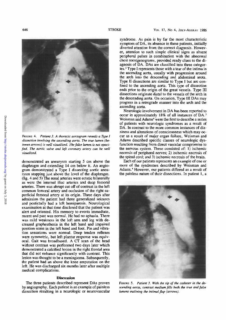
FIGURE 4. Patient 3. A thoracic aortogram reveals a Type Idissection involving the ascending aorta. The true lumen (be-tween arrows) is well visualized. The false lumen is not opaci-fied. The aortic valve and left coronary artery can be wellvisualized.
demonstrated an aneurysm starting 5 cm above thediaphragm and extending 14 cm below it. An angio-gram demonstrated a Type I dissecting aortic aneu-rysm stopping just above the level of the diaphragm,(fig. 4 and 5) The renal arteries were ectatic bilaterallyas were the internal iliac arteries and deep femoralarteries. There was abrupt cut off of contrast in the leftcommon femoral artery and occlusion of the right su-perficial femoral artery at its origin. Three days afteradmission the patient had three generalized seizuresand postictally had a left hemiparesis. Neurologicalexamination at that time disclosed that the patient wasalert and oriented. His memory to events immediate,recent and past was normal. He had no aphasia. Therewas mild weakness in the left arm and leg with de-creased graphesthesia in the left hand and impairedposition sense in the left hand and foot. Pin and vibra-tion sensations were normal. Deep tendon reflexeswere symmetric, but left plantar response was equiv-ocal. Gait was broadbased. A CT scan of the headwithout contrast was performed two days later whichdemonstrated a calcified lesion in the right frontal areathat did not enhance significantly with contrast. Thislesion was thought to be a meningioma. Subsequently,the patient had an above the knee amputation on theleft. He was discharged six months later after multiplemedical complications.
DiscussionThe three patients described represent DAs proven
by angiography. Each patient is an example of painlessdissection resulting in a neurologic or neurovascular
syndrome. As pain is by far the most characteristicsymptom of DA, its absence in these patients, initiallydiverted attention from the correct diagnosis. Howev-er, attention to such simple clinical signs as absentperipheral pulses in combination with the abnormalchest roentgenograms, provided ready clues to the di-agnosis of DA. DAs are classified into three categor-ies.2 Type I represents those with a tear of the intima inthe ascending aorta, usually with progression aroundthe arch into the descending and abdominal arota.Type II dissections are similar to Type I but are con-fined to the ascending aorta. This type of dissectionends prior to the origin of the great vessels. Type IIIdissections originate distal to the vessels of the arch inthe descending aorta. On occasion, Type III DAs mayprogress in a retrograde manner into the arch and theascending aorta.
Neurologic involvement in DA has been reported tooccur in approximately 18% of all instances of DA.3
Weisman and Adams4 were the first to describe a seriesof patients with neurologic syndromes as a result ofDA. In contrast to the more common instances of diz-ziness and alterations of consciousness which may oc-cur as a result of major organ failure, Weisman andAdams described specific classes of neurologic dys-function resulting from direct vascular compromise tothe nervous system. These consisted of: 1) ischemicnecrosis of peripheral nerves; 2) ischemic necrosis ofthe spinal cord; and 3) ischemic necrosis of the brain.
Each of our patients represents an example of one ormore of the syndromes described by Weisman andAdams.4 However, our patients differed as a result ofthe painless nature of their dissections. In patient 1, a
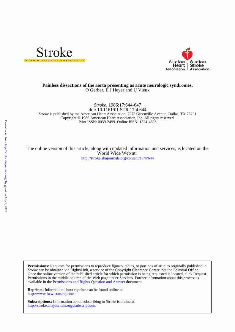
FIGURE 5. Patient 3. With the tip of the catheter in the de-scending aorta, contrast medium fills both the true and falselumens outlining the intimal flap (arrows).
by guest on July 3, 2018http://stroke.ahajournals.org/
Dow
nloaded from

PAINLESS AORTIC DISSECTION/Gerber et al (Al
clinically profound right cerebral syndrome wascaused by occlusion of an innominate artery by thedissection. In patient 2, similarly, innominate occlu-sion occurred as a result of dissection of the aortaresulting in right cerebral ischemia. The active dissec-tion must have initially stopped, only to resume later,and result in thoracic and abdominal aortic dissectionwith a consequent ischemic myelopathy. In patient 3,the motor and sensory involvement of the left leg re-presented an acute ischemic neuropathy. This mostlikely was caused by an embolus from the dissection.The time of occurrence of actual dissection cannot bydetermined. The seizures and the right hemisphericsigns in this patient may have been related to the dis-section, or alternatively were caused by themeningioma.
The incidence of the absence of pain in DA hasvaried in different series. In an early series comprisedof post mortem discovered cases and consequently pre-dominantly of chronic DAs, Baer and Goldburgh5 re-ported that 55% of 44 patients with DA did not have ahistory of pain. According to the large review by Hirst3
14% of 409 patients in whom symptoms were record-ed, did not mention pain. More recent series compiledby clinicians and radiologists and therefore includingmore acute cases2' *•* note an incidence of painless DAof only about 10%.
In the past many individual cases have been reportedas having painless dissecting aneurysms. All of thesepatients have had absence of the characteristic syn-drome, although many cannot be classified as truepainless DAs. In a large number of such patients thediagnosis was established weeks to years after the actu-al dissection.9"13 In such cases pain may not have beenremembered at the time of diagnosis or its occurrencemay have been attributed to another cause. In othersthere existed changes in consciousness, mentation,memory or speech such that these patients could notdescribe a pain syndrome.14' " In still others pain waspresent but not in the classically recognized loca-tions.16 These patients had pain in the groin, hips andelsewhere. Still others had disturbing subjective symp-toms other than pain.10'l7 These included such diversecomplaints as oppressive feelings in the chest and epi-gastric discomfort.
Several explanations have been advanced over theyears for the absence of pain in some patients with DA.None have been entirely satisfactory. These have in-cluded speculation that slow dissection may not resultin pain," that sparing of the adventitia may obviatepain,"'l3 and that circumventing and thus sparing in-tercostal, lumbar and splanchnic vessels may result inpainless dissection.16 Our patients represent examplesof painless DAs presenting with acute neurologic syn-dromes. The first two patients represent acute dissec-tions. The third patient belongs in the category of dis-section of indeterminate duration. In all three patientsthere was clear evidence on general physical examina-tion of sudden occlusion of one or more majorbranches of the aorta. This combination of the absenceof one or more peripheral pulses13 and a vascular neur-
ologic syndrome should lead to a suspicion of DA evenin the absence of pain. In addition, at least two of ourpatients (patients 1 and 3) had findings on chest x-raycompatible with DA.18
ConclusionOur case descriptions are intended to illustrate a
category of patients that may be easily overlooked andtherefore go untreated by those who are primarily con-cerned with the patients' neurological condition.These patients may be seen by a neurologist for anevent or a series of events involving ischemia to thebrain, spinal cord or the limbs and their nerve trunks.
Diagnosis of painless DA in such patients shouldroutinely be pursued by careful attention to some of thecharacteristic and simple non-neurological aspects ofDA. These should include, in particular, examinationof the peripheral pulses, auscultation for aortic insuffi-ciency murmurs and attention to some of the common-ly described chest roentgenographic changes.
References1. WiUius FA, Cragg RW: Cardiac Clinics LXXIX. A talk on dissect-
ing aneurysm of the aorta. Proc of Staff Meeting Mayo Clinic16:41-4, 1941
2. Dalen JE, Howe JP: Dissection of the aorta: current diagnostic andtherapeutic approaches. JAMA 242: 1530-1532, 1979
3. Hirst AE, Johns VJ, KimeSW: Dissecting aneurysm of the aorta: areview of 505 cases. Medicine 37: 217-279, 1958
4. Weisman D, Adams RD: The neurological complications of dis-secting aortic aneurysm. Brain 67: 69-92, 1944
5. Baer S, Goldburgh HL: The varied clinical syndromes produced bydissecting aneurysm. Am Heart J 35: 198-211, 1948
6. Vech RJ, Besterman EMM, Bromley LL, Eastcott HHG, KenyonJR: Acute aortic dissection: historical perspective and current man-agement. Am Heart J 102: 1087-1089, 1981
7. Lindsay J, Hurst JW: Clinical features and prognosis in dissectinganeurysm of the aorta. Circulation 35: 880-887, 1967
8. Slater EE, DeSanctis RW: The clinical recognition of dissectingaortic aneurysm. Am J Med 60: 625-633, 1976
9. Ambos MA, Rothberg M, Lefleur RS, Weiner S, McCanley D:Unsuspected aortic dissection: The chronic "healed" dissection.AJR 132: 221-225, 1979
10. Garcia R, Torbey P, Anbe DT, Drake EH: Painless dissectinganeurysm of the thoracic aorta: report of eight cases masqueradingas gross aortic insufficiency, severe hypertension, myocardial in-farction and mediastinal enlargement. Henry Ford Hosp Med J 25:21-32, 1977
11. Wood FC, Pendergrass EP, Ostrum HW: Dissecting aneurysm ofthe aorta with special reference to its roentgengraphic features. A JRoentgenology and Radium Ther 28: 437-465, 1932
12. Cohen S, littman D: Painless dissecting aneurysm of the aorta. NEngl J Med 271: 143-145, 1964
13. O'Donovan TPB, Osmunndson PJ, Payne WS: Painless dissectinganeurysm of the aorta: report of a case. Circulation 29: 782-786,1964
14. Chase TN, Rosman NP, Price DL: The cerebral syndromes associ-ated with dissecting aneurysm of the aorta. A clinicopathologicalstudy. Brain 91: 173-192, 1968
15. Rosenberg GA: Transient global amnesia with a dissecting aorticaneurysm. Arch Neurol 36: 255, 1979
16. Amer NC, Schaeffer HC, Domingo RT, Sawyer PN, WesolowskiS A: Aortic dissection presenting as iliac occlusion — an aid to earlydiagnosis. N Engl J Med 266: 1040, 1962
17. Hamburger M, Ferris EB: Dissecting aneurysm: study of six recentcases. Am Heart J 16: 1-13, 1938
18. Smith DC, Jang GC: Radiological diagnosis of aortic dissection.In: Doroghazi RM, Slater EE. Aortic dissection. McGraw-Hill,NY, pp 71-132, 1983
by guest on July 3, 2018http://stroke.ahajournals.org/
Dow
nloaded from
O Gerber, E J Heyer and U VieuxPainless dissections of the aorta presenting as acute neurologic syndromes.
Print ISSN: 0039-2499. Online ISSN: 1524-4628 Copyright © 1986 American Heart Association, Inc. All rights reserved.
is published by the American Heart Association, 7272 Greenville Avenue, Dallas, TX 75231Stroke doi: 10.1161/01.STR.17.4.644
1986;17:644-647Stroke.
http://stroke.ahajournals.org/content/17/4/644World Wide Web at:
The online version of this article, along with updated information and services, is located on the
http://stroke.ahajournals.org//subscriptions/
is online at: Stroke Information about subscribing to Subscriptions:
http://www.lww.com/reprints Information about reprints can be found online at: Reprints:
document. Permissions and Rights Question and Answer available in the
Permissions in the middle column of the Web page under Services. Further information about this process isOnce the online version of the published article for which permission is being requested is located, click Request
can be obtained via RightsLink, a service of the Copyright Clearance Center, not the Editorial Office.Stroke Requests for permissions to reproduce figures, tables, or portions of articles originally published inPermissions:
by guest on July 3, 2018http://stroke.ahajournals.org/
Dow
nloaded from