7 overview of resections around the shoulder...
TRANSCRIPT

BACKGROUND■ The upper extremity is involved by bone and soft tissue neo-plasms only one third as often as the lower extremity. Thescapula and proximal humerus are common sites of primarysarcoma, including osteosarcoma and Ewing sarcoma in chil-dren and chondrosarcoma in adults. Metastatic tumors, espe-cially hypernephroma, also have a propensity for the proximalhumerus. When soft tissue tumors occur in the upper extrem-ity, they tend to favor the shoulder girdle and may secondarilyinvolve the scapula, proximal humerus, or clavicle. The axillais another site around the shoulder girdle where primary softtissue tumors may develop or where metastases can spread toand replace the local lymph nodes. The axilla is a relatively“silent” area, where tumors may grow to large sizes beforethey become symptomatic and are detected.■ The shoulder girdle consists of the proximal humerus, thescapula, and the distal third of the clavicle, as well as the sur-rounding soft tissues. Each bone may be involved by a primarymalignant bone tumor or metastases, with or without soft tis-sue extension. The bones of the shoulder girdle also may besecondarily involved by a soft tissue sarcoma, which requiresresection and reconstruction techniques similar to those of aprimary bone tumor (FIG 1).■ Until the mid-20th century, forequarter amputation wasthe treatment for malignant tumors of the shoulder girdle.Today, about 95% of patients with sarcomas of the shouldergirdle can be treated safely by limb-sparing resection such asthe Tikhoff–Linberg resection and its modifications.6 The re-lation of the neurovascular bundle to the tumor and otherstructures of the shoulder girdle is the most significantanatomic factor in determining resectability, removal of thetumor, and reconstruction.■ The resection and reconstruction of tumors of the shouldergirdle consists of three components: (1) surgical resection ofthe tumor following oncologic principles; (2) reconstruction ofthe skeletal defect (ie, endoprosthetic replacement); and (3) softtissue reconstruction using multiple muscle transfers to coverthe skeletal reconstruction and provide a functional extremity.The goals of all shoulder girdle reconstructions are to providea stable shoulder and to preserve normal elbow and hand func-tion. The extent of tumor resection and remaining motorgroups available for reconstruction dictate the degree of shoul-der motion and function that are retained.
Historical Background■ Some of the earliest discussions concerning limb-sparingsurgery focused on techniques for resection of tumors aboutthe scapula. Initial reports of shoulder girdle resections wereconfined to the individual bones or portions of the scapula.The first reported scapular resection was a partial scapulec-tomy performed by Liston in 18197 for an ossified aneurysmaltumor. Between this time and the mid-1960s, several other au-thors discussed limb-sparing resections about the shoulder
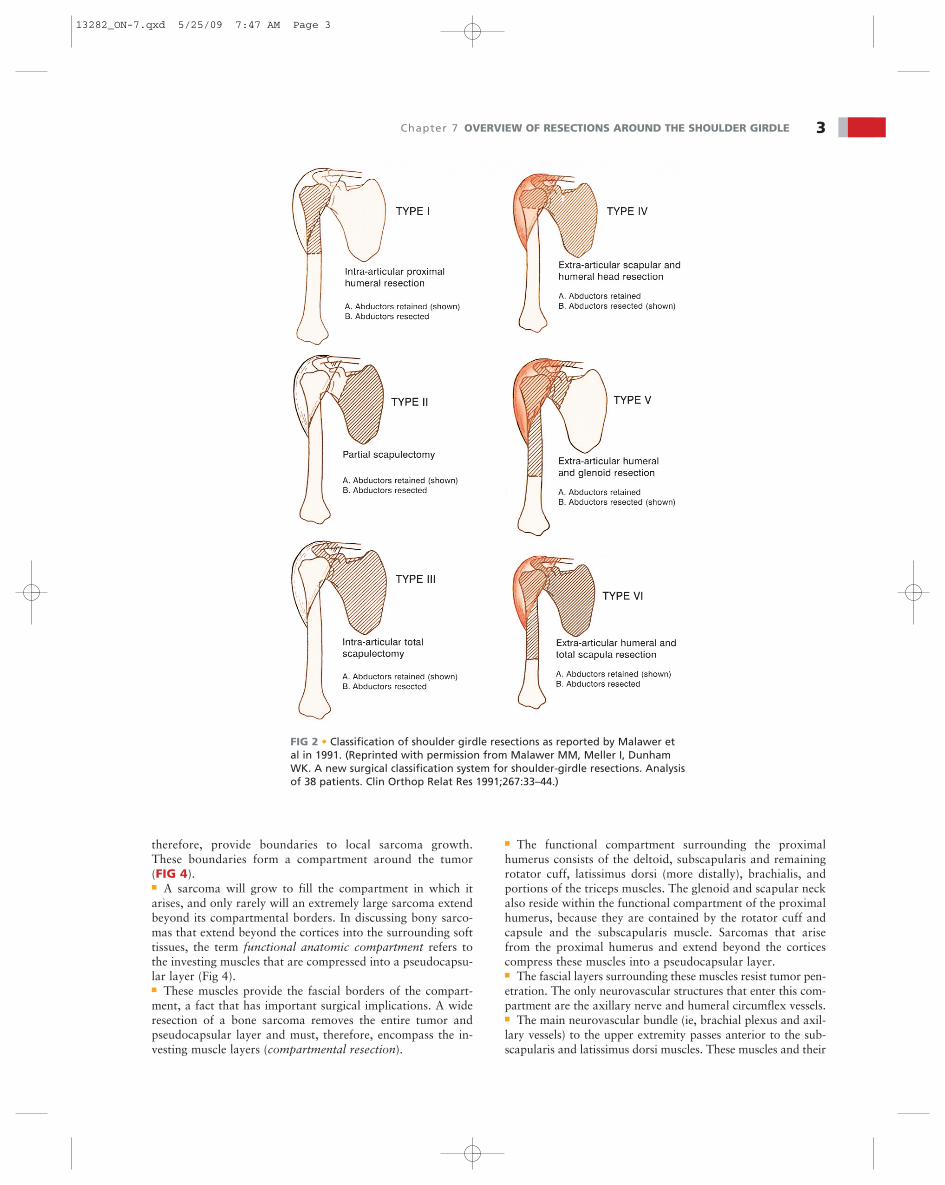
girdle.4,11–15, 19 In 1965, Papioannou and Francis16 reported26 scapulectomies and discussed the indications and limita-tions of the procedure.■ The Tikhoff–Linberg interscapulothoracic resection ortriple-bone resection was described in the Russian literatureby Baumann1 in 1914. He referred to a 1908 report byPranishkov that described the removal of the scapula, thehead of the humerus, the outer one third of the clavicle, andthe surrounding soft tissue for a sarcoma of the scapula. Theshoulder was suspended from the remaining clavicle by metalsutures. Tikhoff and Baumann performed three such opera-tions between 1908 and 1913, and Tikhoff was named as theoriginator of the procedure. The technique became estab-lished in the Western surgical community only after Linberg’sEnglish publication in 1926.6■ Classically, most shoulder girdle resections were done forlow-grade tumors of the scapula and for periscapular soft tissuesarcomas. Before 1970, most patients with high-grade spindlecell sarcomas (eg, osteosarcoma, chondrosarcoma) involvingthe shoulder girdle were treated with a forequarter amputation.In 1977, Marcove et al12 were the first to report limb-sparingsurgery for high-grade sarcomas arising from the proximalhumerus. These authors reported performing an en bloc extra-articular resection that included the proximal humerus, glenoid,overlying rotator cuff, lateral two thirds of the clavicle, deltoid,coracobrachialis, and proximal biceps. Local tumor control andsurvival rates were similar to those achieved with a forequarteramputation. Resection, however, preserved a functional handand elbow. These early results were confirmed by other sur-geons. After the 1980s, osteosarcoma, chondrosarcoma, andEwing sarcoma of the proximal humerus became the tumorsmost commonly treated with a Tikhoff–Linberg resection. Avariety of new techniques and modifications of shoulder girdleresections have been developed. Most have been reported as“Tikhoff–Linberg” or “modified Tikhoff–Linberg” resections.These eponyms are not accurate descriptions, however, becausethe Tikhoff–Linberg procedure was not intended to refer to sar-comas of the humerus.■ As the popularity of limb-sparing surgery for shoulder girdlesarcomas grew, the extent of resection necessary for various tu-mors, particularly indications for an extra-articular resection,remained a matter of debate. The best method for reconstruc-tion also came under considerable discussion. In response,Malawer et al. developed a surgical classification system(FIG 2) based on tumor location, extent, grade, and pathologictype. This system was intended to provide guidelines regardingthe extent of resection necessary for primary bone sarcomasand soft tissue sarcomas that secondarily involve the bones ofthe shoulder girdle.
SURGICAL CLASSIFICATION SYSTEM■ The current surgical classification system was described byMalawer and associates8 in 1991 (Fig 2). It is based on the
Chapter 7James C. Wittig, Martin M. Malawer, and Kristen Kellar-Graney
Overview of ResectionsAround the Shoulder Girdle
1
13282_ON-7.qxd 5/25/09 7:47 AM Page 1
2 Part 4 ONCOLOGY • Section I I SHOULDER GIRDLE AND UPPER EXTREMITIES
current concepts of surgical margins, the relation of the tumorto anatomic compartments (ie, intracompartmental vs. extra-compartmental), the status of the glenohumeral joint, themagnitude of the individual surgical procedure, and preciseconsiderations of the functionally important soft tissue com-ponents. It includes six categories:
■ Type I: intra-articular proximal humeral resection■ Type II: partial scapular resection■ Type III: intra-articular total scapulectomy■ Type IV: extra-articular total scapulectomy and humeralhead resection (classic Tikhoff–Linberg resection)■ Type V: extra-articular humeral and glenoid resection■ Type VI: extra-articular humeral and total scapularresection.
■ Each type is subdivided according to the status of the abduc-tor mechanism (the deltoid muscle and rotator cuff):
■ Abductors intact■ Abductors partially or completely resected
■ Type A resections, in which the abductors are preserved,usually are recommended for high-grade spindle cell bone sar-comas that are entirely intracompartmental (ie, containedwithin either the proximal humerus or scapula bone). This is arare situation, however. This type of resection also is recom-mended for low grade-bone sarcomas, selected metastatic car-cinomas, and, often, round cell sarcomas.■ Type B resections, in which the abductors are resected, areextracompartmental resections and are the most common typeof resection performed for high-grade spindle cell sarcomas.■ All six of these types of shoulder girdle resections and theirindications are described briefly in the following section. The
surgical techniques for each resection and reconstruction aredescribed in ON-8, 10, and 11–13.
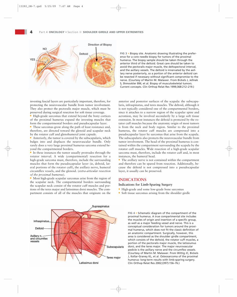
GUIDELINES FOR SHOULDER GIRDLE RESECTIONLocal Growth and Transarticular Involvement byShoulder Girdle Tumors■ The shoulder joint appears to be more prone than otherjoints to intra-articular or pericapsular (ligamentous) involve-ment by high-grade bone sarcomas.■ Four basic mechanisms underlie tumor spread across theshoulder joint: direct capsular extension; tumor extensionalong the long head of the biceps tendon; fracture hematomafrom a pathologic fracture; and poorly planned biopsy (FIG 3).■ These mechanisms place patients undergoing intra-articularresections for high-grade sarcomas at greater risk for local re-currence than those undergoing extra-articular resections.Therefore, it often is necessary to perform an extra-articularresection for high-grade bone sarcomas of the proximalhumerus or scapula.■ Most tumors arise from the metaphyseal portion of theproximal humerus. They extend beyond the cortices andspread underneath the deltoid muscle, subscapularis muscle,and remaining rotator cuff muscles. As the tumor grows, theextraosseous component spreads along the long head of thebiceps tendon, along the glenohumeral ligaments, and under-neath the rotator cuff, heading toward the glenoid or directlycrossing the glenohumeral joint. The deltoid, subscapularismuscle, and remaining rotator cuff muscles are compressedinto a pseudocapsular layer. These muscles form compart-mental boundaries around the tumor. The axillary nerve andcircumflex vessels enter this compartment. The major neu-rovascular bundle is displaced by the tumor; however, in mostinstances the fascia overlying the subscapularis muscle as wellas the axillary sheath that contains the blood vessels andnerves protect the major neurovascular bundle from tumorinvolvement or encasement.■ Similarly, most scapular sarcomas originate from themetaphyseal portion of the scapula or the scapula neck, andgrow centripetally into the soft tissues. They form a soft tis-sue mass that extends outward and usually is contained bythe subscapularis and other rotator cuff muscles. These tu-mors follow the path of least resistance and are directed to-ward the glenohumeral joint and proximal humerus.Eventually, the tumor contaminates these structures. Thesubscapularis muscle and its investing fascia function as abarrier and protect the axillary vessels and brachial plexusfrom tumor invasion. These neurovascular structures usuallyare displaced by the adjacent tumor that lies deep to the sub-scapularis muscle.
Functional Anatomic Compartment of the Shoulder Girdle■ Sarcomas grow locally in a centripetal manner and com-press surrounding tissues (muscles) into a pseudocapsularlayer. The pseudocapsular layer contains microscopic finger-like projections of tumor, which are referred to as satellitenodules.■ Sarcomas spread locally along the path of least resistance.Surrounding fascial layers resist tumor penetration and,
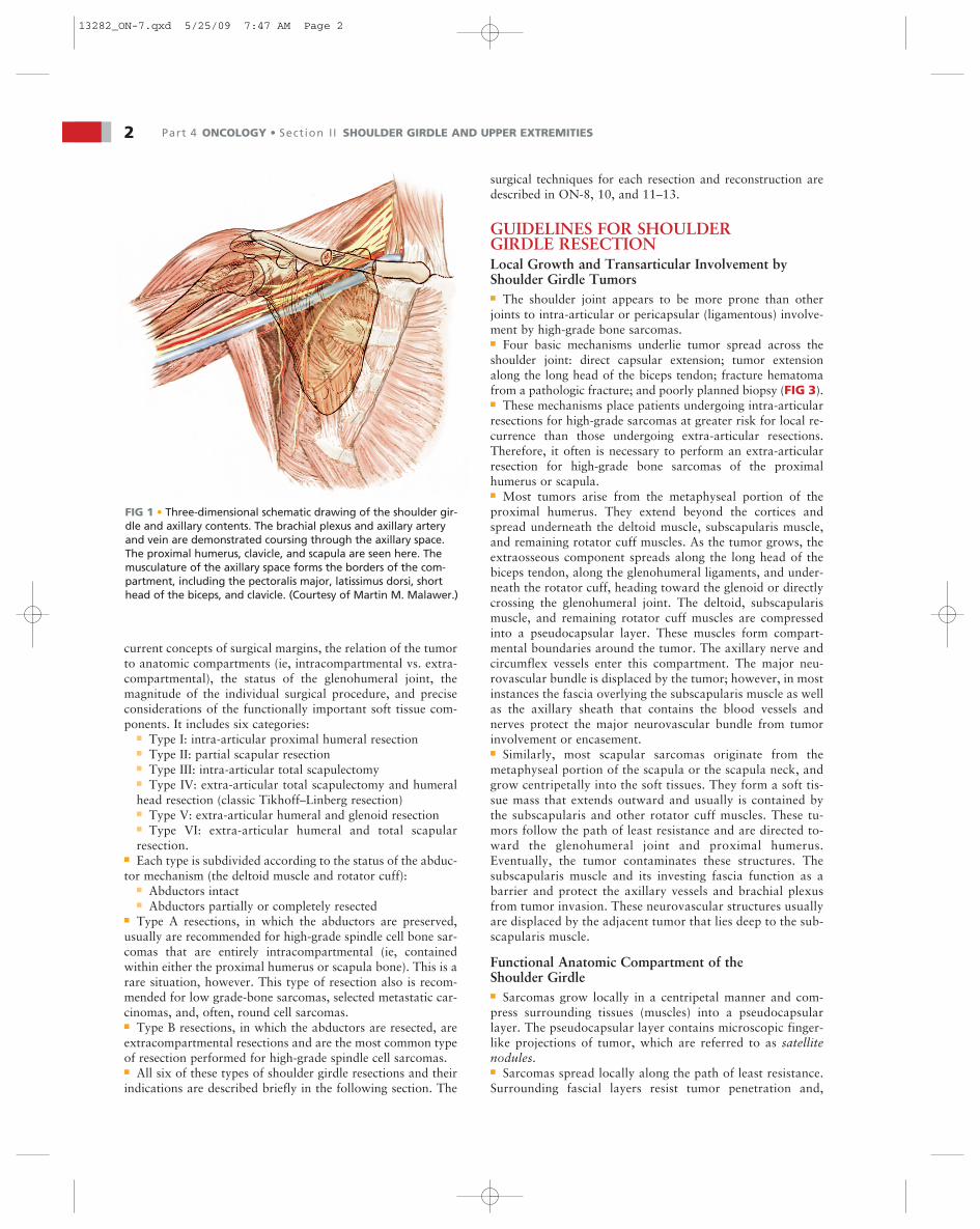
FIG 1 • Three-dimensional schematic drawing of the shoulder gir-dle and axillary contents. The brachial plexus and axillary arteryand vein are demonstrated coursing through the axillary space.The proximal humerus, clavicle, and scapula are seen here. Themusculature of the axillary space forms the borders of the com-partment, including the pectoralis major, latissimus dorsi, shorthead of the biceps, and clavicle. (Courtesy of Martin M. Malawer.)
13282_ON-7.qxd 5/25/09 7:47 AM Page 2
therefore, provide boundaries to local sarcoma growth.These boundaries form a compartment around the tumor(FIG 4).■ A sarcoma will grow to fill the compartment in which itarises, and only rarely will an extremely large sarcoma extendbeyond its compartmental borders. In discussing bony sarco-mas that extend beyond the cortices into the surrounding softtissues, the term functional anatomic compartment refers tothe investing muscles that are compressed into a pseudocapsu-lar layer (Fig 4).■ These muscles provide the fascial borders of the compart-ment, a fact that has important surgical implications. A wideresection of a bone sarcoma removes the entire tumor andpseudocapsular layer and must, therefore, encompass the in-vesting muscle layers (compartmental resection).
■ The functional compartment surrounding the proximalhumerus consists of the deltoid, subscapularis and remainingrotator cuff, latissimus dorsi (more distally), brachialis, andportions of the triceps muscles. The glenoid and scapular neckalso reside within the functional compartment of the proximalhumerus, because they are contained by the rotator cuff andcapsule and the subscapularis muscle. Sarcomas that arisefrom the proximal humerus and extend beyond the corticescompress these muscles into a pseudocapsular layer.■ The fascial layers surrounding these muscles resist tumor pen-etration. The only neurovascular structures that enter this com-partment are the axillary nerve and humeral circumflex vessels.■ The main neurovascular bundle (ie, brachial plexus and axil-lary vessels) to the upper extremity passes anterior to the sub-scapularis and latissimus dorsi muscles. These muscles and their
Chapter 7 OVERVIEW OF RESECTIONS AROUND THE SHOULDER GIRDLE 3
FIG 2 • Classification of shoulder girdle resections as reported by Malawer etal in 1991. (Reprinted with permission from Malawer MM, Meller I, DunhamWK. A new surgical classification system for shoulder-girdle resections. Analysisof 38 patients. Clin Orthop Relat Res 1991;267:33–44.)
13282_ON-7.qxd 5/25/09 7:47 AM Page 3
investing fascial layers are particularly important, therefore, forprotecting the neurovascular bundle from tumor involvement.They also protect the pectoralis major muscle, which must bepreserved during surgical resection for soft tissue coverage.■ High-grade sarcomas that extend beyond the bony corticesof the proximal humerus expand the investing muscles thatform the compartmental borders and pseudocapsular layer.■ These sarcomas grow along the path of least resistance and,therefore, are directed toward the glenoid and scapular neckby the rotator cuff and glenohumeral joint capsule.■ Anteriorly, the tumor is covered by the subscapularis, whichbulges into and displaces the neurovascular bundle. Onlyrarely does a very large proximal humerus sarcoma extend be-yond the compartmental borders.■ In these instances the tumor usually protrudes through therotator interval. A wide (compartmental) resection for ahigh-grade sarcoma must, therefore, include the surroundingmuscles that form the pseudocapsular layer (ie, deltoid, lat-eral portions of the rotator cuff), the axillary nerve, humeralcircumflex vessels, and the glenoid. (extra-articular resectionof the proximal humerus).■ Most high-grade scapular sarcomas arise from the region ofthe scapular neck. The compartmental borders surroundingthe scapular neck consist of the rotator cuff muscles and por-tions of the teres major and latissimus dorsi muscles. The com-partment consists of all of the muscles that originate on the
anterior and posterior surfaces of the scapula: the subscapu-laris, infraspinatus, and teres muscles. The deltoid, although itis not typically considered one of the compartmental borders,since it attaches to a narrow region of the scapular spine andacromion, may be involved secondarily by a large soft tissueextension. In most instances the deltoid is protected by the ro-tator cuff muscles because the anatomic origin of most tumorsis from the neck and body region. Similar to the proximalhumerus, the rotator cuff muscles are compressed into apseudocapsular layer by sarcomas that arise from the scapula.The subscapularis also protects the neurovascular bundle fromtumor involvement. The head of the proximal humerus is con-tained within the compartment surrounding the scapula by therotator cuff muscles. Wide resection of a high-grade scapularsarcoma must, therefore, include the rotator cuff and, in mostinstances, the humeral head.■ The axillary nerve is not contained within the compartmentand therefore can be spared from resection. Additionally, be-cause the deltoid is not compressed into a pseudocapsularlayer, it usually can be preserved.
INDICATIONSIndications for Limb-Sparing Surgery■ High-grade and some low-grade bone sarcomas■ Soft tissue sarcomas arising from the shoulder girdle
4 Part 4 ONCOLOGY • Section I I SHOULDER GIRDLE AND UPPER EXTREMITIES
FIG 3 • Biopsy site. Anatomic drawing illustrating the prefer-ence for a core needle biopsy for tumors of the proximalhumerus. The biopsy sample should be taken through theanterior third of the deltoid. Great care should be taken toavoid the pectoralis major muscle, the deltopectoral interval,and the axillary vessels. The deltoid is innervated by the axil-lary nerve posteriorly, so a portion of the anterior deltoid canbe resected if necessary without significant compromise to thenerve. (Courtesy of Martin M. Malawer. From Bickels J, JellnekS, Shmookler BM, et al. Biopsy of musculoskeletal tumors.Current concepts. Clin Orthop Relat Res 1999;368:212–219.)
FIG 4 • Schematic diagram of the compartment of theproximal humerus. A true compartmental site includesthe muscles of origin and insertion of a specific group,as well as a major feeding vessel and nerve. This is aconceptual consideration for tumors around the proxi-mal humerus, which does not fit the classic definition ofan anatomic compartment. Surgically, however, thisarea is considered as the shoulder girdle compartment,which consists of the deltoid, the rotator cuff muscles, aportion of the pectoralis major muscle, the latissiumusdorsi, and the teres major. The major neurovascularpedicle is the axillary nerve and the circumflex vessels.(Courtesy of Martin M. Malawer. From Wittig JC, BickelsJ, Kellar-Graney KL, et al. Osteosarcoma of the proximalhumerus: long-term results with limb-sparing surgery.Clin Orthop Relat Res 2002;(397):156–76.)
13282_ON-7.qxd 5/25/09 7:47 AM Page 4

■ Metastatic carcinomas: isolated metastasis or metastaticlesions that have caused significant bony destruction■ Occasionally, benign–aggressive tumors also may requirethese treatment techniques.■ Selection of patients for limb-sparing surgery is based on theanatomic location of the tumor and a thorough understandingof the natural history of sarcomas and other malignancies.
Contraindications for Limb-Sparing Surgery■ Absolute contraindications for limb-sparing procedures in-clude tumor involvement of the neurovascular bundle, or apatient’s inability or unwillingness to tolerate a limb-sparingoperation.■ Relative contraindications may include chest wall extension,pathologic fracture, previous infection, lymph node involve-ment, or a complicated, inappropriately placed biopsy that hasresulted in extensive hematoma, which has resulted in tissuecontamination.
Biopsy Site■ One of the most common causes for forequarter amputationis an inappropriately placed biopsy site that has resulted incontamination of the pectoralis muscles, neurovascular struc-tures, and chest wall. Extreme care must be taken with biopsyplacement and technique (see Fig 3).
Vascular Involvement■ Fortunately, most tumors of the proximal humerus are sepa-rated from the anterior vessels by the subscapularis, latissimusdorsi, and coracobrachialis muscles. It is rare for the axillary orbrachial artery to be involved with tumor, although a large softtissue component may cause displacement and compression.■ In general, if the vessels appear to be involved with tumor,the adjacent brachial plexus also is involved, and a limb-sparing procedure may be contraindicated.
Nerve Involvement■ The three major cords of the brachial plexus follow theartery and vein and rarely are involved with tumor. The axil-lary nerve may be involved by neoplasm as it passes fromanterior to posterior along the inferior glenohumeral joint cap-sule. Resection of the axillary nerve usually is required forstage IIB tumors of the proximal humerus.■ The musculocutaneous and radial nerves rarely are involved.The deficit created by resecting the radial nerve is greater thanthat for the musculocutaneous nerve, but this should not be anindication for amputation.■ If resection will lead to a major functional loss and a closemargin (increasing the risk of local recurrence), amputationshould be considered. Direct tumor extension into or en-casement of the brachial plexus necessitates a forequarteramputation.
Lymph Nodes■ Bone sarcomas rarely involve adjacent lymph nodes; never-theless, axillary nodes should be evaluated and may requirebiopsy. In the rare incidence of lymph node involvement doc-umented by biopsy, a forequarter amputation may be the bestmethod for removing all gross disease.■ Alternatively, a lymph node dissection in conjunction with alimb-sparing procedure may be considered. Malawer has
found, based on two cases, that local control and long-termsurvival can be obtained by this method (unpublished data).
Chest Wall Involvement■ Tumors of the shoulder girdle with large extraosseous com-ponents occasionally may involve the chest wall, ribs, and in-tercostal muscles.■ Chest wall involvement should be evaluated preoperativelywith physical examination and imaging studies; however, suchinvolvement often is not determined until the time of surgery.It is not an absolute indication for forequarter amputation; alimb-sparing procedure combined with a chest wall resectionmay be performed, depending on the involvement of adjacentsoft tissues and neurovascular structures.
Previous Resection■ The local recurrence rate is increased in cases in which awide resection is attempted (1) following a previous inade-quate resection around the shoulder girdle or (2) when atumor already has recurred locally. This possibility must be aconsideration especially with tumors of the scapula andclavicle and of soft tissue tumors that involve the proximalhumerus.
Infection■ In patients with high-grade sarcomas, limb-sparing proce-dures performed in an area of infection are extremely risky,because these patients must receive postoperative adjuvantchemotherapy. If an infection cannot be eradicated with theprimary resection, amputation is advisable.
SURGICAL MANAGEMENTPreoperative Planning
Physical Examination■ The physical examination is important for assessing tumorresectability and for estimating the extent of resection thatmay be required. Physical examination is important in deter-mining tumor extension into the glenohumeral joint, neu-rovascular involvement, or tumor invasion of the chest wall. Iftumor has invaded the joint, shoulder range of motion usuallyis reduced, and the patient may complain about discomfortand pain.■ Neurovascular involvement or compression may be sug-gested by an abnormal neurovascular examination or by de-creased or absent pulses.■ Tumors that move freely with respect to the chest wall usu-ally are separated from it by at least a thin tissue plane throughwhich it is safe to dissect.
Determining Tumor Resectability■ High-grade tumors arising from the shoulder girdle regionoften are large and encroach on the neurovascular bundle.Tumors that encase or invade the brachial plexus are consid-ered unresectable. In many cases it is difficult to determine,both clinically and radiologically, which tumors involve orencase the neurovascular structures directly as opposed tomerely displacing these structures. Although most tumors thatdisplace the neurovascular structures are resectable, some areunresectable, and it can be difficult to determine clinicallywhich are in this category.
Chapter 7 OVERVIEW OF RESECTIONS AROUND THE SHOULDER GIRDLE 5
13282_ON-7.qxd 5/25/09 7:47 AM Page 5

■ We have found the clinical triad of intractable pain, motordeficit, and a venogram showing obliteration of the axillaryvein to be very reliable in predicting brachial plexus inva-sion. No single imaging study is available that accurately vi-sualizes the brachial plexus. MRI and CT scans typicallyshow a large tumor juxtaposed to the neurovascular bundle(FIG 5).■ Venography, however, is extremely accurate in predictingbrachial plexus invasion. The axillary vein, axillary artery, andbrachial plexus travel in intimate association within a singlefascial sheath, the axillary sheath.■ The major nerves and cords travel along the periphery of thesheath; therefore only complete obliteration—not just com-pression—of the brachial or axillary vein denotes direct tumorextension in and around the nerves and also indicates sec-ondary involvement of the venous wall. This progression alsoexplains the clinical triad of pain, motor loss, and venousobstruction.■ Tumors that invade or encase the brachial plexus obliteratethe axillary vein because of that vein’s thin walls and lowintraluminal pressure. In these instances arteriographydemonstrates displacement of the axillary artery; however, theaxillary artery remains patent because of its thick walls andhigh intraluminal pressures.
■ The final decision regarding the need for a forequarter am-putation should be reserved until surgical exploration of thebrachial plexus has been performed.
Prosthetic Reconstruction■ When endoprosthetic reconstruction was developed in the1940s, attention initially was focused on reconstruction ofskeletal defects of the lower extremity. Use of the techniquewas broadened gradually to include defects of the upper ex-tremity and shoulder girdle.■ The MRS shoulder prosthesis has undergone several de-sign changes and improvements since that time. The currentcomponents for proximal humerus and scapular replace-ment are shown in Chapters ON-10 and 8, respectively. TheMRS is used in conjunction with both intra- and extra-articular resections, and results are highly predictable andsuccessful.■ Reported rates of fracture, infection, nonunion, reopera-tion, and tumor recurrence are lower, and length of immo-bilization is shorter with extremity endoprosthetic recons-truction than with allograft, composite reconstruction, orarthrodesis.■ Survival of the MRS proximal humeral prosthesis is reportedto be 95% to 100% at 10 years.
6 Part 4 ONCOLOGY • Section I I SHOULDER GIRDLE AND UPPER EXTREMITIES
A B
C
D
E F G
FIG 5 • Imaging studies of the shoulder girdle and axil-lary space demonstrating bony and soft tissue findings. A.CT scan showing a tumor arising from the glenoid and in-volving the glenohumeral joint. CT scans are the bestmodality for observing bony detail. B. Coronal MRI scanshowing direct tumor extension. C,D. A large soft tissueaxillary tumor protruding anteriorly through the pec-toralis major and the skin. This is a high-grade fungatingsoft tissue sarcoma. MRI is the best scan for evaluation ofsoft tissue masses in relation to other soft tissue struc-tures. E. MRI scan of the axillary space (coronal view)showing a secondary skipped lesion along the axillaryvein, coming from a high-grade soft tissue sarcoma lowerin the axilla. Metastatic lesions of the axilla and lymphnodes are a common source of large axillary masses andare best evaluated by MRI scans. F. Angiography and em-bolization of metastatic renal cell carcinoma (hyper-nephroma) to the distal clavicle. Following embolization,there is no evidence of a tumor blush. Embolization oftenis performed for large high-grade soft tissue sarcomasprior to a resection. G. On this axillary venogram, the axil-lary vein is occluded by thrombosis, and there is a smallbackward filling from the innominate vein. This is themost pathognomonic finding of brachial plexus involve-ment seen during the time of surgery. Brachial plexus in-volvement often correlates with the clinical findings ofneurologic deficits, numbness, or muscle weakness of theaffected extremity.
13282_ON-7.qxd 5/25/09 7:47 AM Page 6

Chapter 7 OVERVIEW OF RESECTIONS AROUND THE SHOULDER GIRDLE 7
SKELETAL RECONSTRUCTION FOLLOWING HUMERUS AND SCAPULARESECTIONS
TECH
NIQ
UES
■ Special prosthetic replacements are recommended forskeletal reconstruction following proximal humerus andtotal scapula resections, although the utilitarian ap-proach may be used with any method of reconstruction(TECH FIG 1).
■ Soft tissue reconstruction is accomplished using a dualsuspension technique that employs static and dynamicmethods of prosthetic stabilization and soft tissue andmotor reconstruction.
■ Static methods of stabilization include the use of heavynonabsorbable sutures, Dacron tapes, or Gore-Tex grafts,depending on the site of tumor resection and the pros-thesis that is being used. This method offers secure fixa-tion and stabilization of the prosthesis until the softtissues heal and scar to each other.
■ Dynamic methods of stabilization and reconstruction in-clude multiple muscle rotation flaps and muscle transfersthat eventually heal, scar to each other and the prosthe-sis, and provide the necessary motor units for a func-tional extremity.
■ Soft tissue reconstruction follows skeletal reconstructionand static stabilization. The short head of the biceps is
attached with a tenodesis proximally to the coracoid(intra-articular proximal humerus reconstruction), or tothe clavicle (extra-articular proximal humerus recon-struction) or pectoralis major (total scapula reconstruc-tion). The pectoralis minor also is tenodesed back to itsorigin, when possible, or to the scapula to protect theneurovascular structures. The pectoralis major is repairedto its humeral insertion or, in cases requiring extra-articular proximal humerus reconstruction, transferredto cover the prosthesis with soft tissues. The latissimusdorsi may be transferred laterally to function as an exter-nal rotator following extra-articular proximal humerusresection.
■ In total scapula reconstruction, the periscapular musclesare tenodesed to the prosthesis with heavy nonab-sorbable sutures or tapes in a manner that covers the en-tire prosthesis with muscle. Following isolated axillarytumor resection, the distal (humeral) transected edge ofthe latissimus dorsi muscle is rotated into the defect andsutured to the superficial surface of the subscapularismuscle to fill the dead space. Large-bore closed suctiondrains are routinely placed prior to skin closure.
A
TECH FIG 1 • A. Utilitarian incision. This incision has been developed based on the extensive ex-perience of surgeons performing resections around the shoulder girdle. It consists of three com-ponents. Dashed line A indicates the anterior approach, an extended deltopectoral incision com-ing from the midclavicle through the deltopectoral interval and distally over the medial aspectof the arm, curving in a posterior direction. Dashed line B is a posterior incision that is somewhatcurved in nature, to allow development of a large posterior fasciocutaneous flap for exposure ofthe entire scapula and rhomboid region. Dashed line C is an incision that connects A and Bthrough the axillary folds. This permits resection of large axillary tumors or performance of fore-quarter amputations. (continued)
13282_ON-7.qxd 5/25/09 7:47 AM Page 7

8 Part 4 ONCOLOGY • Section I I SHOULDER GIRDLE AND UPPER EXTREMITIES
TECH FIG 1 • (continued) B. The initial steps of the anterior approach of the utilitarianshoulder girdle incision. The key to this approach is the release of the pectoralis majorfrom its insertion on the humerus (1–2 cm away). With the pectoralis major now re-flected onto the chest wall, the entire axillary space can be exposed. This is termed thefirst layer of musculature of the axillary space. The second muscular layer of the axillaryspace is then visualized. With the pectoralis major layer retracted, the axillary space iscompletely covered by fascia, similar to the peritoneum. This covers two muscles, theshort head of the biceps and the pectoralis minor, which attach to the coracoid process,which must be released. With these two muscles released, the axillary space and infra-clavicular component of the brachial plexus (ie, the axillary vein and artery through itsentire length) can be explored completely. If necessary, a portion of the clavicle can beresected to exposed the subclavian artery and vein. (Courtesy of Martin M. Malawer.)
B
TEC
HN
IQU
ES
PEARLS AND PITFALLSPreoperative evaluation ■ Physical examination and radiologic imaging modalities are useful for predicting whether a
tumor is resectable. The scapula and proximal humerus should move freely from the chest wall.Chronic swelling in the distal extremity, intractable pain, motor loss, and a venogram thatdemonstrates obliteration of the axillary vein strongly suggest that the tumor is unresectable.The final determination regarding the need for a forequarter amputation is madeintraoperatively, after anterior exposure and exploration of the brachial plexus and neu-rovascular structures.
Neurovascular exploration ■ The key to a safe and adequate resection of all types of neoplasms around the shoulder girdle and mobilization lies in adequate visualization, exposure, dissection, mobilization and preservation of all vital
neurovascular structures. Full exposure is facilitated by releasing the pectoralis major musclefrom its humeral insertion and the strap muscles from the coracoid process.
Type of resection ■ High-grade sarcomas that arise from the proximal humerus or scapula are likely to contaminateor cross the glenohumeral joint, either grossly or microscopically. An extra-articular type ofresection is used for most high-grade sarcomas arising from the scapula or proximal humerus.Clavicular tumors, although less common, require a slightly different surgical approach (FIG 6).
Soft tissue reconstruction ■ Soft tissue reconstruction is just as important as skeletal reconstruction during limb-sparingsurgery if a functional extremity is to be provided. Static and dynamic methods of soft tissuereconstruction and stabilization are used. Static methods rely on heavy nonabsorbable sutures,Dacron tapes, and Gore-Tex grafts. Dynamic methods rely on multiple muscle transfers androtational muscle flaps.
13282_ON-7.qxd 5/25/09 7:47 AM Page 8
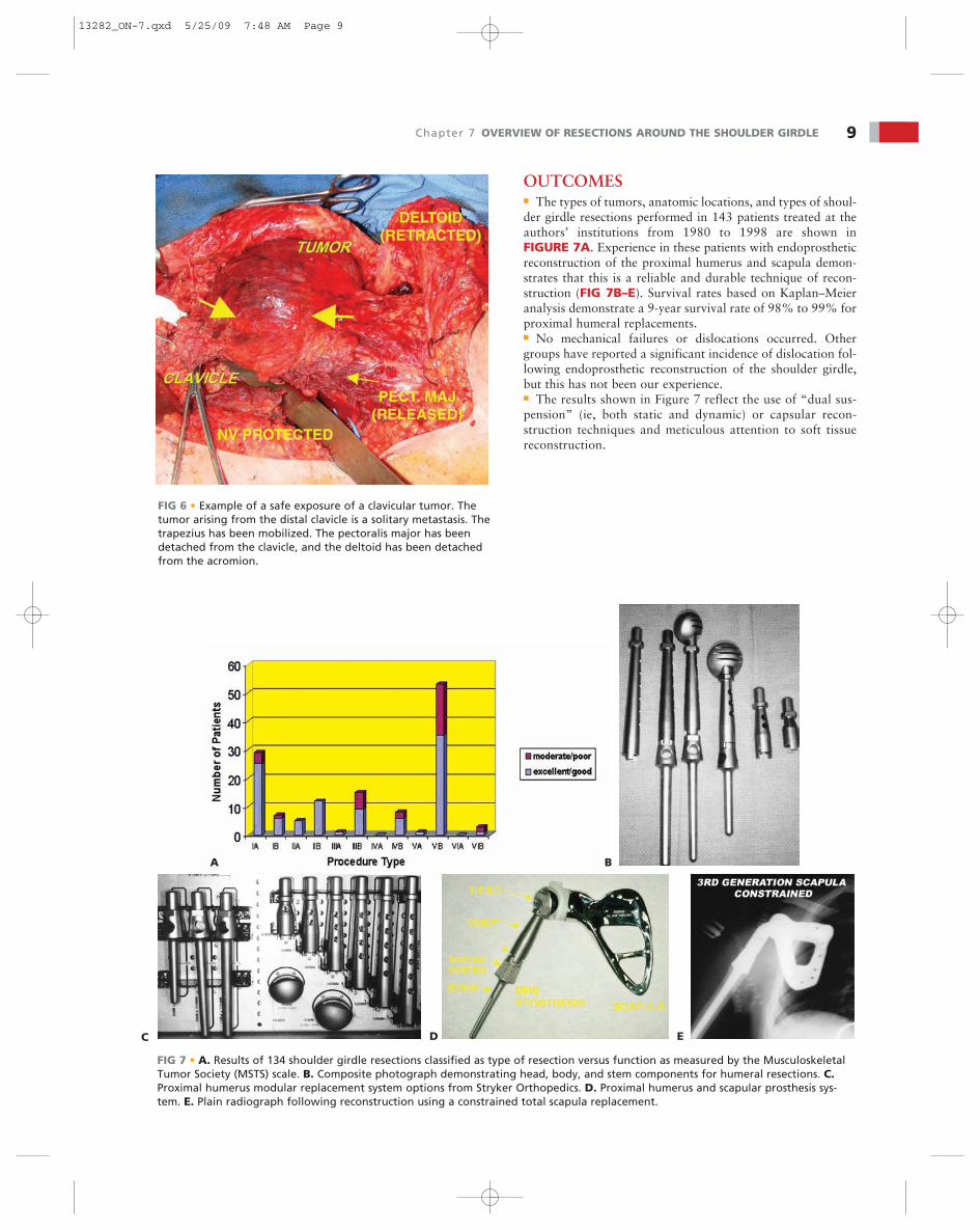
OUTCOMES■ The types of tumors, anatomic locations, and types of shoul-der girdle resections performed in 143 patients treated at theauthors’ institutions from 1980 to 1998 are shown inFIGURE 7A. Experience in these patients with endoprostheticreconstruction of the proximal humerus and scapula demon-strates that this is a reliable and durable technique of recon-struction (FIG 7B–E). Survival rates based on Kaplan–Meieranalysis demonstrate a 9-year survival rate of 98% to 99% forproximal humeral replacements.■ No mechanical failures or dislocations occurred. Othergroups have reported a significant incidence of dislocation fol-lowing endoprosthetic reconstruction of the shoulder girdle,but this has not been our experience.■ The results shown in Figure 7 reflect the use of “dual sus-pension” (ie, both static and dynamic) or capsular recon-struction techniques and meticulous attention to soft tissuereconstruction.
Chapter 7 OVERVIEW OF RESECTIONS AROUND THE SHOULDER GIRDLE 9
FIG 6 • Example of a safe exposure of a clavicular tumor. Thetumor arising from the distal clavicle is a solitary metastasis. Thetrapezius has been mobilized. The pectoralis major has beendetached from the clavicle, and the deltoid has been detachedfrom the acromion.
A B
C D E
FIG 7 • A. Results of 134 shoulder girdle resections classified as type of resection versus function as measured by the MusculoskeletalTumor Society (MSTS) scale. B. Composite photograph demonstrating head, body, and stem components for humeral resections. C.Proximal humerus modular replacement system options from Stryker Orthopedics. D. Proximal humerus and scapular prosthesis sys-tem. E. Plain radiograph following reconstruction using a constrained total scapula replacement.
13282_ON-7.qxd 5/25/09 7:48 AM Page 9
REFERENCES1. Baumann PK. Resection of the upper extremity in the region of the
shoulder joint. Khirurgh Arkh Velyaminova 1914;30:145.2. Dahlin DC. Bone Tumors: General Aspects and Data on 6,221 Cases,
ed 3. Springfield, IL: Charles C Thomas, 1978.3. Francis KC, Worcester JN. Radical resection for tumors of the shoul-
der with preservation of a functional extremity. J Bone Joint Surg Am1962;44A;1423–1430.
4. Guerra A, Capanna R, Biagini R, et al. Extra-articular resection ofthe shoulder (Tikhoff–Linberg). Ital J Orthop Traumatol 1985;11:151–157.
5. Henshaw RM, Jones V, Malawer MM. Endoprosthetic replacementwith the modular replacement system: survival analysis of the first100 implants with a minimum 2-year follow-up. Presented at theCombined Meeting of the American and European MusculoskeletalTumor Societies, Washington, DC, May 6–10, 1998.
6. Linberg BE. Interscapulo-thoracic resection for malignant tumors ofthe shoulder girdle region. J Bone Joint Surg 1928;10:344.
7. Liston R. Ossified aneurysmal tumor of the subscapular artery. EduilMed J 1820;16:66–70.
8. Malawer MM. Tumors of the shoulder girdle: technique of resectionand description of a surgical classification. Orthop Clin N Am1991;22:7–35.
9. Malawer MM, Link M, Donaldson S. Sarcomas of bone. In: DeVitaVT, Hellman S, Rosenberg SA, eds. Cancer: Principles and Practice ofOncology, ed 3. Philadelphia: JB Lippincott, 1984:1418–1468.
10. Malawer MM, Sugarbaker PH, Lambert MH, et al. TheTikhoff–Linberg procedure and its modifications. In: Sugarbaker PH,ed. Atlas of Sarcoma Surgery. Philadelphia: JB Lippincott, 1984.
11. Marcove RC. Neoplasms of the shoulder girdle. Orthop Clin N Am1975;6:541–552.
12. Marcove RC, Lewis MM, Huvos AG. En-bloc upper humeral inter-scapulothoracic resection. The Tikhoff–Linberg procedure. ClinOrthop 1977;124:219–228.
13. Mussey RD. Removal by dissection of the entire shoulder blade andcollar bone. Am J Med Sci 1837;21:390–394.
14. Pack GT, Baldwin JC. The Tikhoff–Linberg resection of the shouldergirdle. Case report. Surgery 1955;38:755–757.
15. Pack GT, Crampton RS. The Tikhoff–Linberg resection of the shoul-der girdle. Clin Orthop 1961;19:148–161.
16. Papioannou AN, Francis KC. Scapulectomy for the treatment of pri-mary malignant tumors of the scapula. Clin Orthop 1965;41:125
17. Rosenberg SA, Suit FD, Baker LH. Sarcomas of soft tissue. In:Devita VT, Hellman S, Rosenberg SA, eds. Cancer: Principles andPractice of Oncology, ed 2. Philadelphia: JB Lippincott; 1985:1243–1293.
18. Samilson RL, Morris JM, Thompson RW. Tumors of the scapula. Areview of the literature and an analysis of 31 cases. Clin Orthop1968;58:105–115.
19. Syme J. Excision of the Scapula. Edinburgh: Edmonston and Douglas,1864.
20. Wittig JC, Bickels J, Wodajo F, et al. Utilitarian shoulder approachfor malignant tumor resection. Orthopedics 2002;5:479–484.
10 Part 4 ONCOLOGY • Section I I SHOULDER GIRDLE AND UPPER EXTREMITIES
13282_ON-7.qxd 5/25/09 7:48 AM Page 10