76 12 october 1968 hypnosis for asthma sou - bmj.com · 76 12 october 1968 hypnosis for asthma mbic...
TRANSCRIPT
76 12 October 1968 Hypnosis for Asthma MBIC SOUnine centres is given below, with the names of the participatingphysicians in parentheses, together with (1) the independent clinicalassessors, and (2) the administrative, nursing, and technical staff whotook a major part in running the trial during its four years. Thebreathing exercises were specially devised for the trial by Dr. P. J. D.Heaf and Dr. G. P. Maher-Loughnan in the Chest Department atUniversity College Hospital, London. The illustrations in thebooklet containing details of the breathing exercises were drawn byMiss C. Schmolle. The criteria used for classifying the type ofasthma were recommended by Dr. J. Pepys, who made the finaldecision in cases of doubt. The secretarial work was undertakenby Mrs. D. Williams, who also painstakingly transcribed the monthlydiary recordings and helped with the analysis of results.
Chest Clinic, Knightswood Hospital, Glasgow (Dr. J. A. Crocket): (1) Dr.C. D. Anderson, Dr. C. Johnston; (2) Sister MacLeod, Staff Nurse Mitchell,Dr. J. F. Boyd, Staff Nurse McRae, Mrs. Corrigan.Ransom Hospital, Mansfield (Dr. D. Davies): (1) Dr. W. H. R. Smith-
(2) Miss B. Buck.Worcester Royal Infirmary (Dr. S. Z. Kalinowski): (1) Dr. E. N. Moyes;
(2) Mrs. M. J. Rowlands, Sister A. Hartley, Sister R. James, Mr. G. H.Green.Hitchin Chest Clinic, Lister Hospital (Dr. N. Macdonald): (1) Dr. F. A. H.
Simmonds, Dr. H. Bruckner, Dr. H. Lawrence; (2) Mrs. H. Snowden, SisterB. Wood, Staff Nurse M. Ellis.
Colindale Hospital, London (Dr. G. P. Maher-Loughnan): (1) Dr. W. E.Snell, Dr. M. K. Sarin; (2) Mrs. M. J. Curry, Mr. B. J. Newman.
University College Hospital, London (Dr. M. K. McAllen): (1) Dr. P. J. D.Heaf; (2) Miss P. McInroy.Birmingham Chest Cliftic (Dr. J. Morrison Smith): (1) Dr. V. H. Springett,
Dr. H. E. Thomas ; (2) Mrs. J. Morris, Mrs. Y. Jones.Luton Chest Clinic (Dr. J. Brian Shaw: (1) Dr. S. G. Maddock, Dr. J.
Clifford-Firth, Dr. L. Ghosh; (2) Dr. H. Banks-Smith, Mrs. C. Teale, SisterK. Kennedy, Sister E. M. Waldron, Sister D. Givan.Chest Clinic, St. Helen's Hospital, Ipswich (Dr. C. J. Stewart): (1) Dr. D.
van Zwanenberg, Dr. D. P. F. Embleton ; (2) Mrs. E. Hart, Mrs. B. Wade,Dr. M. Dixon, Dr. A. Lintott.
REFERENCES
Maher-Loughnan, G. P., Macdonald, N., Mason, A. A., and Fry, L.(1962). Brit. med. Y., 2, 371.
Moore, N. (1965). 7. psychosom. Res., 9, 257.Smith, J. Morrison, and Burns, C. L. C. (1960). Brit. 7. Dis. Chest, 54,
78.
Osteoporosis of Lumbar Vertebrae and Calcification of AbdominalAorta in Women Living in Durban
C. E. DENT,* M.D., F.R.C.P., F.R.S.; H. E. ENGELBRECHT,t M.B., D.M.R.D.; R. C. GODFREY4 M.B., M.R.C.P.
[WITH SPECIAL PLATE FACING PAGE 941
Brit. med. J., 1968, 4, 76-79
Summary: To try to establish whether mechanical stressand muscular activity in earlier life influence the
incidence and severity of spinal osteoporosis in old agelateral x-ray films of the lumbar vertebrae were obtainedfrom three matched groups, each of 100 women 50 to 90years old. Group A was of rural Bantu accustomed tocarrying heavy loads on their heads. Group B was ofurban Bantu, mainly in domestic service. Group C wasof women of European origin.
Severe osteoporosis occurred in three cases from groupA, two from group B, and 14 from group C. Lesserdegrees of osteoporosis could not be assessed preciselyenough for inclusion in these figures. Evenly biconcavevertebral bodies, strongly suggestive of osteomalacia, wereseen in 10 from group A, five from group B, and one fromgroup C. In many Bantu subjects the fifth lumbarvertebra appeared flattened though of good radiodensityand with no marked changes in the other vertebrae.Twenty-eight of these were from group A, 16 from groupB, and none from group C.About a third of each group showed severe degenera-
tive changes in the spine; another third showed milderchanges. More cases of spondylolisthesis occurred in theBantu groups than in the white group. Severe calcifica-tion in the abdominal aorta was noted in 24 women ingroup C. Mild signs occurred in 35 further womenfrom group C, in six from group B, and in onlyone from group A.
Introduction
Osteoporosis is seen most commonly in old age, and by someit is regarded as an integral part of the normal ageing process(reviewed by Rose, 1967). Other factors, especially hormonal,are known to play a significant part in the development of
osteoporosis. However, prolonged immobilization, as afterparalysis or after fracture, can also produce severe osteoporosisof the affected part. No cl ar evidence exists about the part thatmight be played by lesser degrees of immobilization or of minorchanges of activity over very long periods, nor is it clear howfar the reverse may be true-namely, the possibly beneficialeffect of great activity on new bone formation.
In the work described here an attempt has been made toestablish whether the incidence and severity of osteoporosis ofthe spine appearing in old age is influenced in any way bymechanical stress and muscular activity during earlier life. Todo this we chose to examine the lumbar vertebrae of matchedgroups of women, 50 to 90 years old, from two different racesin Durban, South Africa, where there are excellent medicalfacilities for this purpose. Many differences exist between theways of life of Bantu and of white women in and aroundDurban, but one of the most striking concerns the amount ofmechanical stress to which their spines are subjected duringchildhood, young adulthood, and middle age. It is the practiceof rural Bantu women to carry loads of all kinds on theirheads. Such loads, usually of firewood or buckets full ofwater, may approach 2 cwt. (100 kg.) and they are often carriedfor distances of several miles. They begin to carry loads inthis way from about 8 years of age, so the effect, if any, ofweight-bearing covers an important period of skeletal matura-tion and growth. As the study proceeded we also decided torecord some other features shown on the x-ray films.Nordin (1966) studied the incidence of osteoporosis in about
1,000 normal male and female adults of various ages in 10different countries. That study was intended particularly tonote the effects of diet and of age. We are unaware of any studywhich is concerned only with the possible effect of exercise andwhich has used a more narrowly demarcated human popula-tion.
* Professor of Human Metabolism, University College Hospital MedicalSchool, London W.C.i.
t X-ray Department, King Edward VIII Hospital, Durban.* Department of Medicine, King Edward VIII Hospital, Durban. Nowat National Hospital for Nervous Diseases, London.
Case Material
The groups were made up as follows:Group A: 100 Bantu women who had spent their entire lives in
rural areas working on the farms and who often carried heavy loadson their heads.
on 12 March 2019 by guest. P
rotected by copyright.http://w
ww
.bmj.com
/B
r Med J: first published as 10.1136/bm
j.4.5623.76 on 12 October 1968. D
ownloaded from

12 October 1968 Spinal Osteoporosis-Dent et al.
Group B: 100 Bantu women who had migrated from the countryto Durban, and had worked as domestic servants for the majorpart of their lives.
Group C: 100 white women of European descent who hadresided for most of their lives in the city of Durban.
Unfortunately the distinction between groups A and B wasnot as sharp as was hoped, mainly because most domesticservants were unable to give the exact age at which they hadmigrated to an urban environment. Even after moving manyhad continued to carry heavy loads-for example, dustbins-on their heads, and preferred to use this method also for smallerloads such as cans of beer.
Method of Selection of Patients
Bantu Patients.-Between November 1966 and March 1967the female wards of King Edward VIII Hospital were visitedregularly. Suitable cases for the survey were pointed out bythe sisters. The interpreter was a medical social worker wellversed in Zulu history and custom. Hospital notes wereinspected, and if the diagnosis was suitable a patient was askedwhether she would be willing to take part. The followingquestions were asked through the interpreter:
(1) Age of patient: This was never memorized as such, butwas discovered mainly by means of a historical questionary whichcomprised about 50 items oovering the years 1880-1910; theinterpreter felt that she was able to be accurate to within two years.Particularly useful events proved to be the rhinderpest plague of1897, the Anglo-Boer War (1899-1902), and the snow blizzard of1905.
(2). Previous work of patient-that is, farm worker or domesticservant, and whether much heavy carrying.
(3) A rough history of the dietary habit. It was found thatthis consisted either of a predominantly cereal diet with very littlemeat or milk or, in the case of the domestic workers, a good mixeddiet.
(4) Whether or not there was a positive history of back pain.White Patients.-These patients were all living in Durban,
and were having a normal European diet. The same historywas taken from them. Nearly all were of British origin, andtheir admissions were for various causes unconnected withskeletal disease.The patient was measured (crown-pubis, pubis-heel, and
span) and sent for x-ray examination. Patients were excludedif they had been immobilized for more than a week, or were
suffering from systemic disease or receiving drugs known toaffect bone metabolism. Most of the Bantu patients in factwere suffering from pneumonia or acute amoebic dysentery.The survey was continued until 100 suitable patients had
been found for each group. To boost the numbers of patientsin group B, an old people's home was visited and 30 suitable
25
20 -
C
4'
a
*4.-0
z
150
10
5
0
Group A
0n 0in0o 0e0in 00-- l_- eeD or0 un ..oI I I I I I I I f.0 --.o - -.O - .0 - -0
in i.0.o r r- osoe
Age groups (years)
Group B
0.0 N
.0
-.0 .0
Age distribution of the three groups.
subjects were found. The x-ray films were then sorted out intotheir three respective groups.Age (Mean) of the Three Groups.-The age distribution of
the three groups is shown in the Chart. The average age was68.5 years for group A, 66.3 years for group B, and 70.4 yearsfor group C.
X-ray FilmsOne lateral view of the lumbar spine only was taken for each
patient. Specialized techniques were not used to standardizethe bone density. Conventional radiographic equipment andtechnique were employed with standard kilovoltage, milli-amperage, and exposure-time variation according to the thick-ness of the patient.The complete series of 300 films was examined both in South
Africa and in London. For the London assessment the filmswere arranged in random order and the observations madewithout knowledge of the group from which each film wastaken. It was found that there were six features which meritedspecial attention.
Osteoporosis
In the present series, where no attempt was made tostandardize the technical aspects of the x-ray films other thanto ensure a good film suitably centred, we decided to rely on apurely visual assessment of the vertebral shape and structurewith no attempt to assess radiodensity of the bones. Theradiological features of established osteoporosis of the spinewere well known (Cooke, 1955; Jesserer, 1963 ; Rose, 1964;Dent and Watson, 1966).
In less-advanced cases, before shape changes in the vertebralbodies have occurred, it is hard to assess the degree of osteo-porosis, and even complex methods which aim to quantitate thebone density (Schraer, 1958; Nordin et al., 1962) are not
Numbers of Patients with Pathological Changes Seen on Lateral LumbarX-ray
GroupA GrOUBdIGopX-ray Signs of Gs(Rural (Urbanized
Bantu) Bantu) Wites)Definite osteoporosis .. 3 2 140steomalacia . . 10 5 1Flattening of5th lumbar vertebra 28 160Degenerative changes (mild) 31 31 27Degenerative changes (severe) 26 38 31Spondylolisthesis L5-S1 or L45 8 8 4Aortic calcification (mild) 1 6 35Aortic calcification (severe) 0 0 I 24
wholly satisfactory (Dent and Watson, 1966). There weremany films in which the presence of osteoporosis could besuspected by an apparent rarefaction of the bones but in which
there were no more specific changes such asSchmorl's nodes and wedging of the
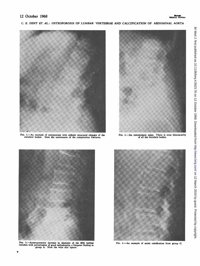
Group C vertebral bodies. We finally decided togroup the films into three categories only:(1) normal, (2) ambiguous, and (3) definitevertebral body changes due to osteoporosis., Lr~~Fig. I (Special Plate) gives an example ofa definitely osteoporotic spine from theseries.Our results are shown in the Table. It
will be noted that there is an increasedincidence of definite osteoporosis among thewhite patients. There was no clear differ-ence between rural Bantu and urbanizedBantu. The mean age of the three osteo-porotic patients in group A was 76.6 years,of the two in group B 65.5 years, and of the14 in group C 75.4 years.
Eleven of the 14 osteoporotic patientsfrom group C had a pubis-heel measure-
BRmsHMEDICAL JOURNAL 77
in 0 in 0 LO 0 Ln 0 LA 0 Ln 0r- aD co ol Lo .0 .0 r- r- CO CK) 0,
I I I I i I I I I I I I- .-o - .0 '.0 - 'o - .O - Or.. r- ao CO Ln Ln -o o r- r- w w
on 12 March 2019 by guest. P
rotected by copyright.http://w
ww
.bmj.com
/B
r Med J: first published as 10.1136/bm
j.4.5623.76 on 12 October 1968. D
ownloaded from

Spinal Osteoporosis-Dent et al.ment more than 5 cm. greater than the crown-pubis measure-
ment. All the osteoporotic Bantu patients had much morethan this degree of shortening, but little significance can beattached to measurements in African Negroes, whose limbs arealways relatively longer than those of Caucasians. For example,in this series of cases the average difference between the crown-pubis and pubis-heel measurement was 6.65 cm. in the Bantu,and only 3.68 cm. in the Whites (excluding those cases withdefinite osteoporosis).
Osteomalacia
A number of films in this series showed an even biconcavityof the vertebral bodies typical of osteomalacia. Fig. 2 (SpecialPlate) shows a gross example of this change, and the Tablegives the number of such films found in this series. Thoughthe figures are not large, it is of interest that only one case
occurred in the well-nourished white patients and 10 casesin the rural Bantu. The diet of the rural Bantu consists mainlyof cereals with very little meat or milk, and the vitamin Dcontent of this diet is low. However, there was always plentyof sunshine to help synthesize the vitamin on the exposed skin.
Flattening of Fifth Lumbar Vertebra
In 44 of the 200 Bantu women (28 of the rural and 16 ofthe urbanized), there was a characteristic change in the shapeof the fifth lumbar vertebra (Special Plate, Fig. 3). It will benoted that the bone density is good but that there is a flatteningand increase in the anteroposterior diameter. In some casesthere was also anterior wedging. Along with this it was notedthat the disc space was well preserved and even perhapswidened. A further common finding was a marked loss of thenormal lumbar curve. One of us (H. E. E.) has a wide experi-ence of Bantu spines, and has noted that these changes are very
common among patients beyond middle age. Whether thesechanges are related to the high stresses to which their spinesare exposed during earlier life (Jonck, 1964) remains an open
question, but we do note that there was more of the flatteningin group A than in group B. We had to be careful not toassess this as one of the changes in shape due to osteoporosis.
Degenerative Changes
Osteophyte formation, disc narrowing, and disc calcificationwere used as guides to the degree of degenerative disease presentin this series of films. There was very little difference in theincidence of such changes in the three groups. The degreeof severity was closely related to increasing age.
Spondylolisthesis
Both of the Bantu groups contained more cases of spondylo-listhesis than the white group. The increased incidence maybe due only to the greater amount of trauma to which theBantu spine is subjected. However, it is well established thatthe pelvis of the Bantu women is tilted forward in comparisonwith white women, and the greater anterior lumbosacral anglemay have some bearing on the tendency to spondylolisthesis.
Aortic Calcification
Here we found the most significant differences between thegroups. All the severe aortic calcification (Special Plate, Fig.4) and most of the lesser degrees of this were found among
the white patients of group C. There was no tendency for thesevere calcification to occur in the more osteoporotic spines.
MEDICAL JouRNA
Discussion
The original project was aimed to be a pilot survey to deter-mine the long-term and possibly beneficial effects of heavyweight-bearing on the bones of the spine. It was supposedthat, as can occur with long bones, the stresses and strains ofweight-bearing might lead to the formation in early adult lifeof a much stronger spine than usual. If so, this should protectthe spine in later life from the development of " senile " osteo-porosis, since many authors now (references quote by Dent andWatson, 1966) believe that osteoporosis is a slow inevitableprocess of thinning of the skeleton beginning around 30 yearsof age, but of very variable rate from person to person, andnot usually manifesting symptoms and signs until after 60 yearsof age. At this latter stage it is easy to apply the name " senile "or even, if in a woman, " postmenopausal," to the osteoporosisthen present.
We were anxious to test this theory by comparing the x-rayfilms of the spines of women who had regularly carried heavyweights on their heads with those of suitable control groupsof less active subjects. We were in some trouble here, as it wasnot possible to be certain, say, that a given woman of 70 yearsof age had or had not been carrying heavy weights on her headtill the age of 30 years, as the memory of such women wassometimes imperfect. We decided to choose a group of urbanAfricans of comparable age to compare with the similar groupof rural Africans. The rural people were certainly very activein the sense we wanted, but it is likely that many of the urbanpeople were also pretty active, and certainly they too were accus-tomed to carrying less heavy loads on their heads. Also somemight have migrated from the countryside more recently thanwe were told. A group of white women of similar age distribu-tion was also collected for comparison. The whole surveywas limited to women, as they suffer so much more fromosteoporosis than do men, and, further, if lack of exercise wasan important contributory reason for this rather than the usualmuch maligned menopause, this effect should show better inwomen, since they tend to vegetate more than men in old age.Furthermore, the situation in Bantu men is complicated by thecommon occurrence of severe osteoporosis associated withhaemosiderosis (Seftel et al., 1966).Our results show that the two groups of African women were
almost the same with regard to the degree of osteoporosis. Therewas a much lower incidence in both these groups than in thegroup of white women. This is an important finding, not quitein the way we had originally expected, and it needs detailedstudy in a further survey directed differently from this one, forthe social and dietary habits of the Africans and Whites are sodifferent that at this stage it is not possible to say what thedifference found in their spines is due to.
Quite incidental to the original project was our finding ofvery marked differences in the amount of calcification seen inthe abdominal aorta on the x-ray films of the lateral lumbarvertebrae. This should not have surprised us, as there is aconsiderable literature on the apparent immunity of unwestern-ized Africans to degenerative arterial disease (discussed byDavidson and Passmore, 1959). However, the size of thedifference did surprise us, also the possibility that the smallnumber of calcified aortae found in the urban Africans mightrepresent a real difference with the rural African group, whoshowed only one case of mild aortic calcification. There wasno positive correlation between the patients with aortic calcifica-tion and those with severe osteoporosis, as was observed byBernstein et al. (1966), who took similar x-ray views in theirsurvey of the inhabitants of Dakota in high and low fluorideareas.
The assessment of the degree of osteoporosis was done blindlyby one of us after assessment had been carried out with know-ledge of the diagnosis by the other two. There was goodagreement. We had intended to repeat the process to see howwe agreed among ourselves on a further fully blind assessment
78 12 October 1968
on 12 March 2019 by guest. P
rotected by copyright.http://w
ww
.bmj.com
/B
r Med J: first published as 10.1136/bm
j.4.5623.76 on 12 October 1968. D
ownloaded from

12 October 1968 Spinal Osteoporosis-Dent et al. BRmTISH 79
This was not possible, however, for we soon realized we coulddistinguish the x-ray films of most of the whites from those ofthe Africans by the different incidence of aortic calcificationand the rather characteristic shape of the lower lumbarvertebrae of many of the Africans.
We wish to thank the Medical Research Council for their supportand for a grant to one of us (R. C. G.). Thanks are due to manymembers of the staff of King Edward VIII Hospital, Durban, formaking this project possible. Professor E. B. Adams put manyfacilities at our disposal, including the medical social worker, MissNgwane, to whom we are especially grateful. The high qualityof the x-ray films was entirely due to Professor B. Sachs andthe x-ray department of the Addington and King Edward VIIHospitals, Durban. The extra work we imposed on them was bornein the normal course of their workings. We thank the consultants
of these hospitals for letting us use cases under their care, and alsoMiss Y. Brown, director of the Lamontville Old People's Home,Durban.
REFERENCES
Bernstein, D. S., Sadowsky, N. Hegsted, D. M., Guri, C. D., and Stare,F. J. (1966). 7. Amer. med. Ass., 198, 499.
Nordin, B. E. C. (1966). Clin. Orthop. No. 45, p. 17.Cooke, A. M. (1955). Lancet, 1, 929.Davidson, S., and Passmore, R. (1966). Human Nutrition and Dietetics,
3rd ed., p. 400, Edinburgh.Dent, C. E., and Watson, L. (1966). Postgrad. med. 7., October Suppl.Jesserer, H. (1963). In Ostjoporose, edited by E. Blaschker, Berlin.Jonck, L. M. (1964). S. Afr. 7. Radiol., 2, 25.Nordin, B. E. C., Barnett, E., MacGregor, J., and Nisbet, J. (1962).
Brit. med. 7., 1, 1793.Rose, G. A. (1964). Clin. Radiol., 15, 75.Rose, G. A. (1967). Sci. Basis Med., p. 252.Schraer, H. (1958). 7. Pediat., 52, 416.Seftel, H. C., et al. (1966). Brit. med. 7., 1, 642.
Prognosis in Tetraplegia
J. R. SILVER, M.B., B.S., M.R.C.P.ED.; N. 0. K. GIBBON, M.CH., F.R.C.S.
Brit. med.JY., 1968, 4, 79-83
C ummary: A total of 141 cases of traumatic tetraplegiawere admitted to the Liverpool Paraplegic Centre
between 1947 and 1967. Most of the deaths occurredwithin three months of injury, and comparison with othercentres suggests that the early mortality could be reducedby more use of mechanical respirators.
Urological complaints and pressure sores are hazardsthat can be overcome by careful attention to nursing pro-cedures. Later deaths are more common among patientstransferred from the unit to hostels or hospitals thanamong patients transferred home. Specialist units forthese patients improve the quality of their lives as wellas their expectation of life. Electronic equipment canalso play a large part in helping tetraplegics to play a partin community life.
Introduction
A total of 626 patients were admitted to the LiverpoolRegional Paraplegic Centre between its opening in January1947 and May 1967. Among these were 196 tetraplegics (179males and 17 females), their ages ranging from 11 to 75 years.The small number of female patients in this series is partlybecause there has been a four-bedded female ward only since1961, but it is also because women are less liable to traumatictetraplegia, as they do not indulge to the same extent as menin the dangerous activities of motor-cycling and outdoor con-struction work, nor temperamentally are they so prone to reck-less driving and diving.The total numbers of tetraplegics admitted to the centre
compared with the total admissions year by year are set outin Fig. 1, which shows that there has been a progressiveincrease in the total number of patients admitted. Previousto 1953 very few tetraplegics were admitted, but since 1954the proportion has averaged about 33 % of the total admissions,without any obvious trend in either direction. In commonwith other centres (Walsh, 1967), tetraplegics absorb a rela-tively high proportion of the resources of the centre, and wehave therefore turned our attention recently to the specialproblems affecting patients with cervical involvement.
C
The cause of the tetraplegia was trauma in 141 cases andmultiple sclerosis in 45. There were 10 miscellaneous medicalconditions, such as angioma of the cord, extradural abscess,and transverse myelitis. The prognosis and management of
70
60
50 -
40-U
06 30z
20
10
0I','0- rn r
e nLO
*nco N
n vo0° 0, 07C-0- 0- 0- 0-or0-CY 0-O 0- 0- 0- 0- 0r-
FIG. I.-Total tetraplegics admitted year by year compared withtotal admissions. Yearly average is 30%.
multiple sclerosis are dealt with elsewhere (MacAlpine et al.,1955, 1965); this paper is devoted to a consideration of theprognosis in the 141 cases of traumatic tetraplegia.
Breithaupt et al. (1961) have shown that the prognosis differsbetween complete and incomplete tetraplegia, and so we havetaken this into account in presenting our statistics (Table I).The majority of the patients had been seen for routine
follow-up investigations, intravenous pyelography, residualurine check, blood urea, etc., during the last year, and inthose who had not (49 patients) a personal follow-up letterwas dispatched both to the patient's general practitioner andto the patient. Further information was received on 44patients, a most gratifying response, so that up-to-dateinformation was available on 135 patients, only six patientsbeing lost to follow-up.
on 12 March 2019 by guest. P
rotected by copyright.http://w
ww
.bmj.com
/B
r Med J: first published as 10.1136/bm
j.4.5623.76 on 12 October 1968. D
ownloaded from
12 October 1968 uEDICALOIJNAC. E. DENT ET AL.: OSTEOPOROSIS OF LUMBAR VERTEBRAE AND CALCIFICATION OF ABDOMINAL AORTA
FIG. 1.-An example of osteoporosis with definite structural changes of thevertebral bodies. Note the unevenness of the compression fractures.
FIG. 3.-Anteroposterior increase in diameter of the fifth lumbarvertebra with preservation of good radiodensity, a frequent finding in
group A. Note the wide disc spaces.
FIG. 2.-An osteomalacic spine. There is even bioconcavityof all the vertebral bodies.
FIG. 4.-An example of aortic calcification from group C.
*
on 12 March 2019 by guest. P
rotected by copyright.http://w
ww
.bmj.com
/B
r Med J: first published as 10.1136/bm
j.4.5623.76 on 12 October 1968. D
ownloaded from