8 glaser encephalitis - continuing medical education - … glaser... · · 2013-03-20scarlet...
TRANSCRIPT
Carol Glaser, DVM, MPVM, MD Encephalitis and Special Investigations Section
Division of Communicable Disease Control California Department of Public Health
& Department of Pediatrics
Division of Pediatric Infectious Diseases University of California, San Francisco
• Background Encephalitis California Encephalitis Project (CEP)
• Case vignettes Highlights of agent-specific findings with focus on
diagnostics (rather than Rx) CEP experience and lessons learned, particularly as
it relates to diagnostic testing Present variety of cases-
some relatively common where diagnostic problems arose and
other rare, but important, causes Handout slightly different > lecture
• “Little House on the Prairie” author wrote that her sister Mary was robbed of her sight by scarlet fever
• The journal Pediatrics asserts that it wasn’t scarlet fever, it was viral meningoencephalitis
Mary’s meningoencephalitis likely caused optic neuritis, inflammation of her optic
nerves, which resulted in her vision loss

• Wide range of incidence rates depending on country, age-group etc.,
• 0.7-13.8/100,000 • Generally higher pediatric population >
adults • Higher in tropical areas > “Western”
countries • Comparable to ‘purulent meningitis’
Jmor F et al., Jour Virol 2008 Granerod J et al., Lancet Infect Dis 2010 Michael BD et al., Epilepsia, 2010
One of the most challenging syndromes for clinicians to diagnose and manage:
• Severity of syndrome with high morbidity/mortality
• Vast number of infectious agents • Large number of non-infectious mimickers • Specific pathogen/underlying cause is
identified < 50% of cases

• Not a single disease entity
• Often an uncommon presentation of a common infection
• But sometimes a rare infection
• Lots of misconceptions about diagnostic testing
• Togavirus: EEE, VEE, WEE • Flavivirus: SLE, WN, JV, Dengue • Bunyaviruses: LaCrosse, • Paramyxoviridae: mumps, measles • Arenaviruses: LCM, Machupo, etc • Enteroviruses: Polio, coxsacki, etc • Reoviruses: CTF • Rhabdovirus: rabies • Filoviridae: Ebola, Marburg • Retroviridae: HIV • Herpes: HSV1/2,VZV,EBV,CMV,HHV6 • Adenovirus
• Rickettsial • Bacterial • Fungal • Parasites • Prion
• Non-infectious “mimickers”

• 1998 – 2011
• Viral and Rickettsial Disease Laboratory, State of CA
• Funding from CDC Emerging Infections Program
• Cases referred from MDs throughout CA Not population-based (e.g., large sampling
throughout CA) Biased toward more severe and diagnostically
difficult cases
• TN and NY had similar programs
• Hospitalized w/ encephalopathy (depressed or ALOC > 24 hours)
AND
• 1 or more of the following: fever (38o C) seizure(s) focal neurological findings CSF pleocytosis EEG findings c/w encephalitis abnormal neuroimaging
• Exclusions: <6 months old or immunocompromised
• CSF
• Acute serum
• Respiratory sample (NP/throat swab)
• Convalescent serum (10-14 days > acute serum)
• Brain tissue if available
• Molecular, serologic, isolation
• Multiple specimen types (CSF, sera, respiratory, brain if available)
• Core testing: Arboviruses (WNV, SLE, WEE) Herpesviruses (HSV1, HSV2, VZ, EBV, HHV6) Enteroviruses Respiratory viruses (Flu A/B, Paraflu 1-3, adenovirus, HMPV) Mycoplasma pneumoniae
• Expanded testing - exposures, clinical symptomatology, laboratory
• 10 year old, previously healthy, white female
Admitted with 2 day history fever and upper respiratory illness, increasing lethargy and somnolence
Admission exam - inattentive, drooling, and “difficulty finding words”
• Exposure history: Owns dog and cat Residence in rural area No sick contacts No recent travel
• Admit labs/Neuroimaging LP: WBC = 90 cells/mm3 (75%L, 14%M), Protein = 26 mg/ml, Glucose = 59 mg/ml CT Scan: Left frontal lobe enhancement,
mass effect
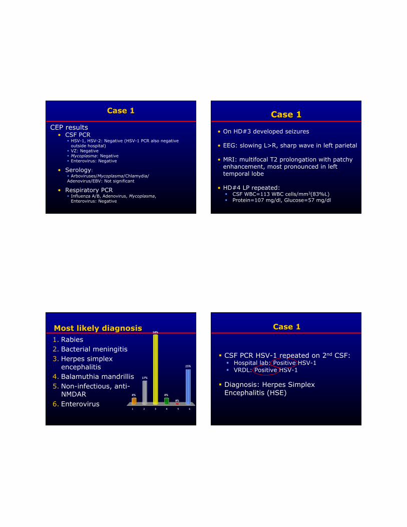
CEP results • CSF PCR
HSV-1, HSV-2: Negative (HSV-1 PCR also negative outside hospital)
VZ: Negative Mycoplasma: Negative Enterovirus: Negative
• Serology: Arboviruses/Mycoplasma/Chlamydia/ Adenovirus/EBV: Not significant
• Respiratory PCR Influenza A/B, Adenovirus, Mycoplasma,
Enterovirus: Negative
• On HD#3 developed seizures
• EEG: slowing L>R, sharp wave in left parietal
• MRI: multifocal T2 prolongation with patchy enhancement, most pronounced in left temporal lobe
• HD#4 LP repeated: CSF WBC=113 WBC cells/mm3(83%L) Protein=107 mg/dl, Glucose=57 mg/dl
1. Rabies 2. Bacterial meningitis 3. Herpes simplex
encephalitis 4. Balamuthia mandrillis 5. Non-infectious, anti-
NMDAR 6. Enterovirus
CSF PCR HSV-1 repeated on 2nd CSF: Hospital lab: Positive HSV-1 VRDL: Positive HSV-1
Diagnosis: Herpes Simplex Encephalitis (HSE)

• HSV-1 considered to be leading cause of encephalitis
• Acute necrotizing encephalitis • PCR: considered sensitive and
specific - Tunkel AR et al., Clin Inf Dis, 2008
• CEP: 80 cases --~ 20% had initial PCR negative (biased toward more difficult cases)
• Of those with false negative 1st CSF, CSFs were relatively bland: Initial CSF lab values:
Median CSF WBC=17 WBCs/mm3 (range: 0-330)
Median CSF Protein=34 mg/dL (range: 22-87)
• False negative PCRs tend to occur early in course of illness (e.g., w/in 72 hours of onset) or CSF fluid relatively “bland’’
3/11 (27%) patients tested within 72 hours: negative-repeat LP 5-11 day later positive
-Weil AA et al, CID, 2002
8/33 (24%) negative first 3 days5 cases repeat LP and 4 were positive
-De Tiege X et al, CID, 2003
2/15 (13%) of HSE cases were initially negative HSV CSF PCR but results became positive in repeat CSF analyses
-Elbers JM et al, Pediatrics, 2007

• 10 year old male — 3 days prior to admission with fever, right
sided weakness, and slurred speech
On admission febrile lethargy change in behavior confusion somnolence
• Exposure history — Owns dog, cat and lizard — Raccoons in yard — Plays football — No international travel, traveled to Central
Valley
• Admit labs, neuroimaging — LP: WBC = 27 WBCs/mm3 (100% Mo) — Protein = 23 mg/dL, Glucose = 54 mg/ml — 1st MRI: bilateral thalamic enhancement, L>R
• CSF PCR HSV1, HSV2: Negative VZ: Negative Mycoplasma: Negative Enterovirus: Negative
Respiratory — Enterovirus: Negative — Other viruses: Negative
• Serology: Arboviruses (SLE, WEE, WNV), Mycoplasma,
Chlamydia, adenovirus, EBV: Not significant
• Quickly deteriorated within first few days of hospitalization — Intubated — Comatose — Non-interactive — Occasional
— Repeat MRI: area of enhancement involvement with more extensive involvement of brainstem region

• A. Rabies • B. Bacterial meningitis • C. Herpes simplex encephalitis • D. Balamuthia mandrillis • E. Non-infectious, anti-NMDAR • F. Enterovirus
1. Rabies 2. Bacterial meningitis 3. Herpes simplex
encephalitis 4. Balamuthia mandrillis 5. Non-infectious, anti-
NMDAR 6. Enterovirus
• Brain biopsy — “necrotizing encephalitis”, no
intranuclear cells, no organism
• Multiple studies done on biopsy — All negative — EXCEPT for weakly positive Entero
PCR — Prelim sequence data suggestive
EV 71
Most common infection identified:
• 26% of all confirmed etiologies: 98 patients confirmed by positive PCR in CSF
• Median age = 14.0 years (mean=20.5 years)
• 63% pediatric, 37% adult
• CSF: median WBC = 58 cells (0-2655), Protein=40 mg/dl (20-473), glucose=64 mg/dl (20-122)
• Relatively short length of stay, median= 6 days
• Most with good outcomes, however 5+ fatalities (e.g. Enterovirus 71)
Fowlkes AL et al., JID, 2008
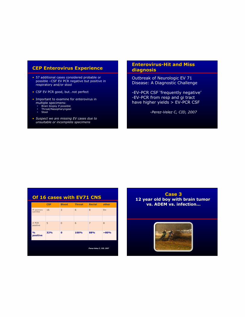
• 57 additional cases considered probable or possible –CSF EV PCR negative but positive in respiratory and/or stool
• CSF EV PCR good, but…not perfect
• Important to examine for enterovirus in multiple specimens: Brain biopsy if possible Throat/Nasopharyngeal Stool
• Suspect we are missing EV cases due to unsuitable or incomplete specimens
Outbreak of Neurologic EV 71 Disease: A Diagnostic Challenge
-EV-PCR CSF ‘frequently negative’ -EV-PCR from resp and gi tract have higher yields > EV-PCR CSF
-Perez-Velez C, CID, 2007
CSF Blood Throat Rectal other
# specimens submitted
16 3 6 8 9+
# PCR positive
5 0 6 7 8
% positive
32% 0 100% 88% ~80%
Perez-Velez C, CID, 2007

• 12 year old previously healthy Hispanic male
Presented with one week of progressively worsening headache, vomiting, decreased appetite, poor sleep, lethargy, and hallucinations
CT concerning for brain tumor
Intubated for increased ICP and ALOC
• Exposure history Bird at home Motorcycling – dusty conditions
• Admission laboratory results LP: WBC = 78 cells/mm3 (78%L, 20%M),
Protein = 42 mg/ml, Glucose = 74 mg/ml MRI suggestive of ADEM
Negative bacterial, fungal, & AFB stains & cultures Negative Coccidioides, Blasto, Histo, Cocci, &
Cysticercosis
• Treatment Decadron, Vancomycin,
Cefotaxime, Flagyl
• Discharged one week later At baseline on 1 week oral
prednisone taper with dx of ADEM
• Symptoms returned 2 weeks after stopping oral prednisone Headache, nausea, vomiting, and
lethargy • Repeat CT
Worsening enhancement and edema • Recurrent ADEM?
Initial plan was for longer course of steroids
Changed after repeat MRI- concerning for lymphoma

• Laboratory results LP: WBC = 298 cells/mm3 (89%L, 5%M),
Protein = 69 mg/ml, Glucose = 40 mg/ml Repeat infectious studies-bacterial, fungal, and
TB studies negative Biopsy #1- angiocentric lymphoid and plasma
cell infiltrates Full body CT, PET, & bone scans normal
• Treatment Prednisone
1. Rabies 2. Bacterial meningitis 3. Herpes simplex
encephalitis 4. Balamuthia mandrillis 5. Non-infectious, anti-
NMDAR 6. Enterovirus
• A. Rabies • B. Bacterial meningitis • C. Herpes simplex encephalitis • D. Balamuthia mandrillis • E. Non-infectious, anti-NMDAR • F. Enterovirus

• Symptoms returned - California Encephalitis Project contacted
• Balamuthia mandrillaris testing positive
• Rx with ‘cocktail’ of different Rx with initial improvement but died a few weeks later
• Type of free-living amoeba (living single-celled organisms) Dozen of types of free-living amoeba, few are pathogenic
• Two distinct CNS presentations of free-living amoeba: Fulminant, rapid progressive, fatal encephalitis — Naegleria fowlerii (diving in brackish water) Granulomatous amoebic encephalitis
– Acanthoemba sp., – Balamuthia mandrillis
• Considered to be a rare cause of encephalitis

• Subacute to chronic presentation Imaging: ring-enhancing, hydrocephalus, or parenchymal mass Often lymphocytic CSF pleocytosis Insidious onset -- headache, nausea, low-grade fever, lethargy, & confusion
• Can mimic Brain tumor ADEM Mycobacterium tuberculosis Neurocysticercosis Viral encephalitis
• Found in soil and water • Worldwide distribution • Inhalation or direct contamination of skin lesion
• 15 confirmed cases identified by CEP since 1990: (probably additional cases but no brain tissue)
• Positive serology often the ‘tip-off’ but brain biopsy needed for confirmation
• Demographics
Median age = 19 years (1-84 years) 82% Hispanic
Is this because of exposure vs. genetic susceptibility vs. ? 91% male Most immunocompetent
• CSF: lymphocytic or neutrophilic predominance, normal-high protein, normal-low glucose
• All had abnormal neuroimaging: ring-enhancing, mass lesions, hydrocephalus
• Exposure/risk factor: Not clear but most with soil exposure such as
gardener, construction worker, jeeping/motorcycling in dusty area
• Most died but 2 still living (one lost-to-follow-up)
Consult with CDC National Center for Emerging and Zoonotic Infectious Diseases Waterborne Disease Prevention Branch, CDC (or if in California, consult with California State Health Dept) • Testing
Brain material for IHC and molecular (serology can be helpful as well) • Treatment recommendations
Combination of Sulfadiazine, Flucytosine, Azole, Azithromycin and Pentamidine
CEP cases survivors
Case 4
• 72 y/o male with possible encephalitis, referred to CEP
Onset of headache/fever
5 days later hospitalized with lethargy, somnolence and ascending paralysis
Clinician thought it was probably a stroke but wanted to rule out encephalitis
Exposure history:
Born and raised in the Philippines and had been there 10 months prior to onset of illness
US resident for 10 years
No known mosquito bites
No known animal exposures
Admit labs/Neuroimaging:
LP: WBC = 10 WBCs/mm3, Protein = 172 mg/dL, Glucose = 60 mg/dL
MRI: mild atrophy (appropriate for age), otherwise normal
Died 11 days after hospitalization
No autopsy, cause of death: “cerebral vascular accident”
CEP results • CSF PCR
Herpes consensus: NEG Mycoplasma: NEG Enterovirus: NEG
• Serology Arboviruses/Mycoplasma/Chlamydia/Adenovirus/
EBV: Negative
• Respiratory PCR Influenza A/B, Adenovirus, Mycoplasma,
enterovirus: Negative

1. Rabies 2. Bacterial meningitis 3. Herpes simplex
encephalitis 4. Balamuthia mandrillis 5. Non-infectious, anti-
NMDAR 6. Enterovirus
• A. Rabies • B. Bacterial meningitis • C. Herpes simplex encephalitis • D. Balamuthia mandrillis • E. Non-infectious, anti-NMDAR • F. Enterovirus
• CEP deceased “core testing” includes rabies testing: • Rabies antibody positive and rabies PCR
positive from “throat swab” (contaminated with saliva)
• Sequenced strain: canine strain/Philippines
• Patient originally from Philippines, was there 10 months prior to onset of illness
Etiology • Family: Rhabdoviridae
Negative-stranded RNA genome • Genus: Lyssavirus • Envelope virus, bullet-shaped
Epidemiology
• Only 1-2 recognized cases/year in the United States…so why is it so important? “uniformly fatal” without vaccine [until
recently] Tremendous ‘angst’ : ~40,000 persons
receive post-exposure prophylaxis (PEP) in the US
Many encephalitis cases are ‘rule out’ rabies …on the other hand, cases are missed
World: 50,000-100,000 cases/year
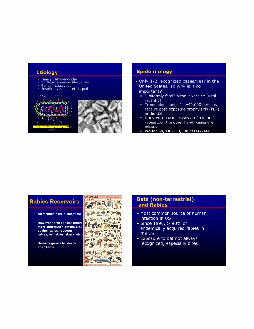
Rabies Reservoirs
• All mammals are susceptible
• However some species much more important > others, e.g., canine rabies, raccoon rabies, bat rabies, skunk, etc.
• Humans generally “dead-end” hosts
Bats (non-terrestrial) and Rabies
• Most common source of human infection in US
• Since 1990, > 90% of endemically acquired rabies in the US
• Exposure to bat not always recognized, especially bites
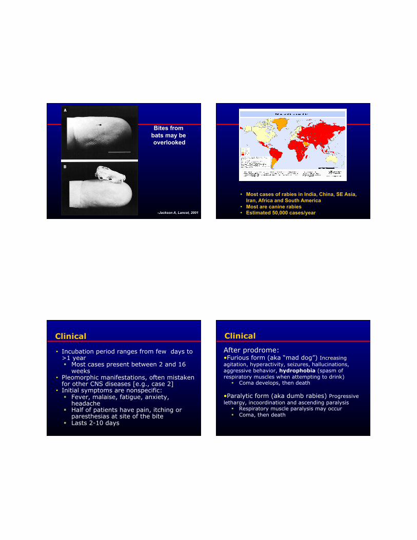
Bites from bats may be overlooked
-Jackson A, Lancet, 2001
• Most cases of rabies in India, China, SE Asia, Iran, Africa and South America
• Most are canine rabies • Estimated 50,000 cases/year
Clinical
• Incubation period ranges from few days to >1 year Most cases present between 2 and 16
weeks • Pleomorphic manifestations, often mistaken
for other CNS diseases [e.g., case 2] • Initial symptoms are nonspecific:
Fever, malaise, fatigue, anxiety, headache
Half of patients have pain, itching or paresthesias at site of the bite
Lasts 2-10 days
Clinical
After prodrome: • Furious form (aka “mad dog”) Increasing agitation, hyperactivity, seizures, hallucinations, aggressive behavior, hydrophobia (spasm of respiratory muscles when attempting to drink)
Coma develops, then death
• Paralytic form (aka dumb rabies) Progressive lethargy, incoordination and ascending paralysis
Respiratory muscle paralysis may occur Coma, then death

Diagnosis of Rabies
• Always consider in case of acute onset, rapidly progressive encephalitis
• Diagnosis before death is ‘tricky’, testing includes: Testing for viral antigen by DFA in nuchal skin or corneal
cells Testing for viral RNA by PCR of saliva, neural tissue, or
CSF Serology for antibodies in the blood Growing virus isolated from saliva or CSF in cell culture
• Testing after death includes: Testing for viral antigen by DFA in brain tissue
Prevention and Treatment issues these issues often confused…note the differences
• Rabies Pre-exposure prophylaxis Given to ‘high risk’ individuals such as veterinarians,
animal control workers, spleunkers before exposure 3 doses vaccine
• Rabies Post-exposure prophylaxis (PEP) Given following a bite from rabid (or suspected rabid)
animal Rabies Immune globulin (RIG) and 4 doses vaccine (day
0,3,7,14)
Highly effective for prevention • Rabies “Treatment”
No known effective Rx; once symptoms develop; vaccine and RIG of no benefit
Experimental treatment
PEP - Yes or No?
• Type of exposure (bite, non-bite) If bite: provoked vs. unprovoked Assess other circumstances of exposure,
e.g., behavior of animal • Severity of wound • Animal species involved • Animal health and vaccination history • Local animal rabies epidemiology • Animal available for observation / testing • Urgent but not “emergency”, consult local public
health c
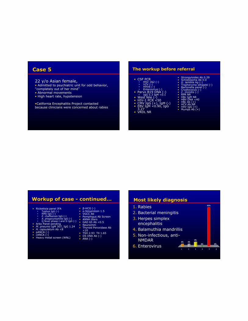
22 y/o Asian female, • Admitted to psychiatric unit for odd behavior, “completely out of her mind” • Abnormal movements • High heart rate, hypotension
• California Encephalitis Project contacted because clinicians were concerned about rabies
• CSF PCR HSV 1&2 (-) VZV (-) HHV6 (-) Enterovirus (-)
• Parvo B19 DNA (-) • IgG 5.3 IgM <0.1
• West Nile (-) • HIV-1 PCR <50 • CMV IgG (+), IgM (-) • EBV IgM <0.90, IgG
3.27 • VRDL NR
• Strongyloides Ab 0.29 • Schistosoma Ab 0.0 • G. lamblia Ag (-) • Tropheryma whipplei (-) • Bartonella panel (-) • Cryptococus (-) • C. immitis (-) • RPR NR • HBc IgM NR • HBV DNA <40 • HBc Ab (+) • HCV Ab NR • HAV IgG (+) • Mumps Ab (+)
• Rickettsia panel IFA Typhus IgG (-) RMS IgG (-) E. chaffeensis IgG (-) A. phagocytophilla IgG (-) Q fever phase I and II IgG (-)
• Arbo Panel pending • M. pneumo IgM 307, IgG 1.24 • H. capsulatum Ab <8 • pANCA (-) • cANCA (-) • Heavy metal screen (WNL)
• β-HCG (-) • α-fetoprotein 1.5 • VGCC Ab • Pemphigus Ab Screen • ANNA titers • GAD 65 Ab <0.5 • Neuroimm • Thyroid Peroxidase Ab
<10 • TSH 2.93 T4 1.65 • DS DNA Ab (-) • ANA (-)
1. Rabies 2. Bacterial meningitis 3. Herpes simplex
encephalitis 4. Balamuthia mandrillis 5. Non-infectious, anti-
NMDAR 6. Enterovirus
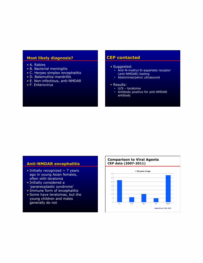
• A. Rabies • B. Bacterial meningitis • C. Herpes simplex encephalitis • D. Balamuthia mandrillis • E. Non-infectious, anti-NMDAR • F. Enterovirus
• Suggested: Anti-N-methyl-D-aspartate receptor
(anti-NMDAR) testing Abdominal/pelvic ultrasound
Results: U/S – teratoma Antibody positive for anti-NMDAR
antibody
• Initially recognized ~ 7 years ago in young Asian females, often with teratoma
• Initially considered a ‘paraneoplastic syndrome’
• Immune form of encephalitis • Some have teratomas, but the
young children and males generally do not
Comparison to Viral Agents CEP data (2007-2011)
0
5
10
15
20
25
30
35
EV WNV HSV-1 VZV NMDAR
< 30 years of age
-Gable M et al, CID, 2012

• Herpes simplex: Limitations of CSF PCR testing, consider
re-tap if temporal lobe involvement
• Enterovirus Another leading cause of encephalitis More often in pediatrics but still important
in adults Multiple specimen types needed to
optimize yield
• Balamuthia mandrillis
Although not common, probably not so rare Consider testing in patients with parenchymal
lesions especially if CSF profile “MTB/fungal-ish”
• Rabies
Should be considered in any rapidly progressive encephalitis
Probably also being missed Changing paradigm-not all fatal
• Bacterial meningitis — Prior antibiotics influence culture,
role for PCR in these cases
• Anti-NMDAR encephalitis
THE leading entity in CEP, consider in patients with movement disorders, seizures and/or autonomic instability
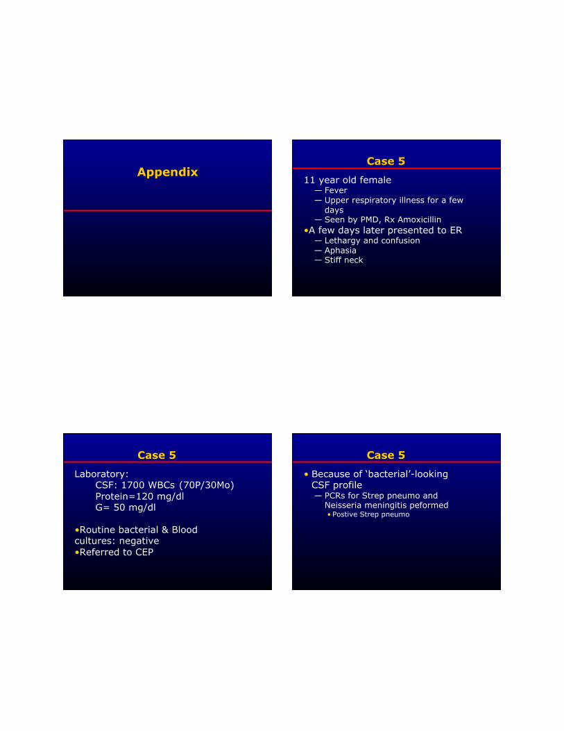
11 year old female — Fever — Upper respiratory illness for a few
days — Seen by PMD, Rx Amoxicillin
• A few days later presented to ER — Lethargy and confusion — Aphasia — Stiff neck
Laboratory: CSF: 1700 WBCs (70P/30Mo) Protein=120 mg/dl G= 50 mg/dl
• Routine bacterial & Blood cultures: negative • Referred to CEP
• Because of ‘bacterial’-looking CSF profile — PCRs for Strep pneumo and
Neisseria meningitis peformed • Postive Strep pneumo

Organism Incidence (cases per 100,000)
1986 1995 2006-2007
H. influenzae 2.9 0.2 0.08
S. pneumoniae 1.1 1.1 0.81
N. meningitidis 0.9 0.6 0.19
Group B streptococcus
0.4 0.3 0.25
L. monocytogenes 0.2 0.2 0.05
1986: Wenger et al. J Infect Dis 1990;162:1316-23 1995: Schuchat A, et al. NEJM 1997;337:970-7
2006-7: Thigpen M, et al. NEJM 2011; 364:2016-25.
• Estimated 3,000–6,000 cases per year in U.S.
• #1 cause of bacterial meningitis in US
• Case-fatality rate ~30%, up to 80% in the elderly
• Neurologic sequelae common among survivors
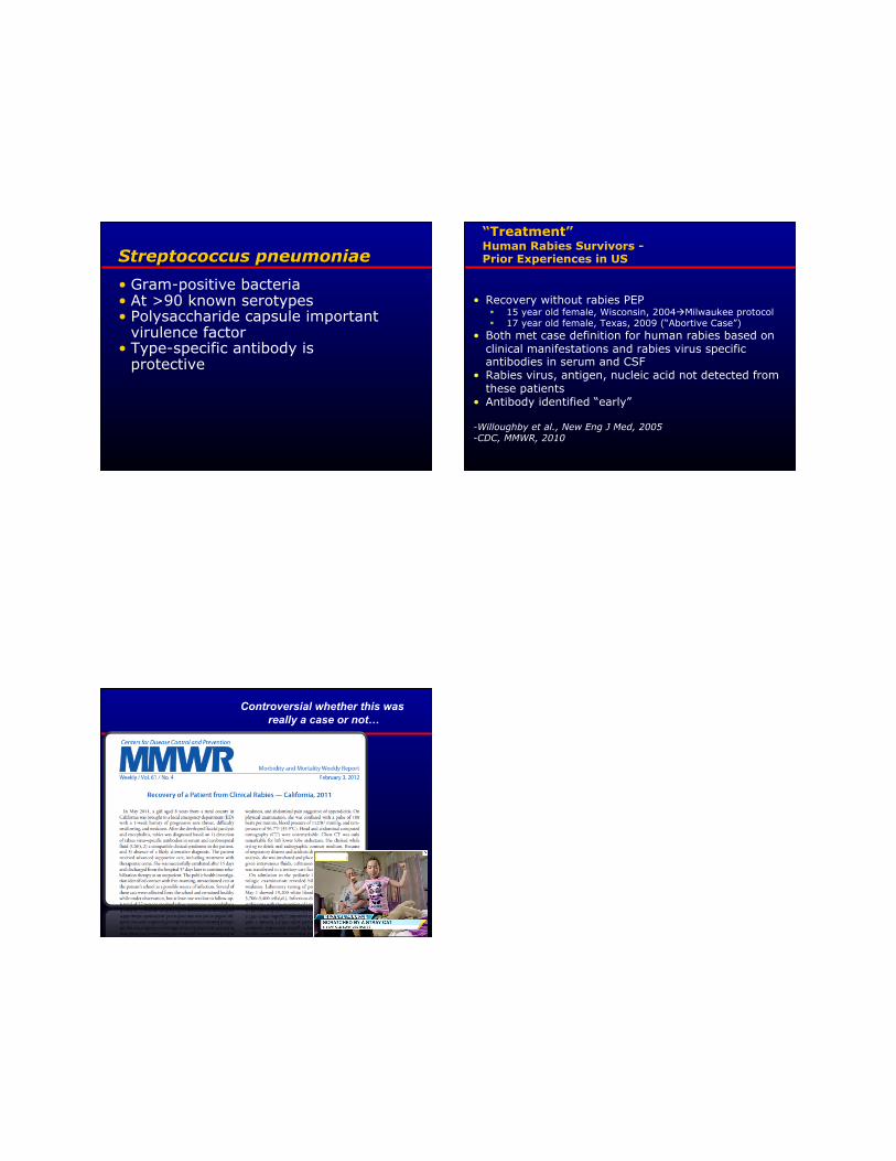
• Gram-positive bacteria • At >90 known serotypes • Polysaccharide capsule important
virulence factor • Type-specific antibody is
protective
“Treatment” Human Rabies Survivors - Prior Experiences in US
• Recovery without rabies PEP 15 year old female, Wisconsin, 2004Milwaukee protocol 17 year old female, Texas, 2009 (“Abortive Case”)
• Both met case definition for human rabies based on clinical manifestations and rabies virus specific antibodies in serum and CSF
• Rabies virus, antigen, nucleic acid not detected from these patients
• Antibody identified “early”
-Willoughby et al., New Eng J Med, 2005 -CDC, MMWR, 2010
Controversial whether this was really a case or not…