8/2/2017 radiomics in clinical...
TRANSCRIPT

8/2/2017
1
Radiomics in Clinical Trials Laurence Court
Departments of Radiation Oncology and Imaging Physics
University of Texas MD Anderson Cancer Center
Conflicts of Interest
• Radiomics projects funded by the NCI
• Other projects funded by the NCI, CPRIT, Varian, Elekta
2
Significant growth in radiomics
Figure from Philippe Lambin, MAASTRO
8/2/2017
2
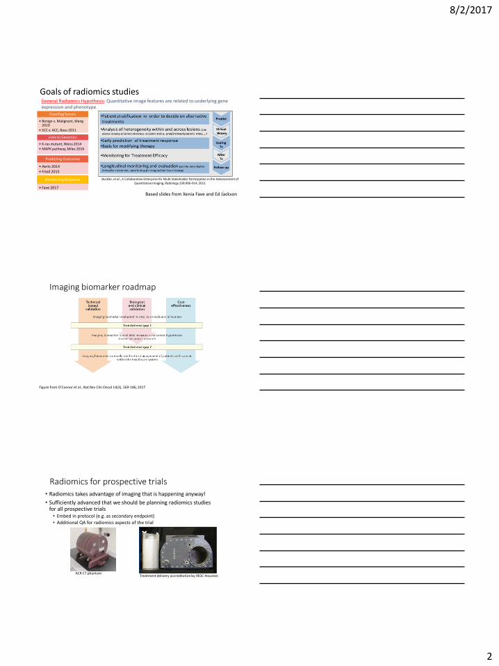
Goals of radiomics studies
Buckler, et al., A Collaborative Enterprise for Multi-Stakeholder Participation in the Advancement of Quantitative Imaging, Radiology 258:906-914, 2011
Based slides from Xenia Fave and Ed Jackson
General Radiomics Hypothesis: Quantitative image features are related to underlying gene expression and phenotype
4
Classifing Tumors
• Benign v. Malignant, Wang 2010
• SCC v. ACC, Basu 2011
Predicting Outcomes
• Aerts 2014 • Fried 2015
Links to Genomics
• K-ras mutant, Weiss 2014
• MAPK pathway, Miles 2016
Monitoring Response
• Fave 2017
Imaging biomarker roadmap
Figure from O’Connor et al., Nat Rev Clin Oncol 14(3), 169-186, 2017
Radiomics for prospective trials • Radiomics takes advantage of imaging that is happening anyway!
• Sufficiently advanced that we should be planning radiomics studies for all prospective trials • Embed in protocol (e.g. as secondary endpoint)
• Additional QA for radiomics aspects of the trial
Treatment delivery accreditation by IROC-Houston ACR CT phantom

8/2/2017
3
Radiomics workflow
Figure adapted from Aerts et al, Nature Communications 2015
Imaging Pre-processing Feature extraction Analysis Segmentation
Includes: • Imaging protocol • Motion
management
7
Including feature selection
Imaging protocols should be harmonized • Texture phantom
• Acquired 17 scans from GE, Philips, Siemens and Toshiba scanners scattered throughout the Texas Medical Center
Dennis Mackin et al, Investigative Radiology 50(11), 757-765, 2015 Data from Dennis Mackin
Harmonization of PET imaging
Harmonize protocols
Figures from Robert Jeraj, University of Wisconsin. Also see Lin et al, JNM 2016
• Two scanners: Discovery (GE) and Gemini (Philips) • Harmonize GE scanner to the Philips scanner
9

8/2/2017
4
Minimize inter-scanner variability
• Minimize the number of scanners (to one…..) • Aerts et al, Defining a Radiomics Response
Phenotype: A Pilot Study using targeted therapy in NSCLC, Scientific Reports 6, 33860, 2016 • 47 patients • Radiomics data could predict EGFR-mutation status
and associated gefitinib response
• Minimize the number of scanners (to a few) • Fried et al, Prognostic Value and Reproducibility
of Pretreatment CT texture features in stage III non-small cell lung cancer, IJROBP 90(4), 834-842, 2014
But care is still needed…….. • 110 NSCLC patients with advanced stage disease
• Randomized to treatment with IMRT or PSPT to 66 or 74 Gy
• Received concurrent chemotherapy
• Imaged with a 4DCT weekly during treatment (5-9 4DCTs per patient)
• Most on one of two CT scanners (both GE, one was widebore, one was PET-CT)
• Texture signature was a function of CT scanner
Data from Xenia Fave, UT MD Anderson Cancer Center (published in Scientific Reports 7, 588, 2017 11
CT Model Dependence can be mitigated by image preprocessing • Scanner independence, p-value of Wilcoxon rank sum test
• No features were ALWAYS significantly associated with CT model
• Suggests that image preprocessing can be used to remove differences between CT scanners
• 24 features were ALWAYS independent of CT model
• Included features from all 4 categories
• For 31 features that were sometimes dependent,
• 2 features were fixed with bit depth resampling
• 28 features with smoothing
• 27 with smoothing and bit depth resampling
• 6 no preprocessing
12
+

8/2/2017
5
Radiomics workflow
Figure adapted from Aerts et al, Nature Communications 2015
Imaging Pre-processing Feature extraction Analysis Segmentation
Includes: • Imaging protocol • Motion
management
13
Including feature selection
Segmentation
Based on Hunter et al, Med Phys 40, 121916, 2013 14
Increasing pixel removal
• PET texture – tracer uptake heterogeneity: • Metabolism • Hypoxia • Cellular proliferation • Vascularization • Necrosis
• CT texture - tissue density heterogeneity: • Vascularization • Necrosis • Relative fat, air, water content
• But - we don’t understand the meaning of the radiomics texture features in terms of pathophysiology
• Reproducibility/robustness is important • If using pre-drawn contours, consider the original task
Hatt et al, Eur J Nucl Mol Imaging, 2016
• Benchmark standards (standard images etc) • Open source algorithms • Standardization
• Phantoms • Best (consensus) practices • Accurate reporting • Data sharing 15

8/2/2017
6
Resources • Court et al, Computational resources for radiomics, Translational Cancer Research 5(4), 340-348, 2016
• Larue et al, Quantitative radiomics studies for tissue characterization: A review of technology and methodological procedures, Brit. J. Radiol. 90, 20160665, 2017
• www.Radiomics.world – Radiomics Quality Score
www.radiomics.world
Final thoughts • Radiomics:
• Is correlated to biology
• Can improve accuracy of prediction
• Can calculate probability of certain diagnosis
• Can be used in clinical trials
• (and is low cost and non-invasive)
• Re-analyse your image data whenever possible
• Plan radiomics studies in all your prospective trials
• Embed radiomics studies into the protocol (e.g. as a secondary endpoint or hypothesis generative approach monitoring treatment response)
• Careful consideration of the imaging protocols
• Full transparency on everything else
• Imaging
• Feature extraction (details)
• Analysis
Partially based on slides by Philippe Lambin, MAASTRO Fried et al, IJROBP 94, 368-376, 2015
Research group and collaborators
Our group (past and present)
• Joy Zhang
• Jinzhong Yang
• Dennis Mackin
• Luke Hunter
• David Fried
• Xenia Fave
• Joonsang Lee
• Rachel Ger
Physics
• Osama Mawlawi
• Peter Balter
Radiation Oncology and Radiology
• Zhongxing Liao
• Steven Lin
• Daniel Gomez
• Chaan Ng
• Joe Chang
• Dave Fuller
Statistics
• Shouhao Zhou
• Susan Tucker
• Francesco Stingo
• Arvind Rao
• Center for Radiation Oncology Research 18