8|the appendicular skeleton · 2018-10-02 · and feet, and the bones that anchor the limbs to the...
TRANSCRIPT

8 | THE APPENDICULARSKELETON
Figure 8.1 Dancer The appendicular skeleton consists of the upper and lower limb bones, the bones of the handsand feet, and the bones that anchor the limbs to the axial skeleton. (credit: Melissa Dooley/flickr)
Introduction
Chapter Objectives
After studying this chapter, you will be able to:
• Discuss the bones of the pectoral and pelvic girdles, and describe how these unite the limbs with the axialskeleton
• Describe the bones of the upper limb, including the bones of the arm, forearm, wrist, and hand• Identify the features of the pelvis and explain how these differ between the adult male and female pelvis• Describe the bones of the lower limb, including the bones of the thigh, leg, ankle, and foot• Describe the embryonic formation and growth of the limb bones
Your skeleton provides the internal supporting structure of the body. The adult axial skeleton consists of 80 bones that formthe head and body trunk. Attached to this are the limbs, whose 126 bones constitute the appendicular skeleton. These bonesare divided into two groups: the bones that are located within the limbs themselves, and the girdle bones that attach thelimbs to the axial skeleton. The bones of the shoulder region form the pectoral girdle, which anchors the upper limb to thethoracic cage of the axial skeleton. The lower limb is attached to the vertebral column by the pelvic girdle.
CHAPTER 8 | THE APPENDICULAR SKELETON 293

Because of our upright stance, different functional demands are placed upon the upper and lower limbs. Thus, the bonesof the lower limbs are adapted for weight-bearing support and stability, as well as for body locomotion via walking orrunning. In contrast, our upper limbs are not required for these functions. Instead, our upper limbs are highly mobile andcan be utilized for a wide variety of activities. The large range of upper limb movements, coupled with the ability to easilymanipulate objects with our hands and opposable thumbs, has allowed humans to construct the modern world in which welive.
8.1 | The Pectoral Girdle
By the end of this section, you will be able to:
• Describe the bones that form the pectoral girdle
• List the functions of the pectoral girdle
The appendicular skeleton includes all of the limb bones, plus the bones that unite each limb with the axial skeleton (Figure8.2). The bones that attach each upper limb to the axial skeleton form the pectoral girdle (shoulder girdle). This consists oftwo bones, the scapula and clavicle (Figure 8.3). The clavicle (collarbone) is an S-shaped bone located on the anterior sideof the shoulder. It is attached on its medial end to the sternum of the thoracic cage, which is part of the axial skeleton. Thelateral end of the clavicle articulates (joins) with the scapula just above the shoulder joint. You can easily palpate, or feelwith your fingers, the entire length of your clavicle.
294 CHAPTER 8 | THE APPENDICULAR SKELETON
This content is available for free at http://cnx.org/content/col11496/1.6
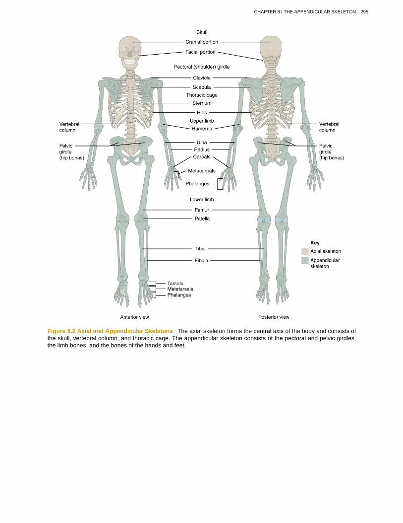
Figure 8.2 Axial and Appendicular Skeletons The axial skeleton forms the central axis of the body and consists ofthe skull, vertebral column, and thoracic cage. The appendicular skeleton consists of the pectoral and pelvic girdles,the limb bones, and the bones of the hands and feet.
CHAPTER 8 | THE APPENDICULAR SKELETON 295

Figure 8.3 Pectoral Girdle The pectoral girdle consists of the clavicle and the scapula, which serve to attach theupper limb to the sternum of the axial skeleton.
The scapula (shoulder blade) lies on the posterior aspect of the shoulder. It is supported by the clavicle, which alsoarticulates with the humerus (arm bone) to form the shoulder joint. The scapula is a flat, triangular-shaped bone with aprominent ridge running across its posterior surface. This ridge extends out laterally, where it forms the bony tip of theshoulder and joins with the lateral end of the clavicle. By following along the clavicle, you can palpate out to the bony tipof the shoulder, and from there, you can move back across your posterior shoulder to follow the ridge of the scapula. Moveyour shoulder around and feel how the clavicle and scapula move together as a unit. Both of these bones serve as importantattachment sites for muscles that aid with movements of the shoulder and arm.
The right and left pectoral girdles are not joined to each other, allowing each to operate independently. In addition, theclavicle of each pectoral girdle is anchored to the axial skeleton by a single, highly mobile joint. This allows for theextensive mobility of the entire pectoral girdle, which in turn enhances movements of the shoulder and upper limb.
ClavicleThe clavicle is the only long bone that lies in a horizontal position in the body (see Figure 8.3). The clavicle has severalimportant functions. First, anchored by muscles from above, it serves as a strut that extends laterally to support the scapula.This in turn holds the shoulder joint superiorly and laterally from the body trunk, allowing for maximal freedom of motion
296 CHAPTER 8 | THE APPENDICULAR SKELETON
This content is available for free at http://cnx.org/content/col11496/1.6

for the upper limb. The clavicle also transmits forces acting on the upper limb to the sternum and axial skeleton. Finally, itserves to protect the underlying nerves and blood vessels as they pass between the trunk of the body and the upper limb.
The clavicle has three regions: the medial end, the lateral end, and the shaft. The medial end, known as the sternalend of the clavicle, has a triangular shape and articulates with the manubrium portion of the sternum. This formsthe sternoclavicular joint, which is the only bony articulation between the pectoral girdle of the upper limb and theaxial skeleton. This joint allows considerable mobility, enabling the clavicle and scapula to move in upward/downwardand anterior/posterior directions during shoulder movements. The sternoclavicular joint is indirectly supported by thecostoclavicular ligament (costo- = “rib”), which spans the sternal end of the clavicle and the underlying first rib. The lateralor acromial end of the clavicle articulates with the acromion of the scapula, the portion of the scapula that forms the bonytip of the shoulder. There are some sex differences in the morphology of the clavicle. In women, the clavicle tends to beshorter, thinner, and less curved. In men, the clavicle is heavier and longer, and has a greater curvature and rougher surfaceswhere muscles attach, features that are more pronounced in manual workers.
The clavicle is the most commonly fractured bone in the body. Such breaks often occur because of the force exerted onthe clavicle when a person falls onto his or her outstretched arms, or when the lateral shoulder receives a strong blow.Because the sternoclavicular joint is strong and rarely dislocated, excessive force results in the breaking of the clavicle,usually between the middle and lateral portions of the bone. If the fracture is complete, the shoulder and lateral claviclefragment will drop due to the weight of the upper limb, causing the person to support the sagging limb with their otherhand. Muscles acting across the shoulder will also pull the shoulder and lateral clavicle anteriorly and medially, causingthe clavicle fragments to override. The clavicle overlies many important blood vessels and nerves for the upper limb, butfortunately, due to the anterior displacement of a broken clavicle, these structures are rarely affected when the clavicle isfractured.
ScapulaThe scapula is also part of the pectoral girdle and thus plays an important role in anchoring the upper limb to the body. Thescapula is located on the posterior side of the shoulder. It is surrounded by muscles on both its anterior (deep) and posterior(superficial) sides, and thus does not articulate with the ribs of the thoracic cage.
The scapula has several important landmarks (Figure 8.4). The three margins or borders of the scapula, named for theirpositions within the body, are the superior border of the scapula, the medial border of the scapula, and the lateralborder of the scapula. The suprascapular notch is located lateral to the midpoint of the superior border. The cornersof the triangular scapula, at either end of the medial border, are the superior angle of the scapula, located between themedial and superior borders, and the inferior angle of the scapula, located between the medial and lateral borders. Theinferior angle is the most inferior portion of the scapula, and is particularly important because it serves as the attachmentpoint for several powerful muscles involved in shoulder and upper limb movements. The remaining corner of the scapula,between the superior and lateral borders, is the location of the glenoid cavity (glenoid fossa). This shallow depressionarticulates with the humerus bone of the arm to form the glenohumeral joint (shoulder joint). The small bony bumpslocated immediately above and below the glenoid cavity are the supraglenoid tubercle and the infraglenoid tubercle,respectively. These provide attachments for muscles of the arm.
CHAPTER 8 | THE APPENDICULAR SKELETON 297
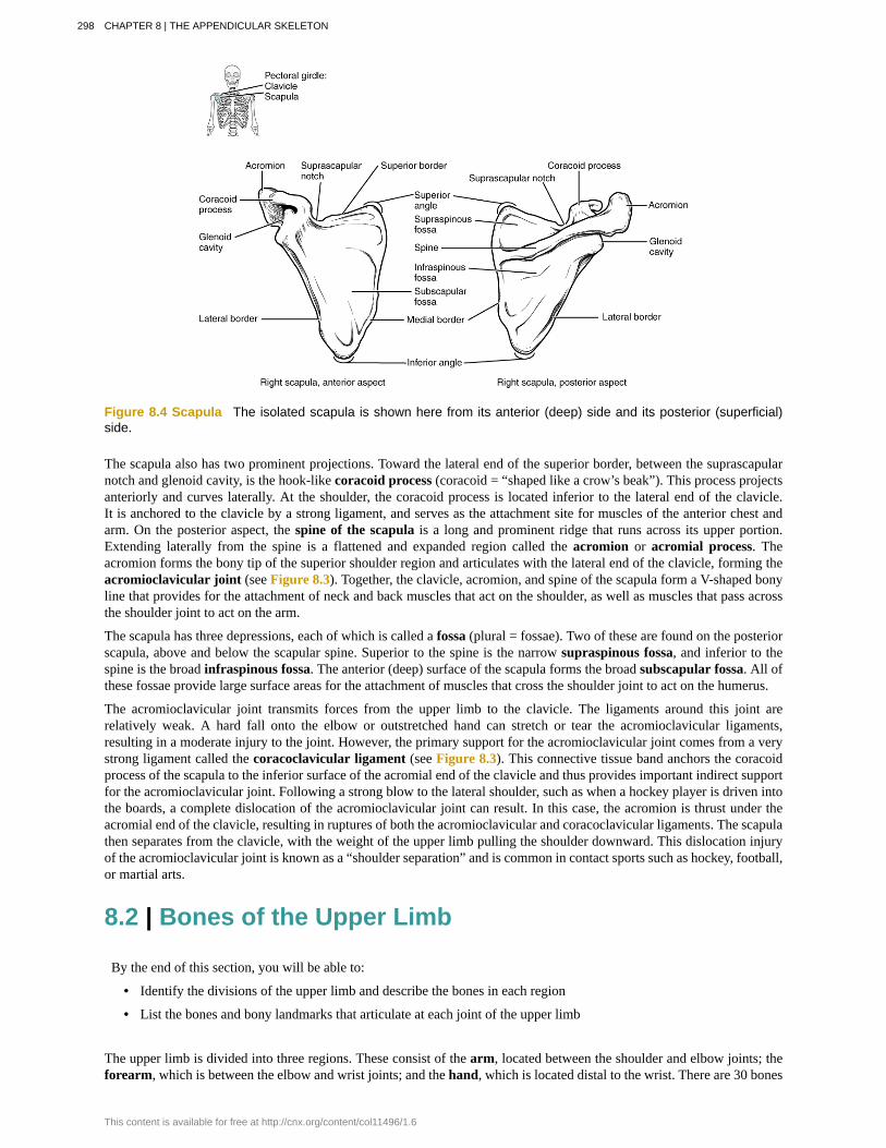
Figure 8.4 Scapula The isolated scapula is shown here from its anterior (deep) side and its posterior (superficial)side.
The scapula also has two prominent projections. Toward the lateral end of the superior border, between the suprascapularnotch and glenoid cavity, is the hook-like coracoid process (coracoid = “shaped like a crow’s beak”). This process projectsanteriorly and curves laterally. At the shoulder, the coracoid process is located inferior to the lateral end of the clavicle.It is anchored to the clavicle by a strong ligament, and serves as the attachment site for muscles of the anterior chest andarm. On the posterior aspect, the spine of the scapula is a long and prominent ridge that runs across its upper portion.Extending laterally from the spine is a flattened and expanded region called the acromion or acromial process. Theacromion forms the bony tip of the superior shoulder region and articulates with the lateral end of the clavicle, forming theacromioclavicular joint (see Figure 8.3). Together, the clavicle, acromion, and spine of the scapula form a V-shaped bonyline that provides for the attachment of neck and back muscles that act on the shoulder, as well as muscles that pass acrossthe shoulder joint to act on the arm.
The scapula has three depressions, each of which is called a fossa (plural = fossae). Two of these are found on the posteriorscapula, above and below the scapular spine. Superior to the spine is the narrow supraspinous fossa, and inferior to thespine is the broad infraspinous fossa. The anterior (deep) surface of the scapula forms the broad subscapular fossa. All ofthese fossae provide large surface areas for the attachment of muscles that cross the shoulder joint to act on the humerus.
The acromioclavicular joint transmits forces from the upper limb to the clavicle. The ligaments around this joint arerelatively weak. A hard fall onto the elbow or outstretched hand can stretch or tear the acromioclavicular ligaments,resulting in a moderate injury to the joint. However, the primary support for the acromioclavicular joint comes from a verystrong ligament called the coracoclavicular ligament (see Figure 8.3). This connective tissue band anchors the coracoidprocess of the scapula to the inferior surface of the acromial end of the clavicle and thus provides important indirect supportfor the acromioclavicular joint. Following a strong blow to the lateral shoulder, such as when a hockey player is driven intothe boards, a complete dislocation of the acromioclavicular joint can result. In this case, the acromion is thrust under theacromial end of the clavicle, resulting in ruptures of both the acromioclavicular and coracoclavicular ligaments. The scapulathen separates from the clavicle, with the weight of the upper limb pulling the shoulder downward. This dislocation injuryof the acromioclavicular joint is known as a “shoulder separation” and is common in contact sports such as hockey, football,or martial arts.
8.2 | Bones of the Upper Limb
By the end of this section, you will be able to:
• Identify the divisions of the upper limb and describe the bones in each region
• List the bones and bony landmarks that articulate at each joint of the upper limb
The upper limb is divided into three regions. These consist of the arm, located between the shoulder and elbow joints; theforearm, which is between the elbow and wrist joints; and the hand, which is located distal to the wrist. There are 30 bones
298 CHAPTER 8 | THE APPENDICULAR SKELETON
This content is available for free at http://cnx.org/content/col11496/1.6
in each upper limb (see Figure 8.2). The humerus is the single bone of the upper arm, and the ulna (medially) and theradius (laterally) are the paired bones of the forearm. The base of the hand contains eight bones, each called a carpal bone,and the palm of the hand is formed by five bones, each called a metacarpal bone. The fingers and thumb contain a total of14 bones, each of which is a phalanx bone of the hand.
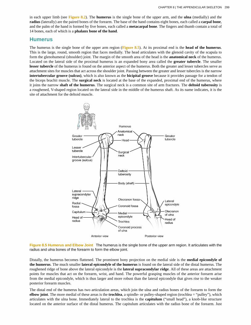
HumerusThe humerus is the single bone of the upper arm region (Figure 8.5). At its proximal end is the head of the humerus.This is the large, round, smooth region that faces medially. The head articulates with the glenoid cavity of the scapula toform the glenohumeral (shoulder) joint. The margin of the smooth area of the head is the anatomical neck of the humerus.Located on the lateral side of the proximal humerus is an expanded bony area called the greater tubercle. The smallerlesser tubercle of the humerus is found on the anterior aspect of the humerus. Both the greater and lesser tubercles serve asattachment sites for muscles that act across the shoulder joint. Passing between the greater and lesser tubercles is the narrowintertubercular groove (sulcus), which is also known as the bicipital groove because it provides passage for a tendon ofthe biceps brachii muscle. The surgical neck is located at the base of the expanded, proximal end of the humerus, whereit joins the narrow shaft of the humerus. The surgical neck is a common site of arm fractures. The deltoid tuberosity isa roughened, V-shaped region located on the lateral side in the middle of the humerus shaft. As its name indicates, it is thesite of attachment for the deltoid muscle.
Figure 8.5 Humerus and Elbow Joint The humerus is the single bone of the upper arm region. It articulates with theradius and ulna bones of the forearm to form the elbow joint.
Distally, the humerus becomes flattened. The prominent bony projection on the medial side is the medial epicondyle ofthe humerus. The much smaller lateral epicondyle of the humerus is found on the lateral side of the distal humerus. Theroughened ridge of bone above the lateral epicondyle is the lateral supracondylar ridge. All of these areas are attachmentpoints for muscles that act on the forearm, wrist, and hand. The powerful grasping muscles of the anterior forearm arisefrom the medial epicondyle, which is thus larger and more robust than the lateral epicondyle that gives rise to the weakerposterior forearm muscles.
The distal end of the humerus has two articulation areas, which join the ulna and radius bones of the forearm to form theelbow joint. The more medial of these areas is the trochlea, a spindle- or pulley-shaped region (trochlea = “pulley”), whicharticulates with the ulna bone. Immediately lateral to the trochlea is the capitulum (“small head”), a knob-like structurelocated on the anterior surface of the distal humerus. The capitulum articulates with the radius bone of the forearm. Just
CHAPTER 8 | THE APPENDICULAR SKELETON 299
above these bony areas are two small depressions. These spaces accommodate the forearm bones when the elbow is fullybent (flexed). Superior to the trochlea is the coronoid fossa, which receives the coronoid process of the ulna, and abovethe capitulum is the radial fossa, which receives the head of the radius when the elbow is flexed. Similarly, the posteriorhumerus has the olecranon fossa, a larger depression that receives the olecranon process of the ulna when the forearm isfully extended.
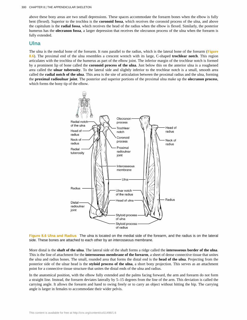
UlnaThe ulna is the medial bone of the forearm. It runs parallel to the radius, which is the lateral bone of the forearm (Figure8.6). The proximal end of the ulna resembles a crescent wrench with its large, C-shaped trochlear notch. This regionarticulates with the trochlea of the humerus as part of the elbow joint. The inferior margin of the trochlear notch is formedby a prominent lip of bone called the coronoid process of the ulna. Just below this on the anterior ulna is a roughenedarea called the ulnar tuberosity. To the lateral side and slightly inferior to the trochlear notch is a small, smooth areacalled the radial notch of the ulna. This area is the site of articulation between the proximal radius and the ulna, formingthe proximal radioulnar joint. The posterior and superior portions of the proximal ulna make up the olecranon process,which forms the bony tip of the elbow.
Figure 8.6 Ulna and Radius The ulna is located on the medial side of the forearm, and the radius is on the lateralside. These bones are attached to each other by an interosseous membrane.
More distal is the shaft of the ulna. The lateral side of the shaft forms a ridge called the interosseous border of the ulna.This is the line of attachment for the interosseous membrane of the forearm, a sheet of dense connective tissue that unitesthe ulna and radius bones. The small, rounded area that forms the distal end is the head of the ulna. Projecting from theposterior side of the ulnar head is the styloid process of the ulna, a short bony projection. This serves as an attachmentpoint for a connective tissue structure that unites the distal ends of the ulna and radius.
In the anatomical position, with the elbow fully extended and the palms facing forward, the arm and forearm do not forma straight line. Instead, the forearm deviates laterally by 5–15 degrees from the line of the arm. This deviation is called thecarrying angle. It allows the forearm and hand to swing freely or to carry an object without hitting the hip. The carryingangle is larger in females to accommodate their wider pelvis.
300 CHAPTER 8 | THE APPENDICULAR SKELETON
This content is available for free at http://cnx.org/content/col11496/1.6

RadiusThe radius runs parallel to the ulna, on the lateral (thumb) side of the forearm (see Figure 8.6). The head of the radiusis a disc-shaped structure that forms the proximal end. The small depression on the surface of the head articulates with thecapitulum of the humerus as part of the elbow joint, whereas the smooth, outer margin of the head articulates with the radialnotch of the ulna at the proximal radioulnar joint. The neck of the radius is the narrowed region immediately below theexpanded head. Inferior to this point on the medial side is the radial tuberosity, an oval-shaped, bony protuberance thatserves as a muscle attachment point. The shaft of the radius is slightly curved and has a small ridge along its medial side.This ridge forms the interosseous border of the radius, which, like the similar border of the ulna, is the line of attachmentfor the interosseous membrane that unites the two forearm bones. The distal end of the radius has a smooth surface forarticulation with two carpal bones to form the radiocarpal joint or wrist joint (Figure 8.7 and Figure 8.8). On the medialside of the distal radius is the ulnar notch of the radius. This shallow depression articulates with the head of the ulna,which together form the distal radioulnar joint. The lateral end of the radius has a pointed projection called the styloidprocess of the radius. This provides attachment for ligaments that support the lateral side of the wrist joint. Comparedto the styloid process of the ulna, the styloid process of the radius projects more distally, thereby limiting the range ofmovement for lateral deviations of the hand at the wrist joint.
Watch this video (http://openstaxcollege.org/l/fractures) to see how fractures of the distal radius bone can affectthe wrist joint. Explain the problems that may occur if a fracture of the distal radius involves the joint surface of theradiocarpal joint of the wrist.
Carpal BonesThe wrist and base of the hand are formed by a series of eight small carpal bones (see Figure 8.7). The carpal bones arearranged in two rows, forming a proximal row of four carpal bones and a distal row of four carpal bones. The bones in theproximal row, running from the lateral (thumb) side to the medial side, are the scaphoid (“boat-shaped”), lunate (“moon-shaped”), triquetrum (“three-cornered”), and pisiform (“pea-shaped”) bones. The small, rounded pisiform bone articulateswith the anterior surface of the triquetrum bone. The pisiform thus projects anteriorly, where it forms the bony bump thatcan be felt at the medial base of your hand. The distal bones (lateral to medial) are the trapezium (“table”), trapezoid(“resembles a table”), capitate (“head-shaped”), and hamate (“hooked bone”) bones. The hamate bone is characterized bya prominent bony extension on its anterior side called the hook of the hamate bone.
A helpful mnemonic for remembering the arrangement of the carpal bones is “So Long To Pinky, Here Comes The Thumb.”This mnemonic starts on the lateral side and names the proximal bones from lateral to medial (scaphoid, lunate, triquetrum,pisiform), then makes a U-turn to name the distal bones from medial to lateral (hamate, capitate, trapezoid, trapezium).Thus, it starts and finishes on the lateral side.
CHAPTER 8 | THE APPENDICULAR SKELETON 301
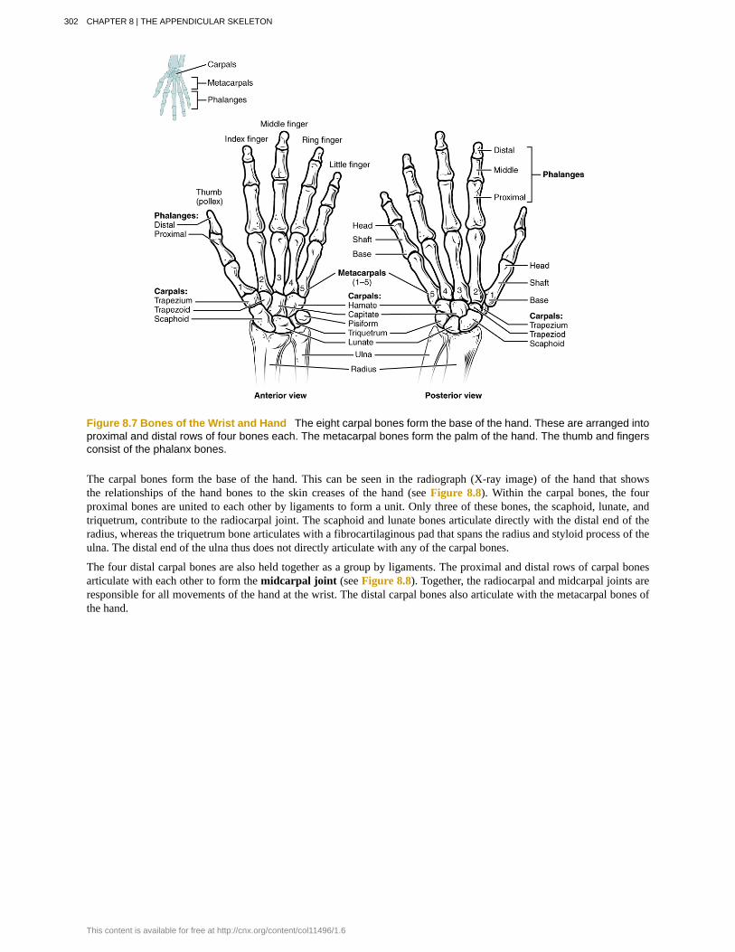
Figure 8.7 Bones of the Wrist and Hand The eight carpal bones form the base of the hand. These are arranged intoproximal and distal rows of four bones each. The metacarpal bones form the palm of the hand. The thumb and fingersconsist of the phalanx bones.
The carpal bones form the base of the hand. This can be seen in the radiograph (X-ray image) of the hand that showsthe relationships of the hand bones to the skin creases of the hand (see Figure 8.8). Within the carpal bones, the fourproximal bones are united to each other by ligaments to form a unit. Only three of these bones, the scaphoid, lunate, andtriquetrum, contribute to the radiocarpal joint. The scaphoid and lunate bones articulate directly with the distal end of theradius, whereas the triquetrum bone articulates with a fibrocartilaginous pad that spans the radius and styloid process of theulna. The distal end of the ulna thus does not directly articulate with any of the carpal bones.
The four distal carpal bones are also held together as a group by ligaments. The proximal and distal rows of carpal bonesarticulate with each other to form the midcarpal joint (see Figure 8.8). Together, the radiocarpal and midcarpal joints areresponsible for all movements of the hand at the wrist. The distal carpal bones also articulate with the metacarpal bones ofthe hand.
302 CHAPTER 8 | THE APPENDICULAR SKELETON
This content is available for free at http://cnx.org/content/col11496/1.6
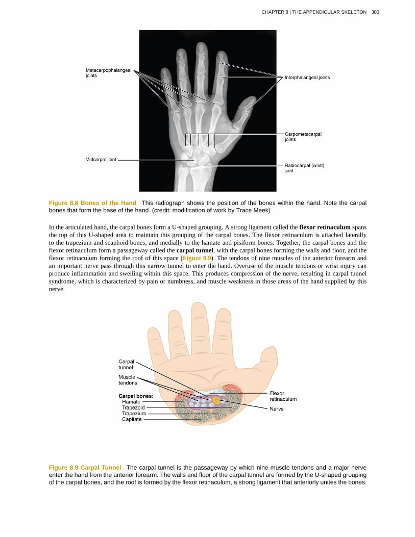
Figure 8.8 Bones of the Hand This radiograph shows the position of the bones within the hand. Note the carpalbones that form the base of the hand. (credit: modification of work by Trace Meek)
In the articulated hand, the carpal bones form a U-shaped grouping. A strong ligament called the flexor retinaculum spansthe top of this U-shaped area to maintain this grouping of the carpal bones. The flexor retinaculum is attached laterallyto the trapezium and scaphoid bones, and medially to the hamate and pisiform bones. Together, the carpal bones and theflexor retinaculum form a passageway called the carpal tunnel, with the carpal bones forming the walls and floor, and theflexor retinaculum forming the roof of this space (Figure 8.9). The tendons of nine muscles of the anterior forearm andan important nerve pass through this narrow tunnel to enter the hand. Overuse of the muscle tendons or wrist injury canproduce inflammation and swelling within this space. This produces compression of the nerve, resulting in carpal tunnelsyndrome, which is characterized by pain or numbness, and muscle weakness in those areas of the hand supplied by thisnerve.
Figure 8.9 Carpal Tunnel The carpal tunnel is the passageway by which nine muscle tendons and a major nerveenter the hand from the anterior forearm. The walls and floor of the carpal tunnel are formed by the U-shaped groupingof the carpal bones, and the roof is formed by the flexor retinaculum, a strong ligament that anteriorly unites the bones.
CHAPTER 8 | THE APPENDICULAR SKELETON 303
Metacarpal BonesThe palm of the hand contains five elongated metacarpal bones. These bones lie between the carpal bones of the wrist andthe bones of the fingers and thumb (see Figure 8.7). The proximal end of each metacarpal bone articulates with one of thedistal carpal bones. Each of these articulations is a carpometacarpal joint (see Figure 8.8). The expanded distal end ofeach metacarpal bone articulates at the metacarpophalangeal joint with the proximal phalanx bone of the thumb or oneof the fingers. The distal end also forms the knuckles of the hand, at the base of the fingers. The metacarpal bones arenumbered 1–5, beginning at the thumb.
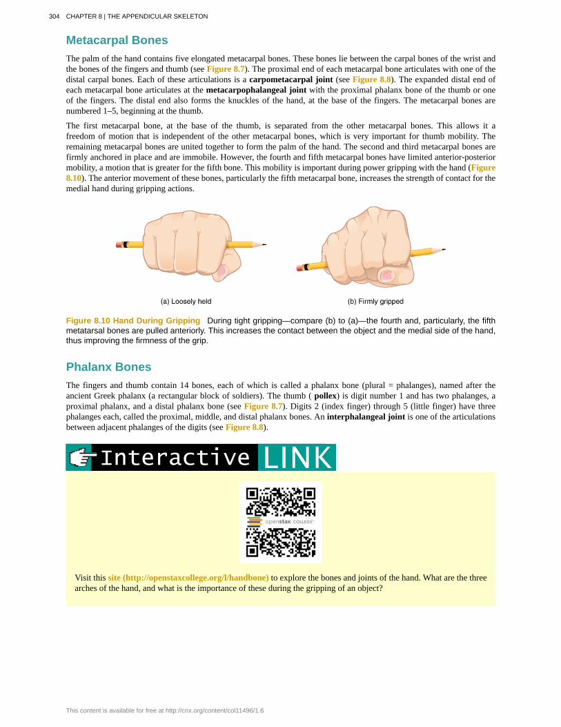
The first metacarpal bone, at the base of the thumb, is separated from the other metacarpal bones. This allows it afreedom of motion that is independent of the other metacarpal bones, which is very important for thumb mobility. Theremaining metacarpal bones are united together to form the palm of the hand. The second and third metacarpal bones arefirmly anchored in place and are immobile. However, the fourth and fifth metacarpal bones have limited anterior-posteriormobility, a motion that is greater for the fifth bone. This mobility is important during power gripping with the hand (Figure8.10). The anterior movement of these bones, particularly the fifth metacarpal bone, increases the strength of contact for themedial hand during gripping actions.
Figure 8.10 Hand During Gripping During tight gripping—compare (b) to (a)—the fourth and, particularly, the fifthmetatarsal bones are pulled anteriorly. This increases the contact between the object and the medial side of the hand,thus improving the firmness of the grip.
Phalanx BonesThe fingers and thumb contain 14 bones, each of which is called a phalanx bone (plural = phalanges), named after theancient Greek phalanx (a rectangular block of soldiers). The thumb ( pollex) is digit number 1 and has two phalanges, aproximal phalanx, and a distal phalanx bone (see Figure 8.7). Digits 2 (index finger) through 5 (little finger) have threephalanges each, called the proximal, middle, and distal phalanx bones. An interphalangeal joint is one of the articulationsbetween adjacent phalanges of the digits (see Figure 8.8).
Visit this site (http://openstaxcollege.org/l/handbone) to explore the bones and joints of the hand. What are the threearches of the hand, and what is the importance of these during the gripping of an object?
304 CHAPTER 8 | THE APPENDICULAR SKELETON
This content is available for free at http://cnx.org/content/col11496/1.6
Appendicular System: Fractures of Upper Limb BonesDue to our constant use of the hands and the rest of our upper limbs, an injury to any of these areas will cause asignificant loss of functional ability. Many fractures result from a hard fall onto an outstretched hand. The resultingtransmission of force up the limb may result in a fracture of the humerus, radius, or scaphoid bones. These injuries areespecially common in elderly people whose bones are weakened due to osteoporosis.
Falls onto the hand or elbow, or direct blows to the arm, can result in fractures of the humerus (Figure 8.11). Followinga fall, fractures at the surgical neck, the region at which the expanded proximal end of the humerus joins with the shaft,can result in an impacted fracture, in which the distal portion of the humerus is driven into the proximal portion. Fallsor blows to the arm can also produce transverse or spiral fractures of the humeral shaft.
In children, a fall onto the tip of the elbow frequently results in a distal humerus fracture. In these, the olecranon ofthe ulna is driven upward, resulting in a fracture across the distal humerus, above both epicondyles (supracondylarfracture), or a fracture between the epicondyles, thus separating one or both of the epicondyles from the body of thehumerus (intercondylar fracture). With these injuries, the immediate concern is possible compression of the artery tothe forearm due to swelling of the surrounding tissues. If compression occurs, the resulting ischemia (lack of oxygen)due to reduced blood flow can quickly produce irreparable damage to the forearm muscles. In addition, four majornerves for shoulder and upper limb muscles are closely associated with different regions of the humerus, and thus,humeral fractures may also damage these nerves.
Another frequent injury following a fall onto an outstretched hand is a Colles fracture (“col-lees”) of the distal radius(see Figure 8.11). This involves a complete transverse fracture across the distal radius that drives the separated distalfragment of the radius posteriorly and superiorly. This injury results in a characteristic “dinner fork” bend of theforearm just above the wrist due to the posterior displacement of the hand. This is the most frequent forearm fractureand is a common injury in persons over the age of 50, particularly in older women with osteoporosis. It also commonlyoccurs following a high-speed fall onto the hand during activities such as snowboarding or skating.
The most commonly fractured carpal bone is the scaphoid, often resulting from a fall onto the hand. Deep pain at thelateral wrist may yield an initial diagnosis of a wrist sprain, but a radiograph taken several weeks after the injury, aftertissue swelling has subsided, will reveal the fracture. Due to the poor blood supply to the scaphoid bone, healing willbe slow and there is the danger of bone necrosis and subsequent degenerative joint disease of the wrist.
CHAPTER 8 | THE APPENDICULAR SKELETON 305
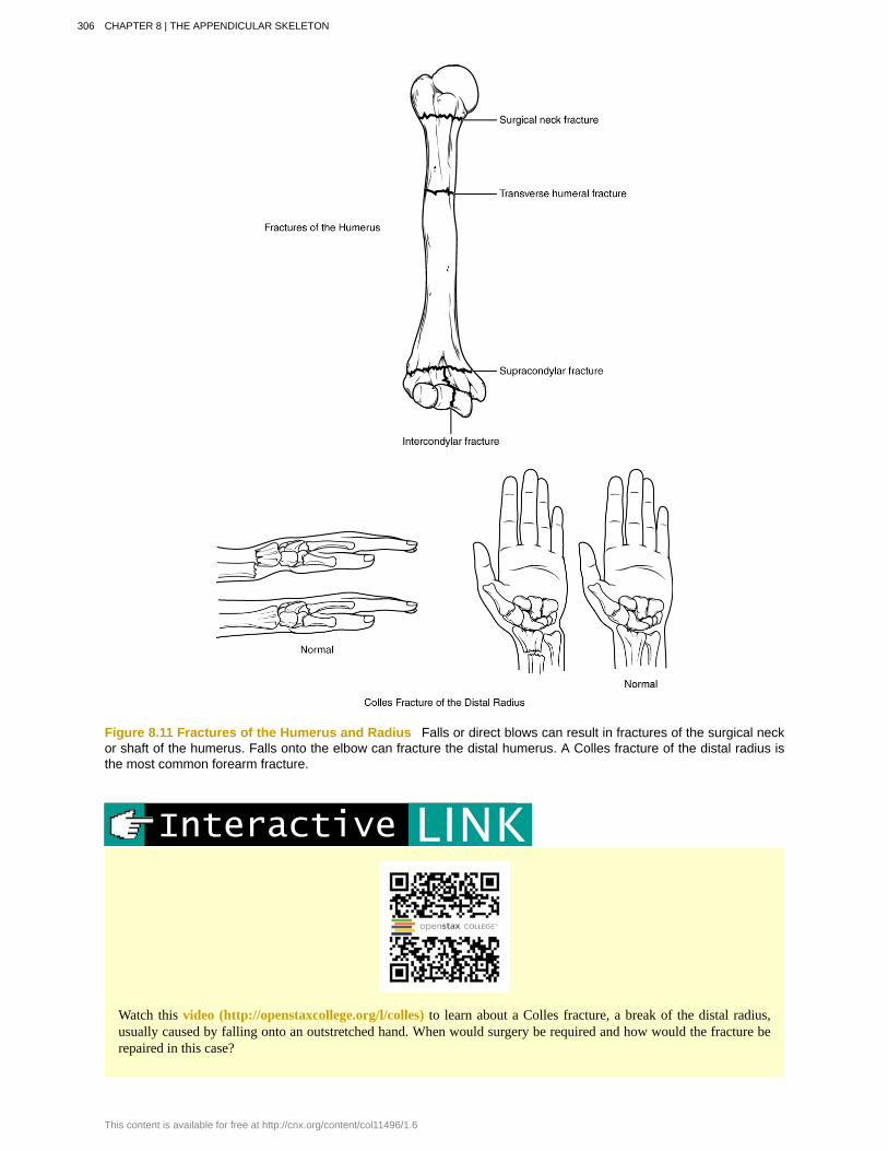
Figure 8.11 Fractures of the Humerus and Radius Falls or direct blows can result in fractures of the surgical neckor shaft of the humerus. Falls onto the elbow can fracture the distal humerus. A Colles fracture of the distal radius isthe most common forearm fracture.
Watch this video (http://openstaxcollege.org/l/colles) to learn about a Colles fracture, a break of the distal radius,usually caused by falling onto an outstretched hand. When would surgery be required and how would the fracture berepaired in this case?
306 CHAPTER 8 | THE APPENDICULAR SKELETON
This content is available for free at http://cnx.org/content/col11496/1.6
8.3 | The Pelvic Girdle and Pelvis
By the end of this section, you will be able to:
• Define the pelvic girdle and describe the bones and ligaments of the pelvis
• Explain the three regions of the hip bone and identify their bony landmarks
• Describe the openings of the pelvis and the boundaries of the greater and lesser pelvis
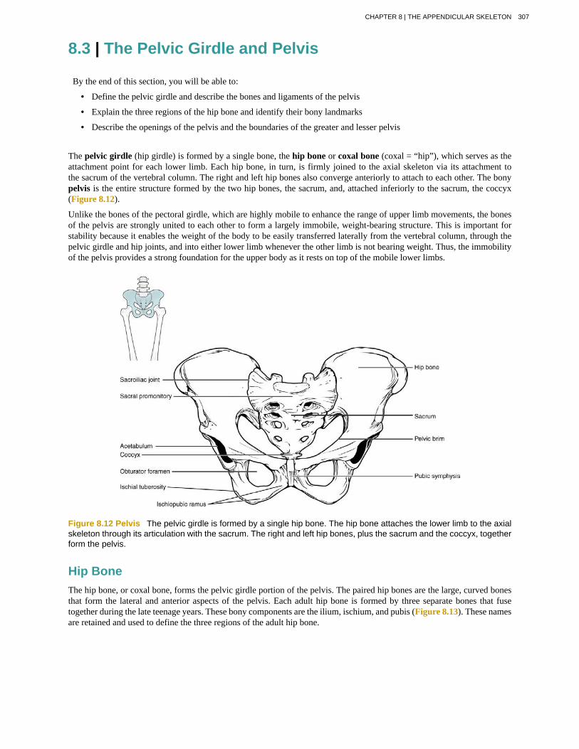
The pelvic girdle (hip girdle) is formed by a single bone, the hip bone or coxal bone (coxal = “hip”), which serves as theattachment point for each lower limb. Each hip bone, in turn, is firmly joined to the axial skeleton via its attachment tothe sacrum of the vertebral column. The right and left hip bones also converge anteriorly to attach to each other. The bonypelvis is the entire structure formed by the two hip bones, the sacrum, and, attached inferiorly to the sacrum, the coccyx(Figure 8.12).
Unlike the bones of the pectoral girdle, which are highly mobile to enhance the range of upper limb movements, the bonesof the pelvis are strongly united to each other to form a largely immobile, weight-bearing structure. This is important forstability because it enables the weight of the body to be easily transferred laterally from the vertebral column, through thepelvic girdle and hip joints, and into either lower limb whenever the other limb is not bearing weight. Thus, the immobilityof the pelvis provides a strong foundation for the upper body as it rests on top of the mobile lower limbs.
Figure 8.12 Pelvis The pelvic girdle is formed by a single hip bone. The hip bone attaches the lower limb to the axialskeleton through its articulation with the sacrum. The right and left hip bones, plus the sacrum and the coccyx, togetherform the pelvis.
Hip BoneThe hip bone, or coxal bone, forms the pelvic girdle portion of the pelvis. The paired hip bones are the large, curved bonesthat form the lateral and anterior aspects of the pelvis. Each adult hip bone is formed by three separate bones that fusetogether during the late teenage years. These bony components are the ilium, ischium, and pubis (Figure 8.13). These namesare retained and used to define the three regions of the adult hip bone.
CHAPTER 8 | THE APPENDICULAR SKELETON 307

Figure 8.13 The Hip Bone The adult hip bone consists of three regions. The ilium forms the large, fan-shapedsuperior portion, the ischium forms the posteroinferior portion, and the pubis forms the anteromedial portion.
The ilium is the fan-like, superior region that forms the largest part of the hip bone. It is firmly united to the sacrum atthe largely immobile sacroiliac joint (see Figure 8.12). The ischium forms the posteroinferior region of each hip bone. Itsupports the body when sitting. The pubis forms the anterior portion of the hip bone. The pubis curves medially, where itjoins to the pubis of the opposite hip bone at a specialized joint called the pubic symphysis.
IliumWhen you place your hands on your waist, you can feel the arching, superior margin of the ilium along your waistline(see Figure 8.13). This curved, superior margin of the ilium is the iliac crest. The rounded, anterior termination of theiliac crest is the anterior superior iliac spine. This important bony landmark can be felt at your anterolateral hip. Inferiorto the anterior superior iliac spine is a rounded protuberance called the anterior inferior iliac spine. Both of these iliacspines serve as attachment points for muscles of the thigh. Posteriorly, the iliac crest curves downward to terminate asthe posterior superior iliac spine. Muscles and ligaments surround but do not cover this bony landmark, thus sometimesproducing a depression seen as a “dimple” located on the lower back. More inferiorly is the posterior inferior iliac spine.This is located at the inferior end of a large, roughened area called the auricular surface of the ilium. The auricular surfacearticulates with the auricular surface of the sacrum to form the sacroiliac joint. Both the posterior superior and posteriorinferior iliac spines serve as attachment points for the muscles and very strong ligaments that support the sacroiliac joint.
The shallow depression located on the anteromedial (internal) surface of the upper ilium is called the iliac fossa. The inferiormargin of this space is formed by the arcuate line of the ilium, the ridge formed by the pronounced change in curvaturebetween the upper and lower portions of the ilium. The large, inverted U-shaped indentation located on the posterior marginof the lower ilium is called the greater sciatic notch.
IschiumThe ischium forms the posterolateral portion of the hip bone (see Figure 8.13). The large, roughened area of the inferiorischium is the ischial tuberosity. This serves as the attachment for the posterior thigh muscles and also carries the weightof the body when sitting. You can feel the ischial tuberosity if you wiggle your pelvis against the seat of a chair. Projectingsuperiorly and anteriorly from the ischial tuberosity is a narrow segment of bone called the ischial ramus. The slightlycurved posterior margin of the ischium above the ischial tuberosity is the lesser sciatic notch. The bony projectionseparating the lesser sciatic notch and greater sciatic notch is the ischial spine.
PubisThe pubis forms the anterior portion of the hip bone (see Figure 8.13). The enlarged medial portion of the pubis is the pubicbody. Located superiorly on the pubic body is a small bump called the pubic tubercle. The superior pubic ramus is thesegment of bone that passes laterally from the pubic body to join the ilium. The narrow ridge running along the superiormargin of the superior pubic ramus is the pectineal line of the pubis.
308 CHAPTER 8 | THE APPENDICULAR SKELETON
This content is available for free at http://cnx.org/content/col11496/1.6
The pubic body is joined to the pubic body of the opposite hip bone by the pubic symphysis. Extending downward andlaterally from the body is the inferior pubic ramus. The pubic arch is the bony structure formed by the pubic symphysis,and the bodies and inferior pubic rami of the adjacent pubic bones. The inferior pubic ramus extends downward to jointhe ischial ramus. Together, these form the single ischiopubic ramus, which extends from the pubic body to the ischialtuberosity. The inverted V-shape formed as the ischiopubic rami from both sides come together at the pubic symphysis iscalled the subpubic angle.
PelvisThe pelvis consists of four bones: the right and left hip bones, the sacrum, and the coccyx (see Figure 8.12). The pelvishas several important functions. Its primary role is to support the weight of the upper body when sitting and to transferthis weight to the lower limbs when standing. It serves as an attachment point for trunk and lower limb muscles, and alsoprotects the internal pelvic organs. When standing in the anatomical position, the pelvis is tilted anteriorly. In this position,the anterior superior iliac spines and the pubic tubercles lie in the same vertical plane, and the anterior (internal) surface ofthe sacrum faces forward and downward.
The three areas of each hip bone, the ilium, pubis, and ischium, converge centrally to form a deep, cup-shaped cavity calledthe acetabulum. This is located on the lateral side of the hip bone and is part of the hip joint. The large opening in theanteroinferior hip bone between the ischium and pubis is the obturator foramen. This space is largely filled in by a layerof connective tissue and serves for the attachment of muscles on both its internal and external surfaces.
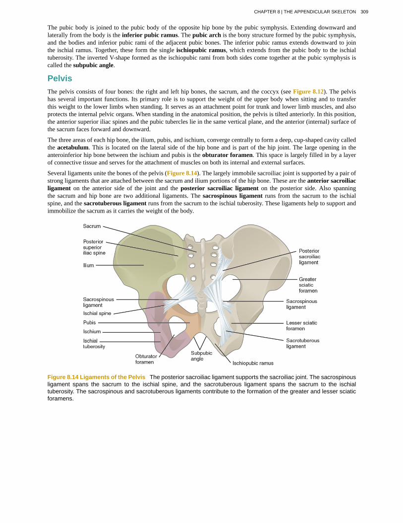
Several ligaments unite the bones of the pelvis (Figure 8.14). The largely immobile sacroiliac joint is supported by a pair ofstrong ligaments that are attached between the sacrum and ilium portions of the hip bone. These are the anterior sacroiliacligament on the anterior side of the joint and the posterior sacroiliac ligament on the posterior side. Also spanningthe sacrum and hip bone are two additional ligaments. The sacrospinous ligament runs from the sacrum to the ischialspine, and the sacrotuberous ligament runs from the sacrum to the ischial tuberosity. These ligaments help to support andimmobilize the sacrum as it carries the weight of the body.
Figure 8.14 Ligaments of the Pelvis The posterior sacroiliac ligament supports the sacroiliac joint. The sacrospinousligament spans the sacrum to the ischial spine, and the sacrotuberous ligament spans the sacrum to the ischialtuberosity. The sacrospinous and sacrotuberous ligaments contribute to the formation of the greater and lesser sciaticforamens.
CHAPTER 8 | THE APPENDICULAR SKELETON 309
Watch this video (http://openstaxcollege.org/l/3Dpelvis) for a 3-D view of the pelvis and its associated ligaments.What is the large opening in the bony pelvis, located between the ischium and pubic regions, and what two parts of thepubis contribute to the formation of this opening?
The sacrospinous and sacrotuberous ligaments also help to define two openings on the posterolateral sides of the pelvisthrough which muscles, nerves, and blood vessels for the lower limb exit. The superior opening is the greater sciaticforamen. This large opening is formed by the greater sciatic notch of the hip bone, the sacrum, and the sacrospinousligament. The smaller, more inferior lesser sciatic foramen is formed by the lesser sciatic notch of the hip bone, togetherwith the sacrospinous and sacrotuberous ligaments.
The space enclosed by the bony pelvis is divided into two regions (Figure 8.15). The broad, superior region, definedlaterally by the large, fan-like portion of the upper hip bone, is called the greater pelvis (greater pelvic cavity; false pelvis).This broad area is occupied by portions of the small and large intestines, and because it is more closely associated with theabdominal cavity, it is sometimes referred to as the false pelvis. More inferiorly, the narrow, rounded space of the lesserpelvis (lesser pelvic cavity; true pelvis) contains the bladder and other pelvic organs, and thus is also known as the truepelvis. The pelvic brim (also known as the pelvic inlet) forms the superior margin of the lesser pelvis, separating it fromthe greater pelvis. The pelvic brim is defined by a line formed by the upper margin of the pubic symphysis anteriorly, andthe pectineal line of the pubis, the arcuate line of the ilium, and the sacral promontory (the anterior margin of the superiorsacrum) posteriorly. The inferior limit of the lesser pelvic cavity is called the pelvic outlet. This large opening is defined bythe inferior margin of the pubic symphysis anteriorly, and the ischiopubic ramus, the ischial tuberosity, the sacrotuberousligament, and the inferior tip of the coccyx posteriorly. Because of the anterior tilt of the pelvis, the lesser pelvis is alsoangled, giving it an anterosuperior (pelvic inlet) to posteroinferior (pelvic outlet) orientation.
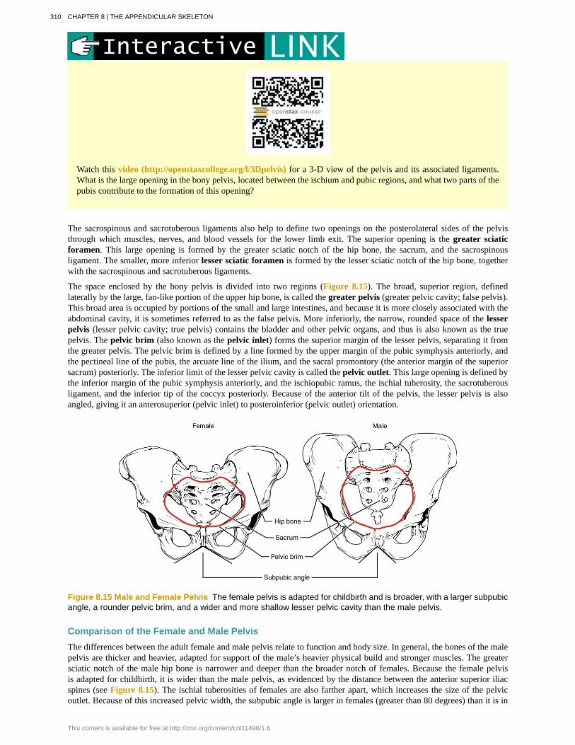
Figure 8.15 Male and Female Pelvis The female pelvis is adapted for childbirth and is broader, with a larger subpubicangle, a rounder pelvic brim, and a wider and more shallow lesser pelvic cavity than the male pelvis.
Comparison of the Female and Male PelvisThe differences between the adult female and male pelvis relate to function and body size. In general, the bones of the malepelvis are thicker and heavier, adapted for support of the male’s heavier physical build and stronger muscles. The greatersciatic notch of the male hip bone is narrower and deeper than the broader notch of females. Because the female pelvisis adapted for childbirth, it is wider than the male pelvis, as evidenced by the distance between the anterior superior iliacspines (see Figure 8.15). The ischial tuberosities of females are also farther apart, which increases the size of the pelvicoutlet. Because of this increased pelvic width, the subpubic angle is larger in females (greater than 80 degrees) than it is in
310 CHAPTER 8 | THE APPENDICULAR SKELETON
This content is available for free at http://cnx.org/content/col11496/1.6

males (less than 70 degrees). The female sacrum is wider, shorter, and less curved, and the sacral promontory projects lessinto the pelvic cavity, thus giving the female pelvic inlet (pelvic brim) a more rounded or oval shape compared to males.The lesser pelvic cavity of females is also wider and more shallow than the narrower, deeper, and tapering lesser pelvis ofmales. Because of the obvious differences between female and male hip bones, this is the one bone of the body that allowsfor the most accurate sex determination. Table 8.1 provides an overview of the general differences between the female andmale pelvis.
Overview of Differences between the Female and Male PelvisFemale pelvis Male pelvis
Pelvic weight Bones of the pelvis are lighter andthinner
Bones of the pelvis are thicker andheavier
Pelvic inlet shape Pelvic inlet has a round or oval shape Pelvic inlet is heart-shaped
Lesser pelvic cavityshape Lesser pelvic cavity is shorter and wider Lesser pelvic cavity is longer and
narrower
Subpubic angle Subpubic angle is greater than 80degrees Subpubic angle is less than 70 degrees
Pelvic outlet shape Pelvic outlet is rounded and larger Pelvic outlet is smaller
Table 8.1
Forensic Pathology and Forensic AnthropologyA forensic pathologist (also known as a medical examiner) is a medically trained physician who has been specificallytrained in pathology to examine the bodies of the deceased to determine the cause of death. A forensic pathologistapplies his or her understanding of disease as well as toxins, blood and DNA analysis, firearms and ballistics, andother factors to assess the cause and manner of death. At times, a forensic pathologist will be called to testify underoath in situations that involve a possible crime. Forensic pathology is a field that has received much media attentionon television shows or following a high-profile death.
While forensic pathologists are responsible for determining whether the cause of someone’s death was natural,a suicide, accidental, or a homicide, there are times when uncovering the cause of death is more complex, andother skills are needed. Forensic anthropology brings the tools and knowledge of physical anthropology and humanosteology (the study of the skeleton) to the task of investigating a death. A forensic anthropologist assists medicaland legal professionals in identifying human remains. The science behind forensic anthropology involves the study ofarchaeological excavation; the examination of hair; an understanding of plants, insects, and footprints; the ability todetermine how much time has elapsed since the person died; the analysis of past medical history and toxicology; theability to determine whether there are any postmortem injuries or alterations of the skeleton; and the identification ofthe decedent (deceased person) using skeletal and dental evidence.
Due to the extensive knowledge and understanding of excavation techniques, a forensic anthropologist is an integraland invaluable team member to have on-site when investigating a crime scene, especially when the recovery of humanskeletal remains is involved. When remains are bought to a forensic anthropologist for examination, he or she must firstdetermine whether the remains are in fact human. Once the remains have been identified as belonging to a person andnot to an animal, the next step is to approximate the individual’s age, sex, race, and height. The forensic anthropologistdoes not determine the cause of death, but rather provides information to the forensic pathologist, who will use all ofthe data collected to make a final determination regarding the cause of death.
CHAPTER 8 | THE APPENDICULAR SKELETON 311
8.4 | Bones of the Lower Limb
By the end of this section, you will be able to:
• Identify the divisions of the lower limb and describe the bones of each region
• Describe the bones and bony landmarks that articulate at each joint of the lower limb
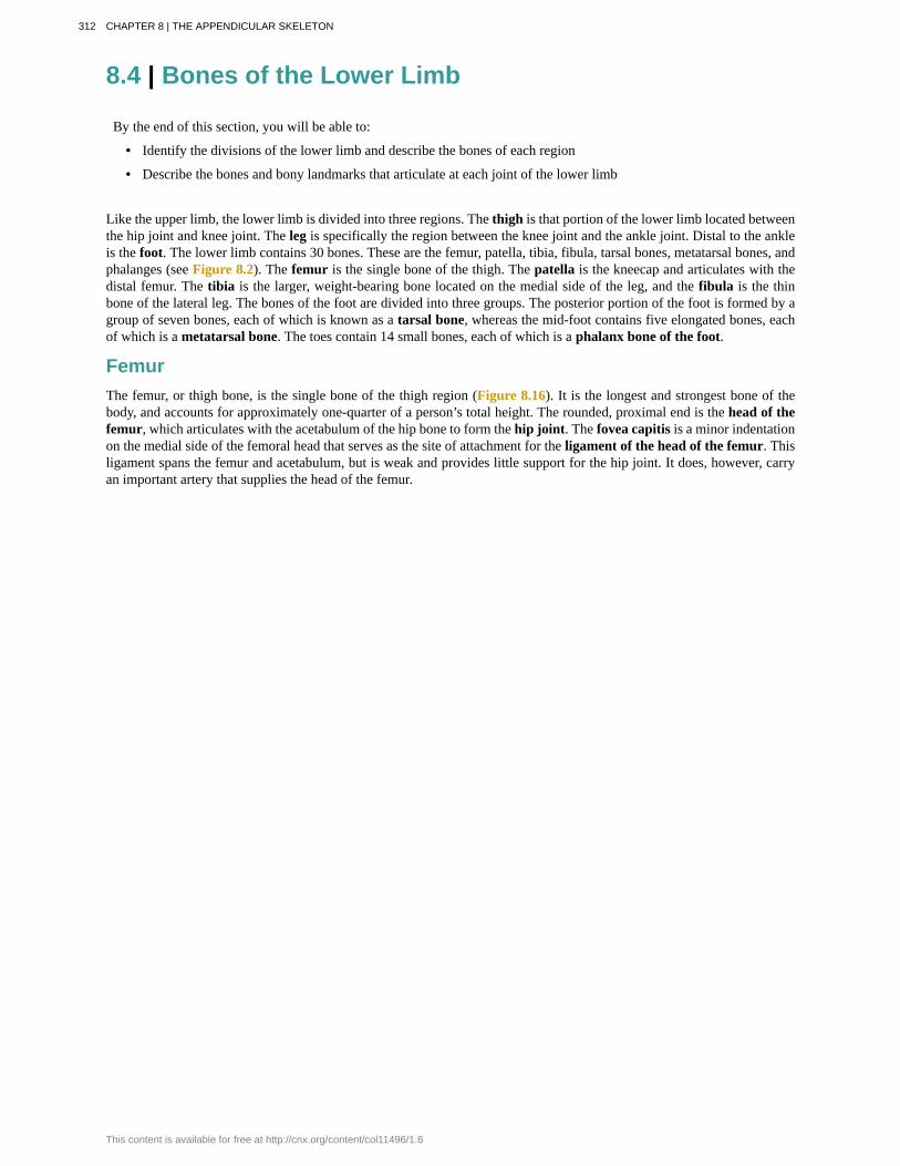
Like the upper limb, the lower limb is divided into three regions. The thigh is that portion of the lower limb located betweenthe hip joint and knee joint. The leg is specifically the region between the knee joint and the ankle joint. Distal to the ankleis the foot. The lower limb contains 30 bones. These are the femur, patella, tibia, fibula, tarsal bones, metatarsal bones, andphalanges (see Figure 8.2). The femur is the single bone of the thigh. The patella is the kneecap and articulates with thedistal femur. The tibia is the larger, weight-bearing bone located on the medial side of the leg, and the fibula is the thinbone of the lateral leg. The bones of the foot are divided into three groups. The posterior portion of the foot is formed by agroup of seven bones, each of which is known as a tarsal bone, whereas the mid-foot contains five elongated bones, eachof which is a metatarsal bone. The toes contain 14 small bones, each of which is a phalanx bone of the foot.
FemurThe femur, or thigh bone, is the single bone of the thigh region (Figure 8.16). It is the longest and strongest bone of thebody, and accounts for approximately one-quarter of a person’s total height. The rounded, proximal end is the head of thefemur, which articulates with the acetabulum of the hip bone to form the hip joint. The fovea capitis is a minor indentationon the medial side of the femoral head that serves as the site of attachment for the ligament of the head of the femur. Thisligament spans the femur and acetabulum, but is weak and provides little support for the hip joint. It does, however, carryan important artery that supplies the head of the femur.
312 CHAPTER 8 | THE APPENDICULAR SKELETON
This content is available for free at http://cnx.org/content/col11496/1.6
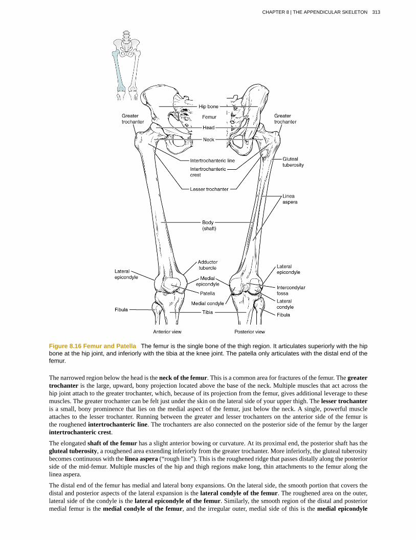
Figure 8.16 Femur and Patella The femur is the single bone of the thigh region. It articulates superiorly with the hipbone at the hip joint, and inferiorly with the tibia at the knee joint. The patella only articulates with the distal end of thefemur.
The narrowed region below the head is the neck of the femur. This is a common area for fractures of the femur. The greatertrochanter is the large, upward, bony projection located above the base of the neck. Multiple muscles that act across thehip joint attach to the greater trochanter, which, because of its projection from the femur, gives additional leverage to thesemuscles. The greater trochanter can be felt just under the skin on the lateral side of your upper thigh. The lesser trochanteris a small, bony prominence that lies on the medial aspect of the femur, just below the neck. A single, powerful muscleattaches to the lesser trochanter. Running between the greater and lesser trochanters on the anterior side of the femur isthe roughened intertrochanteric line. The trochanters are also connected on the posterior side of the femur by the largerintertrochanteric crest.
The elongated shaft of the femur has a slight anterior bowing or curvature. At its proximal end, the posterior shaft has thegluteal tuberosity, a roughened area extending inferiorly from the greater trochanter. More inferiorly, the gluteal tuberositybecomes continuous with the linea aspera (“rough line”). This is the roughened ridge that passes distally along the posteriorside of the mid-femur. Multiple muscles of the hip and thigh regions make long, thin attachments to the femur along thelinea aspera.
The distal end of the femur has medial and lateral bony expansions. On the lateral side, the smooth portion that covers thedistal and posterior aspects of the lateral expansion is the lateral condyle of the femur. The roughened area on the outer,lateral side of the condyle is the lateral epicondyle of the femur. Similarly, the smooth region of the distal and posteriormedial femur is the medial condyle of the femur, and the irregular outer, medial side of this is the medial epicondyle
CHAPTER 8 | THE APPENDICULAR SKELETON 313
of the femur. The lateral and medial condyles articulate with the tibia to form the knee joint. The epicondyles provideattachment for muscles and supporting ligaments of the knee. The adductor tubercle is a small bump located at the superiormargin of the medial epicondyle. Posteriorly, the medial and lateral condyles are separated by a deep depression called theintercondylar fossa. Anteriorly, the smooth surfaces of the condyles join together to form a wide groove called the patellarsurface, which provides for articulation with the patella bone. The combination of the medial and lateral condyles with thepatellar surface gives the distal end of the femur a horseshoe (U) shape.
Watch this video (http://openstaxcollege.org/l/midfemur) to view how a fracture of the mid-femur is surgicallyrepaired. How are the two portions of the broken femur stabilized during surgical repair of a fractured femur?
PatellaThe patella (kneecap) is largest sesamoid bone of the body (see Figure 8.16). A sesamoid bone is a bone that is incorporatedinto the tendon of a muscle where that tendon crosses a joint. The sesamoid bone articulates with the underlying bones toprevent damage to the muscle tendon due to rubbing against the bones during movements of the joint. The patella is foundin the tendon of the quadriceps femoris muscle, the large muscle of the anterior thigh that passes across the anterior kneeto attach to the tibia. The patella articulates with the patellar surface of the femur and thus prevents rubbing of the muscletendon against the distal femur. The patella also lifts the tendon away from the knee joint, which increases the leveragepower of the quadriceps femoris muscle as it acts across the knee. The patella does not articulate with the tibia.
Visit this site (http://openstaxcollege.org/l/kneesurgery) to perform a virtual knee replacement surgery. Theprosthetic knee components must be properly aligned to function properly. How is this alignment ensured?
314 CHAPTER 8 | THE APPENDICULAR SKELETON
This content is available for free at http://cnx.org/content/col11496/1.6
Runner’s KneeRunner’s knee, also known as patellofemoral syndrome, is the most common overuse injury among runners. It is mostfrequent in adolescents and young adults, and is more common in females. It often results from excessive running,particularly downhill, but may also occur in athletes who do a lot of knee bending, such as jumpers, skiers, cyclists,weight lifters, and soccer players. It is felt as a dull, aching pain around the front of the knee and deep to the patella.The pain may be felt when walking or running, going up or down stairs, kneeling or squatting, or after sitting with theknee bent for an extended period.
Patellofemoral syndrome may be initiated by a variety of causes, including individual variations in the shape andmovement of the patella, a direct blow to the patella, or flat feet or improper shoes that cause excessive turning in orout of the feet or leg. These factors may cause in an imbalance in the muscle pull that acts on the patella, resulting inan abnormal tracking of the patella that allows it to deviate too far toward the lateral side of the patellar surface on thedistal femur.
Because the hips are wider than the knee region, the femur has a diagonal orientation within the thigh, in contrast tothe vertically oriented tibia of the leg (Figure 8.17). The Q-angle is a measure of how far the femur is angled laterallyaway from vertical. The Q-angle is normally 10–15 degrees, with females typically having a larger Q-angle due totheir wider pelvis. During extension of the knee, the quadriceps femoris muscle pulls the patella both superiorly andlaterally, with the lateral pull greater in women due to their large Q-angle. This makes women more vulnerable todeveloping patellofemoral syndrome than men. Normally, the large lip on the lateral side of the patellar surface of thefemur compensates for the lateral pull on the patella, and thus helps to maintain its proper tracking.
However, if the pull produced by the medial and lateral sides of the quadriceps femoris muscle is not properlybalanced, abnormal tracking of the patella toward the lateral side may occur. With continued use, this produces painand could result in damage to the articulating surfaces of the patella and femur, and the possible future development ofarthritis. Treatment generally involves stopping the activity that produces knee pain for a period of time, followed by agradual resumption of activity. Proper strengthening of the quadriceps femoris muscle to correct for imbalances is alsoimportant to help prevent reoccurrence.
CHAPTER 8 | THE APPENDICULAR SKELETON 315
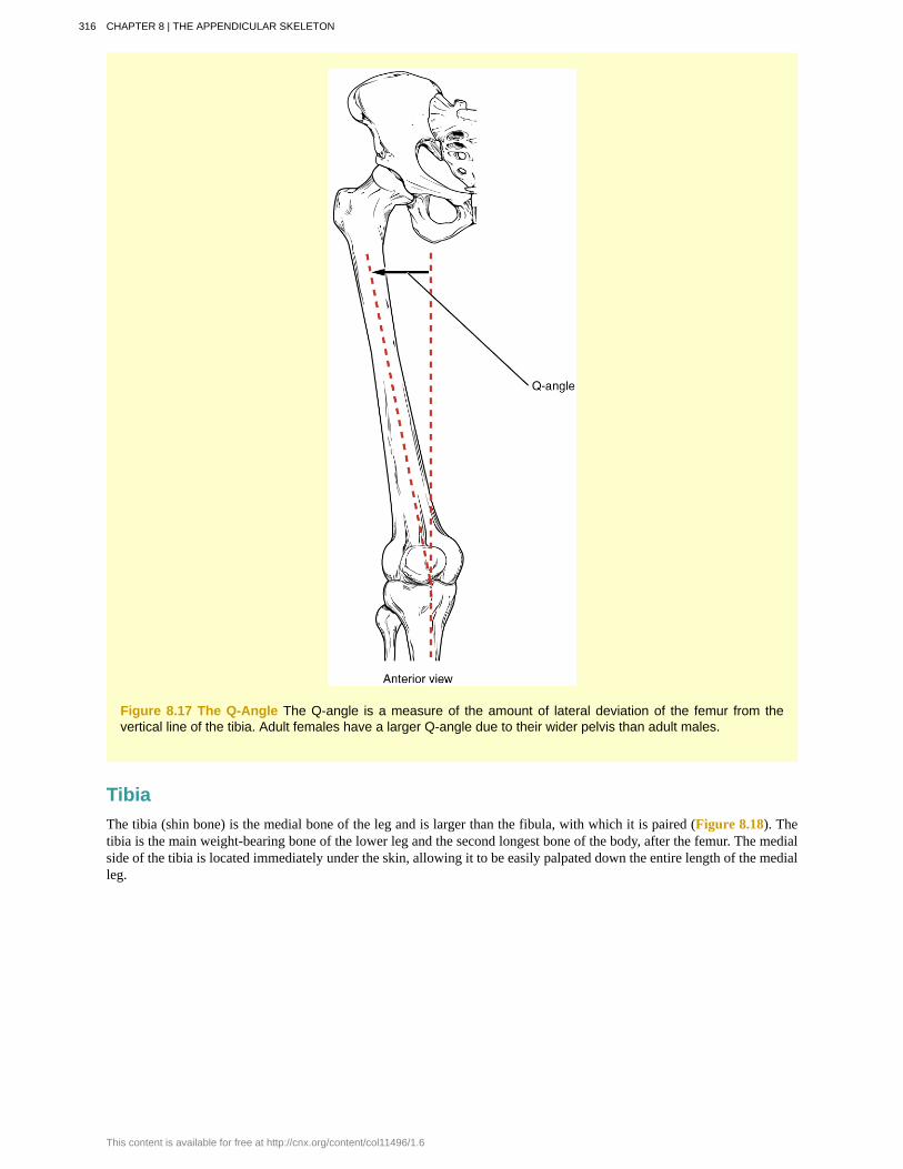
Figure 8.17 The Q-Angle The Q-angle is a measure of the amount of lateral deviation of the femur from thevertical line of the tibia. Adult females have a larger Q-angle due to their wider pelvis than adult males.
TibiaThe tibia (shin bone) is the medial bone of the leg and is larger than the fibula, with which it is paired (Figure 8.18). Thetibia is the main weight-bearing bone of the lower leg and the second longest bone of the body, after the femur. The medialside of the tibia is located immediately under the skin, allowing it to be easily palpated down the entire length of the medialleg.
316 CHAPTER 8 | THE APPENDICULAR SKELETON
This content is available for free at http://cnx.org/content/col11496/1.6
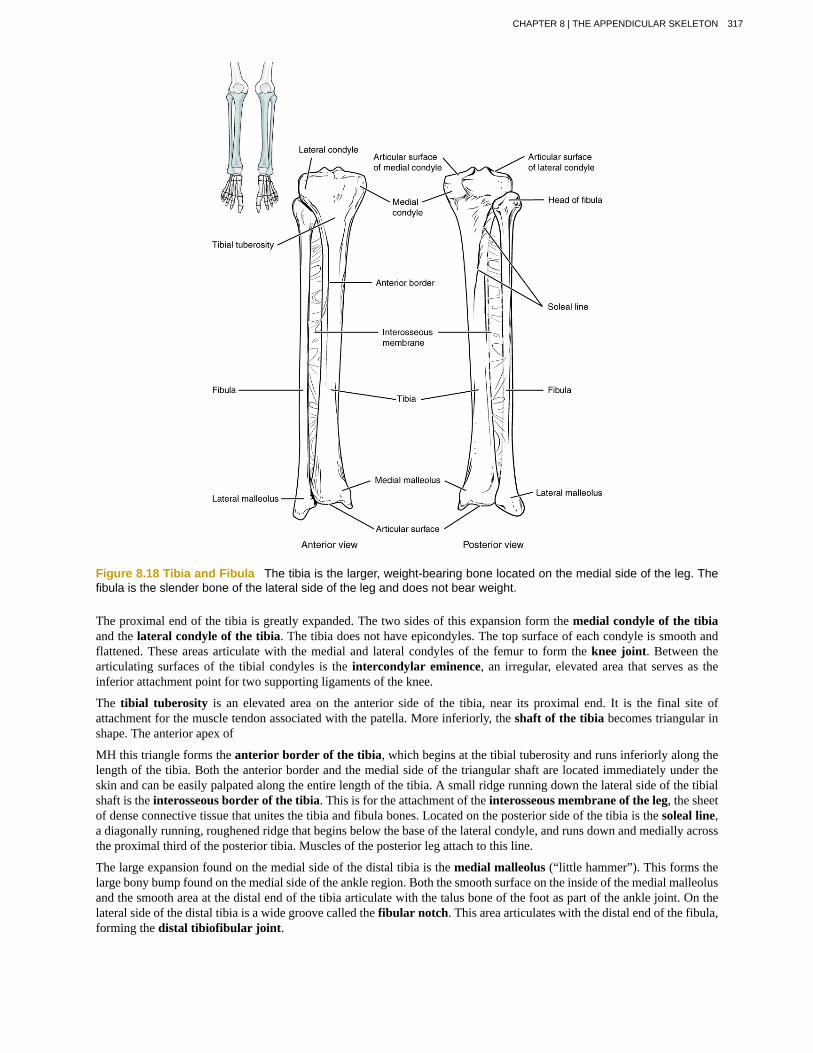
Figure 8.18 Tibia and Fibula The tibia is the larger, weight-bearing bone located on the medial side of the leg. Thefibula is the slender bone of the lateral side of the leg and does not bear weight.
The proximal end of the tibia is greatly expanded. The two sides of this expansion form the medial condyle of the tibiaand the lateral condyle of the tibia. The tibia does not have epicondyles. The top surface of each condyle is smooth andflattened. These areas articulate with the medial and lateral condyles of the femur to form the knee joint. Between thearticulating surfaces of the tibial condyles is the intercondylar eminence, an irregular, elevated area that serves as theinferior attachment point for two supporting ligaments of the knee.
The tibial tuberosity is an elevated area on the anterior side of the tibia, near its proximal end. It is the final site ofattachment for the muscle tendon associated with the patella. More inferiorly, the shaft of the tibia becomes triangular inshape. The anterior apex of
MH this triangle forms the anterior border of the tibia, which begins at the tibial tuberosity and runs inferiorly along thelength of the tibia. Both the anterior border and the medial side of the triangular shaft are located immediately under theskin and can be easily palpated along the entire length of the tibia. A small ridge running down the lateral side of the tibialshaft is the interosseous border of the tibia. This is for the attachment of the interosseous membrane of the leg, the sheetof dense connective tissue that unites the tibia and fibula bones. Located on the posterior side of the tibia is the soleal line,a diagonally running, roughened ridge that begins below the base of the lateral condyle, and runs down and medially acrossthe proximal third of the posterior tibia. Muscles of the posterior leg attach to this line.
The large expansion found on the medial side of the distal tibia is the medial malleolus (“little hammer”). This forms thelarge bony bump found on the medial side of the ankle region. Both the smooth surface on the inside of the medial malleolusand the smooth area at the distal end of the tibia articulate with the talus bone of the foot as part of the ankle joint. On thelateral side of the distal tibia is a wide groove called the fibular notch. This area articulates with the distal end of the fibula,forming the distal tibiofibular joint.
CHAPTER 8 | THE APPENDICULAR SKELETON 317

FibulaThe fibula is the slender bone located on the lateral side of the leg (see Figure 8.18). The fibula does not bear weight. Itserves primarily for muscle attachments and thus is largely surrounded by muscles. Only the proximal and distal ends of thefibula can be palpated.
The head of the fibula is the small, knob-like, proximal end of the fibula. It articulates with the inferior aspect of the lateraltibial condyle, forming the proximal tibiofibular joint. The thin shaft of the fibula has the interosseous border of thefibula, a narrow ridge running down its medial side for the attachment of the interosseous membrane that spans the fibulaand tibia. The distal end of the fibula forms the lateral malleolus, which forms the easily palpated bony bump on the lateralside of the ankle. The deep (medial) side of the lateral malleolus articulates with the talus bone of the foot as part of theankle joint. The distal fibula also articulates with the fibular notch of the tibia.
Tarsal BonesThe posterior half of the foot is formed by seven tarsal bones (Figure 8.19). The most superior bone is the talus. This hasa relatively square-shaped, upper surface that articulates with the tibia and fibula to form the ankle joint. Three areas ofarticulation form the ankle joint: The superomedial surface of the talus bone articulates with the medial malleolus of thetibia, the top of the talus articulates with the distal end of the tibia, and the lateral side of the talus articulates with the lateralmalleolus of the fibula. Inferiorly, the talus articulates with the calcaneus (heel bone), the largest bone of the foot, whichforms the heel. Body weight is transferred from the tibia to the talus to the calcaneus, which rests on the ground. The medialcalcaneus has a prominent bony extension called the sustentaculum tali (“support for the talus”) that supports the medialside of the talus bone.
Figure 8.19 Bones of the Foot The bones of the foot are divided into three groups. The posterior foot is formed bythe seven tarsal bones. The mid-foot has the five metatarsal bones. The toes contain the phalanges.
The cuboid bone articulates with the anterior end of the calcaneus bone. The cuboid has a deep groove running acrossits inferior surface, which provides passage for a muscle tendon. The talus bone articulates anteriorly with the navicularbone, which in turn articulates anteriorly with the three cuneiform (“wedge-shaped”) bones. These bones are the medialcuneiform, the intermediate cuneiform, and the lateral cuneiform. Each of these bones has a broad superior surface anda narrow inferior surface, which together produce the transverse (medial-lateral) curvature of the foot. The navicular andlateral cuneiform bones also articulate with the medial side of the cuboid bone.
318 CHAPTER 8 | THE APPENDICULAR SKELETON
This content is available for free at http://cnx.org/content/col11496/1.6

Use this tutorial (http://openstaxcollege.org/l/footbones) to review the bones of the foot. Which tarsal bones are inthe proximal, intermediate, and distal groups?
Metatarsal BonesThe anterior half of the foot is formed by the five metatarsal bones, which are located between the tarsal bones of theposterior foot and the phalanges of the toes (see Figure 8.19). These elongated bones are numbered 1–5, starting withthe medial side of the foot. The first metatarsal bone is shorter and thicker than the others. The second metatarsal is thelongest. The base of the metatarsal bone is the proximal end of each metatarsal bone. These articulate with the cuboidor cuneiform bones. The base of the fifth metatarsal has a large, lateral expansion that provides for muscle attachments.This expanded base of the fifth metatarsal can be felt as a bony bump at the midpoint along the lateral border of the foot.The expanded distal end of each metatarsal is the head of the metatarsal bone. Each metatarsal bone articulates with theproximal phalanx of a toe to form a metatarsophalangeal joint. The heads of the metatarsal bones also rest on the groundand form the ball (anterior end) of the foot.
PhalangesThe toes contain a total of 14 phalanx bones (phalanges), arranged in a similar manner as the phalanges of the fingers(see Figure 8.19). The toes are numbered 1–5, starting with the big toe ( hallux). The big toe has two phalanx bones, theproximal and distal phalanges. The remaining toes all have proximal, middle, and distal phalanges. A joint between adjacentphalanx bones is called an interphalangeal joint.
View this link (http://openstaxcollege.org/l/bunion) to learn about a bunion, a localized swelling on the medial sideof the foot, next to the first metatarsophalangeal joint, at the base of the big toe. What is a bunion and what type ofshoe is most likely to cause this to develop?
Arches of the FootWhen the foot comes into contact with the ground during walking, running, or jumping activities, the impact of the bodyweight puts a tremendous amount of pressure and force on the foot. During running, the force applied to each foot as itcontacts the ground can be up to 2.5 times your body weight. The bones, joints, ligaments, and muscles of the foot absorbthis force, thus greatly reducing the amount of shock that is passed superiorly into the lower limb and body. The arches ofthe foot play an important role in this shock-absorbing ability. When weight is applied to the foot, these arches will flattensomewhat, thus absorbing energy. When the weight is removed, the arch rebounds, giving “spring” to the step. The archesalso serve to distribute body weight side to side and to either end of the foot.
CHAPTER 8 | THE APPENDICULAR SKELETON 319
The foot has a transverse arch, a medial longitudinal arch, and a lateral longitudinal arch (see Figure 8.19). The transversearch forms the medial-lateral curvature of the mid-foot. It is formed by the wedge shapes of the cuneiform bones and bases(proximal ends) of the first to fourth metatarsal bones. This arch helps to distribute body weight from side to side within thefoot, thus allowing the foot to accommodate uneven terrain.
The longitudinal arches run down the length of the foot. The lateral longitudinal arch is relatively flat, whereas the mediallongitudinal arch is larger (taller). The longitudinal arches are formed by the tarsal bones posteriorly and the metatarsalbones anteriorly. These arches are supported at either end, where they contact the ground. Posteriorly, this support isprovided by the calcaneus bone and anteriorly by the heads (distal ends) of the metatarsal bones. The talus bone, whichreceives the weight of the body, is located at the top of the longitudinal arches. Body weight is then conveyed from the talusto the ground by the anterior and posterior ends of these arches. Strong ligaments unite the adjacent foot bones to preventdisruption of the arches during weight bearing. On the bottom of the foot, additional ligaments tie together the anterior andposterior ends of the arches. These ligaments have elasticity, which allows them to stretch somewhat during weight bearing,thus allowing the longitudinal arches to spread. The stretching of these ligaments stores energy within the foot, rather thanpassing these forces into the leg. Contraction of the foot muscles also plays an important role in this energy absorption.When the weight is removed, the elastic ligaments recoil and pull the ends of the arches closer together. This recovery ofthe arches releases the stored energy and improves the energy efficiency of walking.
Stretching of the ligaments that support the longitudinal arches can lead to pain. This can occur in overweight individuals,with people who have jobs that involve standing for long periods of time (such as a waitress), or walking or running longdistances. If stretching of the ligaments is prolonged, excessive, or repeated, it can result in a gradual lengthening of thesupporting ligaments, with subsequent depression or collapse of the longitudinal arches, particularly on the medial side ofthe foot. This condition is called pes planus (“flat foot” or “fallen arches”).
8.5 | Development of the Appendicular Skeleton
By the end of this section, you will be able to:
• Describe the growth and development of the embryonic limb buds
• Discuss the appearance of primary and secondary ossification centers
Embryologically, the appendicular skeleton arises from mesenchyme, a type of embryonic tissue that can differentiate intomany types of tissues, including bone or muscle tissue. Mesenchyme gives rise to the bones of the upper and lower limbs, aswell as to the pectoral and pelvic girdles. Development of the limbs begins near the end of the fourth embryonic week, withthe upper limbs appearing first. Thereafter, the development of the upper and lower limbs follows similar patterns, with thelower limbs lagging behind the upper limbs by a few days.
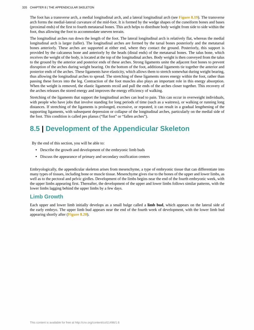
Limb GrowthEach upper and lower limb initially develops as a small bulge called a limb bud, which appears on the lateral side ofthe early embryo. The upper limb bud appears near the end of the fourth week of development, with the lower limb budappearing shortly after (Figure 8.20).
320 CHAPTER 8 | THE APPENDICULAR SKELETON
This content is available for free at http://cnx.org/content/col11496/1.6

Figure 8.20 Embryo at Seven Weeks Limb buds are visible in an embryo at the end of the seventh week ofdevelopment (embryo derived from an ectopic pregnancy). (credit: Ed Uthman/flickr)
Initially, the limb buds consist of a core of mesenchyme covered by a layer of ectoderm. The ectoderm at the end of the limbbud thickens to form a narrow crest called the apical ectodermal ridge. This ridge stimulates the underlying mesenchymeto rapidly proliferate, producing the outgrowth of the developing limb. As the limb bud elongates, cells located farther fromthe apical ectodermal ridge slow their rates of cell division and begin to differentiate. In this way, the limb develops along aproximal-to-distal axis.
During the sixth week of development, the distal ends of the upper and lower limb buds expand and flatten into a paddleshape. This region will become the hand or foot. The wrist or ankle areas then appear as a constriction that develops at thebase of the paddle. Shortly after this, a second constriction on the limb bud appears at the future site of the elbow or knee.Within the paddle, areas of tissue undergo cell death, producing separations between the growing fingers and toes. Alsoduring the sixth week of development, mesenchyme within the limb buds begins to differentiate into hyaline cartilage thatwill form models of the future limb bones.
The early outgrowth of the upper and lower limb buds initially has the limbs positioned so that the regions that will becomethe palm of the hand or the bottom of the foot are facing medially toward the body, with the future thumb or big toe bothoriented toward the head. During the seventh week of development, the upper limb rotates laterally by 90 degrees, so thatthe palm of the hand faces anteriorly and the thumb points laterally. In contrast, the lower limb undergoes a 90-degreemedial rotation, thus bringing the big toe to the medial side of the foot.
Watch this animation (http://openstaxcollege.org/l/limbbuds) to follow the development and growth of the upperand lower limb buds. On what days of embryonic development do these events occur: (a) first appearance of the upperlimb bud (limb ridge); (b) the flattening of the distal limb to form the handplate or footplate; and (c) the beginning oflimb rotation?
CHAPTER 8 | THE APPENDICULAR SKELETON 321
Ossification of Appendicular BonesAll of the girdle and limb bones, except for the clavicle, develop by the process of endochondral ossification. This processbegins as the mesenchyme within the limb bud differentiates into hyaline cartilage to form cartilage models for futurebones. By the twelfth week, a primary ossification center will have appeared in the diaphysis (shaft) region of the longbones, initiating the process that converts the cartilage model into bone. A secondary ossification center will appear in eachepiphysis (expanded end) of these bones at a later time, usually after birth. The primary and secondary ossification centersare separated by the epiphyseal plate, a layer of growing hyaline cartilage. This plate is located between the diaphysis andeach epiphysis. It continues to grow and is responsible for the lengthening of the bone. The epiphyseal plate is retained formany years, until the bone reaches its final, adult size, at which time the epiphyseal plate disappears and the epiphysis fusesto the diaphysis. (Seek additional content on ossification in the chapter on bone tissue.)
Small bones, such as the phalanges, will develop only one secondary ossification center and will thus have only a singleepiphyseal plate. Large bones, such as the femur, will develop several secondary ossification centers, with an epiphysealplate associated with each secondary center. Thus, ossification of the femur begins at the end of the seventh week with theappearance of the primary ossification center in the diaphysis, which rapidly expands to ossify the shaft of the bone prior tobirth. Secondary ossification centers develop at later times. Ossification of the distal end of the femur, to form the condylesand epicondyles, begins shortly before birth. Secondary ossification centers also appear in the femoral head late in the firstyear after birth, in the greater trochanter during the fourth year, and in the lesser trochanter between the ages of 9 and 10years. Once these areas have ossified, their fusion to the diaphysis and the disappearance of each epiphyseal plate follow areversed sequence. Thus, the lesser trochanter is the first to fuse, doing so at the onset of puberty (around 11 years of age),followed by the greater trochanter approximately 1 year later. The femoral head fuses between the ages of 14–17 years,whereas the distal condyles of the femur are the last to fuse, between the ages of 16–19 years. Knowledge of the age atwhich different epiphyseal plates disappear is important when interpreting radiographs taken of children. Since the cartilageof an epiphyseal plate is less dense than bone, the plate will appear dark in a radiograph image. Thus, a normal epiphysealplate may be mistaken for a bone fracture.
The clavicle is the one appendicular skeleton bone that does not develop via endochondral ossification. Instead, the clavicledevelops through the process of intramembranous ossification. During this process, mesenchymal cells differentiate directlyinto bone-producing cells, which produce the clavicle directly, without first making a cartilage model. Because of this earlyproduction of bone, the clavicle is the first bone of the body to begin ossification, with ossification centers appearing duringthe fifth week of development. However, ossification of the clavicle is not complete until age 25.
322 CHAPTER 8 | THE APPENDICULAR SKELETON
This content is available for free at http://cnx.org/content/col11496/1.6

Appendicular System: Congenital ClubfootClubfoot, also known as talipes, is a congenital (present at birth) disorder of unknown cause and is the most commondeformity of the lower limb. It affects the foot and ankle, causing the foot to be twisted inward at a sharp angle, likethe head of a golf club (Figure 8.21). Clubfoot has a frequency of about 1 out of every 1,000 births, and is twice aslikely to occur in a male child as in a female child. In 50 percent of cases, both feet are affected.
Figure 8.21 Clubfoot Clubfoot is a common deformity of the ankle and foot that is present at birth. Most casesare corrected without surgery, and affected individuals will grow up to lead normal, active lives. (credit: James W.Hanson)
At birth, children with a clubfoot have the heel turned inward and the anterior foot twisted so that the lateral sideof the foot is facing inferiorly, commonly due to ligaments or leg muscles attached to the foot that are shortened orabnormally tight. These pull the foot into an abnormal position, resulting in bone deformities. Other symptoms mayinclude bending of the ankle that lifts the heel of the foot and an extremely high foot arch. Due to the limited range ofmotion in the affected foot, it is difficult to place the foot into the correct position. Additionally, the affected foot maybe shorter than normal, and the calf muscles are usually underdeveloped on the affected side. Despite the appearance,this is not a painful condition for newborns. However, it must be treated early to avoid future pain and impairedwalking ability.
Although the cause of clubfoot is idiopathic (unknown), evidence indicates that fetal position within the uterus is nota contributing factor. Genetic factors are involved, because clubfoot tends to run within families. Cigarette smokingduring pregnancy has been linked to the development of clubfoot, particularly in families with a history of clubfoot.
Previously, clubfoot required extensive surgery. Today, 90 percent of cases are successfully treated without surgeryusing new corrective casting techniques. The best chance for a full recovery requires that clubfoot treatment beginduring the first 2 weeks after birth. Corrective casting gently stretches the foot, which is followed by the application ofa holding cast to keep the foot in the proper position. This stretching and casting is repeated weekly for several weeks.In severe cases, surgery may also be required, after which the foot typically remains in a cast for 6 to 8 weeks. After thecast is removed following either surgical or nonsurgical treatment, the child will be required to wear a brace part-time(at night) for up to 4 years. In addition, special exercises will be prescribed, and the child must also wear special shoes.Close monitoring by the parents and adherence to postoperative instructions are imperative in minimizing the risk ofrelapse.
Despite these difficulties, treatment for clubfoot is usually successful, and the child will grow up to lead a normal,active life. Numerous examples of individuals born with a clubfoot who went on to successful careers include DudleyMoore (comedian and actor), Damon Wayans (comedian and actor), Troy Aikman (three-time Super Bowl-winningquarterback), Kristi Yamaguchi (Olympic gold medalist in figure skating), Mia Hamm (two-time Olympic goldmedalist in soccer), and Charles Woodson (Heisman trophy and Super Bowl winner).
CHAPTER 8 | THE APPENDICULAR SKELETON 323
acetabulum
acromial end of the clavicle
acromial process
acromioclavicular joint
acromion
adductor tubercle
anatomical neck
ankle joint
anterior border of the tibia
anterior inferior iliac spine
anterior sacroiliac ligament
anterior superior iliac spine
apical ectodermal ridge
arcuate line of the ilium
arm
auricular surface of the ilium
base of the metatarsal bone
bicipital groove
calcaneus
capitate
capitulum
carpal bone
carpal tunnel
carpometacarpal joint
clavicle
KEY TERMSlarge, cup-shaped cavity located on the lateral side of the hip bone; formed by the junction of the ilium,
pubis, and ischium portions of the hip bone
lateral end of the clavicle that articulates with the acromion of the scapula
acromion of the scapula
articulation between the acromion of the scapula and the acromial end of the clavicle
flattened bony process that extends laterally from the scapular spine to form the bony tip of the shoulder
small, bony bump located on the superior aspect of the medial epicondyle of the femur
line on the humerus located around the outside margin of the humeral head
joint that separates the leg and foot portions of the lower limb; formed by the articulations between the talusbone of the foot inferiorly, and the distal end of the tibia, medial malleolus of the tibia, and lateral malleolus of thefibula superiorly
narrow, anterior margin of the tibia that extends inferiorly from the tibial tuberosity
small, bony projection located on the anterior margin of the ilium, below the anteriorsuperior iliac spine
strong ligament between the sacrum and the ilium portions of the hip bone that supportsthe anterior side of the sacroiliac joint
rounded, anterior end of the iliac crest
enlarged ridge of ectoderm at the distal end of a limb bud that stimulates growth andelongation of the limb
smooth ridge located at the inferior margin of the iliac fossa; forms the lateral portion of thepelvic brim
region of the upper limb located between the shoulder and elbow joints; contains the humerus bone
roughened area located on the posterior, medial side of the ilium of the hip bone;articulates with the auricular surface of the sacrum to form the sacroiliac joint
expanded, proximal end of each metatarsal bone
intertubercular groove; narrow groove located between the greater and lesser tubercles of the humerus
heel bone; posterior, inferior tarsal bone that forms the heel of the foot
from the lateral side, the third of the four distal carpal bones; articulates with the scaphoid and lunateproximally, the trapezoid laterally, the hamate medially, and primarily with the third metacarpal distally
knob-like bony structure located anteriorly on the lateral, distal end of the humerus
one of the eight small bones that form the wrist and base of the hand; these are grouped as a proximal rowconsisting of (from lateral to medial) the scaphoid, lunate, triquetrum, and pisiform bones, and a distal rowcontaining (from lateral to medial) the trapezium, trapezoid, capitate, and hamate bones
passageway between the anterior forearm and hand formed by the carpal bones and flexor retinaculum
articulation between one of the carpal bones in the distal row and a metacarpal bone of thehand
collarbone; elongated bone that articulates with the manubrium of the sternum medially and the acromion ofthe scapula laterally
324 CHAPTER 8 | THE APPENDICULAR SKELETON
This content is available for free at http://cnx.org/content/col11496/1.6
coracoclavicular ligament
coracoid process
coronoid fossa
coronoid process of the ulna
costoclavicular ligament
coxal bone
cuboid
deltoid tuberosity
distal radioulnar joint
distal tibiofibular joint
elbow joint
femur
fibula
fibular notch
flexor retinaculum
foot
forearm
fossa
fovea capitis
glenohumeral joint
glenoid cavity
gluteal tuberosity
greater pelvis
greater sciatic foramen
greater sciatic notch
greater trochanter
strong band of connective tissue that anchors the coracoid process of the scapula to thelateral clavicle; provides important indirect support for the acromioclavicular joint
short, hook-like process that projects anteriorly and laterally from the superior margin of thescapula
depression on the anterior surface of the humerus above the trochlea; this space receives the coronoidprocess of the ulna when the elbow is maximally flexed
projecting bony lip located on the anterior, proximal ulna; forms the inferior marginof the trochlear notch
band of connective tissue that unites the medial clavicle with the first rib
hip bone
tarsal bone that articulates posteriorly with the calcaneus bone, medially with the lateral cuneiform bone, andanteriorly with the fourth and fifth metatarsal bones
roughened, V-shaped region located laterally on the mid-shaft of the humerus
articulation between the head of the ulna and the ulnar notch of the radius
articulation between the distal fibula and the fibular notch of the tibia
joint located between the upper arm and forearm regions of the upper limb; formed by the articulationsbetween the trochlea of the humerus and the trochlear notch of the ulna, and the capitulum of the humerus and thehead of the radius
thigh bone; the single bone of the thigh
thin, non-weight-bearing bone found on the lateral side of the leg
wide groove on the lateral side of the distal tibia for articulation with the fibula at the distal tibiofibularjoint
strong band of connective tissue at the anterior wrist that spans the top of the U-shaped grouping ofthe carpal bones to form the roof of the carpal tunnel
portion of the lower limb located distal to the ankle joint
region of the upper limb located between the elbow and wrist joints; contains the radius and ulna bones
(plural = fossae) shallow depression on the surface of a bone
minor indentation on the head of the femur that serves as the site of attachment for the ligament to thehead of the femur
shoulder joint; formed by the articulation between the glenoid cavity of the scapula and the headof the humerus
(also, glenoid fossa) shallow depression located on the lateral scapula, between the superior and lateralborders
roughened area on the posterior side of the proximal femur, extending inferiorly from the base ofthe greater trochanter
(also, greater pelvic cavity or false pelvis) broad space above the pelvic brim defined laterally by thefan-like portion of the upper ilium
pelvic opening formed by the greater sciatic notch of the hip bone, the sacrum, and thesacrospinous ligament
large, U-shaped indentation located on the posterior margin of the ilium, superior to the ischialspine
large, bony expansion of the femur that projects superiorly from the base of the femoral neck
CHAPTER 8 | THE APPENDICULAR SKELETON 325

greater tubercle
hallux
hamate
hand
head of the femur
head of the fibula
head of the humerus
head of the metatarsal bone
head of the radius
head of the ulna
hip bone
hip joint
hook of the hamate bone
humerus
iliac crest
iliac fossa
ilium
inferior angle of the scapula
inferior pubic ramus
infraglenoid tubercle
infraspinous fossa
intercondylar eminence
intercondylar fossa
intermediate cuneiform
interosseous border of the fibula
interosseous border of the radius
enlarged prominence located on the lateral side of the proximal humerus
big toe; digit 1 of the foot
from the lateral side, the fourth of the four distal carpal bones; articulates with the lunate and triquetrumproximally, the fourth and fifth metacarpals distally, and the capitate laterally
region of the upper limb distal to the wrist joint
rounded, proximal end of the femur that articulates with the acetabulum of the hip bone to form thehip joint
small, knob-like, proximal end of the fibula; articulates with the inferior aspect of the lateralcondyle of the tibia
smooth, rounded region on the medial side of the proximal humerus; articulates with theglenoid fossa of the scapula to form the glenohumeral (shoulder) joint
expanded, distal end of each metatarsal bone
disc-shaped structure that forms the proximal end of the radius; articulates with the capitulum ofthe humerus as part of the elbow joint, and with the radial notch of the ulna as part of the proximal radioulnar joint
small, rounded distal end of the ulna; articulates with the ulnar notch of the distal radius, forming thedistal radioulnar joint
coxal bone; single bone that forms the pelvic girdle; consists of three areas, the ilium, ischium, and pubis
joint located at the proximal end of the lower limb; formed by the articulation between the acetabulum of thehip bone and the head of the femur
bony extension located on the anterior side of the hamate carpal bone
single bone of the upper arm
curved, superior margin of the ilium
shallow depression found on the anterior and medial surfaces of the upper ilium
superior portion of the hip bone
inferior corner of the scapula located where the medial and lateral borders meet
narrow segment of bone that passes inferiorly and laterally from the pubic body; joins with theischial ramus to form the ischiopubic ramus
small bump or roughened area located on the lateral border of the scapula, near the inferiormargin of the glenoid cavity
broad depression located on the posterior scapula, inferior to the spine
irregular elevation on the superior end of the tibia, between the articulating surfaces of themedial and lateral condyles
deep depression on the posterior side of the distal femur that separates the medial and lateralcondyles
middle of the three cuneiform tarsal bones; articulates posteriorly with the navicular bone,medially with the medial cuneiform bone, laterally with the lateral cuneiform bone, and anteriorly with the secondmetatarsal bone
small ridge running down the medial side of the fibular shaft; for attachment ofthe interosseous membrane between the fibula and tibia
narrow ridge located on the medial side of the radial shaft; for attachment of theinterosseous membrane between the ulna and radius bones
326 CHAPTER 8 | THE APPENDICULAR SKELETON
This content is available for free at http://cnx.org/content/col11496/1.6

interosseous border of the tibia
interosseous border of the ulna
interosseous membrane of the forearm
interosseous membrane of the leg
interphalangeal joint
intertrochanteric crest
intertrochanteric line
intertubercular groove (sulcus)
ischial ramus
ischial spine
ischial tuberosity
ischiopubic ramus
ischium
knee joint
lateral border of the scapula
lateral condyle of the femur
lateral condyle of the tibia
lateral cuneiform
lateral epicondyle of the femur
lateral epicondyle of the humerus
lateral malleolus
lateral supracondylar ridge
leg
lesser pelvis
lesser sciatic foramen
small ridge running down the lateral side of the tibial shaft; for attachment of theinterosseous membrane between the tibia and fibula
narrow ridge located on the lateral side of the ulnar shaft; for attachment of theinterosseous membrane between the ulna and radius
sheet of dense connective tissue that unites the radius and ulna bones
sheet of dense connective tissue that unites the shafts of the tibia and fibulabones
articulation between adjacent phalanx bones of the hand or foot digits
short, prominent ridge running between the greater and lesser trochanters on the posterior sideof the proximal femur
small ridge running between the greater and lesser trochanters on the anterior side of theproximal femur
bicipital groove; narrow groove located between the greater and lesser tubercles ofthe humerus
bony extension projecting anteriorly and superiorly from the ischial tuberosity; joins with the inferiorpubic ramus to form the ischiopubic ramus
pointed, bony projection from the posterior margin of the ischium that separates the greater sciatic notchand lesser sciatic notch
large, roughened protuberance that forms the posteroinferior portion of the hip bone; weight-bearing region of the pelvis when sitting
narrow extension of bone that connects the ischial tuberosity to the pubic body; formed by thejunction of the ischial ramus and inferior pubic ramus
posteroinferior portion of the hip bone
joint that separates the thigh and leg portions of the lower limb; formed by the articulations between themedial and lateral condyles of the femur, and the medial and lateral condyles of the tibia
diagonally oriented lateral margin of the scapula
smooth, articulating surface that forms the distal and posterior sides of the lateralexpansion of the distal femur
lateral, expanded region of the proximal tibia that includes the smooth surface thatarticulates with the lateral condyle of the femur as part of the knee joint
most lateral of the three cuneiform tarsal bones; articulates posteriorly with the navicular bone,medially with the intermediate cuneiform bone, laterally with the cuboid bone, and anteriorly with the thirdmetatarsal bone
roughened area of the femur located on the lateral side of the lateral condyle
small projection located on the lateral side of the distal humerus
expanded distal end of the fibula
narrow, bony ridge located along the lateral side of the distal humerus, superior to thelateral epicondyle
portion of the lower limb located between the knee and ankle joints
(also, lesser pelvic cavity or true pelvis) narrow space located within the pelvis, defined superiorly by thepelvic brim (pelvic inlet) and inferiorly by the pelvic outlet
pelvic opening formed by the lesser sciatic notch of the hip bone, the sacrospinous ligament,and the sacrotuberous ligament
CHAPTER 8 | THE APPENDICULAR SKELETON 327
lesser sciatic notch
lesser trochanter
lesser tubercle
ligament of the head of the femur
limb bud
linea aspera
lunate
medial border of the scapula
medial condyle of the femur
medial condyle of the tibia
medial cuneiform
medial epicondyle of the femur
medial epicondyle of the humerus
medial malleolus
metacarpal bone
metacarpophalangeal joint
metatarsal bone
metatarsophalangeal joint
midcarpal joint
navicular
neck of the femur
neck of the radius
obturator foramen
olecranon fossa
olecranon process
patella
patellar surface
shallow indentation along the posterior margin of the ischium, inferior to the ischial spine
small, bony projection on the medial side of the proximal femur, at the base of the femoral neck
small, bony prominence located on anterior side of the proximal humerus
ligament that spans the acetabulum of the hip bone and the fovea capitis of thefemoral head
small elevation that appears on the lateral side of the embryo during the fourth or fifth week of development,which gives rise to an upper or lower limb
longitudinally running bony ridge located in the middle third of the posterior femur
from the lateral side, the second of the four proximal carpal bones; articulates with the radius proximally, thecapitate and hamate distally, the scaphoid laterally, and the triquetrum medially
elongated, medial margin of the scapula
smooth, articulating surface that forms the distal and posterior sides of the medialexpansion of the distal femur
medial, expanded region of the proximal tibia that includes the smooth surface thatarticulates with the medial condyle of the femur as part of the knee joint
most medial of the three cuneiform tarsal bones; articulates posteriorly with the navicular bone,laterally with the intermediate cuneiform bone, and anteriorly with the first and second metatarsal bones
roughened area of the distal femur located on the medial side of the medial condyle
enlarged projection located on the medial side of the distal humerus
bony expansion located on the medial side of the distal tibia
one of the five long bones that form the palm of the hand; numbered 1–5, starting on the lateral(thumb) side of the hand
articulation between the distal end of a metacarpal bone of the hand and a proximalphalanx bone of the thumb or a finger
one of the five elongated bones that forms the anterior half of the foot; numbered 1–5, starting on themedial side of the foot
articulation between a metatarsal bone of the foot and the proximal phalanx bone of a toe
articulation between the proximal and distal rows of the carpal bones; contributes to movements of thehand at the wrist
tarsal bone that articulates posteriorly with the talus bone, laterally with the cuboid bone, and anteriorly withthe medial, intermediate, and lateral cuneiform bones
narrowed region located inferior to the head of the femur
narrowed region immediately distal to the head of the radius
large opening located in the anterior hip bone, between the pubis and ischium regions
large depression located on the posterior side of the distal humerus; this space receives the olecranonprocess of the ulna when the elbow is fully extended
expanded posterior and superior portions of the proximal ulna; forms the bony tip of the elbow
kneecap; the largest sesamoid bone of the body; articulates with the distal femur
smooth groove located on the anterior side of the distal femur, between the medial and lateralcondyles; site of articulation for the patella
328 CHAPTER 8 | THE APPENDICULAR SKELETON
This content is available for free at http://cnx.org/content/col11496/1.6
pectineal line
pectoral girdle
pelvic brim
pelvic girdle
pelvic inlet
pelvic outlet
pelvis
phalanx bone of the foot
phalanx bone of the hand
pisiform
pollex
posterior inferior iliac spine
posterior sacroiliac ligament
posterior superior iliac spine
proximal radioulnar joint
proximal tibiofibular joint
pubic arch
pubic body
pubic symphysis
pubic tubercle
pubis
radial fossa
radial notch of the ulna
radial tuberosity
radiocarpal joint
radius
narrow ridge located on the superior surface of the superior pubic ramus
shoulder girdle; the set of bones, consisting of the scapula and clavicle, which attaches each upper limbto the axial skeleton
pelvic inlet; the dividing line between the greater and lesser pelvic regions; formed by the superior marginof the pubic symphysis, the pectineal lines of each pubis, the arcuate lines of each ilium, and the sacral promontory
hip girdle; consists of a single hip bone, which attaches a lower limb to the sacrum of the axial skeleton
pelvic brim
inferior opening of the lesser pelvis; formed by the inferior margin of the pubic symphysis, right and leftischiopubic rami and sacrotuberous ligaments, and the tip of the coccyx
ring of bone consisting of the right and left hip bones, the sacrum, and the coccyx
(plural = phalanges) one of the 14 bones that form the toes; these include the proximal anddistal phalanges of the big toe, and the proximal, middle, and distal phalanx bones of toes two through five
(plural = phalanges) one of the 14 bones that form the thumb and fingers; these includethe proximal and distal phalanges of the thumb, and the proximal, middle, and distal phalanx bones of the fingerstwo through five
from the lateral side, the fourth of the four proximal carpal bones; articulates with the anterior surface of thetriquetrum
(also, thumb) digit 1 of the hand
small, bony projection located at the inferior margin of the auricular surface on theposterior ilium
strong ligament spanning the sacrum and ilium of the hip bone that supports theposterior side of the sacroiliac joint
rounded, posterior end of the iliac crest
articulation formed by the radial notch of the ulna and the head of the radius
articulation between the head of the fibula and the inferior aspect of the lateral condyle ofthe tibia
bony structure formed by the pubic symphysis, and the bodies and inferior pubic rami of the right and leftpubic bones
enlarged, medial portion of the pubis region of the hip bone
joint formed by the articulation between the pubic bodies of the right and left hip bones
small bump located on the superior aspect of the pubic body
anterior portion of the hip bone
small depression located on the anterior humerus above the capitulum; this space receives the head of theradius when the elbow is maximally flexed
small, smooth area on the lateral side of the proximal ulna; articulates with the head of theradius as part of the proximal radioulnar joint
oval-shaped, roughened protuberance located on the medial side of the proximal radius
wrist joint, located between the forearm and hand regions of the upper limb; articulation formedproximally by the distal end of the radius and the fibrocartilaginous pad that unites the distal radius and ulna bone,and distally by the scaphoid, lunate, and triquetrum carpal bones
bone located on the lateral side of the forearm
CHAPTER 8 | THE APPENDICULAR SKELETON 329
sacroiliac joint
sacrospinous ligament
sacrotuberous ligament
scaphoid
scapula
shaft of the femur
shaft of the fibula
shaft of the humerus
shaft of the radius
shaft of the tibia
shaft of the ulna
soleal line
spine of the scapula
sternal end of the clavicle
sternoclavicular joint
styloid process of the radius
styloid process of the ulna
subpubic angle
subscapular fossa
superior angle of the scapula
superior border of the scapula
superior pubic ramus
supraglenoid tubercle
suprascapular notch
supraspinous fossa
surgical neck
sustentaculum tali
talus
tarsal bone
thigh
tibial tuberosity
joint formed by the articulation between the auricular surfaces of the sacrum and ilium
ligament that spans the sacrum to the ischial spine of the hip bone
ligament that spans the sacrum to the ischial tuberosity of the hip bone
from the lateral side, the first of the four proximal carpal bones; articulates with the radius proximally, thetrapezoid, trapezium, and capitate distally, and the lunate medially
shoulder blade bone located on the posterior side of the shoulder
cylindrically shaped region that forms the central portion of the femur
elongated, slender portion located between the expanded ends of the fibula
narrow, elongated, central region of the humerus
narrow, elongated, central region of the radius
triangular-shaped, central portion of the tibia
narrow, elongated, central region of the ulna
small, diagonally running ridge located on the posterior side of the proximal tibia
prominent ridge passing mediolaterally across the upper portion of the posterior scapularsurface
medial end of the clavicle that articulates with the manubrium of the sternum
articulation between the manubrium of the sternum and the sternal end of the clavicle; formsthe only bony attachment between the pectoral girdle of the upper limb and the axial skeleton
pointed projection located on the lateral end of the distal radius
short, bony projection located on the medial end of the distal ulna
inverted V-shape formed by the convergence of the right and left ischiopubic rami; this angle isgreater than 80 degrees in females and less than 70 degrees in males
broad depression located on the anterior (deep) surface of the scapula
corner of the scapula between the superior and medial borders of the scapula
superior margin of the scapula
narrow segment of bone that passes laterally from the pubic body to join the ilium
small bump located at the superior margin of the glenoid cavity
small notch located along the superior border of the scapula, medial to the coracoid process
narrow depression located on the posterior scapula, superior to the spine
region of the humerus where the expanded, proximal end joins with the narrower shaft
bony ledge extending from the medial side of the calcaneus bone
tarsal bone that articulates superiorly with the tibia and fibula at the ankle joint; also articulates inferiorly with thecalcaneus bone and anteriorly with the navicular bone
one of the seven bones that make up the posterior foot; includes the calcaneus, talus, navicular, cuboid,medial cuneiform, intermediate cuneiform, and lateral cuneiform bones
portion of the lower limb located between the hip and knee joints
elevated area on the anterior surface of the proximal tibia
330 CHAPTER 8 | THE APPENDICULAR SKELETON
This content is available for free at http://cnx.org/content/col11496/1.6
tibia
trapezium
trapezoid
triquetrum
trochlear notch
trochlea
ulnar notch of the radius
ulnar tuberosity
ulna
shin bone; the large, weight-bearing bone located on the medial side of the leg
from the lateral side, the first of the four distal carpal bones; articulates with the scaphoid proximally, thefirst and second metacarpals distally, and the trapezoid medially
from the lateral side, the second of the four distal carpal bones; articulates with the scaphoid proximally, thesecond metacarpal distally, the trapezium laterally, and the capitate medially
from the lateral side, the third of the four proximal carpal bones; articulates with the lunate laterally, thehamate distally, and has a facet for the pisiform
large, C-shaped depression located on the anterior side of the proximal ulna; articulates at the elbowwith the trochlea of the humerus
pulley-shaped region located medially at the distal end of the humerus; articulates at the elbow with thetrochlear notch of the ulna
shallow, smooth area located on the medial side of the distal radius; articulates with thehead of the ulna at the distal radioulnar joint
roughened area located on the anterior, proximal ulna inferior to the coronoid process
bone located on the medial side of the forearm
CHAPTER REVIEW
8.1 The Pectoral Girdle
The pectoral girdle, consisting of the clavicle and the scapula, attaches each upper limb to the axial skeleton. The clavicle isan anterior bone whose sternal end articulates with the manubrium of the sternum at the sternoclavicular joint. The sternalend is also anchored to the first rib by the costoclavicular ligament. The acromial end of the clavicle articulates with theacromion of the scapula at the acromioclavicular joint. This end is also anchored to the coracoid process of the scapula bythe coracoclavicular ligament, which provides indirect support for the acromioclavicular joint. The clavicle supports thescapula, transmits the weight and forces from the upper limb to the body trunk, and protects the underlying nerves and bloodvessels.
The scapula lies on the posterior aspect of the pectoral girdle. It mediates the attachment of the upper limb to the clavicle,and contributes to the formation of the glenohumeral (shoulder) joint. This triangular bone has three sides called the medial,lateral, and superior borders. The suprascapular notch is located on the superior border. The scapula also has three corners,two of which are the superior and inferior angles. The third corner is occupied by the glenoid cavity. Posteriorly, the spineseparates the supraspinous and infraspinous fossae, and then extends laterally as the acromion. The subscapular fossa islocated on the anterior surface of the scapula. The coracoid process projects anteriorly, passing inferior to the lateral end ofthe clavicle.
8.2 Bones of the Upper Limb
Each upper limb is divided into three regions and contains a total of 30 bones. The upper arm is the region located betweenthe shoulder and elbow joints. This area contains the humerus. The proximal humerus consists of the head, which articulateswith the scapula at the glenohumeral joint, the greater and lesser tubercles separated by the intertubercular (bicipital) groove,and the anatomical and surgical necks. The humeral shaft has the roughened area of the deltoid tuberosity on its lateral side.The distal humerus is flattened, forming a lateral supracondylar ridge that terminates at the small lateral epicondyle. Themedial side of the distal humerus has the large, medial epicondyle. The articulating surfaces of the distal humerus consist ofthe trochlea medially and the capitulum laterally. Depressions on the humerus that accommodate the forearm bones duringbending (flexing) and straightening (extending) of the elbow include the coronoid fossa, the radial fossa, and the olecranonfossa.
The forearm is the region of the upper limb located between the elbow and wrist joints. This region contains two bones, theulna medially and the radius on the lateral (thumb) side. The elbow joint is formed by the articulation between the trochleaof the humerus and the trochlear notch of the ulna, plus the articulation between the capitulum of the humerus and the headof the radius. The proximal radioulnar joint is the articulation between the head of the radius and the radial notch of theulna. The proximal ulna also has the olecranon process, forming an expanded posterior region, and the coronoid processand ulnar tuberosity on its anterior aspect. On the proximal radius, the narrowed region below the head is the neck; distalto this is the radial tuberosity. The shaft portions of both the ulna and radius have an interosseous border, whereas the distal
CHAPTER 8 | THE APPENDICULAR SKELETON 331
ends of each bone have a pointed styloid process. The distal radioulnar joint is found between the head of the ulna and theulnar notch of the radius. The distal end of the radius articulates with the proximal carpal bones, but the ulna does not.
The base of the hand is formed by eight carpal bones. The carpal bones are united into two rows of bones. The proximal rowcontains (from lateral to medial) the scaphoid, lunate, triquetrum, and pisiform bones. The scaphoid, lunate, and triquetrumbones contribute to the formation of the radiocarpal joint. The distal row of carpal bones contains (from medial to lateral)the hamate, capitate, trapezoid, and trapezium bones (“So Long To Pinky, Here Comes The Thumb”). The anterior hamatehas a prominent bony hook. The proximal and distal carpal rows articulate with each other at the midcarpal joint. The carpalbones, together with the flexor retinaculum, also form the carpal tunnel of the wrist.
The five metacarpal bones form the palm of the hand. The metacarpal bones are numbered 1–5, starting with the thumbside. The first metacarpal bone is freely mobile, but the other bones are united as a group. The digits are also numbered1–5, with the thumb being number 1. The fingers and thumb contain a total of 14 phalanges (phalanx bones). The thumbcontains a proximal and a distal phalanx, whereas the remaining digits each contain proximal, middle, and distal phalanges.
8.3 The Pelvic Girdle and Pelvis
The pelvic girdle, consisting of a hip bone, serves to attach a lower limb to the axial skeleton. The hip bone articulatesposteriorly at the sacroiliac joint with the sacrum, which is part of the axial skeleton. The right and left hip bones convergeanteriorly and articulate with each other at the pubic symphysis. The combination of the hip bone, the sacrum, and thecoccyx forms the pelvis. The pelvis has a pronounced anterior tilt. The primary function of the pelvis is to support the upperbody and transfer body weight to the lower limbs. It also serves as the site of attachment for multiple muscles.
The hip bone consists of three regions: the ilium, ischium, and pubis. The ilium forms the large, fan-like region of the hipbone. The superior margin of this area is the iliac crest. Located at either end of the iliac crest are the anterior superiorand posterior superior iliac spines. Inferior to these are the anterior inferior and posterior inferior iliac spines. The auricularsurface of the ilium articulates with the sacrum to form the sacroiliac joint. The medial surface of the upper ilium formsthe iliac fossa, with the arcuate line marking the inferior limit of this area. The posterior margin of the ilium has the largegreater sciatic notch.
The posterolateral portion of the hip bone is the ischium. It has the expanded ischial tuberosity, which supports body weightwhen sitting. The ischial ramus projects anteriorly and superiorly. The posterior margin of the ischium has the shallow lessersciatic notch and the ischial spine, which separates the greater and lesser sciatic notches.
The pubis forms the anterior portion of the hip bone. The body of the pubis articulates with the pubis of the opposite hipbone at the pubic symphysis. The superior margin of the pubic body has the pubic tubercle. The pubis is joined to theilium by the superior pubic ramus, the superior surface of which forms the pectineal line. The inferior pubic ramus projectsinferiorly and laterally. The pubic arch is formed by the pubic symphysis, the bodies of the adjacent pubic bones, and thetwo inferior pubic rami. The inferior pubic ramus joins the ischial ramus to form the ischiopubic ramus. The subpubic angleis formed by the medial convergence of the right and left ischiopubic rami.
The lateral side of the hip bone has the cup-like acetabulum, which is part of the hip joint. The large anterior opening is theobturator foramen. The sacroiliac joint is supported by the anterior and posterior sacroiliac ligaments. The sacrum is alsojoined to the hip bone by the sacrospinous ligament, which attaches to the ischial spine, and the sacrotuberous ligament,which attaches to the ischial tuberosity. The sacrospinous and sacrotuberous ligaments contribute to the formation of thegreater and lesser sciatic foramina.
The broad space of the upper pelvis is the greater pelvis, and the narrow, inferior space is the lesser pelvis. These areas areseparated by the pelvic brim (pelvic inlet). The inferior opening of the pelvis is the pelvic outlet. Compared to the male, thefemale pelvis is wider to accommodate childbirth, has a larger subpubic angle, and a broader greater sciatic notch.
8.4 Bones of the Lower Limb
The lower limb is divided into three regions. These are the thigh, located between the hip and knee joints; the leg, locatedbetween the knee and ankle joints; and distal to the ankle, the foot. There are 30 bones in each lower limb. These are thefemur, patella, tibia, fibula, seven tarsal bones, five metatarsal bones, and 14 phalanges.
The femur is the single bone of the thigh. Its rounded head articulates with the acetabulum of the hip bone to form thehip joint. The head has the fovea capitis for attachment of the ligament of the head of the femur. The narrow neck joinsinferiorly with the greater and lesser trochanters. Passing between these bony expansions are the intertrochanteric line on theanterior femur and the larger intertrochanteric crest on the posterior femur. On the posterior shaft of the femur is the glutealtuberosity proximally and the linea aspera in the mid-shaft region. The expanded distal end consists of three articulatingsurfaces: the medial and lateral condyles, and the patellar surface. The outside margins of the condyles are the medial andlateral epicondyles. The adductor tubercle is on the superior aspect of the medial epicondyle.
The patella is a sesamoid bone located within a muscle tendon. It articulates with the patellar surface on the anterior side ofthe distal femur, thereby protecting the muscle tendon from rubbing against the femur.
332 CHAPTER 8 | THE APPENDICULAR SKELETON
This content is available for free at http://cnx.org/content/col11496/1.6
The leg contains the large tibia on the medial side and the slender fibula on the lateral side. The tibia bears the weightof the body, whereas the fibula does not bear weight. The interosseous border of each bone is the attachment site for theinterosseous membrane of the leg, the connective tissue sheet that unites the tibia and fibula.
The proximal tibia consists of the expanded medial and lateral condyles, which articulate with the medial and lateralcondyles of the femur to form the knee joint. Between the tibial condyles is the intercondylar eminence. On the anteriorside of the proximal tibia is the tibial tuberosity, which is continuous inferiorly with the anterior border of the tibia. On theposterior side, the proximal tibia has the curved soleal line. The bony expansion on the medial side of the distal tibia is themedial malleolus. The groove on the lateral side of the distal tibia is the fibular notch.
The head of the fibula forms the proximal end and articulates with the underside of the lateral condyle of the tibia. Thedistal fibula articulates with the fibular notch of the tibia. The expanded distal end of the fibula is the lateral malleolus.
The posterior foot is formed by the seven tarsal bones. The talus articulates superiorly with the distal tibia, the medialmalleolus of the tibia, and the lateral malleolus of the fibula to form the ankle joint. The talus articulates inferiorly with thecalcaneus bone. The sustentaculum tali of the calcaneus helps to support the talus. Anterior to the talus is the navicular bone,and anterior to this are the medial, intermediate, and lateral cuneiform bones. The cuboid bone is anterior to the calcaneus.
The five metatarsal bones form the anterior foot. The base of these bones articulate with the cuboid or cuneiform bones.The metatarsal heads, at their distal ends, articulate with the proximal phalanges of the toes. The big toe (toe number 1) hasproximal and distal phalanx bones. The remaining toes have proximal, middle, and distal phalanges.
8.5 Development of the Appendicular Skeleton
The bones of the appendicular skeleton arise from embryonic mesenchyme. Limb buds appear at the end of the fourth week.The apical ectodermal ridge, located at the end of the limb bud, stimulates growth and elongation of the limb. During thesixth week, the distal end of the limb bud becomes paddle-shaped, and selective cell death separates the developing fingersand toes. At the same time, mesenchyme within the limb bud begins to differentiate into hyaline cartilage, forming modelsfor future bones. During the seventh week, the upper limbs rotate laterally and the lower limbs rotate medially, bringing thelimbs into their final positions.
Endochondral ossification, the process that converts the hyaline cartilage model into bone, begins in most appendicularbones by the twelfth fetal week. This begins as a primary ossification center in the diaphysis, followed by the laterappearance of one or more secondary ossifications centers in the regions of the epiphyses. Each secondary ossificationcenter is separated from the primary ossification center by an epiphyseal plate. Continued growth of the epiphysealplate cartilage provides for bone lengthening. Disappearance of the epiphyseal plate is followed by fusion of the bonycomponents to form a single, adult bone.
The clavicle develops via intramembranous ossification, in which mesenchyme is converted directly into bone tissue.Ossification within the clavicle begins during the fifth week of development and continues until 25 years of age.
INTERACTIVE LINK QUESTIONS1. Watch this video (http://openstaxcollege.org/l/fractures) to see how fractures of the distal radius bone canaffect the wrist joint. Explain the problems that may occur ifa fracture of the distal radius involves the joint surface of theradiocarpal joint of the wrist.
2. Visit this site (http://openstaxcollege.org/l/handbone)to explore the bones and joints of the hand. What are thethree arches of the hand, and what is the importance of theseduring the gripping of an object?
3. Watch this video (http://openstaxcollege.org/l/colles) tolearn about a Colles fracture, a break of the distal radius,usually caused by falling onto an outstretched hand. Whenwould surgery be required and how would the fracture berepaired in this case?
4. Watch this video (http://openstaxcollege.org/l/3Dpelvis) for a 3-D view of the pelvis and its associatedligaments. What is the large opening in the bony pelvis,located between the ischium and pubic regions, and whattwo parts of the pubis contribute to the formation of thisopening?
5. Watch this video (http://openstaxcollege.org/l/midfemur) to view how a fracture of the mid-femur issurgically repaired. How are the two portions of the brokenfemur stabilized during surgical repair of a fractured femur?
6. Visit this site (http://openstaxcollege.org/l/kneesurgery) to perform a virtual knee replacement surgery.The prosthetic knee components must be properly aligned tofunction properly. How is this alignment ensured?
7. Use this tutorial (http://openstaxcollege.org/l/footbones) to review the bones of the foot. Which tarsalbones are in the proximal, intermediate, and distal groups?
8. View this link (http://openstaxcollege.org/l/bunion) tolearn about a bunion, a localized swelling on the medial sideof the foot, next to the first metatarsophalangeal joint, at thebase of the big toe. What is a bunion and what type of shoeis most likely to cause this to develop?
9. Watch this animation (http://openstaxcollege.org/l/limbbuds) to follow the development and growth of theupper and lower limb buds. On what days of embryonicdevelopment do these events occur: (a) first appearance ofthe upper limb bud (limb ridge); (b) the flattening of the
CHAPTER 8 | THE APPENDICULAR SKELETON 333
distal limb to form the handplate or footplate; and (c) thebeginning of limb rotation?
REVIEW QUESTIONS10. Which part of the clavicle articulates with themanubrium?
a. shaftb. sternal endc. acromial endd. coracoid process
11. A shoulder separation results from injury to the________.
a. glenohumeral jointb. costoclavicular jointc. acromioclavicular jointd. sternoclavicular joint
12. Which feature lies between the spine and superior borderof the scapula?
a. suprascapular notchb. glenoid cavityc. superior angled. supraspinous fossa
13. What structure is an extension of the spine of thescapula?
a. acromionb. coracoid processc. supraglenoid tubercled. glenoid cavity
14. Name the short, hook-like bony process of the scapulathat projects anteriorly.
a. acromial processb. claviclec. coracoid processd. glenoid fossa
15. How many bones are there in the upper limbs combined?
a. 20b. 30c. 40d. 60
16. Which bony landmark is located on the lateral side ofthe proximal humerus?
a. greater tubercleb. trochleac. lateral epicondyled. lesser tubercle
17. Which region of the humerus articulates with the radiusas part of the elbow joint?
a. trochleab. styloid processc. capitulumd. olecranon process
18. Which is the lateral-most carpal bone of the proximalrow?
a. trapeziumb. hamatec. pisiformd. scaphoid
19. The radius bone ________.a. is found on the medial side of the forearmb. has a head that articulates with the radial notch of
the ulnac. does not articulate with any of the carpal bonesd. has the radial tuberosity located near its distal end
20. How many bones fuse in adulthood to form the hipbone?
a. 2b. 3c. 4d. 5
21. Which component forms the superior part of the hipbone?
a. iliumb. pubisc. ischiumd. sacrum
22. Which of the following supports body weight whensitting?
a. iliac crestb. ischial tuberosityc. ischiopubic ramusd. pubic body
23. The ischial spine is found between which of thefollowing structures?
a. inferior pubic ramus and ischial ramusb. pectineal line and arcuate linec. lesser sciatic notch and greater sciatic notchd. anterior superior iliac spine and posterior superior
iliac spine
24. The pelvis ________.a. has a subpubic angle that is larger in femalesb. consists of the two hip bones, but does not include
the sacrum or coccyxc. has an obturator foramen, an opening that is
defined in part by the sacrospinous andsacrotuberous ligaments
d. has a space located inferior to the pelvic brimcalled the greater pelvis
25. Which bony landmark of the femur serves as a site formuscle attachments?
a. fovea capitisb. lesser trochanterc. headd. medial condyle
26. What structure contributes to the knee joint?a. lateral malleolus of the fibulab. tibial tuberosityc. medial condyle of the tibiad. lateral epicondyle of the femur
27. Which tarsal bone articulates with the tibia and fibula?
a. calcaneus
334 CHAPTER 8 | THE APPENDICULAR SKELETON
This content is available for free at http://cnx.org/content/col11496/1.6
b. cuboidc. naviculard. talus
28. What is the total number of bones found in the foot andtoes?
a. 7b. 14c. 26d. 30
29. The tibia ________.a. has an expanded distal end called the lateral
malleolusb. is not a weight-bearing bonec. is firmly anchored to the fibula by an interosseous
membraned. can be palpated (felt) under the skin only at its
proximal and distal ends
30. Which event takes place during the seventh week ofdevelopment?
a. appearance of the upper and lower limb buds
b. flattening of the distal limb bud into a paddleshape
c. the first appearance of hyaline cartilage models offuture bones
d. the rotation of the limbs
31. During endochondral ossification of a long bone,________.
a. a primary ossification center will develop withinthe epiphysis
b. mesenchyme will differentiate directly into bonetissue
c. growth of the epiphyseal plate will produce bonelengthening
d. all epiphyseal plates will disappear before birth
32. The clavicle ________.a. develops via intramembranous ossificationb. develops via endochondral ossificationc. is the last bone of the body to begin ossificationd. is fully ossified at the time of birth
CRITICAL THINKING QUESTIONS33. Describe the shape and palpable line formed by theclavicle and scapula.
34. Discuss two possible injuries of the pectoral girdle thatmay occur following a strong blow to the shoulder or a hardfall onto an outstretched hand.
35. Your friend runs out of gas and you have to help pushhis car. Discuss the sequence of bones and joints that conveythe forces passing from your hand, through your upper limband your pectoral girdle, and to your axial skeleton.
36. Name the bones in the wrist and hand, and describe orsketch out their locations and articulations.
37. Describe the articulations and ligaments that unite thefour bones of the pelvis to each other.
38. Discuss the ways in which the female pelvis is adaptedfor childbirth.
39. Define the regions of the lower limb, name the bonesfound in each region, and describe the bony landmarks thatarticulate together to form the hip, knee, and ankle joints.
40. The talus bone of the foot receives the weight of thebody from the tibia. The talus bone then distributes thisweight toward the ground in two directions: one-half of thebody weight is passed in a posterior direction and one-halfof the weight is passed in an anterior direction. Describethe arrangement of the tarsal and metatarsal bones that areinvolved in both the posterior and anterior distribution ofbody weight.
41. How can a radiograph of a child’s femur be used todetermine the approximate age of that child?
42. How does the development of the clavicle differ fromthe development of other appendicular skeleton bones?
CHAPTER 8 | THE APPENDICULAR SKELETON 335