9.15 k1 d parry
TRANSCRIPT
Unravelling the Working of the Animal Body: A Biophysical Approach
David A D ParryInstitute of Fundamental Sciences
Massey UniversityNew Zealand
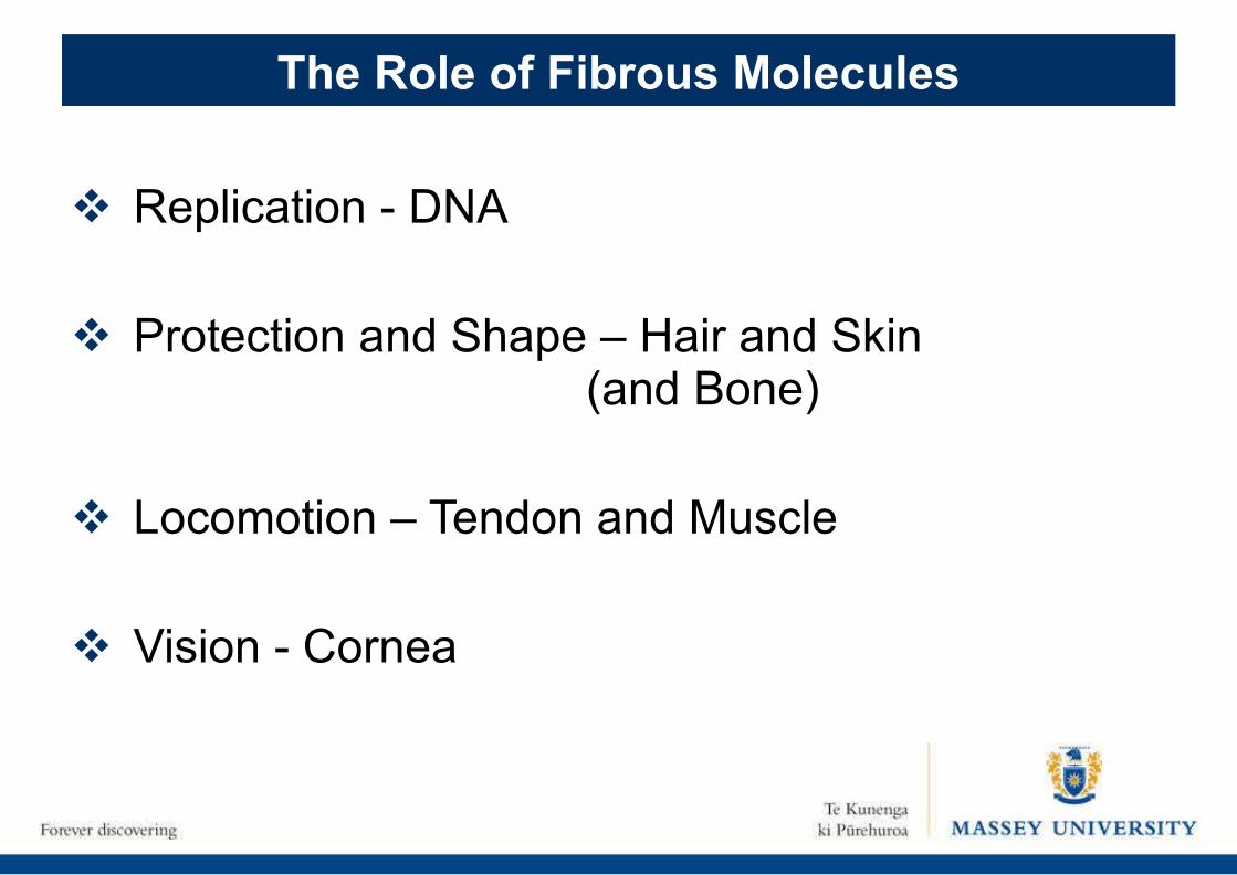
Replication - DNA
Protection and Shape – Hair and Skin (and Bone)
Locomotion – Tendon and Muscle
Vision - Cornea
The Role of Fibrous Molecules
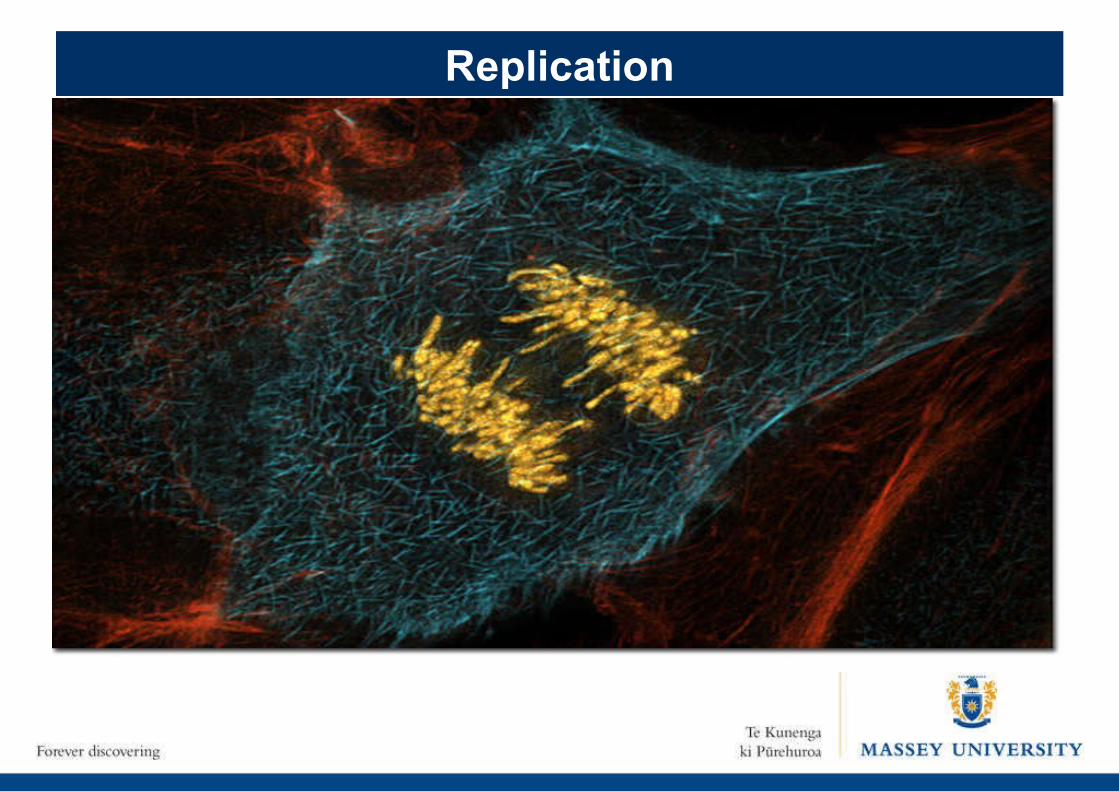
Replication
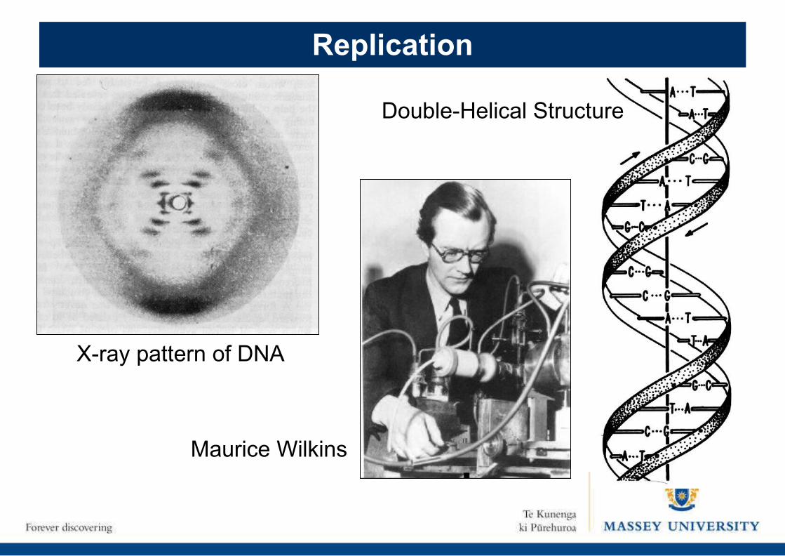
Replication
X-ray pattern of DNA
Maurice Wilkins
Double-Helical Structure
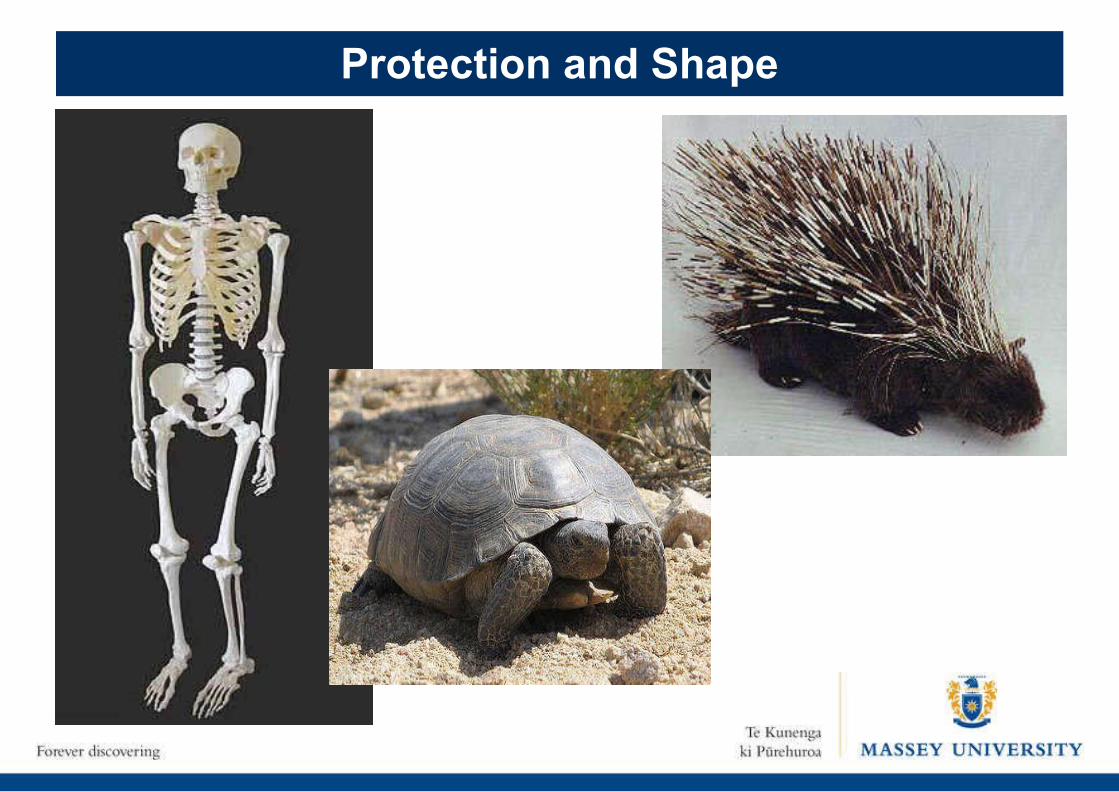
Protection and Shape
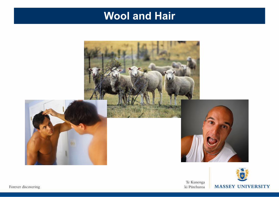
Wool and Hair
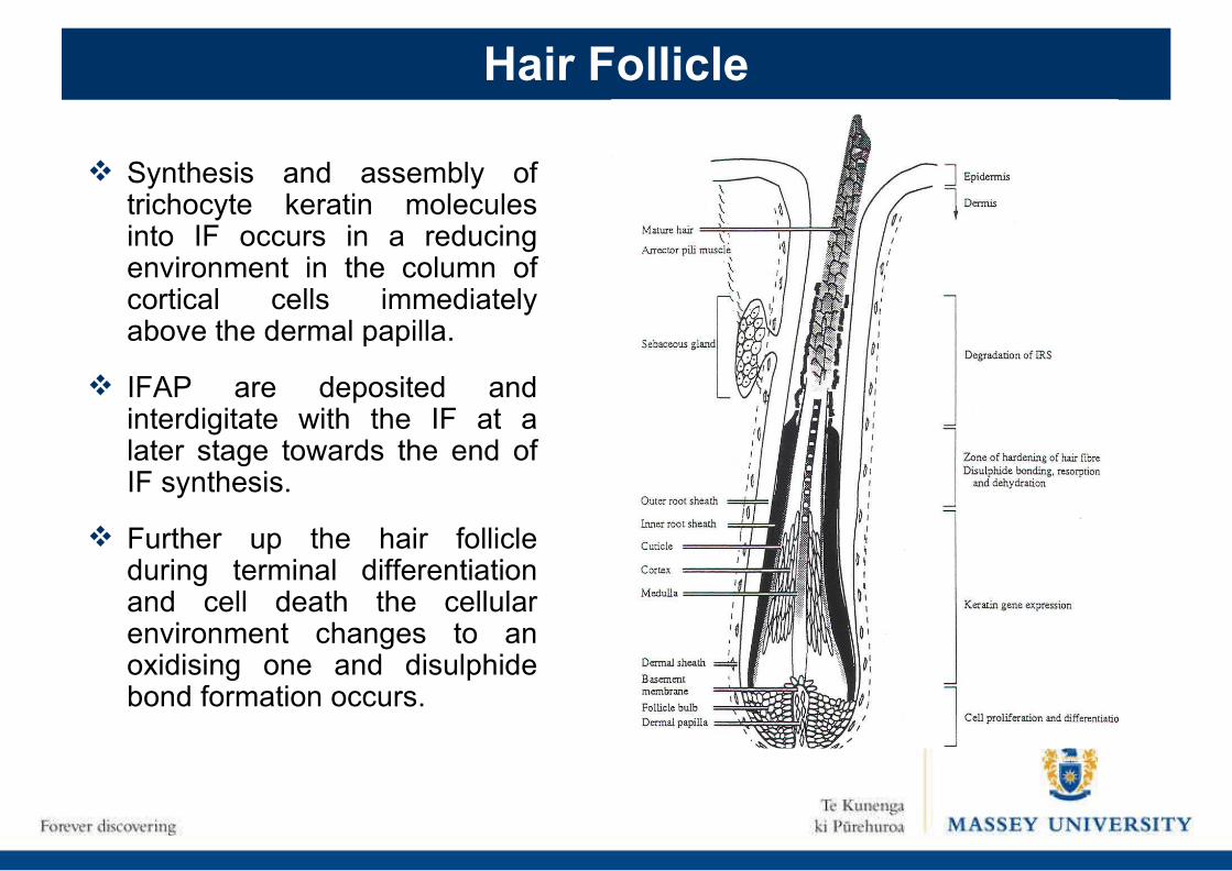
Synthesis and assembly of trichocyte keratin molecules into IF occurs in a reducing environment in the column of cortical cells immediately above the dermal papilla.
IFAP are deposited and interdigitate with the IF at a later stage towards the end of IF synthesis.
Further up the hair follicle during terminal differentiation and cell death the cellular environment changes to an oxidising one and disulphide bond formation occurs.
Hair Follicle
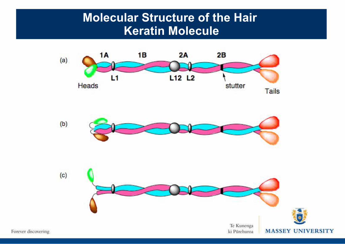
Molecular Structure of the Hair Keratin Molecule
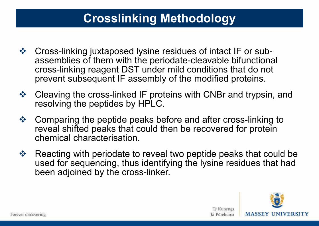
Crosslinking Methodology
Cross-linking juxtaposed lysine residues of intact IF or sub-assemblies of them with the periodate-cleavable bifunctional cross-linking reagent DST under mild conditions that do not prevent subsequent IF assembly of the modified proteins.
Cleaving the cross-linked IF proteins with CNBr and trypsin, and resolving the peptides by HPLC.
Comparing the peptide peaks before and after cross-linking to reveal shifted peaks that could then be recovered for protein chemical characterisation.
Reacting with periodate to reveal two peptide peaks that could be used for sequencing, thus identifying the lysine residues that had been adjoined by the cross-linker.
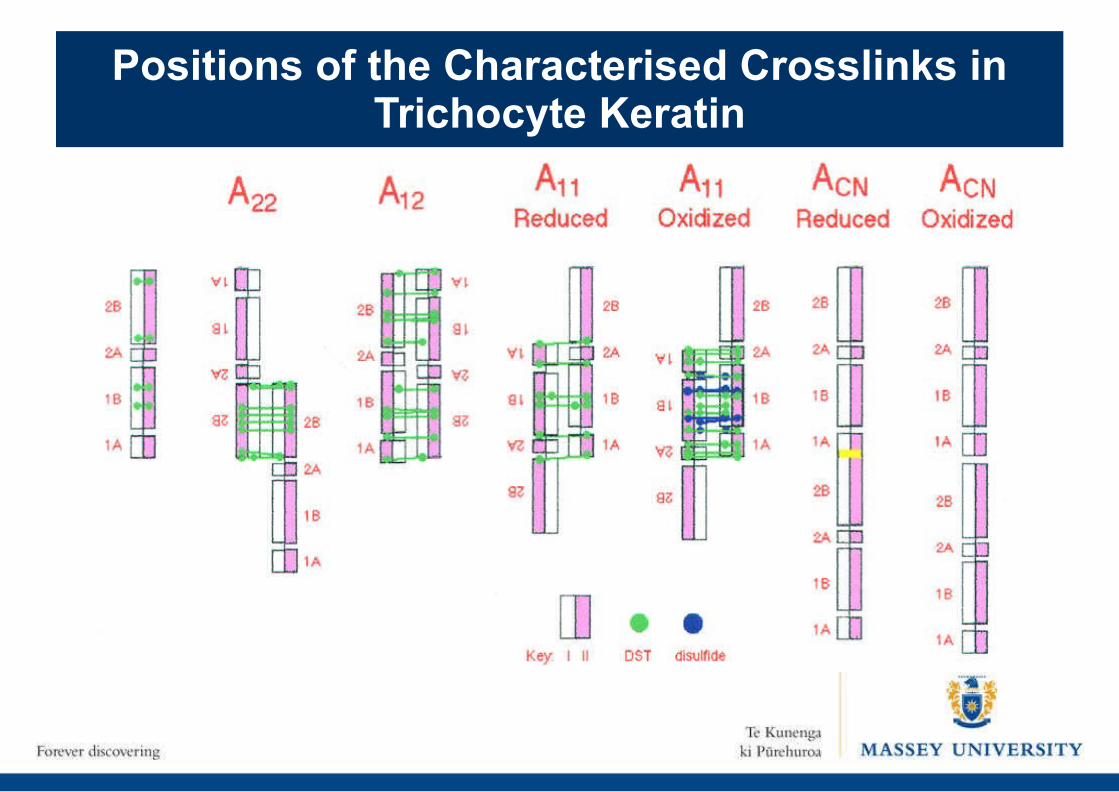
Positions of the Characterised Crosslinks in Trichocyte Keratin
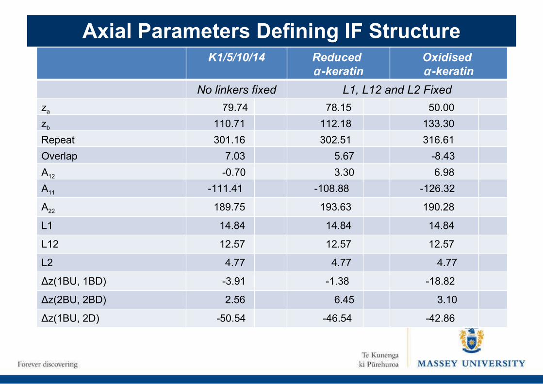
Axial Parameters Defining IF StructureK1/5/10/14 Reduced
α-keratinOxidised α-keratin
No linkers fixed L1, L12 and L2 Fixed
za 79.74 78.15 50.00
zb 110.71 112.18 133.30
Repeat 301.16 302.51 316.61
Overlap 7.03
5.67 -8.43
A12 -0.70 3.30 6.98
A11 -111.41 -108.88 -126.32
A22 189.75 193.63
190.28
L1 14.84 14.84 14.84
L12 12.57 12.57
12.57
L2 4.77 4.77 4.77
Δz(1BU, 1BD) -3.91 -1.38 -18.82
Δz(2BU, 2BD) 2.56 6.45 3.10
Δz(1BU, 2D) -50.54 -46.54 -42.86
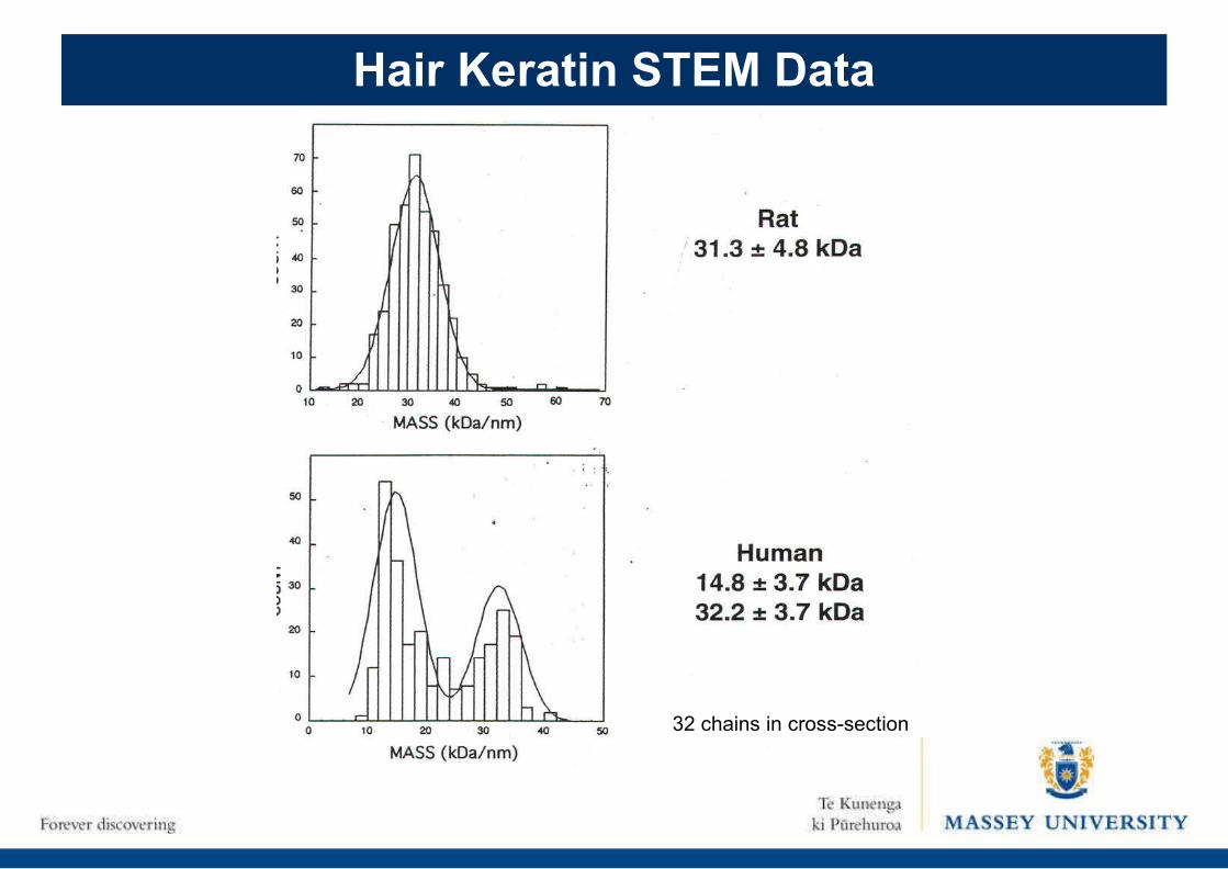
Hair Keratin STEM Data
32 chains in cross-section
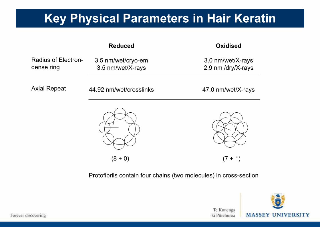
Key Physical Parameters in Hair Keratin
Reduced
3.5 nm/wet/cryo-em3.5 nm/wet/X-rays
44.92 nm/wet/crosslinks
Oxidised
3.0 nm/wet/X-rays2.9 nm /dry/X-rays
47.0 nm/wet/X-rays
Radius of Electron-dense ring
Axial Repeat
r
(8 + 0)
r
(7 + 1)
Protofibrils contain four chains (two molecules) in cross-section
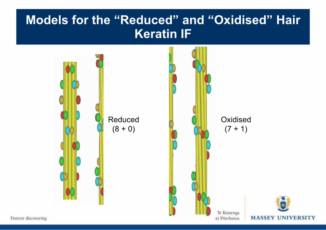
Models for the “Reduced” and “Oxidised” Hair Keratin IF
Reduced(8 + 0)
Oxidised(7 + 1)
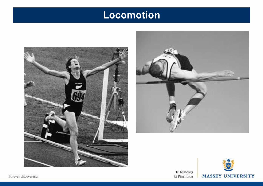
Locomotion
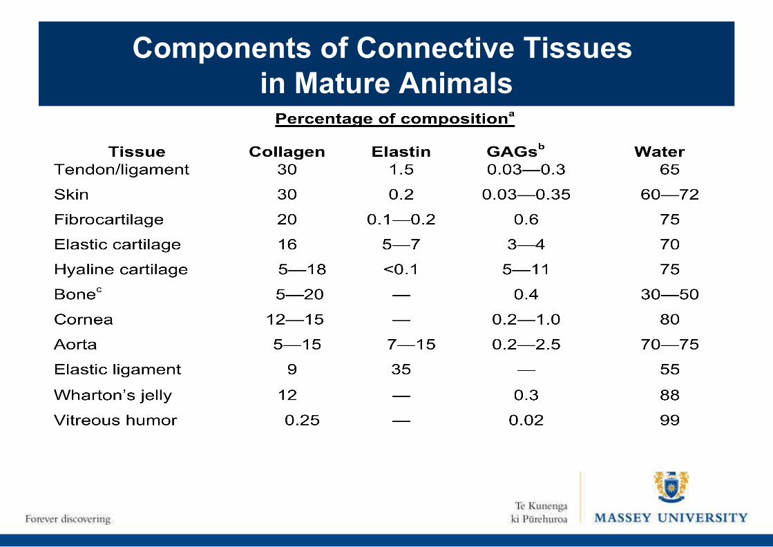
Components of Connective Tissues in Mature Animals
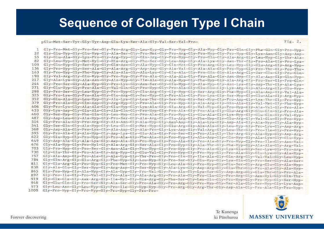
Sequence of Collagen Type I Chain
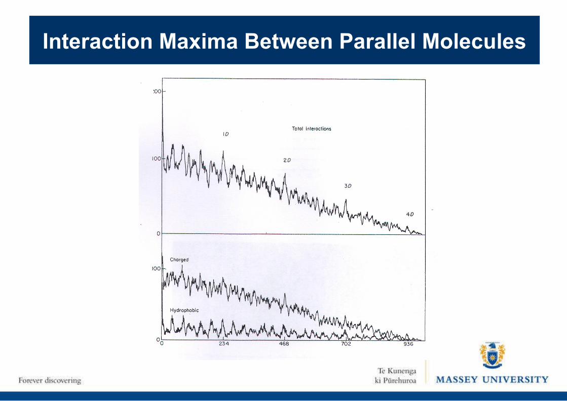
Interaction Maxima Between Parallel Molecules
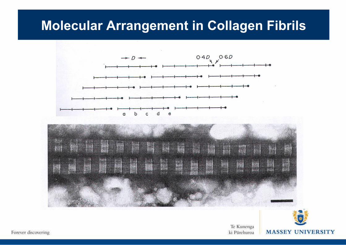
Molecular Arrangement in Collagen Fibrils
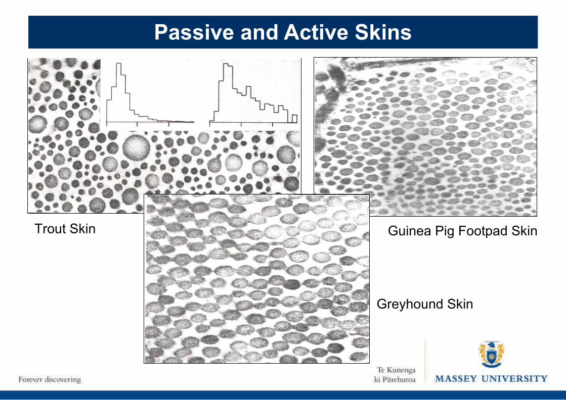
Passive and Active Skins
Greyhound Skin
Guinea Pig Footpad SkinTrout Skin
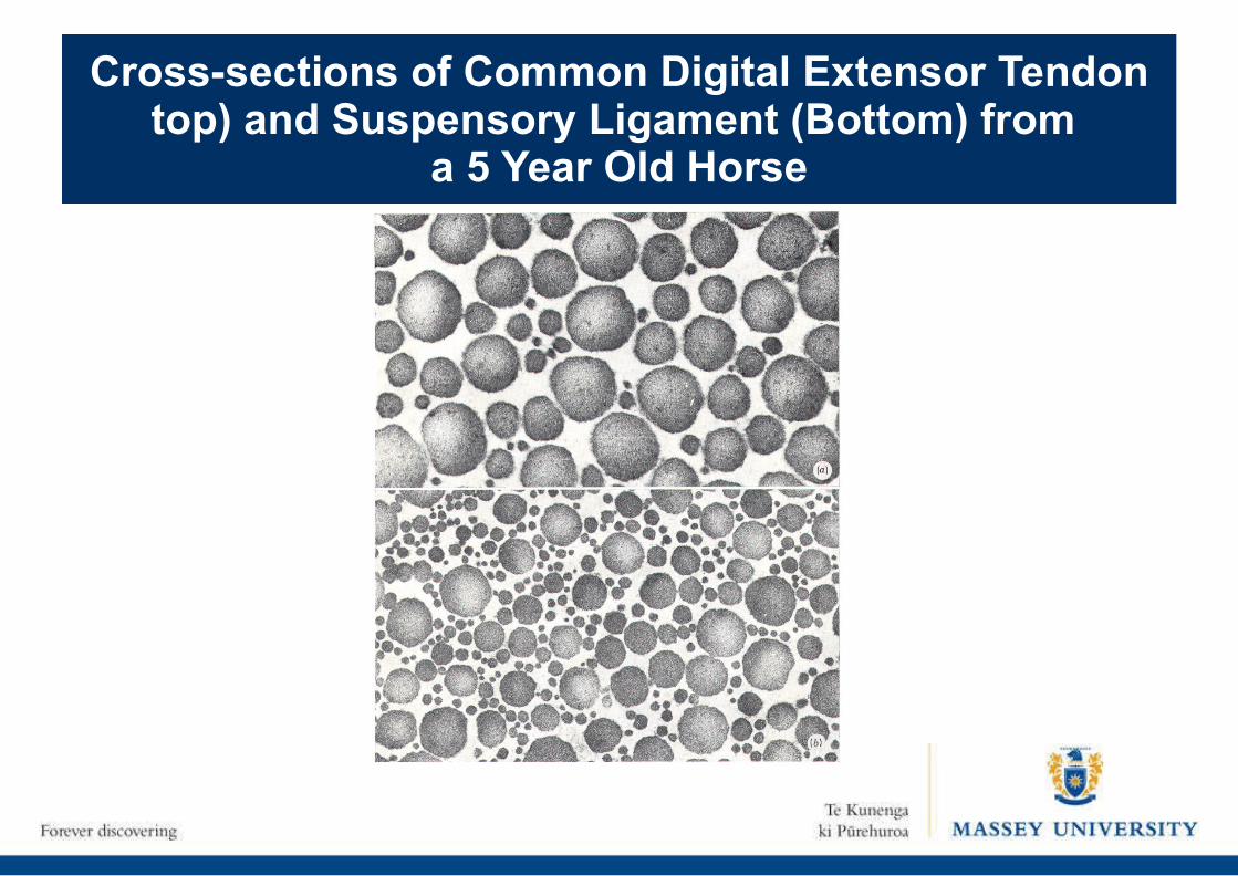
Cross-sections of Common Digital Extensor Tendon top) and Suspensory Ligament (Bottom) from
a 5 Year Old Horse
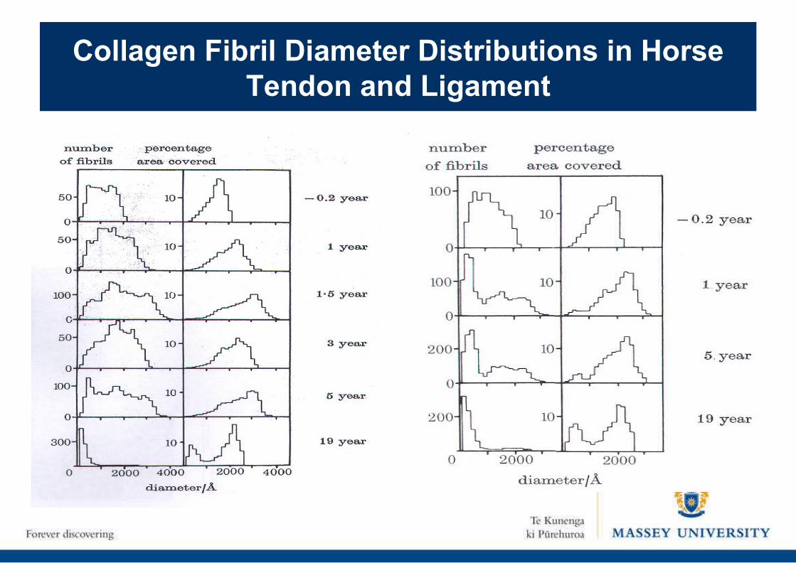
Collagen Fibril Diameter Distributions in Horse Tendon and Ligament
X-ray Picture of Relaxed and Contracting Muscle
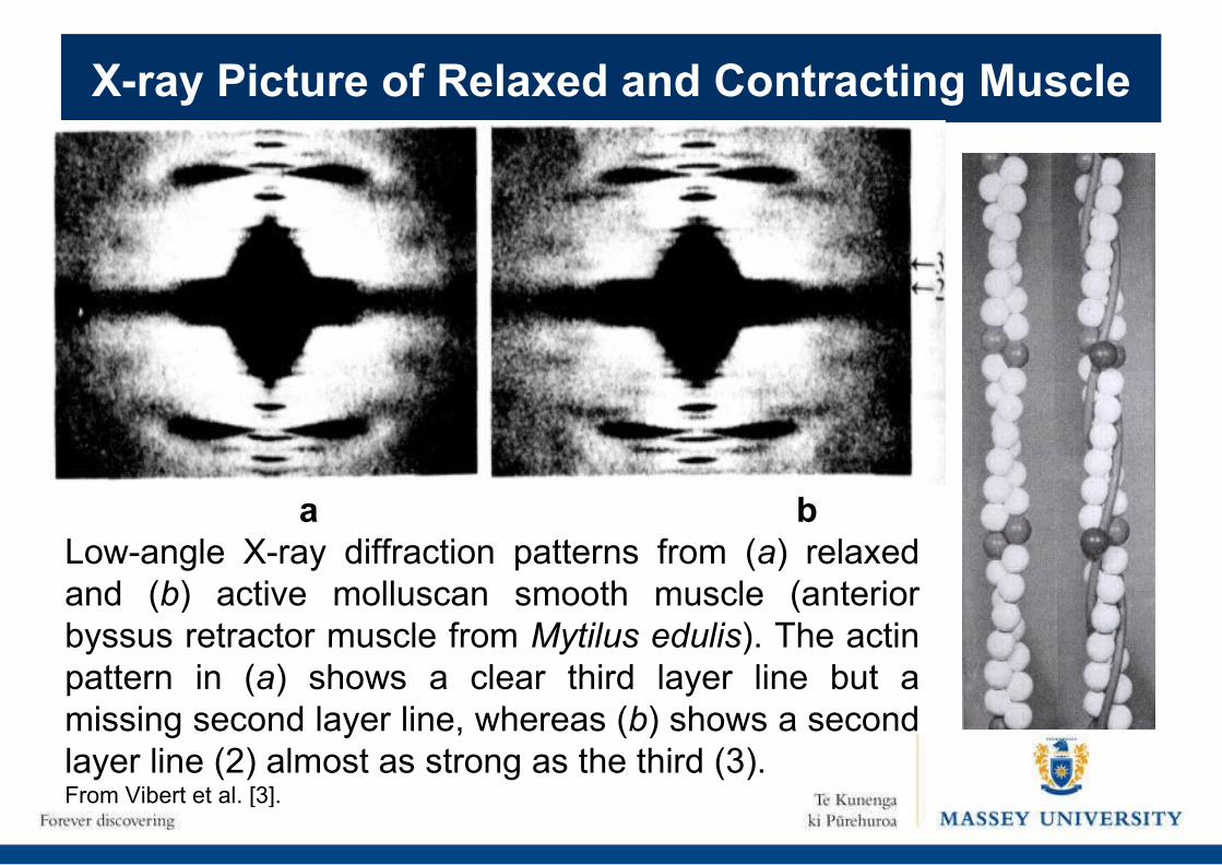
a bLow-angle X-ray diffraction patterns from (a) relaxed and (b) active molluscan smooth muscle (anterior byssus retractor muscle from Mytilus edulis). The actin pattern in (a) shows a clear third layer line but a missing second layer line, whereas (b) shows a second layer line (2) almost as strong as the third (3). From Vibert et al. [3].
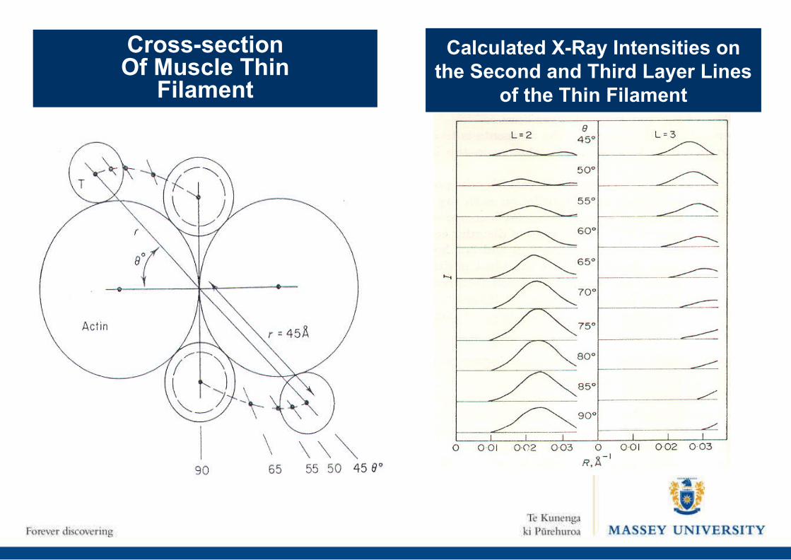
Cross-sectionOf Muscle Thin
Filament
Calculated X-Ray Intensities on the Second and Third Layer Lines
of the Thin Filament
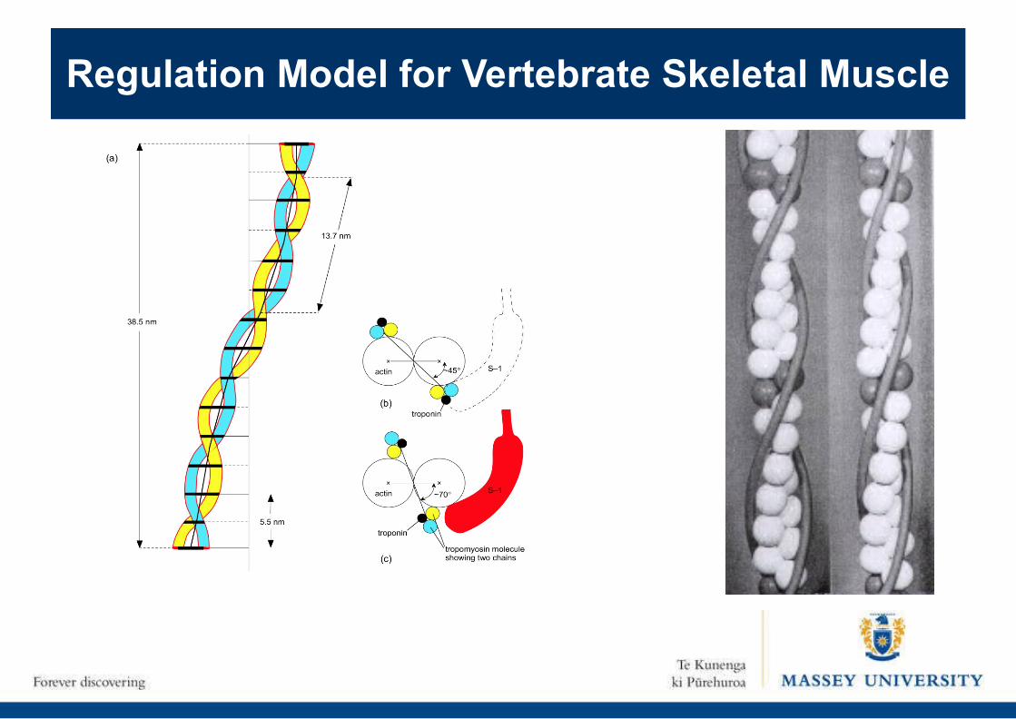
Regulation Model for Vertebrate Skeletal Muscle
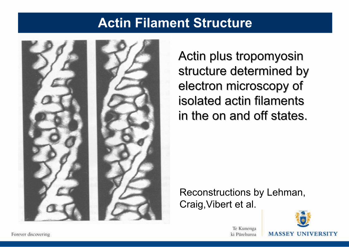
Actin plus tropomyosin Actin plus tropomyosin structure determined by structure determined by electron microscopy of electron microscopy of isolated actin filaments isolated actin filaments in the on and off states.in the on and off states.
Reconstructions by Lehman, Craig,Vibert et al.
Actin Filament Structure
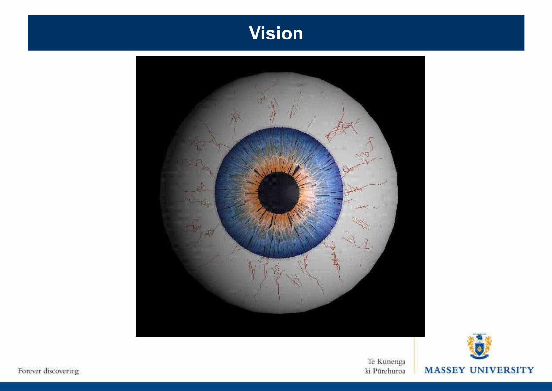
Vision
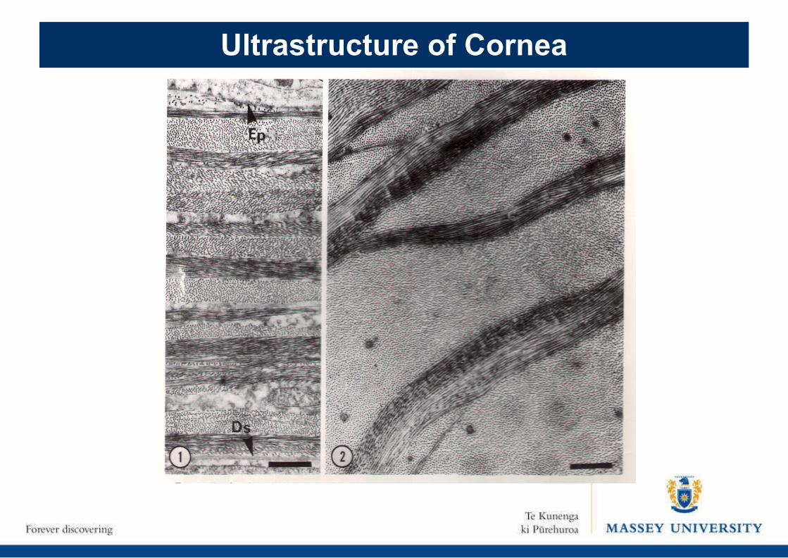
Ultrastructure of Cornea
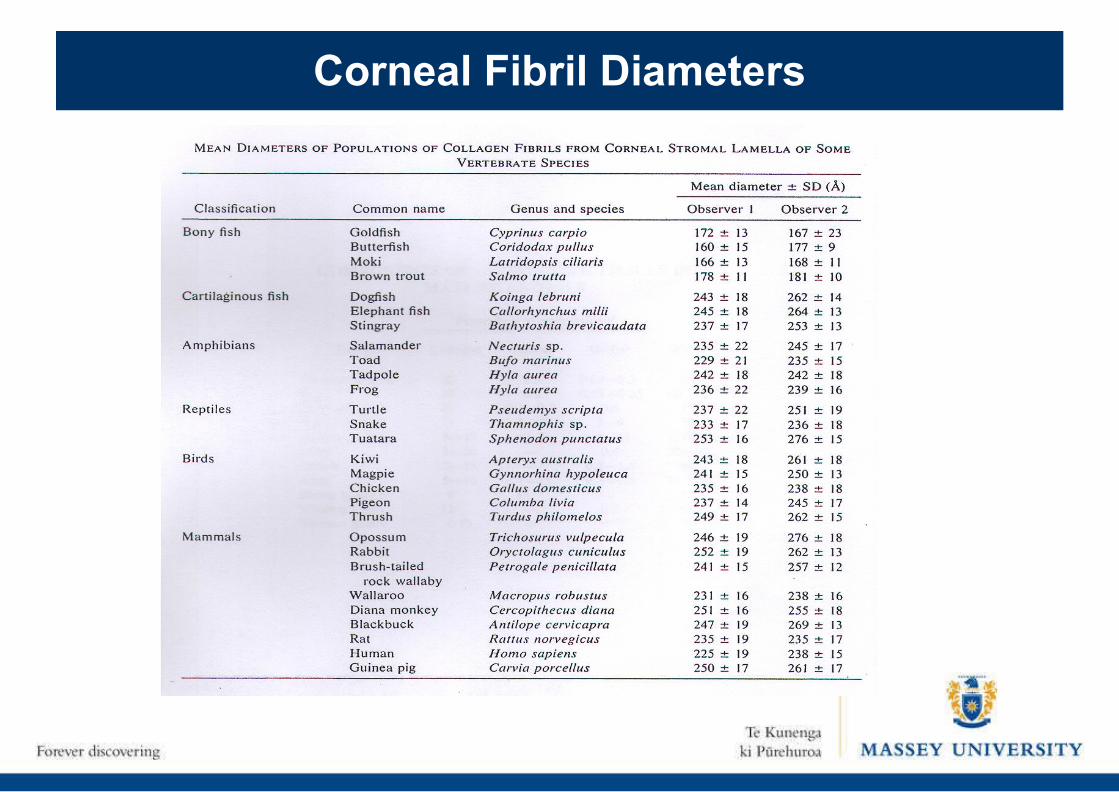
Corneal Fibril Diameters
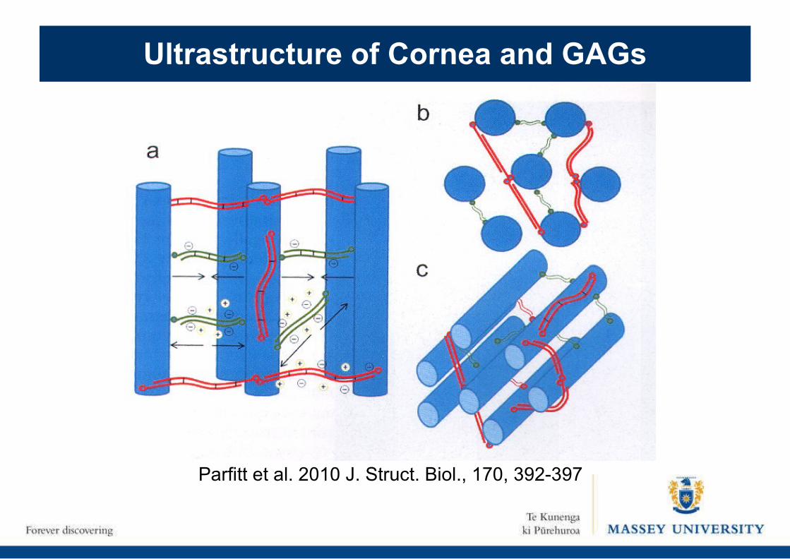
Parfitt et al. 2010 J. Struct. Biol., 170, 392-397
Ultrastructure of Cornea and GAGs
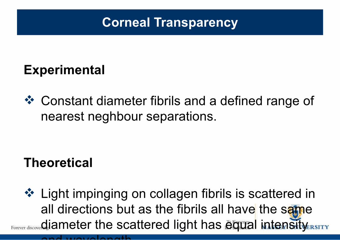
Corneal Transparency
Experimental
Constant diameter fibrils and a defined range of nearest neghbour separations.
Theoretical
Light impinging on collagen fibrils is scattered in all directions but as the fibrils all have the same diameter the scattered light has equal intensity and wavelength.
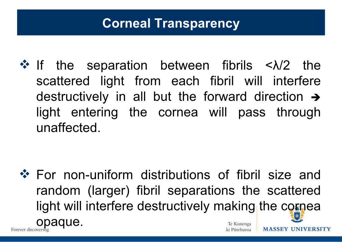
If the separation between fibrils <λ/2 the scattered light from each fibril will interfere destructively in all but the forward direction
light entering the cornea will pass through unaffected.
For non-uniform distributions of fibril size and random (larger) fibril separations the scattered light will interfere destructively making the cornea opaque.
Corneal Transparency
Unraveling the Working of the Animal Body: A Biophysical Approach
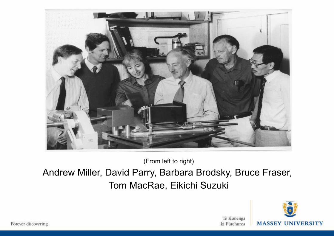
(From left to right)
Andrew Miller, David Parry, Barbara Brodsky, Bruce Fraser, Tom MacRae, Eikichi Suzuki

David HulmesAlan Craig
John Squire
Carolyn Cohen
Tammy LynchPeter Steinert