a 360-degree overview of paediatric nafld: recent insights filevalerio nobili 1,⇑, gianluca...
TRANSCRIPT
Review
A 360-de gree overview of paedia tric NAFLD: Recent insight s
Valerio Nobili 1,⇑, Gianluca Svegliati-Baroni 2, Anna Alisi 1, Luca Miele 3, Luca Valenti 4, Pietro Vajro 5
1Hepato-me tabolic Disease Unit and Liver Research Unit, ‘‘Bambino Gesù’’ Children’s Hospital, IRCCS, P.le S. Onofrio 4, 00165 Rome, Italy;2Departme nt of Gastroen terology, Polytechnic University of Marche , Ancona, Italy; 3Departme nt of Medical Sciences, Policlinic o Gemelli Hospital,Catholic University of Rome, Italy; 4Departme nt of Pathoph ysiology and Transplan tation, Section of Internal Medicin e, Università degli Study di
Milano, Fondazion e IRCCS Cà Granda Ospedale Maggiore Policlinico, Milan, Italy; 5Departme nt of Medicin e and Surgery, University of Salerno, Italy
Summary
Non-alcoh olic fatty liver disease (NAFLD) is a multi- faceted disor- der, which ranges from simple steatosis to non-alc oholic steato- hepatitis (NASH) with/with out fibrosis. The effects of specificrisk factors, such as obesity and sedentary lifestyle , on predispos- ing genetic settings eventuall y lead to the developmen t of NAFLD in childre n. The complex interplay between genes and environ- ment in NAFLD pathogenesis is sustained by multiple mecha- nisms that involve liver crosstalk with other organs and tissues,especially gut and adipose tissue. Unfortunat ely, natural history of paediatric NAFLD is lacking, and the etiopathog enesis is still in the process of being defined. Potential early predic tors and suitable non-invasi ve diagnostic tools can be discovered based on the pathogenet ic mechanisms and histological patterns. This will also help design novel treatment s and a comprehen sive and successful managemen t strategy for patients.
In this review, we discuss the recent advances made in genet- ics, etiopathog enesis, diagnosis, and therapeutic managemen t of NAFLD, focusing especially on the obesity-rela ted steatoti c liver condition.� 2013 Europea n Associat ion for the Study of the Liver. Published by Elsevier B.V. All rights reserved.
Introducti on
Non-alcoh olic fatty liver disease (NAFLD) is a major health prob- lem that integrates several liver conditions ranging from simple fatty liver to non-alcoh olic steatohepatiti s (NASH), which is sometimes associated with fibrosis that may evolve into cirrhosis and result into hepatoc ellular carcinoma [1,2]. Furthermore, as underlined in a recent meta-analy sis, NAFLD is also associat ed with increased overall morbidity and mortality for cardiovascu lar disease [3–5].
Journal of Hepatology 20
Keywords: NAFLD; NASH; Fibrosis; Children.Received 9 July 2012; received in revised form 14 November 2012; accepted 4December 2012 ⇑ Corresponding author. Tel.: +39 06 68 59 22 43; fax: +39 06 68 59 21 92.E-mail address: nobili66@yahoo .it (V. Nobili).
Key Points
• Analysis of genetic background may help paediatricsidentify children susceptible to NAFLD
• Novel findings about pathogenetic mechanisms leading to paediatric NAFLD may improve therapeuticperformance
• Adequate diagnostic and therapeutic managementis crucial to prevent and counteract progression ofpaediatric NAFLD
As the prevalenc e of obesity in childre n increases , so does the prevalence of paediatric NAFLD [4]. This rise is worrisome because of its close association with the develop ment of meta- bolic syndrome (MetS) [6–8].
Although etiopathog enesis of paedia tric NAFLD remains unknown, it is conceivabl e that, such as in adults, a network of interactions among multiple factors is involved in both the devel- opment and progres sion of the disease [9,10]. However, as obes- ity-related NAFLD in children is rarely influenced by secondar ycauses (e.g., severe weight loss, drug, and alcohol consump tion),it may be an excellent model to elucidate the factual origins of primary NAFLD, including the contribution of susceptib ility genes and environmen t [11,12].
NAFLD is suspected mainly by the analysis of anthropome tri- cal and bioche mical parameters and/or ultrasoun d liver bright- ness [13]. Histological analysis of liver biopsy remains the masterful method to differentiat e simple steatosi s from NASH and to perform staging and grading of the disease in children [14]. However , recentl y, advances have been made in the fieldof non-invasi ve evaluation of paediatri c NAFLD [15].
There are no current specific therapeut ic indicatio ns for the treatment of paediatric NAFLD. However, as overwei ght/obesity is often coupled with NAFLD, weight loss by diet and exercise programs is widely accepted as the first line of interventio n in children [16]. Poor adheren ce to lifestyle modifications often fails to halt or revert the occurrence of liver damage during pathogen- esis of paediatri c NASH. Therefore, at this time, developing possi- ble multi-target ed therapies to halt disease progres sion, restoring
13 vol. 58 j 1218–1229

JOURNAL OF HEPATOLOGY
liver cell homeostasi s and repairing the damage are all funda- mental objectives in the treatment. In fact, during the last 5 years,a series of fairly novel pharmacol ogical treatments have been tested, and a wide range of newer drugs are now being develop ed [17].Here we provide an extensive overview of NAFLD in children while discuss ing the most up-to-da te literature in the field. Par- ticularly, we focus on the many interesting breakthro ughs in genetics, etiopathogen esis, diagnosis, and therapeuti c manage- ment of paedia tric obesity-rela ted liver disease.
Epidemiol ogy
The epidemics of overweight and obesity in paediatric population has reached worldwid e proportion over the last two decades [4,13]. As a consequenc e, NAFLD has now become the most com- mon cause of chronic liver disease in children and adolesce nts in the USA [18], and most probably, in the rest of the industriali zed countries. The true prevalenc e of NAFLD, however, remains lar- gely elusive. In fact, extrapola tion of correct epidemiol ogical data is hampered by the large variability of available non-invasi ve and invasive diagnost ic tests. None of these tests are exempt from criticisms in terms of misdiagno sis and/or invasiven ess and costs.
Serum alanine aminotra nsferase (ALT) activity is a widely accessibl e and cheap test for the screening and initial evaluation of NAFLD. The sensitivity of this biochemical marker, however,remains low because some adult and paediatric patien ts with biopsy-pro ven NAFLD may present ALT values in the normal range [19]. In an Italian study, 72 obese children presented with ultrasonogra phic fatty liver (50% of cases) and/or hypertransam- inasemia (25% of cases) [20].
To add to the difficulties, histology – the gold standard for the diagnosis of NASH – may be subject to the higher probabili ty of staging sampling error [14]. Furtherm ore, epidemiologic al data of a paedia tric population may be influenced by a series of crossed risk factors such as peri-pube rtal age, male gender (forNAFLD, but not for NASH), hispanic ethnicity, non-blac k race and individu al genetic predisp osition. NAFLD and/or its progres- sion to severe disease have, in fact, been linked to several inher- ited variants that will be discussed extensi vely in the next paragraph on genetic risk factors [21]. Familial clustering of obes- ity, insulin resistance (IR), NAFLD or type 2 diabetes are frequent and should raise suspicion of NAFLD in children from such fami- lies [22,23].
Obesity and MetS are the major risk factors for paedia tric NAFLD. NAFLD prevalence is higher in overweight (gender and age specific BMI >85th percentile ) or obese (>95th percentile )children as compared with normal weight age matched pairs.Today, it seems clear that waist circumferenc e (i.e., abdominal fat or central obesity) plays a pivotal role and correlates with NAFLD diagnosis more than BMI alone [24].
Population -based studies conducte d with ALT or ultrasono g- raphy in several countries have shown that paediatric NAFLD prevalenc e remains in a range of 2.6–7.1% of children [10]. An autoptic study in Californ ia, conducte d in children deceased for accidents, showed a prevalence of histological NAFLD ranging from 0.7% in 2–4 year old to 17.3% in 15–19 year old subjects [25]. In cohorts of children of various national ities selected for overweigh t or obesity, the prevalenc e of elevated ALT is higher and ranges from 8 to 42%, whereas the prevalence of bright liver
Journal of Hepato logy 2013
ranges from 1.7 to 77% [26]. In an autoptic study by Schwimmer et al ., prevalenc e of histologi cal NAFLD in obese individu als was 38% [25].
Genetic risk factors
In recent years, an exciting step towards the understand ing of the pathogenesi s and risk factors in the develop ment and progression of paediatric NAFLD has been taken as a result of genetic studies.It is indeed well known that NAFLD has a major genetic compo- nent [23]. Because of the lower numbers of confoun ding factors (e.g., the duratio n of disease, presence of obesity , lifestyle habits,co-morbid ities, and drugs) and the more importan t role of inher- ited factors in early-onset disease, this is especially true for chil- dren [27].
The major advance has come from the finding, first obtained by genome-wid e association studies (GWAS) conducte d in the general adult population [28,29], that genetic variants of the pat- atin-like phospholip ase domain-co ntaining protein-3 (PNPLA3)and, in particular, the common rs738409 C>G single nucleoti de polymorphi sm (SNP) encoding the I148M variant, are not only associated with hepatic fat content and increased serum liver enzymes but also increase the risk of NASH and fibrosis progres- sion [30–32]. The I148M PNPLA3 variant influenced liver triglyc- eride content without apparent ly affecting body mass, serum lipid levels and systemic IR [31,33]. PNPLA3 is regulated by the lipogenic program [34] and is highly expressed in the liver and adipose tissue at the level of the endoplasm ic reticulum and the surface of lipid droplets, where it seems to be involved in the metabol ism of triglycerid es [35]. Although the mechanism and physiologic al substrates remain unclear, the common I148M variant disrupts the activity of the enzyme, thereby alter- ing lipid catabolism, and it might also acquire new and still unknown function s [35]. Indeed, the association of I148M variant with progres sive liver disease and hepatocell ular carcinoma is independen t of the predispos ition to increased steatosis, thus suggesting that it influences the regulation of pro-inflammatory lipid mediators [27,30,32].
The associat ion between I148M variant and both liver enzymes and steatosis was soon confirmed in obese children of different ethnicities [36–39] and in one family study in Italian trios [30], indicatin g that it exerts its effect early in life and that the magnitud e of the associat ion between I148M PNPLA3 variantand serum levels of liver enzymes was related to the size of abdominal fat [40] and to the high dietary carbohydra te and sugar consumption [41]. In addition, an interac tion was reported between dietary fatty acids compositio n, PNPLA3 genotype and hepatic fat accumu lation. Indeed, the omega-6/om ega-3 poly- insaturated fatty acids ratio was correlated with hepati c fat frac- tion and ALT levels only in children homozyg ous for 148M PNPLA3 variant and at risk of steatosis [42].
Furthermo re, PNPLA3 genotype influenced the histological severity of NASH alteration s and fibrosis in obese paediatri cpatients who underwent biopsy because of persisten tly altered liver enzymes. Interestingly, the association with fibrosis was stronger in children than in adults, in that each 148M allele increased the risk of fibrosis by almost twofold [27].
A more recent GWAS, conducte d in a larger population , was able to identify a wider set of genetic variants influencing steato- sis besides I148M of PNPLA3 [43]. Of these variants, rs2854 116
vol. 58 j 1218–1229 1219

Review
SNP of glucokin ase regulator (GCKR), involved in the regulation of uptake of monosacc harides and lipogenesis , was confirmed to predispose to fatty liver and dyslipidemia in obese childre n and adolescents independ ent of PNPLA3 [44], although the effect on histological progression of liver disease remains unknown, espe- cially in view of the ameliora ting effect on IR.Besides, additiona l SNPs of genes implicated in NASH patho- genesis have been shown to influence liver damage and fibrosisprogression in candidate gene case-contr ol studies including pae- diatric patients. These include genetic variants regulating insulin receptor activity, namely the ectoenzyme nucleotide pyrophos- phate phosphod iesterase 1 (ENPP1) Lys121Gln and the insulin receptor substrate-1 (IRS-1) Gly972Ar g function al SNPs [45], thus underscoring the causal role of IR in the progress ion of liver dam- age in NAFLD. The mangane se superoxide dismuta se (SOD2) C47T rs4880 SNP, regulating SOD2 mitochond rial import and antioxi- dant activity [46], and the Kruppel-l ike factor 6 (KLF6) IVS1- 27G>A SNP, regulating alternati ve splicing isoforms of the tran- scription factor KLF6, involved in the regulation of metabolism in hepatoc ytes and fibrogenesis in hepatic stellate cells, seem to be involved as well [47]. In contrast , variants in the hemochro ma- tosis gene (HFE), regulating iron metabolism and in the apolipo- protein-C3 (APOC3), regulating very low density lipoprotein metabolism, were not confirmed to influence susceptibil ity stea- tosis and NASH [48,49].
Finally, there is a growing awarene ss that the expressi on of some genetic variants may be age-depe ndent, i.e., the phenotype may be more (or less) marked or involve differen t traits during the developmen tal age. For example, a common variant (rs13412852) influencing the expression of lipin-1 (LPIN1),another lipid phospha tase involved in adipoge nesis and regulat- ing the flux of free fatty acids between the adipose tissue and the liver, whose expressi on is deregulated during steatosis [50],was associated with lipid levels, NASH severity, and hepatic fibro-sis in children with NAFLD, whereas it influenced body mass in adults of the same ethnici ty [51]. In Table 1, we have summari zed genetic polymo rphisms influencing the suscepti bility to paediat- ric NASH.
Table 1. Genetic variants influencing the susceptibility to NAFLD and NASH during t
Gene Function SNP
PNPLA3, patatin-like phospholipase domain containing 3
Lipid remodelling, lipogenesis
rs738409 C
GCKR, glucokinase regulatory protein Glucose uptake, lipogenesis
rs780094 C
SOD2, superoxide dismutase 2, mitochondrial
Anti-oxidant response rs4880 C>
ENPP1, ectonucleotide pyrophosphatase/phosphodiesterase 1
Insulin signalling rs1044498
IRS1, insulin receptor substrate 1 Insulin signalling rs1801278KLF6, Kruppel-like factor 6 Fibrogenesis, glucose
metabolism and lipogenesisrs3750861
LPIN1, lipin-1 Lipogenesis, adipogenesis rs1341285
SNP, single nucleotide polymorphism; MAF, minor allele frequency, in European healthThe number of arrows is related to the strength of both the genetic association and the
1220 Journal of Hepatology 2013
Metabolic risk factors and organ crosstalk for NAFLD development
It is widely accepted that genetic susceptib ility, epigenetic mech- anisms, physica l inactivity , and excess caloric intake by diet influ-ence visceral fat accumulati on inducing an obese and metabolicall y dysfunctional phenotyp e [52]. This pattern, charac- terized by abdom inal obesity, IR, impair ed glucose tolerance,dyslipidemia, and hypertension , defines a subject with MetS.NAFLD is generally associat ed with at least one of these MetS fea- tures in adults, and emerging evidence confirms the existen ce of a similar relation ship in children as well [52,53,12]. An Italian study demonstra ted that 65.8% of 120 childre n (age range: 3–18 years) with biopsy-p roven NAFLD presented with MetS, which was also associated with fibrosis grade [54]. Approx imately 26%of 254 childre n adolesce nts (age range: 6–17 years) enrolled in the Non-alc oholic Steatohepa titis Clinical Research Network (NASH CRN) met specified criteria for MetS diagnosis [55]. These data, as well as other contribut ions [6–8], highlight the potential role of NAFLD as co-factor of other obesity-rela ted co-morb idities in a paediatric population.
In the last decade, the concept that increased consumptio n of obesogenic food may have a role in the pathogenesi s of NAFLD has spread. In fact, apart from the impact of the genetic back- ground, hypercalori c diets (particularly those enriched in fat and fructose/ sucrose) may act by favouring the occurren ce of sys- temic IR and, in turn, a danger ous hepatic free fatty acid (FFA)accumulati on or causing visceral fat deposition and consequen thepatic IR, accountable for developmen t of fatty liver [56,57].According to the ‘multiple hits’ hypothesis, IR and FFA accumula- tion may predisp ose the fatty liver to secondary hits, including the imbalanc e of product ion/release of hormones derived from adipose tissue (adipocytokines), oxidative stress, activation of specific nuclear receptors and fibrogenesis [58,59]. In order to counteract the IR, the number of pancreatic b cells increases,resulting in a compens atory hypersecr etion of insulin. This over- flow of circulati ng insulin accelera tes liver fat storage leading to NAFLD.
he developmental age.
Protein variant
Activity MAF Effect on steatosis
Effect on
NASH>G Ile148Met Increased
lipogenesis ?0.19 ↑↑ ↑↑
>T Pro446Leu Loss of regulatory function
0.45 ↑ ?
T Ala16Val Loss of mitochondrial import
0.49 none ↑
A>C Lys121Gln Loss of inhibitory function
0.29 none ↑
G>C Gly972Arg Loss of function 0.05 none ↑ G>A IVS1-27G>A Altered splicing 0.06 ? ↑
2 C>T Non coding SNP
Unknown 0.35 ↑ ↑
fibrosing
y subjects. available evidence.
vol. 58 j 1218–1229
PAMPs
DAMPs
Proteins
Intestinal epithelium
Carbohydrates
Lipids
Bacteria
Diet
Macrophages
Adipocytokines
Adipose tissue
FFAsLiver
Hepatocytes
HSCs
Kupffer cells
Insulin
Pancreas
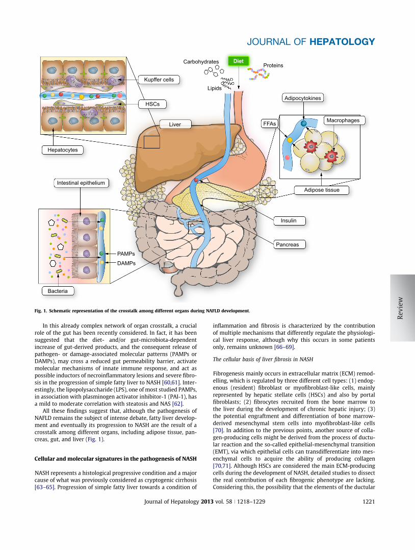
Fig. 1. Schematic represen tation of the crosstalk amon g differen t organs during NAFLD development .
JOURNAL OF HEPATOLOGY
In this already complex network of organ crosstalk, a crucial role of the gut has been recently considered . In fact, it has been suggested that the diet- and/or gut-mic robiota-depe ndent increase of gut-deriv ed products , and the consequent release of pathogen- or damage-assoc iated molecular patterns (PAMPs or DAMPs), may cross a reduced gut permeability barrier, activate molecula r mechanisms of innate immune response, and act as possible inductor s of necroinflammatory lesions and severe fibro-sis in the progression of simple fatty liver to NASH [60,61]. Inter- estingly, the lipopolys accharide (LPS), one of most studied PAMPs,in association with plasminog en activator inhibito r-1 (PAI-1), has a mild to moderat e correlat ion with steatosis and NAS [62].
All these findings suggest that, although the pathogenesi s of NAFLD remains the subject of intense debate, fatty liver develop- ment and eventuall y its progression to NASH are the result of acrosstalk among differen t organs, includin g adipose tissue, pan- creas, gut, and liver (Fig. 1).
Cellular and molecula r signatur es in the pathogene sis of NASH
NASH represents a histological progressive condit ion and a major cause of what was previously considered as cryptogeni c cirrhos is [63–65]. Progression of simple fatty liver towards a condition of
Journal of Hepato logy 2013
inflammation and fibrosis is characte rized by the contribution of multiple mechanisms that differently regulate the physiologi- cal liver response, although why this occurs in some patients only, remain s unknown [66–69].
The cellular basis of liver fibrosis in NASH
Fibrogenesi s mainly occurs in extracellula r matrix (ECM) remod- elling, which is regulate d by three differen t cell types: (1) endog- enous (resident) fibroblast or myofibroblast-like cells, mainly represente d by hepatic stellate cells (HSCs) and also by portal fibroblasts; (2) fibrocytes recruite d from the bone marrow to the liver during the developmen t of chronic hepatic injury; (3)the potential engraftment and differen tiation of bone marrow- derived mesenc hymal stem cells into myofibroblast-like cells [70]. In addition to the previous points, another source of colla- gen-produci ng cells might be derived from the process of ductu- lar reaction and the so-called epithelial-m esenchymal transition (EMT), via which epithelia l cells can transdiffere ntiate into mes- enchymal cells to acquire the ability of producing collagen [70,71]. Although HSCs are considered the main ECM-produ cing cells during the developmen t of NASH, detailed studies to dissect the real contribution of each fibrogenic phenotype are lacking.Considering this, the possibilit y that the elements of the ductular
vol. 58 j 1218–1229 1221

Review
reaction might undergo EMT has been recently questioned [71].However, the element s of the ductular reaction represent the hepatic progenitor cell (HPC) compartment of the liver, and it has been recentl y shown that the HPC compartment , especially in children with NASH, was expanded and independ ently associ- ated with the degree of fibrosis [72].Role of adipocytok ines
Key players in the progression from simple steatosis to NASH are the so-called adipocyt okines includin g adiponec tin, leptin, resi- stin, tumour necrosi s factor (TNF)a and interleu kins (ILs)secreted by adipocytes or the inflammatory cells that infiltratethe adipose tissue in insulin -resistant conditions.
Leptin downregulat es insulin signallin g in the liver and reduces fibrosis in experimen tal models of fibrosis and NASH.Leptin may activate HSC through the NADPH oxydase system [73–77]. In contrast , it has been clearly demonstra ted in a previ- ous study that adiponec tin can counteract the IR effect of leptin,exert anti-inflammatory actions and inhibit HSC prolifera tion,thus interferi ng with fibrogenesis [78].
Cytokines, particular ly TNF a, IL-6, and IL-1, are involved in the recruitment and activation of Kupffer cells and transfor mation of HSC into myofibroblasts. Levels of TNF a and IL-6 are often ele- vated in the liver and blood of patients with NASH, and inhibition of these cytokines has been shown to improve NAFLD in rodents [79,80].
Role of oxidative stress and lipotoxici ty
In a normal liver, antioxidant systems such as superoxid e dismu- tase and catalase efficiently remove reactive oxygen species (ROS) produ ced during cellular metabol ism and maintain normal cell homeostasi s [81]. Oxidati ve stress is increased in patients with NASH [82]. Excess of non-esterified fatty acids (NEFA) sup- ply to the liver increases mitochondria l and peroxiso mal oxida- tion, promote microsomal induction of CYP4A1 and CYP2E1,and cause elevat ed product ion of ROS [83]. Among ROS produc- ing systems, the NADPH oxidase (NADPH) complex plays a major role in hepatic fibrogenesis and collagen production. This system also plays a crucial role in ROS production in both Kupffer cells and HSCs during hepatic fibrogenesis [84,85]. Kupffer cells in the liver mainly produce ROS through the phagoc ytic form of NADPH, which plays an important role in host defence and inflammation [86,87], HSCs express the non-phagoc ytic form that plays an importan t role in regulati ng cell signallin g and can be stimulated by fibrogenic molecule s such as leptin [88,89].
The pathogenesis of NASH also takes into considerati on the concept of lipotoxici ty. The most recent hypothesis for this is that excessive intra-hep atic lipid accumulati on triggers a local necro- inflammatory response, subsequ ently producing free radicals that can result in damage to the cell membran e and DNA [90–92].Thus, the cytotoxic products of lipid peroxidation induce apopto- sis signals in hepatocytes and play a role in liver fibrogenesis by modulating HSCs behavio ur in a paracrine manner [93,94].
Role of nuclear receptors in the pathogenesi s of NASH
Nuclear recept ors (NRs) are ligand-act ivated transcript ion factors that regulate the expression of specific genes controlling a broad range of cellular and metabol ic functions [95,96].
1222 Journal of Hepatology 2013
Among NRs, the peroxisome proliferator- activated receptors (PPARs) have been studied the most because of the presence of pharmacolog ical ligands [96]. PPARs are involved in the activa- tion of HSCs [97,98]. In addition to PPARs, other NRs, such as the farnesoid X recepto r (FXR), xenobiot ic sensors (CAR and PXR), liver X receptor (LXR), and hepatocyte nuclear factor 4(HNF4), have also been implicated in the pathogenesi s of NAFLD,but their role in the develop ment of NASH remains to be estab- lished [99].
Diagnostic tools
Considering the lack of specific symptoms and/or signs of NAFLD,early detection of NAFLD in childre n may be effective to identify subjects with potential silent progress ive fatty liver [5]. In addi- tion, the screening for NAFLD should be recommend ed to over- weight and obese childre n [100–102].
In the first assessmen t of children with suspicion of fatty liver,clinicians should take an accurate clinical history with particular attention to informa tion regarding the nutritional habits and pos- sible drug consumptio n.
Routine laboratory tests
ALT elevation is common in boys and girls with fatty liver [103,104]. In epidemiol ogical studies, elevat ed ALT seems to be associated with MetS in children and adolescents [105,106]. In a tertiary centre setting, NAFLD associat ed hypertran saminas- emia was responsib le for about 20% of all cases [107].
Recently, the American Academy of Paediatrics recommend ed screening with liver function tests (ALT, AST) in children aged 10 years in case of BMI P95th percent ile or between 85th and 94th percentile with risk factors. The biochemistry test panel should also include lipid profiling and fasting glucose determina- tion [14,108 ].
Some considerati ons concerning the significance of liver enzymes are mandatory. Serum aminotra nsferases that are influenced by dietary habits and hyperalime ntation, may reflect,in some case the presence of steatosis [109]. However, as the serum levels of aminotra nsferases may be reduced even in the presence of NASH and fibrosis, liver function test cannot repre- sent the severity of NAFLD [110]. Anothe r major issue with ami- notransferases lies in the variability among centres and the need of ‘biology-base d thresholds’ to increase sensitivity, as recentl yunderlined in the SAFETY study [111]. In this study, the thresh- olds of 25.8 U/L in boys and 22.1 U/L in girls have been pro- posed as a reliable index for suspicion of a chronic liver disease. Obese children who become hepatopat ic should defi-nitely be tested for causes of liver diseases other than NAFLD,particularly for those condition s that are rapidly progres sive if not adequate ly treated (e.g., autoimm une hepatitis and Wilson disease). The so-called NASH trash bin should also be carefully taken into considerati on, especially in early-onset NAFLD among young children [14].
US and radiology
Liver ultrasoun d (US) can detect fatty liver when steatosis involves >30% of hepatocytes [112]. US has several advantag es for use as a screening tool: relative low cost, large diffusion in
vol. 58 j 1218–1229

JOURNAL OF HEPATOLOGY
medical communit y and feasibility. Recently, a large prospective paediatric cohort showed a good correlation between ultrasono- graphic steatosis score and the severity of steatosis on liver biopsy [113]. Moreover, US has been used to assess the outcome of efficacy in paediatric trials with good compliance between the outcomes of children and parents [114].Computed tomograp hy (CT) scan is not recommen dable in paediatric setting because of the unjustified radiation exposur einvolved in the process. In contrast , magneti c resonance imaging (MRI) has been demon strated to have good sensitivity in quanti- fication of fat in the liver [115,116].
Progressi on indexes
The main challenge for paediatric hepatolo gists is the identifica- tion of children with higher probabili ty of progres sion to a more severe form of liver disease.
Ultrasound- guided or assisted needle liver biopsy remains the gold standard method for defining diagnosis, severity, and rate of progression of NASH to its clinical sequelae. Because it is an inva- sive tool for monitori ng therapeuti c responses and disease evolu- tion, researc h on clinical parameters or serum markers that can identify subjects with steatohepatit is and who are prone to fur- ther progression has been undertaken .
Several studies have aimed at identifying clinical parameters that can predict the progress ion of a liver disease. The presenc eof necro-inflammation at liver biopsy is associated with a possi- ble rapid progression of fibrosis [54]. Results of clinical studies have indicated BMI, lipid profile, IR, and fasting glucose as predic- tors of NAFLD in children [14]. Moreover, waist circumferenc ealone seems to be correlated with liver fibrosis in the paediatric setting [117]. In conclusion, simple clinical parameters such as age (expression of length of disease), IR surrogate clinical marker acanthosis nigricans, anthropome trical data (BMI, waist), lipid,and glucose profile can allow a clinician to recognise children with potential severe NAFLD. The Paediatric NAFLD Fibrosis Index (PNFI) algorithm was developed on the basis of waist circumfer- ence, triglycerid es, and age [118].
The need of a simple and reliable tool for the assessment and monitorin g of liver fibrosis has booste d the research of ‘non-inva- sive’ markers. Unfortunatel y, limited sample size, lack of ade- quate informa tion regarding the control groups and quality of liver specime n, and lack of validation in large and independ ent cohorts have togethe r reduced the application of this tool in clin- ical practice [119].
Several molecule s are involved in ECM remodelling during fibrogenesis: the matrix metallopro teinases (MMPs) that partici- pate in ECM degradati on; their specific tissue inhibitors, the tis- sue inhibitor of metalloprote inases-1 (TIMPs), and various cytokine s that stimulate HSC conversio n into myofibroblast-like cells that synthesiz e hyaluro nic acid (HA), which has been found as a reliable marker of fibrosis deposition in children [120].
However , a recent paper suggests that the more reliable serum marker of steatohe patitis is cytokerati n-18 (CK18) frag- ment levels, which are increased in the bloodstre am accordin gto the presence and severity of NASH [121].
Recently, the enhanced liver fibrosis (ELF) panel of serum markers (tissue inhibit or of matrix metallopr oteinase 1, HA, and aminoter minal peptide of pro-coll agen III) was tested in apaediatric cohort to show the capability of predicti on of fibrosis[122].
Journal of Hepato logy 2013
In a recent study, the combin ation of ELF and PNFI showed good accuracy in predictin g fibrosis in childre n with histologi- cally confirmed NAFLD [123].
Transient elastography appears to be a more promising tool for non-invasi ve assessment of fibrosis, and its role in paediatric NAFLD has been recently reviewed elsewhe re, unfortunatel y the lack of capability to discriminate between intermedia te degrees of fibrosis represents the major limitation to avoid liver biopsy for the diagnosis of fibrosis in NASH [124].
Treatments
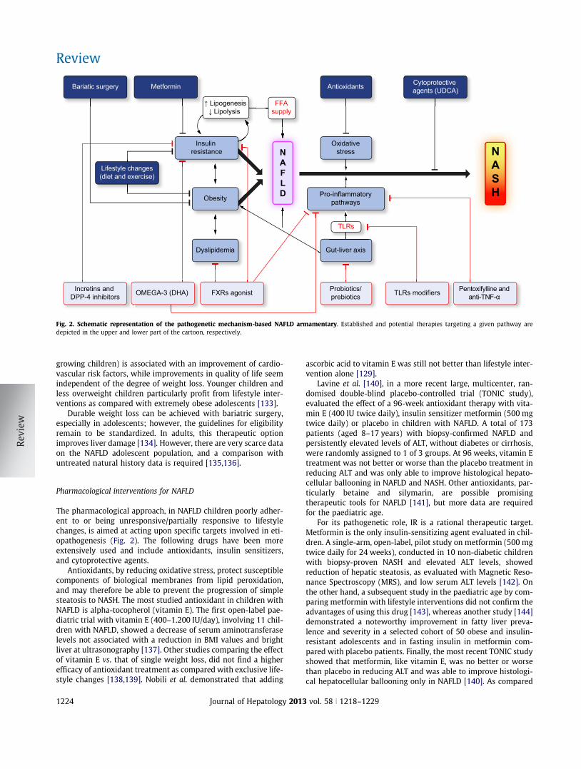
The aim of this section is to evaluate establi shed and other pos- sible novel NAFLD treatment options in the paediatric age group by providing recent evidence from the literature . Fig. 2 illustratesa synop tic summary of most pathogeneti c mechanism-ba sed treatments .
Diet and lifestyle changes
The goal of lifestyle intervent ions is a gradua l and controll ed weight loss achieved by diet and physica l exercise. Unfortunatel y,this aim is difficult to achieve and the results are disappoint ing,with an extremely low percentage of individual s who are able to steadily lose weight and exercise regularly [16]. Weight loss in NAFLD patients on diet improves hepatic insulin sensitivit yby reducing hepatic FFA supply, improves extra- hepatic insulin sensitivity through better glucose utilization and reduces ROS generation and adipose tissue inflammation. Exercise and out- door activities are also promoted because they may improve sub- strate utilization in muscles and contribute to obtaining better insulin sensitivit y, irrespecti ve of weight loss [125].
Weight loss (approximately 5–10% of the basal weight) should be gradua l, since extreme slimming diets may lead to the onset of severe metabol ic disorders and promote liver damage. Indeed,some studies in childre n confirmed an improvem ent in serum aminotran sferase levels and several metabolic parameters including lipids, fasting glucose, insulin, and insulin sensitivit yindices [126–128]. Interestin gly, Nobili et al . showed that arepeated biopsy at 24 months displa yed significant improvemen tof liver histology with reductio n of the grade of steatosis, hepatic lobular inflammation, hepatocyte ballooning, and NAFLD activity score [129].
Currently , there are no precise evidence -based guidelines establishing the optimal dietary interventio ns. Reduct ion in sugar/sucrose and in soft drinks rich in fructose, most probably not only acts through a reductio n in IR and lipogene sis but also counteracts the recentl y evidence d hepatic pro-inflammatory/ fibrogenetic role of fructose [130].
Diet in childhood must be balanced to allow a healthy and harmonic growth, includin g wellness of bone structure s. Reduced dietary intake of saturated/ trans fat, increased intake of polyun- saturated fat (omega-3) [131], and increased fibres intake [132]have been proposed as other valuab le measure s. Diets rich in fibres, however, should be approache d with caution because poor information is available on the possible side effects of fibres when used in large amounts in children. Unfortunatel y, lifestyle inter- vention has only a very moderate effect on weight loss, with <10% success rate 2 years after the onset of intervent ion. A reduc- tion of >0.5 SDS-BMI (indicating stable weight over 1 year in
vol. 58 j 1218–1229 1223
Bariatic surgery Metformin
Dyslipidemia
Antioxidants Cytoprotective agents (UDCA)
Incretins and DPP-4 inhibitors
Probiotics/prebioticsOMEGA-3 (DHA) FXRs agonist TLRs modifiers Pentoxifylline and
anti-TNF-α
Gut-liver axis
Oxidativestress N
ASHPro-inflammatory
pathways
Lifestyle changes(diet and exercise)
TLRs
FFAsupply
NAFLD
↑ Lipogenesis↓ Lipolysis
Obesity
Insulin resistance
Fig. 2. Schematic representation of the pathogenetic mechanism-based NAFLD armamentary . Established and potential therapies targeting a given pathway are depicted in the upper and lower part of the cartoon, respe ctively.
Review
growing children) is associated with an improvem ent of cardio- vascular risk factors, while improvem ents in quality of life seem independen t of the degree of weight loss. Younger children and less overweight children particular ly profit from lifestyle inter- ventions as compared with extremely obese adolesce nts [133].
Durable weight loss can be achieved with bariatric surgery ,especially in adolesce nts; however, the guidelines for eligibili ty remain to be standardize d. In adults, this therapeut ic option improves liver damage [134]. Howeve r, there are very scarce data on the NAFLD adolesce nt population , and a comparison with untreated natural history data is required [135,136].
Pharmacolo gical interventio ns for NAFLD
The pharm acological approach, in NAFLD children poorly adher- ent to or being unresponsi ve/partiall y responsi ve to lifestyle changes, is aimed at acting upon specific targets involved in eti- opathogene sis (Fig. 2). The follow ing drugs have been more extensively used and include antioxida nts, insulin sensitizers,and cytoprotect ive agents.
Antioxidan ts, by reducing oxidative stress, protect susceptib le component s of biologic al membranes from lipid peroxidat ion,and may therefore be able to prevent the progression of simple steatosis to NASH. The most studied antioxida nt in children with NAFLD is alpha-tocop herol (vitamin E). The first open-label pae- diatric trial with vitamin E (400–1.200 IU/day), involving 11 chil- dren with NAFLD, showed a decrease of serum aminotran sferase levels not associated with a reducti on in BMI values and bright liver at ultrasonogra phy [137]. Other studies comparing the effect of vitamin E vs. that of single weight loss, did not find a higher efficacy of antioxidant treatment as compared with exclusive life- style changes [138,139]. Nobili et al . demons trated that adding
1224 Journal of Hepatology 2013
ascorbic acid to vitamin E was still not better than lifestyle inter- vention alone [129].
Lavine et al . [140], in a more recent large, multicenter, ran- domised double-blin d placebo-c ontrolled trial (TONIC study),evaluated the effect of a 96-week antioxida nt therapy with vita- min E (400 IU twice daily), insulin sensitiz er metformi n (500 mg twice daily) or placebo in children with NAFLD. A total of 173 patients (aged 8–17 years) with biopsy-co nfirmed NAFLD and persistently elevated levels of ALT, without diabet es or cirrhosis,were randomly assigned to 1 of 3 groups. At 96 weeks, vitamin Etreatment was not better or worse than the placebo treatmen t in reducing ALT and was only able to improve histological hepato- cellular ballooning in NAFLD and NASH. Other antioxidan ts, par- ticularly betaine and silymarin, are possible promising therapeutic tools for NAFLD [141], but more data are required for the paediatric age.
For its pathogeneti c role, IR is a rational therapeut ic target.Metformin is the only insulin-sens itizing agent evaluated in chil- dren. A single-arm, open-label , pilot study on metformi n (500 mg twice daily for 24 weeks), conduc ted in 10 non-di abetic childre nwith biopsy-proven NASH and elevated ALT levels, showed reduction of hepatic steatosis, as evaluate d with Magnetic Reso- nance Spectroscop y (MRS), and low serum ALT levels [142]. On the other hand, a subsequ ent study in the paediatric age by com- paring metformi n with lifestyle interventio ns did not confirm the advantages of using this drug [143], whereas another study [144]demonstrate d a noteworthy improvem ent in fatty liver preva- lence and severity in a selected cohort of 50 obese and insulin- resistant adolesce nts and in fasting insulin in metformi n com- pared with placebo patients. Finally, the most recent TONIC study showed that metformi n, like vitamin E, was no better or worse than placebo in reducing ALT and was able to improve histologi- cal hepatocellu lar ballooning only in NAFLD [140]. As compared
vol. 58 j 1218–1229

JOURNAL OF HEPATOLOGY
to placebo, both vitamin E and metformi n significantly improved ballooning, but neither of them led to a significant improvem ent in other histological features.There are still insufficient data about the possibility of use of the glitazones, drugs believed to have a role in the treatmen t of adult NAFLD [145], in NAFLD-aff ected children.
UDCA (ursodeoxycholic acid) is a hydrophilic bile acid that normally constitu tes 3% of the human biliary pool [146]. UDCA might theoretica lly interfere with the progres sion of NAFLD/ NASH through the following different pathways : (1) protectin ghepatocyt es from mitochondr ial membrane injury mediated by bile salts, (2) playing an immunomo dulatory function , and (3)activating anti-apo ptotic signalling pathway s. A pilot randomised controlled trial involving 31 NAFLD-af fected children [147]showed that conventiona l dosage of UDCA is not effective on ALT levels and ultrasonogr aphic bright liver improvemen t.
Data from NAFLD animal model studies suggests that gut mic- robiota manipula tion with probioti cs reduces liver inflammation and improves gut epithelial barrier function, thus representing anovel advantag eous therapeuti c approach in NAFLD patients [148–150].
In human NAFLD, Loguercio et al . evaluated the effects of chronic therapy with a probiotic (VSL#3) in patients affected by several types of chronic hepatop aties, includi ng NAFLD. Results of the study suggested that probioti cs warrant considerat ion as an additiona l beneficial therapy in some types of chronic liver disease, such as NAFLD [151]. In obese children with persistin ghypertran saminasemi a and ultrasono graphic bright liver, a dou- ble-blind , placebo-c ontrolled, short-term pilot study showed that after 8 weeks of treatment , patients receiving probiotics therapy vs. placebo attained a significant improvem ent of serum ALT and antipeptid oglycan-poly saccharide antibodies levels, a surrogat etest for gut microbiota dysbiosis evaluation , independ ently of weight, waist, and abdominal fat changes [152]. Overall, these results, in association with their excellent tolerability, suggest the use of probiotics as a promising therapeuti c tool in paedia tric NAFLD.
Polyuns aturated fatty acids (PUFAs) are fatty acids that con- tain more than one double bond in their backbon e. This class includes many importan t compoun ds, such as essential fatty
Table 2. Novel potential NAFLD treatment targets still partially explored in adults and/or children.
Drug class Target/mechanism of action
References
Probiotics Gut-liver axis Vajro et al., 2011Omega-3 DHA (docosahexaenoic acid)
Dyslipidemia/insulin resistance
Nobili et al., 2011
Pentoxifylline and anti-TNF-α
TNF-α pathway Li et al., 2011
Incretins and dipeptidyl dipeptidase (DPP)-4 inhibitors
Insulin resistance Nguyen et al., 2012Shirakawa et al., 2011
Agonist of the farnesoid X receptor (FXR)
FXRs pathway Fuchs et al., 2012
Toll like receptors in Kuppfer cells and natural killer T cells
Miura et al., 2010
Bariatric surgery Obesity Pardee et al., 2009Weiner et al., 2010
Pro-inflammatory pathway(TLRs) modifiers
Journal of Hepato logy 2013
acids, like omega-3 and omega -6 acids. The term ‘essen tial fatty acid’ refers to fatty acids that the body needs, but cannot produce.Essential fatty acids serve multiple functions; in each of them, the balance between dietary x-3 and x-6 strongly affects function.PUFAs can be found in several natural foods. PUFAs have a variety of health benefits when they are consumed in moderation and as a part of a diet high in fibre [153].
Recent pharmacol ogical studies in NAFLD animal models and in human adults, focusing on the effect of oral treatmen t with omega-3 fatty acids, showed both anti-inflammatory and insu- lin-sensitiz ing propert ies, suggesting a role of this lipid in the treatment of NAFLD [153]. In NAFLD children, a recent double- blind RCT [131] investig ated the effect of x3-docosah exaenoic acid (DHA) supplemen tation on the decrease of liver fat content,evaluated by ultrasonogra phic bright liver after 6 months of treatment. DHA taken orally for 6 months improves bright liver and insulin sensitivity, without significant differen ces between doses of 250 and 500 mg/day. Because x3 fatty acids are well tol- erated by the paediatric population, therapy with DHA warrants considerati on in the management of paediatri c patient with NAFLD. Random ised placebo-c ontrolled trials are therefore needed.
Promising novel therapeu tic approache s
Here, we discuss other interesting approache s that have hitherto been explored only in NAFLD animal models or in adults (Table 2),and which will perhaps become the object of study in the paedi- atric population in future.
Incretin mimet ics and DPP-4 inhibitors increase insulin secre- tion through different pharm acological mecha nisms. Exenatide and liragluti de are glucagon -like peptide (GLP)-1 receptor ago- nists, resistant to DPP-4 degradation [141]. Sitaglip tin is a selec- tive DPP-4 inhibitor that enhances GLP-1 and glucose-depen dent insulinotro pic peptide (GIP) serum levels. An extra- pancreatic protective role of sitaglipti n in diet-induc ed adipose tissue inflammation and hepatic steatosis has also been shown in dia- betic mice [154].
FXRs are NRs seemin gly involved in the NAFLD pathogenesi s[155]. Bile acids act as endogen ous ligands for these receptors,mediating a variety of functions as follow s: (1) control of lipids homeostasi s with a beneficial effect on dyslipid emia; (2) glucose metabolism regulation (mechanism is still unknown, but FXR ablation in mice led to glucose intolerance); (3) reduction of hepatic inflammation and fibrogenesis through different mecha- nisms. Therefore, FXR agonist might have a role in the pharmaco- logical therapy of NAFLD/NASH . However, future studies are expected, especially in adults, due to their narrow therapeuti crange or poor safety profile [156].
Cysteamine bitartrate is a potent antioxida nt that readily tra- verses cellular membran es and can increase the levels of all adiponectin multimers [157]. In a recent pilot study, enteric- coated cysteamine was shown to be of potential benefit in NAFLD-affect ed childre n [158].
Some studies in NASH- affected adults showed a role of pen- toxifylline, a phosphod iesterase inhibitor that exerts its immuno- modulatory functions antagoniz ing the pathway of TNF- a, in reduction of serum ALT levels and in histological features such as steatosis, lobular inflammation, and fibrosis stage [159]. More research is needed, especially in paedia tric patients, where the
vol. 58 j 1218–1229 1225

Review
treatment seems interesting especially in view of the good toler- ability reported in NASH–affected adults [160].TLRs are a group of recepto rs that recognise PAMPs and DAMPs. TLR2, TLR4, and TLR9 seem to be involved in NAFLD path- ogenesis [61,161]. TLRs stimulati on results in activatio n of the transcript ional factor NF- jB, crucial for the inflammatory response and implicated in the progre ssion of NAFLD to NASH.Upon stimulatio n, hepatic immun e cells produce various media- tors (cytokines and chemoki nes) that alter lipid metabol ism,insulin signalling, cell survival, and fibrogenesis. For their ability to antagonize TLRs pathway in Kupffer cells and natural killer Tcells, antagonist s of TLRs may represent a novel tool in NAFLD therapy [10,161].
Conclusions
NAFLD in childre n is a new global challenge for liver disease researchers and an importan t burden for healthy systems.
Paediatric NAFLD has a cause-and -effect complex relations hip with several metabol ic abnormaliti es that increases the risk of MetS and cardiovascu lar diseases. Therefore, fundament al research studies are urgently required to investig ate the mecha- nisms by which these inherite d or acquired traits influenceNAFLD. These advances may have a high clinical translationa lity in diagnosis as well as in therapy. In fact, although diagnosis of NAFLD in childre n is currently based upon invasive and non-inva- sive approache s, concom itant efforts should be directed in the identification of novel tools for large-scale screening of paediatric populations at risk. Finally, as no exact guidelin e exists on NAFLD optimal therapy, in case of poor complian ce to diet and lifestyle changes and/or in case of partial or no response, integrated multi-discip linary programs for prevention and multi-target ed treatment in childre n are required to comba t the escalation and progression of this disease.
Conflict of interest
The authors declared that they do not have anything to disclose regarding funding or conflict of interest with respect to this manuscript.
References
[1] Brunt EM. Pathology of nonalcoholic fatty liver disease. Nat Rev Gastroen- terol Hepatol 2010;7:195–203.
[2] Day CP. Non-alcoho lic fatty liver disease: a massive proble m. Clin Med 2011;11:176 –178.
[3] Musso G, Gambino R, Cassader M, Pagano G. Meta-analys is: natural history of non-alcoholic fatty liver disease (NAFLD) and diagnosti c accuracy of non- invasive tests for liver disease severity. Ann Med 2011;43:617–649.
[4] Alisi A, Manco M, Vania A, Nobil i V. Pediatric nonalcoholic fatty liver disease in 2009. J Pediatr 2009;155:469–474.
[5] Feldstein AE, Charatcharoenw itthaya P, Treeprasert suk S, Benson JT,Enders FB, Angulo P. The natural history of non-alcoholic fatty liver disease in children: a follow-up study for up to 20 years. Gut 2009;58:1538–1544.
[6] Pacifico L, Nobil i V, Anania C, Verdecchia P, Chiesa C. Pediatric nonalco holic fatty liver disease, metabolic syndrome and cardiovascular risk. World JGastroentero l 2011;17:3082–3091.
[7] Schwimm er JB, Pardee PE, Lavine JE, Blumkin AK, Cook S. Cardiovascular risk factors and the metabolic syndrome in pediatric nonalcoholic fatty liver disease. Circulation 2008;118:277–283.
1226 Journal of Hepatology 2013
[8] Kelishadi R, Cook SR, Adibi A, Faghihimani Z, Ghatrehsamani S, Beihag hi A,et al. Association of the components of the metabolic syndrome with non- alcoholic fatty liver disease among normal-weight, overweight and obese children and adolescents. Diabeto l Metab Syndr 2009;1:29.
[9] Alisi A, Locatelli M, Nobil i V. Nona lcoholic fatty liver disease in children.Curr Opin Clin Nutr Metab Care 2010;13:397–402.
[10] Alisi A, Feldste in AE, Villani A, Raponi M, Nobili V. Pediatric nonalcoholic fatty liver disease: a multidisciplin ary approach. Nat Rev Gastroenterol Hepatol 2012;9:152–161.
[11] Mager DR, Patterson C, So S, Rogenstein CD, Wykes LJ, Roberts EA. Dietary and physical activity patterns in children with fatty liver. Eur J Clin Nutr 2010;64:628–635.
[12] Alisi A, Cianfarani S, Man co M, Agostoni C, Nobili V. Non-alcoholic fatty liver disease and metabolic syndrome in adolescen ts: pathogenetic role of genetic background and intrauterine environment. Ann Med 2012;44:29–40.
[13] Mencin AA, Lavine JE. Advances in pediatric nonalco holic fatty liver disease.Pediatr Clin North Am 2011;58:1375–1392.
[14] Vajro P, Lenta S, Socha P, Dhawan A, McKiernan P, Baumann U, et al.Diagnosis of nonalcoholic fatty liver disease in children and adolescen ts:position paper of the ESPGHAN Hepatology Committee. J Pediatr Gastro- enterol Nutr 2012;54:700–713.
[15] Adams LA, Feldstein AE. Non-invasive diagnosis of nonalcoholic fatty liver and nonalcoholic steatohepatitis. J Dig Dis 2011;12:10–16.
[16] Nobili V, Alisi A, Raponi M. Pediatric non-alcoholic fatty liver disease:preventive and therapeutic value of lifestyle intervention. World J Gastro- enterol 2009;15:6017–6022.
[17] Alisi A, Nobil i V. Nonalcoholic fatty liver disease: targeted therapy in children – what is the right way? Nat Rev Gastroenter ol Hepatol 2011;8:425–426.
[18] Loomba R, Sirlin CB, Schwimm er JB, Lavine JE. Advances in pediatric nonalcoholic fatty liver disease. Hepatology 2009;50 :1282–1293.
[19] Manco M, Alisi A, Nobili V. Risk of severe liver disease in NAFLD with normal ALT levels: a pediatric report. Hepatolo gy 2008;48:2087–2088.
[20] Franzese A, Vajro P, Argenziano A, Puzziello A, Iannucci MP, Saviano MC,et al. Liver involvement in obese children. Ultrasonography and liver enzyme levels at diagnosis and during follow-up in an Italian population.Dig Dis Sci 1997;42:1428–1432.
[21] Daly AK, Ballestri S, Carulli L, Loria P, Day CP. Genetic determinants of susceptibility and severity in nonalcoholic fatty liver disease. Expert Rev Gastroenterol Hepatol 2011;5:253–263.
[22] Schwimmer JB, Celedon MA, Lavine JE, Salem R, Campbell N, Schork NJ,et al. Heritability of non alcoholic fatty liver disease. Gastroentero logy 2009;136:1585 –1592.
[23] Lin YC, Chang PF, Yeh SJ, Liu K, Chen HC. Risk fac tors for liver steatosis in obese children and adolescen ts. Pediatr Neonatol 2010;51:149–154.
[24] Willner IR, Waters B, Patil SR, Reuben A, More lli J, Riely CA. Ninety patients with non alcoholic steatohepatitis: insulin resis tance, familial tendency,and severity of disease. Am J Gastroenterol 2001;96:2957–2961.
[25] Schwimmer JB, Deutsch R, Kahen T, Lavine JE, Stanley C, Behling C.Prevalence of fatty liver in children and adolescents. Pediatrics 2006;118:1388–1393.
[26] Pacifico L, Poggiogalle E, Cantisani V, Menichini G, Ricci P, Ferraro F, et al.Pediatric nonalcoholic fatty liver disease: a clinical and laborat ory challenge. World J Hepatol 2010;2:275–288.
[27] Valenti L, Alisi A, Galmozzi E, Bartuli A, Del Menico B, Alterio A, et al. I148 Mpatatin-like phospholipase domain-containing 3 gene variant and severity of pediatric nonalcoholic fatty liver disease. Hepatology 2010;52:1274–1280.
[28] Romeo S, Kozlitina J, Xing C, Pertsem lidis A, Cox D, Pennacchio LA, et al.Genetic variation in PNPLA3 confers susceptibility to nonalcoholic fatty liver disease. Nat Genet 2008;40:1461–1465.
[29] Yuan X, Waterworth D, Perry JR, Lim N, Song K, Chambers JC, et al.Population-base d genome-wide association studies reveal six loci influ-encing plasma levels of liver enzymes. Am J Hum Genet 2008;83:520–528.
[30] Valenti L, Al-Serri A, Daly AK, Galmoz zi E, Rametta R, Dong iovanni P, et al.Homozygosity for the PNPLA3/adiponutrin I148M polymorphism influ-ences liver fibrosis in patients with nonalcoholic fatty liver disease.Hepatology 2010;51:1209–1217.
[31] Sookoian S, Pirola CJ. Meta-analysis of the influence of I148M variant of patatin-like pho spholipase domain containing 3 gene (PNPLA3) on the susceptibility and histolo gical severity of nonalcoholic fatty liver disease.Hepatology 2011;53:1883–1894.
[32] Valenti L, Alisi A, Nobili V. I148M PNPLA3 variant and progressive liver disease: a new paradigm in hepatology. Hepatology 2012;56:1883–1889.
vol. 58 j 1218–1229

JOURNAL OF HEPATOLOGY
[33] Speliotes EK, Butler JL, Palmer CD, Voight BF, Hirschhorn JN. PNPLA3variants specifically confer increased risk for histolo gic nonalcoholic fatty liver disease but not metabolic disease. He patology 2010;52:904–912.
[34] Huang Y, He S, Li JZ, Seo YK, Osborne TF, Cohen JC, et al. A feed-forward loop amplifies nutritional regulation of PNPLA3. Proc Natl Acad Sci U S A2010;107:7892–7897.
[35] He S, McPhaul C, Li JZ, Garuti R, Kinch LN, Grishin NV, et al. A sequence variation (I148M) in PNPlA3 associated with nonalcoholic fatty liver disease disrupts triglyceride hydrolysis. J Biol Chem 2009;285:6706–6715.
[36] Romeo S, Sentinelli F, Cambuli VM, Incani M, Congiu T, Matta V, et al. The 148M allele of the PNPLA3 gene is associated with indices of liver damage early in life. J Hepatol 2010;53:335–338.
[37] Lin YC, Chang PF, Hu FC, Yang WS, Chang MH, Ni YH. A commo n variant in the PNPLA3 gene is a risk factor for non-alcoho lic fatty liver disease in obese Taiwanese children. J Pediatr 2011;158:74 0–744.
[38] Santoro N, Kursawe R, D’Adamo E, Dykas DJ, Zhang CK, Bale AE, et al. Acommon variant in the patatin-like pho spholipase 3 gene (PNPLA3) is associated with fatty liver disease in obese children and adolescents.Hepatology 2010;52:1281–1290.
[39] Goran MI, Walker R, Le KA, Mahurkar S, Vikman S, Davis JN, et al. Effects of PNPLA3 on liver fat and metabolic profile in Hispanic children and adolescents. Diabetes 2010;59:3127–3130.
[40] Miraglia Del Giudice E, Grando ne A, Cirillo G, Santoro N, Amato A, Brienza C,et al. The association of PNPLA3 variant s with liver enzymes in childhood obesit y is driven by the interaction with abdominal fat. PLoS One 2011;6:e279 33.
[41] Davis JN, Le KA, Walker RW, Vikman S, Spruijt-Metz D, Weigensberg MJ,et al. Increased hepatic fat in overweight Hispanic youth influenced by interaction between genetic variation in PNPLA3 and high dietary carbo- hydrate and sugar consumption. Am J Clin Nutr 2011;92:1522–1527.
[42] Santoro N, Savoye M, Kim G, Marotto K, Shaw MM, Pierpont B, et al. Hepatic fat accumulation is modulated by the interaction betwe en the rs738409 variant in the PNPL A3 gene and the dietary omega6/omega3 PUFA intake.PLoS One 2012;7:e378 27.
[43] Speliotes EK, Yerges-Armstrong LM, Wu J, Hernaez R, Kim LJ, Palmer CD,et al. Genome-wide association analysis identifies variants associated with nonalcoholic fatty liver disease that have distinct effects on metabolic traits. PLoS Genet 2011;7:e1001324.
[44] Santoro N, Zhang CK, Zhao H, Pakstis AJ, Kim G, Kursawe R, et al. Variant in the glucokinase regulatory protein (GCKR) gene is associated with fatty liver in obese children and adolescents. Hepatology 2012;55:781 –789.
[45] Dongiovanni P, Valenti L, Rametta R, Daly AK, Nobili V, Mozzi E, et al.Genetic variants regulating insulin receptor signaling are associated with the severity of liver damage in patients with nonalcoholic fatty liver disease. Gut 2010;59:267–273.
[46] Al-Serri A, Anstee QM, Valenti L, Nobili V, Leathart JB, Dongiovanni P, et al.The SOD2 C47T polymorphism influences NAFLD fibrosis severity: evidence from case–control and intra-familial allele association studies. J Hepatol 2012;56:448–454.
[47] Miele L, Beale G, Patman G, Nobili V, Leathart J, Grieco A, et al. The Kruppel- like factor 6 genotype is associated with fibrosis in nonalcoholic fatty liver disease. Gastroenter ology 2008;135:282–291.
[48] Valenti L, Nobili V, Al-Serr i A, Ram etta R, Leathart JB, Zappa MA, et al. The APOC3 T-455C and C-482T promoter region polymorphisms are not associated with the severity of liver damage independently of PNPLA3 I148M genotype in patients with nonalcoholic fatty liver. J Hepatol 2011;55:1409–1414.
[49] Manco M, Alisi A, Real JM, Equitani F, Devito R, Valenti L, et al. Early interplay of intra-hepatic iron and insulin resis tance in children with non- alcoholic fatty liver disease. J Hepatol 2011;55:647–653.
[50] Alisi A, Da Sacco L, Bruscalupi G, Piemonte F, Panera N, De Vito R, et al.Mirnome analysis reveals novel molecular determinants in the pathogen- esis of diet-ind uced nonalcoholic fatty liver disease. Lab Invest 2010;91:283–293.
[51] Valenti L, Motta BM, Alisi A, Sartorelli R, Bonaiuto G, Dongiovanni P, et al.LPIN1 rs13412852 polymorphism in pediatric non-alcoholic fatty liver disease. J Pediatr Gastroenterol Nutr 2012;54:588 –593.
[52] Larter CZ, Chitturi S, Heydet D, Farrell GC. A fresh look at NASH pathogenesis. Part 1: the metabolic movers. J Gastroenterol Hepatol 2010;:672–690.
[53] Sundaram SS, Zeitler P, Nadeau K. The metabolic syndrome and nonalco- holic fatty liver disease in children. Curr Opin Pediatr 2009;21:529–535.
[54] Manco M, Marcellini M, Devito R, Comparcola D, Sartor elli MR, Nobil i V.Metabolic syndrome and liver histology in paediatric non-alcoholic steatohepatitis. Int J Obes (Lond) 2008;32:381–387.
Journal of Hepato logy 2013
[55] Patton HM, Yates K, Unalp-Arida A, Behling CA, Huang TT, Rosenthal P, et al.Association between metabolic syndrome and liver histolo gy among children with nonalco holic Fatty liver disease. Am J Gastroentero l2010;105:2093–2102.
[56] Lim JS, Mietus-Snyder M, Valente A, Schwarz JM, Lustig RH. The role of fructose in the pathogenesis of NA FLD and the metabolic syndrome. Nat Rev Gastroenterol Hepatol 2010;7:2 51–264.
[57] Kraegen EW, Cooney GJ. Free fatty acids and skeletal muscle insulin resistance. Curr Opin Lipidol 2008;19:235–241.
[58] Tilg H, Moschen AR. Insulin resistance, inflammation, and non-alcoholic fatty liver disease. Trends Endocrinol Metab 2008;19:371–379.
[59] Malaguarnera M, Di Rosa M, Nicoletti F, Malaguarnera L. Molecular mechanisms involved in NAFLD progressio n. J Mol Med 2009;87:679–695.
[60] Baffy G. Kupffer cells in non-alcoholic fatty liver disease: the emerging view. J Hepatol 2009;51:212–223.
[61] Alisi A, Carsetti R, Nobili V. Pathogen- or damage-a ssociated molecular patterns during nonalcoholic fatty liver disease development. Hepatolo gy 2011;54:1500–1502.
[62] Alisi A, Manco M, Devito R, Piemonte F, Nobili V. Endotoxin and plasminogen activator inhibitor-1 serum levels associated with nonalco- holic steatohepatitis in childre n. J Pediatr Gastroentero l Nutr 2010;50:645–649.
[63] Angulo P. Nonalcoholic fatty liver disease. N Engl J Med 2002;346:1221–1231.
[64] Brunt EM, Kleiner DE, Wilson LA, Belt P, Neuschwander-Tetri BA. Nonal- coholic fatty liver disease (NAFLD) activity score and the histopathologic diagnosis in NAFLD: distinct clinicopathologic meanings. Hepatolo gy 2011;53:810–820.
[65] Kleiner DE, Brunt EM, Van Natta M, Behling C, Contos MJ, Cummings OW,et al. Design and validat ion of a histological scoring system for nonalcoholic fatty liver disease. Hepatology 2005;41:1313–1321.
[66] Caldwell SH, Crespo DM. The spectrum expanded: crypto genic cirrhosis and the natural history of non-alcoholic fatty liver disease. J Hepatol 2004;40:578–584.
[67] Powell EE, Cooksley WG, Hanson R, Searle J, Halliday JW, Powell LW. The natural history of nonalcoholic steatohepatitis: a follow-up study of forty- two patients for up to 21 years. Hepatology 1990;11:74–80.
[68] Marra F, Gastaldelli A, Svegliati Baroni G, Tell G, Tiribelli C. Molecular basis and mechanisms of progre ssion of non-alcoholic steatohepatitis. Trends Mol Med 2008;14:72–81.
[69] Vanni E, Bugianesi E, Kotron en A, De Minicis S, Yki-Jarvinen H, Svegliati- Baroni G. From the metabolic syndrome to NAFLD or vice versa? Dig Liver Dis 2010;42:320–330.
[70] De Minicis S, Svegliati-Baroni G. Fibrogenesis in nonalcoholic steatohepa- titis. Expert Rev Gastroentero l Hepatol 2011;5:179–187.
[71] Kisseleva T, Brenner DA. Is it the end of the line for the EMT? Hepatolo gy 2011;53:1433–1435.
[72] Nobili V, Carpino G, Alisi A, Franchitto A, Alpini G, De Vito R, et al. Hepatic progenitor cells activation, fibrosis and adipokine s production in pediatric nonalcoholic fatty liver disease. Hepatolo gy 2012;56:214 2–2153.
[73] Adachi T, Togashi H, Suzuki A, Kasai S, Ito J, Sugahara K, et al. NAD(P)Hoxidase plays a crucial role in PDGF-induced proliferation of hepatic stellate cells. Hepatolo gy 2005;41:1272–1281.
[74] Bataller R, Schwabe RF, Choi YH, Yang L, Paik YH, Lindquist J, et al. NADPH oxidase signal transduces angiotensin II in hepatic stellate cells and is critical in hepatic fibrosis. J Clin Invest 2003;112:1383–1394.
[75] De Minicis S, Seki E, Oesterreicher C, Schnabl B, Schwabe RF, Brenner DA.Reduced nicotinamide adenine dinucleotide phosphate oxidase mediates fibrotic and inflammatory effects of leptin on hepatic stellate cells.Hepatology 2008;48:2016–2026.
[76] Friedman JM, Halaas JL. Leptin and the regulation of body weight in mammals. Nature 1998;395:763–770.
[77] Leclercq IA, Fa rrell GC, Schriemer R, Robertson GR. Leptin is essential for the hepatic fibrogenic response to chronic liver injury. J Hepatol 2002;37:206–213.
[78] Adachi M, Brenner DA. High molecular weight adiponectin inhibits proliferation of hepatic stellate cells via activation of adenosine mono- phosphate-activated protein kinase. Hepatology 2008;47:677–685.
[79] Marra F, Bertolani C. Adipokines in liver diseases. Hepatolo gy 2009;50:957–969.
[80] Tilg H. The role of cytokines in non-alcoholic fatty liver disease. Dig Dis 2010;28:179–185.
[81] Baskol G, Baskol M, Kocer D. Oxidative stress and antio xidant defenses in serum of patients with non-alcoholic steatohepatitis. Clin Biochem 2007;40:776–780.
vol. 58 j 1218–1229 1227

Review
[82] Chalasani N, Deeg MA, Crabb DW. Systemic levels of lipid peroxidation andits metabolic and dietary correlates in patients with nonalcoholic steato- hepatitis. Am J Gastroentero l 2004;99:1497–1502.
[83] Leclercq IA, Fa rrell GC, Field J, Bell DR , Gonzalez FJ, Robertson GR. CYP2E1 and CYP4A as microsomal catalysts of lipid peroxides in murine nonalco- holic steatohepatitis . J Clin Invest 2000;105:1067–1075.
[84] De Minicis S, Bataller R, Brenner DA. NADPH oxidase in the liver: defensive,offensive, or fibrogenic? Gastroentero logy 2006;131:272–275.
[85] De Minicis S, Seki E, Paik YH, Oste rreicher CH, Kodama Y, Kluwe J, et al. Role and cellula r source of nicotinamide adenine dinucleotide phosphate oxidase in hepatic fibrosis. Hepatolo gy 2010;52:1420–1430.
[86] Mizrahi A, Molshansk i-Mor S, Weinbaum C, Zheng Y, Hirshberg M, Pick E.Activation of the phagocyte NADPH oxidase by rac guanine nucleotide exchange factors in conjunction with ATP and nucleoside diphosphate kinase. J Biol Chem 2005;280:3802–3811.
[87] Wheeler MD, Kono H, Yin M, Nakagami M, Uesugi T, Arteel GE, et al. The role of Kupffer cell oxidant production in early ethanol-ind uced liver disease. Free Radic Biol Med 2001;31:1544–1549.
[88] Bataller R, Sancho-Bru P, Gines P, Brenner DA. Liver fibrogenesis: a new role for the renin–angiotensin system. Antioxid Redox Signa l 2005;7:1346–1355.
[89] Berson A, De Beco V, Letteron P, Robin MA, Moreau C, El Kahwaji J, et al.Steatohepatitis-inducing drugs cause mitochondrial dysfunction and lipid peroxidation in rat hepatocytes. Gastroentero logy 1998;114:764–774.
[90] Sanyal A. Nonalcoholic steatohepatitis. Indian J Gastroenterol 2001;20:C64–C70.
[91] Weltman MD, Farrell GC, Hall P, Ingelman-Sundb erg M, Liddle C. Hepatic cytochrome P450 2E1 is increased in patients with nonalco holic steato- hepatitis. Hepatology 1998;27:128–133.
[92] Seki S, Kitada T, Yamada T, Sakaguchi H, Nakatani K, Wakasa K. In situ detection of lipid peroxidation and oxidative DNA damage in non-alcoholic fatty liver diseases. J Hepatol 2002;37:56–62.
[93] Feldstein AE, Werneburg NW, Canbay A, Guicciardi ME, Bronk SF,Rydzewski R, et al. Free fatty acids promote hepatic lipotoxicity by stimulating TNF-alpha expression via a lysosomal pathway. Hepatolo gy 2004;40:185–194.
[94] Svegliati Baroni G, D’Ambr osio L, Ferretti G, Casini A, Di Sario A, Salzano R,et al. Fib rogenic effect of oxidative stress on rat hepatic stellate cells.Hepatology 1998;27:720–726.
[95] Karpen SJ. Nuclear receptor regulation of hepatic function. J Hepatol 2002;36:832–850.
[96] Shulman AI, Mangelsdorf DJ. Retinoid x receptor heterodimers in the metabolic syndrome. N Engl J Med 2005;353:604–615.
[97] Kallwitz ER, McLachlan A, Cotler SJ. Role of peroxisome proliferators- activated receptors in the pathogenesis and treatmen t of nonalcoholic fatty liver disease. World J Gastroentero l 2008;14:22–28.
[98] Browning J, Horton J. Molecular mediators of hepatic steatosis and liver injury. J Clin Invest 2004;114:147–152.
[99] Trauner M, Halilbasic E. Nuclear receptors as new perspective for the management of liver diseases. Gastroentero logy 2011;140, 1120–5 e1–12.
[100] Barlow S, The Expert Committee. Expert committee recommendations on the assessment, preventio n, and treatment of child and adolescent overweight and obesit y, summary report. Pediatri cs 2007;120:S 164–S192.
[101] August GP, Caprio S, Fennoy I, Freemark M, Kaufman FR, Lustig RH, et al.Prevention and treatment of pediatric obesity: an endocrine society clinical practice guideline based on expert opinion. J Clin Endocrinol Metab 2008;93:4576–4599.
[102] Schwimmer JB, McGreal N, Deutsch R, Finegold MJ, Lavine JE. Influence of gender, race, and ethnicity on suspected fatty liver in obese adolescents.Pediatrics 2005;115:e561–e565.
[103] Bedogni G, Gastaldelli A, Manco M, De Col A, Agosti F, Tiribelli C, et al.Relationship between fatty liver and glucose metabolism: a cross-sectional study in 571 obese children. Nutr Metab Cardiovasc Dis 2012;22:120–126.
[104] Patel DA, Srinivasan SR, Chen W, Berenson GS. Serum alanine aminotrans- ferase and its association with metabolic syndrome in children: the bogalusa heart study. Metab Syndr Relat Disord 2011;9:211–216.
[105] Calcaterra V, Muratori T, Klersy C, Albe rtini R, Caramagna C, Brizzi V, et al.Early-onset metabolic syndrome in prepubertal obese children and the possible role of alanine aminotransfe rase as marker of metabolic syn- drome. Ann Nutr Metab 2011;58:307–314.
[106] Nobili V, Reale A, Alisi A, Morino G, Trenta I, Pisani M, et al. Elevated serum ALT in children presenting to the emergency unit: relationship with NAFLD.Dig Liver Dis 2009;41:749–752.
[107] Kechagias S, Ernersson A, Dahlqvist O, Lundberg P, Lindström T, Nystrom FH. Fast Food Study Group. Fast-food-based hyper-alimentation can induce
1228 Journal of Hepatology 2013
rapid and profound elevation of serum alanine aminotransferase in healthy subjects. Gut 2008;57:649–654.
[108] Rodríguez G, Gallego S, Breidenassel C, Moreno LA, Gottrand F. Is liver transaminases assessment an appropriate tool for the screening of non- alcoholic fatty liver disease in at risk obese children and adolescen ts? Nutr Hosp 2010;25:712–717.
[109] Kechagias S, Ernerss on A, Dahlqvist O, Lundberg P, Lindström T, Nystrom FH, et al. Fast-food-based hyper-alimentati on can induce rapid and profound elevation of serum alanin e aminotransfe rase in healthy subjects.Gut 2008;57:649–654.
[110] Mofrad P, Contos MJ, Haque M, Sargeant C, Fisher RA, Luketic VA,et al. Clinical and histologic spectrum of nonalcoholic fatty liver disease associated with normal ALT values. Hepatology 2003;37:1286–1292.
[111] Schwimm er JB, Dunn W, Norman GJ, Pardee PE, Middleton MS, Kerkar N,et al. SAFETY study: alanine aminotransferase cutoff values are set too high for reliable detection of pediatric chronic liver disease. Gastroentero logy 2010;138:1357–1364.
[112] Saadeh S, Younossi ZM, Remer EM, Gramlich T, Ong JP, Hurley M, et al. The utility of radiologica l imaging in nonalcoholic fatty liver disease. Gastro- enter ology 2002;123:745–750.
[113] Shann on A, Alkhouri N, Carte r-Kent C, Monti L, Devito R, Lopez R, et al.Ult rasonographic quantitative estimation of hepatic steatosis in children with NAFLD. J Pediatr Gastroenterol Nutr 2011;53:190–195.
[114] Pacifico L, Celestre M, Anania C, Paolantonio P, Chiesa C, Laghi A. MRI and ultraso und for hepatic fat quantification: relationships to clinical and metabolic characteristics of pediatric nonalcoholic fatty liver disease. Acta Paediat r 2007;96:542–547.
[115] Schwen zer NF, Springer F, Schraml C, Stefan N, Machann J, Schick F.Noninva sive assessment and quantification of liver steatosis by ultrasound,compute d tomography and magnetic resonance. J Hepatol 2009;51:433–445.
[116] Alisi A, de Vito R, Monti L, Nobili V. Liver fibrosis in paediatric liver diseases. Best Pract Res Clin Gastroenterol 2011;25:259–268.
[117] Man co M, Bedogni G, Marcellini M, Devito R, Ciampalini P, Sartorelli MR,et al. Waist circumfere nce correlates with liver fibrosis in children with non alcoholic steatohepatitis. Gut 2008;57:1283–1287.
[118] Nobil i V, Alisi A, Vania A, Tiribelli C, Pietrobattist a A, Bedogni G. The pediat ric NAFLD fibrosis index: a predictor of liver fibrosis in children with non-alcoho lic fatty liver disease. BMC Med 2009;7:21.
[119] Mie le L, Forgione A, Gasbarrini G, Grieco A. Noninvasive assessment of fibrosis in non-alcoholic fatty liver disease (NAFLD) and non-alcoholic steatohepatitis (NASH). Transl Res 2007;149:114–125.
[120] Nobili V, Alisi A, Torre G, De Vito R, Pietrobattista A, Morino G, et al.Hyaluronic acid predicts hepatic fibrosis in children with nonalcoholic fatty liver disease. Transl Res 2010;156:229–234.
[121] Fe ldstein AE, Wieckowska A, Lopez AR, Liu YC, Zein NN, McCullough AJ.Cytoker atin-18 fragment levels as noninvasive biomarkers for nonalcoholic steatohepatitis : a multicentre validation study. Hepatology 2009;50:1072–1078.
[122] Nobil i V, Parkes J, Bottazzo G, Marcellini M, Cross R, Newman D, et al.Perfor mance of ELF serum markers in predicting fibrosis stage in pediatric nonalco holic fatty liver disease. Gastroenterology 2009;136:160–167.
[123] Alkhouri N, Carter-Kent C, Lopez R, Rosenberg WM, Pinzani M, Bedogni G,et al. A combination of the pediatric NAFLD fibrosis index and enhan ced liver fibrosis test identifies children with fibrosis. Clin Gastroentero lHepatol 2011;9:150–155.
[124] Nobil i V, Monti L, Alisi A, Lo Zupone C, Pietrobattista A, Tomà P. Transient elasto graphy for assessment of fibrosis in paediatric liver disease. Pediatr Radiol 2011;41:1232–1238.
[125] McCur dy LE, Winterbottom KE, Mehta SS, Roberts JR. Using nature and outdoor activity to improve children’s health. Curr Probl Pediatr Adolesc Health Care 2010;40:102–117.
[126] Nobil i V, Marcellini M, Devito R, Ciampalini P, Piemonte F, Comparcola D,et al. NAFLD in children: a prospective clinical-patholo gical study and effect of lifestyle advice. Hepatology 2006;44:458–465.
[127] Reinehr T, Schmidt C, Toschke AM, Andler W. Lifestyle intervention in obese children with non-alcoholic fatty liver disease: 2-year follow-up study. Arch Dis Child 2009;94:437–442.
[128] Koot BG, van der Baan-Slootweg OH, Tamminga-Smeulders CL, Rijcken TH,Korevaar JC, van Aalderen WM, et al. Lifestyle intervention for non- alcoho lic fatty liver disease: prospective cohort study of its efficacy and fac tors related to improvement. Arch Dis Child 2011;96:669–674.
[129] Nobil i V, Manco M, Devito R, Di Ciommo V, Comparcola D, Sartorelli MR,et al. Lifestyle intervention and antioxidant therapy in children with
vol. 58 j 1218–1229

JOURNAL OF HEPATOLOGY
nonalcoholic fatty liver disease: a randomized, controlled trial. Hepatology 2008;48:119–128.[130] Abdelmalek MF, Suzuki A, Guy C, Unalp-Arida A, Colvin R, Johnson RJ, et al.Increased fructose consumption is associated with fibrosis severity in patients with nonalcoholic fatty liver disease. Hepatology 2010;51:1961–1971.
[131] Nobili V, Bedogni G, Alisi A, Pietrobattist a A, Alt erio A, Tiribelli C, et al.Docosahexaenoic acid supplementa tion decreases liver fat content in children with non-alcoholic fatty liver disease: double-blind randomized controlled clinical trial. Arch Dis Child 2011;96:350–353.
[132] Zelber-Sagi S, Ratziu V, Oren R. Nutrition and physical activity in NAFLD: an overview of the epidemiological evidence. World J Gastroenterol 2011;17:3377–3389.
[133] Reinehr T. Effectiveness of lifestyle intervention in overweight children.Proc Nutr Soc 2011;70:494–505.
[134] Weiner RA. Surgical treatment of non-alcoho lic steatohepatitis and non- alcoholic fatty liver disease. Dig Dis 2010;28:274–279.
[135] Pardee PE, Lavine JE, Schwimm er JB. Diagnosis and treatment of pediatric non-alcoholic steatohepatitis and the implications for bariatric surgery.Semin Pediatr Surg 2009;18:144–151.
[136] Fullmer MA, Abrams SH, Hrovat K, Mooney L, Scheimann AO, Hillman JB,et al. Nutritional strategy for adolescen ts undergoing bariatric surgery:report of a working group of the Nutr ition Committee of NA SPGHAN/ NACHRI. J Pediatr Gastroenterol Nutr 2012;54:125–135.
[137] Lavine JE, Vitamin E. Treatment of nonalcoholic steatohepatitis in children:a pilot study. J Pediatr 2000;136:7 34–738.
[138] Vajro P, Mandato C, Franzese A, Ciccimarra E, Lucariello S, Savoia M, et al.Vitamin E treatmen t in pediatric obesity-related liver disease: a random- ized study. J Pediatr Gastroenter ol Nutr 2004;38:48–55.
[139] Wang CL, Liang L, Fu JF, Zou CC, Hong F, Xue JZ, et al. Effect of lifestyle intervention on non-alcoholic fatty liver disease in Chinese obese children.World J Gastroenterol 2008;14:1598–1602.
[140] Lavine JE, Schwimmer JB, Van Natta ML. Effect of vitamin E or metformin for treatmen t of nonalco holic fatty liver disease in children and adoles- cents: the TONIC randomized controlled trial. JAMA 2011;305:1659–1668.
[141] Nguyen TA, Sanyal AJ. Pathophysiology guided treatment of nonalcoholic steatohepatitis. J Gastroentero l Hepatol 2012;27:5 8–64.
[142] Schwimmer JB, Middleton MS, Deutsch R, Lavine JE. A phase 2 clinical trial of metformin as a treatmen t for non-diabetic paediatric non-alcoholic steatohepatitis. Aliment Pharmacol Ther 2005;21:871–879.
[143] Nobili V, Manco M, Ciampalini P, Alisi A, Devito R, Bugianesi E, et al.Metformin use in children with nonalcoholic fatty liver disease: an open- label, 24-month, observationa l pilot study. Clin Ther 2008;30:11 68–1176.
[144] Nadeau KJ, Ehlers LB, Zeitler PS, Love-Osborne K. Treatment of non- alcoholic fatty liver disease with metformin versus lifestyle intervention in insulin-resistant adolescents. Pediatr Diabetes 2009;10:5–13.
[145] Diehl AM. Hepatic complications of obesity. Gastroenterol Clin North Am 2010;39:57–68.
Journal of Hepato logy 2013
[146] Perez MJ, Briz O. Bile-acid-induced cell injury and protection. World JGastroentero l 2009;15:1677–1689.
[147] Vajro P, Franzese A, Valerio G, Iannucci MP, Aragione N. Lack of efficacy of ursodeox ycholic acid for the treatment of liver abnormalities in obese children. J Pediatr 2000;136:739–743.
[148] Cani PD, Bibiloni R, Knauf C, Waget A, Neyrinck AM, Delzenne NM, et al.Changes in gut microbio ta control metabolic endotoxemia-induced inflam-mation in highfat diet-induced obesity and diabetes in mice. Diabetes 2008;57:1470–1481.
[149] Esposito E, Iacono A, Bianco G, Autore G, Cuzzocrea S, Vajro P, et al.Probiotics redu ce the inflammatory respons e induced by a high-f at diet in the liver of young rats. J Nutr 2009;139:905–911.
[150] Iacono A, Raso GM, Canani RB, Calignano A, Meli R. Probiotics as an emerging therapeutic strategy to treat NAFLD: focus on molecular and biochemical mechanisms. J Nutr Biochem 2011;22:699–711.
[151] Loguercio C, Federico A, Tuccillo C, Terracciano F, D’Auria MV, De Simone C,et al. Beneficial effects of a probiotic VSL#3 on parameters of liver dysfunction in chronic liver diseases. J Clin Gastroentero l 2005;39:540–543.
[152] Vajro P, Mandato C, Licenziati MR, Franzese A, Vitale DF, Lenta S, et al.Effects of Lactobacillus rhamnosus strain GG in pediatric obesity-rel ated liver disease. J Pediatr Gastroentero l Nutr 2011;52:740–743.
[153] Masterton GS, Plevris JN, Hayes PC. Review article: omega-3 fatty acids – apromising novel thera py for non-alcoholic fatty liver disease. Aliment Pharmacol Ther 2010;31:679–692.
[154] Shirakawa J, Fujii H, Ohnum a K, Sato K, Ito Y, Kaji M, et al. Diet-induced adipose tissue inflammation and liver steatosis are prevented by DPP-4 inhibition in diabetic mice. Diabetes 2011;60:1246–1257.
[155] Fuchs M. Non-alcoholic fatty liver disease: the bile acid-activated farnesoid x receptor as an emerging treatmen t target. J Lipids 2012;2012:934396.
[156] Stefano Fiorucci S, Men carelli A, Distrutti E, Zampella A. Farnesoid Xreceptor: from medicina l chemistr y to clinical applications. Future Med Chem 2012;4:877–891.
[157] Dohil R, Schmeltzer S, Cabrera BL, Wang T, Durelle J, Duke KB, et al. Enteric- coated cysteamine for the treatmen t of paediatric non-alcoholic fatty liver disease. Aliment Pharmacol Ther 2011;33:1036–1044.
[158] Dohil R, Meyer L, Schmeltzer S, Cabrera BL, Lavine JE, Phillips SA. The effect of cysteamine bitartrate on adipone ctin multimerization in non-alcoholic fatty liver disease and healthy subjects. J Pediatr 2012;161:639–645.
[159] Li W, Zheng L, Sheng C, Cheng X, Qing L, Qu S. Systematic review on the treatmen t of pentoxifylline in patients with non-alcoholic fatty liver disease. Lipids Health Dis 2011;10:49.
[160] Zein CO, Yerian LM, Gogate P, Lopez R, Kirwan JP, Feldstein AE, et al.Pentoxifylline improves nonalcoholic steatohepatitis : a rand omized pla- cebo-controlled trial. Hepatology 2011;54:161 0–1619.
[161] Miura K, Seki E, Ohnishi H, Brenner DA. Role of toll-like receptors and their downstream molecole in the development of nonalcoholic fatty liver disease. Gastroentero l Res Pract 2010;2010:362847.
vol. 58 j 1218–1229 1229