a 71 year old man with peripheral edema pleural effusion ascites and breathlessness
TRANSCRIPT

A 71 year old man with peripheral edema, pleural effusion, ascites and
breathlessness
Day 1-
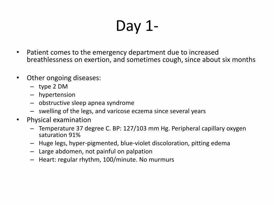
• Patient comes to the emergency department due to increased breathlessness on exertion, and sometimes cough, since about six months
• Other ongoing diseases: – type 2 DM– hypertension– obstructive sleep apnea syndrome– swelling of the legs, and varicose eczema since several years
• Physical examination– Temperature 37 degree C. BP: 127/103 mm Hg. Peripheral capillary oxygen
saturation 91%– Huge legs, hyper-pigmented, blue-violet discoloration, pitting edema– Large abdomen, not painful on palpation– Heart: regular rhythm, 100/minute. No murmurs

Day 1-
• Medicines: Metformin, furosemide• Social history: He is retired and lives alone. Non-smoker• Investigations
– Chest X-ray: Bilateral pleural effusion; no opacities; no dilatation of pulmonary vessels – ECG: sinus rhythm 100/min. – S-N-terminal pBNP: 208 (<230) ng/L– Prothrombin time (INR): 1.5 (0.9-1.2)– P-albumin: 33 (34-45) g/L– P-bilirubin: 47 (5-25) umol/L– P-CRP: 10 (<5) mg/L– B-hemoglobin: 133 (130-170) g/L– P-sodium: 142 (137-145) mmol/L– P-potassium: 4.2 (3,5-5) mmol/L– P-creatinine: 76 (60-105) umol/L– P-ALAT: normal– P-ALP: normal
Day1-
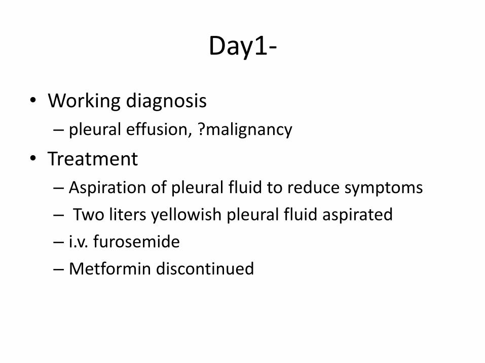
• Working diagnosis
– pleural effusion, ?malignancy
• Treatment
– Aspiration of pleural fluid to reduce symptoms
– Two liters yellowish pleural fluid aspirated
– i.v. furosemide
– Metformin discontinued

Day 7-
• (new) symptom:– Fresh blood with stool. Constipation and blood in stool since
several months.
• Digital rectal examination:– ?bleeding hemorrhoids. No malignancy
• Computer tomography of the abdomen:– Increased number of enlarged lymphnodes (< 2 cm), no ascites.
• Trans-thoracic echocardiography:– Normal RV and LV function. Altered E/A ratio suggestive of
diastolic dysfunction
• Consultation with dermatologist:– Varicose veins with eczematous changes and inflammation.
Also, ?arterial ischemia

Day 12-more investigations and consultations
• Rectoscopy– No hemorrhpids, rather rectal prolapse. No
malignancy. No changes in the mucus membrane
• CT colon– No tumor in the colon or any other abdominal organs
• CT-thorax– No pulmonary embolism. No tumor
• Consultation with vascular surgeon– No pain in the legs. Good ankle pressure. No sign of
ischemia.
Day 18
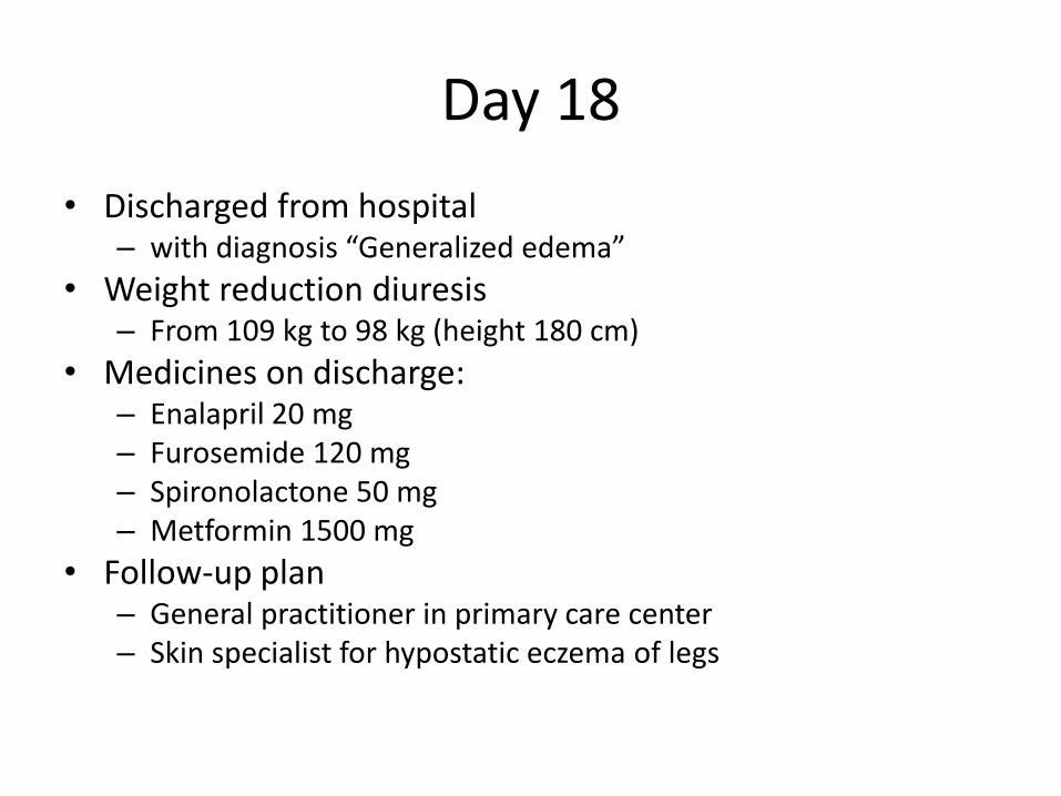
• Discharged from hospital– with diagnosis “Generalized edema”
• Weight reduction diuresis– From 109 kg to 98 kg (height 180 cm)
• Medicines on discharge:– Enalapril 20 mg– Furosemide 120 mg– Spironolactone 50 mg– Metformin 1500 mg
• Follow-up plan– General practitioner in primary care center – Skin specialist for hypostatic eczema of legs

Day 29-
• Patient comes to the emergency department again• Presenting symptoms
– Increased breathlessness, heaviness in the chest. Can walk 1 km, by stopping 6 times. – Weight increased from 98 kg t0 107 kg– Did not take enalapril, and perhaps other medicines as prescribed before
• Medicines according to the records– Enalapril, Furosemide, Spironolactone, Metformin– Plus Bisoprolol 5 mg
• Physical examination– Breathlessness at rest. Peripheral capillary oxygen saturation 96%– Blood pressure 125/75– Lungs: reduced percussion tone at the bases. Some crepitations.
• Laboratory analyses– S-N-terminal pBNP 296 (<230) ng/L, D-dimer 18.8, prothrombin time (INR) 1.5, P-albumin 32, – P-Sodium, P-Potassium, S-creatinine were normal
• Working diagnosis– Decompensation of heart failure, ? Pulmonary embolism

Day 29-
• CT-Thorax– No pulmonary embolism. Bilateral pleural effusion, no
other signs of heart failure. A little fluid in the pericardium. Enlarged medisatinal lymphnodes (some upto 3 cm). Malignancy suspected
• CT- abdomen– Moderate ascites. – Five liter yellowish ascites fluid aspirated– P-albumin 34, ascites fluid albumin 23.8 (ratio 0.70)
• New CT abdomen after aspiration of ascites– Increased sagital diameter of vena cava. Signs of right
sided heart failure. No sign of portal vein thrombosis.

Day 29-
• Ultrasound examination of the liver:– Veins of the liver are dilated. Lower edge of the liver is
round. Minimal ascites. No sign of portal vein thrombosis. – Diameter of inferior vena cava is increased with reduced
variation of the size with respiratory cycles– Findings as in right heart failure. – Cholelithiasis and signs of chronic cholecystitis
• New echocardiography– Normal RV and LV function. No wall hypertrophy. No
valvular lesion. Normal movement of the septum, normal flow pattern and flow velocities that tells against constrictive pericarditis.
Day 31Patient deteriorates acutely
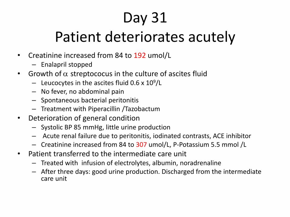
• Creatinine increased from 84 to 192 umol/L– Enalapril stopped
• Growth of a streptococus in the culture of ascites fluid– Leucocytes in the ascites fluid 0.6 x 10⁹/L– No fever, no abdominal pain– Spontaneous bacterial peritonitis– Treatment with Piperacillin /Tazobactum
• Deterioration of general condition– Systolic BP 85 mmHg, little urine production– Acute renal failure due to peritonitis, iodinated contrasts, ACE inhibitor– Creatinine increased from 84 to 307 umol/L, P-Potassium 5.5 mmol /L
• Patient transferred to the intermediate care unit– Treated with infusion of electrolytes, albumin, noradrenaline– After three days: good urine production. Discharged from the intermediate
care unit
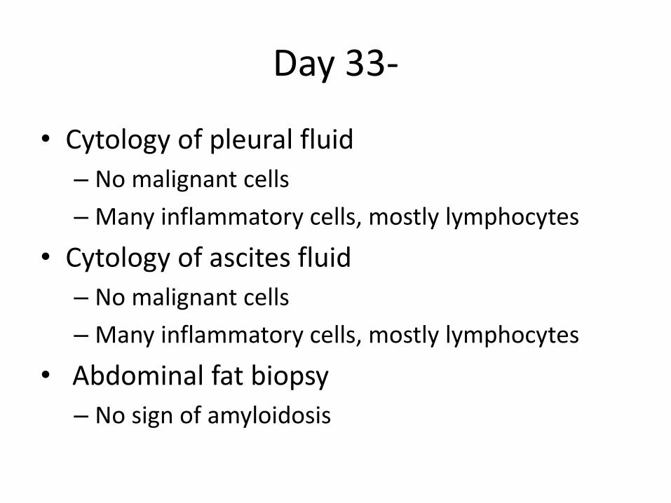
Day 33-
• Cytology of pleural fluid
– No malignant cells
– Many inflammatory cells, mostly lymphocytes
• Cytology of ascites fluid
– No malignant cells
– Many inflammatory cells, mostly lymphocytes
• Abdominal fat biopsy
– No sign of amyloidosis

Day 43-
• Continued treatment with spironolactone. Successive weight loss
• Garstroscopy
– Petechial changes in the fundus as in NSAID use
– Suspect pyloric stenosis
– Treatment with PPI started

Day 43-?Lymphoma
• Malignant lymphoma is unlikely
– No palpable lymph nodes
– No fever or sweating
– Normal LD
• Consultation with thoracic surgeon
– The lymph nodes in the mediastinum are not unusual in size or number. No need of mediastinoscopy
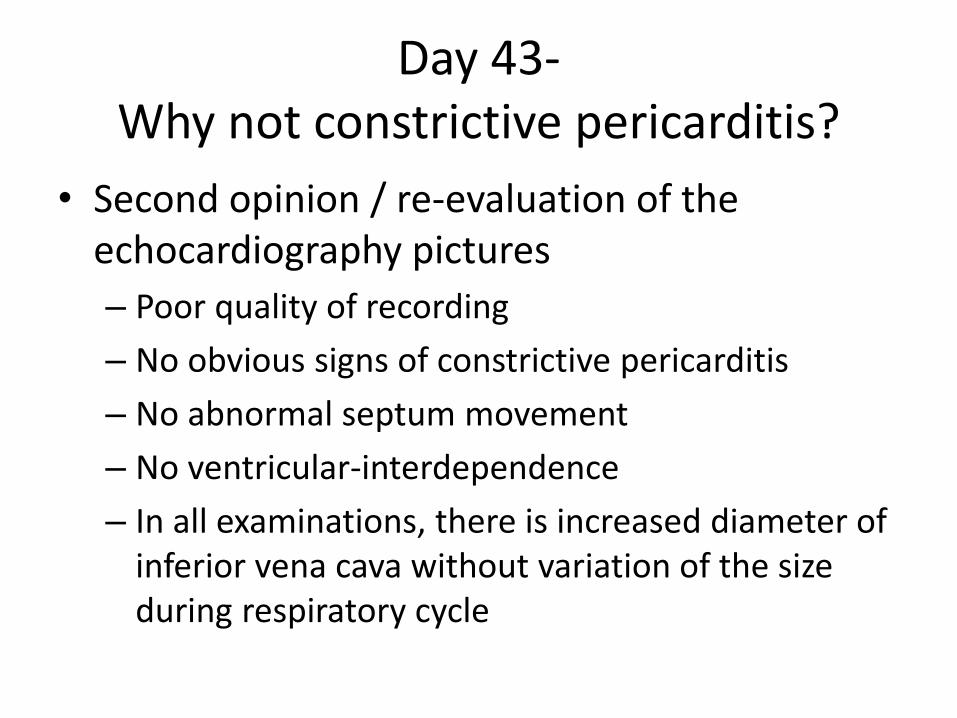
Day 43-Why not constrictive pericarditis?
• Second opinion / re-evaluation of the echocardiography pictures
– Poor quality of recording
– No obvious signs of constrictive pericarditis
– No abnormal septum movement
– No ventricular-interdependence
– In all examinations, there is increased diameter of inferior vena cava without variation of the size during respiratory cycle

Day 43-Why not constrictive pericarditis?
• Cardiac CT scan
– Relatively little, tubular LV. LV-EF 60%.
– Atypical movement of septum during heart cycle. ?Increased pressure in the pulmonary circulation.
– Increased size of the right ventricle, right atrium
– No calcification of the pericardium. No obvious thickening of the pericardium.
– No significant stenosis of the coronary arteries.
– No typical finding as in constrictive pericarditis.

“If the map does not agree with the ground, the map is wrong”
Gordon Livingstone, M.D. in “Too Soon Old, Too Late Smart”

Day 56-Why not constrictive pericarditis?
• Re-evaluation of the CT thorax, CT abdomen, and cardiac CT scan:– The size of the lymph nodes have not increased during
two months. Most are < 10 mm
– Slight thickening of pericardium. Pleural adehesionsand atelectasis
– Small diameter of LV, LA. Increased size of the RV is a visual illusion.
– Inferior vena cava is dilated
– Size of the cardiac chambers may suggest constrictive or restrictive pathology

Day 56-A diagnostic procedure was performed
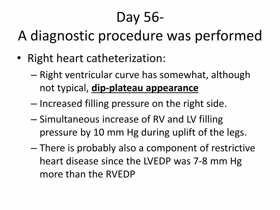
Day 56-A diagnostic procedure was performed
• Right heart catheterization:
– Right ventricular curve has somewhat, although not typical, dip-plateau appearance
– Increased filling pressure on the right side.
– Simultaneous increase of RV and LV filling pressure by 10 mm Hg during uplift of the legs.
– There is probably also a component of restrictive heart disease since the LVEDP was 7-8 mm Hg more than the RVEDP
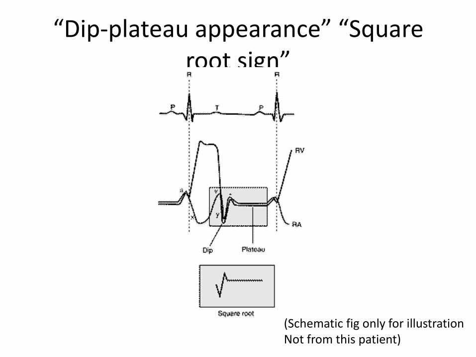
“Dip-plateau appearance” “Square root sign”
(Schematic fig only for illustrationNot from this patient)

Day 56-Why not constrictive pericarditis?
• New transthoracic echocardiography– Pericardium: somewhat increased echogenicity– No significant increase in the systolic pulmonary
arterial pressure– Pathological variation in the diastolic flow across AV
valves during respiration with variation > 25%.– Dilated inferior vena cava with reduced variation in
size during respiratory cycle suggesting increased pressure in the right atrium ( about 15 mm Hg)
– E/A 1.2, E/é 6– Some of these findings are suggestive of constrictive
pericarditis.
Day 56-Planned for elective pericardiectomy
• Rheumatology consultation– No obvious rheumatic-, systemic-disease or
vasculitis. Laboratory tests for IgG4 related diseases may be helpful. Let the pathologists examine for IgG4 positive plasma cells in the pericardial tissue.
• Myocardial biopsy– Staining for amyloid is negative. Non-specific
hypertrophy of cardiac myocytes. No sign of primary muscle disease. No IgG4 positive cells.
Day 67-
• Pericardiectomy
– Thick and inflamed pericardial fat. Thick pericardium that is non-elastic, and constrictive. Severe adhesions. Heart freed from pericardium, and most of the pericardium removed. CVP reduced from 26 to 12

Day 71-
• Patient deteriorates again– Increased breathlessness, respiratory rate 25/min,
peripheral capillary oxygen saturation 94% with 2 L oxygen /min
– Disoriented– Temperature 39.7 degree C, BP 120/70 mm Hg, pulse
130/min,
• Transferred to ICU • Sepsis
– Treated with piperacillin + gentamycin– Recovers over several days

Day 82-
• Discharged from the hospital• Follow up after one week
– Much better. Can walk 500 m at a stretch
• Histology of pericardium– “signs of mainly chronic, and also acute pericarditis. Severe
fibrosis. In some areas, there are granulocytes. Pericardial tissue shows exudates on both sides, and infiltration by many lymphocytes , and in some areas numerous plasma cells. No significant eosinophilia. Numerous cells (over 50 per HPF) positive for IgG4. Since there are numerous plasma cells, the percentage of IgG4 positive plasma cells is not especially high. Moderate probability of an IgG4 related disease.
Final diagnosis
IgG4-related constrictive pericarditis

Day 137
• Follow up at rheumatology out patient dept– Feels much stronger– Medicines: Furosemide 40 mg, Spironolactone 25 mg, metformin 1000
mg.– Pulse 80/min, BP 120/70 mmHg, some pitting edema at the ankles,
heart/lungs: normal findings– CRP 3 mg/L, SR 44 mm, Hb 123, electrolytes normal– S-IgG4 1.48 (1.25) g/L– C3 1.3 (0.67-1.29) g/L– S-anti-citrullinated protein antibody IgG <7– Anti-proteinase 3 antibody IgG [ANCA] 21 (<20) kE/L – Anti-myeloperoxidase antibody IgG [ANCA], not detected– Anti-nuclear antibody, not detected
– Patient put on prednisolone 7.5 mg /day

Rigid pericardium encircles heartSystolic contraction normal
Inhibits diastolic filling of both ventricles
Stroke volume
Cardiac output
Blood pressure
Heart rate
Venous pressure
Systemic
JVP
Hepatomegaly
Ascites
Peripheral edema
Pulmonary congestion
Crepitations
Constrictive pericarditis

Growing list of IgG4-related diseases
• Lynphadenopathy• Autoimmune pancreatitis• Sclerosing cholangitis• Salivary and lacrimal gland diseases• Retroperitoneal fibrosis• Aortitis, periaortitis• Fibrous forms of Hashimoto´s thyroiditis• Riedel´s thyroiditis• IgG4 related lung disease (may mimic sarcoidosis)• Tubulointerstitial nephritis• Cutaneous pseudolymphoma• IgG4 hepatitis• Lymphoplasmacytic gastrisit• Sclerosing mastitis• IgG4-related hypophysitis• Pachymeningitis• Nasopharyngeal disease
Uppsala, 2015-01-16
Md. Shahidul Islam, MD, PhDSenior consultant physician, Internal medicine, Uppsala, University HospitalAssociate professor, Department of Clinical Science and Education, Södersjukhuset. Karolinska Institutet, SE-118 83 [email protected]