a bone sample cleaning method using trypsin for the isolation of dna
TRANSCRIPT

Legal Medicine 13 (2011) 304–308
Contents lists available at SciVerse ScienceDirect
Legal Medicine
journal homepage: www.elsevier .com/ locate/ legalmed
Brief Communication
A bone sample cleaning method using trypsin for the isolation of DNA
Richard Li ⇑, Lidissy LirianoForensic Science Program, Department of Science, John Jay College of Criminal Justice, The City University of New York, New York, NY 10019, USA
a r t i c l e i n f o a b s t r a c t
Article history:Received 11 March 2011Received in revised form 27 June 2011Accepted 11 July 2011Available online 22 September 2011
Keywords:BoneTrypsinForensicDNA
1344-6223/$ - see front matter � 2011 Elsevier Irelandoi:10.1016/j.legalmed.2011.07.001
⇑ Corresponding author. Tel.: +1 646 557 4886.E-mail address: [email protected] (R. Li).
Cleaning the surface of bone samples is a necessary step to remove contaminants prior to isolating DNAfor forensic DNA analysis. In this study, a simple trypsin method for cleaning bone samples prior to DNAisolation was developed. Cleaning the surface of human bone samples was achieved by the application oftrypsin solution. Light microscopy and scanning electron microscopy results indicated that trypsintreatment was effective in removing the outer surface of bone samples. The yield of DNA isolated fromtrypsin-treated bone samples was sufficient for subsequent short tandem repeat (STR) analysis. STR anal-ysis revealed no adverse effect on the DNA profile after the trypsin treatment. The data suggest that thistrypsin method can potentially be an alternative cleaning method to mechanical cleaning methods.
� 2011 Elsevier Ireland Ltd. All rights reserved.
1. Introduction
Human skeletal remains are useful specimens for forensic DNAanalysis in a variety of situations when other tissue types are notavailable, such as when analyzing buried or decomposed skeletalremains [1–4].
Prior to isolating DNA from bone samples, cleaning of the boneis required to remove the potential of having co-mingled remainsand contamination by physical contact [5–7]. Additionally, poly-merase chain reaction (PCR) inhibitors and bacterial contaminationthat interfere with forensic DNA analysis must be removed duringthe cleaning process [8]. The outer surface of the bone fragmentusually is cleaned using manual mechanical cleaning methods,such as sanding using sandpaper [9,10] and sanding discs attachedto a rotary tool [11–13].
However, the bone dust generated by sanding the bone canpotentially increase the risk for cross-contamination between sam-ples [14]. Moreover, the sanding method cannot easily be used toprocess multiple samples simultaneously, and it is difficult toadapt for automation. Therefore, the processing of bone specimensis more labor-intensive and time-consuming than other types ofspecimens, such as bodily fluids and soft tissues.
To address these issues, we used an alternative enzymaticcleaning method using trypsin solution. Trypsin has been utilizedin enzymatic maceration methods for processing bone samples inanthropological laboratories [15,16]. In our previous study, thetrypsin maceration technique of bone samples was characterized[17]. Our data suggest that this method can be used in the initial
d Ltd. All rights reserved.
sample preparation for cleaning the outer surface of human bonesamples prior to DNA isolation. In this study, the application ofthe trypsin cleaning method for DNA isolation was studied inhuman bone samples. Additionally, the DNA yields and the forensicDNA profiles of trypsin-treated bone samples were examined.
2. Materials and methods
2.1. Sample preparation
Sampling was carried out in a sterilized laminar flow cabinet.The cortical portion of a human radius (within 7 days postmortem)used in this study was cut into approximately 1 g bone fragments.Soft tissues on the bone surfaces were mechanically removed usinga surgical scraper [18]. The bone fragments were rinsed with 100%ethanol and left to air-dry.
2.2. Trypsin digestion
Trypsin was obtained from Fisher Scientific. The procedures fortrypsin treatment were those developed previously [17]. Thestandard assay employed approximately 1 g of bone fragment foreach treatment. The reaction was initiated by adding 5 ml of tryp-sin solution (30 lg/ll) to the bone sample, which was incubated at55 �C with gentle agitation. After two to six hours of incubation,the supernatant was removed. The bone sample was rinsed withwater and air dried. Bone surface morphology was then observedusing light microscopy and scanning electron microscopy. Fortrypsin treatment longer than two hours, the trypsin solutionwas replaced in two hour intervals. The control samples consistedof bone fragments without trypsin treatment.
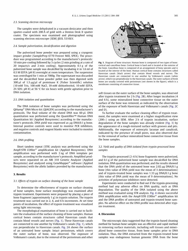
Fig. 1. Diagram of bone structure. Human bone is composed of two types of bone:cortical and cancellous bone. Cortical bone is hard and is located at the exterior ofthe bone. Cancellous bone is composed of an arrangement of bony spicules and islocated at the interior of the bone. Human cortical bone contains structures calledHaversian canals (black arrow) that contain blood vessels and nerves. TheHaversian canals are connected to one another by Volkmann’s canals (whitearrow), which run perpendicular to the Haversian canals. The outer surfaces of freshbones are usually covered with periosteum (not shown in the figure), which is amembrane consisting of connective tissue.
R. Li, L. Liriano / Legal Medicine 13 (2011) 304–308 305
2.3. Scanning electron microscopy
The samples were dehydrated in a vacuum desiccator and thensputter-coated with 200 Å of gold with a Denton Desk II sputtercoater. The specimen was examined and photographed usingscanning electron microscope (SEM) JEOL 5310LV.
2.4. Sample pulverization, decalcification and digestion
The pulverized bone powder was prepared using a cryogenicimpact grinder (SamplePrep 6770 Freezer Mill, SPEX). The proce-dure was programmed according to the manufacturer’s protocols:10 min pre-cooling followed by 3 cycles (2 min grinding at a rate of20 impacts/s and 2 min cooling). Approximately 0.1 g of thepulverized bone sample was decalcified by incubating it in 1.6 ml of0.5 M EDTA (pH 8) overnight at room temperature. The suspensionwas centrifuged for 1 min at 7000g. The supernatant was discardedand the decalcified bone powder pellet was then digested with300 ll of 1.5 lg/ll of proteinase K (Fisher Scientific) solution(10 mM Tris, 100 mM NaCl, 39 mM dithiothreitol, 10 mM EDTA,2% SDS; pH 8) at 56 �C for six hours with gentle agitation prior toDNA isolation.
2.5. DNA isolation and quantitation
The DNA isolation of bone samples was performed using theQIAamp� DNA Micro kit (QIAGEN) according to the manufacturer’sprotocols. The final volume of the eluted DNA was 60 ll. DNAquantitation was performed using the Quantifiler™ Human DNAQuantitation kit (Applied Biosystems) according to the manufac-turer’s protocols. DNA yield was expressed as lg DNA/0.1 g bone.Internal positive controls were used to monitor PCR inhibitors,and negative controls and reagent blanks were included to monitorcontamination.
2.6. DNA profiling
Short tandem repeat (STR) analysis was performed using theAmpF‘STR COfiler� amplification kit (Applied Biosystems). DNAamplification was performed with 2 ng of DNA template persample, according to the manufacturer’s protocols. Amplified prod-ucts were separated on an ABI 310 Genetic Analyzer (AppliedBiosystems) and analyzed using GeneMapper� software (AppliedBiosystems) with the allelic ladders provided by the manufacturer.
3. Results
3.1. Effects of trypsin on surface cleaning of the bone sample
To determine the effectiveness of trypsin on surface cleaningof bone samples, bone surface morphology was examined aftertrypsin treatment. Experiments were prepared by placing a piece ofbone fragment (approximately 1 g) in trypsin solution. The trypsintreatment was carried out in 2, 4, and 6 h increments. At set timepoints of incubation, the effect of trypsin treatment was visualizedusing light microscopy.
The morphological examination of bone is carried out to facili-tate the evaluation of the surface cleaning of bone samples. Humancortical bones contain structures called Haversian canals thatcontain blood vessels and nerves (Fig. 1). These Haversian canalsare connected to one another by Volkmann’s canals (Fig. 1), whichrun perpendicular to Haversian canals. Fig. 2A shows the surfaceof an untreated bone sample. Intact periosteum, which coversthe outer surface of bone, was observed. The exposure ofVolkmann’s canals, due to the removal of the periosteum and other
soft tissues on the outer surface of the bone samples, was observedafter trypsin treatment for 2 h (Fig. 2B). After longer incubation (4and 6 h), some mineralized bone connective tissue on the outersurface of the bone was removed, as indicated by the observationof the exposure of both Haversian and Volkmann’s canals (Fig. 2Cand D).
To further evaluate the surface cleaning effect of trypsin treat-ment, the samples were examined at a higher magnification view(200�) using an SEM. After 2 h of trypsin incubation, surfacedegradation of the bone samples was already evident (Fig. 3) bythe appearance of a rough-textured surface with grooves and pits.Additionally, the exposure of osteocytic lacunae and canaliculi,indicated by the presence of small pores, was also observed dueto the removal of surface mineralized bone connective tissue fromthe bone samples.
3.2. Yield and quality of DNA isolated from trypsin-processed bonesamples
The trypsin-processed (2–6 h) bone fragments were pulverized,and 0.1 g of the pulverized bone sample was decalcified for DNAisolation. DNA quantitation was performed, and the results showedthat the DNA yield of the untreated bone was 2.02 lg DNA/0.1 gbone (this value of DNA yield was the mean of 3 determinations)and of trypsin-treated bone samples was 1.33 lg DNA/0.1 g bone(this value of DNA yield was the mean of 9 determinations). Noactivities of polymerase inhibitors were detected.
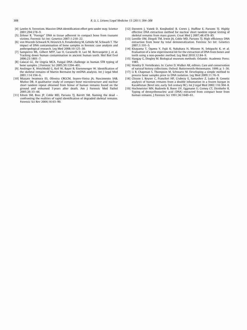
Experiments were conducted to find out if the trypsin-processingmethod had any adverse effect on DNA quality, such as DNAdegradation. The quality of the DNA isolated using the abovemethod was evaluated using STR analysis. Fig. 4 shows successfulDNA amplification with the AmpF‘STR COfiler� amplification kitand the DNA profiles of untreated and trypsin-treated bone sam-ples. No adverse effect on the DNA profile was detected after tryp-sin treatment.
4. Discussion
The microscopic data suggested that the trypsin-based cleaningmethod for human bone samples was an effective and rapid methodin removing surface materials, including soft tissues and miner-alized bone connective tissue, from bone samples prior to DNAisolation. Thus, the DNA extracted from the trypsin-treated bonesamples was endogenous human genomic DNA from the bone
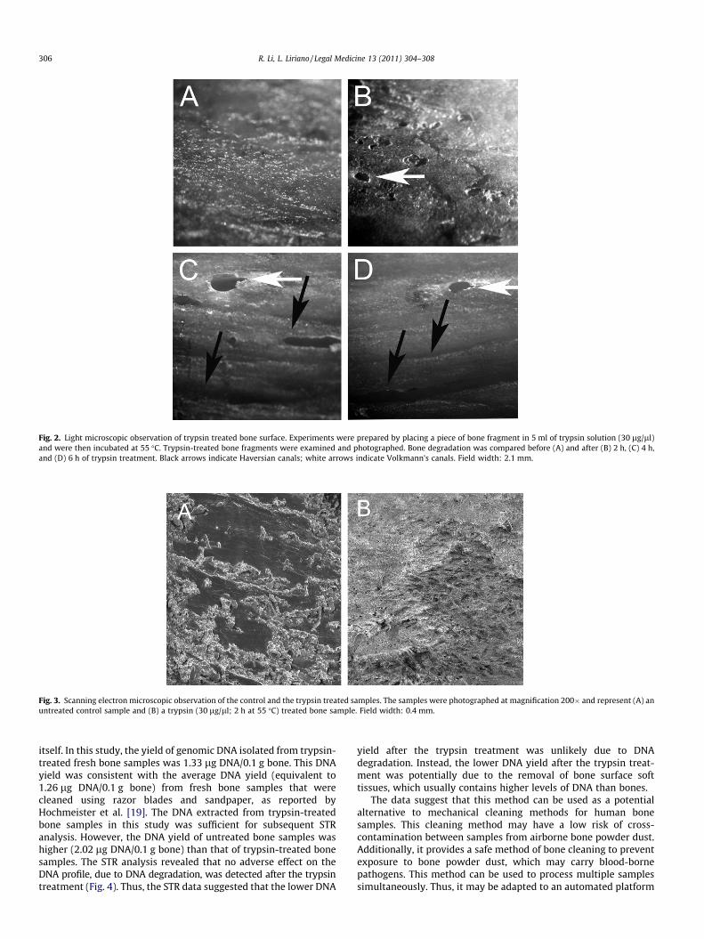
Fig. 2. Light microscopic observation of trypsin treated bone surface. Experiments were prepared by placing a piece of bone fragment in 5 ml of trypsin solution (30 lg/ll)and were then incubated at 55 �C. Trypsin-treated bone fragments were examined and photographed. Bone degradation was compared before (A) and after (B) 2 h, (C) 4 h,and (D) 6 h of trypsin treatment. Black arrows indicate Haversian canals; white arrows indicate Volkmann’s canals. Field width: 2.1 mm.
Fig. 3. Scanning electron microscopic observation of the control and the trypsin treated samples. The samples were photographed at magnification 200� and represent (A) anuntreated control sample and (B) a trypsin (30 lg/ll; 2 h at 55 �C) treated bone sample. Field width: 0.4 mm.
306 R. Li, L. Liriano / Legal Medicine 13 (2011) 304–308
itself. In this study, the yield of genomic DNA isolated from trypsin-treated fresh bone samples was 1.33 lg DNA/0.1 g bone. This DNAyield was consistent with the average DNA yield (equivalent to1.26 lg DNA/0.1 g bone) from fresh bone samples that werecleaned using razor blades and sandpaper, as reported byHochmeister et al. [19]. The DNA extracted from trypsin-treatedbone samples in this study was sufficient for subsequent STRanalysis. However, the DNA yield of untreated bone samples washigher (2.02 lg DNA/0.1 g bone) than that of trypsin-treated bonesamples. The STR analysis revealed that no adverse effect on theDNA profile, due to DNA degradation, was detected after the trypsintreatment (Fig. 4). Thus, the STR data suggested that the lower DNA
yield after the trypsin treatment was unlikely due to DNAdegradation. Instead, the lower DNA yield after the trypsin treat-ment was potentially due to the removal of bone surface softtissues, which usually contains higher levels of DNA than bones.
The data suggest that this method can be used as a potentialalternative to mechanical cleaning methods for human bonesamples. This cleaning method may have a low risk of cross-contamination between samples from airborne bone powder dust.Additionally, it provides a safe method of bone cleaning to preventexposure to bone powder dust, which may carry blood-bornepathogens. This method can be used to process multiple samplessimultaneously. Thus, it may be adapted to an automated platform
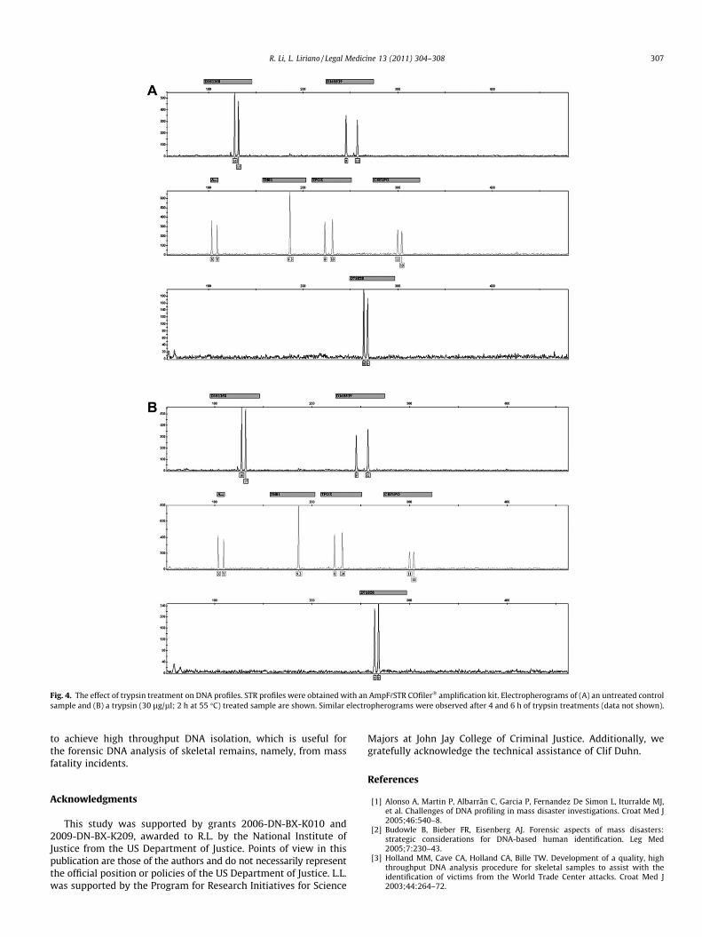
Fig. 4. The effect of trypsin treatment on DNA profiles. STR profiles were obtained with an AmpF‘STR COfiler� amplification kit. Electropherograms of (A) an untreated controlsample and (B) a trypsin (30 lg/ll; 2 h at 55 �C) treated sample are shown. Similar electropherograms were observed after 4 and 6 h of trypsin treatments (data not shown).
R. Li, L. Liriano / Legal Medicine 13 (2011) 304–308 307
to achieve high throughput DNA isolation, which is useful forthe forensic DNA analysis of skeletal remains, namely, from massfatality incidents.
Acknowledgments
This study was supported by grants 2006-DN-BX-K010 and2009-DN-BX-K209, awarded to R.L. by the National Institute ofJustice from the US Department of Justice. Points of view in thispublication are those of the authors and do not necessarily representthe official position or policies of the US Department of Justice. L.L.was supported by the Program for Research Initiatives for Science
Majors at John Jay College of Criminal Justice. Additionally, wegratefully acknowledge the technical assistance of Clif Duhn.
References
[1] Alonso A, Martin P, Albarrãn C, Garcia P, Fernandez De Simon L, Iturralde MJ,et al. Challenges of DNA profiling in mass disaster investigations. Croat Med J2005;46:540–8.
[2] Budowle B, Bieber FR, Eisenberg AJ. Forensic aspects of mass disasters:strategic considerations for DNA-based human identification. Leg Med2005;7:230–43.
[3] Holland MM, Cave CA, Holland CA, Bille TW. Development of a quality, highthroughput DNA analysis procedure for skeletal samples to assist with theidentification of victims from the World Trade Center attacks. Croat Med J2003;44:264–72.
308 R. Li, L. Liriano / Legal Medicine 13 (2011) 304–308
[4] Lawler A. Terrorism. Massive DNA identification effort gets under way. Science2001;294:278–9.
[5] Zehner R. ‘‘Foreign’’ DNA in tissue adherent to compact bone from tsunamivictims. Forensic Sci Int: Genetics 2007;1:218–22.
[6] von Wurmb-Schwark N, Heinrich A, Freudenberg M, Gebühr M, Schwark T. Theimpact of DNA contamination of bone samples in forensic case analysis andanthropological research. Leg Med 2008;10:125–30.
[7] Sampietro ML, Gilbert MTP, Lao O, Caramelli D, Lari M, Bertranpetit J, et al.Tracking down human contamination in ancient human teeth. Mol Biol Evol2006;23:1801–7.
[8] Calacal GC, De Ungria MCA. Fungal DNA challenge in human STR typing ofbone samples. J Forensic Sci 2005;50:1394–401.
[9] Anslinger K, Weichhold G, Keil W, Bayer B, Eisenmenger W. Identification ofthe skeletal remains of Martin Bormann by mtDNA analysis. Int J Legal Med2001;114:194–6.
[10] Miazato Iwamura ES, Oliveira CRGCM, Soares-Vieira JA, Nascimento SAB,Muñoz DR. A qualitative study of compact bone microstructure and nuclearshort tandem repeat obtained from femur of human remains found on theground and exhumed 3 years after death. Am J Forensic Med Pathol2005;26:33–44.
[11] Edson SM, Ross JP, Coble MD, Parsons TJ, Barritt SM. Naming the dead –confronting the realities of rapid identification of degraded skeletal remains.Forensic Sci Rev 2004;16:63–90.
[12] Davoren J, Vanek D, Konjhodzic R, Crews J, Huffine E, Parsons TJ. Highlyeffective DNA extraction method for nuclear short tandem repeat testing ofskeletal remains from mass graves. Croat Med J 2007;48:478–85.
[13] Loreille OM, Diegoli TM, Irwin JA, Coble MD, Parsons TJ. High efficiency DNAextraction from bone by total demineralization. Forensic Sci Int: Genetics2007;1:191–5.
[14] Kitayama T, Ogawa Y, Fujii K, Nakahara H, Mizuno N, Sekiguchi K, et al.Evaluation of a new experimental kit for the extraction of DNA from bones andteeth using a non-powder method. Leg Med 2010;12:84–9.
[15] Hangay G, Dingley M. Biological museum methods. Orlando: Academic Press;1985.
[16] Hendry D. Vertebrates. In: Carter D, Walker AK, editors. Care and conservationof natural history collections. Oxford: Butterworth-Heinemann; 1999. p. 1–36.
[17] Li R, Chapman S, Thompson M, Schwartz M. Developing a simple method toprocess bone samples prior to DNA isolation. Leg Med 2009;11:76–9.
[18] Clisson I, Keyser C, Francfort HP, Crubezy E, Samashev Z, Ludes B. Geneticanalysis of human remains from a double inhumation in a frozen kurgan inKazakhstan (Berel site, early 3rd century BC). Int J Legal Med 2002;116:304–8.
[19] Hochmeister MN, Budowle B, Borer UV, Eggmann U, Comey CT, Dirnhofer R.Typing of deoxyribonucleic acid (DNA) extracted from compact bone fromhuman remains. J Forensic Sci 1991;36:1649–61.