a burkholderia pseudomallei type iii secreted protein ...jb.asm.org/content/185/16/4992.full.pdf ·...
TRANSCRIPT

JOURNAL OF BACTERIOLOGY, Aug. 2003, p. 4992–4996 Vol. 185, No. 160021-9193/03/$08.00�0 DOI: 10.1128/JB.185.16.4992–4996.2003Copyright © 2003, American Society for Microbiology. All Rights Reserved.
NOTES
A Burkholderia pseudomallei Type III Secreted Protein, BopE,Facilitates Bacterial Invasion of Epithelial Cells and Exhibits
Guanine Nucleotide Exchange Factor ActivityMark P. Stevens,1 Andrea Friebel,2 Lowrie A. Taylor,1 Michael W. Wood,1
Philip J. Brown,1† Wolf-Dietrich Hardt,2and Edouard E. Galyov1*
Division of Environmental Microbiology, Institute for Animal Health, Compton Laboratory, BerkshireRG20 7NN, United Kingdom,1 and Institute of Microbiology, Swiss Federal Institute of
Technology Zurich, ETH Zentrum, CH-8092 Zurich, Switzerland2
Received 24 March 2003/Accepted 12 May 2003
We report the characterization of BopE, a type III secreted protein that is encoded adjacent to the Burk-holderia pseudomallei bsa locus and is homologous to Salmonella enterica SopE/SopE2. Inactivation of bopE im-paired bacterial entry into HeLa cells, indicating that BopE facilitates invasion. Consistent with this notion,BopE expressed in eukaryotic cells induced rearrangements in the subcortical actin cytoskeleton, and purifiedBopE exhibited guanine nucleotide exchange factor activity for Cdc42 and Rac1 in vitro.
Burkholderia pseudomallei is the etiological agent of melioid-osis, a severe invasive infection of humans and animals that isendemic in subtropical areas (3, 6). Melioidosis has a remark-able capacity for latency. Development of disease 26 yearsafter geographical exposure has been reported (21), and re-lapse is common even in patients treated with antibiotics (4).This is believed to result from the ability of B. pseudomallei toinvade nonphagocytic host cells and to survive and replicatewithin phagocytes, where antibiotics may be less effective (14,16, 25). The mechanism by which B. pseudomallei enters epi-thelial cells is poorly understood.
We and others have identified a putative type III proteinsecretion apparatus in B. pseudomallei (Bsa) similar to the Sal-monella enterica Inv/Spa/Prg and Shigella flexneri Ipa/Mxi/Spasystems (1, 26, 31). Type III secretion systems are key virulencedeterminants of Salmonella, Shigella, and other gram-negativefacultative intracellular pathogens and serve to inject bacterialproteins into target cells (reviewed in references 5, 10, 13, and29). A subset of type III secretion system secreted proteins(translocators) is believed to interact with the eukaryotic cellmembrane and mediate the delivery of secreted effector pro-teins. Once inside host cells the effector proteins subvert hostcell processes to the benefit of the bacteria (reviewed in ref-erences 5 and 13).
Research in our laboratory and elsewhere has identified anumber of Salmonella Inv/Spa/Prg secreted effector proteins
(Sops) and shown that several of these are delivered into eu-karyotic cells by mechanisms dependent on secreted translo-cator proteins (Sips) (11, 34, 35). Mutations that disrupt theInv/Spa/Prg apparatus and selected sip and sop genes inhibitbacterial invasion of epithelial cells and Salmonella-inducedenteritis (reviewed in references 10, 33, and 36). Some Sopeffector proteins possess eukaryote-like enzymatic activities. Inparticular, it has been shown that the SopE and SopE2 pro-teins promote bacterial invasion (2, 35) by acting as guaninenucleotide exchange factors (GEFs) for RhoGTPases that reg-ulate the actin network (12, 27, 30). SopE acts as a GEF forCdc42, Rac1, and Rab5 (8, 9, 12, 23, 27); however, SopE2efficiently activates Cdc42 but not Rac1 (8, 30), indicating thatSopE and SopE2 may activate different signaling cascadesduring Salmonella infection. Mutation of Salmonella sopE andsopE2 reduces the induction of intestinal inflammatory andsecretory responses in calves, suggesting that they play a role inSalmonella-induced enteritis (36, 37).
Recently we reported that mutations affecting putative com-ponents of the B. pseudomallei Bsa secretion and translocationapparatus impair intracellular survival of B. pseudomallei inmurine macrophage-like cells and prevent escape of the bac-teria from endocytic vesicles (31). Here we have investigatedthe role of a putative Bsa-secreted protein (BopE) that shareshomology with the Salmonella SopE/SopE2 proteins. BopE is27% identical over 168 amino acids to SopE and 28% identicalover 139 amino acids to SopE2.
BopE is secreted by the Bsa type III secretion apparatus. Tostudy expression and secretion of BopE, a BopE-glutathione-S-transferase fusion protein was generated and polyclonal an-tiserum was raised against BopE in rabbits. A DNA fragmentencoding the domain of BopE proposed to be required forGEF activity (amino acid residues 78 to 261) was amplified
* Corresponding author. Mailing address: Division of Environmen-tal Microbiology, Institute for Animal Health, Compton Laboratory,Berkshire RG20 7NN, United Kingdom. Phone: 44 1635 577291. Fax:44 1635 577243. E-mail: [email protected].
† Present address: Nuffield Department of Clinical Laboratory Sci-ences, University of Oxford, John Radcliffe Hospital, Oxford OX39DU, United Kingdom.
4992
on May 1, 2018 by guest
http://jb.asm.org/
Dow
nloaded from

using the primers BopEGexBam (5�-CGGCAGCTATGGATCCACGGGCGACGCGAAAC-3�) and BopEGexE1 (5�-CCACGCTGAATTCTCACGCGCCGTCC-3�) and the productcloned into pGEX-2T (Amersham Biosciences, Little Chal-font, Buckinghamshire, England) via EcoRI and BamHI sites(underlined) in the primers. Following expression in Esche-richia coli BL21(DE3) under isopropyl-�-D-thiogalactoside in-duction, the fusion protein was purified using glutathioneSepharose 4B resin and BopE78-261 released from glutathione-S-transferase by thrombin digestion. A 12-week-old New Zea-land White rabbit was immunized subcutaneously four timesat 2-week intervals with ca. 100 �g of purified BopE78-261 inFreund’s incomplete adjuvant and serum collected 12 daysafter the final booster.
The BopE-specific antiserum was used to detect BopE inwhole-cell and secreted protein fractions of B. pseudomalleistrain 10276 and defined bsaZ, bipD, and bopE mutant strainsdescribed previously (31). BsaZ and BipD are homologous tothe Salmonella SpaS and SipD proteins involved in secretionand translocation of Sop proteins, respectively. Bacteria weregrown to stationary phase in Luria-Bertani broth, and culturesupernatants were passed through 0.22-�m-pore-size filtersprior to precipitation of secreted proteins with trichloroaceticacid (10% [vol/vol]). Approximately 25 �g of total protein (Fig.1A) or secreted protein (Fig. 1B) was resolved by 4-to-15%sodium dodecyl sulfate-polyacrylamide gel electrophoresis andtransferred to Immobilon-P membrane (Millipore, Bedford,Mass.). A 1:100 dilution of rabbit polyclonal antiserum toBopE78-261 was used, and bound antibody was detected with ananti-rabbit alkaline phosphatase conjugate. As expected, BopE
was detected in whole-cell extracts of all strains except the10276 bopE mutant (Fig. 1A). BopE secretion was dependenton the Bsa type III secretion apparatus, as no secretion wasobserved in a bsaZ mutant (Fig. 1B). In contrast, BopE secre-tion was elevated in B. pseudomallei lacking the putative trans-locator BipD (Fig. 1B). These data are consistent with theobservation that Salmonella sip mutants secrete elevated levelsof selected Sops (15, 35). Thus, our data suggest that the B.pseudomallei Bsa type III secretion apparatus is functional andthat BopE is type III secreted.
BopE facilitates invasion of nonphagocytic cells. B. pseudo-mallei can invade and survive within nonphagocytic cells (14,16). To assess the role of BopE in bacterial invasion, we quan-tified intracellular B. pseudomallei following infection of HeLacells by strains 10276, 10276 bipD, and 10276 bopE by using akanamycin protection assay. Previously we have been unable todetect significant invasion of HeLa cells by B. pseudomalleistrain 10276 (31); however, we have found that invasion effi-ciency can be improved by centrifugation of the bacteria ontocell monolayers at 300 � g at the onset of infection. HeLa cellsmaintained in RPMI 1640 containing 10% (vol/vol) fetal calfserum were infected at a multiplicity of 10 with B. pseudomalleistrains grown to stationary phase in Luria-Bertani broth at37°C in a humidified 5% CO2 atmosphere. One hour afterbacterial inoculation, monolayers were washed three times andoverlaid with medium containing kanamycin (250 �g/ml) to killextracellular bacteria. After 6 h viable intracellular bacteriawere released by gentle lysis using 0.1% Triton X-100 andenumerated by plating of serial dilutions. We detected a sta-tistically significant reduction in invasion of HeLa cells by the10276 bopE mutant compared to that of the wild type (P �0.0464) (Fig. 2), indicating that BopE, like Salmonella SopE/SopE2, facilitates bacterial invasion of nonphagocytic cells.SopE acts in concert with other type III secreted proteins topromote Salmonella invasion (38). Recently it was reported
FIG. 1. Western blot analysis of BopE expression and secretion byB. pseudomallei 10276 wild type and bsaZ, bipD, and bopE mutantstrains. Approximately 25 �g of total protein (A) or secreted protein(B) was probed with rabbit polyclonal antiserum to BopE78-261 anddetected with an anti-rabbit alkaline phosphatase conjugate. Molecu-lar mass markers are shown on the left.
FIG. 2. Invasion of HeLa cells by B. pseudomallei 10276 wild typeand bipD and bopE mutant strains. HeLa cells (5 � 105) were infectedat a multiplicity of infection 10 in triplicate for each assay and theresults represent the arithmetic means (error bars show standard er-rors of the means) of results of four independent assays.
VOL. 185, 2003 NOTES 4993
on May 1, 2018 by guest
http://jb.asm.org/
Dow
nloaded from
that the effector protein SopB, which possesses phosphatidyl-inositol phosphatase activity, influences Salmonella invasion(24, 39). It is likely that other type III secreted proteins influ-ence invasion of nonphagocytic cells by B. pseudomallei. Con-sistent with this hypothesis we detected a highly significantreduction in invasion of HeLa cells by the 10276 bipD mutant(P � 0.0058). B. pseudomallei is a Centers for Disease Controland Prevention category B critical biological agent, and wewere unable to trans-complement the bopE mutation owing torestrictions on genetic modification of the organism.
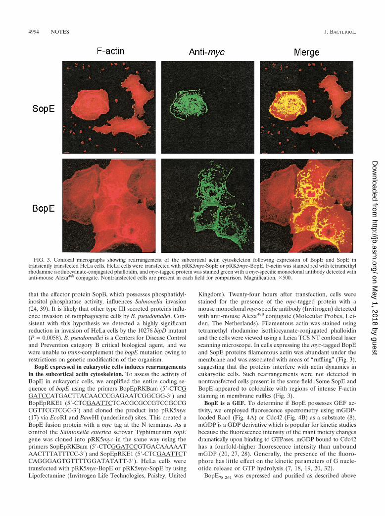
BopE expressed in eukaryotic cells induces rearrangementsin the subcortical actin cytoskeleton. To assess the activity ofBopE in eukaryotic cells, we amplified the entire coding se-quence of bopE using the primers BopEpRKBam (5�-CTCGGATCCATGACTTACAACCCGAGAATCGGCGG-3�) andBopEpRKE1 (5�-CTCGAATTCTCACGCGCCGTCCGCCGCGTTCGTCGC-3�) and cloned the product into pRK5myc(17) via EcoRI and BamHI (underlined) sites. This created aBopE fusion protein with a myc tag at the N terminus. As acontrol the Salmonella enterica serovar Typhimurium sopEgene was cloned into pRK5myc in the same way using theprimers SopEpRKBam (5�-CTCGGATCCGTGACAAAAATAACTTTATTTCC-3�) and SopEpRKE1 (5�-CTCGAATTCTCAGGGAGTGTTTTGGATATATT-3�). HeLa cells weretransfected with pRK5myc-BopE or pRK5myc-SopE by usingLipofectamine (Invitrogen Life Technologies, Paisley, United
Kingdom). Twenty-four hours after transfection, cells werestained for the presence of the myc-tagged protein with amouse monoclonal myc-specific antibody (Invitrogen) detectedwith anti-mouse Alexa488 conjugate (Molecular Probes, Lei-den, The Netherlands). Filamentous actin was stained usingtetramethyl rhodamine isothiocyanate-conjugated phalloidinand the cells were viewed using a Leica TCS NT confocal laserscanning microscope. In cells expressing the myc-tagged BopEand SopE proteins filamentous actin was abundant under themembrane and was associated with areas of “ruffling” (Fig. 3),suggesting that the proteins interfere with actin dynamics ineukaryotic cells. Such rearrangements were not detected innontransfected cells present in the same field. Some SopE andBopE appeared to colocalize with regions of intense F-actinstaining in membrane ruffles (Fig. 3).
BopE is a GEF. To determine if BopE possesses GEF ac-tivity, we employed fluorescence spectrometry using mGDP-loaded Rac1 (Fig. 4A) or Cdc42 (Fig. 4B) as a substrate (8).mGDP is a GDP derivative which is popular for kinetic studiesbecause the fluorescence intensity of the mant moiety changesdramatically upon binding to GTPases. mGDP bound to Cdc42has a fourfold-higher fluorescence intensity than unboundmGDP (20, 27, 28). Generally, the presence of the fluoro-phore has little effect on the kinetic parameters of G nucle-otide release or GTP hydrolysis (7, 18, 19, 20, 32).
BopE78-261 was expressed and purified as described above
FIG. 3. Confocal micrographs showing rearrangement of the subcortical actin cytoskeleton following expression of BopE and SopE intransiently transfected HeLa cells. HeLa cells were transfected with pRK5myc-SopE or pRK5myc-BopE. F-actin was stained red with tetramethylrhodamine isothiocyanate-conjugated phalloidin, and myc-tagged protein was stained green with a myc-specific monoclonal antibody detected withanti-mouse Alexa488 conjugate. Nontransfected cells are present in each field for comparison. Magnification, �500.
4994 NOTES J. BACTERIOL.
on May 1, 2018 by guest
http://jb.asm.org/
Dow
nloaded from

and mGDP-Rac1 or mGDP-Cdc42 was prepared as describedpreviously (8, 9). In the assay buffer (no EDTA or BopE)mGDP dissociation from Rac1 was very slow. In contrast, fastdissociation of the mGDP-Rac1 complex was observed in thepresence of 25 nM BopE and even faster in the presence of 250nM BopE (Fig. 4A) (kobs � 0.48 s�1). Similar observationswere made using mGDP-Cdc42 as a substrate (Fig. 4B). Thesedata demonstrate that BopE is an efficient GEF for Cdc42 andRac1. The observed G-nucleotide exchange rates are lowerthan those observed with SopE from Salmonella serovar Ty-phimurium (8) but range in the same order of magnitude.
Taken together our observations suggest that B. pseudomal-lei enters epithelial cells by a mechanism dependent at least inpart upon the Bsa type III protein secretion apparatus and oneof its secreted proteins, BopE. It is likely that BopE is trans-located into the host cell cytosol, where it may promote mem-brane ruffling by acting as a GEF for Cdc42 and Rac1. InSalmonella several Inv/Spa/Prg-secreted proteins (SopE, SopE2,and SopB) act in concert to promote bacterial uptake by non-phagocytic cells (38, 39), and Salmonella invasion probablyevolved through the acquisition of new sequence elements(22). Given that a B. pseudomallei bipD mutant was impaired ininvasion of HeLa cells to a greater extent than a bopE mutant,it is likely that other Bsa-secreted proteins may be involved inbacterial uptake. We are investigating the role of other puta-tive type III secreted proteins in the host-cell interactions ofB. pseudomallei.
REFERENCES
1. Attree, O., and I. Attree. 2001. A second type III secretion system in Burk-holderia pseudomallei: who is the real culprit? Microbiology 147:3197–3199.
2. Bakshi, C. S., V. P. Singh, M. W. Wood, P. W. Jones, T. S. Wallis, and E. E.Galyov. 2000. Identification of SopE2, a Salmonella secreted protein which ishighly homologous to SopE and involved in bacterial invasion of epithelialcells. J. Bacteriol. 82:2341–2344.
3. Brett, P. J., and D. E. Woods. 2000. Pathogenesis of and immunity tomelioidosis. Acta Trop. 74:201–210.
4. Chaowagul, W., Y. Suputtamongkol, D. A. B. Dance, A. Rajchanuvong, J.Pattaraarechachai, and N. J. White. 1993. Relapse in melioidosis: incidenceand risk factors. J. Infect. Dis. 168:1181–1185.
5. Cornelis, G. R., and F. van Gijsegem. 2000. Assembly and function of typeIII secretion systems. Annu. Rev. Microbiol. 54:735–774.
6. Dance, D. A. 2000. Melioidosis is an emerging global problem. Acta Trop.74:115–119.
7. Franken, S. M., A. J. Scheidig, U. Krengel, H. Rensland, A. Lautwein, M.Geyer, K. Scheffzek, R. S. Goody, H. R. Kalbitzer, E. F. Pai, et al. 1993.Three-dimensional structures and properties of a transforming and a non-transforming glycine-12 mutant of p21H-ras. Biochemistry 32:8411–8420.
8. Friebel, A., H. Ilchmann, M. Aepfelbacher, K. Ehrbar, W. Machleidt, andW.-D. Hardt. 2001. SopE and SopE2 from Salmonella typhimurium activatedifferent sets of RhoGTPases of the host cell. J. Biol. Chem. 276:34035–34040.
9. Friebel, A., and W.-D. Hardt. 2000. Purification and biochemical activity ofSalmonella exchange factor SopE. Methods Enzymol. 325:82–91.
10. Galan, J. E. 2001. Salmonella interactions with host cells: type III secretionat work. Annu. Rev. Cell. Dev. Biol. 17:53–86.
11. Galyov, E. E., M. W. Wood, R. Rosqvist, P. B. Mullan, P. R. Watson, S.Hedges, and T. S. Wallis. 1997. A secreted effector protein of Salmonelladublin is translocated into eukaryotic cells and mediates inflammation andfluid secretion in infected ileal mucosa. Mol. Microbiol. 25:903–912.
12. Hardt, W.-D., L. M. Chen, K. E. Schuebel, X. R. Bustelo, and J. E. Galan.1998. S. typhimurium encodes an activator of Rho GTPases that inducesmembrane ruffling and nuclear responses in host cells. Cell 93:815–826.
13. Hueck, C. J. 1998. Type III protein secretion systems in bacterial pathogensof animals and plants. Microbiol. Mol. Biol. Rev. 62:379–433.
14. Jones, A. L., T. J. Beveridge, and D. E. Woods. 1996. Intracellular survival ofBurkholderia pseudomallei. Infect. Immun. 64:782–790.
15. Kaniga, K., D. Trollinger, and J. E. Galan. 1995. Identification of two targetsof the type III protein secretion system encoded by the inv and spa loci ofSalmonella typhimurium that have homology to the Shigella IpaD and IpaAproteins. J. Bacteriol. 177:7078–7085.
16. Kespichayawattana, W., S. Rattanachetkul, T. Wanun, P. Utaisincharoen,and S. Sirisinha. 2000. Burkholderia pseudomallei induces cell fusion andactin-associated membrane protrusion: a possible mechanism of cell-to-cellspreading. Infect. Immun. 68:5377–5384.
17. Lamarche, N., N. Tapon, L. Stowers, P. D. Burbelo, P. Aspenstrom, T.Bridges, J. Chant, and A. Hall. 1996. Rac and Cdc42 induce actin polymer-ization and G1 cell cycle progression independently of p65PAK and theJNK/SAPK MAP kinase cascade. Cell 87:519–529.
18. Lenzen, C., R. H. Cool, and A. Wittinghofer. 1995. Analysis of intrinsic and
FIG. 4. BopE acts as a GEF for the RhoGTPases Rac1 and Cdc42. The multiple turnover kinetics of guanine nucleotide exchange by BopEwas analyzed by measuring the release of mantGDP from 10 �M Rac1-191�mantGDP (A) or Cdc42 Hs1-192�mantGDP (B) in the presence of 1 mMGDP and 25 nM or 250 nM BopE using fluorescence spectrometry (excitation wavelength, 366 nm; emission wavelength, 440 nm; step size, 1; band-pass, 4) at 20°C in a buffer containing 40 mM HEPES–NaOH (pH 7.4), 100 mM NaCl, and 5 mM MgCl2. Spontaneous dissociation of theRhoGTPase�mantGDP complex in the assay buffer or in assay buffer supplemented with 10 mM EDTA was measured as the control.
VOL. 185, 2003 NOTES 4995
on May 1, 2018 by guest
http://jb.asm.org/
Dow
nloaded from

CDC25-stimulated guanine nucleotide exchange of p21ras-nucleotide com-plexes by fluorescence measurements. Methods Enzymol. 255:95–109.
19. Lenzen, C., R. H. Cool, H. Prinz, J. Kuhlmann, and A. Wittinghofer. 1998.Kinetic analysis by fluorescence of the interaction between Ras and thecatalytic domain of the guanine nucleotide exchange factor Cdc25Mm. Bio-chemistry 37:7420–7430.
20. Leonard, D. A., T. Evans, M. Hart, R. A. Cerione, and D. Manor. 1994.Investigation of the GTP-binding/GTPase cycle of Cdc42Hs using fluores-cence spectroscopy. Biochemistry 33:12323–12328.
21. Mays, E. E., and E. A. Ricketts. 1975. Melioidosis: recrudescence associatedwith bronchogenic carcinoma twenty-six years following initial geographicexposure. Chest 68:261–263.
22. Mirold, S., K. Ehrbar, A. Weissmuller, R. Prager, H. Tschape, H. Russmann,and W.-D. Hardt. 2001. Salmonella host cell invasion emerged by acquisitionof a mosaic of separate genetic elements, including Salmonella pathogenicityisland 1 (SPI1), SPI5, and sopE2. J. Bacteriol. 183:2348–2358.
23. Mukherjee, K., S. Parashuraman, M. Raje, and A. Mukhopadhyay. 2001.SopE acts as an Rab5-specific nucleotide exchange factor and recruits non-prenylated Rab5 on Salmonella-containing phagosomes to promote fusionwith early endosomes. J. Biol. Chem. 276:23607–23615.
24. Norris, F. A., M. P. Wilson, T. S. Wallis, E. E. Galyov, and P. W. Majerus.1998. SopB, a protein required for virulence of Salmonella dublin, is aninositol phosphatase. Proc. Natl. Acad. Sci. USA 95:14057–14059.
25. Pruksachartvuthi, S., N. Aswapokee, and K. Thankerngpol. 1990. Survival ofPseudomonas pseudomallei in human phagocytes. J. Med. Microbiol. 31:109–114.
26. Rainbow, L., C. A. Hart, and C. Winstanley. 2002. Distribution of type IIIsecretion gene clusters in Burkholderia pseudomallei, B. thailandensis andB. mallei. J. Med. Microbiol. 51:374–384.
27. Rudolph, M. G., C. Weise, S. Mirold, B. Hillenbrand, B. Bader, A. Witting-hofer, and W.-D. Hardt. 1999. Biochemical analysis of SopE from Salmonellatyphimurium, a highly efficient guanosine nucleotide exchange factor forRhoGTPases. J. Biol. Chem. 274:30501–30509.
28. Rudolph, M. G., P. Bayer, A. Abo, J. Kuhlmann, I. R. Vetter, and A. Wit-tinghofer. 1998. The Cdc42/Rac interactive binding region motif of theWiskott Aldrich syndrome protein (WASP) is necessary but not sufficient fortight binding to Cdc42 and structure formation. J. Biol. Chem. 273:18067–18076.
29. Sansonetti, P. J. 2001. Rupture, invasion and inflammatory destruction ofthe intestinal barrier by Shigella, making sense of prokaryote-eukaryotecross-talks. FEMS Microbiol. Rev. 25:3–14.
30. Stender, S., A. Friebel, S. Linder, M. Rohde, S. Mirold, and W.-D. Hardt.2000. Identification of SopE2 from Salmonella typhimurium, a conservedguanine nucleotide exchange factor for Cdc42 of the host cell. Mol. Micro-biol. 36:1206–1221.
31. Stevens, M. P., M. W. Wood, L. A. Taylor, P. Monaghan, P. Hawes, P. W.Jones, T. S. Wallis, and E. E. Galyov. 2002. An Inv/Mxi-Spa-like type IIIprotein secretion system in Burkholderia pseudomallei modulates intracellu-lar behavior of the pathogen. Mol. Microbiol. 46:649–659.
32. Tan, Y. C., H. Wu, W. N. Wang, Y. Zheng, and Z. X. Wang. 2002. Charac-terization of the interactions between the small GTPase RhoA and its gua-nine nucleotide exchange factors. Anal. Biochem. 310:156–162.
33. Wallis, T. S., and E. E. Galyov. 2000. Molecular basis of Salmonella-inducedenteritis. Mol. Microbiol. 36:997–1005.
34. Wood, M. W., M. A. Jones, P. R. Watson, A. M. Siber, B. A. McCormick, S.Hedges, R. Rosqvist, T. S. Wallis, and E. E. Galyov. 2000. The secretedeffector protein of Salmonella dublin, SopA, is translocated into eukaryoticcells and influences the induction of enteritis. Cell. Microbiol. 2:293–303.
35. Wood, M. W., R. Rosqvist, P. B. Mullan, M. H. Edwards, and E. E. Galyov.1996. SopE, a secreted protein of Salmonella dublin, is translocated into thetarget eukaryotic cell via a Sip-dependent mechanism and promotes bacterialentry. Mol. Microbiol. 22:327–338.
36. Zhang, S., R. A. Kingsley, R. L. Santos, H. Andrews-Polymenis, M. Raffa-tellu, J. Figueiredo, J. Nunes, R. M. Tsolis, L. G. Adams, and A. J. Baumler.2003. Molecular pathogenesis of Salmonella enterica serotype Typhimurium-induced diarrhea. Infect. Immun. 71:1–12.
37. Zhang, S., R. L. Santos, R. M. Tsolis, S. Mirold, W.-D. Hardt, L. G. Adams,and A. J. Baumler. 2002. Phage mediated horizontal transfer of the sopE1gene increases enteropathogenicity of Salmonella enterica serotype Typhi-murium for calves. FEMS Microbiol. Lett. 217:243–247.
38. Zhou, D., and J. Galan. 2001. Salmonella entry into host cells: the work inconcert of type III secreted effector proteins. Microbes Infect. 3:1293–1298.
39. Zhou, D., L. M. Chen, L. Hernandez, S. B. Shears, and J. E. Galan. 2001. ASalmonella inositol polyphosphatase acts in conjunction with other bacterialeffectors to promote host cell actin cytoskeleton rearrangements and bacte-rial internalization. Mol. Microbiol. 39:248–259.
4996 NOTES J. BACTERIOL.
on May 1, 2018 by guest
http://jb.asm.org/
Dow
nloaded from