a c-mnc oncogene and an immunoglobulin switch region
TRANSCRIPT

Proc. Nat!. Acad. Sci. USAVol. 80, pp. 7269-7273, December 1983Genetics
DNA sequences near the site of reciprocal recombination betweena c-mnc oncogene and an immunoglobulin switch region
(switch enzyme recognition sequence/c-myc gene regulation)
W DUNNICK, BRITON E. SHELL, AND CHARLOTTrE DERYDepartment of Microbiology and Immunology, University of Michigan Medical School, Ann Arbor, MI 48109
Communicated by Robert Perry, July 25, 1983
ABSTRACT The chromosomal translocations found in manyB-cell tumors result in the joining of a c-myc oncogene with animmunoglobulin heavy chain switch region. This finding is strikingbecause the natural function of switch regions is to mediate DNArearrangements important to the maturation of immune re-sponses. These normal switch rearrangements are probably me-diated by specific enzymes. In this paper we report the isolationof the two reciprocal products of a recombination between a c-myc gene on murine chromosome 15 and an immunoglobulin switchregion (SAS72b) on chromosome 12. We have determined the se-quences of these DNA molecules near the recombination sites andshow that the recombination is nearly perfectly reciprocal, witha seven-nucleotide deletion. An examination of the sequences re-ported in this paper, and of sequences published by other authors,shows a correlation between the points of recombination for c-ayc-S segment rearrangements and for normal heavy chain switches.We suggest that this correlation implies a role for switch recom-bination enzymes in creating substrates for the c-myc recombi-nation. The c-myc gene also seems to share some limited homologyto sequences thought to be important in heavy chain switching.Finally, we discuss a working model that accounts for some char-acteristics of c-myc-S segment recombinations. The model alsosuggests -a mechanism for increased transcriptional activity of therearranged c-myc oncogene in B-cell tumors.
A large portion of B-cell tumors (lymphomas and plasmacyto-mas) have chromosomal translocations (1, 2). Several workershave shown that these translocations in murine plasmacytomasresult in the joining of c-myc gene on chromosome 15 to im-munoglobulin heavy chain (chromosome 12) or light chain(chromosome 6) genes (3-8). Similar translocations are found inhuman B-cell tumors involving c-myc on chromosome 8 andimmunoglobulin genes on chromosomes 14, 2, or 22 (4, 7,- 9-11). c-myc is a ubiquitous eukaryotic gene, homologous to a viralgene (v-myc) in the avian retrovirus MC29 (12, 13). Because thepresence of v-myc in the virus is strictly correlated with theability of the virus to rapidly induce tumors in chickens, themyc gene is termed an oncogene. In plasmacytoma DNA, therecombination between c-myc and an immunoglobulin gene in-volves loss of the 5' end of c-myc from the new complex geneand results in increased transcription of c-myc (7, 14, 15). Thetranslocated c-myc gene in B-cell tumor DNA is often joined tosequences important in immunoglobulin heavy chain switch-ing.
During the differentiation of B cells, a switch from IgM toIgG or IgA synthesis takes place. This protein switch reflectsthe shuffling of a heavy chain variable region (V) gene betweena au constant region (C) gene and a CG or C, gene (Fig. 1).. (Inthe mouse, there are four slightly different C., genes: yv, y2a,
y2b, and y3.) This gene switch is a deletion event that usesswitch sequences that' lie to the 5' end of each heavy chain Cgene (16-19). Switch (S) sequences are known to consist of 50-200 copies of short sequences repeated in tandem (20-24).However, the exact signals used in the DNA deletion are justbeginning to be understood (22, 25). The c-myc translocationnearly always recombines with SJ, or S,,. Adams, Cory, and theircolleagues (7, 26) have shown, by identification and cloning ofrestriction fragments containing both the 5' end of the c-mycgene and the 5' end of switch sequences, that the c-myc-S seg-ment recombination is a reciprocal event. Beyond this, themechanism of the recombination and its relationship to normalheavy chain switching is unknown.We have molecularly cloned both products of a c-myc-S seg-
ment reciprocal recombination from the plasmacytoma P3. Wereport DNA sequences around the c-myc-S segment recom-bination sites and show that the recombination is essentiallyreciprocal at the nucleotide level. Our interpretation of thesesequences favors the use of immunoglobulin switch enzymes inc-myc rearrangements. Finally, we suggest a mechanism thataccounts for the increased transcription of rearranged c-mycgenes. This mechanism relates to the possible oncogenic roleof c-myc.
MATERIALS AND METHODSMolecular Cloning. Clone yM27-3 was derived from P3.X27
DNA, a cell line subcloned from the P3 line. Clone yM52 wasderived from P3.26Bu4, a P3 line isolated by Margulies et al.(27) and given to us by M. Scharff. In both cases, plasmacytoma.DNA was partially digested with EcoRI and ligated to Charon4a arms (28). Phage libraries of about 400,000 plaques werescreened (29, 30) with the 6.0-kilobase (kb) EcoRI fragment fromclone 'yM14 (21). Positive plaques were purified twice and grownin bulk for detailed analysis. The 12-kb EcoRI insert of yM27-3 was subcloned into the EcoRI site of pBR325 and designatedpX27-3.
Southern Hybridization Analysis. High molecular weightDNA was prepared from cell lines and from BALB/cJ kidneysand livers as described by Steffen and Weinberg (31). DNAsamples were cut with restriction endonucleases, fractionatedon 0.8% agarose gels, and blotted onto nitrocellulose paper (32).The immobilized DNA was hybridized to nick-translated DNA(33) in 6x standard saline citrate at 650C as described (21). Thefinal wash was in lx standard saline citrate/0.5% sodium do-decyl sulfate at 650C. Probes used included pyl/IF2.E.6.0, agenomic clone in pBR325 that includes S,, S-.1, and Cyl se-quences and is the 6.0-kb EcoRI fragment from the clone yM14
Abbreviations: NIARD, non-immunoglobulin-associated rearrangingDNA; V, C, and S, variable, constant, and switch regions of immuno-globulin genes; kb, kilobase(s).
7269
The publication costs of this article were defrayed in part by page chargepayment. This article must therefore be hereby marked "advertise-ment" in accordance with 18 U.S.C. §1734 solely to indicate this fact.

7DProc. Nati. Acad. Sci. USA 80 (1983)
(21); pSy 2a-1, a Sy2a probe (R. Lang and K. B. Marcu, per-sonal communication); pjl4, a S,, probe (34); pa2SBH3.4, a probethat includes most of the murine c-myc introns, exons, and 3'flanking sequences (35); PX9-5, a probe for the 5' exon of themurine c-myc gene (14); and M13/73, a probe that includes Sy1and C,1. sequences (21).
Restriction enzyme cleavage maps for EcoRl, BamHI, XbaI, Kpn I, and HindIII were constructed by single and doubledigests and a best-fit analysis.DNA Sequences. DNA sequences were obtained 'by the di-
deoxy method (36) in phages M13mp8 and M13mp9 (37).
RESULTSImmunoglobulin gene expression is nearly always associatedwith DNA rearrangement. Using a probe that included 1L andyl switch sequences and yi constant region sequences, we de-tected at least three rearranged EcoRI fragments in P3 plas-macytoma DNA (Fig. 1, lane 3). One fragment (8.5 kb) is prob-ably associated with the yi heavy chain gene expressed by P3cells. We obtained molecular clones of two other fragments (12and 14 kb). The EcoRI insert of the plasmid clone pX27-3 co-migrates with the upper band in the 12-kb EcoRI doublet fromP3 DNA (lanes 1-3), and the EcoRI insert of the Charon 4aclone yM52 comigrates with the 14-kb EcoRI fragment from P3DNA (lanes 3 and 4). Both the 8.5- and 14-kb EcoRI fragmentshybridized to a Sy2a probe, as did S.2b (6.6-kb) and Sy2a (4.6-kb) germ-line fragments (lanes 5 and 6).To understand better the composition of the rearranged switch
segments, we analyzed the two clones containing the 12- and14-kb EcoRI fragments by Southern hybridization using S.,S.d, and S.2a probes. We also used probes for the murinec-myc gene [non-immunoglobulin-associated rearranging DNA(NIARD); ref. 35] and the 5' exon of c-myc (14). These analyses(Fig. 2) showed that the 14-kb EcoRI fragment (yM52) con-tained a 5.6-kb BamHI fragment that included most of the c-myc exons and introns and a 2.8-kb .BamHI fragment that in-cluded some of the 5' exon from the c-myc gene, Sy2a or S.2bsequences, and S, sequences. The 12-kb EcoRI fragment in-cluded, in a 3.6-kb Xba I fragment, both S. sequences and c-
1 2 3 4 5 6kb
14 -
12--
8.5
Ui
_m_
FIG. 1. Identification of cloned DNA inserts from P3 plasmacy-toma DNA. DNA samples were cut with EcoRP, fractionated on 0.8%agarose gels, blotted onto nitrocellulose paper, and hybridized with nick-translated pyl/IF2.E.6.0 (lanes 1-4) or pS'y2a-1 (lanes 5 and 6). DNAsamples were pX27-3 (lanes 1 and 2), P3 (lanes 3 and 5), yM52 (lane 4),and BALB/cJ kidney and liver (lane 6). Because the probes used in-cludedradiolabeled plasmid DNA, plasmid vector hybridization at about5kb can be seen in lanes 1 and 2. Lanes 1-4 and 5 and 6 are from dif-ferent experiments and thus have slightly differentelectrophoretic mi-gration distances. The 14- and 8.5-kb fragments in lane 5 are shown bydots.
a2 3 l4 5 6 7 8 91o 11 12113 14 15
kb_- vector
- 5.6
- 3.6
_ - 2.8= . .....
b H HH H HHIF2S SV1Cy1-ic-I-- .Charon 4a left arm
Ay
x xxxS,, 5'c-myc pBR325B B B B B
3'c-myc S,, S,2b 4-.- I' I Charon 4a right arm
FIG. 2. (a) Southern hybridization analysis of c-myc and switch re-gion content of recombinant DNA clones. Probes were M13/73 for Szl(lanes 1-3), pSy2a-1 forS72a (lanes 4-6), pa25BH3.4 forNIARD (lanes7-9), pjl4 forS,, (lanes 10-12), and PX9-5 forthe 5' exonofc-myc (lanes13-15). DNA samples were HindIlI-digested yM14 (lanes 3, 6, 9, 12,and 13), Xba I-digested pX27-3 (lanes 2, 5, 8, 11, and 15), and BamHI-digested yM52DNA (lanes 1, 4,7, 10, and 14). pBR325 vectorDNAfromthe clone pX27-3 hybridizes to radiolabeled vector sequences in severalofthe lanes. (b) Restriction enzyme cleavage sites andhybridizing frag-ments (noted by thin lines) for yM14 (Upper), pX27-3 (Middle), and yM52(Lower). H, HindI; X, Xba I; B, BamHI.
myc 5' exon sequences. However, it lacked the bulk of the c-myc gene. As a control for these hybridization analyses we useda Charon 4a clone called yM14 (21). This fragment hybridizesto the 54 probe but not to the S72a probe. Furthermore, se-quences in yM52 hybridize to the Sy2a probe but not to the SY4probe.We constructed enzyme cleavage maps for both the 14-kb
and the 12-kb EcoRI fragments (Fig. 3). These maps, along withthe hybridization experiments presented in Fig. 2 and the re-striction maps published by others (6, 7, 26,35, 38, 39), suggestthe following composition for these two rearranged fragments.The 14-kb EcoRI fragment includes most of the murine c-mycgene and 3' flanking sequences. It includes the recombination,probably within the 5' exon of c-myc, to S. and Sy2b se-quences. The clone ends with the EcoRI site between S.2b andCGAb. The c-myc and switch sequences are transcriptionally inthe opposite sense, in a head-to-head fashion. These results withP3 DNA segments confirm those of other workers studyingsimilar segments derived from other plasmacytomas (4, 7, 14,26, 35, 38, 39).The 12-kb fragment seems to include DNA reciprocal to that
in the 14-kb fragment. It includes some of the c-myc 5' exonand all of the 5' flanking sequences to the EcoRI site. These5' c-myc sequences are joined to, S. sequences. Again, the DNAsegments are transcriptionally in the opposite sense, being joinedtail-to-tail.
These results were also confirmed by direct sequence anal-ysis. Most of these sequences, which are in 99% agreement withpublished sequences (14, 20, 23, 34), are not presented, but theareas analyzed are shown in Fig. 3. Some sequences near re-combination sites are presented in Fig. 4. These sequences
7270 Genetics: Dunnick et al.
VW
:----:=--A:.--- Is
Ailolom _,

Proc. Nati. Acad. Sci. USA 80 (1983) 7271
SV2b CY2bK K BEB B K
B BKII
X XHc-myc SF SV2b
B K BEI v%
H H H X' X H
Sb d5
B BKI I IH H H
d.
12 kb EcoRl YM27-3
1 kb
c-ozaB BIL
_- -r I IX H XH
S S
I 'IuI I IXH H H X
SF S c-myc
E SFA
XH H
FIG. 3. Restriction enzyme cleavage mapsclones and corresponding germ-line DNA. (a)gene (23). (b) yM52 EcoRI insert (this paper).forSau3A fragments in phage M13 is shown byregion. The letters Vb" and "d" represent portioFig. 4. (c) Murine germ-line c-myc gene (6, 7, 23 EcoRI insert (this paper). This clone contaiifiagments that were not ordered. Sequencements and Hindm fiagments in phage M13 isexpanded region. The letter 'a" represents theshown in Fig. 4. (e) Murine germ-line tL gene ('cleavage sites: H, HindJ; X, Xba I; E, EcoRI; ]
Kpn I.
demonstrate that the c-myc-S,. recombirfectly reciprocal. The recombination in tisuIts in a deletion of only seven base pairquence; the rest of the gene in either thefragment is retained. Because of the tandeof the S,. region, it is impractical to detenrecombination is truly reciprocal. However,the recombination leaves S,. in the 12-kb
7M52 14 kb EcoRi
S, in the 14-kb fragment seems to be at the same position withinthe repeat unit. Both recombinations seem to occur after theG-G-G-G-T subunit of the S,. repeat (Fig. 5a). Because of one-or two-base-pair homologies between c-myc and S,. at the re-combination sites, it is possible that the two recombinations tookplace a few nucleotides apart. The S,-S,2b recombination alsoseems to have taken place at a homologous site within the S,,repeat unit.
DISCUSSIONK B E The chromosomal translocations associated with many B-cell
tumors result in the physical joining of a c-myc oncogene withi H XH H H an immunoglobulin gene. The c-myc gene is joined to the switch
region of an immunoglobulin gene in a head-to-head fashion (3-7, 26). Thus, the 5' exons and flanking sequences of both genes
K B E are absent from this new complex gene. Cory et al. (26) haveg X , shown that the 5' part of the c-myc gene, absent from the bulkx x of the rearranged c-myc gene, is not lost from the genome. Part
of the 5' exon of the c-myc gene and the c-myc 5' flanking se-everal H--------J quences are found as a rearranged fragment associated with im-
B CF munoglobulin switch sequences, which suggests that the c-myc-H IMHMH S region recombination is a reciprocal event. At the nucleotide
level, we have shown that the recombination in P3 DNA be-s for recombinant DNA tween c-myc and S,. sequences is reciprocal (with a seven-nu-Murine germ-line y2b cleotide deletion). L. Stanton, J.-Q. Yang, L. Harris, L. Eck-DNA sequence strategy hardt, B. Burstein, and K. Marcu (personal communication)rarrows in the expanded have also identified and cloned the two reciprocal products ofnsof sequence shown in the c-myc-Sr2a recombination in the MPC-il plasmacytoma.06,35,38,39). (d) yM27-is, at,least nineH(nd)ly-It also appears to be reciprocal at the nucleotide level with a
strategy of Sau3Afrned similar small deletion in the c-myc gene.shown by arrows in the One can hypothesize that c-myc rearrangements are eitherportion of the sequence random DNA rearrangements or are facilitated by switch re-20). Restriction enzyme combination enzymes. In P3 plasmacytoma DNA, the produc-B, BamHI; S, Sau3A; K, tive SA. to Syl rearrangement, the S,, to c-myc rearrangement,
and the S,, to S,2b rearrangement use a homologous positionin the S. repeat unit (Fig. 5a). Analysis of other published se-
nation is nearly per- quence data reveals a similar correlation between normal switcheshe 5' c-myc exon re- and c-myc rearrangements. The recombination sites in S. seg-s of c-myc coding se- ments for both productive S;C-Sa rearrangements and c-myc-Sa> 12- or 14-kb EcoRI rearrangements in tumors that produce a heavy chains are shownImly repeated nature (Fig. 5b). The relative position of recombination for these twomine whether the S,. rearrangements is usually within a few nucleotides. In the tu-the position at which mor in which the c-myc and heavy chain switch recombinationfragment and enters sites are the most widely separated (J558), both sites fall in the
8.
c. 3' c-uy
b.
d.
e. Sy2b
-+-Sp 4 c-yc 5'-+GAGCTGAGCTGAGCTGGGGTGAGCTGAGCTGGGGTGCAGGGTTGGGGAGAGTGGGCGGC 380
c-uyc 5-
GTCAGTC
c-- C-
GCGGCGAGGGTTGCGGCCGTGATGTTGGAGAGCTGAGGTGAGCTGAGCTGGGGTGAGGT+- 3' c-yc + Sp+
Spw Sy2b +GAGCTGAGCTGGGGTGAGCTGAGCTGGGGTGGGGCAGGTGGGAGTGTGAGGGACCAGAC
GGGACCAGTCTCAGCAGCTAGGAGGGAGCT.--------------. 228
FIG. 4. Sequences near c-myc and switch segment recombination sites. (a) Sequence of the S,,-5' c-myc recombination site in pX27-3. This se-quence is a portion of that derived from the M13 clone designated by an arrow and the letter "a" in Fig. 3d. The numbering of the c-myc part ofthis sequence corresponds to the system used by Stanton et al. (14). (b) The sequence of the 3' c-myc-8S, recombination site in yM52. This sequenceis a portion of that derived from the M13 clone designated by an arrow and the letter "b" in Fig. 3b. (c) The published sequence (anti-sense strand)of murine c-myc (14) is shown as a dashed line when it agrees with our sequence. The seven-nucleotide deletion in c-myc is noted. (d) The sequenceof the S,,.2b recombination in yM52. This sequence is a portion of that derived from the M13 clone designated by an arrow and the letter "d" inFig. 3b. (e) Identity of the sequence of the germ-line S,2b segment (23, 34) to our sequence is indicated by a dashed line. The numbering is thatof Kataoka et al. (23).
a.
b.E B
C.E B
x
e.
Genetics: Dunnick et al.
Proc. Nati. Acad. Sci. USA 80 (1983)
a. GGGGT GAGCT GAGCT GAGCT Su repeata
.-G-______ _____ P3 Su-5'cycycb
-PA,+~----- ---IG - -- P3 3'c-uc-Suc
P3 Sp-Sy2bd-----
--------- ----- ----- IF2 SUSylIe-ATTA +
b. GAGCT AAGCT GGAAT AAGCT GGGNT GAGCT Sa repeatfG GG GG T G
+f--- ---- ---C- ----- 379 11603 p-a
.---- -T.------ 1160 1603 cc-g
AAT
--a-- --C- --A-- ?A-CGG - --- ---A- ?----- ----- -A -----A----?
--A-- ---A- AA-C- ----- A- ---AC 1079
1167 "-a h
M1167 a-a h
ll 67 a-a h
11167 c-myc-a 8
GAGCT GAGCTA--- 4; .... .....I...-C- A---- 1044 J558 Hii-czA.1GCTGAGCT
A---- -TA-- ----- -it+-----C- A---- 1044 J558 comc-a J
c. CGgCCTCTCAGgTTGGUtc ac
TCAGTgGCACGGTC consensusa8
-tcTG----TCgc---- -T-C-aCAG ----- 217 J558J
--g-- --G-Tg----G -----Cf---G---T 364 P3
-tgG---G---a---T--G(TYC---gAG-A---- 437 1111~~T
--A----C-G-T---CCCC ---Aag----AA-- 894 1l679GA ++++T A
T-c ac-----.G ----aa--T---AG 1009 K6038
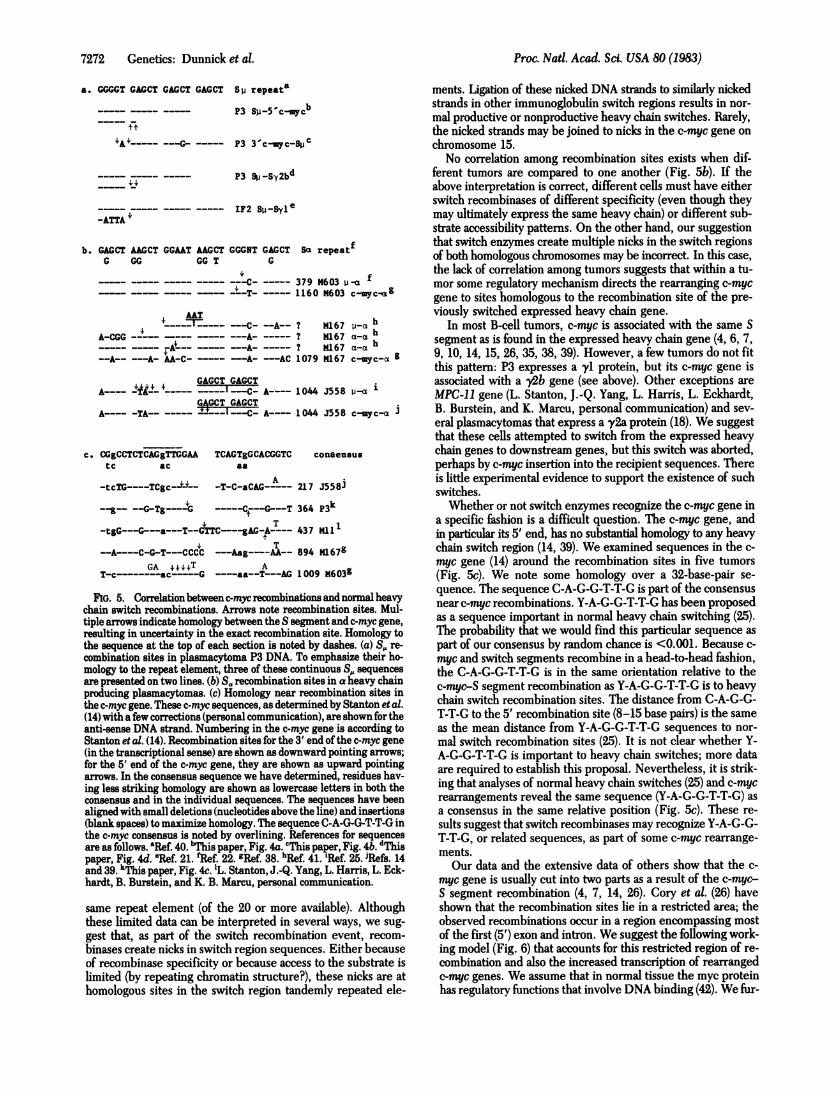
FIG. 5. Correlationbetweenc-mycrecombinations and normal heavychain switch recombinations. Arrows note recombination sites. Mul-tiple arrows indicate homology between the S segment and c-myc gene,resulting in uncertainty in the exact recombination site. Homology tothe sequence at the top of each section is noted by dashes. (a) S,. re-combination sites in plasmacytoma P3 DNA. To emphasize their ho-mology to the repeat element, three of these continuous S,. sequencesare presented on two lines. (b) Sa recombination sites in a heavy chainproducing plasmacytomas. (c) Homology near recombination sites inthe c-myc gene. These c-myc sequences, as determined by Stanton et al.(14) with afew corrections (personal communication), are shown fortheanti-sense DNA strand. Numbering in the c-myc gene is according toStanton et al. (14). Recombination sites for the 3' end of the c-myc gene(in the transcriptional sense) are shown as downward pointing arrows;for the 5' end of the c-myc gene, they are shown as upward pointing
arrows. In the consensus sequence we have determined, residues hav-ing less striking homology are shown as lowercase letters in both theconsensus and in the individual sequences. The sequences have beenalignedwith small deletions (nucleotides above the line) and insertions(blank spaces) to maximize homology. The sequence C-A-G-G-T-T-G inthe c-myc consensus is noted by overlining. References for sequencesare as follows. aRef. 40. 'TiMs paper, Fig. 4a. cThis paper, Fig. 4b. dThispaper, Fig. 4d. 'Ref. 21. fRef. 22. gRef. 38. hRef 41. 'Ref. 25. iRefs. 14and 39. k~jis paper, Fig. 4c. 'L. Stanton, J.-Q. Yang, L. Harris, L. Eck-hardt, B. Burstein, and K. B. Marcu, personal communication.
same repeat element (of the 20 or more available). Althoughthese limited data can be interpreted in several ways, we sug-gest that, as part of the switch recombination event, recom-binases create nicks in switch region sequences. Either becauseof recombinase specificity or because access to the substrate islimited (by repeating chromatin structure?), these nicks are athomologous sites in the switch region tandemly repeated ele-
ments. Ligation of these nicked DNA strands to similarly nickedstrands in other immunoglobulin switch regions results in nor-mal productive or nonproductive heavy chain switches. Rarely,the nicked strands may be joined to nicks in the c-myc gene onchromosome 15.No correlation among recombination sites exists when dif-
ferent tumors are compared to one another (Fig. 5b). If theabove interpretation is correct, different cells must have eitherswitch recombinases of different specificity (even though theymay ultimately express the same heavy chain) or different sub-strate accessibility patterns. On the other hand, our suggestionthat switch enzymes create multiple nicks in the switch regionsof both homologous chromosomes may be incorrect. In this case,the lack of correlation among tumors suggests that within a tu-mor some regulatory mechanism directs the rearranging c-mycgene to sites homologous to the recombination site of the pre-viously switched expressed heavy chain gene.
In most B-cell tumors, c-myc is associated with the same Ssegment as is found in the expressed heavy chain gene (4, 6, 7,9, 10, 14, 15, 26, 35, 38, 39). However, a few tumors do not fitthis pattern: P3 expresses a yl protein, but its c-myc gene isassociated with a y2b gene (see above). Other exceptions areMPC-li gene (L. Stanton, J.-Q. Yang, L. Harris, L. Eckhardt,B. Burstein, and K. Marcu, personal communication) and sev-eral plasmacytomas that express a y2a protein (18). We suggestthat these cells attempted to switch from the expressed heavychain genes to downstream genes, but this switch was aborted,perhaps by c-myc insertion into the recipient sequences. Thereis little experimental evidence to support the existence of suchswitches.Whether or not switch enzymes recognize the c-myc gene in
a specific fashion is a difficult question. The c-myc gene, andin particular its 5' end, has no substantial homology to any heavychain switch region (14, 39). We examined sequences in the c-myc gene (14) around the recombination sites in five tumors(Fig. Sc). We note some homology over a 32-base-pair se-quence. The sequence C-A-G-G-T-T-G is part of the consensusnear c-myc recombinations. Y-A-G-G-T-T-G has been proposedas a sequence important in normal heavy chain switching (25).The probability that we would find this particular sequence aspart of our consensus by random chance is <0.001. Because c-myc and switch segments recombine in a head-to-head fashion,the C-A-G-G-T-T-G is in the same orientation relative to thec-myc-S segment recombination as Y-A-G-G-T-T-G is to heavychain switch recombination sites. The distance from C-A-G-G-T-T-G to the 5' recombination site (8-15 base pairs) is the sameas the mean distance from Y-A-G-G-T-T-G sequences to nor-mal switch recombination sites (25). It is not clear whether Y-A-G-G-T-T-G is important to heavy chain switches; more dataare required to establish this proposal. Nevertheless, it is strik-ing that analyses of normal heavy chain switches (25) and c-mycrearrangements reveal the same sequence (Y-A-G-G-T-T-G) asa consensus in the same relative position (Fig. 5c). These re-sults suggest that switch recombinases may recognize Y-A-G-G-T-T-G, or related sequences, as part of some c-myc rearrange-ments.Our data and the extensive data of others show that the c-
myc gene is usually cut into two parts as a result of the c-myc-S segment recombination (4, 7, 14, 26). Cory et al. (26) haveshown that the recombination sites lie in a restricted area; theobserved recombinations occur in a region encompassing mostof the first (5') exon and intron. We suggest the following work-ing model (Fig. 6) that accounts for this restricted region of re-combination and also the increased transcription of rearrangedc-myc genes. We assume that in normal tissue the myc proteinhas regulatory finctions that involve DNA binding (42). We fur-
7272 Genetics: Dunnick et al.

Proc. Natl. Acad. Sci. USA 80 (1983) 7273
Gene
mRNAProtein
Protein
Gene
NORMAL TISSUE
MNME<m
Auto-
other regulatedregulatory
_Jlevels
functions
B CELL TUMORS
InNo trans-
~~JcriptionRearranged y \
mRNA Excessive
Protein ot levelsProtein regulator
functions
SOME B CELL TUMORS (e.g. PC3741)Gene (operator _
mutation)mRNA &Fyr::ivpProteinProtein otherI
regulatorfunctions
FIG. 6. Working model for c-myc gene regulation. We show the pro-tein in the unfolded form to emphasize that it is encoded by the secondand third exons. We also show the folded form of the protein to em-phasize its function.
ther assume that the myc protein also binds to the 5' end of thec-myc gene and inhibits its own transcription. The translocationin B-cell tumors separates the myc protein coding segments (inthe second and third exons only; see ref. 14) from its operatorin or near the 5'-most exon (7, 14, 26). Thus, transcription ofthe c-myc gene proceeds unregulated, albeit from an adven-titious promoter that may or may not rely on proximity to im-munoglobulin genes (7, 14). The excess of myc protein has twoconsequences. First, it shuts off the unrearranged gene com-
pletely, a phenomenon already noted by Stanton et al. (14). Sec-ond, it provides an overabundance of regulatory proteins thatmay result in oncogenesis. Recombinations that take place 5'of the first exon may not separate the operator from the bulkof the c-myc gene; recombinations that take place 3' of the firstintron may remove transcription signals, translation signals, or
protein domains important in oncogenesis. Thus, c-myc recom-binations that lead to tumors are restricted to a small region.This model predicts the existence of operator constitutive mu-tations (43) that do not bind the myc protein. This may be thegenotype of plasmacytomas with high levels of transcriptionbut no c-myc rearrangement (e.g., PC3741) (4, 35). myc proteinvariants that do not bind operator are not likely to be detectedbecause they would probably have to be homozygous to be ef-fective. In addition to autoregulation, c-myc expression may beregulated by enhancing factors present only in B cells.We are deeply indebted to Ken Marcu and his colleagues for intel-
lectual stimulation, for generous gifts of probes, and for sending us databefore publication. We thank J. L. Claflin and D. Friedman for readingthe manuscript. This work was supported by grants to W. D. from theNational Institute of Allergy and Infectious Diseases (AI 17778), fromthe Michigan Memorial Phoenix Project, and from the Horace H.Rackham Graduate School, and by Institutional Research Grant IN-40Uto the University of Michigan from the American Cancer Society. B.E. S.is a Predoctoral Fellow of the National Science Foundation.
1. Klein, G. (1981) Nature (London) 294, 313-318.2. Rowley, J. D. (1982) Science 216, 749-751.3. Harris, L. J., D'Eustachio, P., Ruddle, F. H. & Marcu, K. B. (1982)
Proc. Natl. Acad. Sci. USA 79, 6622-6626.4. Marcu, K. B., Harris, L. J., Stanton, L. W., Watt, E. R. & Croce,
C. M. (1983) Proc. Natl. Acad. Sci. USA 80, 519-523.
5. Crews, S., Barth, R., Hood, L., Prehn, J. & Calame, K. (1982)Science 218, 1319-1321.
6. Shen-Ong, G. L. C., Keath, E. J., Piccoli, S. P. & Cole, M. D.(1982) Cell 31, 443-452.
7. Adams, J. M., Gerondakis, S., Webb, E., Corcoran, L. M. & Cory,S. (1983) Proc. Nati. Acad. Sci. USA 80, 1982-1986.
8. Mushinski, J. F., Bauer, S. R., Potter, M. & Reddy, E. P. (1983)Proc. Natl. Acad. Sci. USA 80, 1073-1077.
9. Taub, R., Kirsch, I., Morton, C., Lenoir, G., Swan, D., Tronick,S., Aaronson, S. & Leder, P. (1982) Proc. Nati. Acad. Sci. USA 79,7837-7841.
10. Dalla-Favera, R., Bregni, M., Erikson, J., Patterson, D., Gallo,R. C. & Croce, C. M. (1982) Proc. Nati. Acad. Sci. USA 79, 7824-7827.
11. de la Chapelle, A., Lenoir, G., Boue, J., Boue, A., Gallano, P.,Huerre, C., Szajnert, M.-F., Jeanpierre, M., Lalouel, J.-M. &Kaplan, J.-C. (1983) Nucleic Acids Res. 11, 1132-1142.
12. Sheiness, D. & Bishop, J. (1979) J. Virol. 31, 514-521.13. Duesberg, P. H. (1979) Cold Spring Harbor Symp. Quant. Biol.
44, 13-30.14. Stanton, L. W., Watt, R. & Marcu, K. B. (1983) Nature (London)
303, 401-406.15. Erikson, J., Ar-Rushdi, A., Drivinga, H. L., Nowell, P. C. & Croce,
C. M. (1983) Proc. Nati. Acad. Sci. USA 80, 820-824.16. Honjo, T. & Kataoka, T. (1978) Proc Natl. Acad. Sci. USA 75, 2140-
2144.17. Rabbitts, T. H., Forster, A., Dunnick, W & Bentley, D. L. (1980)
Nature (London) 283, 351-356.18. Coleclough, C., Cooper, D. & Perry, R P. (1980) Proc, Natl. Acad.
Sci. USA 77, 1422-1426.19. Cory, S., Jackson, J. & Adams, J. M. (1980) Nature (London) 285,
450-456.20. Sakano, H., Maki, R., Kurosawa, Y., Roeder, W. & Tonegawa, S.
(1980) Nature (London) 286, 676-683.21. Dunnick, W, Rabbitts, T. H. & Milstein, C. (1980) Nature (Lon-
don) 286, 669-675.22. Davis, M. M., Kim, S. K. & Hood, L. E. (1980) Science 209,1360-
1365.23. Kataoka, T., Miyata, T. & Honjo, T. (1981) Cell 23, 357-368.24. Shimizu, A., Takahashi, N., Yaoita, Y. & Honjo, T. (1982) Cell 28,
499-506.25. Marcu, K. B., Lang, R. G., Stanton, L. W. & Harris, L. J. (1982)
Nature (London) 298, 87-89.26. Cory, S., Gerondakis, S. & Adams, J. M. (1983) EMBOJ.2, 697-
704.27. Margulies, D. H., Kuehl, W. M. & Scharff, M. D. (1976) Cell 8,
405-415.28. Blattner, F. R., Williams, B. G., Blechl, A. E., Denniston-
Thompson, K., Faber, H. E., Furlong, L.-A., Grunwald, D. J.,Kiefer, D. O., Moore, D. D., Sheldon, E. L. & Smithies, 0. (1977)Science 196, 161-164.
29. Hohn, B. (1979) Methods Enzymol. 68, 299-309.30. Benton, W. D. & Davis, R. W. (1977) Science 196, 180-182.31. Steffen, D. & Weinberg, R. A. (1978) Cell 15, 1003-1010.32. Southern, E. M. (1975)J. Mol. Biol. 98, 503-517.33. Rigby, P. W. J., Dieckmann, M., Rhodes, C. & Berg, P. (1977)J.
Mol. Biol. 113, 237-251.34. Lang, R. B., Stanton, L. W. & Marcu, K. B. (1982) Nucleic Acids
Res. 10, 611-630.35. Harris, L. J., Lang, R. B. & Marcu, K. B. (1982) Proc. Natl. Acad.
Sci. USA 79, 4175-4179.36. Sanger, F., Coulson, A. R., Barrell, B. G., Smith, A. J. H. & Roe,
B. A. (1980) J. Mol. Biol. 143, 161-178.37. Messing, J. (1981) in Third Cleveland Symposium on Macromol-
ecules: Recombinant DNA, ed. Walton, A. (Elsevier, Amster-dam), pp. 143-153.
38. Calame, K., Kim, S., Laley, D., Hill, R., Davis, M. & Hood, L.(1982) Proc. Natl. Acad. Sci. USA 79, 6994-6998.
39. Adams, J. M., Gerondakis, S., Webb, E., Mitchell, J., Bernard,0. & Cory, S. (1982) Proc. Natl. Acad. Sci. USA 79, 6966-6970.
40. Nikaido, T., Nakai, S. & Honjo, T. (1981) Nature (London) 292,845-847.
41. Kim, S., Davis, M., Sinn, E., Patten, P. & Hood, L. (1981) Cell27, 573-581.
42. Donner, P., Greiser-Wilke, I. & Moelling, K. (1982) Nature (Lon-don) 296, 262-266.
43. Jacob, F. & Monod, J. (1961) J. Mol. Biol. 3, 318-356.
Genetics: Dunnick et al.