a case of an ectopic thyroid gland at the lateral neck ... · ryngeal pouch and contributes 1% to...
TRANSCRIPT

INTRODUCTION
Embryologically the thyroid gland is derived from 2 anlages:a large median endodermal anlage and two lateral anlages.The median anlage produces most of the thyroid parenchy-ma, while the lateral anlage is derived from the fourth pha-ryngeal pouch and contributes 1% to 30% of the thyroidweight (1). Commonly, failure in the descent of the mediananlage results in a lingual thyroid gland. In rare cases, fail-ure of the lateral anlage to fuse with the median anlage canresult in lateral ectopic thyroid gland (2).
We present a case report of a 54-yr-old woman with pre-sumed metastatic papillary thyroid carcinoma (PTC) of lat-eral neck nodes, as was expected from clinical and comput-ed tomography (CT) findings. Total thyroidectomy with leftmodified radical neck dissection (MRND) was performed,but histological examination of the mass at the lateral neckshowed ectopic thyroid tissue with a hyperplastic pattern.There was also no malignancy in other lymph nodes.
CASE REPORT
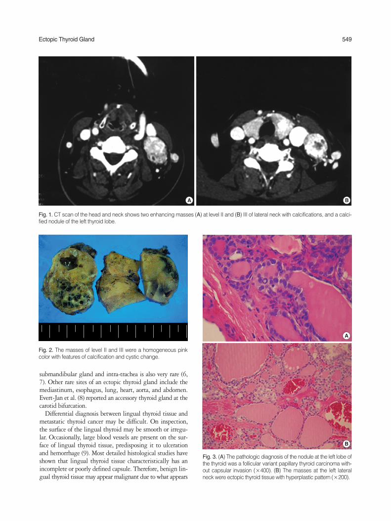
A 54-yr-old female presented with palpable masses in theleft lateral neck region. Physical examination revealed twofirm, non-tender, mobile masses of 3×3 cm size and 4×4cm, respectively. The thyroid gland was not palpable in itsnormal position. A CT scan of the head and neck with con-trast was done, which showed a mass (2×2 cm) with calci-fication on the left lobe of the thyroid, and also revealed that
the two palpable masses at the left lateral neck were enhancedwith calcification on the left level II (3.5×4.5 cm) and III(3.5×4 cm) (Fig. 1). Fine-needle aspiration biopsy (FNAB)of the mass at left lateral level II resulted in fibroadipose tis-sues, but suggested lateral neck metastasis of PTC, as evi-denced by calcification and cystic change on CT scan, and aFNAB of the calcified nodule at the left lobe of thyroid con-sisted of intranuclear inclusion, which was indicative of PTC.
The patient underwent total thyroidectomy with left MR-ND under the presumptive diagnosis of lateral neck metas-tasis of PTC. The two masses at level II and III had calcifica-tion and cystic change, which is sufficient evidence of neckmetastasis. However, unlike nodal metastasis of PTC, whichis gray, these masses were homogeneous and pink (Fig. 2).The pathologic diagnosis of the nodule at the left lobe of thethyroid was PTC without capsular invasion, and the massin the left lateral neck was an ectopic thyroid gland with ahyperplastic pattern (Fig. 3). On pathologic examination,four nodes of the central compartment and five nodes of leftlevel IV and V were not malignant.
DISCUSSION
Ectopic thyroid gland is defined as thyroid tissue not locat-ed anterolaterally in the second to fourth tracheal cartilages.The lingual location is most common, accounting for 90%of reported cases (2-4). Bradley et al. (5) reported multifocalpapillary thyroid carcinoma occurring very rarely in a lin-gual thyroid. An ectopic thyroid gland in the region of the
548
Jae-Young Choi and Jeong-Hoon Kim
Department of Surgery, Kosin University College ofMedicine, Busan, Korea
Address for correspondenceJeong Hoon Kim, M.D.Department of Surgery, Kosin University College ofMedicine, 34 Amnam-dong, Seo-gu, Busan 602-702,Korea Tel : +82.51-990-6462, Fax : +82.51-246-6093 E-mail : [email protected]
J Korean Med Sci 2008; 23: 548-50ISSN 1011-8934DOI: 10.3346/jkms.2008.23.3.548
Copyright � The Korean Academyof Medical Sciences
A Case of an Ectopic Thyroid Gland at the Lateral Neck Masquerading as a Metastatic Papillary Thyroid Carcinoma
Ectopic thyroid glands generally occur in the midline as a result of abnormal medianmigration, and their presence lateral to the midline is rare. We present one case ofan ectopic thyroid gland masquerading as a lateral neck metastasis of a papillarythyroid carcinoma (PTC). In this case of a 54-yr-old woman with left PTC, we sus-pected left lateral neck metastasis on preoperative neck computed tomography.The patient underwent total thyroidectomy, central compartment neck dissection,and left modified radical neck dissection (MRND). The patient was diagnosed ashaving an accessory thyroid gland on the lateral neck on the final pathologic report.Surgeons should be aware of the existence of an ectopic thyroid gland in unusuallocations.
Key Words : Ectopic Thyroid; Carcinoma, Papillary
Received : 30 March 2007Accepted : 18 September 2007
Ectopic Thyroid Gland 549
submandibular gland and intra-trachea is also very rare (6,7). Other rare sites of an ectopic thyroid gland include themediastinum, esophagus, lung, heart, aorta, and abdomen.Evert-Jan et al. (8) reported an accessory thyroid gland at thecarotid bifurcation.
Differential diagnosis between lingual thyroid tissue andmetastatic thyroid cancer may be difficult. On inspection,the surface of the lingual thyroid may be smooth or irregu-lar. Occasionally, large blood vessels are present on the sur-face of lingual thyroid tissue, predisposing it to ulcerationand hemorrhage (9). Most detailed histological studies haveshown that lingual thyroid tissue characteristically has anincomplete or poorly defined capsule. Therefore, benign lin-gual thyroid tissue may appear malignant due to what appears
Fig. 1. CT scan of the head and neck shows two enhancing masses (A) at level II and (B) III of lateral neck with calcifications, and a calci-fied nodule of the left thyroid lobe.
A B
Fig. 2. The masses of level II and III were a homogeneous pinkcolor with features of calcification and cystic change.
Fig. 3. (A) The pathologic diagnosis of the nodule at the left lobe ofthe thyroid was a follicular variant papillary thyroid carcinoma with-out capsular invasion (×400). (B) The masses at the left lateralneck were ectopic thyroid tissue with hyperplastic pattern (×200).
A
B

550 J.-Y. Choi, J.-H. Kim
to be an invasion into the muscle, but this only signifies adefect in the capsule, resulting in intermingling of the glan-dular and the muscular elements.
Basaria et al. (3) reported ectopic lingual thyroid mas-querading as thyroid cancer metastases. They performed atotal thyroidectomy and radioiodine therapy due to PTC.One year after the operation, a diagnostic radioiodine scanshowed a significant residual uptake at the level of the chin,thought to be consistent with a metastasis at the base of thetongue. The patient received radioactive iodine therapy, butdid not improve. Incisional biopsy was done, and histologi-cal examination showed numerous follicular structures, anda diagnosis of ectopic thyroid was made. Interestingly, akinetic analysis of iodine turnover in lingual thyroid tissueshowed a biological half-life of 1.3 days compared with 100days in normal thyroid tissue (10). This phenomenon mayexplain why the lingual thyroid in the present patient wasso resistant to ablation, compared with the remnant tissuein the thyroid bed.
In our case report, we described a patient with PTC whohad two ectopic lateral thyroid glands in the neck masquerad-ing as a metastatic focus. In fact, ectopic thyroid glands ofthe midline, like lingual thyroid, are not easily mistaken formetastatic PTC, because it is almost impossible for metasta-sis of PTC at level I of the neck to skip over levels II, III, andIV. However, if PTC is combined with lateral neck masseswith calcification, why did someone suggest it was an ectopicthyroid at the lateral neck? If preoperatively diagnosed as anectopic thyroid, selective neck dissection may be enough.However, since it is impossible for suspected calcified mass-es of the lateral neck to be an ectopic thyroid, MRND mustbe performed in such cases.
Currently, there are a relatively large number of reports onectopic thyroid, but there are few articles that present ectopicthyroid coexisting simultaneously with PTC. If a preopera-tive diagnostic method could be developed that can differ-entiate between ectopic thyroid tissue and metastatic thy-
roid cancer, an accurate assessment of the extent of operationwould be made.
REFERENCES
1. Mansberger AR, Wei JP. Surgical embryology and anatomy of thethyroid and parathyroid glands. Surg Clin North Am 1993; 73: 727-46.
2. Paresi RJ Jr, Saha D. Hashimoto’s thyroiditis presenting as an enlarg-ing submandibular mass in a patient with a lingual thyroid. Oto-laryngol Head Neck Surg 2005; 132: 806-8.
3. Basaria S, Westra WH, Cooper DS. Ectopic lingual thyroid mas-querading as thyroid cancer metastases. J Clin Endocrinol Metab2001; 86: 392-5.
4. Aktolun C, Demir H, Berk F, Metin Kir K. Diagnosis of completeectopic lingual thyroid with Tc-99m pertechnetate scintigraphy. ClinNucl Med 2001; 26: 933-5.
5. Goldstein B, Westra WH, Califano J. Multifocal papillary thyroidcarcinoma arising in a lingual thyroid. Arch Otolaryngol Head NeckSurg 2002; 128: 1198-200.
6. Kumar R, Sharma S, Marwah A, Moorthy D, Dhanwal D, MalhotraA. Ectopic goiter masquerading as submandibular gland swelling:a case report and review of the literature. Clin Nucl Med 2001; 26:306-9.
7. Pritchyk KM, Thompson LD, Malekzadeh S. Endoscopic manage-ment of intratracheal ectopic thyroid. Otolaryngology Head Necksurg 2004; 130: 630-2.
8. Hollander EJ, Visser MJ, van Baalen JM. Accessory thyroid glandat carotid bifurcation presenting as a carotid body tumor: case reportand review of the literature. J Vasc Surg 2004; 39: 260-2.
9. Kansal P, Sakati N, Rifai A, Woodhouse N. Lingual thyroid. Diag-nosis and treatment. Arch Intern Med 1987; 147: 2046-8.
10. Carter LC, Uthman A, Drinnan AJ, Loree TR. Diagnostic dilemmainvolving calcification in the parapharyngeal space: metastatic thy-roid carcinoma masquerading as a deep lobe parotid mass. OralSurg Oral Med Oral Pathol Oral Radiol Endod 1997; 84: 697-702.