a case of subacute sclerosing panencephalitis with...
TRANSCRIPT

OLGU SUNUMU/CASE REPORT
ABSTRACT
The purpose of this study was to report a SSPE patient presenting with bilateral optic atrophy, maculopathy, and widesp-read chorioretinitis. A 14-year-old girl was admitted to our hospital with the complaint of sudden painless visual loss in both eyes with a 1-month interval. Best corrected visual acuity was finger counting level in both eyes. Bilateral optic atrophy, macular scar, and chorioretinitis affecting the whole posterior segment were observed. Encephalopathy and convulsions developed 2 months after the presentation of eye findings. She was diagnosed with SSPE according to clinical, electroen-cephalographic, and cerebrospinal fluid findings. SSPE should be considered in the differential diagnosis of optic atrophy and atypical widespread chorioretinitis in young patients, especially in countries where SSPE has a higher incidence rate.
Key Words: SSPE, optic atrophy, chorioretinitis.
ÖZ
Bu çalışmanın amacı, her iki gözde optik atrofi, makülopati ve yaygın koryoretinit bulgularıyla başlayan bir SSPE olgusunu sunmaktır. On dört yaşında bayan hasta, her iki gözde bir ay ara ile gelişen, ağrısız ani görme kaybı şikayeti ile hastanemi-ze başvurdu. Düzeltilmiş uzak görme keskinliği her iki gözde el hareketlerini seviyesindeydi. Arka segmet muayenesinde, her iki gözde optik atrofi, makülada skar ve tüm arka kutbu etkilemiş koryoretinit bulguları izlendi. 2 ay sonra hastada konvulsiyonlar ve ensefalopati tablosu gelişti. Klinik tablo, elektroensefalografik değerlendirme ve beyin omurilik sıvısının incelenmesi sonucunda elde edilen bulgularla hastaya SSPE tanısı kondu. Özellikle SSPE insidansının daha yüksek olduğu ülkelerde, optik atrofi ve atipik yaygın koryoretinit izlenen genç hastalarda, SSPE tanısı akılda tutulmalıdır.
Anahtar Kelimeler: SSPE, optik atrofi, koryoretinit.
Geliş Tarihi - Received: 24.11.2011 Kabul Tarihi - Accepted: 12.01.2012
Ret-Vit 2012;20:230-232
Yazışma Adresi / Correspondence Adress: MD., Nagihan UĞURLUYıldırım Beyazıt University, Atatürk Research and Training Hospital, Depart-
ment of Ophthalmology, Ankara/TURKEY
Phone: +90 0312 291 25 25E-Mail: [email protected]
1- MD. Yıldırım Beyazıt University, Atatürk Research and Training Hos-pital, Department of Ophthalmology, Ankara/TURKEY
UĞURLU N., [email protected] MD. Assistant Professor, Yıldırım Beyazıt University, School of Medi-
cine, Department of Ophthalmology, Ankara ÇAĞIL N., [email protected] MD., Dıskapı Children’s Education and Training Hematology Oncology
Hospital, Department of Pediatric Neurology, Ankara/TURKEY ARHAN E., [email protected] MD. Associated Professor, Dıskapı Children’s Education and Training
Hematology Oncology Hospital, Department of Pediatric Neurology, Ankara/TURKEY
KÖSE G., [email protected] MD. Professor, Ufuk University, School of Medicine, Department of
Ophthalmology, Ankara/TURKEY ŞENGÜN A., [email protected]
A Case of Subacute Sclerosing Panencephalitis with Widespread Chorioretinitis
Yaygın Koryoretinitli Bir Subakut Sklerozan Panensefalit Olgusu
Nagihan UĞURLU1, Nurullah ÇAĞIL2, Ebru ARHAN3, Gülşen KÖSE4, Ahmet ŞENGÜN5
INTRODUCTION
Subacute sclerosing panencephalitis (SSPE) is a rare progressive neurodegenerative disorder caused by persis-tent infection of the brain by an aberrant measles virus called SSPE virus.1 SSPE is a slow virus infection and characteristic clinical manifestations include personality and behavioral changes, cognitive decline, myoclonic seizures, paresis, and eventually obtundation, stupor, and coma. Ocular involvement occurs in up to 50% of cases and optic neuritis, papilledema, chorioretinitis, homonymous visual field defects, and cortical blindness are characteristic ocular findings.1-3 Herein we describe a 14-year-old girl with sudden loss of vision as an initial symptom of SSPE.
Ret-Vit 2011;20:230-232 Uğurlu et al. 231
CASE REPORT
A 14-year-old girl was admitted to our clinic with the complaint of sudden painless loss of vision in both eyes. Visual loss was encountered in her right eye first, and then after 1 month a considerable visual loss occurred in the second eye. Best corrected visual acuity was at finger counting level in both eyes. Her anterior segment examination and intraocular pres-sures were normal. Her fundus examination showed bilateral optic atrophy, sclerosis, and a beaten bronze appearance on the macula.
In addition, epiretinal membrane and patches of reti-na pigment epithelial hyperpigmentation were noted. There were no signs of active retinal vasculitis. Wide-spread scars as a result of necrotizing retinitis and small islands of hemorrhagic foci were also observed at the peripheral retina.
FA revealed hypofluorescent spots at hyperpigment-ed areas of the macula. The OCT revealed widespread destruction of all retinal layers and thinning of the retina. ERG and EOG examinations suggested wide-spread rod cone degeneration. No systemic or neuro-logical abnormalities were recorded during her physi-cal examination. Her family history was negative for ocular or any other systemic or neurological distur-bances. Her school performance was good. Laboratory investigations revealed an increased white blood cell count (18.5/mm3) and ESR (26 mm/h). Liver and kid-ney function tests, anti-double-stranded DNA anti-body, anti-nuclear antibody, blood and urine culture, tuberculin test, chest X-ray, and abdominal ultra-sound were all normal. Serology for toxoplasmosis, cytomegalovirus, and Borreliosis, VDRL, and FTA was negative. Visual evoked potentials showed re-duced amplitude and delayed P100 latency.
Eight weeks after presentation, she developed con-vulsions and encephalopathy. Lumbar puncture (LP) was performed. Cerebrospinal fluid (CSF) showed 2 cells/mm3 (all lymphocytes), 31 mg/mL protein, and 56 mg/dL glucose and negative results for Gram and acid-fast stains and polymerase chain reaction (PCR) for herpes simplex virus (HSV) and Epstein-Barr vi-rus (EBV). Results of borrelia serology were negative. Antimeasles IgG titers were elevated in both serum and CSF. Immunofixation electrophoresis of CSF re-vealed an oligoclonal band.
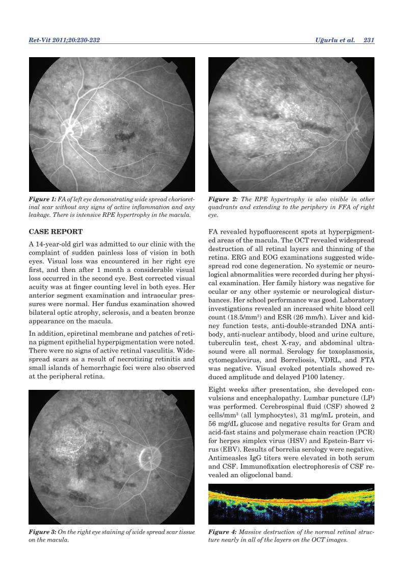
Figure 1: FA of left eye demonstrating wide spread chorioret-inal scar without any signs of active inflammation and any leakage. There is intensive RPE hypertrophy in the macula.
Figure 3: On the right eye staining of wide spread scar tissue on the macula.
Figure 2: The RPE hypertrophy is also visible in other quadrants and extending to the periphery in FFA of right eye.
Figure 4: Massive destruction of the normal retinal struc-ture nearly in all of the layers on the OCT images.

232 A Case of Subacute Sclerosing Panencephalitis with ...
On T2-weighted imaging of magnetic resonance im-aging (MRI) study, increased signal intensity of the white matter in the right parietooccipital region was seen. EEG showed diffusely slow background activity and periodic slow waves. These periodic generalized slow waves did not disappear after intravenous diaz-epam injection. The diagnosis of SSPE was confirmed by these EEG and CSF examination findings. She was hospitalized and isoprinosine (100 mg/kg) and carbamazepine (10 mg/kg) were started.
DISCUSSION
Our case is an unusual presentation of SSPE as se-vere visual loss preceded the typical neurological signs and symptoms. Differential diagnosis of wide-spread chorioretinitis, maculopathy, and optic atro-phy with neurological involvement was made by ex-cluding syphilitic retinitis and Lyme disease.
Ocular involvement of syphilis may develop at any stage of the disease. Since it can affect all the struc-tures in the eye, it is called the ‘great mimicker’. It causes a wide range of clinical manifestations such as episcleritis, panuveitis, chorioretinitis, macular ede-ma, optic neuritis, and optic atrophy. The serum ex-amination, which revealed negative results for both non-treponemal (VDRL) and treponemal (FTA) tests, excluded the diagnoses of syphilis.
Lyme disease is another disease that has a diverse range of ocular manifestations with CNS involve-ment. Therefore, it should be kept in mind in cas-es of chorioretinitis with neurological symptoms. Negative results of the serum and CSF examination for Borrelia serology also excluded the diagnosis of Lyme disease.
SSPE is a rare but feared sequela of a common vi-ral infection.1,2 Various neuroophthalmic and retinal findings may be observed in SSPE patients.4-10 In 10% of cases, ophthalmic manifestations precede the neu-rological symptoms.3 This is explained by the pres-ence of possible measles virus acquired neurotropism in the retina.11 Necrotizing retinitis is reported as the most characteristic lesion but, in contrast to the present case, it is usually observed as focal areas of retinitis in the macula. Optic atrophy is also a rare presenting sign of SSPE and is an indicator of CNS involvement.12,13
The present case is unusual, presenting with optic nerve involvement that precedes the more charac-teristic neurological symptoms. Although SSPE is a rare disease, its incidence is higher in our country compared with more developed countries, and so it should be considered in the differential diagnosis of atypical chorioretinitis with optic nerve involvement especially in young patients even in the absence of other neurological symptoms.
KAYNAKLAR/REFERENCES1. Garg RK. Subacute sclerosing panencephalitis. Postgrad Med
J 2002;78:63-70.
2. Green SH, Wirtschhafter JD. Ophthalmoscopic findings in subacute sclerosing panencephalitis. Br J Ophthalmol 1973;57:780-7.
3. Takayama S, Iwasaki Y, Yamanouchi H, et al. Characteris-tic clinical features in a case of fulminant subacute sclerosing panencephalitis. Brain Dev 1994;16:132-5.
4. Babu RB, Biswas J. Bilateral macular retinitis as the present-ing feature of subacute sclerosing panencephalitis. J Neur-oophthalmol 2007;27:288-91.
5. Zako M, Kataoka T, Ohno-Jinno A, et al. Analysis of progres-sive ophthalmic lesion in a patient with subacute sclerosing panencephalitis. Eur J Ophthalmol 2008;18:155-8.
6. Sharma S, Chanana B, Gulati S, et al. Acute bilateral vision loss as the presenting feature of subacute sclerosing panen-cephalitis. Indian J Pediatr 2009;76:952-3.
7. Caruso JM, Robbins-Tien D, Brown WD, et al. Atypical chorioretinitis as an early presentation of subacute scle-rosing panencephalitis. J Pediatr Ophthalmol Strabismus 2000;37:119-22.
8. Floegel I, Haas A, El-Shabrawi Y. Acute multifocal placoid pigment epitheliopathy-like lesion as an early presentation of subacute sclerosing panencephalitis. Am J Ophthalmol 2003;135:103-5.
9. Yuksel D, Sonmez PA, Yilmaz D, et al. Ocular findings in subacute sclerosing panencephalitis. Ocul Immunol Inflamm 2011;19:135-8.
10. Tomoda A, Miike T, Miyagawa S, et al. Subacute sclerosing panencephalitis and chorioretinitis. Brain Dev 1997;19:55-7.
11. Park DW, Boldt HC, Massicotte SJ, et al. Subacute scleros-ing panencephalitis manifesting as viral retinitis: clinical and histopathologic findings. Am J Ophthalmol. 1997;123:533-42.
12. Berker N, Batman C, Guven A, et al. Optic atrophy and macu-lar degeneration as initial presentations of subacute scleros-ing panencephalitis.Am J Ophthalmol 2004;138:879-81.
13. Tandon R, Khanna S, Sharma MC, et al. Subacute sclerosing panencephalitis presenting as optic neuritis. Indian J Oph-thalmol 1999;47:250-2.